operative delivery - wisdom
TRANSCRIPT

OPERATIVE DELIVERY
Owahnah Sherene Quammie & Hilary Williams

Operative or instrumental vaginal delivery: Owahnah
Forceps delivery
Ventouse
Caesarean section: Hilary

ASSISTED VAGINAL DELIVERY The use of any surgical procedure to facilitate vaginal delivery
Uses traction to expedite delivery in second stage of labour
Used in prolonged 2nd stage to ↓ maternal/foetal death and/or complications
Both are equally effective in some cases and preference is largely based on clinician choice and experience
Assisted vaginal delivery
ForcepsVacuum
Extraction
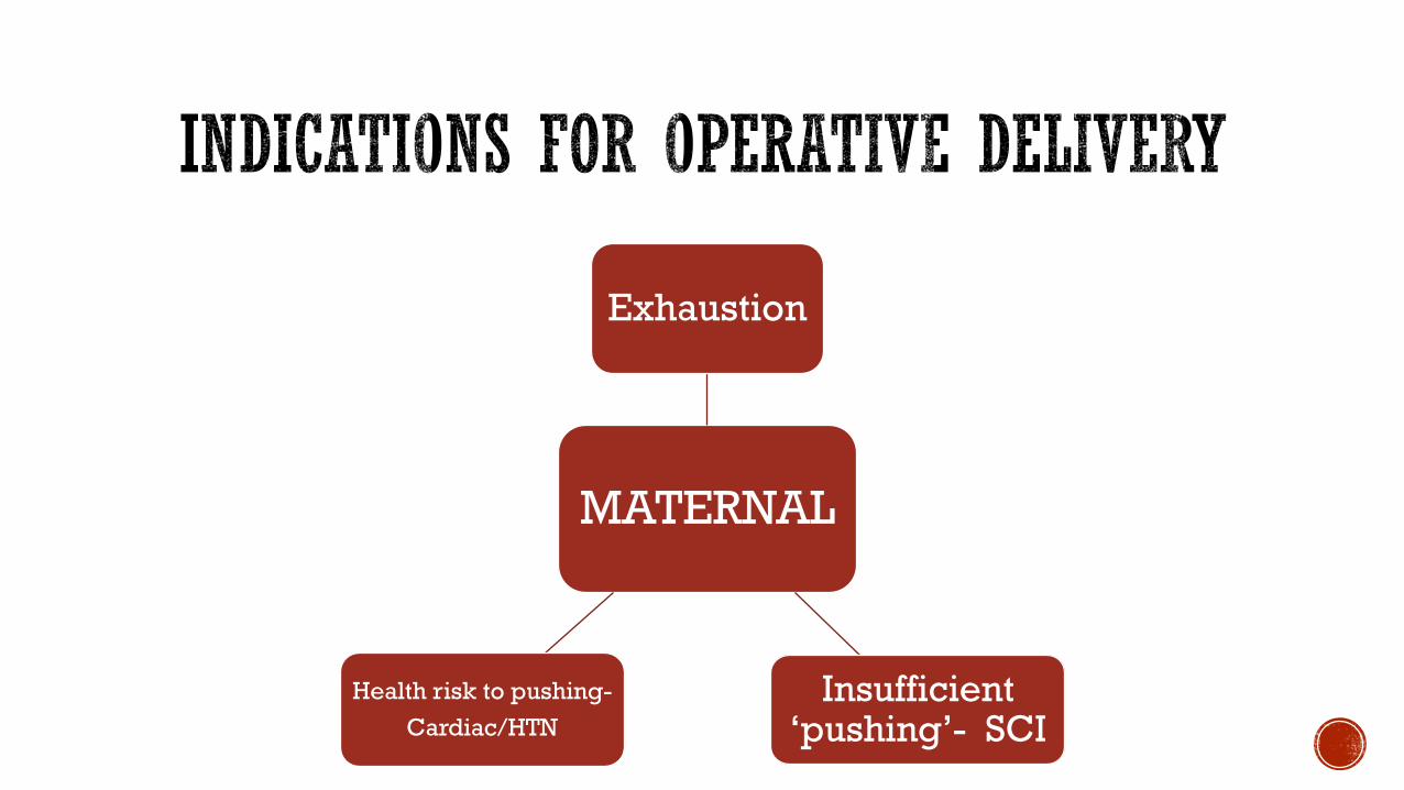
MATERNAL
Exhaustion
Insufficient ‘pushing’- SCI
Health risk to pushing-
Cardiac/HTN

FOETUS
Fetal distress
• HR abnormalities
• Acidosis on FBS
• Meconium

Prolonged 2nd Stage
Nulliparous•3hrs + regional anaesthesia
•2hrs – regional anasthesia
Multiparous 2hrs + regional anaesthesia
1hr – regional anaesthesia

CRITERIA FOR INSTRUMENTAL VAGINAL DELIVERY 1
MOTHER
Consent from mother
Adequate anaesthesia/analgesia. Low forceps Perineal nerve block
Midforceps Epidural block, a pudendal nerve block or GA
Empty Bladder (catheterization)
Full cervical Dilation and Rupture of membranes (ROM)
Lithotomy position (legs separated, flexed, and supported in raised stirrups)
FOETUS
Fully engaged head (at spines or below) with no head palpable abdominally
Known position and attitude of foetal head (caput?)
Vertex presentation

FORCEPS: A BRIEF HISTORY 2
Controversial introduction into obstetric practice during 18th century Europe by the Chamberlen family
‘The labouring woman was blindfold lest she should see the “secret.”’
‘Only the Chamberlen’s were allowed in the locked lying-in room, from which the terrified relatives heard peculiar noises, ringing bells, and other sinister sounds as the
“secret” went to work’
Dr. Peter Chamberlen’s obstetric instruments 2

CASE: FORCEPS DELIVERY
A 28 year old nulliparous woman at term, has been in the 2nd stage of labour for over 3 hours. She initially didn’t want any anesthesia but decided on an epidural with good effect. Her contractions are regular averaging 3 in ten minutes
On vaginal examination the fetal head in in the OP position and just below the ischial spines. The consultant has discussed and consented the patient for a likely operational delivery
Which type of forceps delivery is indicated in this patient?
A. low cavity delivery with Wrigley’s forceps
B. Rotational forceps delivery with Keilland’s forceps
C. Mid-cavity delivery with Simpson’s forceps

LOW-CAVITY/OUTLET FORCEPS DELIVERY:WRIGLEY’S 3
Short and light
Used when the head is at the vaginal introitus but is being held back by the perineum OUTLET
Head has advanced beyond ischial spines LOW
Mother in lithotomy position
Bladder emptied/catheterised
Aseptic cleaning and draping of perineum
Adequate Analgesia (Pudendal + LA)
Assemble then insert forceps
Pelvic curve of forceps should be over the malar
aspect of baby’s head towards the cheeks
Traction in time with uterine contractions at an initial
angle of 60°

MID-CAVITY OUTLET FORCEPS DELIVERY:SIMPSON’S 3
Used when the sagittal suture is in the anteroposterior plane which is usually OA
If the head is not palpable abdominally but is at or just below level of ischial spines
Trial of Forceps- done in a theatre setting if risk of unsuccessful delivery to quickly transition to CS
Head position:
If OT then non-rotational forceps contraindicated
Use rotational forceps e.g. Keilland
a. Pelvic curvature
b. Cephalic curvature
c. Locking handles

ROTATIONAL FORCEPS DELIVERY:KEILLAND’S 4 Can be placed directly onto baby’s head if in OP for
gentle rotation into OA
The reduced pelvic curve allows rotation about the
axis of the handle
Delivery should ideally be done in theatre setting to
quickly perform CS if needed
Once rotated delivery continues as with mid-cavity
forceps
Successful use requires adequate skill and should only
be used by experienced obstetricians
Can be associated with ↑ injury to mother than with rotational
ventouse
a. No/slight pelvic
curvature
b. Cephalic curve
c. Sliding shanks

COMPLICATIONS
MATERNAL
Injury to vagina or cervix Haemorrhage (PPH)
↑ analgesic requirements
FOETAL
Foetal trauma
Cerebral compression
Cephalohaematoma
Skull fractures
Cervical spine injuries
Facial nerve palsy
Markings/indentations
Facial bruising/lacerations

CASE: FORCEPS DELIVERY
A 28 year old nulliparous woman at term, has been in the 2nd stage of labour for over 3 hours. She initially didn’t want any anesthesia but decided on an epidural with good effect. Her contractions are regular averaging 3 in ten minutes
On vaginal examination the fetal head in in the OP position and just below the ischial spines. The consultant has discussed and consented the patient for a likely operational delivery
Which type of forceps delivery is indicated in this patient?
A. low cavity delivery with Wrigley’s forceps
B. Rotational forceps delivery with Keilland’s forceps
C. Mid-cavity delivery with Simpson’s forceps

VENTOUSE
Developed by Malmström in 1954
First type of cup used was metal (M-CUP) but current models are made of plastic or silicone
Consists of a Cup with handle
Tube- attached to cup and to suction device
Theoretical advantage that less pelvic space is required in comparison to forceps
Not used before 36 Weeks

1. The cup placed in the midline overlying, or just anterior to, the posterior fontanelle in order to encourage flexion of the head
2. Held firmly against the scalp and low suction applied 100mmHgEnsure no vaginal skin is trapped beneath cup
3. Suction pressure increased to 500-600mmHg
Downward traction in time with uterine contractions and suction released between contractions
4. Procedure stopped if the cup detaches 3 times or there is no descent of the head
5. Episiotomy ideally avoided as perineum provides pressure on vacuum cup to keep attached to head
VENTOUSE DELIVERY1

If the head is in a transverse or posterior position a manoeuvrable cup, e.g. OmniCupis used as it allows accurate placement over the fexion point
The hard cups (e.g. Malmström,OmniCup) have a lower failure rate than the soft cups (e.g. Silc,
Silastic)
lower detachment rate, 13% versus 33%.
OmniCup vacuum extractor5

COMPLICATIONS
Maternal injury to reproductive tract PPH
Failed delivery more common with soft cups
Scalp lacerations
Chignon- temporary swelling post ventouse delivery
Cephalohaematoma- bleeding into scalp
Retinal haemorrhages
Subgaleal haemorrhage following poor application of the cup
Prolonged extraction
Multiple cup detachments and reatchments

FORCEPS VENTOUSE
Vaginal Delivery
↑ ↓
Caesarean Section ↑ ↓
Maternal Injury
Pain peri and post delivery
↑ ↓
Anaesthesia ↑ ↓
Cephalohaematomas
Subgaleal haemorrhage and
Retinal haemorrhages
↓ ↑
Neonatal morbidity ↔︎ ↔︎
Concerns over appearance
of baby
↓ ↑

1. Norwitz ER, Schorge JO. Obstetrics and Gynaecology at a Glance. 4th edition. Oxford: John Wiley and Sons. 2013
2. Dunn PM. The Chamberlen family (1560–1728) and obstetric forceps. Archives of Disease in Childhood - Fetal and Neonatal Edition 1999;81:F232-F234
3. Oats J, Abraham Suzanne. Fundamentals of Obstetrics and Gynaecology. 10th edition. Elsevier. 2017
4. Magowan BA, Owen P, Thomson A. Clinical Obstetrics and gynaecology. 3rd edition. Elselvier. 2014
5. Impey L, Child T. Obstetrics and Gynaecology. 4th edition. Oxford: John Wiley and Sons 2012

Delivery of baby through abdominal surgery
https://www.gponline.com/journals-watch-incontinence-sinusitis/article/11256521

WHO recommend only if medically necessary
Global rates doubled between 2003-2018: now 21%
Locally2:
28.500 deliveries in Welsh maternity units in 2017-18
27% by Caesarean section

1794, USA Mother & baby survived: Dr Jesse Bennett operates on wife3
1865, UK 85% mortality rate
Reduced through introduction of:
Aseptic surgery
Antibiotics
Anaesthesia,
Transfusion
Pfannensteil/Joseph Cohen cut

Placenta praevia (minor or major)
Abnormal lie –IECV
Pelvic deformity/cephalopelvic disproportion
Previous classical section3
Morbidly adherent placenta
Pre eclampsia
IUGR small baby with placental insufficiency
Concurrent HIV/HCV or third trimester maternal herpes
Mother anxious despite perinatal mental health support4

Relative:
Breech at 36 weeks4
Diabetes
Older nulliparous
Previous section

Elective: suit patient or staff
Classific
ation
Time limit Urgency Indication
Category
1
30 minutes imminent threat to life Abruption
Abnormal foetal heart rate
Cord prolapse
Scar rupture
Prolonged Bradycardia
Category
2
60/75(nice
is 75)
No immediate threat to life Failure to progress
Pathological CTG
Category
3
Scheduled Pre term: early not urgent Pre eclampsia
IUGR
Failed induction
Category
4
None Equates to elective Term breech
Maternal infection
Placenta praevia

Consent
VTE prophylaxis: TED stockings/LMWH/hydration
FBC to assess Hb.
H2 receptor agonist: gastric aspiration
Foleys catheter to drain bladder and minimise risk of damage
Tilt bed 15⁰ to ease pressure on IVC and reduce risk of hypotension
Clean and prepare skin: hair removal if necessary
Anaesthesia:
Spinal/epidural anaesthetic

Transverse abdominal incision*5
3 cm above pubic symphysis (Joseph Cohen)
Blunt opening of lower tissue layers using scissors not knife
Tear from either end of incision
Part rectus abdominus muscle
Reflect utero-vesicular peritoneum
Protect bladder using retractor
*Classical CS still used if adhesions/fibroids/anterior placenta praevia


Allow uterus to relax
Push on cervix (from vagina) to release head
Try to keep foetal head flexed and use flat of hand where possible
Insert balloon catheter to release vacuum if head remains engaged
Wrigleys forceps if head difficult5
Delay cord clamp 1 minute

Remove placenta
5IU oxytocin to contract uterus and minimise blood loss
Syntocinon helps to deliver placenta
IM ergometrine helps prevent PPH
Close uterus-secure corners first to close area around uterine arteries
Double suture uterus
Do not suture peritoneal layers
Close skin using suture

1:1 until airway safe if general anaesthetic has been given
Hourly observations if opioid analgesia given, otherwise 2 hourly
Remove catheter 12 hours post epidural
Analgesia: diamorphine
Vigilance re pyrexia, wound dehiscence/infection
Early mobilization to reduce VTE risk

2% risk foetal laceration
9/1000 women need HDU/ITU care postoperatively
Mortality 1/5000
4% women experience stress incontinence following C-section4
8% infection: endometritis/UTI/Wound
VTE

G1+1 P1. 38 weeks
Previous forceps delivery following induction-18 months ago
Normal labour, reassuring CTG
Twin one born by spontaneous vaginal delivery
Syntocinon administered 45 minutes later. Dose increased to maximum
Contractions slow
What happens next? A. Natural delivery B. Operative vaginal C. C-section?

1. https://www.gponline.com/journals-watch-incontinence-sinusitis/article/1125652
2. Http://gov.wales/statistics-and-research/maternity-statistics/?lang=en
3. http://content.time.com/time/magazine/article/0,9171,815000,00.html
4. NICE guidance https://www.nice.org.uk/guidance/cg132/chapter/1-Guidance#ftn.footnote_2
5. http://www.wisdom.wales.nhs.uk/sitesplus/documents/1183/Caesarean%20Section%20Techniques_ABMU%20Maternity%20Guideline%202018.doc.pdf
6. Abdominal surgical incisions for caesarean section. Cochrane Database Syst Rev. 2007 Jan 24; (1):CD004453. Epub 2007 Jan 24. [Cochrane Database Syst Rev. 2007]
7. Hacker and moore's essentials of obstetrics and gynecology, 5th ed. (2009, 06). Scitech Book News, 33 Retrieved from https://search.proquest.com/docview/200159257?accountid=14680