omt in the child with growing pains - academy of...
TRANSCRIPT

OMT in the child with growing pains
Doris Newman, DO
Associate Professor of OPP
Director of Rural/Underserved Medicine
Nova Southeastern University College of Osteopathic Medicine
October 2, 2013
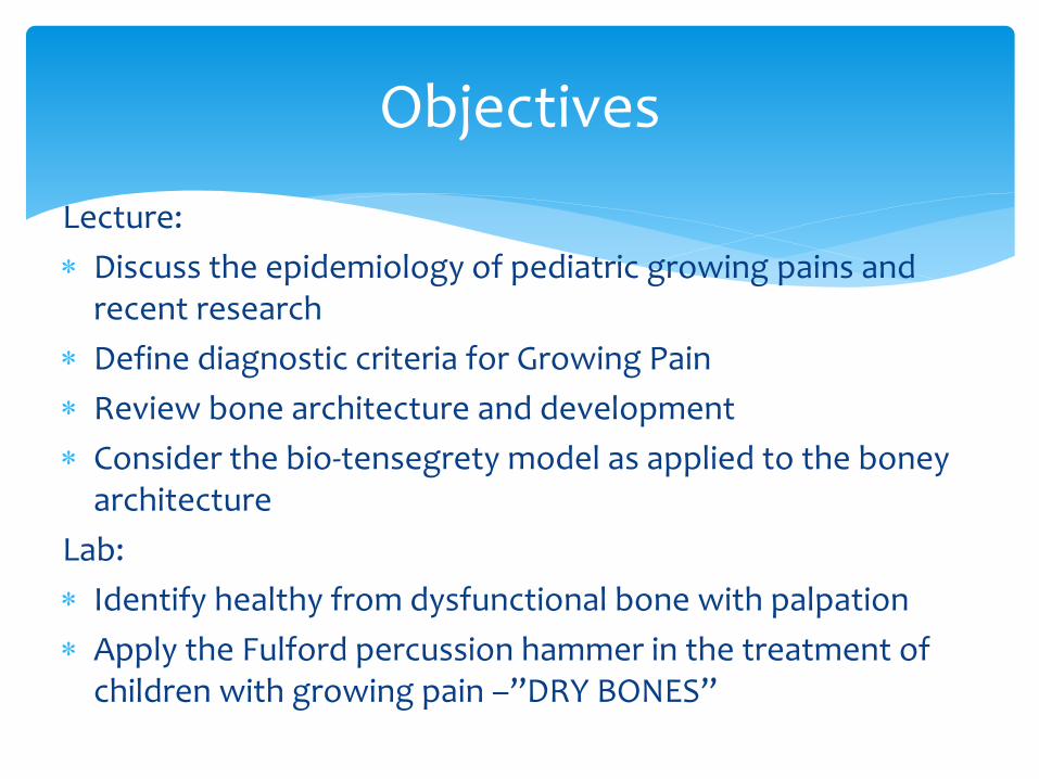
Lecture:
Discuss the epidemiology of pediatric growing pains and recent research
Define diagnostic criteria for Growing Pain
Review bone architecture and development
Consider the bio-tensegrety model as applied to the boney architecture
Lab:
Identify healthy from dysfunctional bone with palpation
Apply the Fulford percussion hammer in the treatment of children with growing pain –”DRY BONES”
Objectives

In every
case of
Disease
there was an Abnormality
in the
Bony structure

To know
All of a Bone
in it’s
Entirety
would close both ends
of an
Eternity

Most common cause of recurrent
childhood musculoskeletal pain
3-37% of children ages 3 – 12 years
32% of children ages 4 – 6 years
Associated with:
Increased prevalence of other pain syndromes
abdominal pain; headaches
5% of children with GP are heavier
Overuse syndrome
Growing Pains Epidemiology

Inclusion Exclusions
Nature of Pain Intermittent, some pain-free days
Persistent; increasing in intensity
Nights
Unilateral or Bilateral Bilateral Unilateral
Location Anterior thigh, calf, posterior knee, in muscles
Joint pain
Onset of pain Later afternoon or evening
Pain still present next morning
Physical Examination Normal Swelling, erythema, tenderness, localized trauma, infection, reduced joint ROM, limping
Tests Normal Elevated ESR, X-Rays, Bone Scan
Diagnostic Criteria

Date First Author Sample size
Research design
Findings New Theory
2004 Hashkes, PJ GP = 44 No GP = 46
Case Control Dolorimeter (pressure)
GP group had lower pain threshold
GP may be a variant of non-inflammatory pain syndrome
2005 Friedland, O GP = 39 No GP = 38
Case control; US bone speed of tibia and radius
GP group had reduced tibial bone speed
GP may represent a local overuse syndrome
2005 Hashkes, PJ GP = 11 No GP = 12
Case Control Bone Scintigraphy, tibia
GP group did not have altered vascular perfusion
GP is not assoc with altered vascular perfusion
Growing Pains Etiology-Theory
Development (From A. Evans Review Article in 2008)

Date First Author
Sample size
Research design
Findings New Theory
2010 Uziel, Y GP = 44 No GP = 38
5-year follow-up Outcome Trial
GP has a benign prognosis; GP patients did not exhibit other pain syndromes at 5 years
Probably represents a pain amplification syndrome of early childhood
Research since 2008…
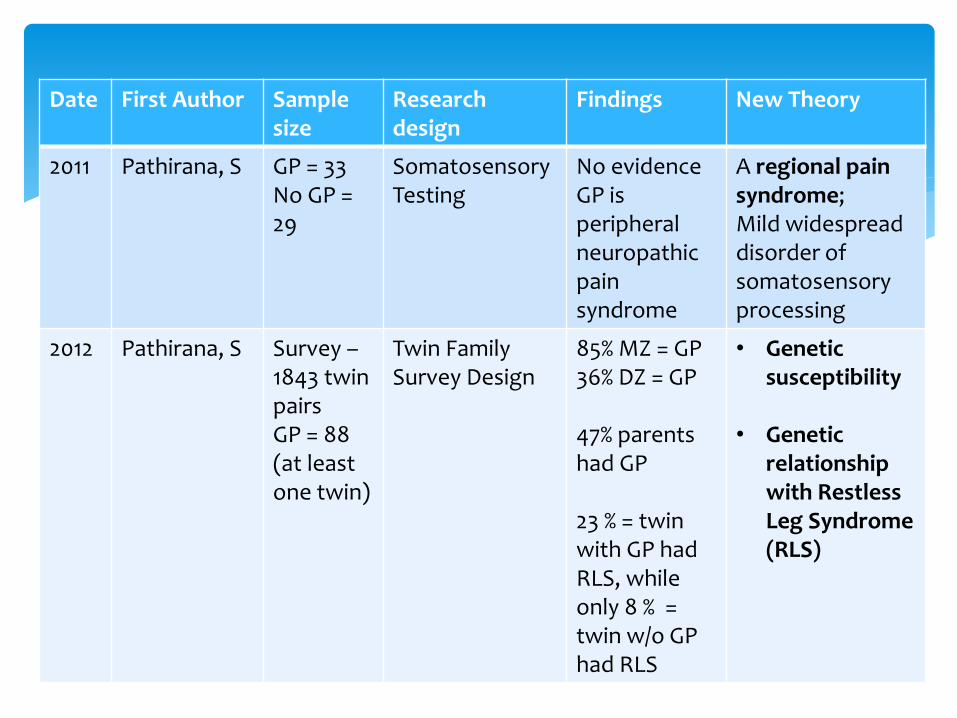
Date First Author Sample size
Research design
Findings New Theory
2011 Pathirana, S GP = 33 No GP = 29
Somatosensory Testing
No evidence GP is peripheral neuropathic pain syndrome
A regional pain syndrome; Mild widespread disorder of somatosensory processing
2012 Pathirana, S Survey – 1843 twin pairs GP = 88 (at least one twin)
Twin Family Survey Design
85% MZ = GP 36% DZ = GP 47% parents had GP 23 % = twin with GP had RLS, while only 8 % = twin w/o GP had RLS
• Genetic susceptibility
• Genetic
relationship with Restless Leg Syndrome (RLS)

Continues to be a puzzle
Literature – plentiful, anecdotal
Main theories:
Anatomical
postural, scoliosis, genu valgum, pes planus
Fatigue (overuse syndrome)
Psychological (pain amplification)
Non-inflammatory, regional pain syndrome or disorder of the somatosensory system
Genetic component
So, current hypotheses…after 190 since first described in 1823….

Osteoblasts
Derived from unspecialized mesenchymal cells
Basophilic, mononuclear, cuboidal in shape
Synthesizes and secretes collagen fibers and other organic components
Build the extracellular matrix of bone tissue; become trapped in their own secretions and become
Bony Development A very active process…

Osteoblasts becoming Osteocytes embedded in bony matrix
Osteocytes
Ellipsoid cell bond, few organelles, are mononuclear
Maintain the metabolism
Exchange of nutrients and waste with the blood = RESPIRATION!!
Possess fine, dendritic processes which project into the matrix
gap junctions and communication

Large cells
15-20 nuclei
Derived from fusion of monocytes within bone marrow
Lie in close contact with bone surface in Howship’s Lacunae
Function –
local removal of bone during resorption via secretion of lysosomes and acids and digests protein and mineral components; VERY ACTIVE PROCESS!!
Osteoclasts

Coordinated effort of
Osteoclasts
Secrete acids and enzymes that secrete the hard bone matrix by tunneling into the bone, creating channels that allow in capillaries and osteoblasts
Osteoblasts
Fill the channels with concentric deposits of new bone matrix
5-10% of all your bone is dissolved and replaced each year
Regulates serum levels of calcium
Bone Remodeling Video
http://www.youtube.com/watch?feature=player_detailpage&v=78RBpWSOl08 (Video developed by Amgen)
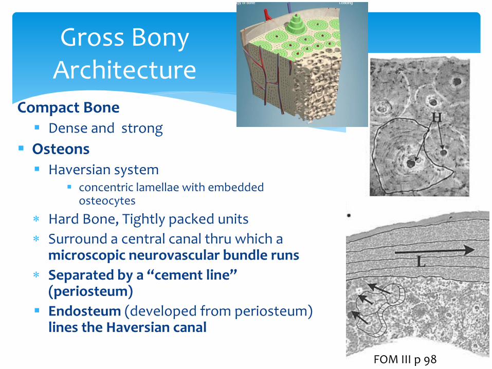
Gross Bony Architecture
Compact Bone Dense and strong
Osteons Haversian system
concentric lamellae with embedded osteocytes
Hard Bone, Tightly packed units
Surround a central canal thru which a microscopic neurovascular bundle runs
Separated by a “cement line” (periosteum)
Endosteum (developed from periosteum) lines the Haversian canal
FOM III p 98

Development of Periosteum/Endosteum
http://www.youtube.com/watch?v=X6E5Rz9tOKE&list=PLRrCFhmVYxNkMN675bc5H17WjN3YizLwt&feature=player_detailpage
Periosteum:
Nociceptive nerves
Provides nourishment to the osteum through the blood supply
Attached to bone by strong Sharpey’s fibers (outer and lamellar layers)
Acts as the attachments sites for muscles and tendons
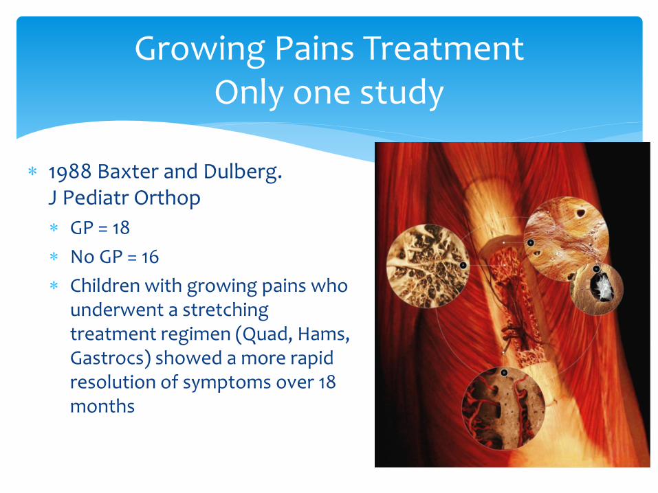
1988 Baxter and Dulberg. J Pediatr Orthop
GP = 18
No GP = 16
Children with growing pains who underwent a stretching treatment regimen (Quad, Hams, Gastrocs) showed a more rapid resolution of symptoms over 18 months
Growing Pains Treatment Only one study

Cardiovascular flow
Lymphatic flow
Expansion and Recoil of alveoli
Axoplasmotic flow of nerve conduction
Cerebral Spinal Fluid fluxuations
Inherent motility of the cranial system
BONY REMODELING fluctuations
And more….
Consider the Numerous Biological Waveforms

Addressing the Strain via the biological waveforms
Waves are encoders and carriers of information
Two waves are “In Phase”
When they overlap with peaks and troughs that are the same
“Constructive interference”
Can enhance the waves
“Out of Phase”
One wave peaks while the other troughs
“Destructive interference”
Can dampen down the waves
Can negate the strain pattern
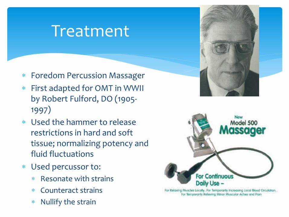
Treatment
Foredom Percussion Massager
First adapted for OMT in WWII by Robert Fulford, DO (1905-1997)
Used the hammer to release restrictions in hard and soft tissue; normalizing potency and fluid fluctuations
Used percussor to:
Resonate with strains
Counteract strains
Nullify the strain

Hands-on Lab
Patient Evaluation
Cranial Rhythm from the lower extremities
Boney health
Periosteum
Fascial Strains
Compare above and below the knee on each side
Ankle, Knee, Hip, Pelvis
Identify SI dysfunctions
Treatment
BLT
Fibula
Tibia
Knee Joint
Apply the percussor to the lower extremities
Ankle
Knee
Pelvis
Thorax

Osteopathic Assessment
Either lay your hands on top of the ankles OR
Lift the ankle Be careful not to inhibit the
lower extremity cranial rhythm
Check for rate and amplitude of internal and external rotation in the cranial field of the extremities
GP children have poor amplitude and decreased rate
GP children have tibias that feel like “dry dust”
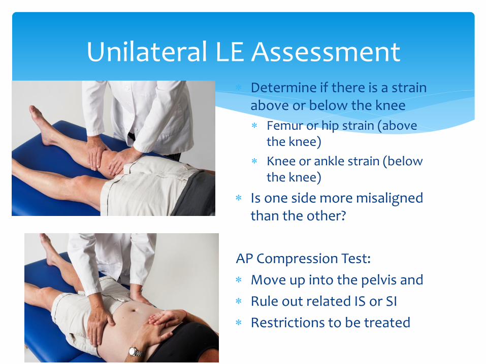
Unilateral LE Assessment Determine if there is a strain
above or below the knee
Femur or hip strain (above the knee)
Knee or ankle strain (below the knee)
Is one side more misaligned than the other?
AP Compression Test:
Move up into the pelvis and
Rule out related IS or SI
Restrictions to be treated

Fibula BLT
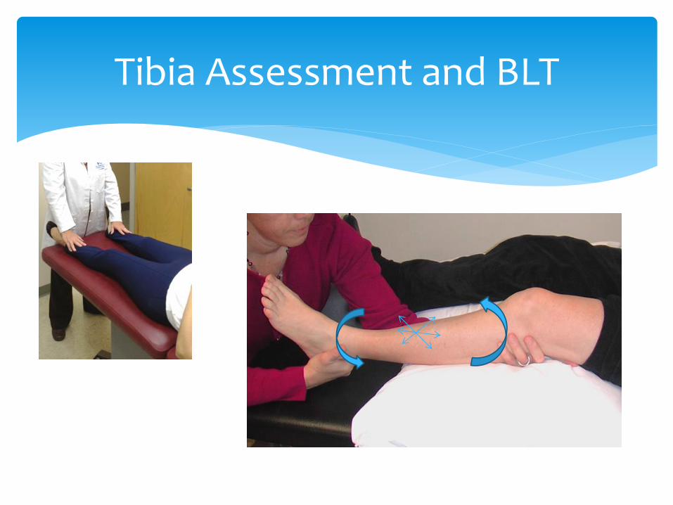
Tibia Assessment and BLT

Treatment of GP with “Fulford” Percussor

Capobianco, A. Clinical Case Studies to Support a Percussor Generated Vibratory Component in OMT. The Cranial Letter:2012;65:11-17.
Chila, A. Exec Ed. Foundations of Osteopathic Medicine, 3rd Ed., Lippincott Williams & Wilkins, Philadelphia, PA, 2011.
Evans, AM, et al. Prevalence of “Growing Pains” in Young Children. J Pediatr 2004;145:255-8.
Evans, AM. Review - Growing pains: contemporary knowledge and recommended practice. Journal of Foot and Ankle Research 2008,1:4 doi:10.1186/1757-1146-1-4.
Hankinson, D. The Message in the Bones; AAO Convocation March 2012 Lecture Presentation
Sadler, TW Editor. Langman’s Medical Embryology. 11th Ed. North America. Lippincott Williams and Wilkins, Philadelphia, PA, 2010.
Primal Anatomy 3D images via NSU HPD Library Resources
Uziel, Y, et al. Five-Year Outcome of Children with “growing pains”: Correlations with pain threshold. J Pediatr 2010;156:838-40. May 2010
References