contents2017.koa.or.kr/file/simposium.pdfcontents oct. 20th. 2017. ... saudi arabia 136 14:45-15:00...
TRANSCRIPT


Contents
■ Oct. 19th
. 2017. Thu | Grand Ballroom 1 ■
08:00-08:10 Opening Ceremony
08:10-09:20 LCP
Sung Taek Jung
08:10-08:25Combined Pemberton and femoral varus osteotomies in Legg-Calvé-Perthes disease
Ting-Ming Wang / NATIONAL TAIWAN UNIVERSITY HOPITAL, TAIWAN14
08:25-08:40Treatment Options for Perthes disease
Saw Aik / UNIVERSITY MALAYA MEDICAL CENTRE, MALAYSIA16
08:40-08:55Tonnis Triple Osteotomy for Containment of Perthes Disease
Yukun Wang / BEIJING JISHUITAN HOSPITAL, CHINA18
08:55-09:10Our treatment strategy for children with Legg-Calvé-Perthes disease
Ryosuke Yamaguchi / FUKUOKA CHILDREN'S HOSPITAL, JAPAN20
09:10-09:20 Discussion
09:40-10:10 Coffee Break
10:10-11:103D printing in reconstruction surgery
Ye Yeon Won ・ Yang Soo Kim
10:10-10:22Additive manufacturing and FE Simulation for Biomechanics
Kohei Murase / NAGOYA UNIVERSITY, JAPAN22
10:22-10:34Corrective osteotomy or fracture reduction by 3D mirroring
Ahmet Mehmet DemirtaŞ / ANKARA UNIVERSITY FACULTY OF MEDICINE, TURKEY
10:34-10:46Digital surgery techniques used in craniofacial bone reconstruction
Zhigang Cai / PEKING UNIVERSITY, CHINA23
10:46-10:583D Printing in Musculoskeletal Oncology
Yang Guk Chung / CATHOLIC UNIV.25
10:58-11:10 Discussion
11:10-12:10Periprosthetic fracture of the femur: Reduction and Fixation
Byung Woo Min
11:10~11:20Periprosthetic Fracture of the Femur: Decision making (is the stem stable?)
Byung Woo Min / KEIMYUNG UNIV.34
11:20~11:35Biomechanical challenges of periprosthetic fractures
Toru Sato / NATIONAL HOSPITAL ORGANIZATION OKAYAMA MEDICAL CENTER, JAPAN36
11:35~11:50Principles of reduction and fixation
Tak Wing Lau / THE UNIVERSITY OF HONG KONG, QUEEN MARY HOSPITAL, HONG KONG37
11:50~12:05Case-based lecture-fracture around the femoral stem
Takeshi Sawaguchi / KANAZAWA UNIVERSITY, TOYOMA MUNICIPAL HOSPITAL, JAPAN38
12:05~12:10 Discussion
12:30-13:30 Lunch

Contents
■ Oct. 19th
. 2017. Thu | Grand Ballroom 2 ■
08:00-08:10 Opening Ceremony
08:10-09:40Treatment of Sports-related Injuries & Diseases
Sang Hun Ko ・ Chul Won Ha
08:10-08:25
The evaluation and conservative treatment of internal impingement of shoulder for throwing
athlete
Toru Morihara / KYOTO PREFECTURAL UNIVERSITY, JAPAN60
08:25-08:40Shoulder injuries among japanase professional baseball players
Shin Yokoya / HIROSHIMA UNIVERSITY, JAPAN63
08:40-08:55Biological application in ACL surgery
Chih-Hwa Chen / TAIPEI MEDICAL UNIV HOSPITAL, TAIWAN65
08:55-09:10Trochleoplasty In Patella Instability...A Necessary Evil?
James Hui / NATIONAL UNIVERSITY SINGAPORE HOSPITAL, SINGAPORE
09:10-09:25
Does cutting the lateral retinaculum and reconstructing MPFL result in improved patellofemoral
incongruency?
Hua Feng / BEIJING JISHUITAN HOSPITAL, CHINA67
09:25-09:40 Discussion
09:40-10:10 Coffee Break
10:10-12:10TFCC Injury and DRUJ Instability
Soo Hong Han
10:10-10:25Distal Radioulnar Joint Functional Anatomy
Il-Jung Park / CATHOLIC UNIV.70
10:25-10:40Treatment of Distal radioulnar joint instability
Jong Pil Kim / DANKOOK UNIV.77
10:40-10:55DRUJ Instability: My preferred management
Abhijeet L. Wahegaonkar / JEHANGIR HOSPITAL, INDIA
10:55-11:10Arthroscopic Repair for the TFCC foveal Tear
Bo Liu / BEIJING JISHUITAN HOSPITAL, CHINA84
11:10-11:25The Surgical treatment for TFCC foveal tear- Open vs Arthroscopic repair
Yukio Abe / SAISEKAI SHIMONOSEKI GENERAL HOSPITAL, JAPAN85
11:25-11:40Arthroscopic TFCC reconstruction with tendon graft
Wing Lim Tse / PRINCE OF WALES HOSPITAL, HONG KONG92
11:40-11:55Surgical treatment of ECU tendinopathy associated with TFCC Injury
Young Keun Lee / CHONBUK NATIONAL UNIV.93
11:55-12:10 Discussion
12:30-13:30 Lunch
■ Oct. 19th
. 2017. Thu | Grand Ballroom 1 ■
13:30-14:30Cutting Edge Technology in the Field of ASAMI Society of Asian Countries
Chang Hoon Jeong
13:30-13:40
Comparison of Joint Distraction and Non-distraction using an Ilizarov External Fixator in the
Treatment of Ankle Fractures in Older Patients
Koji Nozaka / AKITA UNIVERSITY GRADUATE SCHOOL OF MEDICINE, JAPAN42
13:40-13:50Treatment of the lower limb deformities by a multi-axial external fixation system
Masaki Matsushita / NAGOYA UNIVERSITY SCHOOL OF MEDICINE, JAPAN44
13:50-14:00
Novel Management of Larger Bone Defect: Combination of Biomaterials and Distraction
Osteogenesis Technique
Gang Li / THE CHINESE UNIVERSITY of HONG KONG, PRINCE OF WALES HOSPITAL, HONG KONG46
14:00-14:10Lower Limb Reconstruction in paediatric Orthopedics
Andrew Lim Kean Seng / NATIONAL UNIVERSITY HOSPITAL OF SINGAPORE, SINGAPORE48
14:10-14:20Game changers in Limb lengthening and Deformity Correction Field
Dong Hoon Lee / YONSEI UNIV.49
14:20-14:30 Discussion
14:30-15:30Sacro-pelvic bone cancer surgery
Hyun Guy Kang
14:30-14:45Resection and reconstruction for pelvic ring cancer
Tetsuo Hotta / NIIGATA UNIVERSITY HOSPITAL, JAPAN54
14:45-15:00Sacrectomy: Modern surgical technique
Hwan Seong Cho / SEOUL NATIONAL UNIV.56
15:00-15:15Computer-assisted pelvic ring cancer surgery
Kwok Chuen Wong / PRINCE OF WALES HOSPITAL, HONG KONG57
15:18-15:30 Discussion

Contents
■ Oct. 20th. 2017. Fri | Grand Ballroom 1 ■
08:30-09:30 International Perspective in Recent Shoulder Updates I
Chang Hyuk Choi ・ Yong Min Chun
08:30-08:42Arthroscopic Reconstruction of the Acromioclavicular Joint
James Tan Chung Hui / KHOO TECK PUAT HOSPITAL, SINGAPORE116
08:42-08:54L- Shaped Arthroscopic Posterior Capsular Release In Frozen Shoulder
Mohamed Gamal Morsy / ALEXANDRIA UNIVERSITY, EGYPT117
08:54-09:06Management options in Young Arthritic shoulder- now and in future?
Roshan Wade / GSMC & KEM HOSPITAL, INDIA
09:06-09:18
Anatomical Medial Patellofermoral Ligament Insertion to the Patella: More Than a Cadaveric
Study
Teo Seow Hui / OSAKA POLICE HOSPITAL, MALAYSIA118
09:18-09:30 Discussion
09:30-10:00 Coffee Break
10:00-11:00 International Perspective: Injury of Shoulder and Elbow
Jin Young Park ・ Joo Han Oh
10:00-10:12Arthroscopic AC Joint Reconstruction and Management of Concomitant Injuries
JEREMY JAMES C. MUNJI / DELOS SANTOS MEDICAL CENTER, PHILIPPINES120
10:12-10:24Traumatic posteromedial varus instability of elbow: My technique
Ming Xiang / SICHUAN PROVINCIAL ORTHOPAEDICS HOSPITAL CHENGDU, CHINA121
10:24-10:36Strategic approach of first time dislocation of shoulder
Peter Wai Pan Yau / THE UNIVERSITY OF HONG KONG, HONG KONG123
10:36-10:48Arthroscopic treatment for recurrent shoulder dislocation: Vietnamese Experience
Trần Trung Dũng / ST PAUL UNIVERSITY HOSPITAL, VIETNAM125
10:48-11:00 Discussion
11:00-12:00 International Perspective in Recent Shoulder Updates II
Yong Girl Rhee ・ Jae Chul Yoo
11:00-11:12
Completion repair shows better healing characteristics in comparison with insitu repair in the
partial thickness bursal rotator cuff tear
Arel Gereli / ACIBADEM UNIVERSITY SCHOOL OF MEDICINE, TURKEY128
11:12-11:24
No relationship between critical shoulder angle and glenoid erosion after shoulder
hemiarthroplasty: a comparative radiographic study
Simone Cerciello / CASA DI CURA VILLA BETANIA GIOMI, ITALY130
11:24-11:36
Biceps tendon tenotomy or tenodesis, what is the evidence?what i do
Hossein Saremi / BESAT HOSPITAL HAMEDAN UNIVERSITY OF MEDICAL SCIENCES HAMADAN, IRAN
132
11:36-11:48
Bone integrity and morphology of the coracoid process after the coracoid transfer for the
recurrent anterior shoulder instability
Makoto Tanaka / OSAKA POLICE HOSPITAL, JAPAN133
11:48-12:00 Discussion
■ Oct. 19th
. 2017. Thu | Grand Ballroom 2 ■
13:30-14:30건강보험과 ABC 원가분석
강승백, 한승범
13:30-13:45건강보험 보장성 강화 대책
정통령 / 보건복지부 보험급여과장
13:45-14:00문재인 케어와 정형외과
손영래 / 보건복지부 의료자원정책과장
14:00-14:15원가기반 수가결정방법
정성출 / 갈렙ABC컨설팅 대표이사
14:15-14:30 Discussion
14:30-15:30Updates on peripheral nerve surgery
Jong Woong Park ・ Kanit Sananpanich
14:30~14:40Distal nerve transfer for peripheral nerve injury in BPI and tetraplegia
Kanit Sananpanich / CHIANG MAI UNIVERSITY HOSPITAL, THAILAND102
14:40~14:50Treatment Strategies for Neuromas and neuropathic pain
Abhijeet L. Wahegaonkar / JEHANGIR HOSPITAL, INDIA
14:50~15:00Cutting-edge technology for the enhanced neural regeneration
Jong Woong Park / KOREA UNIV.104
15:00~15:10Conduits for Peripheral Nerve Regeneration
Joo Yup Lee / CATHOLIC UNIV.106
15:10~15:20The role of stem cell transplantation for peripheral nerve regeneration
Jae Kwang Kim / ULSAN UNIV.113
15:20~15:30 Discussion

Contents
■ Oct. 20th. 2017. Fri | Grand Ballroom 2 ■
08:00-09:30Current issues in knee arthroplasty
Myung Chul Lee ・ Choong Hyeok Choi
08:00-08:10Tibial preservation in Fixed & mobile medial UKA: rational & technique.
Aree Tanavalee / CHULALONGKORN HOSPITAL, THAILAND150
08:10-08:20MIS TKA: are you still there?
Aree Tanavalee / CHULALONGKORN HOSPITAL, THAILAND152
08:20-08:40Kinematics after TKA—Normal or Durable?
Tzai-Chiu Yu / R AND D CENTER OF JOINT RECONSTRUCTION, TZU-CHI MEDICAL CENTE, TAIWAN154
08:40-08:55Infected TKA: Update on Diagnosis and Treatment
Chong Bum Chang / SEOUL NATIONAL UNIV.156
08:55-09:10Revision TKA: Managing bone defect
Seung-Beom Han / KOREA UNIV.162
09:10-09:30 Discussion
09:30-10:00 Coffee Break
10:00-11:00New surgical trend in cervical spine
Hak Sun Kim ・ Jin Sup Yeom
10:00-10:12
Distraction Arthrodesis of the C1-C2 Facet Joint with Preservation of the C2 Root for the
Management of Intractable Occipital Neuralgia Caused by C2 Root Compression
QuanYou Li / CYANBIAN UNIVERSITY HOSPITAL, CHINA168
10:12-10:24
The Usefulness of Dynamic MRI in Cervical Myelopathy Caused by OPLL for Selective Surgical
Decompression
Yehlen Francis Reyes Saligumba / ST. LUKE'S MEDICAL CENTER GLOBAL CITY, PHILIPPINES170
10:24-10:36Free-hand placement of C7 laminar screws: accuracy and safety in 43 consecutive patients
Feng Shen / QINGDAO UNIVERSITY HOSPITAL, CHINA172
10:36-10:48ACDF with Total En Bloc Resection of Uncinate
Michael Nelson Perez Lim / EAST RAMON MAGSAYSAY MEMORIAL MEDICAL CENTER, PHILIPPINES174
10:48-11:00 Discussion
11:00-11:40 Latest Trends in Spinal Surgery
Jae Hyup Lee
11:00~11:30Surgical Treatment of metastatic spinal tumors
Masato Tanaka / OKAYAMA UNIVERSITY HOSPITAL, JAPAN176
11:30~11:40 Discussion
■ Oct. 20th. 2017. Fri | Grand Ballroom 1 ■
12:00-12:20 Lunchoen Symposium (Room A, B)
12:00-12:20 The 62nd Regular General Assembly(Room B) & Lunch(Room A, B)
13:30-14:00 Presentation of Scientific Award Paper(Room B)
14:00-14:30 President Lecture(Room B)
14:30-15:30 limb reconstruction with microsurgical technique
Sang Hyun Lee ・ Joo Yeoup Lee
14:30-14:45
A Modified Technique for Harvesting the Reverse Sural Artery Flap from the Upper Part of the
Leg: Inclusion of a Gastrocnemius Muscle Cuff Around the Sural Pedicle
Nedhal. A. Alqumber / PRINCE SULTAN MILITARY MEDICAL CENTER, SAUDI ARABIA136
14:45-15:00Immediate closure of Gustilo type IIIB open tibia fracture with calf muscle flap
Jong-Woo Kang / KOREA UNIV.137
15:00-15:15
Fixation methods favorite for soft tissue around elbow in complicated fractures around elbow
including ulnar nerve injury
Soo Min Cha / CHUNGNAM NATIONAL UNIV.139
15:15-15:30 Discussion
15:30-16:00 Coffee Break
16:00-17:30 Complications in Total Hip Arthroplasty
Gun Il Im ・ Kee Hyung Rhyu
16:00-16:15Vascular injury after revision arthroplasty of the hip- a case report
Piyush Mukund Sonje / INDIA142
16:15-16:30
Periprosthetic Femoral Fractures after Hip Arthroplasty
Ravi Teja Rudraraju / CENTRE OF EXCELLENCE FOR JOINT REPLACEMENTS, SVS MEDICAL COLLEGE, INDIA
143
16:30-16:45Imaging in Pelvic and Acetabular Surgery – How to Avoid It
Kristoffer Roland U. Roa / SOUTHERN PHILIPPINES MEDICAL CENTER, PHILIPPINES145
16:45-17:00
Mid-term Results of Open Debridement for Labral Tear Using Anterolateral Approach with a
Mini-incision
Ashraf Mohamed Almutasim / Alamal NATIONAL HOSPITAL, SUDAN146
17:00-17:30 Discussion

Contents
■ Oct. 20th. 2017. Fri | Grand Ballroom 2 ■
12:20-13:30 Lunch
13:30-14:00 Presentation of Scientific Award Paper
14:00-14:30 President Lecture
14:30-15:30International Spine Session 1
Jin Sup Yeom
14:30~14:45
Giant Cell Tumor of the Spine: 3 Patients Treated with Intra-lesional Surgery and Denosumab
Chemotherapy Post-excision
Romel P. Estillore / UNIVERSITY OF SANTO TOMAS HOSPITAL, PHILIPPINES178
14:45~15:00
Using intra-operatively ideal entrance point and angle of screws with a set square for lower
cervical pedicle screw placement. Accurate result?
Tran Hoang Manh / KHANH HOA GENERAL HOSPITAL, VIETNAM179
15:00~15:15Do Van Minh / HUE CENTRAL HOSPITAL, VIETNAM
15:15~15:30 Discussion
15:00-16:00 Coffee Break
16:00-18:00International Spine Session 2
Jin Sup Yeom
16:00~16:15
Unusual presentation of tuberculosis in cervical spine: challenges faced by Spine surgeons in
developing country
Dinesh Kafle / TRIVHUVAN UNIVERSITY, NEPAL182
16:15~16:30
TIMING OF SURGERY AND TREATMENT IN TRAUMATIC CENTRAL CORD SYNDROME: OUR
LOCAL EXPERIENCE AND REVIEW OF LITERATURE
Mary Ruth A. Padua / EAST AVENUE MEDICAL CENTER, PHILIPPINES183
16:30~16:45
C5 PALSY AFTER POSTERIOR CERVICAL RECONSTRUCTION BY PEDICLE SCREW FIXATION: 2
CASES REPORT
Quyen Nguyen Ngoc / 108 MILITARY CENTRAL HOSPITAL, VIETNAM184
16:45~17:00
COMBINING YEOM'S AND SHIRAISHI’S TECHNIQUE FOR THE TREATMENT OF CERVICAL
SPONDYLOTIC MYELOPATHY
Nguyen Huu Thuyet / CAN THO UNIVERSITY OF MEDICINE AND PHARMACY, VIETNAM186
17:00~17:15Cervical Tuberculosis With Big Retropharyngeal Abscess: A Case Report
Huynh Chi Hung / PHAM NGOC THACH MEDICAL UNIVERSITY, VIETNAM187
17:15~17:30
A Novel Trajectory of C7 Screws: Evaluation using 3-Dimentional Computed Tomography and
Simulation Program to Compare with a Pre-existing Trajectory
Chee Kean Lee / MAHKOTA MEDICAL CENTRE, MALAYSIA189
17:30~17:45Cervical Spine Alignment – What Have We Understood In The Past Few Years
Hwee Weng Dennis Hey / NATIONAL UNIVERSITY HEALTH SYSTEM, SINGAPORE191
17:45~18:00 Discussion
■ Oct. 20th. 2017. Fri | Grand Ballroom 3 ■
10:00-11:00 Vertebral Bone Resection Using Ultrasonic Aspirator
10:00-10:15Principles and Working Mechanism of Ultrasonic Aspirator
Dongho Lee/ ASAN MEDICAL CENTER
10:15-10:30Usage of SONOPET : Cervical & Posterial Lumbar Surgery
Jae Chul Lee / SOONCHUNHYANG UNIV.
10:30-10:45Usefulness of SONOPET for Complex Thoraco-Lumbar Disorders
Yong Chan Kim/ GANGDONG KYUNGHEE UNIV.
10:45-11:00 Hands-on Session
11:00-12:00 Patient Blood Management in Orthopedic Surgery
Kyu Yeol Lee
11:00-11:25PBM in elective major orthopedic surgery and recent achievement
Gurpal Singh / NATIONAL UNIVERSITY OF SINGAPORE194
11:25-11:50The role of IV iron in PBM Minimal transfusion in orthopedic surgery is possible?
Jong Hoon Park / KOREA UNIV.196
11:50-12:00 Discussion
12:20-13:30 Lunch
13:30-14:00 Presentation of Scientific Award Paper
14:00-14:30 President Lecture
14:30-15:30 Session 1. Quantitative Measurement of Pivot Shift
Nam-Hong Choi ・ Ji-Hoon Bae
14:30-14:42Intraoperative kinematic evaluation of single- or double-bundle anterior cruciate ligament reconstruction using a navigation system
Nobuo Adachi / HIROSHIMA UNIVERSITY, JAPAN202
14:42-14:54
Quantitative Evaluation of Pivot Shift in Double-bundle Anterior Cruciate Ligament
Reconstruction Using Triaxial Accelerometer; Identifying Optimal Conditions to Restore
Anterolateral Rotational Stability
Hideyuki Koga / TOKYO MEDICAL AND DENTAL UNIVERSITY HOSPITAL, JAPAN
203
14:54-15:06Evolution of the Measurement of the rotational instability of the Knee: What’s in, What’s out?
Yung Shu Hang Patrick / THE CHINESE UNIVERSITY OF HONG KONG, HONG KONG205
15:06-15:18How to check the functional instability of ACL injured subjects during sports activities?
Jin-Goo Kim / KONKUK UNIV. 206
15:00-16:00 Coffee Break
16:00-17:00 Session 2. Revision ACL Reconstruction
Kwang-Won Lee ・ Joon Ho Wang
16:00-16:12Slope-decreasing osteotomy in treatment of revision ACL surgery
Hua Feng / BEIJING JISHUITAN HOSPITAL, CHINA212
16:12-16:24One-stage revision ACL reconstruction :Technical strategy and graft optional
Yi-Sheng Chan / CHANG GUNG MENORIAL HOSPITAL, TAIWAN213
16:24-16:36
Effects of remnant tissue preservation on clinical outcomes after anatomic double-bundle
anterior cruciate ligament reconstruction
Eiji Kondo / HOKKAIDO UNIVERSITY, JAPAN215
16:36-16:48Availability of anterolateral ligament in revision ACL reconstruction
Kyoung-Ho Yoon / KYUNGHEE UNIV.217
16:48-17:00 Discussion

The 61st Annual Congress of The Korean Orthopaedic Association
LCP
Oct. 19th. 2017. Thu | Grand Ballroom1
Sung Taek Jung
Contents
■ Oct. 21st. 2017. Sat | Room C-1 ■
08:30-09:30 Ultrasonography in Orthopaedics
Jin Young Park ・ Kyoung Dae Min
08:30-08:42Ultrasonography of the shoulder and elbow-up to date-
Katsumasa Sugimoto / NAGOYA SPORTS CLINIC, JAPAN222
08:42-08:54Ultrasonography after Rotator cuff repair
Sang-Jin Shin / EWHA WOMANS UNIV.223
08:54-09:06Evaluation and Treatment for the Hip Joint Using Ultrasonography
Pil Sung Kim / BUMIN HOSPITAL226
09:06-09:18US in Ankle Instability
Hak Jun Kim / KOREA UNIV.235
09:18-09:30 Discussion
09:30-10:00 Coffee Break
10:00-11:00 Clubfoot
Soo-Sung Park
10:00-10:10
How to manage relapsed clubfeet after Ponseti method in Japan
Daisuke Tamura / OSAKA MEDICAL CENTER AND RESERCH INSTITUTE FOR MATERNAL AND CHILD HEALTH, JAPAN
238
10:10-10:20How to manage residual clubfoot deformity after Ponseti method in Taiwan
Chia Hsieh Chang / CHANGGANG MEMORIAL HOSPITAL, TAIWAN240
10:20-10:30How to manage residual clubfoot deformity after Ponseti method in Singapore
Arjandas Mahadev / KK WOMEN'S AND CHILDREN'S HOSPITAL, SINGAPORE242
10:30-10:40How to manage residual clubfoot deformity after Ponseti method in India
Alaric John Aroojis / CENTRE FOR BONE & JOINT KOKILABEN DHIRUBHAI AMBANI HOSPITAL, INDIA244
10:40-10:50How to manage residual clubfoot deformity after Ponseti method in Bangladesh
Sarwar Ibne Salam / DHAKA MEDICAL COLLEGE HOSPITAL, BANGLADESH245
10:50-11:00 Discussion
11:00-12:00 CP
Hyun Woo Kim
11:00-11:15Decision making: operate or not to operate, when to operate
Abhay Khot / VICTORIAN ORTHOPAEDIC CENTRE, AUSTRALIA248
11:15-11:30Surgeries for correction of crouch gait
Alaric John Aroojis / CENTRE FOR BONE & JOINT KOKILABEN DHIRUBHAI AMBANI HOSPITAL, INDIA249
11:30-11:45Surgeries for spastic hip disease and spine deformity
Jason James Howard / SIDRA MEDICAL AND RESEARCH CENTER, CANADA250
11:45-12:00
Cases presentation for panel discussion
Arjandas Mahadev / KK WOMEN'S AND CHILDREN'S HOSPITAL, SINGAPORESarwar Salam / DHAKA MEDICAL COLLEGE HOSPITAL, BANGLADESH
12:00~ Closing Ceremony

• 14 • • 15 •
■ The 61st Annual Congress of the Korean Orthopaedic Association International Symposium Program & Book of Abstracts ■
Combined Pemberton and femoral varus osteotomies in Legg-Calvé-Perthes disease
Ting-Ming Wang, Ting-Chun Huang, Kuan-Wen Wu, Ken N Kuo
NATIONAL TAIWAN UNIVERSITY HOPITAL, TAIWAN
•••
Abstract
Containment of the femoral head within the acetabulum by conservative or surgical methods has been popularly
accepted as a concept for treatments of LCPD. In patients with onset over the age of 8 years and greater than
lateral pillar B or B/C class, surgical treatment was associated with improved Stulberg outcomes compared with
conservative treatments. To achieve femoral head containment with surgery, one can choose either acetabuloplasty
or proximal femoral osteotomy. However, there are still complications associated with a single procedure alone. In
this study, we proposed a combination of Pemberton osteotomy and femoral varus osteotomy as a novel alternative
treatment.
Methods
This is a retrospective comparative case series in 19 Children with LCPD underwent Pemberton osteotomies with/
without femoral varus osteotomy between July 2002 and January 2012.
The radiographic evaluations performed at minimum 5 years post-operatively included migration index, center-
edge angle, leg length discrepancy, Mose grading and Stulberg classification. The functional evaluations at latest
clinical visit included IOWA hip score, SF36 bodily limitation and pain.
Results
Post-OP femoral head coverage (Center-Edge angle: P<0.001, Migration index: P<0.001), sphericity (Mose
grading: P=0.001), and hip congruency (P=0.006) were all significantly better in Combined Group.
IOWA Hip Score were significantly better in Combined Group than Alone Group (P=0.02). SF36 bodily limitation
and pain did not reveal significant differences between the two groups.
All cases in combined group had leg length discrepancy less than one centimeter.
Conclusions
Combined Group improved femoral head containment, leg length discrepancy, remodeling and functions compared
with alone Group.

• 17 •
International Symposium Program & Book of Abstracts ■
• 16 •
■ The 61st Annual Congress of the Korean Orthopaedic Association
neck or both. Prophylactic epiphysiodesis of the trochanter or trochanteric transfer can be considered for
these conditions. Total hip replacement would be indicated for those prevented with advanced secondary
osteoarthritis at later stage of life.
There has been reports of favourable outcome following hip distraction using external fixator for Perthes
disease. Medical therapy using bisphoshonates has also been shown to reduce the risk of femoral head
deformation. Better understanding of the disease and evidence based studies are needed for these newer
treatment options.
Treatment Options for Perthes disease
Saw Aik
UNIVERSITY MALAYA MEDICAL CENTRE, MALAYSIA
•••
Aetiology of Perthes disease remained unknown despite the fact that it has been recognised a special entity
for more than 100 years. For this reason, treatment is mainly to improve functional outcome or prevent of
long term complications. In most cases, the condition is self-limiting and following reparative phase the
femoral head remained spherical and congruent. However, for cases that became symptomatic at older age
and showing radiological features of extensive head necrosis, risk of subsequent head deformation is very
high. Active treatment for Perthes has been focused on these hips since they were associated with poor
long term prognosis.
Prolonged bed rest and non-weight bearing with crutches have been shown to be not effective in the
management. Concept of containment surgery to increase the coverage for femoral head during the early
stages of injury has been shown to modify the subsequent progress of the disease. Although there has been
no level one study to support the effectiveness of containment surgery, comparative studies have shown
higher percentage of spherical heads in operated compared to non-operated hips. In addition, duration of
disease (especially fragmentation phase) was noted to be shorter following surgery. Proximal femur varus
derotation osteotomy and acetabular directional osteotomy are effective to improve the antero-lateral
coverage of the femoral head, although there is no evidence to indicate which method is better. These
procedures should be performed during fragmentation phase of the condition.
For cases presented late with flattened head, shelf and Chiari osteotomy may help to improve the contact
surface between femora head and the acetabulum to relief pain. In selective cases with showing hinged
abduction, valgus abduction osteotomy or Cheilectomy may offer improvement of hip motion and relief
of symptoms. Trendelenburg gait may be contributed by greater trochanter overgrowth, shortened femoral

• 19 •
International Symposium Program & Book of Abstracts ■
• 18 •
■ The 61st Annual Congress of the Korean Orthopaedic Association
Those who was 8 years or more at operation, Lc or Lb/c if with head subluxation, although after
sophisticated triple osteotomy, were 3.76 times (P<0.001; 95% CI) than as those < 8y or Lb or without
subluxation to have a poor outcome, according to binary logistic regression analyses.
Conclusion: Triple osteotomy is a good procedure for containment of subluxed and/or enlarged femoral
head in Joseph IIa or IIb stage Perthes disease. LLD after operation is minimal.
Tonnis Triple Osteotomy for Containment of Perthes Disease
Yukun Wang
BEIJING JISHUITAN HOSPITAL, CHINA
•••
[ABSTRACT]
Object: To review and introduce the experience of the operative containment of Perthes disease via
Tonnis triple osteotomy in Department of Pediatric Orthopaedics in Jishuitan Hospital.
Method: From March 17, 2011 to April 28, 2015, 67 Perthes cases were managed by triple osteotomy
under the indication of 1st, L-Pillar B with the onset age >= 8y; 2nd, head subluxation of onset age < 8y;
and 3rd, Lc while first presentation. Of these children, 46 were followed at least 22 months. The average
follow-up time was 37.7 (22~61 months). There were only 5 girls, aging from 4y3m to 11 years at a mean
of 7y9m. At the time of operation, the Joseph stages were Ib 6, IIa 19, IIb 20, IIIa 1 cases; the Herring
lateral pillar were b 10, b/c 27, c 9 children respectively. Femoral head subluxation occurred in 36 cases,
and in detail, 9 in Lb, 19 in b/c, and 8 in Lc.
Results: Mean follow-up time was 37.7 months. Stulberg I were rated in 2 children, one was Lc with age
of 6y6m when operated and another boy was Lb without subluxation, although aged 8y while operation.
Stulberg II and III were in 36 and 8 children, respectively. Leg-length discrepancy(LLD) was from 0~15
mm at an average of 3.2mm. Complete peroneal paralysis occurred in a boy. At final follow-up, the
muscle force of the extensor hallucis longus was rated grade II and the extensor digitorum longus was III.
All the others muscles around ankle joint were grade V.

The 61st Annual Congress of The Korean Orthopaedic Association
• 20 •
■ The 61st Annual Congress of the Korean Orthopaedic Association
Our treatment strategy for children with Legg-Calvé-Perthes disease
Ryosuke Yamaguchi1, Tomoyuki Nakamura1, Akifusa Wada2, Toru Yamaguchi1, Haruhisa Yanagida1, Kazuyki Takamura1, Yasuharu Nakashima3
1Department of Orthopaedic and Spine Surgery, Fukuoka Children’s Hospital2Department of Orthopaedic Surgery, Saga Handicapped Children’s Hospital
3Department of Orthopaedic Surgery, Graduate School of Medical Sciences, Kyushu University
•••
Abstract
Legg-Calvé-Perthes disease (LCPD) is a juvenile form of idiopathic osteonecrosis of the femoral head that
can lead to permanent femoral head deformity and subsequent osteoarthritis. Various nonoperative and
operative treatments have been applied based on the concept of containment of the femoral head in the
acetabulum.
In our institution, we had basically inducted Nishio’s brace (non-weight bearing abduction brace) for two
years to almost all LCPD patients until 2009. We consequently found that clinical outcome using Nishio’s
brace demonstrated favorable results for LCPD patients under 8 years old at onset, but unsatisfactory
results for patients over 8 years old or with an extended necrotic lesion. Therefore, we currently
recommend a femoral osteotomy for older LCPD patients with an extended necrotic lesion. Flexion
(anterior rotation) and varus osteotomy (FVO) of the proximal femur has been performed using a locking
compression plate in our institution. The concept of FVO is to rotate the intact area or repaired area in the
posterolateral portion of the femoral head to the superior portion for preventing the progression of femoral
head collapse, in addition to the containment of the femoral head. Non-weight bearing walking with
crutches for a year following the surgery effectively maintains a spherical femoral head even for older
LCPD patients.
Ye Yeon Won / Yang Soo Kim
3D printing in reconstruction surgery
Oct. 19th. 2017. Thu | Grand Ballroom1

• 22 • • 23 •
■ The 61st Annual Congress of the Korean Orthopaedic Association International Symposium Program & Book of Abstracts ■
Additive manufacturing and FE Simulation for Biomechanics
Kohei Murase
NAGOYA UNIVERSITY, JAPAN
•••
Three-dimensional printing technology (Additive Manufacturing: AM) is one of the great epoch
technology in engineering field. Remarkable growth of this innovative technique allows the intricate
fabrication with complicated geometry and structure, to construct from biocompatibility materials.
Medical applications such as custom-made prosthesis and implants, will be the one of most exciting field
for AM. This study introduces AM activities in medical engineering in Japan, for instance, 3D bone shape
fidelity and equivalent mechanical strength for development medical implant, concurrency design and
fatigue predictions which is combined with FE simulations.
Digital surgery techniques used in craniofacial bone reconstruction
Zhigang Cai
PEKING UNIVERSITY, CHINA
•••
Abstract
The craniofacial hard tissue defect caused by head neck ablative tumor surgery, osteomyelitis or severe
trauma would physiologically and psychologically affect patients' life quality. However, the complexity
of this regional anatomy makes it a great challenge for plastic surgeons to reconstruct the facial contour
and rehabilitate the occlusion function. Nowadays, the optional approaches for craniofacial reconstruction
include reconstructive titanium plate, nonvascularized bone grafts, vascularized osteocutaneous flaps
and distraction osteogenesis etc. Improvement in microsurgical techniques, refinement of titanium
fixation systems, and development of digital surgery techniques have revolutionized the craniofacial
reconstruction. Functional and aesthetic rehabilitation of the patients have become a basic goal for
clinicians.
Over the past 30 years, the digital surgery techniques have been widely spread all over the world, more
and more attention has been paid to the individual and functional craniofacial bone reconstruction.
Optimal 3-dimensional configuration of the graft is the crucial factor affecting the facial contour and the
occlusion relationship, which the patients highly concerned. With modern digital surgery techniques,
including computer aided design and computer aided manufacture (CAD/CAM), rapid prototyping (RP),
reverse engineering (RE) and surgical navigation, the individual bone model can be fabricated based on
computed tomography (CT) data, which is valuable for the shaping procedure of the bone graft. Also, the
software programs can enable the clinician to operate virtully before the surgery, progressing from simple
2-dimensional images to sophisticated 3-dimension surgical simulation covering intraoperative procedures

• 24 • • 25 •
■ The 61st Annual Congress of the Korean Orthopaedic Association International Symposium Program & Book of Abstracts ■
such as virtual reality osteotomies, distraction osteogenesis and placement of bone grafts. The surgical
simulation with 3-dimension stereolithographic model helps to establish confidence for the operator,
improve the young clinicians’ surgical skills, and make the operation visualized for patients.
The surgical techniques are usually combined to achieve a better outcome for patients, it can dramatically
improve the safety and precision of the plastic surgery, achieving a designed purpose of both facial
contour recovery and occlusal rehabilitation. With the rapid development of computer techniques, new
digital surgical techniques are seen to be created, so it’s believed that the individual and functional
craniofacial bone reconstruction is to be achieved precisely according to the pre-operation planning in the
future.
3D Printing in Musculoskeletal Oncology
Yang Guk Chung
CATHOLIC UNIV.
•••
1. History
Three dimensional (3D) printing is the additive manufacturing of 3D objects from the digital data.
In addition to 3D printing, the terms of rapid prototyping, solid freeform fabrication and additive
manufacturing are used to describe this technology. The inception of 3D printing can be traced back to
1976 when the inkjet printer was invented. In 1984, Charles Hull invented streolithography (SLA), a
printing process that enables a tangible 3D object to be created from the digital data. Three D model was
manufactured from a picture and allowed users to test a design before investing in a larger manufacturing
program. In 1990s sterolithographic apparatus (SLA) manufactured three dimensional highly complex
building parts with layer by layer accumulation overnight. During 2010s, application of 3D printing
technology was extended to various industrial fields such as production of robotic aircraft, car body,
jewelry and prosthesis for medical usage.
2. 3D printing Technology
Not all 3D printers use the same technology to produce their objects. There are several ways. Selective
laser sintering (SLS) and fused deposition modeling (FDM) used melting or softening of materials to
produce the layers. And in streolithography, liquid materials were laid down and cured. Selective laser
melting (SLM), electronic beam melting (EBM) and direct metal fabrication (DMF) are other different
ways of 3D printing introduced.
3. Current applications
Concurrent application of 3D printing technology in orthopedic oncology includes manufacturing of
patient-specific anatomic models, designing and modeling of patient-specific surgical instruments (PSI),
• 26 • • 27 •
■ The 61st Annual Congress of the Korean Orthopaedic Association International Symposium Program & Book of Abstracts ■
b. Patient-specific instruments (PSIs) for pre-planned bone cutting can also be designed and manufactured
by 3D printing methods and used intra-operatively. With PSIs, more accurate and effective tumor
resection is possible because compared to navigation method or surgical robotics, PSI method does not
require continuous tracing or registration steps, which are sources of errors and time consuming. Using
PSIs also allows a perfect matching of custom made prosthesis to bone defect after tumor resection. (Fig.
3) Proper design and accurate fitting to the remaining bone are required to achieve this goal.
Fig. 3. A: MR image of a patient with an osteosarcoma of the left distal femur B. A surgical jig was made
according to the defined bone resection levels. C. The matching surface contour at the distal resection site
D. A perfect matching of a custom made intercalary prosthesis to bone defect after resection using PSI. (With
permission of KC Wong, Computer Aided Surgery 2012;17:284-93)
c. The most important application of 3 D printing is the manufacturing of custom made implant and
prosthesis. Various types of implants can be manufactured by 3D printing technology (Fig. 4A-
C). Using the ability of 3D printing method to reproduce the complex shapes and structures of the
resected bones, it is possible to manufacture implants customized to each patient’s needs. This kind of
implants are especially useful for reconstruction of pelvic or sacral bone defects after tumor resection,
because the results of conventional methods used for pelvic reconstruction were discouraging with high
complication rate of infections, loosenings, breakages, fractures and functional deficits.
and production of custom made implant or prosthesis.
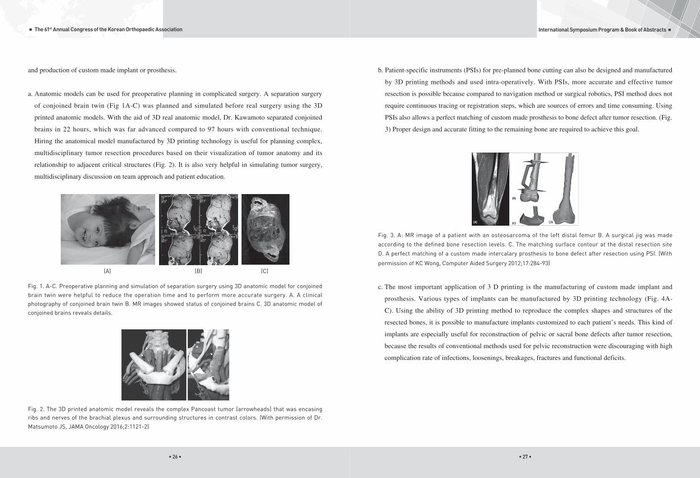
a. Anatomic models can be used for preoperative planning in complicated surgery. A separation surgery
of conjoined brain twin (Fig 1A-C) was planned and simulated before real surgery using the 3D
printed anatomic models. With the aid of 3D real anatomic model, Dr. Kawamoto separated conjoined
brains in 22 hours, which was far advanced compared to 97 hours with conventional technique.
Hiring the anatomical model manufactured by 3D printing technology is useful for planning complex,
multidisciplinary tumor resection procedures based on their visualization of tumor anatomy and its
relationship to adjacent critical structures (Fig. 2). It is also very helpful in simulating tumor surgery,
multidisciplinary discussion on team approach and patient education.
(A) (B) (C)
Fig. 1. A-C. Preoperative planning and simulation of separation surgery using 3D anatomic model for conjoined brain twin were helpful to reduce the operation time and to perform more accurate surgery. A. A clinical photography of conjoined brain twin B. MR images showed status of conjoined brains C. 3D anatomic model of conjoined brains reveals details.
Fig. 2. The 3D printed anatomic model reveals the complex Pancoast tumor (arrowheads) that was encasing ribs and nerves of the brachial plexus and surrounding structures in contrast colors. (With permission of Dr.
Matsumoto JS, JAMA Oncology 2016;2:1121-2)

• 28 • • 29 •
■ The 61st Annual Congress of the Korean Orthopaedic Association International Symposium Program & Book of Abstracts ■
(D) (E) (F)
Fig. 5. Reconstruction with 3 kinds of 3D printed pelvic endoprosthesis after resection of pelvic tumors, iliac
prosthesis (A, B), standard hemipelvic prosthesis (C, D) and screw-rod connected hemipelvic prosthesis (E, F).(
Bone Joint J 2017;99-B:267-75.)
In our experience, scapular reconstruction is one of the good indications of hiring 3D printing technology,
because scapular has complex anatomic and functional elements. A eight year old female patient
with Ewing’s sarcoma involving right scapular body was managed with subtotal scapulectomy and
reconstruction with 3D printed prosthesis. The glenoid and coracoid process portion and distal pole of
scapula were preserved and 3D printed metallic scapular body was implanted into the bone defect site. At
18 months after operation, the patient showed excellent functional outcome and continuous disease free
(CDF) status. (Fig. 6)
(A) (B) (C)
(A) (B) (C)
Fig. 4. Various implants manufactured by 3D printing technology
Recently, Guo et al. reported 35 patients of pelvic tumors treated with 3D printed pelvic prosthesis
made from titanium alloy by electronic beam melting technology. In their series, they used three types
of 3D printed endoprostheses; iliac prosthesis, standard hemipelvic prosthesis and screw-rod connected
hemipelvic prosthesis. The mean musculoskeletal tumor society functional scores were 22.7, 19.8 and 17.7
respectively (Fig. 5A-F). There were 7 delayed wound healings and 2 dislocations of hip as complications.
They concluded that the application of 3D printing technology facilitated the precise matching and
osteointegration between implants and the host bone which resulted in good short-term functional results
without additional complications.
(A) (B) (C)

• 30 • • 31 •
■ The 61st Annual Congress of the Korean Orthopaedic Association International Symposium Program & Book of Abstracts ■
(D) (E) (F)
Fig. 7. Combination of 3D printed custom made prosthesis with bone cutting jig aids to achieve a wide oncological
surgical margin, primary stability of implant and durability of prosthesis (A, B). Proper internal structure (C) and
surface treatment (D, E) appropriate to regional requirement are necessary to obtain good functional result.
Radiograph showed well reconstructed pelvic bone and stable hip joint (F). (With courtesy of Donati DM)
Unsolved problems
However, 3D printing in muscouloskeletal oncology is still in the stage of beginning and many unsolved
problems such as possible fatigue fracture associated with weak biomechanical strength, aseptic loosening
of large segmental reconstructed body with insufficient bone ingrowth, inefficient soft tissue attachment
and difficult assembly with modular joint prosthesis. Restoration of internal structures resistant to repeated
long-standing heavy load on that specific region especially in pelvic and spinal areas is required for long
survival of prosthesis. Rapid and efficient bone ingrowth into the surface of prosthesis from the contacting
host bone is also one of the key elements of successful reconstruction. Even with recent progression,
stable assembly with artificial joint components and durable attachment of muscle, tendon and ligament to
restore the function of reconstructed structures are still remaining issues.
Restoration of durable load-bearing trabecular bone structures which reproduce a much complex shapes
and stress/strain characteristics of pelvic or spinal bones is necessary to prevent stress failure of prosthesis.
At prosthesis-host bone junction, cooperation of ideal porous structures feasible for bone ingrowth and
surface treatment friendly to osseous proliferation is required.
Even with PSIs, resection of bone tumors in complex anatomy such as pelvic bone, sacrum and scapula
can be inaccurate due to complex geometry, limited visibility and restricted working space of those
regions. Designing, manufacturing and precise application of PSIs during operation should be matched to
(D) (E)
Fig. 6. A eight year old female patient with Ewing’s sarcoma involving right scapular body. A. MR image shows a
large tumor involving scapula body with huge extraosseous extension. B. Preoperatively planned resection line.
C. 3D printed scapular body with holes for bone and soft tissue attachment. D. Postoperative radiograph of Rt.
scapula. E. Excellent functional status at 18 months after surgery.
Combination of surgical cutting jig or navigation with 3D printed prosthesis, an accurate tumor resection
and stable reconstruction with perfectly matched implant can be achieved. Donati DM at Instituto
Orthopaedico Rizzoli (Bologna, Italy) developed 3D printed custom made prosthesis with bone cutting jig
and achieved a wide oncological surgical margin, primary stability of implant and durability of prosthesis.
The manufactured implant had wide trabecular space for muscle integration, porous bone prosthesis
bearing surface for bone ingrowth and polished finite parts to avoid muscle friction. (Fig. 7)
(A) (B) (C)

The 61st Annual Congress of The Korean Orthopaedic Association
Sung Taek Jung
• 32 •
■ The 61st Annual Congress of the Korean Orthopaedic Association
surgical approaches used and detailed surface anatomies of specified regions.
Conclusion
To accomplish surgical goals, various factors should be considered, and intimate collaboration between
clinicians with anatomical, functional and biological knowledge and engineers who experts on metal
materials, biomechanics, designing and manufacturing process of prosthesis is essential.
Reasonable cost requirements, acceptable time scales and regulatory approval and supporting for clinical
application are necessary to activate the clinical use of 3D printing technology in orthopedic oncology.
References
1) Cartiaux O, Paul L, Francq BG, Banse X, Docquier P. Improved accuracy with 3D planning and patient-specific instruments
during simulated pelvic bone tumor surgery. Ann Biomed Engineer 2013;42:205-13.
2) Kim D, Lim JY, Shim KW et al. Sacral reconstruction with a 3 D-printed implant after hemisacrectomy in a patient with sacral
osteosarcoma: 1-year follow-up result. Yonsei Med J 2017;58:453-7.
3) Liang H, Ji T, Zhang Y, Wang Y, Guo W. Reconstruction with 3D-printed pelvic endoprosthesis after resection of a pelvic
tumor. Bone Joint J 2017;99-B:267-75.
4) Matsumoto JS, Morris JM, Rose PS. 3-Dimentional printed anatomic models as planning aids in complex oncology surgery.
JAMA Oncology 2016;2:1121-2.
5) Wang B, Xie X, Yin J et al. Reconstruction with modular hemipelvic endoprothesis after pelvic tumor resection: a report of 50
consecutive cases. PLOS one 2015;DOI:10.1371/journal.pone.0127263:1-11.
6) Wong KC, Kumta SM, Geel NV, Demol J. One-step reconstruction with 3D-printed, biomechanically evaluated custom implant
after complex pelvic tumor resection. Computer Aided Surg 2015;20:14-23.
7) Wong KC, Kumta SM, Sze KY, Wong CM. Use of a patient-specific CAD/CAM surgical jig in extremity bone tumor resection
and custom prosthetic reconstruction. Computer Aided Surg 2012;17:284-93.
Oct. 19th. 2017. Thu | Grand Ballroom1
Periprosthetic fracture of the femur: Reduction and Fixation
Byung Woo Min
The 61st Annual Congress of The Korean Orthopaedic Association

• 34 • • 35 •
■ The 61st Annual Congress of the Korean Orthopaedic Association International Symposium Program & Book of Abstracts ■
should be carefully evaluated. Radiographic signs of definite loosening include progressive periprosthetic
or cement mantle luceny, a change in the position of the stem, and component or cement fracture. High
energy trauma associated with comminuted fracture also have high chance of loose stem. Radiographic
signs of probable loosening include greater than 2 mm of periprosthetic or cement mantle lucency around
entire prosthesis, bead shedding, endosteal scalloping, and endosteal bone bridging at the tip of the stem.
CT is occasionally useful for evaluating stem loosening if radiographic findings are inconclusive.
Periprosthetic Fracture of the Femur: Decision making (is the stem stable?)
Byung Woo Min
KEIMYUNG UNIV.
•••
The management of periprosthetic fractures is an issue of increasing importance for orthopaedic
surgeons. Because of the expanding indications for total hip arthroplasty (THA) and an aging population
with increasingly active lifestyles, the incidence of primary and revision THA is increasing, and there
is a corresponding increase in the prevalence of periprosthetic fracture of the femur (PFF). Surgical
management of PFF is technically demanding given the often poor bone quality, altered anatomy, and
need to manage both the prosthesis and fracture. When deciding on how to treat an PFF, the first decision
point surrounds whether or not the stem is well fixed. In general terms, well fixed stems require open
reduction and internal fixation, whereas loose stems require revision arthroplasty. The most commonly
used classification system for periprosthetic fracture around THA is the Vancouver classification
which stratifies these injuries based on the location of the fracture and the stability of the implant. The
stability of the femoral component in the proximal fragment is the cornerstone of this classification.
The strongest risk factor for failure after treatment of PFFs is underestimation of stem stability. The
surgeon, in many cases, misinterpreted the stability of the stem and classified a type B2 fracture as type
B1, and subsequently undertake treatment with plate fixation without revision of the stem. The literature
reports a higher rate of failure for osteosynthesis around prostheses considered to be well-fixed. Rates of
reoperation following ORIF of PFFs are reported from 13% to 23%. Decision making for stem stability
rely on the careful evaluation of high quality standard AP and lateral radiographs of the entire femur
and hip. Radiographs should be critically assessed for signs of implant loosening to distinguish between
type B1 and B2 fractures. Failure to identify an unstable implant is likely to lead to treatment failure if
osteosynthesis rather than revision arthroplasty is performed. Whenever possible, preinjury radiographs
should be obtained for comparison. The implant-bone, cement-implant, and cement-bone interfaces

• 37 •
International Symposium Program & Book of Abstracts ■
• 36 •
■ The 61st Annual Congress of the Korean Orthopaedic Association
Biomechanical challenges of periprosthetic fractures
Toru Sato
NATIONAL HOSPITAL ORGANIZATION OKAYAMA MEDICAL CENTER, JAPAN
•••
Purpose: Figures of peri-THA fractures and treatment options/results were investigated, making a
treatment protocol was considered from the view of biomechanical aspect.
Methods: Thirty-two cases were investigated. Intraoperative fracture cases were excluded. The average
age at injury is 77.8 years old (range 50-91) and all cases were low-energy trauma. Vancouver’s
classification and AO classification for fracture figures were used and investigated a union period and
complications in each treatment options.
Results: All cases that fracture occurred within 10 years after THA were classified into Vancouver type
B1. Beyond 10 years after THA, 6 cases out of 13 were recognized type B2 loosening, but considering
an age and general condition of patients, only osteosynthesis was selected in 4 cases. For osteosynthesis,
a plating system was used in all cases. Locking plates (+cable wire) were 11 cases, locking plates (-cable
wire) in 4, Non-locking plate (+cable wire) in 10, Non-locking (-cable wire) in 5 and 2 cases were revised
a stem. MIPO technique was done in 9 cases, early implant failure was recognized in 2 cases out of 5
cases which were no comminution at fracture site. And one case developed delayed union. In cemented
THA cases, if a fracture line sited at cement border region, delayed union rate was 50%.
Considerations: Cases no comminution at fracture region need an anatomical reduction regardless of
MIPO technique. For this reason, first application of cable system to fix a proximal fragment is mandatory.
An accurate contour is not always necessary because of using locking screws. Peri-THA&TKA fracture
cases were severe osteoporosis related and comminuted fracture pattern. MIPO is good indication for
these fractures.
Principles of reduction and fixation
Tak Wing Lau
THE UNIVERSITY OF HONG KONG, QUEEN MARY HOSPITAL, HONG KONG
•••
Abstract:
Periprosthetic fractures occur in both upper and lower limbs. The principles of fixation are slightly
different from normal shaft fractures in elderly. If the prosthesis is stable, early reduction and stable
fixation is required. If the prosthesis is loosened, it requires a complete revision. In lower limb
periprosthetic fractures, the aim of fracture reduction and stabilization is to allow immediate weight
bearing walking and free joint motion after surgery. Before the surgical fixation, correct classification
can help with the decision of surgical treatment. Vancouver or AO classification could be used. In
periprosthetic fractures of femur, they are usually reduced by semi-open or open technique. Fracture is
usually fixed by an extramedullary implant, usually a plate, reinforced with special implants, e.g. cerclage
wires, cables and locking plate attachment system. Plate should be as long as possible to span the whole
length of femur. In periprosthetic fracture with knee replacement, both plate and nail can be considered.
A retrograde femur intramedullary nail can be used if femoral component has an open-box design. In
upper limb periprosthetic fracture, plating is the treatment of choice. Reduction and fixation follows the
principles of lower limb periprosthetic fracture management.

• 39 •
International Symposium Program & Book of Abstracts ■
• 38 •
■ The 61st Annual Congress of the Korean Orthopaedic Association
In the treatment of periprosthetic femoral fractures, the location and configuration of the fracture, stem
stability, cemented or cementless stem, bone loss should be evaluated. Careful preoperative planning
is mandatory. Vancouver B1 and C can be treated with ORIF using a long plate. In B1 cases, always
prepared to convert to revision. In B2 cases, the stem should be revised and additional plate fixation
sometimes necessary. Former stem fixation mode (cemented or cementless) will influence the revision
procedure. In B3 cases, revision and bone restoration is necessary, it can be done mostly with impaction
grafting with allograft. In very old age patient distal locking stem can be an option.
Case-based lecture-fracture around the femoral stem
Takeshi Sawaguchi
KANAZAWA UNIVERSITY, TOYOMA MUNICIPAL HOSPITAL, JAPAN
•••
Abstract:
With the increase of geriatric population, long life and frequent application of arthroplasty, there is an
increasing number of periprosthetic fractures. The most common classification for periprosthetic femoral
fractures is the Vancouver classification.
In the presentation, mainly Vancouver B cases will be presented and discussed.
Case 1: 74 y.o. female. B1 transverse fracture (cemented stem) treated by MIPO technique with locking
plate.
Case 2: 86 y.o. female. B1 spiral fracture (cemented stem) treated by mini open reduction and MIPO with
locking plate.
Case 3: 55 y.o. male. B2 comminuted fracture (cemented stem) treated with ORIF, locking plate fixation
and stem exchange with cement in cement technique.
Case 4: 79 y.o. female. B2 long comminuted fracture (cemented stem) treated with ORIF, locking plate
fixation and stem revision with a longer cemented stem.
Case 5: 77 y.o. female. B3 fracture with severe osteolysis (cemented stem) treated with revision cemented
stem (only cemented distally) and proximal autogenous bone graft.
Case 6: 87 y.o. female. B3 fracture (cemented stem) treated with revision cemented stem and impaction
grafting with allograft.
Case 7: 90 y.o. female. B3 transverse fracture (cemented stem) treated with distal locking cementless stem
and impaction grafting with allograft.

The 61st Annual Congress of The Korean Orthopaedic Association
Sung Taek Jung
MEMO
Oct. 19th. 2017. Thu | Grand Ballroom1
Cutting Edge Technology in the Field of ASAMI Society of Asian Countries
Chang Hoon Jeong
The 61st Annual Congress of The Korean Orthopaedic Association

• 43 •
International Symposium Program & Book of Abstracts ■
• 42 •
■ The 61st Annual Congress of the Korean Orthopaedic Association
Comparison of Joint Distraction and Non-distraction using an Ilizarov External
Fixator in the Treatment of Ankle Fractures in Older Patients
Koji Nozaka
AKITA UNIVERSITY GRADUATE SCHOOL OF MEDICINE, JAPAN
•••
Background:
Periarticular fracture of the ankle in elderly individuals is likely to become posttraumatic ankle arthritis.
In osteoarthritis (OA), subchondral bone changes alter the joint’s mechanical environment and potentially
influence progression of cartilage degeneration. Joint distraction as a treatment for OA has been shown
to provide pain relief and functional improvement through mechanisms in periarticular fracture of the
ankle that are not well understood. To conduct a retrospective study comparing treatment effects of joint
distraction and joint non-distraction using Ilizarov external fixatior methods among elderly patients with
periarticular fracture of the ankle.
Subjects:
We investigated a total of 54 patients >60 years old who showed fracture of the distal tibia including
tibial plafond fracture or tri- or bimalleolar fracture of the ankle (excluding unimalleolar fractures), upon
admission to our department from among the 601 patients for whom after a surgical treatment for an ankle
fracture using Ilizarov external fixatior who were followed for at least 2 years. Patients were either treated
with distraction (n=26) or non-distraction (n=28). The mean age of patients was 72.4 years (range, 60-
78 years) in the distraction group and 70.2 years (range, 60-84 years) in the non-distraction group. All
patients received partial weight-bearing (as tolerated) 1 day postoperatively, 1/2 partial weight-bearing at
2 weeks postoperatively, and full weight-bearing at 4 weeks postoperatively.
Results:
Bone density (relative to young adult mean, YAM) was 55.8% (range, 28-70%) for the distraction group,
and 61.2% (range, 38-70%) for the non-distraction group. Mean range of motion in the sagittal plane was
45.3°for the distraction group and 38.9° for the non-distraction group. The mean AOFAS score was 94.2
(range, 72–100) for the distraction group and 67.2 (range, 42–100) for the non-distraction group. It was
significantly higher with the distraction group (p<0.05).
Discussion:
In elderly patients with periarticular fracture of the ankle, those who received joint distraction treatment
showed higher in AOFAS score compared to those who received joint non-distraction treatment. Joint
distraction may become an useful option in the treatment of periarticular fracture of the ankle in elderly
individuals.

• 45 •
International Symposium Program & Book of Abstracts ■
• 44 •
■ The 61st Annual Congress of the Korean Orthopaedic Association
group. The preoperative coronal deformity were 20.8 ± 12.2 degrees in monolateral group and 20.1
± 9.7 degrees in circular group, respectively. The amount of correction for coronal deformity and the
duration of the external fixators were similar between both groups. There were two major complications
in the monolateral group, including one compartment syndrome and one regenerate fracture after falling.
Transient peroneal nerve palsy after acute correction was observed in 4 segments. The MAC system
showed a LLD of 10.9 ± 11.7 mm and MAD of 3.2 ± 22.9 mm medial.
Conclusion
The MAC system provided acceptable alternative for the treatment of deformities in lower extremity.
Treatment of the lower limb deformities by a multi-axial external fixation system
Masaki Matsushita1, Hiroshi Kitoh1, Tadashi Hattori2, Hiroshi Kaneko2, Kenichi Mishima1, Naoki Ishiguro1
1Department of Orthopaedic Surgery, Nagoya University Graduate School of Medicine, Japan.2Department of Orthopaedic Surgery, Aichi Children's Health and Medical Center, Japan.
•••
Background
Deformities of the upper and lower limbs can gradually be corrected by external fixators in a less
invasive manner. Monolateral external fixators are simple to apply but have limited capabilities of three-
dimensional deformity correction. Using a multi-axial correction (MAC) monolateral external fixation
system, we have performed corrective osteotomies with or without simultaneous lengthening for various
deformities of the lower limb. We evaluated the final alignment of the treated limbs with the MAC system
and determined the effectiveness of this fixator during corrective osteotomies of the lower limb.
Methods
We retrospectively reviewed the medical records and radiographs of 46 bony segments in 32 patients (mean
age 12.9 years; range 6-23 years) who underwent correction osteotomies of the lower limb with or without
simultaneous lengthening between 2003 and 2016. 19 segments were treated with the MAC system
(monolateral group), while 27 were treated with circular fixators (circular group), including Ilizarov and
Taylor Spatial Frame. Simultaneous lengthening was performed in 11 segments of monolateral group and
20 segments of circular group. 40 segments in 26 patients were congenital deformities and 6 segments in
6 patients were acquired deformities. At the latest follow-up, we measured leg length discrepancy (LLD)
and mechanical axis deviation (MAD).
Results
The average age of surgery was 15.8 ± 4.3 years in monolateral group and 10.8 ± 3.3 years in circular

• 47 •
International Symposium Program & Book of Abstracts ■
• 46 •
■ The 61st Annual Congress of the Korean Orthopaedic Association
Study 2: Pure PCL microspheres and composite PCL and 10% HA microspheres were synthesized using
a modified solvent evaporation method. Bone mesenchymal stem cells isolated from green fluorescent
protein rats (GFP-rBMSCs) were cultured with these microspheres in a rotary bioreactor system. The
formation of the microstructures was confirmed by scanning electron microscopy (SEM). We confirmed
that PCL/HA promotes osteogenic differentiation of rBMSCs in vitro. To investigate the effects of
addition biomaterials on bone consolidation during DO process, PCL/HA (20 mg), PCL (20 mg), or PBS
were then locally administered into the distraction gap in Sprague-Dawley male rat DO model towards
to the end of distraction period and animals were allowed for bone consolidation for 4 weeks after the
distraction completed and then terminated. Weekly x-rays, micro-computed tomography, mechanical
testing, histology, and immunohistochemical examinations were performed to assess the quality of the
newly bone. Results: The microspheres used were of the uniform size and monodisperse. After incubation
with rBMSCs in culture, PCL/HA microspheres showed a better ability of cell adhesion and osteogenic
differentiation comparing to PCL microspheres. In the rat DO model, the bone volume/total tissue volume,
bone mineral density, and mechanical properties of the newly formed bone were significantly higher in the
PCL/HA group compared to the PCL and PBS groups. Histological and immunohistochemical analyses
confirmed improved bone formation and vascularization in the PCL/HA group.
Conclusions: The combined use of biomaterials such as HA/TCP blocks or PCL/HA composite
microspheres in DO is a novel approach for promoting bone regeneration and consolidation, their clinical
applications may reduce the treatment time, pain and suffer of the patients.
Novel Management of Larger Bone Defect: Combination of Biomaterials and Distraction
Osteogenesis Technique
Gang Li
THE CHINESE UNIVERSITY of HONG KONG, PRINCE OF WALES HOSPITAL, HONG KONG
•••
Introduction: Distraction osteogenesis (DO) techniques have been widely accepted and practiced in
orthopaedics, traumatology, and craniofacial surgery over the last two decades, using DO methods, many
previously untreatable conditions have been successfully managed with outstanding clinical outcomes.
The major limitation of DO is relatively long period required for new bone consolidation. Here, we
investigated whether the application of biomaterials, including polycaprolactone (PCL) and hydroxyapatite
(HA) cylinder or composite microspheres could be used to reduce the treatment time and enhance bone
formation in DO.
Study 1: A 1.0cm tibial shaft was removed in the left tibia of 36 rabbits and divided into three groups:
Group A, the defect gap shortened for 1.0-cm; Group B, the defect gap was filled with 1.0-cm porous
hydroxyapatite/tri-calcium phosphates (HA/TCP) cylindrical block; Group C, The 1.0-cm defect gap was
reduced 0.5cm and the remaining 0.5-cm defect gap was filled with 0.5-cm HA/TCP block. The tibia was
then fixed with unilateral lengthener; for groups A and C, lengthening started 7 days after surgery at a rate
of 1.0 mm/day, in two steps. Group A received lengthening for 10 days and Group C for 5 days, there
was no lengthening for Group B. All animals were terminated at day 37 following surgery. The excised
bone specimens were subject to micro-CT, mechanical testing and histological examinations. Results:
Bone mineral density and content and tissue mineral density and content, as well as the mechanical
properties of the regenerates were significantly higher in Group C compared to Groups A and B. Micro
CT and histological examinations also confirmed that the regenerates in Group C had most advanced bone
formation, consolidation and remodeling compared to other groups.

• 48 • • 49 •
■ The 61st Annual Congress of the Korean Orthopaedic Association International Symposium Program & Book of Abstracts ■
Lower Limb Reconstruction in paediatric Orthopedics
Andrew Lim Kean Seng
NATIONAL UNIVERSITY HOSPITAL OF SINGAPORE, SINGAPORE
•••
Abstract
Lower limb reconstruction in paediatric orthopaedics can be challenging.
The spectrum of conditions can range from the simple acquired uniplanar deformity to the complex
congenital multi-planar deformity with limb deficiency.
Early diagnosis will help with appropriate treatment. Growth plate modulation can be useful for selected
cases. The principles of deformity correction are important in limb reconstruction.
External fixator treatment is reserved mainly for the correction of more complex deformity and deficiency.
It is important to understand the bone and soft tissue constrains for every case. Treatment may
occasionally have to be staged and can continue even into skeletal maturity.
Pre-operative counselling and post-operative support for the patient will optimise the outcome of
treatment.
The types and techniques of external fixator treatment will be elucidated for various conditions in
paediatric lower limb reconstruction.
Game changers in Limb lengthening and Deformity Correction Field
Dong Hoon Lee
YONSEI UNIV.
•••
골연장 및 변형교정 분야는 최근 급속히 발전하고 있다. 특히 연장 및 변형교정을 위한 기계적혁신은 고식적인
일리자로프 수술방식에 비하여 환자의 통증과 불편을 줄이고 더 좋은 임상적 결과를 가져왔다. 골연장 분야에서
가장 혁신적인 발전은 내고정 기계를 이용한 연장(lengthening with lengthening nail)이다. 일리자로프와 같
은 외고정장치를 이용한 전통적인 골신연술에 관한 합병증 중 가장 흔한 것은 핀 관련 문제이다. 가장 흔한 합
병증은 pin-site infection으로 minor infection은 2-80%로 보고되었고, major infection도 23%까지 보고되
었다. 외고정장치를 이용한 골신연술이 오랫동안 발달됨에 따라 합병증이 많이 줄어 들었으나 외고정기구를 오
랫동안 착용해야하는 불편과 핀 관련합병증(통증, 감염, 구축, 스트레스) 등은 여전히 문제로 남아있다. 1956
년 Bost와 Larsen이 처음가능성을 보여주고, 이후 Paley와 Herzenberg가 정립한 LON(Lengthening Over
Nail)은 외고정 및 내고정의 장점을 모두 취할 수 있는 좋은 수술법으로 현재까지 널리 이용되고 있으나 이것 역
시 연장 기간 동안 외고정 장치를 착용해야 하므로 외고정으로 인한 문제점을 완전히 극복할 수는 없었고 외고
정 및 내고정장치가 공존하므로 minor pin-site infection이 intramedullary infection으로진행될 수 있는 위
험도 내포하고 있다. 외고정 장치가 없는 순수한 내고정 연장술은 감염률을 낮출 수 있고, 이 외에도 통증 및 핀
으로 인한 ugly scar 등을 줄이고 환자의 정신적인 부담도 줄일 것이라는 기대 하에 1959년부터 개발을 시도하
여 지속적으로 발달해 왔으나 임상적 적용은 미미하였다. 현재까지 비교적 알려진 내연장 금속정은 AlbizziaⓇ
(France), FitboneⓇ(Germany), ISKDⓇ(Intramedullary Skeletal Kinetic Diatractor; Orthofix, USA), 그
리고 PRECICEⓇ nail(Ellipse, USA)이다. 1987년프랑스의 Dr. Guichet가 torsional motion으로 활성화되는
mechanical device(AlbizziaⓇ)를 개발하였는데, 각 rachet 당 0.07mm가 연장되도록 고안되었다. 이후 임상
적용에는 성공하였으나 연장을 위한 최소 20°의 racheting movement를 위하여 실제로는 90도에 가까운 다리
의 회전동작이 필요하였다. 이로 인한 심한 통증으로 비판을 받았고, 수술 후 racheting motion을 위한 전신마
취가 22 - 39% 빈도로 보고 되었다. 이후 독일의 Dr. Baumgart는 motorized electronic nail(FitboneⓇ)을 개
발하여 다리의 회전동작 없이 연장이 가능하게 되어 연장 중 통증이 많이 줄었다고 보고하였다. 이론적으로 전

• 51 •
International Symposium Program & Book of Abstracts ■
• 50 •
■ The 61st Annual Congress of the Korean Orthopaedic Association
기적 작동방식은 연장 속도 및 리듬을 정확하게 조절할 수 있다는 큰 장점이 있으나 FitboneⓇ은 이에 대한 기계
적 신뢰성에 의문을 가지고 있는 의사들이 많은 실정이다. 미국 FDA 승인을 처음 받은 제품은 ISKDⓇ (Orthfix,
USA)이다. 1995년미국의 Dr. Cole이 개발한 ISKDⓇ의 이론적 장점은 3-9° 정도의 작은 회전으로 작동하게 되
는 클러치 원리(clutch mechanism)으로서, 일상적인 움직임만으로도 연장이 가능하고 따라서 Albizzia의 과
도한 회전 동작으로 인한 통증을 줄일 수 있다는 것이었다. 또한 연장속도를 모니터로 측정할 수 있게 하여 환
자 스스로 조절할 수 있게 하였다. 하지만 ISKD의 많은 문제점들이 보고되었는데, 가장 중요한 단점으로 연장
속도 조절의 어려움이 부각되었다. 특히 femur lengthening 시 의도하지 않은 지나치게 빠른 연장(run away)
으로인한 골형성 부전(insufficient bone regenerate)이 25%까지 보고 되었고, 비정상적인 속도조절이 60%까
지 보고되었다. Rozbruch는 ISKD를 이용한 대퇴골 연장에서 평균 1.9mm/day의 연장속도를 보였고, LON보
다 골형성이 좋지 않다고 보고하였다. 그 외에도 느린 연장속도로 인한 premature consolidation, hardware
malfunction 등으로 인한 unexpected additional surgery가 필요하였다고 보고되고 있다. 저자들은 ISKD의
문제점으로 속도 조절 자체의 문제 뿐 아니라 속도 조절이 안될 경우 심한 통증도 지적하였다. 또한 내고정 장
치를 이용한 경골 연장 시 족관절의 첨족 변형(equinus contracture)을 효과적으로 예방할 수 없는 것도 내고
정 연장의 단점 중 하나이다. 현재 전 세계적으로 가장 많이 사용되는 내연장 골수정은 자기장을 이용하여 연
장하게 되는 PRECICEⓇ(Ellipse, USA)이다. 미국에서 FDA 승인 하에 사용되고 있는데, 이 제품은 racheting
motion 없이 연장이 가능하고 이론적으로 정확한 속도조절이 가능하다는 점, 그리고 길이를 줄일 수도 있다
는 점이 장점이나, 여전히 체중 부하를 충분히 할 수 없다는 한계를 가지고 있고 자석의 힘으로 연장을 하므로
rachet mechanism을 가지는 기계보다는 distraction force가 약할 가능성이 있다. Yatin 등은 PRECICEⓇ를
이용한 골연장에서 96%의 연장속도 정확성을 보고하였다. 저자들의 경험으로도 PRECICEⓇ를 이용한 골 연장
시 99%에서 연장 목표를 얻었고, 99%의 속도조절 정확성을 보였다. 또한 통증 조절 역시 우수하여 골연장에서
좋은 대안이 될 것으로 보인다. 내연장 골수정(intramedullary lengthening device)을 이용한 골 연장술은 일
리자로프 방식에 비해 재활, 통증, 흉터, 정신적 스트레스 등의 관점에서 분명한 장점이 있지만, 이 역시 '골 연
장술'이므로 골 연장술 시 발생할 수 있는 모든 합병증의 가능성이 있다. 오히려 외고정 장치가 없기 때문에 골
연장 중 발생하는 문제에 대한 적극적 개입이 어렵다. 따라서 외고정 장치를 이용한 골 연장술보다 더욱 조심
스런 접근이 필요하다. 그 외에도 변형교정 분야에서는 스튜어트 플랫폼(stewart flatform)을 이용한 교정 및
급속교정(acute correction) 기술의 발전이 주목을 받고 있다. 스튜어트 플랫폼을 이용한 외고정 장치를 사용
할 경우 multi-plane deformity같은 복잡한 변형에서도 하나의 프레임(frame)으로, 컴퓨터 프로그램의 도움
을 받아 한번에 모든 변형의 교정이 가능하다. 또한 locking plate의 개발 및 골수강 내 금속정(intramedullary
nail) 기술의 발전으로 예전에는 외고정 장치로만 가능했던 복잡하고 심한 변형에서도 외고정 장치없이 변형을
교정하는 수술기법들이 지속적으로 발전하고 있다.
Reference
1. Burghardt RD, Herzenberg JE, SpechtSC, Paley D. Mechanicalfailure of the Intramedullary Skeletal Kinetic Distractor in
limblengthening. J Bone Joint SurgBr. 2011;93:639–643.
2. Cole J, Justin D, Kasparis T, DeVlught D, Knobloch C. Theintramedullary skeletal kinetic distractor (ISKD): first clinicalresults
of a new intramedullary nail for lengthening of the femur and tibia. Injury. 2001;32(suppl 4):SD129–139.
3. Hankemeier S, Pape H-C, Gosling T, Hufner T, Richter M, Krettek C. Improved comfort in lower limb lengthening with
theintramedullary skeletal kinetic distractor: principles and preliminary clinical experiences. Arch Orthop Trauma Surg.
2004;124:129–133.
4. Kenawey M, Krettek C, Liodakis E, Wiebking U, Hankemeier S.Leg lengthening using intramedullary skeletal kinetic
distractor:results of 57 consecutive applications. Injury. 2011;42:150–155.
5. Kubiak EN, Strauss E, Grant A, Feldman D, Egol KA. [Early complications encountered using a self-lengthening
intramedullarynail for the correction of limb length inequality][in Turkish]. Joint Dis RelatSurg (EklemHastalikCerrahisi).
2007;18:52–57.
6. Schiedel FM, Pip S, Wacker S, Po¨pping J, Tretow H, LeidingerB, Ro¨dl R. Intramedullary limb lengthening with the
IntramedullarySkeletal Kinetic Distractor in the lower limb.J Bone Joint Surg Br. 2011;93:788–792.
7. Simpson AH, Shalaby H, Keenan G. Femoral lengthening withthe Intramedullary Skeletal Kinetic Distractor. J Bone Joint
SurgBr. 2009;91:955–961.
8. Thonse R, Herzenberg JE, Standard SC, Paley D. Limb lengtheningwith a fully implantable, telescopic, intramedullary nail.Oper
Tech Orthop. 2005;15:355–362.
9. ShahabMahboubian DO. Femoral Lengthening with Lengthening over a Nail has Fewer Complications than Intramedullary
Skeletal Kinetic Distraction.ClinOrthopRelat Res 2012; 470:1221–1231
10. Guichet JM, Lascombes P, Grammont PM, PrevotJ. Gradual elongation intramedullary nail for femur (Albizzia). Results ofthe
first 52 cases in 48 patients.J JpnOrthop 1995; 69: 310
11. Jean-Marc Guichet, Barbara Deromedis. Gradual Femoral Lengthening with the Albizzia Intramedullary NailJ Bone Joint Surg
Am. 2003. 85:838-848,
12. Lee DH, Ryu KJ, Song HR, Han SH. Complications of the Intramedullary Skeletal Kinetic Distractor (ISKD) in Distraction
Osteogenesis. Clin Orthop and Related Res. 2014. e-published
13. Yatin M, SR Rozbruch. Precision of the PRECICE(®) Internal Bone Lengthening Nail. Clin Orthop and Related Res. 2014.
e-published
14. Shiedel FM, Rodl R. How precise is the PRECICE compared to the ISKD in intramedullary limb lengthening?. Acta Orthop.
2014. Vol 85: 293-298

The 61st Annual Congress of The Korean Orthopaedic Association
Oct. 19th. 2017. Thu | Grand Ballroom1
MEMO
Hyun Guy Kang
Sacro-pelvic bone cancer surgery

• 55 •
International Symposium Program & Book of Abstracts ■
• 54 •
■ The 61st Annual Congress of the Korean Orthopaedic Association
40% in PG. Limb function was 79 % (ISOLS) due to low emotional acceptance. Vesico-rectal function
was also disturbed even in cases whose S2 nerve roots were completely preserved. Thirteen complications
were observed in 10 cases. Wound trouble was most frequent (8 events) followed by infection (2).
There was no infection in PG of chordoma. We have experienced HIRT for chordoma in 11 cases. Local
recurrence and metastasis occurred in 1 and 2 cases, respectively. Vesico-rectal disturbance was observed
in 6 cases.
DISCUSSION AND CONCLUSION: Posterior approach was preferable to reduce bleeding and to
perform precise osteotomy of the pelvic ring. Safe clearance of the sciatic notch is the most critical
point to achieve less invasive and more reliable wide resection for the local control. S3 nerve root may
be essential to maintain normal vesico-rectal function. HIRT may be also effective at least for sacral
chordoma.
Resection and reconstruction for pelvic ring cancer
Tetsuo Hotta
NIIGATA UNIVERSITY HOSPITAL, JAPAN
•••
ABSTRACT
INTRODUCTION: Surgical treatment of pelvic ring cancers are still challenging because of the
complicated anatomy. Especially, surgical treatment of sacral tumors are most difficult. Massive bleeding,
high local recurrence, and poor functional results are serious problem. Our strategy is as follows. P1;
Resection only, occasionally bone grafting if the ring is disconnected. P2: Resection and reconstruction
with constrained THA, occasionally hip transposition is applied. P3; Resection only. P4; Resection only,
exceptional SI joint and spine fusion after total sacrectomy. We will show the advantage of posterior
approach for the resection of tumors, and introduce the tips of the technique. Case presentation will be
performed by our experience of P4 resection. The results of heavy iron radiation therapy (HIRT) for sacral
chordoma will also be showed as a control.
METHODS: About P4 resection, 21 cases operated from 1997 through 2005 were included. After 2005,
HIRT was mainly performed. Mean age was 52 year-old. Mean follow up period was 135.7 months.
Chordoma was most popular (11 cases) followed by GCT (4), chondrosarcoma (3), and metastasis (3).
These were divided into two groups, anterior approach group (AG) and posterior approach group (PG).
Survival rate, local recurrence, and complication were examined.
RESULTS: Total 10 and 15-year survival rate was 75 and 47 %, respectively. Recurrence rate was 41 %.
Survival rate was worse in AG, but not statistically different. The blood loss of AG and PG were 8,545
and 1,583 ml, which was statistically different. Local recurrence rates of chordoma were 100% in AG and

• 57 •
International Symposium Program & Book of Abstracts ■
• 56 •
■ The 61st Annual Congress of the Korean Orthopaedic Association
Computer-assisted pelvic ring cancer surgery
Kwok Chuen Wong
PRINCE OF WALES HOSPITAL, HONG KONG
•••
Abstract
Conventionally, tumour surgeons analyse two-dimensional imaging information and mentally integrate
and formulate a three-dimensional surgical plan. It is hard to translate the surgical plan to the operating
room in complex cases with distorted surgical anatomy, like in pelvic or sacral tumours. Therefore, there
is always a strong clinical need for better surgical aids to guide surgeons to achieve what was planned for
tumour free margin and bone reconstruction.
Computer Assisted Tumor Surgery (CATS) has been developed and applied in Orthopaedic Oncology for
last decade. The technique may enable surgeons:
1) 3D based surgical planning with multi-modal fused images (including anatomical imaging: CT and MR
and functional PET imaging)
2) exact correlation of imaging information to the real anatomical, pathological structures at the surgery
under navigation guidance;
3) Image-guided bone resection as planned;
4) Accurate matching with prosthetic or allograft reconstruction.
Early results suggested that the technique may be helpful in safe tumour resection and may improve
surgical accuracy by replicating the preoperative planning. The improved surgical accuracy may offer
clinical benefits. Two recent reports from UK group showed that the improved surgical accuracy might
translate into a better oncological outcome in pelvic bone sarcoma surgery by mitigating intralesional
resection with better disease-free survival. The technology may also serve as a useful tool to evaluate
Sacrectomy: Modern surgical technique
Hwan Seong Cho
SEOUL NATIONAL UNIV.
•••
Resection of sacral tumor is one of most difficult surgeries in the field of orthopedic oncology because
complex anatomy in the pelvic cavity makes it difficult to achieve safe resection margin. In addition, wide
resection of sacral tumors may lead to serious functional impairment due to injury of lumbosacral nerve
roots and disruption of load-bearing axis.
Recent advances in diagnostic modalities and surgical technique facilitate better surgical planning and can
help in the performance of preservation of important structures. We reviewed the recent challenges for
elimination of tumor and reduction of functional impairment.

• 58 •
■ The 61st Annual Congress of the Korean Orthopaedic Association
surgical margin and its related oncological results.
The navigation assisted technique requires bulky and costly navigation facilities, the presence of a system
operator in the operating room, and the lack of industrial support of making a reliable navigated saw or
osteotome. A simpler alternative to using 3D-printed Patient Specific Instrument (PSI) has been reported
to replicate bone resections with similar surgical accuracy to navigation assistance. Its exact role remains
to be determined in bone sarcoma surgery.
Given that bone sarcoma is rare, the published reports from different tumour centres could only analyse
relatively small patient population with the heterogeneous histological diagnosis Multicenter comparative
studies with long-term follow-up are necessary to confirm its clinical efficacy.
The 61st Annual Congress of The Korean Orthopaedic Association
Sang Hun Ko / Chul Won Ha
Oct. 19th. 2017. Thu | Grand Ballroom 2
Treatment of Sports-related Injuries & Diseases

• 61 •
International Symposium Program & Book of Abstracts ■
• 60 •
■ The 61st Annual Congress of the Korean Orthopaedic Association
The evaluation and conservative treatment of internal impingement of shoulder for
throwing athlete
Toru Morihara
KYOTO PREFECTURAL UNIVERSITY, JAPAN
•••
Introduction:
Internal shoulder impingement syndrome is frequently encountered in senior high school students and
college students. The rate of positive findings of internal shoulder impingement syndrome in senior high
school baseball players remains unknown. Therefore, the purpose of this study was to examine the rate of
positive findings of internal shoulder impingement syndrome by using the HERT, as well as the utility of
self-check methods derived from the HERT.
Subjects and methods:
Baseball skill camps are held annually in Kyoto Prefecture during the off-season. Study subjects were
515 senior high school baseball players who participated in baseball skill camps in fiscal years 2012 and
2013. All participants were male, and the average age was 16.3 ± 0.6 (16-17) years. Next, all players were
subjected to 2 self-checks, which were similar to the HERT. The test was considered positive when this
induced a subjective symptom consisting of pain in the posterior part of the shoulder joint. The sensitivity,
specificity, accuracy, positive predictive value, and negative predictive value of the HERT in each self-
check were measured. Chi-squared test was performed to examine the difference in proportions among the
4 groups.
Results:
HERT-positive players with positive findings with the self-check methods during the two-year period
accounted for 44 out of 515 players, which represented a rate of 8.5%. The rate of positive findings was
high in the following positions: pitchers, outfielders, catchers, and infielders, in the descending order, but
no significant difference was found (P=0.417). The sensitivity, specificity, and accuracy in comparison
with HERT, as well as positive predictive value and negative predictive value, were 46.2, 87.6%,
84.1%, 34.4%, and 94.5%, respectively for self-check A, and 80.8%, 83.0%, 82.8%, 32.4%, and 97.8%,
respectively for self-check B.
Discussion and conclusions:
It is a self-check test which allows for easy reproduction of the HERT performed by physicians, and
is a useful method which allows players to check and confirm an internal shoulder joint impingement
by themselves. Thus far, we have evaluated the presence or absence of pain in players suffering from
shoulder joint pain who decided by themselves to consult a hospital, or in those who participated in a
systematic health screening and underwent a medical checkup.
1) Neer CS 2nd. Anterior acromioplasty for the chronic impingement syndrome in the shoulder: a
preliminary report. J Bone Joint Surg Am. 1972 ;54(1):41-50.
2) Neer CS. Impingement lesions. Clin Orthop 1983; 173:70-77.
3) Petersson CJ, Gentz CF. Ruptures of the supraspinatus tendon. The significance of distally pointing
acromioclavicular osteophates. Clin Orthop Relat Res. 1983 Apr;(174):143-8.
4) Bigliani LU, Morrison DS, and Ahmad LU. The morphology of the acromion and its relationship to
rotator cuff tears. Orthop Trans 1986; 10:228
5) Aoki M, Ishii S, and Usui M. The slope of the acromion and rotator cuff impingement. Orthop Trans
1986; 10:228
6) Codman EA. The shoulder. the pathology of the subacromial bursa and of the supraspinatus tendon.
Thomas Todd, 1934; 65-107.
7) Hirotaka Sano, Ikuko Wakabayashi, Eiji Itoi. Stress distribution in the supraspinatus tendon with
partial-thickness tears: An analysis using two-dimensional finite element model. Journal of Shoulder
and Elbow Surgery, 15 (1), 2006, 100-105.
8) Gerber
9) Andrews JR, Carson WG Jr, McLeod WD. Glenoid labrum tears related to the long head of the biceps.
Am J Sports Med. 1985;13(5): 337-41.

• 62 • • 63 •
■ The 61st Annual Congress of the Korean Orthopaedic Association International Symposium Program & Book of Abstracts ■
Shoulder injuries among japanase professional baseball players
Shin Yokoya
HIROSHIMA UNIVERSITY, JAPAN
•••
Japanese baseball pitchers usually begin to play baseball in their elementary-school days, and they must
throw a lot of pitches in their high-school days in order to win the summer baseball championship, as
we call the Koshien tournament. Therefore, they often suffer from shoulder pain after they advance in
their career to the professional level. The pathology we see most among professional baseball pitchers is
internal impingement, either with the SLAP lesion or not. Such players usually have posterior shoulder
tightness, weakness around the scapular muscles, and limitation of internal hip rotation. Of course, it is
important to help them regain good condition not only their shoulders, but also the scapula, trunk, and
hip. Moreover, we must pay attention to the concomitant pathology, such as subacromial impingement or
long head tendinitis of biceps brachii. We usually diagnose the pain origin with a block test by injecting
local anesthetics to the glenohumeral joint, subacromial space or bicipital groove with ultrasonographic
guide assist. Next, we often see muscle atrophy of infraspinatus muscles in baseball pitchers due to the
repetitive, eccentric contraction of the infraspinatus muscles. Sometimes, however, the para-labral cyst
close to the suprascapular nerve may cause atrophy. We can diagnose the para-labral cyst easily with
ultrasound from the fossa, between the clavicle and scapular spine. Third, we sometimes see first rib stress
fractures. Baseball players usually twist their necks in the opposite direction of their trunk while throwing
or batting to resist trunk rotation. Such neck muscle contraction, especially anterior and middle scalene
muscles may cause the origin of the stress fractures. These players often complain about pain around
the medial superior part of scapulae, not at the deep part of clavicles. It is easier to make a diagnosis by
the cervical A-P X-ray examination rather than by scapular one. Finally, we often see the thoracic outlet
syndrome. They complain of dullness in their shoulder girdles or numbness and loss of muscle strength in
their upper extremities. We check for tenderness at the cervical plexus, subclavicular fossa, inferior part
10) Snyder SJ, Karzel RP, Del Pizzo W, Ferkel RD, Friedman MJ. SLAP lesions of the shoulder.
Arthroscopy. 1990;6(4):274-9.
11) Jobe FW, Anterior capusulolabral reconstruction of the shoulder in athletes in overhead sports. Am J
Sports Med 1991; 19: 428-434.
12) Walch G, Boileau P, Noel E, Donell ST. Impingement of the deep surface of the supraspinatus tendon
on the posterosuperior glenoid rim: An arthroscopic study.
J Sh oulder Elbow Surg. 1992 Sep;1(5):238-45.
13) Burkhart SS, Morgan CD. The peel-back mechanism: its role in producing and extending posterior
type II SLAP lesions and its effect on SLAP repair rehabilitation.
Arthroscopy. 1998;14(6):637-40
14) Burkhart SS, Morgan CD, Kibler WB. The disabled throwing shoulder: spectrum of pathology Part I:
pathoanatomy and biomechanics.Arthroscopy. 2003;19(4):404-20. Review
15) Burkhart SS, Morgan CD, Kibler WB. The disabled throwing shoulder: spectrum of pathology. Part
II: evaluation and treatment of SLAP lesions in throwers. Arthroscopy. 2003;19(5):531-9. Review.
16) Burkhart SS, Morgan CD, Kibler WB. The disabled throwing shoulder: spectrum of pathology Part
III: The SICK scapula, scapular dyskinesis, the kinetic chain, and rehabilitation. Arthroscopy. 2003;
19(6):641-61. Review.
17) Myers JB Glenohumeral range of motion deficits and posterior shoulder tightness in throwers with
pathologic internal impingement Am J Sports Med. 1998; 26(2):325-37
18) Knesek M Diagnosis and management of superior labral anterior posterior tears in throwing athletes.
Am J Sports Med. 2012
19) Kibler WB. The role of the scapula in athletic shoulder function. Am J Sports Med. 2006; 34(3):385-
391
20) Gerber C, Sebesta A. Impingement of the deep surface of the subscapularis tendon and the reflection
pulley on the anterosuperior glenoid rim: a preliminary report.
J Shoulder Elbow Surg. 2000;9(6):483-90
21) Gerber C, Terrier F, Ganz R. The role of the coracoid process in the chronic impingement syndrome. J
Bone Joint Surg Br. 1985;67(5):703-8.

• 64 • • 65 •
■ The 61st Annual Congress of the Korean Orthopaedic Association International Symposium Program & Book of Abstracts ■
of the coracoid process, or quadrilateral space. The Roos test and Wright test is useful to diagnose this
pathology. Although we usually suggest the scapula-thoracic exercise and muscle strengthening of the
para-scapular muscles, partial rib excision sometimes needs to be performed to relieve such symptoms.
Biological application in ACL surgery
Chih-Hwa Chen
TAIPEI MEDICAL UNIV HOSPITAL, TAIWAN
•••
ACL repair with biological agents have been an option for future treatment of acute ACL injuries. Novel
treatment methods for repair and regeneration of ACL injury with biological approaches have been
developed. Identification of obstacles to native ACL healing is crucial for developments of potentially
solutions using biological strategies. Understanding the mechanisms of this healing process and the nature
and potential of stem cells and progenitor cells for treating ACL injury and the cells involved may lead
the way for novel and biology-based techniques for treatment of ACL injury. Several biological factors
influence the healing after ACL injury process that mainly through the local growth factors and ACL cell
repair mechanisms controlled by stem cells or progenitor cells. Growth factors have demonstrated their
roles in the healing process of ACL injury. These growth factors, including transforming growth factor
(TGF), epidermal growth factor (EGF), vascular endothelial growth factor (VEGF), insulin like growth
factor (IGF), basic fibroblast growth factor (bFGF), and platelet derived growth factor (PDGF), were
proved to potentially regulate the ligament cell activities, promote cell proliferation of ligamentous cells,
induce extracellular matrix deposition, and influence the differentiation of mesenchymal stem cells into
fibroblasts to achieve the repair of ACL tears. The use of platelet rich plasma is considered for improving
the healing of ACL injury. Mesenchymal stems cells have demonstrated their roles in the healing process
of ACL injury. These stem cells, including adipose derived stem cells and ACL derived stem cells for
the repair and regeneration of torn ACL. Successful ACL reconstruction with tendon graft requires solid
tendon to bone healing in the bone tunnels and progressive graft ligamentization for biological, structural
and functional recovery of ACL. Improvement in graft healing to bone is fundamental and decisive to
facilitate early and aggressive rehabilitation after surgery for early return to pre-injury sports ability.
Healing of tendon graft in bone tunnel requires bone ingrowth into the tendon or tendon growth into bone
to achieve incorporating anchorage. Indirect Sharpey fiber and direct fibrocartilage integration of tendon-

• 66 • • 67 •
■ The 61st Annual Congress of the Korean Orthopaedic Association International Symposium Program & Book of Abstracts ■
bone interface provide different anchorage strength and interface property of healing. Theoretically,
interface fibrocartilage formation as translational structure from tendon to bone is more physiological and
functional after implantation of tendon graft into the bone tunnel. Biological enhancement techniques for
tendon graft healing in the bone tunnel have been proposed by means of various biomaterials.
Does cutting the lateral retinaculum and reconstructing MPFL result in improved
patellofemoral incongruency?
Hua Feng
BEIJING JISHUITAN HOSPITAL, CHINA
•••
Patellofemoral malalignment is usually manifested as two major symptoms, patellar instability and
anterior knee pain. It may be caused by a variety of dysplasia skeletal deformities and could be classified
according to the involved planes as saggital(patellar alta), axial(trochear dysplasia, excessive femoral
anterversion and tibial external rotation), coronary(valgus knee) and combined planes(miserable
malalignment syndrome). Torsional deformities in the lower limb frequently remain underrecognized
as the reason for patellofemoral malalignment and are therefore not addressed by so called targeted
therapies. Maltracking of the patella is present as the tracking of the patella deviates from its physiological
kinematics and demonstrates as proximal and/or distal subluxation. Proximal malalignment, often
called J sign, indicates the patella over-lateralisation in extension and centralization in approximate 20
degrees of knee flexion. J sign is a special subset of injury pattern among recurrent patellar instabilities.
Multiple factors including both dynamic(quadriceps muscle) and static factors(skeletal abnormality)
were recognized as the pathogenesis. Distal realignment procedures such as MPFL reconstruction, lateral
release and tibial tubercle transposition, aiming to reduce the patella into the trochlea, failed to address
the underlying torsion deformity and therefore, had a higher recurrent rates due to inability to change
the dynamic orientation of quadricipes which could pull the patella into its original maltracking position
after operation. Postoperative recurrence of J sign produces negative influence on MPFL graft and should
be emphasized and addressed. In another hand, in existence of torsion deformity, distal realignment
procedures could likely produce an iatrogenic squinting patella. In this lecture, excessive femoral torsion
deformity as a common cause of J sign and lateral subluxation will be introduced and its corrective
derotational distal femur osteotomy(D- DFO) and/or derotational HTO(D-HTO) will be presented with

The 61st Annual Congress of The Korean Orthopaedic Association
• 68 •
■ The 61st Annual Congress of the Korean Orthopaedic Association
Soo Hong Han
Oct. 19th. 2017. Thu | Grand Ballroom2
TFCC Injury and DRUJ Instability
the philosophy of putting the trochea underneath the patella. The author’s preliminary clinical outcomes as
well as the relevant literatures demonstrated the D-DFO and/or D-HTO as an encouraging and promising
procedure to: 1)restore the normal patella-femoral congruence, 2) stabilize the patellar and 3) eliminate
the maltracking J sign.

• 71 •
International Symposium Program & Book of Abstracts ■
• 70 •
■ The 61st Annual Congress of the Korean Orthopaedic Association
Distal Radioulnar Joint Functional Anatomy
Il-Jung Park
CATHOLIC UNIV.
•••

• 72 • • 73 •
■ The 61st Annual Congress of the Korean Orthopaedic Association International Symposium Program & Book of Abstracts ■

• 74 • • 75 •
■ The 61st Annual Congress of the Korean Orthopaedic Association International Symposium Program & Book of Abstracts ■

• 76 • • 77 •
■ The 61st Annual Congress of the Korean Orthopaedic Association International Symposium Program & Book of Abstracts ■
Treatment of Distal radioulnar joint instability
Jong Pil Kim, Kwang-Hee Park
DANKOOK UNIV.
•••
Introduction
The distal radioulnar joint (DRUJ) provides the distal link between the radius and the ulna and a pivot
for pronation and supination. Because the osseous structure of the DRUJ gives minimal inherent stability
and surrounding soft tissue structures provides main stability, even feeble injury of the DRUJ can cause
drastic clinical problems such as reduced forearm rotation, pain, instability, and arthritis. The principles
of management for acute DRUJ instability, restoration of the normal anatomy of the radius, followed by
the repair of associated TFCC injuries or ulnar styloid base fractures if necessary. In the chronic setting,
radioulnar ligament reconstruction may be required if assuming that arthrosis has not developed.
Clinical presentation and physical examination
In an acute isolated dislocation of the DRUJ, a deformity with the dislocated ulnar head, local tenderness,
swelling, weakness, and limited motion can be observed. Deep tenderness along the interosseous
membrane and pain at the proximal radioulnar joint may indicate a concomitant Essex-Lopresti injury.
Chronic DRUJ instability sometimes occurs without a history of a distal radius fracture. Patients usually
report ulnar-sided wrist pain of a mechanical nature that is increased with wrist positions such as forearm
rotation or ulnar deviation of the wrist. Localized swelling, crepitus, weakness, a sense of instability,
painful clunk, and loss of rotation due to chronic subluxation. In addition, patients with ulnar impaction
syndrome with a considerably large ulnar positive variance may have instability symptoms in addition to
typical ulnar abutment symptoms. The ulnar fovea sign consists of tenderness when pressure is applied to
the region of the fovea, in the soft depression between the flexor carpi ulnaris (FCU) tendon, ulnar styloid,
and triquetrum. The physician should test the DRUJ stability by stabilizing the radius and translating the
ulna to its volar and dorsal limits. The amount of translation and the firmness of the end point should be

• 78 • • 79 •
■ The 61st Annual Congress of the Korean Orthopaedic Association International Symposium Program & Book of Abstracts ■
is unstable, additional treatment, such as radioulnar pinning in the position of greatest stability or TFCC
repair, should be considered.
Peripheral TFCC tears can be diagnosed and sutured to the capsule using arthroscopic-assisted techniques.
If the arthroscopic repair does not reconnect the TFCC to its anatomic attachments on the fovea or the
ulnar styloid, it may not be as effective as an open repair in this situation. Ulnar-shortening osteotomy
may be considered in conjunction with either an open or an arthroscopic TFCC repair to reduce the loads
on the TFCC, especially in patients with positive ulnar variance. Fractures through the fovea not the base
of the styloid are associated with a higher risk of DRUJ instability because of the increased potential for
disruption of the inserting fibers of the deep limbs of the radioulnar ligaments.
2) Chronic DRUJ instability
Symptomatic dysfunction of the DRUJ is commonly noted after wrist injury with or without a distal
radius or forearm fracture. DRUJ instability after distal radius or forearm malunion usually manifests as
loss of forearm rotation, prominence of the ulnar head, and ulnar-sided wrist pain. Symptoms are caused
by a combination of effects of the malunion on the radiocarpal joint, ulnocarpal joint, and DRUJ. Chronic
DRUJ instability without a fracture occur in a fall on the outstretched hand or an unexpected forcible
rotation of the wrist. In mild instability, pain and weakness occur only with activities that require active
rotation of the forearm during forceful gripping. In the more severe cases, a palpable and painful clunk
may occur during forearm rotation.
Chronic instability rarely improves spontaneously, which represents that non-operative management for
severe, chronic DRUJ instability usually fails. TFCC repair in conjunction with or without a correction of
bony deformity is the best choice when the ligament is repairable. However, a soft tissue reconstructive
procedure is indicated when the TFCC is irreparable and the sigmoid notch is competent. Boyes and
Bunnell and by Hui and Linscheid (Fig. 1) described reconstruction techniques to reconstruct the volar
ulnocarpal ligaments using a distally based strip of the FCU tendon, which provides an indirect radioulnar
link, which proved a buttress effect for proper ligament function. Such techniques are particularly
applicable when ulnocarpal instability is the primary problem, and DRUJ instability is of a lesser concern.
Nonetheless, these reconstructions are occasionally used in conjunction with a Darrach excision of the
distal ulna or to stabilize a previously resected distal ulna.
compared with the contralateral side in neutral, pronation, and supination of the forearm. The ulnocarpal
stress test is useful for provocating symptoms due to articular disc tear or ulnar impaction syndrome.
Imaging studies
Initial radiographic evaluation of the DRUJ includes a standard posteroanterior (PA) view and a true
lateral radiograph of the wrist in neutral forearm rotation. A neutral rotation position is necessary to
standardize ulnar variance measurement. Initial radiographic findings of wide displacement of the DRUJ
and severe radial shortening are the most important risk factors for persistent DRUJ instability. A PA
view with the forearm pronated and the patient making a power grip may reveal a dynamic ulnar positive
variance and may reveal an increase in the DRUJ gap distance. Computed tomography (CT) can be used
to evaluate DRUJ congruency and instability. CT imaging in supination, pronation, and neutral is the most
accurate modality to evaluate the DRUJ for instability. Magnetic resonance imaging (MRI) is the primary
advanced imaging method used for evaluating TFCC injuries. Arthroscopy is the gold standard for the
diagnosis of an avulsion of the foveal tear as well as TFCC articular disc tear.
Treatment
1) Acute DRUJ instability
Although the most frequent cause for DRUJ instability is a distal radius fracture, instability after accurate
reduction and fixation of the distal radius is uncommon. If instability persists after fracture repair, there
are several options to promote a stable joint: (1) temporary immobilization of the forearm in the period of
maximum stability using a sugar tong splint or long-arm cast and (2) percutaneous pinning of the ulna to
the radius. When severe or bidirectional instability exists, ulnar styloid fixation or open or arthroscopic
assisted TFCC repair, combined with radioulnar pinning, should be considered. Also in Galeazzi fracture-
dislocations of the forearm, TFCC Ib injury is present almost inevitably, although there may be a spectrum
of DRUJ instability.
When an isolated DRUJ dislocation is recognized acutely, reduction is accomplished easily, unless there is
interposed soft tissue such as ECU tendon. After reduction, if the joint is stable in an acceptable position
of forearm rotation, it is treated with an above-elbow cast in this position for 3 to 4 weeks followed by use
of a well-molded short-arm cast for 2 to 3 weeks. As injury severity increases with progressive disruption
of the secondary stabilizers including the IOM, the DOB, the ECU subsheath, ulnocarpal ligaments, and
lunotriquetral interosseous ligament, there is progressive instability of the DRUJ. Therefore, if the joint

• 80 • • 81 •
■ The 61st Annual Congress of the Korean Orthopaedic Association International Symposium Program & Book of Abstracts ■
An alternative osteoplasty is an angular osteotomy of the ulna. A closing wedge osteotomy is made in the
distal third of the ulna to tilt the ulnar head toward the sigmoid notch with the forearm in the position of
instability. The osteotomy is fixed with a compression plate.
Fig. 3. Osteoplasty of the sigmoid notch, described by Wallwork and Bain.
3) DRUJ arthritis
Degeneration of the DRUJ can be caused by posttraumatic arthritis, inflammatory arthritis, osteoarthritis,
but rarely by long-standing DRUJ instability. Early stage of arthritis, where osteophytes form along
the proximal margin of the ulnar head whereas the sigmoid notch often is spared, is usually treated by
conservative management. In more advanced arthritis, surgical treatments including resecting all or a
portion of the distal ulna, fusing the joint, or replacing the distal ulna are designed to eliminate the painful
articulation between the distal ulna and radius.
Bowers designed the hemiresection-interposition technique for the treatment of DRUJ arthritis. It may be
useful also in patients with severe DRUJ contractures. However, the instability often can be exacerbated
when DRUJ arthritis is conjoined with DRUJ instability. Thus preservation of the TFCC and ulnocarpal
ligaments is advisable. The concept is to remove all of the articular cartilage and subchondral bone on
the radial and dorsal margins of the distal ulna so as to prevent radioulnar impingement during forearm
rotation.
In 1936, Sauvé and, later, Kapandji described a procedure consisting of a radioulnar joint arthrodesis and
creation of a pseudarthrosis proximal to the fusion. The procedure was developed as an alternative to
resection of the distal ulna with an advantage of retaining support for the ulnar carpus and reducing the
risk of ulnar translation. The Sauvé-Kapandji procedure may be a better option for active young patients.
Fig. 1 Reconstruction of the volar ulnocarpal ligaments
Scheker and associates described a technique for reconstruction of the dorsal radioulnar ligament using a
tendon graft. A technique developed by the senior author reconstructs the anatomic origin and insertion of
the palmar and dorsal radioulnar ligaments (Fig. 2).
Fig. 2. Reconstruction of distal radioulnar ligament.
An osteoplasty can be considered as an isolated procedure or to complement ligament reconstruction to
improve the mechanical buttressing effect of the sigmoid notch’s rim (Fig. 3).

• 82 • • 83 •
■ The 61st Annual Congress of the Korean Orthopaedic Association International Symposium Program & Book of Abstracts ■
8. Schuind F, An KN, Berglund L, Rey R, Cooney WP 3rd, Linscheid RL, Chao EY. The distal radioulnar ligaments: A
biomechanical study. J Hand Surg [Am] 1991;16(6):1106-1114.
9. af Ekenstam F. Anatomy of the distal radioulnar joint. Clin Orthop Relat Res 1992;275:14-18.
10. Xu J, Tang JB. In vivo changes in lengths of the ligaments stabilizing the distal radioulnar joint. J Hand Surg [Am]
2009;34(1):40-45.
11. Tay SC, Tomita K, Berger RA. The “ulnar fovea sign” for defining ulnar wrist pain: An analysis of sensitivity and specificity. J
Hand Surg [Am] 2007;32(4):438-444.
12. Nakamura R, Horii E, Imaeda T, Nakao E, Kato H, Watanabe K. The ulnocarpal stress test in the diagnosis of ulnar-sided wrist
pain. J Hand Surg [Br] 1997;22(6):719-723.
13. Lester B, Halbrecht J, Levy IM, Gaudinez R. “Press test” for office diagnosis of triangular fibrocartilage complex tears of the
wrist. Ann Plast Surg 1995;35(1):41-45.
14. Tomaino MM. The importance of the pronated grip x-ray view in evaluating ulnar variance. J Hand Surg [Am] 2000;25(2):352-
357.
15. Iida A, Omokawa S, Akahane M, Kawamura K, Takayama K, Tanaka Y. Distal radioulnar joint stress radiography for detecting
radioulnar ligament injury. J Hand Surg [Am] 2012;37(5):968-974.
16. Mino DE, Palmer AK, Levinsohn EM. The role of radiography and computerized tomography in the diagnosis of subluxation
and dislocation of the distal radioulnar joint. J Hand Surg [Am] 1983;8(1):23-31.
17. Wechsler RJ, Wehbe MA, Rifkin MD, Edeiken J, Branch HM. Computed tomography diagnosis of distal radioulnar subluxation.
Skeletal Radiol. 1987;16(1):1-5.
18. Lo IK, MacDermid JC, Bennett JD, Bogoch E, King GJ. The radioulnar ratio: a new method of quantifying distal radioulnar
joint subluxation. J Hand Surg [Am] 2001;26(2):236-243.
19. Smith TO, Drew B, Toms AP, Jerosch-Herold C, Chojnowski AJ. Diagnostic accuracy of magnetic resonance imaging and
magnetic resonance arthrography for triangular fibrocartilaginous complex injury: a systematic review and metaanalysis. J Bone
Joint Surg [Am] 2012;94(9):824-832.
20. Kim JK, Koh YD, Do NH. Should an ulnar styloid fracture be fixed following volar plate fixation of a distal radial fracture? J
Bone Joint Surg [Am] 2010;92(1):1-6.
21. Lindau T, Hagberg L, Adlercreutz C, Jonsson K, Aspenberg P. Distal radioulnar instability is an independent worsening factor
in distal radial fractures. Clin Orthop Relat Res 2000;376:229-235.
22. Ruch DS, Papadonikolakis A. Arthroscopically assisted repair of peripheral triangular fibrocartilage complex tears: Factors
affecting outcome. Arthroscopy 2005;21(9):1126-1130.
23. Sennwald GR, Lauterburg M, Zdravkovic V. A new technique of reattachment after traumatic avulsion of the TFCC at its ulnar
insertion. J Hand Surg [Br] 1995;20(2):178-184.
24. Iwasaki N, Nishida K, Motomiya M, Funakoshi T, Minami A. Arthroscopicassisted repair of avulsed triangular fibrocartilage
complex to the fovea of the ulnar head: a 2- to 4-year follow-up study. Arthroscopy 2011;27(10):1371-1378
25. Atzei A, Rozzo A, Luchetti R, Fairplay T. Arthroscopic foveal repair of triangular fibrocartilage complex peripheral tear with
distal radioulnar joint instability. Tech Hand Up Extrem Surg. 2008;12(4):226-35.
26. Adams BD, Berger RA. An anatomic reconstruction of the distal radioulnar ligaments for posttraumatic distal radioulnar joint
instability. J Hand Surg [Am] 2002;27(2):243-251.
27. Friedman SL, Palmer AK. The ulnar impaction syndrome. Hand Clin. 1991;7:295-310.
28. Baek GH, Chung MS, Lee YH, Gong HS, Lee S, Kim HH. Ulnar shortening osteotomy in idiopathic ulnar impaction syndrome.
J Bone Joint Surg Am 2005;87(12):2649-2654.
29. Arimitsu S, Moritomo H, Kitamura T, Berglund LJ, Zhao KD, An KN, Rizzo M. The stabilizing effect of the distal interosseous
membrane on the distal radioulnar joint in an ulnar shortening procedure: a biomechanical study. J Bone Joint Surg Am.
2011;93(21):2022-2030.
Potential complications include instability of the ulnar stump and regeneration of the resected segment
resulting in loss of motion.
Darrach procedure (distal ulnar resection) is particularly effective for a low-demand patient with an
incongruous or degenerative sigmoid notch owing to the sequelae of an intraarticular fracture. According
to Dingman’s study, only resection of the ulna adjacent to the sigmoid notch but preserving the ulnar
styoid is recommended and subperiosteal resection is ideal because patients in whom regeneration had
occurred seemed to have had better results.
Summary
Treatment of acute DRUJ instability has several options including: (1) temporary immobilization of the
forearm using a sugar tong splint or long-arm cast, (2) percutaneous pinning of the ulna to the radius, or
(3) open or arthroscopic assisted TFCC repair combined with or without radioulnar pinning if severe or
bidirectional instability exists. The principles of management for chronic DRUJ instability are restoration
of normal biomechanics of the joint using TFCC repair, distal radioulnar ligament reconstruction,
or volar ulnocarpal ligaments reconstruction using a distally based strip of the FCU tendon, and/or
osteochondroplasty of the sigmoid notch. Early stage of DRUJ arthritis is usually treated by conservative
management, but in more advanced arthritis, surgical treatments including resecting all or a portion of the
distal ulna, fusing the joint, or replacing the distal ulna are commonly designed to eliminate the painful
articulation.
References
1. Pirela-Cruz MA, Goll SR, Klug M, Windler D. Stress computed tomography analysis of the distal radioulnar joint: A diagnostic
tool for determining translational motion. J Hand Surg [Am] 1991;16(1):75-82.
2. af Ekenstam F, Hagert CG. Anatomical studies on the geometry and stability of the distal radioulnar joint. Scand J Plast Reconstr
Surg 1985;19(1):17-25.
3. Stuart PR, Berger RA, Linscheid RL, An KN. The dorsopalmar stability of the distal radioulnar joint. J Hand Surg [Am]
2000;25(4):689-699.
4. Tolat AR, Stanley JK, Trail IA. A cadaveric study of the anatomy and stability of the distal radioulnar joint in the coronal and
transverse planes. J Hand Surg [Br] 1996;21(5):587-594.
5. Wallwork NA, Bain GI. Sigmoid notch osteoplasty for chronic volar instability of the distal radioulnar joint: A case report. J
Hand Surg [Am] 2001;26(3):454-459.
6. Palmer AK, Werner FW. The triangular fibrocartilage complex of the wristanatomy and function. J Hand Surg [Am]
1981;6(2):153-162.
7. Kihara H, Short WH, Werner FW, Fortino MD, Palmer AK. The stabilizing mechanism of the distal radioulnar joint during
pronation and supination. J Hand Surg [Am] 1995;20(6):930-936.

• 85 •
International Symposium Program & Book of Abstracts ■
• 84 •
■ The 61st Annual Congress of the Korean Orthopaedic Association
Arthroscopic Repair for the TFCC foveal Tear
Bo Liu
BEIJING JISHUITAN HOSPITAL, CHINA
•••
Abstract
Recent anatomic and biomechanical findings have shown that the deep fibers of the triangular fibrocartilage
complex (TFCC), which insert at the ulnar fovea, are the key component for the distal radioulnar joint (DRUJ)
stability. Avulsion of the TFCC from the fovea may induce DRUJ instability. The traditional arthroscopic
capsular repair technique of the TFCC to the joint capsule does not reattach the foveal insertion. Recently,
several open and arthroscopic fovea repair techniques were introduced, with promising short term results. We
used an arthroscopic assisted fovea 3htrepair technique to reattach the avulsed TFCC fovea insertion. A reliable
and favorable outcome was achieved. Recently reported open and arthroscopic fovea repair techniques and their
outcomes were reviewed.
The Surgical treatment for TFCC foveal tear- Open vs Arthroscopic repair
Yukio Abe
SAISEKAI SHIMONOSEKI GENERAL HOSPITAL, JAPAN
•••
Introduction
The triangular fibrocartilage complex (TFCC) is a well-known structure that acts as a stabilizer of the distal
radioulnar joint (DRUJ) and a shock absorber of the ulnocarpal joint. Recent anatomical studies have proven that
the distal radioulnar ligament consists of superficial and deep bundles, which attach on the fovea and provides
DRUJ stability. Haugstved et al. demonstrated that the deep ligaments provide greater stability of the DRUJ
than the superficial ligaments in a biomechanical study. This indicates that disruption of the TFCC at the fovea
insertion could lead to DRUJ instability, resulting in disability in daily living. In this situation, a foveal tear might
be considered to be repaired. Several procedures have been recommended to repair a foveal tear, and they can
be divided into open and arthroscopic repairs. The purpose of this study was to compare open with arthroscopic
repair of foveal tears of the TFCC.
Materials and Methods
This was a retrospective study of a group of patients who had complained disability of the wrist and were found
to have a foveal TFCC tear at the time of wrist arthroscopy. Our indication for repair of a TFCC foveal tear was
divided into two criterions. First one was a foveal tear associated with an acute distal radius fracture (DRF),
second one was symptomatic DRUJ instability that had not responded to non-surgical treatment for over 3
months. In this study, however, patients with a foveal tear associated with an acute DRF, and patients who had
scapholunate ligament (SL) injury, were excluded. Furthermore, the patients who had foveal tear with an ulnar
positive variance of more than +1mm were initially treated with ulnar shortening osteotomy, these were also
excluded.

• 86 • • 87 •
■ The 61st Annual Congress of the Korean Orthopaedic Association International Symposium Program & Book of Abstracts ■
Since December 2004 to January 2014, 42 wrists of 42 patients with a TFCC foveal tear were treated surgically.
These included 5 patients with acute DRF, 1 patient associated with SL injury, 7 patients treated with ulnar
shortening osteotomy. Thus 29 wrists of 29 patients with a TFCC foveal tear treated surgically were investigated.
There were 13 men and 16 women, 14 right and 15 left wrists, and 16 dominant and 13 non-dominant hands.
The mean age of the patients was 30 (range 14 to 72) years. Sixteen patients suffered the injury during sports
activities, and twelve patients suffered the injury during working, by a fall, or twisting the wrist. One patient
could not remember the clear history of wrist trauma. Five patients had a history of DRFs that had healed
uneventfully with normal alignment by cast immobilization.
The first 8 patients between December 2004 and October 2008 underwent open repair (group O). Twenty-
one patients between November 2008 and January 2014 were repaired arthroscopically (group A). The mean
duration of symptoms before surgery was 7.1 months, ranging from 3 to 20 months. The follow-up period ranged
from 24 months to 70 months, with an average of 34.4 months.
Clinical and radiological evaluation
All patients complained of ulnar-sided wrist pain with wrist extension and forearm rotation. All patients also felt
ulnar head instability during forceful forearm rotation. Some specific physical examinations for the foveal tear
were examined. A positive fovea sign represented ulnar-sided wrist tenderness in the ulnar fovea. The ulnar head
ballottement test was examined by the piano key sign with neutral forearm rotation and 90-degree flexion of the
elbow, holding the radius and the carpal bones. Obvious palmar and dorsal ulnar head instability compared to
the contralateral wrist was diagnosed as positive in this test. All patients underwent a radiographic evaluation
including neutral rotation posteroanterior and lateral X-rays, and 1.5T coronal plane magnetic resonance imaging
(MRI). On the X-rays, none represented ulnar styloid nonunion or DRUJ arthrosis. The mean ulnar variance
was -0.7 mm (-3.0 mm to + 0.3 mm). Three patients showed a distended DRUJ joint over 1 mm compared to the
contralateral X-ray. One patient showed a small fragment just distal to the fovea. On MRI, 18 patients showed
lack of continuity of TFCC at the fovea, whereas 11 patients seemed to demonstrate continuity at the fovea on
MRI. These were graded by the agreement of two hand surgeons including the first author (Y.A.). All patients
underwent an initial trial of conservative treatment, such as cast immobilization, splinting, and administration of
nonsteroidal anti-inflammatory drugs, all of which failed.
All patients were assessed with wrist arthroscopy by single surgeon (Y.A.) including radio-carpal (RC) and
DRUJ arthroscopy, and they were confirmed to have a foveal tear. According to Atzei’s classification, there were
4 Class 2 (repairable complete tear) and 4 Class 3 (repairable proximal tear) in group O, there were 6 Class 2 and
15 Class 3 in group A. Eleven patients who demonstrated continuity on MRI had fragile scar tissue at the fovea.
Preoperative data
A fovea sign and ulnar head ballottement test were positive in all patients. The mean ulnar variance was -0.6mm
(-2.5mm to +0.3mm) in group O, -0.7mm (-3.0mm to +0.3mm) in group A. Preoperative pain was scored as 10
in all patients with NRS. The mean extension of the wrist was 71.7° (range: 64° - 80°), and mean flexion was
61.0° (range: 48° - 84°) in group O; the mean extension was 72.6° (range: 54° - 86°), and the mean flexion was
59.6° (range: 45° - 81°) in group A. The mean pronation of the forearm was 83.3° (range: 70° - 90°), and the mean
supination was 89.1° (range: 75° - 90°) in group O; the mean pronation was 81.3° (range: 60° - 90°), the mean
supination was 86.9° (range: 45° - 90°) in group A. The mean grip strength was 81.6% (range: 38% - 91%) in
group O and 80.2% (range 38% - 100%) in group A.
Postoperative evaluation
The final evaluation included pain, measurements of wrist and forearm motion, grip strength, ulnar head
instability, the Disabilities of the Arm, Shoulder, and Hand (DASH) questionnaire, and the Mayo modified wrist
score (MMWS). Postoperative pain was evaluated with a numerical rating scale (NRS), and preoperative pain
was scored as 10. Wrist flexion-extension was assessed with a goniometer. Forearm supination and pronation
were assessed with the elbow flexed 90 degrees at the patient’s side. Grip strength was measured with a
calibrated dynamometer and reported as the ratio to the contralateral side. Ulnar head instability was examined
with the ulnar head ballottement test and assessed with Nakamura’s DRUJ instability score (0: no end point in
any direction, 1: at least one end point either in dorsal or palmar, 2: looser than the intact contralateral side, 4:
stable DRUJ).
Statistical analysis
The operating time, NRS score, ROM, grasping power, the ulnar head instability score, and the DASH score
for both procedures were compared using the t-test. The MMWS were compared with the Chi-squared test. A
p-value of <0.05 was regarded as significant.

• 88 • • 89 •
■ The 61st Annual Congress of the Korean Orthopaedic Association International Symposium Program & Book of Abstracts ■
weeks after surgery, and grip strengthening was started at 2 months. The patients were told they could return to
preoperative sports or work 3 to 6 months after surgery.
Results
The average operation time was 89.2 minutes (75-110 minutes) in group O and 55.3 minutes (30-80 minutes) in
group A, significantly shorter than in group O (p=0.002). There was no patient who complained of wrist pain
at the final follow-up in group O; the average NRS was 0. In group A, 3 patients felt mild ulnar-sided wrist pain
during heavy activities. The average NRS was 0.2 (0 to 2). The mean extension of the wrist was 66.6° (range: 60°
- 73°), and the mean flexion was 63.0° (range: 50° - 70°) in group O; the mean extension was 72.9° (range: 60° -
85°), and the mean flexion was 66.3° (range: 50° - 80°) in group A. The mean pronation of the forearm was 83.4°
(range: 80° - 90°), and the mean supination was 90.0° (range: 85° - 95°) in group O; the mean pronation was
83.3° (range: 75° - 90°), and the mean supination was 89.4° (range: 80° - 90°) in group A. The mean grip strength
was 96.9% (range: 92% - 100%) in group O and 97.6% (range: 74% - 115%) in group A. DRUJ instability of
all patients was evaluated as 4 in group O; 18 patients were assessed as 4, and 3 patients were evaluated as 2 in
group A, the average was 3.7. The mean DASH at final follow-up was 7.8 (0 – 15.3) in group O and 5.7 (0 – 14.7)
in group A. The final results according to the MMWS were all excellent in group O, with 18 excellent and 3
good in group A. There were no significant differences between the groups in the t-test (p > 0.05) and the Chi-
square test (p > 0.05) except for the operating time. There were no complications, and no patients needed re-
operation in both groups.
Discussion
In this study, it clarified that transosseous repair for TFCC foveal tear through both open and arthroscopic
approach could provide feasible results. These results suggested that reattachment of the TFCC to the precise
location, which was confirmed with macroscopy, arthroscopy, and image intensifier, is the critical point to
reconstruct the TFCC foveal tear using the open or arthroscopic procedures.
The foveal tear should be diagnosed precisely. The patient complains of a slack sensation during forearm rotation
and loses strong grasping. A positive fovea sign is suggestive but not specific, because this sign may suggest not
only a foveal tear, but a tear from the ulnar styloid and the inflammation at the surrounding structure. The ulnar
head ballottement test is a reliable physical test. It must be evaluated bilaterally; the instability is more evident
if it is examined under general anesthesia. MRI can delineate a foveal detachment clearly. A gradient echo
Surgical technique
The wrist is suspended in vertical traction and examined by arthroscopy. Generally, two dorsal arthroscopic
portals are used: a 3-4 portal and a 4-5 portal to examine the radiocarpal joint. A 2.3-mm arthroscope with
a 30-degree angle is introduced through the 3-4 portal, and a probe, a shaver, and a radiofrequency device
are interchangeably inserted through the 4-5 portal. The 6U portal is used as an outflow portal with the wet
technique. If a foveal tear is present, TFCC tension becomes loose; therefore, loss of the trampoline effect is
recognized. A peripheral tear (ulnar styloid tear) of the TFCC should be also investigated through a hook test.
Then, the TFCC foveal insertion is evaluated through the DRUJ portal. DRUJ arthroscopy can directly visualize
a foveal tear. The TFCC is thoroughly inspected through these portals.
The open repair is started with about a 3-cm straight skin incision on the ulnar side of the ulnar neck. The
tendon sheath of the extensor carpi ulnaris (ECU) tendon is incised, and the ECU tendon is retracted palmarly
or dorsally and freely mobilized. The ulnar wrist capsule is cut longitudinally, exposing the TFCC disc and the
fovea. In general, a foveal tear of the TFCC is easily recognized macroscopically. Two osseous tunnels are made
by inserting 2 parallel 1.5-mm Kirschner wires (K-wire) from the ulnar neck to the foveal region. The 2-suture:
3-0 PDS and 3-0 Vicryl suture are threaded horizontally at the ulnar peripheral lesion of the TFCC and directly
attached to the fovea macroscopically.
The arthroscopic repair is performed through a similar skin incision, and the ECU tendon is freely mobilized.
Two bone tunnels are created in a similar fashion with the direct repair. The location of the bone tunnels is
confirmed with not only RC and DRUJ arthroscopy but also using an image intensifier. The two 21-gauge
needles are inserted with a lasso loop of a 3-0 nylon suture through the two bone tunnels and the torn ulnar edge
of the TFCC in the radiocarpal joint. The two looped sutures are retrieved through the 4-5 portal using blunt
mosquito forceps, and then the 2-suture: 3-0 PDS and 3-0 Vicryl suture are threaded through the loop suture and
introduced into the RC joint. Traction on the looped sutures then pulls the PDS and the Vicryl sutures through
the TFCC and out through the two bone tunnels. The TFCC is tightly sutured to the fovea with this technique.
Postoperative management
After repair, the postoperative protocol was consistent with both procedures. The wrist was fixed with a long-
arm cast for 2 weeks with 90 degrees of elbow flexion and neutral forearm rotation. A short arm cast was applied
for an additional 2 weeks. Gentle range of motion exercise including rotation of the forearm was started at 4
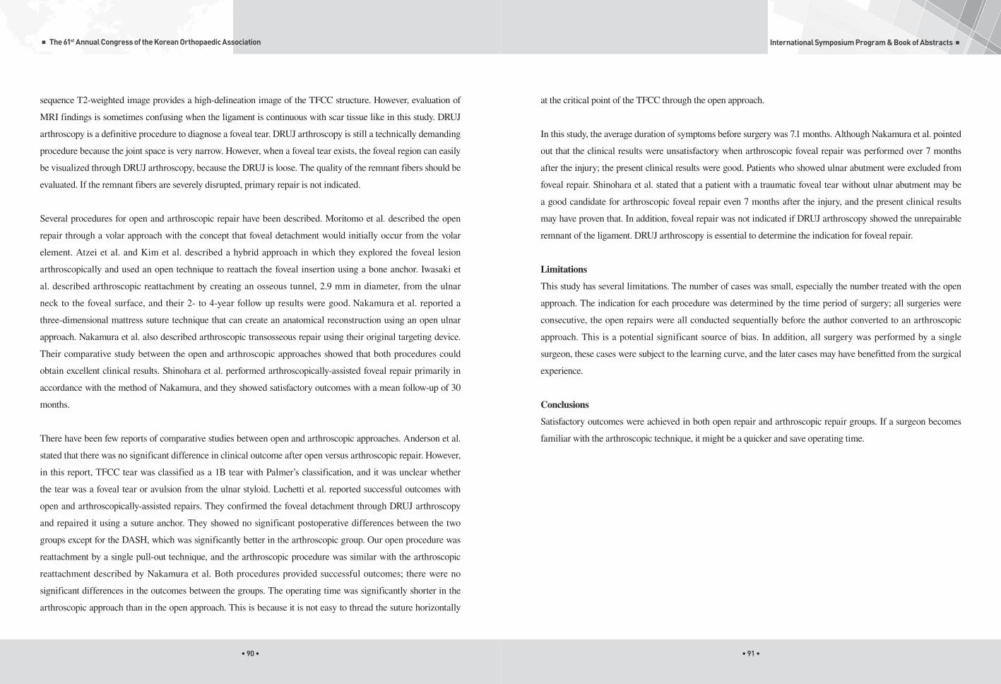
• 90 • • 91 •
■ The 61st Annual Congress of the Korean Orthopaedic Association International Symposium Program & Book of Abstracts ■
at the critical point of the TFCC through the open approach.
In this study, the average duration of symptoms before surgery was 7.1 months. Although Nakamura et al. pointed
out that the clinical results were unsatisfactory when arthroscopic foveal repair was performed over 7 months
after the injury; the present clinical results were good. Patients who showed ulnar abutment were excluded from
foveal repair. Shinohara et al. stated that a patient with a traumatic foveal tear without ulnar abutment may be
a good candidate for arthroscopic foveal repair even 7 months after the injury, and the present clinical results
may have proven that. In addition, foveal repair was not indicated if DRUJ arthroscopy showed the unrepairable
remnant of the ligament. DRUJ arthroscopy is essential to determine the indication for foveal repair.
Limitations
This study has several limitations. The number of cases was small, especially the number treated with the open
approach. The indication for each procedure was determined by the time period of surgery; all surgeries were
consecutive, the open repairs were all conducted sequentially before the author converted to an arthroscopic
approach. This is a potential significant source of bias. In addition, all surgery was performed by a single
surgeon, these cases were subject to the learning curve, and the later cases may have benefitted from the surgical
experience.
Conclusions
Satisfactory outcomes were achieved in both open repair and arthroscopic repair groups. If a surgeon becomes
familiar with the arthroscopic technique, it might be a quicker and save operating time.
sequence T2-weighted image provides a high-delineation image of the TFCC structure. However, evaluation of
MRI findings is sometimes confusing when the ligament is continuous with scar tissue like in this study. DRUJ
arthroscopy is a definitive procedure to diagnose a foveal tear. DRUJ arthroscopy is still a technically demanding
procedure because the joint space is very narrow. However, when a foveal tear exists, the foveal region can easily
be visualized through DRUJ arthroscopy, because the DRUJ is loose. The quality of the remnant fibers should be
evaluated. If the remnant fibers are severely disrupted, primary repair is not indicated.
Several procedures for open and arthroscopic repair have been described. Moritomo et al. described the open
repair through a volar approach with the concept that foveal detachment would initially occur from the volar
element. Atzei et al. and Kim et al. described a hybrid approach in which they explored the foveal lesion
arthroscopically and used an open technique to reattach the foveal insertion using a bone anchor. Iwasaki et
al. described arthroscopic reattachment by creating an osseous tunnel, 2.9 mm in diameter, from the ulnar
neck to the foveal surface, and their 2- to 4-year follow up results were good. Nakamura et al. reported a
three-dimensional mattress suture technique that can create an anatomical reconstruction using an open ulnar
approach. Nakamura et al. also described arthroscopic transosseous repair using their original targeting device.
Their comparative study between the open and arthroscopic approaches showed that both procedures could
obtain excellent clinical results. Shinohara et al. performed arthroscopically-assisted foveal repair primarily in
accordance with the method of Nakamura, and they showed satisfactory outcomes with a mean follow-up of 30
months.
There have been few reports of comparative studies between open and arthroscopic approaches. Anderson et al.
stated that there was no significant difference in clinical outcome after open versus arthroscopic repair. However,
in this report, TFCC tear was classified as a 1B tear with Palmer’s classification, and it was unclear whether
the tear was a foveal tear or avulsion from the ulnar styloid. Luchetti et al. reported successful outcomes with
open and arthroscopically-assisted repairs. They confirmed the foveal detachment through DRUJ arthroscopy
and repaired it using a suture anchor. They showed no significant postoperative differences between the two
groups except for the DASH, which was significantly better in the arthroscopic group. Our open procedure was
reattachment by a single pull-out technique, and the arthroscopic procedure was similar with the arthroscopic
reattachment described by Nakamura et al. Both procedures provided successful outcomes; there were no
significant differences in the outcomes between the groups. The operating time was significantly shorter in the
arthroscopic approach than in the open approach. This is because it is not easy to thread the suture horizontally

• 93 •
International Symposium Program & Book of Abstracts ■
• 92 •
■ The 61st Annual Congress of the Korean Orthopaedic Association
Surgical treatment of ECU tendinopathy associated with TFCC Injury
Young Keun Lee
CHONBUK NATIONAL UNIV.
•••
Introduction
The extensor carpi ulnaris (ECU) tendon and its subsheath (fibro-osseous tunnel) are an important extrinsic
stabilizer of the distal radioulnar joint (DRUJ). Also the floor of the ECU tendon sheath is an important stabilizer
of the triangular fibrocartilage complex (TFCC), which provides intrinsic stability to the DRUJ stability1).
Anatomically, one of the features of ECU tendon is that it occupies a narrow subsheath within the sixth dorsal
compartment, and the subsheath is a separate structure from the overlying extensor retinaculum. Another feature
is that its relative position changes with rotation of the forearm and this feature impacts not only its function but
also its relative stability. Due to these anatomical features, ECU tendon could be easily vulnerable to trauma
such as direct blow, excessive stretch, or repetitive overuse. In addition, because it is an important stabilizer of
the TFCC, we think that both structures may be accompanied by injury and the disease process of ECU tendon
may be difficult to be differentiated from traumatic disruption of the TFCC. Thus accurate diagnosis and proper
treatment with an appropriate time is essential.
Anatomy, pathomechanics and pathoanatomy
ECU muscle arises from the lateral epicondyle, and the tendon of the ECU originates from the muscle fibers 2 to
6 cm proximal to the proximal border of the extensor retinaculum. The tendon is inserted on the fifth metacarpal
base. At ulnar head, ECU tendon passes through a well-defined fibro-osseous tunnel, averaged about 21 mm
long and 6 mm wide2).
ECU tendon is stabilized by bony restraints, subsheath, and extensor retinaculum3). The subsheath is anchored
to distal ulnar and it is essential to maintain the tendon in its normal position3-5). The extensor retinaculum is a
Arthroscopic TFCC reconstruction with tendon graft
Wing Lim Tse, LAU Sun-Wing, HO Pak-Cheong, HUNG Leung-Kim
PRINCE OF WALES HOSPITAL, HONG KONG
•••
Chronic TFCC injury at fovea may result in painful DRUJ instabililty. In neglected case and in patient with ulna
plus variance the result of TFCC repair may not be satisfactory. If significant symptom persist after addressing
the extra-articular malunion of distal radius, correction of ulna plus variance, reconstruction of the deep dorsal
and palmar radio-ulnar ligaments using Adam & Berger’s procedure using palmaris longus tendon graft provide
anatomical restoration of this major static stabilizer of DRUJ. We developed an arthroscopic assisted approach
of this procedure which avoid raising a dorsal DRUJ capsular flap and dislocation of the ulna head that may
further destabilize the joint. Besides, under arthroscopic magnification more accurate placement of the tendon
grafts through the fovea may be achieved. This allow a more aggressive rehabilitation approach that may
achieve better range of movement. However, this require advanced wrist arthroscopic technique and dedicated
instrument. Patient selection is also important: radius malunion and ulna length must be corrected, and presence
of degenerative DRUJ arthritis and infection are contraindications.

• 94 • • 95 •
■ The 61st Annual Congress of the Korean Orthopaedic Association International Symposium Program & Book of Abstracts ■
separate structure from the subsheath, has no attachment to the ulna, but rather on the ulnar aspect of the carpus.
It is not the primary stabilizer of the ECU tendon within its groove, however it compresses the ECU tendon in
the groove in the condition of active wrist flexion.
The ECU muscle’s actions vary dependent on forearm position. During supination, the ECU tendon moves
dorsally closer to the extensor digiti minimi. In full supination, it is subject to maximal traction and exits the
sixth compartment at an angle of 30˚ resulting in a greater contribution to true wrist extension (Fig.1). During
pronation, the ECU tendon lies more in the palmar and ulnar positions of the ulnar head, far from the extensor
digiti minimi and exits the sixths compartment in a straight direction, resulting in diminishing its contribution
to wrist extension5,6) (Fig.2). Therefore tension on the ECU subsheath and retinaculum is greater during the
forearm supination with the wrist in flexion and ulnar deviation7). Understanding these anatomical features of
the ECU tendon is very important to diagnose the ECU tendon pathology and we should recognize that the ECU
subsheath as an integral constituent of the TFCC. Subsheath injury is ususally associated with adjacent soft tissue
disruption, most often the triangular fibrocartilage (TFC).
(Fig.1) (Fig.2)
Melone and Nathan8) reported that because of the complex anatomy of the TFCC, disruption of the TFC
proper, also termed the articular disk, seldom occurred as an isolated injury. Destabilizing injuries of the DRUJ
characteristically disrupted not only the TFC but also the adjacent ECU subsheath and other critical soft tissue
to a variable extent. In this series, 28 (67%) of their 42 cases had associated injury to the ECU subsheath with
subluxation of the ECU tendon. Allende and Le Viet9) also described that the concept of multicomponent injury
of the TFCC. In their series of 28 patients with ECU disruption, 15 (54%) demonstrated substantive concomitant
injury, usually involving the TFC. This clinical study also emphasized that restoration of ulnar wrist stability
requires repair of all pathologic components. Recognition of this variable, often extensive, spectrum of injury at
the ulnar wrist is essential to optimal management of ECU subsheath disruption resulting in ECU tendon and
DRUJ instability. A successful recovery is often contingent on a comprehensive repair of multiple components of
the TFCC.
Clinical Assessment and diagnosis
An accurate clinical history and careful physical examination is critical for diagnosis of ECU tendon disorders.
When evaluating a patient with wrist ulnar side pain, the hallmark of the physical examination pointing to
localized ECU pathology includes the tenderness directly over the ECU tendon and sixth dorsal compartment.
Pain is exacerbated by resisted wrist active extension with ulnar deviation. Ulnar-side wrist pain is also
exacerbated by passive wrist flexion and ulnar deviation with forearm supination. Swelling along the course
of the ECU tendon will be seen. Occasionally crepitus can be detected with palpation of the sixth dorsal
compartment with flexion and extension of the wrist. In the case of the ECU tendon instability, active supination,
flexion and ulnar deviation will produce often visible subluxation of the tendon. ECU tendinosis can coexist with
other conditions in the ulnar side of the wrist, so the diagnosis must not be considered as an exclusion of all other
possible diagnoses. A full clinical and radiological assessment of the other important ulnar-side wrist structures
is mandatory to exclude coexistent pathology in the TFCC, lunotriquetral ligament, distal radioulnar joint or
ulnar styloid.
Plain radiographs are sometime helpful to rule out other regional pathology such as the disorders of the distal
radio-ulnar joint (DRUJ) or TFCC. Occasionally, plain radiographs may demonstrate calcification within the
ECU tendon10). However in most cases it is difficult to diagnose ECU tendinopathy and instability with only plain
radiographs and clinical presentation. Therefore ultrasound (US) and/or magnetic resonance imaging (MRI) are
the imaging modalities of choice in the diagnosis of ECU tendinopathy and instability5,11). MRI may demonstrate
tenosynovitis, recurrent subluxation or dislocation or either partial or complete rupture of the tendon. It is also
able to assess other structures in the ulnar side of the wrist to exclude coexistent pathology in the TFCC, DRUJ,
intercarpal ligament or ulnar styloid that are not easily accessible to US evaluation5,10) (Fig. 3).

• 96 • • 97 •
■ The 61st Annual Congress of the Korean Orthopaedic Association International Symposium Program & Book of Abstracts ■
1) Tenosynovitis
Acute ECU tenosynovitis is defined by inflammation of the tenosynovium of the ECU without significant
stenosis or an underlying bony abnormality of the sixth dorsal compartment. The ECU tendon sheath can be
irritated by repetitive flexion and extension of the wrist, particularly in supination, at the point of angulation of
the tendon as it exits the fibro-osseous tunnel. US demonstrates anechoic, easily compressible fluid surrounding
the tendon without or minimal vascularity on Doppler.
The treatment of acute ECU tenosynovitis includes the stop of use until symptoms subside, the short arm
splinting of the wrist in a position of 30-40 degrees of extension for 2 weeks, and the oral nonsteroidal anti-
inflammatory medication5,10,11). If symptoms persist, corticosteroid injection into the sixth dorsal compartment
is recommended. In rehabilitation, splinting is gradually discontinued if symptoms are resolved and start active/
active assisted wrist range of motion, and extension resistance exercise should be carried out before resuming
unrestricted activities. Graham et al3) recommended to start the strengthening program if the range of motion of
the wrist exceeds 75%.
2) ECU tendinopathy
It develops gradually because in general, it is possible for a patient to continue to use despite the pain or a patient
with a tenosynovitis fails to respond to appropriate treatment. As the disease progresses, tendon thickening
becomes more pronounced resulting in a stenosing tenosynovitis. The tendon can become unstable and dislocated
from attenuation or tearing of its subsheath5,6,10). ECU tendon can be ruptured partially by gliding over the ulnar
ridge of the groove or bony spur at tearing of subsheath.
MRI shows moderate increased signal intensity at the area of tendinopathy and tendon thickening. In the case of
partial tendon tears, it shows clefts or splits within the tendon substance.
Initial treatment is a conservative treatment. If symptoms are not relieved by conservative measures, it can be
also treated by corticosteroids injection into the sheath. Montalvan et al6) described in most their cases, the pain
was resolved within 2 to 24 weeks in tennis players. Futami et al15) reported that 40 of 43 patients recovered in 1
to 9 months using a conservative measures and corticosteroid injection.
(Fig.3)
However some authors, in trauma patients, described that MRI may not be accurate especially in the
investigation of suspected carpal instability because of the post-traumatic changes12,13). Wrist arthroscopy is the
gold standard diagnostic tool for diagnosing carpal instability including TFCC lesions14). Therefore we stress that
wrist arthroscopy should be carried out to get an accurate diagnosis and treatment for all patients undergoing
surgical treatment with ECU tendinopathy (Fig. 4).
(Fig.4)
Treatment of specific conditions
ECU tendinopathy can be classified into tenosynovitis of the tendon sheath, tendinopathy and tendon instability.
These conditions can occur in isolation or synchronously.

• 98 • • 99 •
■ The 61st Annual Congress of the Korean Orthopaedic Association International Symposium Program & Book of Abstracts ■
Summary
The incidence of TFCC rupture associated with ECU tendinopathy is not known exactly. However, since the
ECU subsheath is recognized as an integral part of the TFCC, we think that it is highly likely to occur with ECU
tendinopathy. Therefore when we treat the patients with ECU tendinopathy, the possibility of TFCC combined
injury should always be considered. If surgical treatment is planned, wrist arthroscopy should be performed to
get a more accurate diagnosis of intra-articular pathology and if you get the ECU and DRUJ stability by repair or
reconstruction of the ECU subsheath, TFCC and other intra-articular pathology together, there will be favorable
results.
References
1. Kleinman WB. Distal radius instability and stiffness: common complication of distal radius fractures. Hand Clin 2010;26(2):245-64.
2. Palmer AK, Werner FW, Murphy D, Glisson R. Functional wrist motion: a biomechanical study. J Hand Surg Am. 1985;10(1):39-46.
3. Taleisnik J, Gelberman RH, Miller BW, Szabo RM. The extensor retinaculum of the wrist. J Hand Surg Am. 1984;9(4):495-501.
4. Graham TJ. Pathologies of the extensor carpi ulnaris (ECU) tendon and its investments in the athlete. Hand Clin. 2012;28(3):345-56.
5. Spinner M, Kaplan EB. Extensor carpi ulnaris. Its relationship to the stability of the distal radio-ulnar joint. Clin Orthop Relat Res.
1970;68:124-9.
6. Montalvan B, Parier J, Brasseur JL, Le Viet D, Drape JL. Extensor carpi ulnaris injuries in tennis players: a study of 28 cases. Br J
Sports Med. 2006;40(5):424-9.
7. Campbell D, Campbell R, O’Connor P, Hawkes R. Sports-related extensor carpi ulnaris pathology: a review of functional anatomy,
sports injury and management. Br J Sports Med. 2013;47(17):1105-11.
8. Melone CP Jr, Nathan R. Traumatic disruption of the triangular fibrocartilage complex.
Pathoanatomy. Clin Orthop Relat Res. 1992;275:65-73.
9. Allende C, Le Viet D. Extensor carpi ulnaris problems at the wrist--classification, surgical treatment and results. J Hand Surg Br.
2005;30(3):265-72.
10. Ghatan AC, Puri SG, Morse KW, Hearns KA, von Althann C, Carlson MG. Relative contribution of the subsheath to extensor carpi
ulnaris tendon stability: Implications for surgical reconstruction and rehabilitation. J Hand Surg Am. 2016;41(2):225-32.
11. Hawkes R, O’Connor P, Campbell D. The prevalence, variety and impact of wrist problems in elite professional golfers on the
European Tour. Br J Sports Med. 2013;47(17):1075-9.
12. Cooney WP, Dobyns JH, Linscheid RL. Arthroscopy of the wrist: anatomy and classification of carpal instability. Arthroscopy.
1990;6(2):133-40.
13.Cheng HS, Hung LK, Ho PC, Wong J. An analysis of causes and treatment outcome of chronic wrist pain after distal radial fractures.
Hand Surgery. 2008;13(1):1-10.
14. Andersson JK, Andernord D, Karlsson J, Friden J. Efficacy of magnetic resonance imaging and clinical tests in diagnostics of wrist
ligament injuries: A systemic review. Arthroscopy. 2015;31(10):2014-20.e2.
15. Futami T, Itoman M. Extensor carpi ulnaris syndrome. Findings in 43 patients. Acta Orthop Scand. 1995;66(6):538-9.
16. McCarroll JR. Overuse injuries of the upper extremity in golf. Clin Sports Med. 2001;20(3):469-79.
17. Inoue G, Tamura Y. Surgical treatment for recurrent dislocation of the extensor carpi ulnaris tendon. J Hand Surg Br. 2001;26(6):556-
9.
18. MacLennan AJ, Nemechek NM, Waitayawinyu T, Trumble TE. Diagnosis and anatomic reconstruction of extensor carpi ulnaris
subluxation. J Hand Surg Am. 2008;33(1):59-64.
For patients with recalcitrant symptoms, sixth dorsal compartment release should be considered16). After the
division of retinaculum and subsheath, the tendon is inspected for tearing of sheath, spur and prominent ridges,
which should be repaired or trimmed. The thickened sheath of the ECU tendon was excised. We initially
performed wrist arthroscopy for the diagnosis of intra-articular pathology and carefully repaired the subsheath
and extensor retinaculum. Postoperatively the wrist is immobilized in 30-40 degrees of extension with sugar
tong splint for 2 weeks and additionally it is immobilized with short arm cast for 4 weeks to minimize the risk of
postoperative tendon subluxation.
3) ECU tendon instability
ECU tendon is stabilized by unique fibro-osseous sheath (subsheath) deep to the extensor retinaculum. Therefore,
the instability of the ECU can result in following disruption of the subsheath even if the extensor retinaculum
was intact. Exact mechanism is not clear but is generally seen with forceful supination with wrist flexion and
ulnar deviation15-17). Therefore, during active forearm supination a painful snapping sensation can occur over the
ulnar aspect of the wrist.
Although nonoperative treatment occasionally has been recommended for acute ECU tendon instability,
experience with consistent regimens of conservative treatment is limited and invariably clinical studies fail
to substantiate its efficacy. Most authors concur that operative treatment is a superior method of consistently
achieving through healing and a favorable outcome. Rowland18) operated on an acute case and found that there
was a tear in the subsheath with a considerable gap in the position where the wrist was placed. He suggested
surgical repair in all such patients. Inoue and Tamura9) described three types of disruption of the subsheath
and the differentiation between the types of lesion was impossible clinically. They also recommended surgical
exploration of all symptomatic ECU tendon dislocation and treatment with repair or reconstruction of subsheath.
In chronic ECU tendon instability, numerous authors have described successful operative treatment using a
variety of techniques to restore ECU and DRUJ stability. Retinacular flap19), retinacular graft9,17) and direct suture
of the disrupted subsheath to bone either with bone anchors or through drill holes with or without deepening of
the osseous groove7,9).
When accompanied by a TFCC injury, concurrent repair of the TFC8,9), all have been reported as successful
method of surgical repair. In cases with relatively deficient TFC substance, distally based ECU tenodesis for the
reconstruction of destabilizing ECU and TFC pathology can be a treatment option8).

The 61st Annual Congress of The Korean Orthopaedic Association
• 100 •
■ The 61st Annual Congress of the Korean Orthopaedic Association
19. Burkhart SS, Wood MB, Linscheid RL. Posttraumatic recurrent subluxation of the extensor carpi ulnaris tendon. J Hand Surg Am.
1982;7(1):1-3.
Figure Legends
Figure 1. Intraoperative view of the right wrist, during full supination showing that the extensor carpi ulnaris
(ECU) tendon (blue loops) is subjected to maximal traction and has to adopt an approximately 30˚
angle to reach the base of the 5th metacarpal.
Figure 2. Intraoperative view of the right wrist, during full pronation showing that the ECU tendon (blue loops)
is situated on the inner surface of the ulnar head and having a straight course.
Figure 3. Fat suppressed coronal T2-weighted left wrist MRI images of 45-year-old female with ECU
tendinopathy shows high signal intensity within the TFC (white arrows) representing foveal rupture.
Figure 4. Same patient’s left wrist, radio-carpal arthroscopy images show severe synovitis (A) and unstable TFC
(B). (TFC: triangular fibrocartilage).
Jong Woong Park / Kanit Sananpanich
Oct. 19th. 2017. Thu | Grand Ballroom2
Updates on peripheral nerve surgery

• 103 •
International Symposium Program & Book of Abstracts ■
• 102 •
■ The 61st Annual Congress of the Korean Orthopaedic Association
Conclusions
Nerve transfer from extensor carpi radialis brevis to flexor digitorum profundus branch combined with
brachialis to anterior interosseous nerve offer all digit flexion in C6 spinal cord injury.
Distal nerve transfer for peripheral nerve injury in BPI and tetraplegia
Kanit Sananpanich
CHIANG MAI UNIVERSITY HOSPITAL, THAILAND
•••
Background
In C6 spinal cord injury digit flexion is one of the most required reconstructions. Distal nerve transfers
offer new options for functional recovery. There are three parameters for selection of nerve pairs transfers:
the surgical feasibility of the transfer, the donor-to-recipient axon count ratio, and the distance from the
coaptation site to the motor point. This study investigated pairs of distal nerve transfer for all digit flexion.
Methods
Twenty-two fresh cadaver upper extremities were dissected to determine the anatomic relations and
simulated the transfer of brachialis branch and extensor carpi radialis brevis branch as the donor nerve to
anterior interosseous nerve and flexor digitorum profundus branch of ulnar nerve as the recipient nerve.
Following the simulation, the success of the transfer was estimated by the three parameters. A clinical
case report of complete C6 spinal cord injury was managed by eight pairs of bilateral nerve transfer,
including a new transfer extensor carpi radialis brevis to the flexor digitorum profundus branch of ulnar
nerve.
Results
The newly proposed transfer extensor carpi radialis brevis to the flexor digitorum profundus branch
appears to be superior to the other transfers to regain digit flexion determined by the three parameters
and able to combine with brachialis to anterior interosseous nerve. Our case report demonstrate that good
fingers flexion can be achieved 10 months postoperatively.

• 104 • • 105 •
■ The 61st Annual Congress of the Korean Orthopaedic Association International Symposium Program & Book of Abstracts ■
of an original nerve, especially in the case of pure sensory nerve defect. Furthermore, inside-out vein
grafts provide improved regeneration potential with their abundant laminin, which is known to promote
axonal regeneration.
Recent technological advances, including electrical stimulation to injured or repaired nerves, indicate new
horizons in the field of neural regeneration. Electrical stimulation can be delivered to a target site through
a specifically developed electrode interface for a specific duration. However, the optimal time period and
strength has not been definitively suggested until now. In addition, removal of the electrode interface is
not an easy task.
Low-intensity-focused ultrasound (LIFU) stimulation is another cutting-edge technology for enhanced
neural regeneration. Earlier evidence demonstrated successful axonal stimulation through ultrasound
stimulation. Because ultrasound stimulation is non-invasive, this technology has tremendous potential in
the near future.
Biological approaches to promote neural regeneration include cultured Schwann cell grafts with conduits.
Stem cell implantation is a hopeful approach, and enhancing Schwann cell migration in the early stage
of regeneration under a magnetic field is another interesting idea. Inducing neoangiogenesis in the early
period of regeneration can be promoted by various methods based on the fundamental understanding of
the natural process of neural regeneration.
In this lecture, up-to-date trials to promote neural regeneration in the research bases and clinical trials will
be discussed, however, there is no single best solution developed until now that can completely replace
autogenous nerve grafts.
Cutting-edge technology for the enhanced neural regeneration
Jong Woong Park
KOREA UNIV.
•••
Tension-free, end-to-end neurorrhaphy is the best surgical solution for injured peripheral nerves. However,
it is not always possible. Furthermore, even after simple neurorrhaphy, a long regeneration process
follows. When there is a segmental defect, which cannot be directly repaired, a nerve graft is mandatory.
Although an autogenous nerve graft is the first choice, donor site morbidity is inevitable.
An autogenous sural nerve graft is the most common source for a nerve graft. Although it is an
autogenous tissue without immune-associated problems, non-vascularized nerve grafts have inevitable
limitations in their regeneration potential. According to research performed by Millein this sie, grafted
nerves obtain their nutrition by diffusion from the surrounding tissue. After three days, neoangiogenesis
occurs and active blood perfusion initiates. Wallerian degeneration progresses immediately after injury
and the activity of macrophages is delayed or disturbed, and the entire process of neural regeneration is
significantly delayed.
An autogenous nerve graft may be substituted by a synthetic nerve conduit or allograft that is harvested
and prepared through a decellularization process. Because there are no functioning macrophages or
Schwann cells in these substitutes, their limitations in regeneration potential remain.
Recently, many biological and electronical cutting-edge technologies have been developed in the attempt
to improve regeneration after peripheral nerve injury. A vein graft, muscle graft, or combined vein and
muscle graft can be ideal substitutions for autogenous nerve grafts. According to the author’s basic
research and clinical experience, an approximately 3-cm vein graft has successfully recovered the function

• 106 • • 107 •
■ The 61st Annual Congress of the Korean Orthopaedic Association International Symposium Program & Book of Abstracts ■
외막을 내부로 가져와서 신경의 재생을 촉진하는데 있다. 동물 실험에서 표준적인 정맥 이식 방법과 동일하거
나 나은 결과를 나타내었으며,6) 최근 Jeon 등7)이 임상적으로도 우수한 결과를 보고하고 있다 (Fig. 1). 그러나
inside-out 정맥 이식법이 표준적인 방법에 비하여 더욱 우수한 결과를 얻는다는 근거는 부족하며 더 많은 연
구가 있어야 할 것으로 판단된다.
Fig. 1. Preparation of an inside-out vein graft (From Jeon WJ, et al. Clinical application of inside-out vein grafts for the
treatment of sensory nerve segmental defect. Microsurgery 2011;31:268-73)
2. 근육 (Muscle)
평활근의 내부에는 장축으로 배열된 기저판(basal lamina)이 존재하며, 이 구조는 재생되는 신경의 조직에서
발견되는 신경 내 구조와 유사하기 때문에 이를 이용하여 신경의 재생을 촉진하려는 시도가 진행되고 있다.8) 동
물 실험 및 임상 연구에서 자가 근육이나 변성 근육을 이용하여 신경의 재생의 가능함이 알려지고 있으며, 일부
에서는 자가 신경 이식술에 견줄만한 우수한 신경 재생을 보고하고 있다.9, 10) 근육을 신경 도관으로 이용하는 것
의 문제점은 근육 자체가 도관의 형태를 띄고 있지 않기 때문에 신경이 근육의 밖으로 자라나서 신경 재생의 결
과가 떨어질 수 있으며 신경종을 형성할 수도 있다는 점이다. 또한 정맥 이식에 비하여 공여 근육에 근력 약화
등을 초래할 수 있는 것도 단점으로 지적되고 있다.8) 그러나 Brunelli 등11)은 쥐 실험을 통하여 근육을 정맥 내
에 삽입하여 근육이나 정맥을 단독으로 사용했을 때보다 신경 재생이 우수함을 보고하였으며, 최근 Marcoccio
등12)은 이 방법을 사용하여 임상적으로도 좋은 결과를 발표하였다.
합성 도관 (Synthetic nerve conduits)
자가 신경 이식술의 문제점을 극복하고 신경 결손의 회복을 증진시키기 위하여 다양한 종류의 합성 도관이 개
발되고 있다. 이 신경 도관들은 신경 말단에서 축색의 성장을 유도하고 신경영양 인자(neurotrophic factors)
의 분비를 촉진하며, 외부로부터 섬유 조직이 침투하는 것을 방지한다.13) 처음에는 silicone이 사용되어 신경 재
생의 효과가 있음이 실험적으로 입증되었으나,14) 임상에서 silicone이 이물 반응과 과도한 섬유 조직 형성, 신경
Conduits for Peripheral Nerve Regeneration
Joo Yup Lee
CATHOLIC UNIV.
•••
말초 신경의 손상은 외상이나 종양 절제술 후, 혹은 정형외과 수술의 합병증으로 발생할 수 있으며, 미국에서는
매년 약 200,000건의 신경 봉합술이 시행될 정도로 흔한 손상 중 하나이다. 최근 미세수술의 발달로 신경의 일
차 봉합술이 신경 손상의 표준적인 치료로 시행되고 있으나, 신경단의 간격이 너무 커서 일차 봉합술이 불가능
할 경우에는 자가 신경 이식술(autogenous nerve graft)을 시행할 수 있다.1) 그러나 자가 신경 이식술은 공여
부의 신경 손상이 불가피하며, 길이와 직경을 원하는 대로 조절할 수 없는 단점이 있다. 그래서 자가 신경 이식
술의 대안으로 신경 도관(nerve conduit)을 이용한 말초 신경 재생이 시도되고 있으며, 새로운 신경 도관을 개
발하기 위한 연구가 진행되고 있다. 본 심포지엄에서는 신경 도관 이식의 배경 및 그 종류, 그리고 다양한 신경
도관 이식술에 따른 임상 결과를 알아보기로 한다.
자가 도관 (Autogenous nerve conduits)
1. 정맥 (Vein)
신경의 재생을 위한 자가 정맥 도관 이식술은 1982년 Chiu 등에 의하여 소개된 이래 자가 신경 이식술을 대신
하기 위하여 시도되고 있다.2) Chiu 등은 백서의 좌골신경 결손 모델을 이용하여 자가 정맥 도관 이식술을 시행
한 후 조직학적, 전기 생리학적으로 신경이 재생될 수 있음을 밝혔다. 그 이후 1990년 Chiu와 Strauch 등에 의
하여 자가 정맥 도관에 대한 임상 연구가 시행되었다.3) 이 연구에서 총 15명의 환자에게 손과 전완부에 발생한
외상성 신경종을 절제한 후 3cm 이하의 신경 결손부를 자가 정맥 도관으로 재건하였는데, 비록 자가 신경 이식
술보다 결과는 좋지 않았으나 모든 환자에서 이점 식별력을 회복함을 관찰할 수 있었다. 자가 정맥을 신경 도관
으로 사용하는데 있어 몇 가지 우려가 있는데, 그 중 하나는 정맥의 벽이 얇기 때문에 신경이 재생되기 전에 도
관의 내부가 막혀 버릴 수 있다는 점이다. 그러나 Tseng 등4)은 실험 연구에서 정맥의 내부에 혈종이 발생하기
때문에 도관이 무너지지 않고 신경이 재생되는 기간 동안 유지될 수 있다고 하였다. 또한 Wang 등5)은 정맥을
직접 봉합하지 않고 뒤집어서 봉합하는 inside-out 정맥 이식법을 소개하였다. 이 방법의 장점은 만약에 있을
지도 모르는 정맥 판막으로 인하여 신경의 재생이 방해되는 것을 방지하고, laminin과 collagen이 풍부한 정맥

• 108 • • 109 •
■ The 61st Annual Congress of the Korean Orthopaedic Association International Symposium Program & Book of Abstracts ■
신경이 회복되지 않았다고 하였다. 다른 합성 도관과 비교하여 collagen 신경 도관은 몇 가지 장점을 가지고 있
는데, polyglycolic acid 도관에 비하여 그 내부 구조를 오래 유지할 수 있으며 poly-lactide-ε-caprolactone
도관에 비하여 조작성이 용이하다는 점이다. 최근 Waitayawinyu 등21)은 동물 실험에서 polyglycolic acid 도
관과 collagen 도관을 비교하였는데, collagen은 자가 신경 이식술에 필적할만한 결과를 얻었지만 polyglycolic
acid 도관은 collagen 도관보다 나쁜 결과를 발표하였다. Collagen 도관은 제조 시 가교 과정(cross-
linking process)을 조작하여 분해 시기를 자유롭게 조절할 수 있으며, 신경 도관 내부에 collagen-GAG
(glycosaminoglycan) 등의 기질을 삽입하는 것이 용이하기 때문에 향후 발전이 기대되는 합성 도관 중 하나이
다 (Fig. 2).
Fig. 2. Schematic drawings of collagen conduit repair technique (From Taras JS, et al. Reconstruction of digital nerves
with collagen conduits. J Hand Surg Am 2011;36:1441-6)
3. Poly-lactide-ε-caprolactone (Neurolac)
Poly-lactide-ε-caprolactone은 합성 물질로 인체에서 무해하고 이물 반응을 거의 일으키지 않아 흡수성 신
경 도관으로 사용되고 있다.22) 신경 재생을 유지하기 위하여 내부 구조가 약 10주 동안 강도를 유지한다고 하
며, 1년 내로 완전히 흡수되는 것으로 알려져 있으나 2년까지도 발견될 수 있다고 한다. Den Dunnen 등은 쥐
의 10mm 좌골신경 결손 모델에서 Poly-lactide-ε-caprolactone의 신경 재생 효과를 연구하였다.23) 저자들은
이 합성 도관을 사용하여 매우 우수한 신경 재생 결과를 얻었으며, 심지어 자가 신경 이식술보다 나은 신경 재생
을 보고하였다. 그러나 이 실험 연구는 신경의 형태학적 분석만을 시행하였고, 기능적 평가를 시행하지 않았기
때문에 결과 판단에 신중을 기하여야 할 것이다. 최근에 Shin 등18)은 임상에서 사용 가능한 세 가지 신경 도관의
신경 재생 능력을 동물 실험을 통하여 확인하였다. 이 연구에서 Poly-lactide-ε-caprolactone 이 가장 우수한
신경 재생 효과가 있는 것으로 나타나서 임상적 적용에 희망을 갖게 한다. Bertleff 등24)은 Neurolac을 이용한
임상 시험에서 기존의 방법과 유사한 결과를 보고하여 임상적으로 효과가 있음을 입증하였다. 그러나 이 물질이
2년 이후에도 남아 이물 반응을 일으킬 수 있으며, 벽이 매우 단단하여 8-0 바늘이 통과하기 어려우며, 신경 도
관이 단단하여 피부에서 만져질 수 있다는 점이 문제점으로 지적되고 있다.
을 압박하는 문제 등이 발견되어 사용이 중단되고 흡수성 합성 도관으로 이동하게 되었다.15) 그 후 Polyglycolic
acid (PGA; Neurotube), Collagen (Neuragen), Poly-caprolactone (Neurolac) 등 흡수성 물질을 이용한 신
경 도관이 미국 및 유럽 FDA의 승인을 받아 임상에서 사용되고 있다. 현재 우리나라에서는 아직 보험적용이 가
능한 흡수성 신경 도관은 없으며, 가격이 고가이기 때문에 (개당 500-1500달러) 사용되지 않고 있다. 또한 동
물 실험 및 임상 실험에서 자가 신경 이식술에 필적할만한 결과가 나오고 있지 않다는 점도 합성 도관의 광범위
한 사용을 제한케 하는 이유이다.
1. Polyglycolic acid (Neurotube)
Polyglycolic acid (PGA)는 우리가 일반적으로 사용하는 봉합사인 Dexon과 Vicryl의 주요 성분으로 이미 체내
안전성은 입증되어 있는 물질이다. Polyglycolic acid는 체내에서 가수분해를 통하여 분해되며 흡수되는데 약
90일이 소요된다. 1990년 Mackinnon과 Dellon16)은 흡수성 합성 도관인 polyglycolic acid를 3cm 이하의 감
각신경 결손 부위에 임상적으로 사용하여 자가 신경 이식술과 유사한 신경 재생 결과를 얻을 수 있었다고 발표
하였다. 이 합성 도관은 1999년 미국 FDA에서 Neurotube라는 이름으로 사용이 허가되었으며, Weber 등17)이
polyglycolic acid 도관과 표준적인 신경 봉합 방법들에 대하여 전향적 무작위 시험을 시행한 결과 4mm 이하의
신경 결손에서는 단단 봉합술과, 30mm 이하의 신경 결손에서는 자가 신경 이식술과 동일 결과를 보고하였다.
Neurotube의 장점은 가격이 저렴하며 수술 시 조작이 용이하며 도관의 흡수가 빠르다는 점이지만, 반대로 조기
에 강도를 소실하여 신경의 재생을 충분히 기대할 수 없다는 점도 고려해야 할 것이다. Shin 등18)의 동물 실험
에 의하면 세 종류의 상품화된 합성 도관 중 Neurotube가 가장 나쁜 결과를 얻었으며, 수술 후 12주에 조기 흡
수 및 collapse된 신경 도관을 확인하였다.
2. Collagen (Neuragen)
제 1형 교원질 (type I collagen)은 우리 몸에 다양하게 분포하고 있으며, 말초 신경에서 주된 성분이기 때문에
합성 도관에 널리 이용되고 있다. 현재 Integra 회사에서 bovine dermal type I collagen을 이용한 합성 도관
이 FDA의 허가를 받아 Neuragen 이라는 상품명으로 판매되고 있다. 제 1형 교원질로 만들어진 도관은 우리
몸에서 면역 반응 및 이물질로 인식될 가능성이 적고, 이미 피부 이식재, 상처 치료재 등으로 널리 사용되기 때
문에 체내 안전성은 높다고 할 수 있다. Collagen 신경 도관은 반투과성으로 우리 몸의 대사 과정을 통하여 분
해되며 염증 반응을 일으키지 않는다고 한다. 현재까지 collagen 신경 도관에 대한 임상 연구는 많지 않은 편
이다. Ashley 등19)은 상완 신경총 손상이 있는 5명의 소아 환자에서 2cm 이하의 신경 결손을 치료하기 위하
여 collagen 신경 도관을 사용하고 5명 중 4명에서 수술 후 2년째 좋은 신경 회복을 관찰하였다고 보고하였다.
Lohmeyer 등20)은 수부의 신경 결손 환자에서 collagen 도관을 사용하고 그 결과를 발표하였다. 신경 결손의 크
기는 18mm 이하였으며, 12개월 추시한 6명의 환자들 중 4명은 우수, 1명은 불량, 나머지 한 명은 전혀 감각
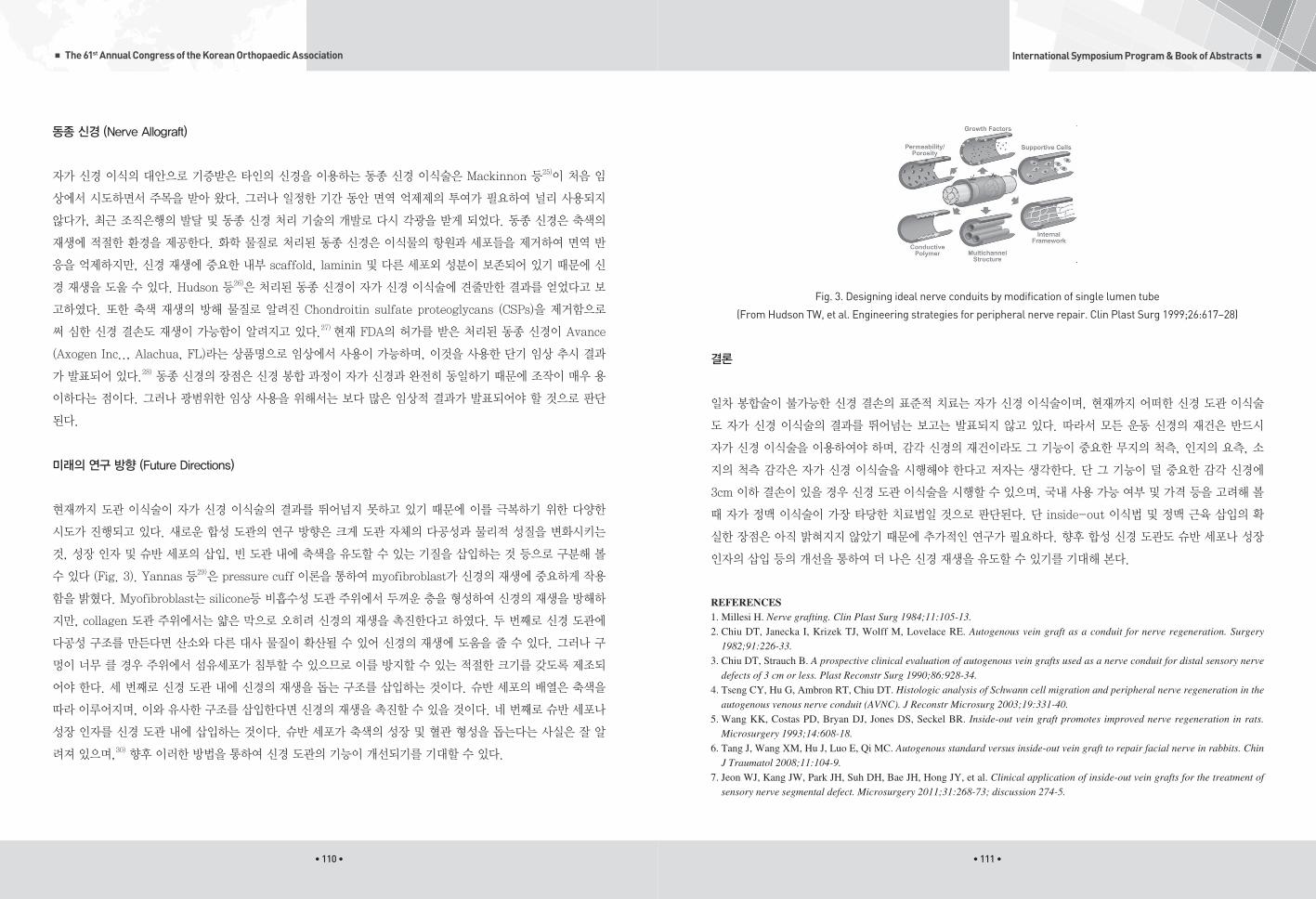
• 110 • • 111 •
■ The 61st Annual Congress of the Korean Orthopaedic Association International Symposium Program & Book of Abstracts ■
Fig. 3. Designing ideal nerve conduits by modification of single lumen tube
(From Hudson TW, et al. Engineering strategies for peripheral nerve repair. Clin Plast Surg 1999;26:617–28)
결론
일차 봉합술이 불가능한 신경 결손의 표준적 치료는 자가 신경 이식술이며, 현재까지 어떠한 신경 도관 이식술
도 자가 신경 이식술의 결과를 뛰어넘는 보고는 발표되지 않고 있다. 따라서 모든 운동 신경의 재건은 반드시
자가 신경 이식술을 이용하여야 하며, 감각 신경의 재건이라도 그 기능이 중요한 무지의 척측, 인지의 요측, 소
지의 척측 감각은 자가 신경 이식술을 시행해야 한다고 저자는 생각한다. 단 그 기능이 덜 중요한 감각 신경에
3cm 이하 결손이 있을 경우 신경 도관 이식술을 시행할 수 있으며, 국내 사용 가능 여부 및 가격 등을 고려해 볼
때 자가 정맥 이식술이 가장 타당한 치료법일 것으로 판단된다. 단 inside-out 이식법 및 정맥 근육 삽입의 확
실한 장점은 아직 밝혀지지 않았기 때문에 추가적인 연구가 필요하다. 향후 합성 신경 도관도 슈반 세포나 성장
인자의 삽입 등의 개선을 통하여 더 나은 신경 재생을 유도할 수 있기를 기대해 본다.
REFERENCES1. Millesi H. Nerve grafting. Clin Plast Surg 1984;11:105-13.
2. Chiu DT, Janecka I, Krizek TJ, Wolff M, Lovelace RE. Autogenous vein graft as a conduit for nerve regeneration. Surgery
1982;91:226-33.
3. Chiu DT, Strauch B. A prospective clinical evaluation of autogenous vein grafts used as a nerve conduit for distal sensory nerve
defects of 3 cm or less. Plast Reconstr Surg 1990;86:928-34.
4. Tseng CY, Hu G, Ambron RT, Chiu DT. Histologic analysis of Schwann cell migration and peripheral nerve regeneration in the
autogenous venous nerve conduit (AVNC). J Reconstr Microsurg 2003;19:331-40.
5. Wang KK, Costas PD, Bryan DJ, Jones DS, Seckel BR. Inside-out vein graft promotes improved nerve regeneration in rats.
Microsurgery 1993;14:608-18.
6. Tang J, Wang XM, Hu J, Luo E, Qi MC. Autogenous standard versus inside-out vein graft to repair facial nerve in rabbits. Chin
J Traumatol 2008;11:104-9.
7. Jeon WJ, Kang JW, Park JH, Suh DH, Bae JH, Hong JY, et al. Clinical application of inside-out vein grafts for the treatment of
sensory nerve segmental defect. Microsurgery 2011;31:268-73; discussion 274-5.
동종 신경 (Nerve Allograft)
자가 신경 이식의 대안으로 기증받은 타인의 신경을 이용하는 동종 신경 이식술은 Mackinnon 등25)이 처음 임
상에서 시도하면서 주목을 받아 왔다. 그러나 일정한 기간 동안 면역 억제제의 투여가 필요하여 널리 사용되지
않다가, 최근 조직은행의 발달 및 동종 신경 처리 기술의 개발로 다시 각광을 받게 되었다. 동종 신경은 축색의
재생에 적절한 환경을 제공한다. 화학 물질로 처리된 동종 신경은 이식물의 항원과 세포들을 제거하여 면역 반
응을 억제하지만, 신경 재생에 중요한 내부 scaffold, laminin 및 다른 세포외 성분이 보존되어 있기 때문에 신
경 재생을 도울 수 있다. Hudson 등26)은 처리된 동종 신경이 자가 신경 이식술에 견줄만한 결과를 얻었다고 보
고하였다. 또한 축색 재생의 방해 물질로 알려진 Chondroitin sulfate proteoglycans (CSPs)을 제거함으로
써 심한 신경 결손도 재생이 가능함이 알려지고 있다.27) 현재 FDA의 허가를 받은 처리된 동종 신경이 Avance
(Axogen Inc.., Alachua, FL)라는 상품명으로 임상에서 사용이 가능하며, 이것을 사용한 단기 임상 추시 결과
가 발표되어 있다.28) 동종 신경의 장점은 신경 봉합 과정이 자가 신경과 완전히 동일하기 때문에 조작이 매우 용
이하다는 점이다. 그러나 광범위한 임상 사용을 위해서는 보다 많은 임상적 결과가 발표되어야 할 것으로 판단
된다.
미래의 연구 방향 (Future Directions)
현재까지 도관 이식술이 자가 신경 이식술의 결과를 뛰어넘지 못하고 있기 때문에 이를 극복하기 위한 다양한
시도가 진행되고 있다. 새로운 합성 도관의 연구 방향은 크게 도관 자체의 다공성과 물리적 성질을 변화시키는
것, 성장 인자 및 슈반 세포의 삽입, 빈 도관 내에 축색을 유도할 수 있는 기질을 삽입하는 것 등으로 구분해 볼
수 있다 (Fig. 3). Yannas 등29)은 pressure cuff 이론을 통하여 myofibroblast가 신경의 재생에 중요하게 작용
함을 밝혔다. Myofibroblast는 silicone등 비흡수성 도관 주위에서 두꺼운 층을 형성하여 신경의 재생을 방해하
지만, collagen 도관 주위에서는 얇은 막으로 오히려 신경의 재생을 촉진한다고 하였다. 두 번째로 신경 도관에
다공성 구조를 만든다면 산소와 다른 대사 물질이 확산될 수 있어 신경의 재생에 도움을 줄 수 있다. 그러나 구
멍이 너무 클 경우 주위에서 섬유세포가 침투할 수 있으므로 이를 방지할 수 있는 적절한 크기를 갖도록 제조되
어야 한다. 세 번째로 신경 도관 내에 신경의 재생을 돕는 구조를 삽입하는 것이다. 슈반 세포의 배열은 축색을
따라 이루어지며, 이와 유사한 구조를 삽입한다면 신경의 재생을 촉진할 수 있을 것이다. 네 번째로 슈반 세포나
성장 인자를 신경 도관 내에 삽입하는 것이다. 슈반 세포가 축색의 성장 및 혈관 형성을 돕는다는 사실은 잘 알
려져 있으며,30) 향후 이러한 방법을 통하여 신경 도관의 기능이 개선되기를 기대할 수 있다.

• 112 • • 113 •
■ The 61st Annual Congress of the Korean Orthopaedic Association International Symposium Program & Book of Abstracts ■
8. Meek MF, Varejao AS, Geuna S. Use of skeletal muscle tissue in peripheral nerve repair: review of the literature. Tissue Eng
2004;10:1027-36.
9. Glasby MA, Gschmeissner SE, Huang CL, De Souza BA. Degenerated muscle grafts used for peripheral nerve repair in
primates. J Hand Surg Br 1986;11:347-51.
10. Pereira JH, Cowley SA, Gschmeissner SE, Bowden RE, Turk JL. Denatured muscle grafts for nerve repair. An experimental
model of nerve damage in leprosy. J Bone Joint Surg Br 1990;72:874-80.
11. Brunelli GA, Battiston B, Vigasio A, Brunelli G, Marocolo D. Bridging nerve defects with combined skeletal muscle and vein
conduits. Microsurgery 1993;14:247-51.
12. Marcoccio I, Vigasio A. Muscle-in-vein nerve guide for secondary reconstruction in digital nerve lesions. J Hand Surg Am
2010;35:1418-26.
13. Hudson TW, Evans GR, Schmidt CE. Engineering strategies for peripheral nerve repair. Orthop Clin North Am 2000;31:485-
98.
14. Lundborg G, Hansson HA. Nerve regeneration through preformed pseudosynovial tubes. A preliminary report of a new
experimental model for studying the regeneration and reorganization capacity of peripheral nerve tissue. J Hand Surg Am
1980;5:35-8.
15. Dahlin LB, Lundborg G. Use of tubes in peripheral nerve repair. Neurosurg Clin N Am 2001;12:341-52.
16. Mackinnon SE, Dellon AL. Clinical nerve reconstruction with a bioabsorbable polyglycolic acid tube. Plast Reconstr Surg
1990;85:419-24.
17. Weber RA, Breidenbach WC, Brown RE, Jabaley ME, Mass DP. A randomized prospective study of polyglycolic acid conduits
for digital nerve reconstruction in humans. Plast Reconstr Surg 2000;106:1036-45; discussion 1046-8.
18. Shin RH, Friedrich PF, Crum BA, Bishop AT, Shin AY. Treatment of a segmental nerve defect in the rat with use
of bioabsorbable synthetic nerve conduits: a comparison of commercially available conduits. J Bone Joint Surg Am
2009;91:2194-204.
19. Ashley WW, Jr., Weatherly T, Park TS. Collagen nerve guides for surgical repair of brachial plexus birth injury. J Neurosurg
2006;105:452-6.
20. Lohmeyer JA, Siemers F, Machens HG, Mailander P. The clinical use of artificial nerve conduits for digital nerve repair: a
prospective cohort study and literature review. J Reconstr Microsurg 2009;25:55-61.
21. Waitayawinyu T, Parisi DM, Miller B, Luria S, Morton HJ, Chin SH, et al. A comparison of polyglycolic acid versus type 1
collagen bioabsorbable nerve conduits in a rat model: an alternative to autografting. J Hand Surg Am 2007;32:1521-9.
22. Meek MF, Jansen K, Steendam R, van Oeveren W, van Wachem PB, van Luyn MJ. In vitro degradation and biocompatibility
of poly(DL-lactide-epsilon-caprolactone) nerve guides. J Biomed Mater Res A 2004;68:43-51.
23. Den Dunnen WF, Meek MF, Robinson PH, Schakernraad JM. Peripheral nerve regeneration through P(DLLA-epsilon-CL)
nerve guides. J Mater Sci Mater Med 1998;9:811-4.
24. Bertleff MJ, Meek MF, Nicolai JP. A prospective clinical evaluation of biodegradable neurolac nerve guides for sensory nerve
repair in the hand. J Hand Surg Am 2005;30:513-8.
25. Mackinnon SE, Doolabh VB, Novak CB, Trulock EP. Clinical outcome following nerve allograft transplantation. Plast
Reconstr Surg 2001;107:1419-29.
26. Hudson TW, Zawko S, Deister C, Lundy S, Hu CY, Lee K, et al. Optimized acellular nerve graft is immunologically tolerated
and supports regeneration. Tissue Eng 2004;10:1641-51.
27. Neubauer D, Graham JB, Muir D. Chondroitinase treatment increases the effective length of acellular nerve grafts. Exp Neurol
2007;207:163-70.
28. Karabekmez FE, Duymaz A, Moran SL. Early clinical outcomes with the use of decellularized nerve allograft for repair of
sensory defects within the hand. Hand (N Y) 2009;4:245-9.
29. Yannas IV, Zhang M, Spilker MH. Standardized criterion to analyze and directly compare various materials and models for
peripheral nerve regeneration. J Biomater Sci Polym Ed 2007;18:943-66.
30. Sinis N, Schaller HE, Schulte-Eversum C, Schlosshauer B, Doser M, Dietz K, et al. Nerve regeneration across a 2-cm gap in
the rat median nerve using a resorbable nerve conduit filled with Schwann cells. J Neurosurg 2005;103:1067-76.
The role of stem cell transplantation for peripheral nerve regeneration
Jae Kwang Kim
ULSAN UNIV.
•••
Peripheral nerve injuries (PNI) are mainly related to trauma, tumor, and iatrogenic lesions, leading
to neurologic deficits and functional disability. Lesions with loss of nerve substance produce serious
problems for the patient. Besides causing pain and morbidity, these injuries usually generate permanent
sequelae, such as sensory deficit and functional dysfunction. These lesions cause damages that
substantially diminish the quality of life of these patients, including physical disability and total or partial
loss of their productive activities, which gives rise to important social and economic consequences.
Nowadays autologous peripheral nerve transplantation represents the gold standard of repair when there
is loss of substance that precludes neurorrhaphy. However, it presents some limitations, such as the need
to perform two surgical procedures at different sites, the consequent greater morbidity and the shortage of
nerve donor sites, besides the resulting sensory deficit in the area from which it was removed. For nerve
discontinuities with a gap, nerve autografts are useful but limited by availability and donor site morbidity.
The various synthetic conduits and acellular allografts are not generally recommended for gaps > 3cm.
Tissue repair requires a complex interaction between cells, extracellular matrix, and trophic factors,
which are all important elements involved in nerve regeneration. Consequently, cell therapy and tissue
engineering have been receiving a great deal of attention in recent decades, and are widely used in
different areas.
Cell transplantation is one of the cell therapy and tissue engineering strategies aimed at the creation of
a favorable microenvironment for tissue regeneration. Stem cells have important characteristics that

• 114 •
■ The 61st Annual Congress of the Korean Orthopaedic Association
differentiate them from other cell types, are undifferentiated precursor cells that have self-renewal ability
and can differentiate into multiple lineages. They are present in several tissues and are responsible for
their regeneration in the event of injuries or lesions.
The self-renewal capacity of stem cells makes it possible to deliver numerous cleavage cells to the damage
site. The stem cells continue proliferating after migrating to the injured nerve tissue, and further differentiate
to the necessary cell type under the appropriate micro environmental conditions. It is confirmed that
Neural Stem Cells (NSC) can be induced to a peripheral neuron, Schwann Cells (SC), or smooth muscle
phenotype upon co-culture with cells from the nervous system. Furthermore, about 5% of BMSCs can
spontaneously transdifferentiate into SCs without specific intervention. However, the differentiation rate
of naive precursor cells in the peripheral nerve is relatively low. Pre-differentiating stem cells toward a
desired phenotype in vitro by chemical induction, biological treatment, gene transfection, or co-culture
with neural cells before injection is an effective method.
Other than differentiation to appropriate cells, stem cells also provide a beneficial micro environment for
neural cell survival and neurogenesis by secreting bioactive neurotrophic molecules. MSCs synthesize and
release a variety of growth factors, such as nerve growth factor (NGF), brain-derived neurotrophic factor
(BDNF), GDNF, neurotrophin-3 (NT-3), VEGF, and ciliary-derived neurotrophic factor (CDNF). Adipose
derived Stem Cells also upregulate protein expression of BDNF, glial growth factor, neuregulin-1, VEGF,
HGF, and insulin-like growth factor. Furthermore, overexpressed neurotrophic factors facilitate the
regeneration of peripheral nerves even beyond the nerve injured region.
Myelination is another major factor that determines the regeneration quality and functional recovery in
PNI. Multiple types of somatic stems cells present the ability to myelinate neuronal cells in the form
of SC-like cells in vitro. SCs play a critical role for myelin sheath structure and function recover by
synthesizing a large amount of myelin proteins. Stem cells differentiated into SC-like cells also show the
capacity of supporting myelination in regenerated nerves in vivo.
The use of cells, whether actual Schwann cells or the stem cells obtained from varied sources,
demonstrates considerable benefits in the repair of peripheral nerves, with great potential to become one
of the most promising options at the clinic.
The 61st Annual Congress of The Korean Orthopaedic Association
Oct. 20th. 2017. Fri | Grand Ballroom1
Chang Hyuk Choi / Yong Min Chun
International Perspective in Recent Shoulder Updates I

• 116 • • 117 •
■ The 61st Annual Congress of the Korean Orthopaedic Association International Symposium Program & Book of Abstracts ■
L- Shaped Arthroscopic Posterior Capsular Release In Frozen Shoulder
Mohamed Gamal Morsy
ALEXANDRIA UNIVERSITY, EGYPT
•••
Abstract:
Arthroscopic capsular release in refractory cases of primary frozen shoulder is a well-established and
acknowledged procedure with successful outcome. Nonetheless, postoperative limitation of internal
rotation is a common complaint that diminishes the postoperative success. The L shaped arthroscopic
posterior capsular release in patients with primary frozen shoulder is a new technique that significantly
improves the postoperative internal rotation range of motion.
Arthroscopic Reconstruction of the Acromioclavicular Joint
James Tan Chung Hui
KHOO TECK PUAT HOSPITAL, SINGAPORE
•••
The acromioclavicular (AC) joint is a vital link between the torso and the upper limb and injury to the
AC joint is often debilitating. With the advancement of medical technology, surgical techniques to repair
and reconstruct the AC joint have evolved and continue to do so, particularly in the realm of arthroscopy-
assisted techniques. However each new solution brings with it a new set of problems. We must remain
mindful of the potential pitfalls as we continue to seek the perfect solution for this deceivingly simple
problem.

• 118 •
■ The 61st Annual Congress of the Korean Orthopaedic Association
Anatomical Medial Patellofermoral Ligament Insertion to the Patella:
More Than a Cadaveric Study
Teo Seow Hui
OSAKA POLICE HOSPITAL, MALAYSIA
•••
Over the past years, the attention of patellar instability disease has been focused on the MPFL
ligament, specifically on its role as the primary medial stabilizer of the patella, and the importance
of its reconstruction in case of dislocation. In recent years, many studies have confirmed not only its
function but also its constancy as a ligamentous structure of the knee. It is placed in the second layer
of the knee capsule, and it goes from the proxmal ex-tremity of patella to the medial part of the femur.
A lot of attention has been paid to under-standing its extreme variability, its anatomical relationships
and especially how and when it should be reconstructed. To date, this ligament has an identified origin
between the adductor tubercle and medial collateral ligament origin, and has been found to insert at the
upper 2/3 of the medial border of the patella. However, there is lack of study and consensus at the pa-tellar
thickness insertion site. Numerous methods on MPFL reconstruction were described with attachment
on different part of the patella, such as bone, bone-fascia tunnel fixation and pre-patellar tissue. A better
understanding of the MPFL insertion site on the patella could improve current surgical technique for
reconstruction. Instead of identifying the footprint only from cadaveric dissection, evaluation of imaging
and histological correlation is utilized. Latest findings will be presented during the talk.
The 61st Annual Congress of The Korean Orthopaedic Association
Jin Young Park / Joo Han Oh
International Perspective: Injury of Shoulder and Elbow
Oct. 20th. 2017. Fri | Grand Ballroom1

• 121 •
International Symposium Program & Book of Abstracts ■
• 120 •
■ The 61st Annual Congress of the Korean Orthopaedic Association
Arthroscopic AC Joint Reconstruction and Management of Concomitant Injuries
JEREMY JAMES C. MUNJI
DELOS SANTOS MEDICAL CENTER, PHILIPPINES
•••
The focus of literature and outcome studies for the Acromioclavicular Joint Injury has always been with regard
to method of fixation and various graft options. This lecture focuses on the management of rotator cuff or labral
injuries that have been seen even on low grade AC Joint separations and options for management that may affect
outcomes even in low-grade lesions. This lecture gives wisdom on offering arthroscopic diagnosis on AC joint
dislocations that could be otherwise treated with an open or mini-open technique.
Traumatic posteromedial varus instability of elbow: My technique
Ming Xiang
SICHUAN PROVINCIAL ORTHOPAEDICS HOSPITAL CHENGDU, CHINA
•••
The function of shoulder and elbow is to Position the upper extremity in space to perform activities with
our hands and the biomechanic study showed loss of 50% of elbow function meaned loss of 80% of
upper extremity global function. The Stability of elbow 50% due to bony articulation and 50% due to
collateral ligaments. The muscle forces at the elbow is dorsal net vector and the coronoid resists posterior
displacement of the ulna and serves as a buttress against varus stress. Anteromedial facet of coronoid
process include anterior articular capsule,brachialis tendon and AMCL. Approximately 58% of the
anteromedial facet of the coronoid protrudes from the proximal ulna shaft, which makes the anteromedial
facet of the coronoid susceptible to injury. Traumatic posteromedial varus instability of elbow is not rare
because gravitational stresses tend to stretch the lateral collateral ligament complex and shear or compress
the anteromedial coronoid in most daily life; An axial force combined with posteromedial rotation, varus
force, and elbow flexion causes the medial trochlea to abut onto the anteromedial facet of the coronoid.
This results in an anteromedial facet fracture with associated disruption of the LCL due to a varus force.
The radial head is usually not fractured in a varus posteromedial instability pattern. The Pathoanatomy of
this injury include anteromedial coronoid process fracture in association with LCL injury and /or MCL
injury. and the coronoid process fractures almost come from O’Driscoll type 2.The special physical
examination is reverse pivot shift test. The goal of treatment is to recover functional ROM without Pain
and keep elbow stability and the strength, So we stabilize the elbow including ligamental and bony
components in order to actively exercises as early as possible. We used Via neurovascular interval
anterior elbow approach to reduce and fix the coronoid process fractures with Herbert screw, K wire, even
mini buttress plate and repair the AMCL with suture anchor if necessary. If there was still unstable after

• 122 • • 123 •
■ The 61st Annual Congress of the Korean Orthopaedic Association International Symposium Program & Book of Abstracts ■
Strategic approach of first time dislocation of shoulder
Peter Wai Pan Yau
THE UNIVERSITY OF HONG KONG, HONG KONG
•••
Anterior dislocation of glenohumeral joint is a common injury in shoulder. There is a bimodal age
distribution in the presentation of first time shoulder dislocation. The first peak occurs in the second
and third decade of life while the second peak appears in the sixth to seventh decade. The prognosis and
treatment are different. A strategic approach is required for proper management of this common injury.
For young patients suffering from first time anterior shoulder dislocation, the main concern is recurrent
shoulder dislocation. The incidence is reported to range from 90 to 100% in patients presenting at or less
than 20 years old. Damage to anterior inferior glenohumeral ligament complex is inevitable. There is a
high incidence of Hill Sachs lesion. Anterior-inferior glenoid bone injury is as high as 20%. Majority of
shoulder surgeons recommend early surgical intervention in terms of arthroscopic assisted Bankart repair
in young patients suffering from first time anterior shoulder dislocation. This approach reduces the chance
of development of “off-track” bipolar bone lesions with repeated recurrent dislocations. More aggressive
surgical intervention (e.g. coracoid process transfer) will be required if significant glenoid bone loss is
present.
For older patients suffering from first time anterior shoulder dislocation, the risk of recurrent dislocation
is low. However, it is notorious that there is a high chance of concomitant rotator cuff tear. After closed
reduction and ruling out of associated humeral and glenoid fracture, an initial period of non-operative
treatment is recommended. Prolonged immobilization should be avoided to minimize the chance of
secondary frozen shoulder. It is important to examine the rotator cuff function three to six weeks after the
initial injury. Presence of pseudoparalysis raises the suspicion of acute rotator cuff tear. Early surgical
reconstruct the medial side, we had to repair the lateral collateral ligament with suture anchor. We must
harvest the ligament graft to reconstruct the LCL in chronic case. In conclusion, we must be familiar with
the bony and soft tissue stabilising structures of elbow and comprehend the pathological mechanism of the
posteromedial varus instability of elbow. So we made a protocol including the one-stage reconstruction
of lateral and medial bony and soft tissue stabilising structures facilitates the early rehabilitation and
emphasis on the importance and durability of rehabilitation

• 124 • • 125 •
■ The 61st Annual Congress of the Korean Orthopaedic Association International Symposium Program & Book of Abstracts ■
repair is recommended to preserve shoulder function. Persistent shoulder pain at four to six months
should be investigated as potential symptomatic rotator cuff tear. Arthroscopic treatment for recurrent shoulder
dislocation: Vietnamese Experience
Trần Trung Dũng2, Manh Nguyen Huu1, Tuyen Nguyen Trung1
ST PAUL UNIVERSITY HOSPITAL, VIETNAM
•••
Abstract
Background: Arthroscopic surgery treatment for recurrent dislocation of shoulder has been known for
a long time and many experienced surgeons have performed with very good results. This study aims to
evaluate the results of arthroscopic treatment for recurrent dislocation of the shoulder in the Bankart lesion
with our own experience.
Material: 32 patients diagnosed with recurrent dislocation of shoulder with Bankart lesion were
determined by patient’s medical records, clinical examination and diagnostic imaging and treated with
arthroscopic surgery method.
Method: Research was done by the prospective method, describing and treating injuries, evaluating
results, inferring comments and viewpoints on its treatment efficiency.
Results: There were 5 patients using one anchor accounting for 15.6%, 7 patients using two anchors
accounting for 21.9%, 20 patients using 3 anchor accounting for 62.5%. Rowe points after surgery was
87.6 points, in that excellent rate: 56,2%, good rate: 34,4%, fair rate: 9.4%.
Conclusions: Anchor placement and stitching techniques is reasonable to recover anteroinferior cartilage
rim and joint in the Bankart lesion with a relatively positive results.

MEMO
The 61st Annual Congress of The Korean Orthopaedic Association
Yong Girl Rhee / Jae Chul Yoo
International Perspective in Recent Shoulder Updates II
Oct. 20th. 2017. Fri | Grand Ballroom1

• 129 •
International Symposium Program & Book of Abstracts ■
• 128 •
■ The 61st Annual Congress of the Korean Orthopaedic Association
Conclusion: Despite the concerns of detaching the intact tendon, completion repair shows better healing
characteristics than insitu technique. Findings of this study indicate that the debridement of remaining
tendon could promote the healing response.
Completion repair shows better healing characteristics in comparison with insitu repair in
the partial thickness bursal rotator cuff tear
Arel Gereli
ACIBADEM UNIVERSITY SCHOOL OF MEDICINE, TURKEY
•••
Abstract
Purpose: Very little information is available regarding to healing capacity of in situ and completion repair
in the treatment of partial thickness rotator cuff tears. The purpose of the study was to analyse the healing
characteristics of both techniques by comparing the Type I collagen and TNF concentrations, number and
diameter of fibroblasts and neovascularization.
Methods: A partial thickness bursal side tear was created in the supraspinatus tendon of 12 adult Spraque-
Dawley rats bilaterally. Three rats were used as a Cham group. Right shoulders were repaired by the insitu
and the left shoulders were repaired by the tear completion technique on the 10th day after detachment
surgery. Rats were sacrificed on the 10th (T1) and 30th (T2) day after repair surgery. Type I collagen and
TNF concentrations, number and diameter of fibroblasts and neovascularization were examined at two
different time lines.
Results: Collagen concentration (ng/mg total protein) was significantly elevated in both groups than
healthy tendon at T1, and then decreased in insitu group while the completion repair continued to increase
at T2 (P=0.03). Mean fibroblast diameter in completion repair group continued to increase in both time
points (p=0.03). Neovascularization was significantly higher in tear completion compared to insitu repair
(p=0.02) at T1. There were no significant differences regarding to TNF concentration (pg/mg total
protein) in both surgical techniques at T2 (p=0,8).

• 130 • • 131 •
■ The 61st Annual Congress of the Korean Orthopaedic Association International Symposium Program & Book of Abstracts ■
0.907, 0.932, and 0.602.
Conclusion
The analysis of the CSA in the two groups showed no significant differences of values between patients
with symptomatic glenoid erosion and patients who did not develop it. However good inter-observer
reliability was found for the CSA method.
No relationship between critical shoulder angle and glenoid erosion after shoulder
hemiarthroplasty: a comparative radiographic study
Simone Cerciello
CASA DI CURA VILLA BETANIA GIOMI, ITALY
•••
Introduction
Glenoid erosion is a common causes of pain and poor function after shoulder hemiarthroplasty often
leading to the need for a revision surgery A decrease in the critical shoulder angle (CSA) leads to
increased loads on the gleno-humeral cartilage and the development of shoulder arthritis. The aims of the
present study were to investigate the inter-observer reliability of the CSA and the relationship between
CSA and symptomatic glenoid erosion after shoulder hemiarthroplasty.
Materials and methods
Twenty-eight patients with symptomatic glenoid erosion after anatomic hemiarthroplasty were compared
to a control group of 30 patients with no signs of symptomatic glenoid erosion. The CSA was measured
by two blinded shoulder surgeons at a mean follow-up of 105.2 and 54.7 months, respectively. The inter-
observer reliability was calculated.
Results
The mean CSA in the control group in neutral, internal, and external rotations was 34°, 33°, and 33°,
respectively. The corresponding values in the study group were 33°, 33°, and 33 (<0.01). The interclass
correlation coefficient between the two examiners was 0.917 (P<0.01), 0.924 (P<0.01), and 0.948 (P<0.01),
respectively. The Mann–Whitney test between the control group and the study group were, respectively,

• 132 • • 133 •
■ The 61st Annual Congress of the Korean Orthopaedic Association International Symposium Program & Book of Abstracts ■
Biceps tendon tenotomy or tenodesis, what is the evidence?what i do
Hossein Saremi
BESAT HOSPITAL HAMEDAN UNIVERSITY OF MEDICAL SCIENCES HAMADAN, IRAN
•••
Biceps tendon pathology usually occures in conjunction with other shoulder pathologies.this tendinopathy
ranges from inflammatory tendinitis to tendinosis and rupture of the tendon .in the literature still
controversy persists regarding management of the biceps pathology .i try to review literature about
pathology and treatment of biceps tendinopathy and then mention my technique of treatment according to
social and cultural expectations in my community.
Bone integrity and morphology of the coracoid process after the coracoid transfer for the
recurrent anterior shoulder instability
Makoto Tanaka
OSAKA POLICE HOSPITAL, JAPAN
•••
Introduction:
The coracoid transfer is reliable procedure for the management of recurrent anterior shoulder instability,
particularly in collision sports players and in cases with significant bone loss. There are two ways of
coracoid transfer. One is Bristow procedure, in which coracoid tip is fixed with one screw in standing
position. The other is Latarjet procedure, in which coracoid tip is fixed with two screws in laying position.
We performed both procedures and assessed the advantages and disadvantages of the two procedures.
Materials and Methods:
We performed open Bankart repair with Bristow procedure or Latarjet procedure for the anterior recurrent
shoulder instability. 48 shoulders with Bristow procedure and 33 shoulders with Latarjet procedure,
evaluated by CT scan at least 6 months after the surgery, were included in this study. We defined the
bone-union insufficiency when bone union was not recognized at more than 6 months after surgery.
Results:
In Bristow procedure, bone-union, bone-union insufficiency and the fracture with the displacement were
recognized in 41 shoulders (85.4%), 5 shoulders (10.4%) and in 2 shoulders (4.2%), respectively. In
Latarjet procedure, bone-union and the fracture with the displacement were recognized in 32 shoulders
(97.0%) and 1 shoulder (3.0%). bone-union insufficiency was not identified in Latarjet procedure.
CT scans were obtained on the first postoperative day (all shoulders), at 6 months (B; 22 shoulders, L; 20
shoulders), and at more than 1 year (B; 21 shoulders, L; 30 shoulders) of follow-up and the CT images

• 134 •
■ The 61st Annual Congress of the Korean Orthopaedic Association
Sang Hyun Lee / Joo Yeoup Lee
limb reconstruction with microsurgical technique
on day 1 were used as a baseline. In Bristow procedure, bone-absorption of the coracoid process was
recognized in 3 out of 22 shoulders at 6 months, 1 out of 20 shoulders after 1 year. In Latarjet procedure,
bone-absorption was identified in all shoulders at both 6 months and 1 year, and severe bone-absorption
was identified in 11 out of 30 shoulders after 1 year.
Conclusion:
Bone-union insufficiencies and fractures were frequently recognized in Bristow procedure, compared
to Latarjet procedure. It could be caused by the weak fixation against the rotation due to a single screw.
As for the morphology of the coracoid process after surgery, bone-absorption was rarely recognized in
Bristow procedure, once the bone integrity was completed. However, bone-absorption was identified in
all cases underwent Latarjet procedure. It seems that bone absorption is caused by the remodeling against
the axial load, but there are several cases with severe bone-absorption. The morphology of the coracoid
process was steady in Bristow procedure, rather than Latarjet procedure.
Acknowledgment:
none.
Keywords: coracoid transfer, bone integrity, bone absorption
The 61st Annual Congress of The Korean Orthopaedic Association
Oct. 20th. 2017. Fri | Grand Ballroom1

• 136 • • 137 •
■ The 61st Annual Congress of the Korean Orthopaedic Association International Symposium Program & Book of Abstracts ■
A Modified Technique for Harvesting the Reverse Sural Artery Flap from the Upper Part of the Leg:Inclusion of a Gastrocnemius Muscle Cuff Around
the Sural Pedicle
Nedhal. A. Alqumber
PRINCE SULTAN MILITARY MEDICAL CENTER, SAUDI ARABIA
•••
Abstract
In the upper part of the leg, the sural nerve and its accompanying median superficial sural artery are buried
between the two heads of the gastrocnemius muscle. Several authors found that the reverse sural artery
flap was safe only if it was taken from the lower leg along the suprafascial course of the sural pedicle.
We presents a modified technique of harvesting the reverse sural artery flap from the proximal part of the
leg: A midline “cuff” of gastrocnemius muscle containing the buried sural pedicle is harvested with the
flap. This modification allowed maintaining a “mesenteric” connection between the sural pedicle and the
overlying fascia in the upper part of the leg. A comparison between the incidence of ischemic events that
occur with reverse sural artery flaps harvested using the standard and the modified techniques confirmed
a more stable blood supply with the latter technique. The arterial and venous drainage of the reverse sural
artery flap is discussed.
Immediate closure of Gustilo type IIIB open tibia fracture with calf muscle flap
Jong-Woo Kang
KOREA UNIV.
•••
Introduction
Even though early flap coverage (within 72 hour after trauma) is important for successful treatment in
Gustiolo IIIB open tibia fracture, it is commonly impossible to cover the soft tissue defect with technically
demanding free flap because of patient’s unstable condition and lack of skilled flap surgeon. Pedicled calf
muscle flap surgery is easier and faster than free flap surgery for repair of tibial soft tissue defect. The
purpose of this study is to evaluate clinical usefulness of immediate (at the time of the initial debridement)
soft tissue coverage with pedicled calf muscle flap in Gustilo IIIB open tibial fracture.
Materials & Methods
Twenty-five patients with Gustilo IIIB open tibial fracture (4 proximal, 16 middle, and 5 distal tibial
fractures) were divided into immediate closure group (at the day of injury, 10 cases) and delayed closure
group (after 3 day from injury, 15 cases) and compared to evaluation of clinical benefits of immediate
flap coverage with calf muscle. Average follow up periods were 4.5 years (12-84) months. Soft tissue
defect were covered with proximal based gastrocnemius muscle for proximal fracture, with proximal
based hemisoleus muscle for middle fracture, and with distal based hemisoleus or gastrocnemius muscle
for distal fracture after initial fracture fixation and initial debridement of contaminated unviable tissue. To
evaluation of clinical usefulness of immediate flap coverage with calf muscle, flap survival rate, incidence
of osteomyelitis, surgical time during flap coverage, number of orthopedic surgery during hospitalization,
Time period to internal conversion of fracture fixation, and length of hospitalization were compared.

• 138 • • 139 •
■ The 61st Annual Congress of the Korean Orthopaedic Association International Symposium Program & Book of Abstracts ■
Results
Flap survival rate was superior in immediate closure group and incidence of osteomyelitis was higher
in delayed closure group. Surgical time during flap coverage was shorter and number of surgery in
hospitalization was fewer in immediate closure group. Time period to internal conversion of fracture
fixation and length of hospitalization were shorter in immediate closure group.
Conclusions
Immediate flap coverage with calf muscle in Gustilo IIIB open tibial fracture is very beneficial surgical
strategy than delayed flap coverage for successful treatment of Gustilo IIIB open tibial fracture.
Key words
: tibia, open fracture, calf muscle flap
Fixation methods favorite for soft tissue around elbow in complicated fractures around elbow
including ulnar nerve injury
Soo Min Cha
CHUNGNAM NATIONAL UNIV.
•••
Primary or secondary microsurgical repair are preferred for treating ulnar nerve injuries. The timing of
nerve repair is classified as primary (within first six to 12 hour), delayed primary (within the first two
to 2.5 weeks) and secondary (after 2.5 to three weeks). Primary microsurgical repair is be superior to
secondary repair; however, there are few studies regarding the repair of partial thickness injury of the
ulnar nerve. Also, ulnar nerve is distinct in the tomographic presence among the major peripheral nerve
in upper extremity, at least around elbow joint. This nerve usually injured concurrently with bony and
ligamentous structures. Surgical results of ulnar nerve injury are often worse than for radial and median
nerve injury. Results are adversely affected in high-level lesions, with low-level ulnar injury generally
having better results. Surgical treatment of ulnar nerve injury has been often evaluated at the level of
wrist and forearm (5–8). However, these articles do not compare injury level. Basar et al. suggested
primary and delayed primary repair using the epiperineural suture technique provides satisfactory results
following end-to-end ulnar nerve injury but a proximal lesion and associated massive soft-tissue injury
was adversely affected results. However, in compromised soft tissue envelop, secondary repair would
be difficult due to the excessive tension due to retraction toward proximally and distally. Thus, in this
condition, anterior transposition of the ulnar nerve would be benefit for minimizing the tension, even
alternating the nerve graft. Smetana BS et al. revealed that Ulnar nerve transposition was found to increase
nerve overlap at an elbow flexion of 30° or greater. No difference was seen between subcutaneous and
submuscular transpositions at all wrist and elbow positions. In situ decompression and mobilization alone
provided an average of 3.5 cm of length gain with the elbow extended. Transposition in conjunction with
clinically feasible wrist and elbow flexion (30° and 60°, respectively) provided 5.2 cm of length gain.

The 61st Annual Congress of The Korean Orthopaedic Association
Oct. 20th. 2017. Fri | Grand Ballroom1
• 140 •
■ The 61st Annual Congress of the Korean Orthopaedic Association
Thus, controlling for mobilization, a statistically significant increase in overlap of approximately 2 cm
was gained from transposition. Inevitablly after 3~4 weeks, nerve graft from sural nerve would be final
options. But, in this nerve graft, transferred nerve both nerve ending would be put on the viable muscle
with the hypothesis that more favorable surrounding tissue would be better in recovery. Finally, concurrent
bony injury should be fixed. Another hypothesis could be suggested at least use of metallic material would
support the healing and regeneration for both recovery of repaired ulnar nerve and compromised soft
tissue envelop.
References
1. Vuursteen PJ, Bloem JJ. Primary versus secondary nerve repair: a review of the literature. Arch Chir Neerl. 1978;30(1):21-8.
2. Ruijs AC, Jaquet JB, Kalmijn S, Giele H, Hovius SE. Median and ulnar nerve injuries: a meta-analysis of predictors of motor
and sensory recovery after modern microsurgical nerve repair. Plast Reconstr Surg. 2005 Aug;116(2):484-94; discussion 495-6.
3. Basar H, Basar B, Erol B, Tetik C. Comparison of ulnar nerve repair according to injury level and type. Int Orthop. 2014
Oct;38(10):2123-8. doi: 10.1007/s00264-014-2430-y. Epub 2014 Jul 11.
4. Smetana BS, Jernigan EW, Rummings WA Jr, Weinhold PS, Draeger RW, Patterson JMM. Submuscular Versus Subcutaneous
Ulnar Nerve Transposition: A Cadaveric Model Evaluating Their Role in Primary Ulnar Nerve Repair at the Elbow. J Hand Surg
Am. 2017 Jul;42(7):571.e1-571.e7. doi: 10.1016/j.jhsa.2017.03.026. Epub 2017 Apr 20.
5. Cha SM, Shin HD, Lee JW. Application of the suture bridge method to olecranon fractures with a poor soft-tissue envelope
around the elbow: Modification of the Cha-Bateman methods for elderly populations. J Shoulder Elbow Surg. 2016
Aug;25(8):1243-50. doi: 10.1016/j.jse.2016.02.011. Epub 2016 Apr 12.
6. Trumble TE, McCallister WV. Repair of peripheral nerve defect in the upper extremity. Hand Clin. 2000;16(1):749e712.
7. Mitchell J, Dunn JC, Kusnezov N, et al. The effect of operative technique on ulnar nerve strain following surgery for cubital
tunnel syndrome. Hand. 2015;10(4):707e711.
8. Choudhry IK, Bracey DN, Hutchinson ID, Li ZG. Comparison of transposition techniques to reduce gap associated with high
ulnar nerve lesions. J Hand Surg Am. 2014;39(12): 2460e2463.
9. Cha SM, Shin HD, Kim KC, Noh CK. Fixation of posterior process fractures of the olecranon using a modified suture bridge
technique: report of 2 cases. J Hand Surg Am. 2014 Dec;39(12):2434-7. doi: 10.1016/j.jhsa.2014.08.015. Epub 2014 Sep 18.
Gun Il Im / Kee Hyung Rhyu
Complications in Total Hip Arthroplasty

• 143 •
International Symposium Program & Book of Abstracts ■
• 142 •
■ The 61st Annual Congress of the Korean Orthopaedic Association
Periprosthetic Femoral Fractures after Hip Arthroplasty
Ravi Teja Rudraraju
CENTRE OF EXCELLENCE FOR JOINT REPLACEMENTS, SVS MEDICAL COLLEGE, INDIA
•••
Purpose: Incidence of periprosthetic femoral fractures after total hip arthroplasty (THA) has been
substantially increased due to the increase in the number of primary and revision THA. Various methods
of operative treatment in accordance with the classification of periprosthetic femoral fracture have been
introduced and applied. However, there is no general consensus as to the best technique for operative
fixation. The purpose of study is to evaluate the radiological and clinical results of surgical treatment of
periprosthetic femoral fractures after hip arthroplasty.
Methods: We retrospectively evaluated the radiological and clinical results of 42 periprosthetic femoral
fractures (20men, 22women, mean age: 68years), who underwent a surgical treatment according to
the management algorithm of the Vancouver classification between 2004 and 2016. The mean follow-
up was60.4months (range, 14-150 months).According to Vancouver classification, type A was present
in 5 hips, type B1 in 17 hips, type B2 in 17 hips, type B3 in 3 hips. They were radiologically evaluated
for bony union, stability of the prosthesis and postoperative complications. The clinical outcomes were
determined by Harris hip scores (HSS) at final follow up.
Results: Bony union was achieved in38of the 42 cases after initial surgical treatment for periprosthetic
femoral fracture and the average time for bony union was 29 weeks (range, 13-61 weeks). Nonunion
was observed in 4cases (9.5%) and all nonunion were occurred after treatment of type B1 fracture.
Periprosthetic infection with stem loosening occurred in 3 hips; therefore they were treated with two stage
revision THA. 2 periprosthetic femoral re-fractures and 2 plate breakages occurred, which were treated
with revision THA in 1 and revision open reduction and plate fixation with bone graft (cortical strut and/
Vascular injury after revision arthroplasty of the hip- a case report
Piyush Mukund Sonje
INDIA
•••
ABSTRACT : 73 year old lady was operated for right side acetabular cup revision In May 2015 (Primary
THR in 1993 and first revision in 1998). In 2015 October she presented in emergency with severe
anaemia, Blood pressure 80/40, HR 120. Patient was admitted for one month in medical dept. After
recovery from severe anaemia, patient was referred to us for recurrent dislocation of hip. After initial
evaluation she was posted for revision total hip arthroplasty for loosening of acetabular component. After
opening hip joint, it was found full of old hematoma which was being carefully removed and suddenly
massive bleeding occured in anterior part of acetabulum. Systolic BP fell down to 40 mm Hg. Vascular
surgeon managed the bleeding from anterior approach. We finished the operation by removing the cup
and stem and replaced them with cement filler in the acetabulum and femur. Total 30 units of pRBC , 9
units of platelets, 25 units of FFP was transfused. Post operatively distal pedal pulse was weak manually
and on Doppler. Left common femoral artery thrombosis was revealed on postoperative CT angiography.
Vascular surgeon did thrombectomy using fogarty catheter and post op bed rest and partial weight bearing
ambulation was given. F/U angiography shows no obstruction.
CONCLUSION : Major vascular injury caused by loosened protruding acetabular screw after revision
THA

• 145 •
International Symposium Program & Book of Abstracts ■
• 144 •
■ The 61st Annual Congress of the Korean Orthopaedic Association
or morselized graft) in 3. The mean HHS was 85 at the final follow-up.
Conclusion: In this study, nonunion was observed in 9.5% and all nonunion were related with treatment
of type B1 fracture. To reduce complications after surgical treatment of periprosthetic femoral fractures,
achievement of adequate and sufficient mechanical fixation and reinforcement for bone deficiency
are necessary. In this respect, using the newly developed plate system and cortical strut graft may be
considered as alternative managements for optimal fixation and augmenting host bone stock.
Imaging in Pelvic and Acetabular Surgery – How to Avoid It
Kristoffer Roland U. Roa
SOUTHERN PHILIPPINES MEDICAL CENTER, PHILIPPINES
•••
ABSTRACT
Ionizing radiation exposure in the trauma setting is probably an underappreciated risk for both surgeons
and patients. Pelvic and Acetabular surgery are difficult tasks to an orthopedic surgeon, usually requiring
prolonged fluoroscopy time during surgery. A 1-minute intraoperative image intensification about the
pelvis is equivalent to about 40 mSv (4 rad, 4,000 mrem) of radiation, or approximately 250 chest x-rays
or a CT scan of the pelvis. The careful surgeon absorbs little direct radiation during image intensification
but is still subject to scatter from the patient’s anatomy. Higher doses are often due to inappropriate
equipment or poor technique.
In developing countries, fluoroscopy is not available all the time which presents a difficult situation for
an orthopedic surgeon. However, we humans always have the ability to adapt to our environment. This
enables us to use the most basic tools that we have within our reach in order to achieve our goal during
surgery.
This paper will present a review of the important aspects in decreasing radiation exposure in the operating
room, and a practical review of the different procedures in pelvic and acetabular surgery, including the
importance of preoperative planning, understanding how the procedure is done (important landmarks), the
images needed for optimal implant placement, and the importance of tactile sense and 3D visualization
during surgery. This will also include the author’s technique in doing pelvic and acetabular surgeries with
less imaging with sample cases.

• 147 •
International Symposium Program & Book of Abstracts ■
• 146 •
■ The 61st Annual Congress of the Korean Orthopaedic Association
Result: There were 20 male (24 hips) and 35 female (44 hips) and the average age was 42.9 years (range,
19-67). The mean follow-up duration was 7.5 years (range, 2.0-9.9). The HHS improved from 70.2 (range,
46-82) preoperatively to 89.8 (range, 70-100) at the last follow-up. The WOMAC score also improved
from 50.3 (range, 29-82) preoperatively to 12.3 (range, 0-40) at the last follow-up. At last follow-
up, clinical improvement showed no significant correlation with preoperative Tönnis grade on plain
radiographs (P > 0.05). Eight patients (9 hips) underwent revision surgery. There was one patient with
early chondrolysis and she had total hip arthroplasty conversion surgery.
Conclusion: Open debridement for labral tear using anterolateral approach with a mini-incision shows
relatively good clinical outcome with average 7.5 years follow-up. Long-term follow-up study should be
performed because of the rate revision surgery is not low.
Key words: labral tear, open debridement, anterolateral approach
Mid-term Results of Open Debridement for Labral Tear Using Anterolateral
Approach with a Mini-incision
Ashraf Mohamed Almutasim
Alamal NATIONAL HOSPITAL, SUDAN
•••
Abstract
Background: Open debridement was commonly used in patients with labral tear until arthroscopic
surgery of the hip joint was widely performed. There are few studies with mid-term or long-term follow-
up about open debridement for labral tear.
Purpose: To evaluate the mid-term results of open debridement for labral tear using anterolateral
approach with a mini-incision.
Study Design: Case series; Level of evidence, 4.
Material and Methods: The retrospective study was done to evaluate clinical and radiologic mid-term
results in 82 patients (100 hips) undergoing open debridement for labral tear with anterolateral approach
between June 2006 to March 2010. Fifty-five patients (68 hips) were included in this study after exclusion
criteria were applied. Perioperative outcome scores including the Harris Hip Score (HHS) and the
Western Ontario and McMaster Universities (WOMAC) Index were used to assess clinical improvement
after surgery. Also we also evaluated the degree of patient’s satisfaction, rates of revision surgery
and conversion to total hip arthroplasty (THA). Tönnis grade on plain radiographs was used to assess
progression of osteoarthritis.

The 61st Annual Congress of The Korean Orthopaedic Association
MEMO
Myung Chul Lee / Choong Hyeok Choi
Current issues in knee arthroplasty
Oct. 20th. 2017. Fri | Grand Ballroom2

• 151 •
International Symposium Program & Book of Abstracts ■
• 150 •
■ The 61st Annual Congress of the Korean Orthopaedic Association
the same technique. The knee is flexed to 90° and thinnest spacer with selected tibial baseplate is inserted.
If the space is too tight, a similar bone cut is made at the posterior femoral condyle until it is adequate.
Next, the femoral sizing is made and the next steps of extension gap evaluation using different spigots for
increment of distal femoral reaming are made until the gap match to the flexion gap. Then, further steps of
bone cuts and preparation are made until finished.
Based on our mid- to long-term experience in 73 fixed bearing medial UKAs using tibial preservation
technique, we found that medial joint line elevation occurred in all cases. As the concept of tibial
preservation in both fixed and mobile bearing UKA is the same, medial joint line elevation should also
occur in mobile bearing UKA. Furthermore, in some cases, the postoperative medial joint line can be
higher than the lateral joint line. Although this surgical modification can affect the biomechanics of the
knee and clinical of outcomes, our study found that it did not have any effect clinical outcomes in fixed
bearing UKA regardless of the amount of elevation. In this series, the survivorship for revision to TKA
was 100%.
Keywords: unicompartmental knee arthroplasty, UKA, joint line, tibial preservation
Tibial preservation in Fixed & mobile medial UKA: rational & technique.
Aree Tanavalee
CHULALONGKORN HOSPITAL, THAILAND
•••
Abstract
In general, the tibial bone cut in fixed and mobile bearing unicompartmental knee arthroplasty (UKA)
should be minimal, of which the resected bone is 2 to 4 mm thick. However, the tibial varus inclination
angle of Asian patients tend to be higher than Caucasian patients. Therefore, cutting the tibial bone
according to the recommended surgical technique in both types of medial UKA in Asian patients may
result in too deep tibial bone cut, and may associate with medial tibial collapse or tibial fracture. We
propose the tibial preservation in fixed & mobile medial UKA in order to preserve more proximal tibial
bone and to prevent medial tibial collapse or fracture.
For fixed bearing UKA, after arthrotomy and adequate exposure of the medial compartment, the tibial
cutting jig is placed along the tibial axis. By free-hand adjusting, the target position of cutting jig should
allow a 90° coronal bone cut with < 5° slope, and 1 mm thick at the lowest part of tibia. Following tibial
cut, the extension gap is then evaluated which should provide adequate space for the thinnest polyethylene
(PE) combined tibial baseplate. If it is too tight, a 1-2 mm distal femoral bone cut is made, and evaluation
of extension space is repeated. Following adequate extension space, the knee is flexed to 90° and the
spacer with same thickness of selected PE thickness (plus tibial baseplate) is inserted for flexion space. If
the space is too tight, a similar bone cut is made at the posterior femoral condyle until it is decent. Then,
the femoral sizing is made and the next steps of bone cuts and preparation are made until finished.
For mobile bearing UKA, the target position of cutting jig may allow 5-7° posterior slope with very thin (1
mm) tibial bone cut. In contrary to the fixed bearing, evaluation of spaces are made from flexion gap, with

• 152 • • 153 •
■ The 61st Annual Congress of the Korean Orthopaedic Association International Symposium Program & Book of Abstracts ■
improved efficiency of postoperative pain control, contemporary postoperative rehabilitation for both
approaches of TKA has moved to the so-called “enhanced recovery after surgery” (ERAS). However, one
should be reminded that the aggressive rehabilitation protocol may increase knee inflammation and may
develop subacute postoperative knee pain. Therefore, a well balance between rapid knee function training
and control of postoperative synovial inflammation should be considered.
Keywords: minimally invasive surgery, MIS, total knee arthroplasty, TKA
MIS TKA: are you still there?
Aree Tanavalee
CHULALONGKORN HOSPITAL, THAILAND
•••
Abstract
Since 2002, total knee arthroplasty (TKA) using minimally invasive surgery (MIS) or MIS-TKA has
been popularized worldwide. Although MIS-TKA is characterized by a 3- to 4-inch skin incision, less
quadriceps injury, no patellar eversion, pain reduction, faster ambulation and shorter hospital stay, current
literature has verified that the significant differences of MIS-TKA from conventional TKA are only shorter
skin incision and faster early knee range of motion, especially, the first 6 to 12 weeks, postoperatively.
Although infection, instability and stiffness are being concerned as the leading causes of early failure after
TKA, reports of mid-term to long-term outcomes of MIS-TKA have shown similar survivorships to those
of conventional TKA. Based on our 15 years of experiences, to achieve consistent satisfactory outcomes
of MIS-TKA, 3 major issues must be addressed, including concepts of MIS approaches, comprehensive
anesthesia & postoperative pain control, and early rehabilitation protocol.
To enhance the MIS approach in TKA, the attempt can be successfully achieved by applying 4 major
principles, including mobile skin window; using a pair of retractors moving around the knee for specific
surgical areas, multiple knee flexion angles; the knee being flexed at multiple angle to accommodate
individual step of surgery, patellar subluxation; avoiding patellar eversion with less tension to the extensor
mechanism, and facilitating instruments; low-profile instruments to avoid skin and soft tissue damage.
Evidences have verified that patients’ satisfaction on postoperative pain relates directly to the efficiency of
anesthesia and postoperative pain control. Current literature demonstrated that contemporary multimodal
pain control, including continuous peripheral nerve blocks and local joint infiltration in TKA provides
similar effectiveness of pain control regardless of MIS or conventional surgical approach. Following the

• 154 • • 155 •
■ The 61st Annual Congress of the Korean Orthopaedic Association International Symposium Program & Book of Abstracts ■
[Results]
1. All the prosthetic knees revealed erratic rolling and gliding with constant posterior tibio-femoral contact
throughout range of motion, which were similar to ACL-deficient knees. In PCL-released group,
relatively more anterior tibio-femoral contact with less femoral rollback was seen.
2. In most knees with posteromedial wear, excessive posterior slope of tibial cut was found. Dynamic
views in all knees exhibited constant posterior tibia-femoral contact throughout ROM and external
subluxation of tibia component in extension phase. The retrieved inserts revealed fatigue failure over
posteromedial aspect. In some knees, rotational mismatch of tibia-femoral articulation and tight PCL
was found following trial reduction.
[Discussion]
The dynamic studies demonstrate that PCL-retaining knees are similar to ACL-deficient knees. Combined
with the pre- and intraoperative evaluation, we postulate that posteromedial wear of the tibial polyethylene
is attributed to external rotary subluxation of the tibial component in extension phase. This external
rotary subluxation is thought to be associated with (1) absence of both ACL and menisci (2) ignorance of
functional meniscal slope leading to excessively posterior slope of tibial cut, and (3) rotational mismatch
of tibia-femoral articulation during gait cycle.
[Conclusion]
Normal physiologic rollback could not be reproduced in PCL-retaining TKAs because of the absence of
ACL. Instead, the PCL-retaining knee often resulted in premature polyethylene wear arising from erratic
motion in both coronal and transverse planes. To enhance prosthetic longevity in primary TKA, the author
thinks it is reasonable to convert a cruciate joint into a non-cruciate joint. In primary TKA, the author
recommends:
1. Resect PCL with 90-degree tibial cut.
2. Match tibial component rotation to femoral rotation in extension.
3. Select a prosthesis with more sagittally conforming insert with anteriorly positioned sulcus.
Kinematics after TKA—Normal or Durable?
Tzai-Chiu Yu
R AND D CENTER OF JOINT RECONSTRUCTION, TZU-CHI MEDICAL CENTE, TAIWAN
•••
[Introduction]
Polyethylene wear is still a challenge issue for the longevity of TKA. The polyethylene material, implant
design affected the polyethylene wear behavior have been studied extensively. Another important factor,
the knee kinematics of patients underwent TKA, was studied extensive from biomechanical aspect but
rare from clinical relevant. In this study, we investigated the relationship between the TKA patients’
kinematics and the polyethylene wear pattern observed from revision retrieval inserts.
[Materials & Methods]
Dynamic weight-bearing lateral views of X-ray at 0°, 30°, 60°, 90°and maximal knee flexion were
evaluated to determine in vivo tibio-femoral contact patterns in various groups of knees. These included
painless knees in 30 elderly subjects, 20 ACL-deficient knees in young subjects, 121 knees receiving
primary PCL-retaining TKA (including 50 knees with PCL release), and 144 revision knees with proved
polyethylene wear.
In addition to dynamic X-ray studies, all patients with polyethylene wear were also evaluated by
preoperative single-leg standing AP, lateral and stress view, and manual test under anesthesia.
Intraoperatively, the rotational alignment of tibia-femoral joint and the motion behavior of the joint
following insertion of new trial insert were closely observed. The retrieved inserts were studied for its
characteristics, including conformity, the location of sulcus and morphologic change of the wear. The
same time, we compared those dynamic x-rays to those knees at least lasting 10 years with good and
excellent clinical score with neglectful poly wear.

• 156 • • 157 •
■ The 61st Annual Congress of the Korean Orthopaedic Association International Symposium Program & Book of Abstracts ■
3. Classification of PJI
• The PJIs could be classified based on onset of symptoms: early, delayed, and late onset. Early and
delayed infections are mostly acquired during implantation, whereas late infections are primarily due to
hematogenous seeding.
Infected TKA: Update on Diagnosis and Treatment
Chong Bum Chang
SEOUL NATIONAL UNIV.
•••
1. Epidemiology, risk factors of prosthetic joint infection (PJI)
• The rate of prosthetic joint infection: 0.5 to 1.0 percent for hip replacements and 0.5 to 2 percent for
knee replacements.
• Kurtz et al. assessed more than 69,000 elective TKA and reported that the rate of infection was highest
during the first two years following surgery (1.5%). The rate of infection 2 to 10 years after joint
replacement was 0.5%.
• The reported major risk factors associated with PJI were early-onset superficial surgical site infection,
nonsurgical trauma to the prosthetic joint, and bacteremia during the previous year. Additionally, several
factors were also associated with PJI: RA, DM, psoriasis, malignancy, poor nutrition, advanced age,
high BMI, prior knee surgery, prior joint infection, prolonged surgery, hematoma formation, and nasal
colonization of MRSA, MSSA.
2. Definition of PJI (by Parvizi & Gehrke, & The International Consensus Group on PJI, 2014)
• Definition of PJI could be difficult. Indeed, there is no gold standard tools for diagnosis of PJI. Thus,
a working group made effort to define PJI based on several clinical variables and the most recent
definition was proposed as below.

• 158 • • 159 •
■ The 61st Annual Congress of the Korean Orthopaedic Association International Symposium Program & Book of Abstracts ■
• Acute Phase Reactant (ESR & CRP)
■ Most initial work-up
■ Cut off values for Dx of PJI (MSIS): ESR 36.5 mm/hr, CRP 23.5 mg/L (2.35 mg/dL)
■ False (+): Recent surgery, coexistent inflammatory joint disease, UTI, URI, C-V diseases
■ If both are normal: infection is ‘unlikely’
• Joint Fluid WBC: WBC counts higher than 1100, 1700, or 4200/mcL had sensitivities of 91, 94, and 84
percent with specificities of 88, 88, and 93 percent in previous different studies. Furthermore, a synovial
fluid leukocyte count differential of > 65 percent neutrophils had high sensitivity and specificity for
diagnosis of PJI (97 and 98 percent, respectively)
• Acute phase reactant (ESR & CRP)
■ Cut off values for Dx of PJI (MSIS): ESR 36.5 mm/hr, CRP 23.5 mg/L (2.35 mg/dL)
■ False (+): Recent surgery, coexistent inflammatory joint disease, UTI, URI, C-V diseases
■ If both are normal: infection is ‘unlikely’
• Leukocyte Esterase (LE) test
■ Detect LE secreted by activated neutrophils, Using ‘urine colorimetric strip’
■ Inexpensive, rapid, and practical method
■ But, false (+) risk for blood stained fluid
• Joint fluid inflammatory markers: IL-1, IL-6, and CRP
• Intraoperative frozen section
■ > 5 PMNs/ HPF (× 400 magnification)
■ Very good “rule-in” but limited “rule-out” test
• Imaging studies: in general, not diagnostic
■ Plain X-rays: abnormal lucency, periosteal rxn
■ Scintigraphy (Tc99, or Ga67 WBC scans): Falsely abnormal for up to 1Y after TKA, High specificity
but very low sensitivity
■ FDG-PET: limited report. may be useful in equivocal case
■ CT, MRI: limited value due to artifact & applicability
• Other system, which was classified based on timing of PJI, suggested treatment plan in each type of PJI.
4. Diagnosis of PJI
• 1st step of PJI diagnosis is confirm the presence of infection, then next is identification of causative
microorganism.
• Symptoms and signs
■ Pain
- Present in > 90 ~ 95%
- Persistent pain, night pain (cf. Aseptic loosening: start-up pain, pain during activity)
■ Fever
- Present in 40 ~ 50%
- Less frequent in delayed onset infection
■ Periarticular swelling
- Present in ~ 40%
- Less frequent in delayed onset infection

• 160 • • 161 •
■ The 61st Annual Congress of the Korean Orthopaedic Association International Symposium Program & Book of Abstracts ■
5. Kurtz SM, Ong KL, Lau E, et al. Prosthetic joint infection risk after TKA in the Medicare population. Clin Orthop Relat Res
2010; 468:52.
6. Namba RS, Inacio MC, Paxton EW. Risk factors associated with deep surgical site infections after primary total knee
arthroplasty: an analysis of 56,216 knees. J Bone Joint Surg Am 2013; 95:775.
7. Osmon DR, Berbari EF, Berendt AR, et al. Diagnosis and management of prosthetic joint infection: clinical practice guidelines
by the Infectious Diseases Society of America. Clin Infect Dis 2013; 56:e1.
8. Parvizi, J., et al. (2014). "Definition of periprosthetic joint infection." J Arthroplasty 29(7): 1331.
9. Workgroup Convened by the Musculoskeletal Infection, S. (2011). "New definition for periprosthetic joint infection." J
Arthroplasty 26(8): 1136.
• Culture Negative PJI: Reasons
■ Recent antibiotic use
■ Inadequate preop. testing
■ Atypical low virulence infection
■ Biofilm protection
• The way improving culture (+) rate
■ Stop using antibiotics (≥14D)
■ Multiple samples from multiple locations
■ Extend incubation upto 14D
■ Biofilm disruption: sonication
■ RT-PCR of bacterial 16S ribosomal RNA
■ Culture or PCR for atypical infection
5. Treatment of PJI
• Treatment of PJI should be planned based on timing of infection, microbiology of infection, stability of
the prosthesis, quality of the soft tissue envelope and individual patient condition.
• Surgical options include one or two stage reimplantation, debridement and retention of prosthesis,
resection arthroplasty or amputation.
• Antibiotic suppression without surgical treatment could be an option for a subset of patient.
• Two-stage reimplantation is still regarded as gold standard method for management of PJI, but, one-
stage reimplantation is advocated in European schools
• Controversial issues in Treatment of PJI involves two stage vs. one stage reimplantation, value of
debridement with prosthesis retention, static spacer vs. mobile spacer in two stage reimplantation,
duration and kind of antibiotics usage, and so on
6. References
1. Aslam S, Reitman C, Darouiche RO. Risk factors for subsequent diagnosis of prosthetic joint infection. Infect Control Hosp
Epidemiol 2010; 31:298.
2. Bejon P, Berendt A, Atkins BL, et al. Two-stage revision for prosthetic joint infection: predictors of outcome and the role of
reimplantation microbiology. J Antimicrob Chemother 2010; 65:569.
3. Berbari EF, Marculescu C, Sia I, et al. Culture-negative prosthetic joint infection. Clin Infect Dis 2007; 45:1113.
4. Deirmengian C, Greenbaum J, Lotke PA, et al. Limited success with open debridement and retention of components in the
treatment of acute Staphylococcus aureus infections after total knee arthroplasty. J Arthroplasty 2003; 18:22.

• 162 • • 163 •
■ The 61st Annual Congress of the Korean Orthopaedic Association International Symposium Program & Book of Abstracts ■
Treatment
1. Defect patterns
1) Cystic : Small trabecular bone defects in the bone-implant interface can be filled with autogenous bone
graft or cement. Bone graft is recommended for cystic defects more than 5 mm in diameter.
2) Epiphyseal : Defects involving the cortical bone of tibial plateau or femoral condyles can be treated by
modular prosthetic augmentations. Stem should be used to transfer load into the medullary canal and
improve fixation.
3) Cavitary : Massive, intracortical, metaphyseal bone defects can be treated by metaphyseal filling
implants or bulk allograft with stemmed implants
4) Segmental: Combined epiphyseal and cavitary patterns. Large portions of distal femur or proximal tibia
are missing. Structural allograft with stemmed implant is recommended in younger patients. Hinge type
prostheses also can be used, because often there is no collateral ligament present.
Revision TKA: Managing bone defect
Seung-Beom Han
KOREA UNIV.
•••
Introduction
Bone defects are often encountered in revision TKA. It affect alignment, stability and longevity of TKA.
Surgeon should assess accurately the extent of bone defect, however, preoperative radiologic evaluation is
not enough. It is important to anticipate bone defect and prepare variable treatment options.
Classification
1. Rand JA, CORR 1991
-Contained Defects: intact cortical rim
-Uncontained Defects: damaged cortical rim (Non-circumferential/Circumferential)
2. Anderson Orthopedic Research Institute (AORI) : most widely used system
Femur Tibia
Type 1 Normal joint line, condyles intact Normal joint line, condyles intact
Type 22A 2B
Damaged metaphyseal boneOne condyleBoth condyle
Damaged metaphyseal boneOne condyleBoth condyle
Type 3 Deficient metaphysealsegment & ligaments
Deficient metaphysealsegment & ligaments
In case of F2, F3, T2 & T3: Stems are required

• 164 • • 165 •
■ The 61st Annual Congress of the Korean Orthopaedic Association International Symposium Program & Book of Abstracts ■
Reference
1. Rand JA. Bone deficiency in total knee arthroplasty : use of metal wedge augmentation. Clinical orthop 1991;271:63-71.
2. Engh G. Revision total knee arthroplasty. Baltimore: Lippincott Williams & Wilkins; 1997. Bone defect classification; pp. 63–
120.
3. Engh GA, Herzwurm PJ, Parks NL. Treatment of major defects of bone with bulk allografts andstemmed components during
revision total knee arthroplasty. J Bone Joint Surg Am 1997;79:1030.
4. Dorr LD, Ranawat CS, Sculco T, et al. Bone graft for tibial defects in total knee arthroplasty. Clin Orthop Relat Res
1986;205:153.
5. Huff TW, Sculco TP. Management of bone loss in revision total knee arthroplasty. J Arthroplasty. 2007;22(7, Supplement):32–6.
6. Mabry TM, Hanssen AD. The role of stems and augments for bone loss in revision knee arthroplasty. J Arthroplasty 2007;22:56-
60.
2. Surgical options
1) Cement filling
Cement filling can be used to treat peripheral small defects (< 50% of bone surface and < 5mm of depth).
Because of poor biomechanical property, cement fracture or loosening can occur when the slope is more
than 20 degrees. It is not recommended for young patient.
2) Metal augmentation
It is used for bone defect of femur and tibia (40% or more of the bone-implant interface is unsupported by
host bone). Because of the various shapes and sizes, it is possible to determine the use of augment during
surgery (inability to achieve stability of the trial implants at the time of trial reduction). There is little risk
of nonunion or collapse.
3) Metaphyseal filling implant (sleeve & cone)
It was designed to fill contained metaphyseal defects of femur and tibia and provide rotational stability.
It is technically easier than allograft and allows immediate weight-bearing. It also reduces the risk of
infection and nonunion.
4) Bone graft
The advantages of bone graft are new bone formation and physiological load transfer. Autologous bone
graft is to fill contained defect (more than 10mm), uncontained bone defect (less than 50% of bone
surface). Structural allograft are used for a stable reconstruction in large or segmental bone defect. But,
there are risks of nonunion and disease transmission.
Conclusion
Bone defect in revision TKA is challenging. Therefore, preoperative planning and preparation of surgical
options are important. Patterns of bone defects in revision TKA can be anatomically categorized, and
management of these can be algorithmically approached depending on bone loss pattern.

The 61st Annual Congress of The Korean Orthopaedic Association
MEMO
Hak Sun Kim / Jin Sup Yeom
New surgical trend in cervical spine
Oct. 20th. 2017. Fri | Grand Ballroom2

• 169 •
International Symposium Program & Book of Abstracts ■
• 168 •
■ The 61st Annual Congress of the Korean Orthopaedic Association
Results: There were 15 patients in the Study group and 8 in the Control group. Although there was no
significant difference in the VAS score for the occipital neuralgia between the 2 groups preoperatively
(8.2 ± 0.9 vs. 7.9 ± 0.6, P = 0.39), it was significantly lower in the Study group at 1, 3, and 6 months
postoperatively (P < 0.01, respectively). At 12 months, it was 0.4 ± 0.6 versus 2.5 ± 2.6 (P = 0.01). There
was no significant difference in improvement in the VAS score for neck pain and neck disability index and
Japanese Orthopedic Association recovery rate, which are minimally influenced by occipital neuralgia.
Conclusions: Our novel technique of distraction arthrodesis with C2 root preservation can be an effective
option for the management of intractable occipital neuralgia caused by C2 root compression.
Distraction Arthrodesis of the C1-C2 Facet Joint with Preservation of the C2 Root for the
Management of Intractable Occipital Neuralgia Caused by C2 Root Compression
QuanYou Li, Su Chan Oh, Sung Shik Kang, K. Daniel Riew*, Jemin Yi, Gun Woo Lee, Ho Sung Han, Hyo Sae Ahn, Bong-Soon Chang, Choon-Ki Lee, Ho-Joong Kim, Jin S. Yeom
CYANBIAN UNIVERSITY HOSPITAL, CHINA
•••
ABSTRACT
Study Design: Prospective observational cohort study
Objectives: To compare the outcomes of our new technique, distraction arthrodesis of C1-C2 facet joint
with C2 root preservation (Study group), to those of conventional C1-C2 fusion with C2 root transection
(Control group) for the management of intractable occipital neuralgia caused by C2 root compression.
Summary of Background Data: We are not aware of any report concerning C2 root decompression
during C1-C2 fusion.
Materials and Methods: Inclusion criteria were visual analogue scale (VAS) score for occipital neuralgia
7 or more; C2 root compression at the collapsed C1-C2 neural foramen; and follow-up 12 months or more.
The Study group underwent surgery with our new technique including (1) C1-C2 facet joint distraction
and bone block insertion while preserving the C2 root; and (2) use of C1 posterior arch screws instead of
conventional lateral mass screws during C1-C2 segmental screw fixation. The Control group underwent
C2 root transection with C1-C2 segmental screw fixation and fusion. We compared the prospectively
collected outcomes data.

• 170 • • 171 •
■ The 61st Annual Congress of the Korean Orthopaedic Association International Symposium Program & Book of Abstracts ■
Conclusion:
Our results demonstrate that the degree of cord compression of cervical OPLL as well as the number of
levels with significant cord compression increase with neck extension.
The Usefulness of Dynamic MRI in Cervical Myelopathy Caused by OPLL for Selective Surgical
Decompression
Yehlen Francis Reyes Saligumba, Gang-Un Kim, Ho Sung Han, Ho-Joong Kim, Sung Shik Kang, Bong-Soon Chang, Choon-Ki Lee, Jin S. Yeom
ST. LUKE'S MEDICAL CENTER GLOBAL CITY, PHILIPPINES
•••
ABSRTACT
Aim:
We aimed to investigate the dynamic effect of the ossification of posterior longitudinal ligament (OPLL)
on cord compression using cervical spine MRI.
Patients & Methods
A total of 76 patients with cervical myelopathy caused by OPLL were enrolled in the study. In each neck
position including flexion, neutral, and extension, three parameters were measured at each disc level
between C2-T1: the spinal canal width, the space available for the spinal cord (SAC), and the spinal cord
diameter (SCD). Using these parameters, the canal stenosis ratio(CSR) and cord occupancy ratio (COR)
at each position were calculated and compared at each position.
Results:
The SAC and SCD were significantly smaller whereas the CSR and COR were significantly larger in neck
extension than in flexion and neutral positions (p < 0.001, respectively). Moreover, the number of levels
with significant cord compression increased significantly with neck extension (p < 0.001).

• 172 • • 173 •
■ The 61st Annual Congress of the Korean Orthopaedic Association International Symposium Program & Book of Abstracts ■
Results. Forty-three consecutive patients were enrolled. There were 26 males and 17 females, and the
age averaged 58.7 ± 13.5 years. A total of 61 C7 laminar screws were used for those patients: twenty-five
patients underwent unilateral C7 laminar screw fixation, and eighteen underwent bilateral fixation. All the
laminar screws were 3.5 mm in diameter and 20 to 26 mm in length (3 20-mm, 13 22-mm, 38 24-mm, and
7 26-mm screws).
Forty-three patients took postoperative CT scans. Of the 61 screws placed for them, 14 screws (23%)
breached the laminar cortical wall, including 3 dorsal and 11 ventral breaches. Of those 14 screws, 11
screws (18%) breached by less than 50% of screw diameter and 3 screws (5%) breached by more than
50% but less than 100% of screw diameter. Nine screws were one of the screws placed bilaterally, and
5 screws were unilaterally placed. No intraoperative neurovascular injury was observed, and none of the
screws with cortical breaches resulted in worsening of neurologic symptoms. None of the patients required
reoperation for any reasons. Over the follow-up period of 20 months, mechanical failure such as loosening
or fractures of screw-rod system has not been observed.
Conclusions. C7 laminar screw may provide a valuable alternative to pedicle screws for C7 fixation in
terms of efficiency and safety.
Key words:
C7, Laminar screw, Intralaminar screw, Translaminar screw, Lower cervical fixation, Cervical spine
Free-hand placement of C7 laminar screws: accuracy and safety in 43 consecutive patients
Feng Shen, Jiwon Park, Hyo Sae Ahn, Quan You Li, Ho-Joong Kim, Bong-Soon Chang, Choon-Ki Lee, and Jin S. Yeom
QINGDAO UNIVERSITY HOSPITAL, CHINA
•••
ABSRTACT
Study Design. Retrospective case study
Objectives. To determine the accuracy and safety of C7 laminar screw placement with a free-hand
technique based on clinical and radiologic outcome
Summary of Background Data. Anatomic feasibility and biomechanical stability of C7 lamina screw
fixation has been demonstrated in previous studies. However, few studies have described the clinical
outcome of C7 laminar screw fixation, and they are limited given small sample size.
Methods. All patients who underwent posterior cervical fixation with C7 laminar screws were chosen
from the prospective surgical patient database of the last author. All screws were placed with a free-
hand technique without radiographic or fluoroscopic guidance. The operating time for each C7 laminar
screw placement was approximately 1 minute. Clinical information and radiologic data of patients were
analyzed. For those who received postoperative CT scans, the accuracy of screw placement was evaluated
by two orthopedic surgeons by assessing the direction (dorsal versus ventral) and degree of laminar
cortical breach.

The 61st Annual Congress of The Korean Orthopaedic Association
• 174 •
■ The 61st Annual Congress of the Korean Orthopaedic Association
Jae Hyup Lee
Latest Trends in Spinal Surgery
ACDF with Total En Bloc Resection of Uncinate
Michael Nelson Perez Lim
EAST RAMON MAGSAYSAY MEMORIAL MEDICAL CENTER, PHILIPPINES
•••
Cervical spine has its peculiar anatomical structures. Those are uncinate process and unco-vertebral
joint, so called joint of Luschka. Arthritic change in unco-vertebral joint results in foraminal stenosis and
compressing nerve root. This condition is cervical spondylotic radiculopathy.
Cervical spondylotic radiculopathy (CSR) has both symptoms of neck and arm pain. When we
consider surgical treatment, both neck and arm pain should be relieved. Therefore, we prefer ACDF
to foraminotomy. By performing ACDF, we can achieve indirect decompression of the nerve roots by
increasing disc height in some patients. But all patients with foraminal stenosis do not have sufficient
foraminal dimension even after the ACDF with increasing disc height. Therefore, direct decompression of
nerve root by resection of uncinate process is necessary in some patients.
For safe resection of uncinate without injury to nerve roots or vertebral artery, we cut uncinate process at
its base as En bloc using osteotome.
Total en bloc resection of uncinate is safe and effective surgical option for cervical spondylotic
radiculopathy.
Oct. 20th. 2017. Fri | Grand Ballroom2

The 61st Annual Congress of The Korean Orthopaedic Association
• 176 •
■ The 61st Annual Congress of the Korean Orthopaedic Association
Surgical Treatment of metastatic spinal tumors
Masato Tanaka
OKAYAMA UNIVERSITY HOSPITAL, JAPAN
•••
Metastatic epidural spinal cord compression (MESCC) is a common complication in patients with a
malignant tumor, but it is difficult to decide the proper time to perform the necessary surgery. Here we
analyzed the prognostic factors for postoperative walking ability.
We retrospectively reviewed the cases of 112 MESCC patients treated surgically at our institute and
divided them into ambulatory (n=88) and non-ambulatory (n=24) groups based on their American Spinal
Injury Association (ASIA) Impairment Scale grades at the final follow-up. We also classified the patients
preoperatively using the revised Tokuhashi score. We assessed the correlation between preoperative or
intraoperative factors and postoperative walking ability in both groups.
Of the 10 patients classified preoperatively as grade A or B, 2 (20%) were ambulatory at the final follow-
up. Of the 102 patients classified preoperatively as grade C, D or E, 86 (84%) were ambulatory at the
final follow-up (p<0.001). There were no significant differences between the groups in the average total
Tokuhashi score.
Our analysis revealed that the severity of paralysis significantly affects neurological recovery in patients
with MESCC. Patients with MESCC should receive surgery before the preoperative ASIA Impairment
Scale grade falls below grade C.
Jin Sup Yeom
International Spine Session 1
Oct. 20th. 2017. Fri | Grand Ballroom2

• 179 •
International Symposium Program & Book of Abstracts ■
• 178 •
■ The 61st Annual Congress of the Korean Orthopaedic Association
Using intra-operatively ideal entrance point and angle of screws with a set square for lower
cervical pedicle screw placement. Accurate result?
Tran Hoang Manh
KHANH HOA GENERAL HOSPITAL, VIETNAM
•••
BACKGROUND CONTEXT:
Cervical pedicle screw (cps) is an excellent technique for spinal fixation, however it is a controversial
technique due to its risks of neurovascular complications. The most important factors impacting the
accuracy of pedicle screws are the entrance point and the angle of screw. cps with ideal entrance point and
angle is usually guided by navigation or screw guide template system. In Vietnam, there are no studies
about using intra-operatively ideal entrance point and angle of screw for cps placement.
PURPOSE:
This study aims to describe the method of using the intra-operatively ideal entrance point of screw and
the angle of pedicle axis and frontal plane (angle PF) with a set square for lower cps placement, and to
determine the accurate position of screws.
METHOD:
Seven spinal trauma patients underwent surgeries of applying the lower cps fixation.
Preoperatively, we utilized CT scanner to determine the ideal entrance point of cps (by Tomomichi
Kajino’s method: Ideal entrance point is defined as an Insertion point of surface lateral mass and pedicle
axis), measure the distances between ideal entrance point and lateral margin of lateral mass (distance L),
and measure angle PF.
Giant Cell Tumor of the Spine: 3 Patients Treated with Intra-lesional Surgery and Denosumab
Chemotherapy Post-excision
Romel P. Estillore
UNIVERSITY OF SANTO TOMAS HOSPITAL, PHILIPPINES
•••
Giant Cell Tumor of the spine poses a great surgical challenge among spine surgeons because of the
difficulty in obtaining clear margins in order to significantly decrease the rate of local recurrence. Local
recurrence is correlated with increased mortality in patients with GCT of the spine. The birth of a new
chemotherapeutic adjuvant, Denosumab, has revolutionized the treatment of Giant Cell Tumors and has
played a significant role in affectations of the spine. Three patients with Giant Cell Tumor of the spine
were treated with intra-lesional surgery and later on underwent a post-surgical Denosumab regimen. Two
patients with thoracic spine and 1 patient with cervical spine involvement (C3) presented with ASIA B
pre-operatively. They were followed up for 1 year with full recovery of function.

The 61st Annual Congress of The Korean Orthopaedic Association
• 180 •
■ The 61st Annual Congress of the Korean Orthopaedic Association
We applied Abumi’s technique to insert cps, but intraoperatively, C - Arm in lateral position determined
sagittal pedicle axis, the set square determined the ideal entrance point (by distance L, it located on sagittal
pedicle axis); after making the entrance hole, we put the straight probe into this hole, the angle of the
frontal plane and straight probe is equal to angle PF, that was measured by a set square.
After having the ideal entrance point and angle of screws, pedicle probe, tap and screws were inserted
into the pedicle.
The accuracy of the pedicle screw placement was evaluated on postoperative CT scanner (Evaluate the
screw position by the classification of Shuichi Kaneyama et al) .
RESULTS:
Seven patients were inserted 39 cps (C3-C7), the mean angle PF and distance L were 43.6±5.0º and
2.4±1.0mm. Postoperative CT scanner showed: 36 screws of Grade 0 (92,3%) and 3 screws of Grade
1(7.7%). There were no identified complications related to cps.
DISCUSSION:
The conventional technique for cps is an excellent technique, with high accuracy results for experienced
surgeons, however this technique is very challenging for inexperienced surgeons to know exactly where is
the entrance point and angle of screws.
Our method of using a set square to determine ideal entrance point and angle of screws has resulted in
higher success rates because of more precise measurements which will be beneficial for inexperienced
surgeons.
CONCLUSION:
The ideal entrance point and pedicle axis angle to the frontal plane on CT Scanner measured
intraoperatively by the set square can improve the accurate position of cps, with 92.3% accuracy rate.
Jin Sup Yeom
International Spine Session 2
Oct. 20th. 2017. Fri | Grand Ballroom2

• 183 •
International Symposium Program & Book of Abstracts ■
• 182 •
■ The 61st Annual Congress of the Korean Orthopaedic Association
Unusual presentation of tuberculosis in cervical spine: challenges faced by Spine surgeons in
developing country
Dinesh Kafle
TRIVHUVAN UNIVERSITY, NEPAL
•••
Tuberculosis of the spine is still prevalent in the developing countries. Myriad of presentations further
complicate the diagnosis. Though uncomplicated tuberculosis is a medical disease managed effectively
with anti-tubercular drugs, morbidity and mortality with pott’s spine is significant in our country. Pott’s
spine of upper cervical spine and cervico-thoracic spine, though uncommon, are frequently encountered
in our institute. Pott’s spine of unusual location usually doesn’t present with classical features associated
with the disease. High degree of suspicion, critical acumen, and expensive investigative modalities are
needed.
Challenges faced by the spine surgeons in developing countries are late presentation of the patients,
limited training and experience dealing with such complicated cases, lack of adequate resources and
operative techniques, and economic constraints. Few such clinical scenarios and challenges we faced will
be discussed.
TIMING OF SURGERY AND TREATMENT IN TRAUMATIC CENTRAL CORD SYNDROME:
OUR LOCAL EXPERIENCE AND REVIEW OF LITERATURE
Mary Ruth A. Padua
EAST AVENUE MEDICAL CENTER, PHILIPPINES
•••
INTRODUCTION. Despite the recommendation for early decompression in traumatic central cord
syndrome (TCCS), logistical limitations in our setting hinder ideal treatment of such cases. The outcomes
and predictors of outcome of these cases remain uncertain in this milieu. This is a retrospective study
that aims to evaluate if the timing of surgery predicts outcomes (neurologic improvement, complications,
functional outcome) in TCCS.
MATERIALS AND METHODS. All patients with traumatic cervical spine injuries seen at the
emergency room from 2014 to present were included in this study. A total of 45 patients with acute TCCS
were included for analysis.
RESULTS. Initial results in one institution showed that 6 out of 15 patients underwent surgical treatment.
All conservatively treated patients improved to full functionality at an average of 12 weeks from injury.
At 12 weeks, improvements of ≥ 2-grade were reported in patients who underwent early surgical
management.
CONCLUSION. Early surgical decompression after traumatic central cord syndrome is associated with at
least two-grade AIS improvement at 12 weeks. Outcomes and predictors of outcomes will be determined
at 6 months after surgery.

• 184 • • 185 •
■ The 61st Annual Congress of the Korean Orthopaedic Association International Symposium Program & Book of Abstracts ■
perform cervical pedicle screw technique. The pedicle screw was correctly inserted from C3 to C6, and
good reduction was gained and the symptoms were improved after surgery. However, the right C5 root
palsy was recognized after 2 days of operation therefore the C4-C5 right foraminotomy was immediately
done. After 5 month follow – ups, the paralysis of C5 nerve root was completely improved and the
patient had good results of treatment. The second case was 61 year old male who underwent cervical
pedicle screw fixation from C2 to C7 for reduction of spondylotic kysosis and the left C5 root palsy
was also seen after 3 days of surgery. The accuracy of screw insertion was confirmed by postoperative
images thus the authors supposed C5 nerve root paralysis was caused by foraminal stenosis. Left C4-5
foraminotomy was taken after a day of diagnosis and severe C5 nerve root compression was seen.
CONCLUSIONS:
This report suggests that improved lordosis of the cervical spinal column can result in traction injury to
the spinal cord and C5 nerve roots and that reoperation may be needed. The foraminotomy at C4-5 level
during the surgery of cervical pedicle screw fixation can be a good method of preventing C5 palsy.
C5 PALSY AFTER POSTERIOR CERVICAL RECONSTRUCTION BY PEDICLE SCREW FIXATION:
2 CASES REPORT
Quyen Nguyen Ngoc
108 MILITARY CENTRAL HOSPITAL, VIETNAM
•••
OBJECTIVE:
To present the cases of 2 patients who developed C5 palsy after cervical pedicle screw insertion for
kyphotic correction and laminectomy for decompression.
SUMMARY OF BACKGROUND DATA:
C5 palsy has been reported to be a major complication of both anterior and posterior decompression
procedures. Although the etiology of C5 palsy and preventive measures remain unclear, it have been
hypothesized that C5 palsy is caused by C5 nerve root impairment induced by potential C4/C5 foraminal
stenosis and posterior shifting of the spinal cord after laminectomy and kyphotic correction.
METHODS:
The authors report two cases of two patients who developed C5 palsy after cervical pedicle screw insertion
for kyphotic correction and laminectomy for decompression. C5 paralysis was defined as deterioration
in muscle power of the deltoid or biceps brachii by at least 1 grade by manual muscle testing. The
laminotomy of C4-5 at the side of C5 palsy was done after C5 paralysis had been recognized.
RESULTS:
The first case was 54 year old male, who had cervical myelopathy with progressive paralysis due to
cervical spondylotic kyphosis. The cervical pedicle screw fixation was chosen for correction of deformity
which was performed by surgeon who had more than 20 year experience about spinal surgery but did not
• 187 •
International Symposium Program & Book of Abstracts ■
• 186 •
■ The 61st Annual Congress of the Korean Orthopaedic Association
COMBINING YEOM'S AND SHIRAISHI’S TECHNIQUE FOR THE TREATMENT OF CERVICAL
SPONDYLOTIC MYELOPATHY
*Nguyen Huu Thuyet, *Huynh Thong Em
CAN THO UNIVERSITY OF MEDICINE AND PHARMACY, VIETNAM
*Jin sup Yeom’s spine fellowDepartment of Orthopaedic Surgery, Seoul National University Bundang Hospital
•••
We designed the treatment of cervical spondylotic myelophathy by combining Yeom’s method for
posterior cervical decompression and fusion with Shiraishi’s minimally invasive technique. We performed
laminar exposure with Shiraishi’s technique, used high speed burr and microscope to prepare lateral mass
screw holes and facet joints. After en bloc laminanectomy decompressions were done, intraaritucular
fusion of the facet joints and posterolateral fusion were performed with Yeom’s technique. According to
our experience, combine Yeom’s and Shiraishi’s techniques can be applied easily and safely for all levels
of subaxial cervical spine.
Cervical Tuberculosis With Big Retropharyngeal Abscess: A Case Report
Huynh Chi Hung
PHAM NGOC THACH MEDICAL UNIVERSITY, VIETNAM
•••
Case:
Cervical Tuberculosis With Big Retropharyngeal Abscess: A Case Report
Introduction
The spinal column occurs in less than 1% of all cases of tuberculosis . Spinal tuberculosis is very
dangerous because it will destroy the vertebra due to neurologic deficit and deformity. The thoracolumbar
junction is the most common area of the spinal column of tuberculosis and it occurs rarely in cervical spine
with serious consequences. Although the development of multidrug resistant tuberculosis and imaging
modalities such as magnetic resonance imaging and computer tomography have made the early diagnosis
and management of spinal tuberculosis better, this disease is challenging. The spinal tuberculosis is still
common in underdeveloped and developing countries.
Case report
We report a case of cervical tuberculosis with big retropharyngeal abscess. A 53 years old male presented
with six months of neck pain, together with increasing swelling, fever, night sweet, loss weight. The major
problem in this patient was dysphagia and weakness that happened more and more serious a few days.
A lateral cervical spine x-ray, we can see the large abscess in front of the cervical spine from C3 to T2.
Magnetic resonance imaging (MRI) showed a mass pre-vertebral with hyper-intensity and narrow of the
pharyngeal. Computer tomography (CT) revealed a destruction of C5 and C6 with large abscess in front.
The patient was treated with antituberculosis drugs (isoniazid, rifampicin, pyrazinamide and
ciprofloxacin). After 3 weeks, patient fells rapid improvement with the ability to eat normally and

• 188 • • 189 •
■ The 61st Annual Congress of the Korean Orthopaedic Association International Symposium Program & Book of Abstracts ■
stronger.
Conlusion
Cervical tuberculosis causes retropharyngeal abscess is rare and dangerous. Therefore, we should
diagnosis and treatment as soon as possible to prevent serious complications.
Acknowledgment:
Professor Vo Van Thanh
Keywords: Cervical tuberculosis, retropharyngeal abscess, antituberculosis
A Novel Trajectory of C7 Screws: Evaluation using 3-Dimentional Computed Tomography and
Simulation Program to Compare with a Pre-existing Trajectory
Chee Kean Lee, MS (Orth), †Quan You Li, MD, *Byungjun Woo, *Sung Shik Kang, MD, *Ho-Joong Kim, MD,*Bong-Soon Chang, MD, †Choon-Ki Lee, MD, †and Jin S. Yeom, MD*
ǂNOCERAL, Department of Orthopaedic Surgery, Faculty of Medicine, University of Malaya, Kuala Lumpur, Malaysia*Spine Center and Department of Orthopaedic Surgery, Seoul National University College of Medicine and Seoul National
University Bundang Hospital, 166 Gumi-ro, Bundang-ku, Sungnam, Gyungki-do, 13620, Republic of Korea†Department of Orthopaedic Surgery, Seoul National University College of Medicine and Seoul National University
Hospital, Seoul, Korea
•••
Abstract
Study Design: Analysis using 3-dimensional screw trajectory software and computed tomographic scans
Objective: To assess the feasibility of a novel trajectory for C7 bilaminar screw and to compare with an
old trajectory.
Summary of Background Data: The old trajectory has a horizontal or downward direction whereas the
novel trajectory has an upward direction.
Methods: Sequential C7 laminar screws were simulated using the new and old trajectories. The success
rate, the causes of failure and the maximum allowable length of each trajectory were compared.
Results: Computed tomographic scans of 100 patients were analyzed. Using the new trajectory, the
success rates of the unilaminar and bilaminar screw were 93% and 83% respectively, which were
significantly better than the old trajectory (80%, p<0.0001 and 70%, p=0.0009 respectively). The causes

• 190 • • 191 •
■ The 61st Annual Congress of the Korean Orthopaedic Association International Symposium Program & Book of Abstracts ■
of failure were similar in both trajectory, which mostly caused by laminar cortical breach. The new
trajectory also showed significant longer maximum allowable length in both unilaminar and bilaminar
screws (both, p<0.0001). With the new trajectory, 70% of unilaminar, 60% of bilaminar-caudal and 32%
of bilaminar-cephalic screws could be extended perfectly into the corresponding lateral mass without any
facet joint violation.
Conclusions: The novel trajectory has higher success rate and longer maximum allowable length than
the old trajectory in both unilaminar and bilaminar screws. In a perfectly matched C7 vertebra size,
anatomical orientation and projection of screw, the novel trajectory screw can be extended into the lateral
mass and hence a lamino-lateral mass screw.
Key Points: C7, laminar screw, trajectory, subaxial cervical spine, lamino-lateral mass screw.
Cervical Spine Alignment – What Have We Understood In The Past Few Years
Hwee Weng Dennis Hey
NATIONAL UNIVERSITY HEALTH SYSTEM, SINGAPORE
•••
Concepts in adult spinal deformity have advanced rapidly over the past few years. In tandem with our
improved knowledge on the thoracic and lumbar spine in various postures, alignment of the cervical
spine is increasingly understood. By convention, the cervical spine is lordotic. Loss of lordosis has been
viewed as pathological and restoration of a lordotic profile ideal. This concept, however, has been lately
challenged, as we better appreciate the behaviour of the cervical spine. Restoration of lordosis may not be
the most ideal strategy during adult spinal deformity surgeries. Key determinants of cervical alignment
are the C7 sagittal vertical axis (SVA) and the T1-slope. By altering these parameters, the alignment of
the cervical spine can be predicted. This would also imply that control over these parameters is crucial to
ensure overall body balance, which is also partly contributed by the cervical spine alignment.

MEMO
The 61st Annual Congress of The Korean Orthopaedic Association
Kyu Yeol Lee
Patient Blood Management in Orthopedic Surgery
Oct. 20th. 2017. Fri | Grand Ballroom3

• 195 •
International Symposium Program & Book of Abstracts ■
• 194 •
■ The 61st Annual Congress of the Korean Orthopaedic Association
parameters included demographic data, transfusion rate, haemoglobin concentrations at different time
points. Impact on patient’s morbidity and mortality included wound complications, length of stay in the
hospital, readmission within 3 months and range of motion.
Results
Data was collected for a total of 849 TKR patients and 111 THR patients. There was 36% drop in
transfusion rates for TKR (p=0.02) and 64%(p<0.001) for THR patients. The average length of stay for
TKR patients fell from 6.4 and 6.3 in 2012 and 2014 compared to 5.8 in 2015 (p = 0.07); 8.1 and 7.7 for
THR in 2012 and 2014 compared to 7.1 in 2015 (p = 0.02). The number of transfusions per patient fell
by 57% for TKR and 119% for THR patients. This translated to a cost savings of $354.67 for TKR and
$627.12 for THR patients. Cost savings to the system totalled $80,000 in 2015 and $81,500 in 2016 for
TKR patients and $16,000 in 2016 for THR patients.
Average blood loss on post-operative day two for TKR patients fell from 380 mls (2012) ǂ 280 mls (2014),
250 mls (2015) (p = 0.02); 720 mls (2012), 670 mls (2014) for THR ǂ 510 mls (2016) post implementation.
Adverse transfusion reactions fell from 5 in 2012 to 2 in 2015 for TKR and 2 in 2013 to 1 in 2015 for
THR. Significant wound complication rates fell 1.7% to 0.8 % for TKR (p=0.03) and 2.3 to 1.1% (p=0.001)
for THR patients. For each patient that avoided a wound complication, there was a saving to the patient of
$21,842.04 ($1500/OT visit excluding implants x 2 and 6 weeks x $448.62/day for hospitalization)
Conclusion
ABT has multiple potential adverse effects. This implementation of a fast track anemia clinic, use of
tranexamic acid and restrictive transfusion thresholds post-operatively is effective in reducing the amount
of ABT required post operatively in patients undergoing TKR with no obvious impact on the patient’s
morbidity and mortality.
PBM in elective major orthopedic surgery and recent achievement
Gurpal Singh
NATIONAL UNIVERSITY OF SINGAPORE
•••
Objective
A cross sectional study done in 2012 revealed transfusion rates of 47.8% for elective THR and 23.7% for
elective TKR in our tertiary institution that were significantly higher than the leading centres worldwide.
Our aim was to reduce blood transfusion rates by at least 10% for elective THR patients and 5% for
elective TKR patients with the implementation of our proposed protocol.
To compare the allogenic blood transfusion rates in 2012 and 2015 after the implementation of a pre-
operative anemia clinic, use of intraoperative tranexamic acid and restrictive post-operative transfusion
thresholds and analysing the impact on patient’s morbidity and mortality.
Materials and Methods
A multidisciplinary patient blood management (PBM) protocol for all patients undergoing elective hip and
knee arthroplasty was drawn up by haematologists and orthopaedic surgeons from our same tertiary centre.
Patients with pre-operative anemia (<11 g/dl for females, <13 g/dl for males) were sent to a fast track
anemia clinic where they would be worked up for anemia and optimized for surgery. Intraoperatively,
tranexamic acid was administered intra-articularly prior to closure. An evidence based restrictive post-
operative allogenic blood transfusion threshold was adopted
In this project, data was collected for who all patients who underwent elective TKR and THR in our
tertiary centre in 2012 and 2014, 2015, before and after the implementation of the protocol. Evaluation

• 196 • • 197 •
■ The 61st Annual Congress of the Korean Orthopaedic Association International Symposium Program & Book of Abstracts ■
such as increased post-operative infections, increased hospital length of stay and increased mortality.
Pulido pointed out that patients receiving allogenic blood transfusion were 2.1 times more likely to
develop peri-prosthetic joint infection compared to patients without transfusion.
Anemia must be corrected before surgery and bleeding should be minimized during surgery. After surgery,
the patient should be able to recover within a short period of time based on his or her competence. For
this purpose, EPO(erythropoietin) or high-dose IV iron may be used before surgery. In order to minimize
bleeding during surgery, careful hemostasis and use of hemostatic agents such as anti-fibrinolytics are
necessary. After surgery, the use of high-dose IV iron and proper fluid therapy will be critical.
We had a retrospective study done with medical records of 17 patients diagnosed with osteosarcoma from
January of 2008 to September, 2016. We compared patients treated before and after 2013, the year in
which PBM(patient blood management) was implemented in my cases. The purpose of this study was to
see if blood transfusion could be reduced in the same procedure. It was not intended to show the effect
of reducing blood transfusion. This is because there is no debate for the positive effect of reducing blood
transfusion already.
PBM strategies used in this study can be categorized as pre-operative, intra-operative, and post-operative
plans. Pre-operatively, we tried to achieve target hemoglobin level within normal range before the time
of surgery by using preoperative infusion of high-dose IV iron and erythropoietin. Tranexamic acid was
used peri-operatively to reduce blood loss, and meticulous hemostasis and surgical techniques were used
intra-operatively. From 2008 and 2012, before the implementation of PBM, there were 10 osteosarcoma
patients who underwent either limb salvage operation or amputation, and all of them received allogenic
blood transfusion. Average amount of transfusion during this period of time was 6.8 pints of blood for
each patient. There are even three patients who have received more than 10 blood transfusions. However,
there have been only 0.2 pint transfusions in 7 osteosarcoma patients since 2013. In other words, there was
a case of transfusion before the operation at the initial stage of PBM and there was no transfusion during
or after the operation.
Peri-operative anemia is one of the main risk factors for transfusion in major surgical procedures and one
of the few, that can be modified pre-operative anemia, even mild degree, is an independent risk factor of
The role of IV iron in PBM Minimal transfusion in orthopedic surgery is possible?
Jong Hoon Park
KOREA UNIV.
•••
The universal thought of blood transfusion in the last century is that blood transfusion save lives. This
belief had continued until recently as a medical common sense. However, despite of this common sense,
in the Nature 2015, there was an interesting article insisting that saving blood saves lives. This article state
that by reducing the blood transfusion by one quarter not only saved 1.6 million dollars per year, but it
also reduced average length of stay, and mortality among them also fell from 5.5 to 3.3%. According to
this article, they just reassured doctors of the guidelines for proper blood transfusion.
There are lots of many reports about the meaning of allogenic blood transfusion. One of these studies,
‘pre-operative anemia and postoperative outcomes in non cardiac surgery: A retrospective cohort study’ is
a representative study that began to point out the problem of blood transfusion.
This study is a retrospective survey of a total 227,425 patients. According to this study, post operative
mortality and morbidity in anemic patients are higher than non anemic patients. The interesting point
is that complications were much greater in patients who had anemia corrected with allogenic blood
transfusion. Recently, the meaning of blood transfusion is now concluded as follow. That is, RBC
transfusions mean only replenishing volumes of lost blood with non-transferrin bound iron. It does not fix
the cause of the anemia, stop the bleeding, and improve tissue oxygenation.
Orthopedic hip and knee surgery used 8% of all transfused units and was the leading cause for blood
transfusion in surgical patients. Beattie WS reported in 2009 that preoperative anemia and increased
transfusion rates were independently associated with an increased risk of peri-operative adverse outcomes,

• 198 • • 199 •
■ The 61st Annual Congress of the Korean Orthopaedic Association International Symposium Program & Book of Abstracts ■
References
A Kotze, L. A. Carter and A. J Scally. Effect of a patient blood management programme on preoperative anaemia, transfusion
rate, and outcome after primary hip or knee arthroplasty: a quality improvement cycle. Britsh J of Anesthesia 2012; 108(6): 943-
52.
Beattie WS, Karkouti K, Wijeysundera DN, Tait G. Risk associated with preoperative anaemia in noncardiac surgery: A single-
center cohort study. Anesthesiology 2009;110:574-81.
Dunne JR, Malone D, Tracy JK, Gannon C, Napolitano LM. Perioperative anaemia: An independent risk factor for infection,
mortality, and resource utilization in surgery. J Surg Res 2002; 102:237-44.
D.R Spahn, O.M Theusinger and A. Hofmann. Patient blood management is a win-win: a wake-up call. Br J Anaesth 2012;
108(6): 889-92.
Gombotz H, Rehak PH, Shander A, Hofmann A. Blood use in elective surgery: The Austrian benchmark study. Transfusion
2007; 47: 1468-80.
Husted H, Holm G, Jacobsen S. Predictors of length of stay and patient satisfaction after hip and knee replacement surgery: fast-
track experience in 712 patients Acta orthop 2008; 79: 168-73,
Keating EM, Meding JB, Faris PM, Ritter MA. Predictors of transfusion risk in elective knee surgery. Clin Orthop Relat Res
1998; 357: 50-9.
No SH, Shin SY, Hwang JY, Jeon YT, Kim CS, DO SH. Effect of intravenous iron combined with low-dose recombinant human
erythropoietin on transfusion requirements in iron-deficient patients undergoing bilateral total knee replacement arthroplasty.
Transfusion 2011; 51: 118-24.
Pape A, Habler O. Alternatives to allogeneic blood transfusions.Best Pract Res Clin Anaesthesiol 2007; 21: 221-39.
Pulido L, Ghanem E, Joshi A, Purtill JJ, Parvizi J. Periprosthetic joint infection: the incidence, timing, and predisposing
factors. Clin Orthop Relate Res 2008; 466: 1710-5.
Shander A, Knight K, Thurer R, Adamson J, Spence R. Prevalence and outcomes af anemia in surgery: a systematic review of
the literature. Am J Med 2004; 116: 58S-69S.
Well AW, Mounter PJ, Chapman CE, Stainsby D, Wallis JP, Where does blood go? Prospective observational study of red cell
transfusion in north England. Br Med J 2002; 325(7368): 803-6.
Yoo YC, Shim JK, Kim JC, Lee JH, Kwak YL. Effect of single recombinant human erythropoietin injection on transfusion
requirements in preoperative anamic patients undergoing valvular heart surgery. Anesthesiology 2011; 115: 929-37.
morbidity and mortality. A long tradition of accepting IDA(iron deficiency anemia) as harmless problem
that can be easily corrected with transfusion. Whenever there is enough time and no contraindication, iron
supplementation should be given in the oral formulation, because of its low cost, easy administration, and
acceptable tolerance. However, if there is poor absorption or poor tolerance, or an accelerated response to
treatment is required, it would be fully justified to use IV iron, which allows a more rapid and complete
bone marrow response and iron store replenishment. With the exception of high molecular weight iron
dextran, IV iron formulations have a favorable benefit-risk-profile in the treatment of iron-deficiency
anemia in different acute and chronic conditions.
Pre-operative anemia management is an essential part of PBM and IV rion therapy has been shown
to increase hemoglobin concentration and reduce the risk of requiring RBC transfusion. According to
NATA(Network for Advancement of Transfusion Alternatives) guidelines, patients hemoglobin level
should be measured 28 days before surgery to allow sufficient time for evaluation and blood management.
If anemia is present, evaluate the underlying causes to determine the appropriate management.
In orthopedic surgery, hematologic evaluation and treatment for anaemia is recommended 3-4 weeks
before the operation. Pre-operative anemia correction with intravenous supplemental, oral iron or
recombinant human erythropoietin therapy and autologous transfusion techniques, such as preoperative
autologous blood donation, an intra- or postoperative cell salvage, have been proposed to reduce the need
for allogenic blood transfusion. However, the significance of a pre-operative correction of anemia and
optimization of red cell mass is easily overlooked in practice. Recent studies demonstrate that a very short-
term treatment with erythropoiesis-stimulating agents and intravenously iron starting only 2-0 days before
surgery is meaningful for reducing peri-operative RBC transfusions. This concept may be important in
orthopedic surgery. A multidisciplinary co-work is very important for the reduction of peri-operative
blood loss. Allogenic blood transfusion is inappropriate if the haemoglobin (Hb) is >8 g/dL in the absence
of symptoms attributable to tissue oxygen deficit or continuing bleeding. Low haemoglobin transfusion
triggers, such as <7.0g/dL in general surgical patients and <8.0g/dL in elderly high-risk patients, are
well tolerated. For optimizing the patient’s outcome, a blood management program and a multimodal,
multidisciplinary effort is needed in orthopaedic surgery. This kind of effort will reduce overall treatment
costs and eliminate the adverse outcomes associated with transfusions.

MEMO
The 61st Annual Congress of The Korean Orthopaedic Association
Nam-Hong Choi / Ji-Hoon Bae
Session 1. Quantitative Measurement of Pivot Shift
Oct. 20th. 2017. Fri | Grand Ballroom3

• 203 •
International Symposium Program & Book of Abstracts ■
• 202 •
■ The 61st Annual Congress of the Korean Orthopaedic Association
Intraoperative kinematic evaluation of single- or double-bundle anterior cruciate ligament reconstruction using a navigation system
Nobuo Adachi MD, Atsuo Nakamae MD, Yasunari Ikuta MD, Masakazu Ishikawa MD, Tomoyuki Nakasa MD
HIROSHIMA UNIVERSITY, JAPAN
•••
Recently, double bundle anterior cruciate ligament (ACL) reconstruction has become very popular,
especially in Korea or Japan. However, biomechanical function of the anteromedial (AM) and
posterolateral (PL) bundle in isolated tibiofemoral rotation remains controversial. We evaluated the
biomechanical function of the AM and PL bundle of the ACL utilizing a computer navigation system,
comparing single-bundle ACL reconstruction.
Intraoperatively, anteroposterior and isolated rotational laxity of the knee was measured with the
computer navigation system. In the first study, those laxities were compared between single- and double
–bundle reconstruction. In the second study, the measurements were performed before reconstruction,
after temporary PL bundle fixation, after AM bundle fixation, and after double-bundle reconstruction.
We continuously measured anterior displacement of the tibia under an anterior drawer stress of 100 N
in neutral tibial rotation. The total range of tibial rotation was also measured under external and internal
rotational torque of 3 Nm.
There was no significant difference between single- and double-bundle reconstruction in terms of anterior
displacement and total range of tibial rotation. In the double-bundle reconstruction, Fixation of either AM
or PL bundle significantly (P < .05) reduced the anteroposterior displacement in all knee flexion angles.
Regarding the total range of tibial rotation under external and internal rotational torque, there was no
significant difference between AM bundle fixation and PL bundle fixation throughout the range of motion.
The total range of tibial rotation was significantly reduced only by double-bundle reconstruction at 20°
and 25° of knee flexion.
Quantitative Evaluation of Pivot Shift in Double-bundle Anterior Cruciate Ligament Reconstruction
Using Triaxial Accelerometer; Identifying Optimal Conditions to Restore
Anterolateral Rotational Stability
Hideyuki Koga
TOKYO MEDICAL AND DENTAL UNIVERSITY HOSPITAL, JAPAN
•••
Anterolateral rotational instability (ALRI) in ACL-injured knee is represented by pivot shift phenomenon.
However, evaluation of pivot shift phenomenon is difficult due to its subjectivity, wide variation of testing
maneuvers, and difficulty in evaluating patients while awake. We evaluated the pivot shift phenomenon
using a triaxial accelerometer by 2 different maneuvers, the pivot shift test as a representative of flexion
maneuver and N-test as a representative of extension maneuver, and in 2 different conditions, awake
and under anesthesia. We found out that the triaxial accelerometer was useful to objectively detect and
quantitatively evaluate the pivot shift phenomenon by both the pivot shift test and N-test under anesthesia.
On the other hand, its use while the patient was awake was likely limited.
Initial graft tension in double-bundle anterior cruciate ligament (ACL) reconstruction has been determined
based on minimum required tension (MRT) by anteroposterior laxity. However, MRT based on rotational
stability seems more critical considering importance of controlling rotational stability. We evaluated
how the anteromedial bundle (AMB) and posterolateral bundle (PLB) contribute to control rotational
instability, as well as to determine MRT based on pivot shift phenomenon in double-bundle ACL
reconstruction using the triaxial accelerometer. We found out that double-bundle ACL reconstruction
better controlled rotational instability with smaller MRT than single-bundle reconstruction. We also found
out that MRT based on the pivot shift phenomenon could be larger than previously-reported MRT based

• 204 • • 205 •
■ The 61st Annual Congress of the Korean Orthopaedic Association International Symposium Program & Book of Abstracts ■
on anteroposterior laxity, and relatively wide variation of MRT among patients indicates that initial graft
tension should be determined individually by objective evaluation of the pivot shift phenomenon.
Residual ALRI revealed by a positive pivot shift test in some patients after ACL reconstruction and
its relation to the patients’ dissatisfaction have been reported. Several factors have been reported to be
associated with ALRI in the ACL injured knee, such as lateral meniscus, anterolateral structures and
iliotibial tract, period from injury to surgery, and joint laxity. However, the knowledge of what exactly are
the risk factors for residual ALRI after ACL reconstruction in the clinical settings is limited. Therefore,
we analyzed possible risk factors for residual ALRI after ACL reconstruction at the time of surgery
using triaxial accelerometer. We identified that large preoperative side-to-side difference of pivot shift
acceleration correlated with residual pivot shift after ACL reconstruction. On the other hand, anterior
instability measured with knee arthrometer did not correlate with residual pivot shift, and neither did age,
sex, joint laxity, preoperative period and meniscus injury. These results suggest that, in patients with high
grade pivot shift, additional anterolateral structure augmentation might have to be considered.
Evolution of the Measurement of the rotational instability of the Knee: What’s in, What’s out?
Yung Shu Hang Patrick
THE CHINESE UNIVERSITY OF HONG KONG, HONG KONG
•••
Over the past 10 years, there is a tremendous growing interests in the different concepts and techniques
in anatomical ACL reconstructions, in order to achieve a very stable particularly the rotational stability
of the knee. These have encouraged the rapid development in different ways in assessing the Knee
rotational stability, which have significantly enhancing the understandings of the knee kinematics. This
presentation is going to present our evolution in the ways that we assess the rotational stability of the
knee, with highlights of some of our innovative approaches in assessment of knee rotational stability after
ACL reconstructions, from the laboratory to on-field scenario, such as the usage of Navigation system,
Knee rotational meter, Motion analysis, weight bearing MRI system, and functional tracking system. The
advantages and disadvantages of individual methods will be discussed.

• 206 • • 207 •
■ The 61st Annual Congress of the Korean Orthopaedic Association International Symposium Program & Book of Abstracts ■
Wilk et al. [4] found that 16% of patients had a LSI of over 90% for quadriceps strength and that the
extension acceleration and deceleration rates at 180°/s and 300°/s, but not isokinetic strength, were
strongly correlated with the timed hop test (r = 0.48 and r = 0.49, respectively) and with the triple cross-
over hop (r = 0.51 and r = 0.49, respectively). They also reported that the isokinetic findings of the knee
flexors were not correlated with any functional tests. In patients who underwent ACL reconstruction,
Keays et al. [5] found that the 6-month follow-up quadriceps strength, but not the hamstring strength,
was significantly correlated with the following functional tests: the shuttle run, the side step, the Carioca,
and the single and triple hop tests. Altogether, these results suggest that patients with ACL injury have
a “quadriceps-avoidance gait” that leads to markedly weakened extensor peak torque. This is thought
to be because of the compensatory role of the hamstring that lends the quadriceps muscles to play a
more prominent role in the functional recovery of the knee after ACL reconstruction than the hamstring
muscles.
However, results of isokinetic strength tests have been shown to significantly correlate with certain knee
mechanics during running, cutting, and one legged-hopping, whilst others have shown they are correlated
with only the knee mechanics of the single leg hop. Although the effect of the outcomes of isokinetic
strength tests on functional performance is unclear, our opinion was that isokinetic knee extensor strength
demonstrating a LSI lower than 15% is appropriate for return to sports.
Because movements such as landing after a jump and pivoting in soccer, handball, and basketball require
extensive eccentric contraction, the feasibility of using only measurements of isokinetic strength to judge
return to sports is questionable. There is also a need to measure endurance of the hamstring and quadriceps
muscles because muscle fatigue can decrease dynamic knee stability and cause ACL re-injury.
FUNCTIONAL ASSESSMENTS
During sports activity, the lower extremity undergoes repetitive motions of acceleration and deceleration,
requiring an extensive and convoluted control from the neuromuscular system; therefore, a calculation of
muscular function that does not take into account neuromuscular control cannot be an accurate reflection
of muscular function. Thus, preexisting functional tests evaluating athletes’ preparedness for return to
sports, which do not measure neuromuscular control, may not accurately measure knee function. To
How to check the functional instability of ACL injured subjects during sports activities?
Jin-Goo Kim
KONKUK UNIV.
•••
INTRODUCTION
The validity of current modes of testing that determines one’s ability to return to sports remains
questioned by many, and a consensus on a gold standard functional test has not been formed. Many
authors have asked whether testing return to sports through simple examinations, such as the single leg
hop test or the vertical jump test, during an outpatients visit alone is reliable; whether quadriceps muscle
strength alone is sufficient or whether additional parameters such as the hamstring/quadriceps ratio should
be used in conjunction; and whether functional performance tests based on closed kinetic chain exercises,
which closely resemble sports activities, are more effective alternatives to current tests. It has also been
suggested that when assessing return to sports a more multidirectional and wholistic approach is required,
assessing not only surgical and rehabilitative factors but also emotional and psychological factors.
MUSCLE STRENGTH
Because muscle strength is vital for functional performance of the knee, restored muscle strength,
specifically the isokinetic strength, is an important factor for deciding whether a patient can safely return
to sports. In our study on ACL reconstructions using either the hamstring autograft or allograft, we found
that the peak extensor torques at the 2-year follow-up were 83% and 81% of the contralateral unaffected
side in patients who received the hamstring autograft and the allograft, respectively [1]. The corresponding
values for peak flexor torque were 87% and 95%, respectively. We reported that standard flexion deficit
was significantly associated with the Carioca test, the co-contraction test, the shuttle run test, and the
single leg hop for distance test, whereas no correlation was observed between the deep flexion deficit and
functional performance tests [2,3].

• 208 • • 209 •
■ The 61st Annual Congress of the Korean Orthopaedic Association International Symposium Program & Book of Abstracts ■
The single leg hop for distance test is used widely as a functional performance test after ACL
reconstruction because it shows a high degree of reliability. Barber et al. [9] reported that their test battery
consisting of the single leg hop for distance test and the single leg vertical jump test provided a more
reliable indicator of knee function after ACL reconstruction than the isokinetic strength test. Noyes et al.
[10] tested four types of hop tests (the single leg hop for distance, the timed hop, the triple hop, and the
cross over hop) and showed that only the single leg hop for distance test showed a significant correlation
with quadriceps isokinetic strength and with the subjective sensation of giving way. Of the types of single
leg hop tests, the single leg hop for distance test and the various types of vertical jump tests are often
included in test batteries for their simplicity. Amongst the vertical jump tests, the most commonly used
test involves the patient, whose hands are placed on hips, to jump as far as possible on flat ground on
one leg, and the furthest distance jumped is measured. Several devices have been developed to measure
vertical jump height, such as the computerized contact mats, which can be used to measure height even
in restricted spaces at one time-point. The single leg hop for distance test on the other hand requires an
anterior jumping space and a manual measurement of distance. Both the vertical jump test and the single
leg hop for distance test have a significant correlation with proprioception in athletes, and the former has
been shown to have a high level of test-retest reliability.
Because of its usability even in restricted spaces, the Y- balance test (YBT) is also widely used, along with
the single leg hop for distance and the vertical jump tests. Derived from the star excursion balance test, the
YBT is a relatively simple and reproducible test for balance and dynamic control. Reduced performance
and a high LSI as determined through the YBT have been shown to be associated with increased risk
of lower body injury. The Y-shaped YBT KitTM (Perform Better, West Warwick, RI, USA) requires
the patient to balance on one leg whilst stretching and tapping the far ground as far as possible with the
other leg in three separate directions (anterior, posterolateral, and posteromedial) and finally to bring
the leg back in to its original position. The furthest dis- tance reached, normalized to the contralateral
side, is measured in each direction. Therefore, the test evaluates muscle strength, proprioception, and
neuromuscular control.
Since the outcomes of the vertical jump test and the YBT have been shown to agree with those of
preexisting and validated functional knee scores, further developing them so that they are indicative of
extensor power would make them even more ideal functional knee scores for return to sports.
this end, researchers are working to develop better functional performance assessments. The following
functional performance tests were recommended by Lephart et al. [6]: the co-contraction test, which
reproduces rotation movements that induce tibial translation; the Carioca test, which reproduces the pivot
shift phenomenon; and the Shuttle run test, which reproduces the acceleration and deceleration forces that
are common in sports activities. We reported that these tests show a high level of test-retest reliability
when performed in healthy individuals and that the results show a normal distribution and a significant
association with the Tegner activity scale (with the assumption that the three tests indeed reflect the
daily activities of patients) [7]. We also found that the 6-month follow-up IKDC subjective knee scores
and Tegner activity scale were both significantly correlated with functional performance tests (the co-
contraction, the Carioca, and the Shuttle run tests) in patients who underwent ACL reconstruction [7].
Lephart et al. [6] showed that those who could not return to pre-injury level of sports showed worse
outcomes of these functional performance tests than those who could return to pre-injury level of sports.
Interestingly, we reported that the results of the 1-year follow-up co-contraction and Carioca tests (but
not those of the isokinetic test, the single leg hop for distance test, and the shuttle run test) significantly
differed between the two groups, and emphasized that rotational stability in dynamic situations is an
important factor when assessing return to sports after ACL reconstruction [8].
Although the functional performance tests mentioned so far closely resemble the knee mechanics in sport
activities, these tests require space, take time, and demand the expertise of an experienced examiner.
Further, because most functional performance tests involve separate processes for testing and evaluating,
patients are required to make at least two hospital visits. Thus, cost-effective tests that can be performed
in restricted areas during a single out-patients visit are needed. To this end, here we suggest a test battery
consisting of the single leg hop for distance test, the vertical jump test, and the Y balance test. When the
healthy contralateral side is used as an internal control, a side-to-side difference within a range of 15%
to 20% has been shown to be appropriate for return to sports; this test battery also measures performance
of the affected side relative to the contralateral side. Further, assessing single leg performance is useful
because unilateral deficits masked by bilateral leg movements in sports can be detected. The use of LSI as
a threshold index in assessing return to sports has been validated through a number of studies that show
that LSI significantly improves with appropriate rehabilitation. We chose an LSI threshold of 85% or
greater to determine the patient’s preparedness for return to sports.

The 61st Annual Congress of The Korean Orthopaedic Association
• 210 •
■ The 61st Annual Congress of the Korean Orthopaedic Association
Improvements in the single leg hop for distance test, the vertical jump test, and the YBT through advanced
digital sensor and internet technology may lead to easier and real-time measurements of knee performance
in restricted in-door settings.
REFERENCES
1. Kim JG, Yang SJ, Lee YS, Shim JC, Ra HJ, Choi JY. The effects of hamstring harvesting on outcomes in anterior cruciate
ligament-reconstructed patients: a comparative study between hamstring-harvested and -unharvested patients. Arthroscopy
2011;27:1226-34.
2. Ko MS, Yang SJ, Ha JK, Choi JY, Kim JG. Correlation between hamstring flexor power restoration and functional performance
test: 2-year follow-up after ACL reconstruction using hamstring autograft. Knee Surg Relat Res 2012;24:113-9.
3. Choi JY, Ha JK, Kim YW, Shim JC, Yang SJ, Kim JG. Relationships among tendon regeneration on MRI, flexor strength,
and functional performance after anterior cruciate ligament reconstruction with hamstring autograft. Am J Sports Med. 2012
Jan;40(1):152-62
4. Wilk KE, Romaniello WT, Soscia SM, Arrigo CA, Andrews JR. The relationship between subjective knee scores, isokinetic
testing, and functional testing in the ACL-reconstructed knee. J Orthop Sports Phys Ther 1994;20:60-73.
5. Keays SL, Bullock-Saxton JE, Newcombe P, Keays AC. The rela- tionship between knee strength and functional stability before
and after anterior cruciate ligament reconstruction. J Orthop Res 2003;21:231-7.
6. Lephart SM, Perrin DH, Fu FH, Gieck JH, McCue FC, Irrgang JJ. Relationship between selected physical characteristics and
functional capacity in the anterior cruciate ligament-insufficient athlete. J Orthop Sports Phys Ther 1992;16:174-81.
7. Kong DH, Yang SJ, Ha JK, Jang SH, Seo JG, Kim JG. Validation of functional performance tests after anterior cruciate ligament
reconstruction. Knee Surg Relat Res 2012;24:40-5.
8. Jang SH, Kim JG, Ha JK, Wang BG, Yang SJ. Functional perfor- mance tests as indicators of returning to sports after anterior
cruciate ligament reconstruction. Knee 2014;21:95-101.
9. Barber-Westin SD, Noyes FR. Factors used to determine return to unrestricted sports activities after anterior cruciate ligament
reconstruction. Arthroscopy 2011;27:1697-705.
10. Noyes FR, Barber SD, Mangine RE. Abnormal lower limb sym- metry determined by function hop tests after anterior cruciate
ligament rupture. Am J Sports Med 1991;19:513-8.
Session 2. Revision ACL Reconstruction
Kwang-Won Lee / Joon Ho Wang
Oct. 20th. 2017. Fri | Grand Ballroom3

• 213 •
International Symposium Program & Book of Abstracts ■
• 212 •
■ The 61st Annual Congress of the Korean Orthopaedic Association
Slope-decreasing osteotomy in treatment of revision ACL surgery
Hua Feng
BEIJING JISHUITAN HOSPITAL, CHINA
•••
Identification of risk factors and causes for failed ACL reconstruction is an important step in the
development of surgical algorithms. Recently in the literature, there has been a great deal of focus on
anatomic risk factors. Most notably, the posterior tibial slope(PTS) has been considered as a potential
risk factor for primary ACL injury and failed ACL surgery and PTS>12°was commonly considered as an
indication for slope-decreasing osteotomy.
Two kinds of techniques for slope-decreasing osteotomy were available, traditional HTO( including
medial open and lateral closing) and anterior closing wedge high tibial ostetotomy (ACW HTO) .Traditional
HTO is perferred in circumstances with moderately increased( 5°-7°) PTS of <12°. For cases of excessive
larger PTS(>15°), we used ACW HTO in revision ACL and primary ACL reconstruction surgeries.
As a whole, clinical scenarios that should be taken into account for ACW HTO are: 1) revision ACL
surgery with larger PTS>15°, 2)failed ACLR with passive anterior translation of tibial in combination with
larger PTS, 3) high grade pivot shift with irrepairable posterior horn of lateral meniscus and larger PTS, 4)
ACL failure with larger PTS and ligament revision was not considered in sedentary middle-aged patients,
5)ACL graft failure that could not be attributed to technical errors, such as predominant malpositioned
tunnels and high-dose irradiated allografts,et al.
One-stage revision ACL reconstruction :Technical strategy and graft optional
Yi-Sheng Chan
CHANG GUNG MENORIAL HOSPITAL, TAIWAN
•••
Indication: If the femoral and tibial tunnels from the previous procedure are acceptable or the previous
tunnels are grossly malpositioned such that they can be avoided completely when drilling new tunnels,
then the revision ACL reconstruction can be done in a single operation. Completely inaccurate tunnel
placement is commonly seen in vertically malpositioned femoral tunnels prepared with a transtibial
technique such that a new anatomic femoral socket can be drilled without risk of convergence with the old
tunnel. If this is the case, the old graft can remain in situ, with the new graft offering additional sagittal
and rotational stability to the knee. Partially overlapping tunnels are the most problematic and should be
reassessed on a case-by-case basis to determine the need for a single versus staged approach. Posterior
tibial tunnels with significant widening may be best treated with a staged approach rather than accepting
significant malposition, whereas anterior but relatively anatomic tibial tunnels with expansion may be
effectively managed by filling with a large graft in a single-stage setting
Single-Stage Procedure:
Step 1: Diagnostic Arthroscopy and Socket Characterization
Step 2: Hardware Removal
Step 3: Tunnel Preparation including femoral tunnel preparation / tibial tunnel preparation
Step 4: Graft Fixation and Postoperative Course.
Graft Choice:
There is no perfect graft choice for primary or revision ACL reconstruction. Both allograft and autograft
options are reasonable. Allografts eliminate concerns of donor site morbidity and may be particularly

• 214 • • 215 •
■ The 61st Annual Congress of the Korean Orthopaedic Association International Symposium Program & Book of Abstracts ■
useful in the setting of multiligament knee reconstructive surgery. Furthermore, grafts such as the Achilles
tendon offer a large cross-sectional area and may be useful to fill large but well-positioned tunnels
in a single-stage revision ACL reconstruction. However, there is a small risk of disease transmission
with allografts that is not present with autografts. Allografts are frequently used in revision ACL
reconstruction, especially if autograft options are limited or compromised by the initial procedure.
Achilles tendon, and tibialis posterior, other available options include the quadriceps tendon, hamstring
tendons, peroneus longus tendon, and fascia lata. Many surgeons favor autograft because of these
additional risks. Patellar tendon or hamstring autograft options may not be viable in revision surgery. In
these settings, quadriceps autograft may be favorable due to its large cross-sectional area. The patient
should have a full understanding of the risks and benefits inherent with both graft types before surgery. In
cases of substantial tunnel expansion and partial tunnel malposition, a staged approach may be required
independent of graft selection.
All the important steps and technical pitfalls will be presented in the lecture!
Effects of remnant tissue preservation on clinical outcomes after anatomic double-bundle anterior
cruciate ligament reconstruction
Eiji Kondo
HOKKAIDO UNIVERSITY, JAPAN
•••
Abstract
Recently, preservation of the anterior cruciate ligament (ACL) remnant tissue has attracted notice in
the field of single-bundle ACL reconstruction. Remnant preservation has been expected to have several
potential advantages to improve postoperative knee stability, such as enhanced graft coverage, accelerated
cell repopulation and revascularization, maintenance of the native broad tibial enthesis, and reduction of
bone tunnel enlargement, although these points are arguable. Recently, we reported that preservation of
the ACL remnant tissue enhanced cell proliferation, revascularization, and regeneration of proprioceptive
organs in the reconstructed ACL and reduced anterior translation using sheep ACL reconstruction model.
(Takahashi et al. AJSM 2016) However, the effect of remnant tissue preservation on postoperative knee
stability has not yet been established in single-bundle ACL reconstruction because the reported clinical
results have been inconsistent.
Several biomechanical studies have shown that double-bundle reconstruction produces better stability
in the knee, especially during rotatory loads, compared with the commonly performed single-bundle
reconstruction. (Kondo et al. AJSM 2010, 2011) However, no previous studies have shown clinical
evidence regarding the utility of ACL remnant tissue preservation in double-bundle ACL reconstruction
as of yet. To verify whether preservation of the ACL remnant tissue can actually improve proprioceptive
function and enhance revascularization, we should conduct a comparative trial with a sufficient number

• 216 • • 217 •
■ The 61st Annual Congress of the Korean Orthopaedic Association International Symposium Program & Book of Abstracts ■
of patients to compare the 2 ACL reconstruction procedures with and without the remnant preservation in
terms of proprioception and revascularization of the graft. Recently, we have developed a new remnant-
preserving technique for anatomic double-bundle ACL reconstruction using the semitendinosus tendon.
(Yasuda et al. Arthroscopy 2012) Then, we compared clinical results after anatomic double-bundle ACL
reconstruction procedures that preserve the remnant tissue and those that resect the remnant tissue. (Kondo
et al. AJSM 2015) In this symposium, the surgical procedure, and clinical results of anatomic double
bundle ACL reconstruction with ligament remnant tissue preservation are explained.
There is no perfect graft choice for primary or revision ACL reconstruction. Both allograft and autograft
options are reasonable. Allografts eliminate concerns of donor site morbidity and may be particularly
Availability of anterolateral ligament in revision ACL reconstruction
Kyoung-Ho Yoon
KYUNGHEE UNIV.
•••
Introduction
Failure rates of 3% to 15% have been reported in primary anterior cruciate ligament (ACL) reconstruction.
This lead to vast revision ACL reconstructions performed annually. The success of revision surgery relies
on determining the etiology of failure for the primary operation. Potential causes of failure include re-
trauma, technical error, biologic etiology and etc.
Mode of failure
Early postoperative period trauma was most common in traumatic failure. Trauma before graft
incorporation and imperfect neuromuscular control may be cause of failure in early postoperative
period. Soccer is reported as most common cause of re-trauma. Early ROM exercise after anatomic ACL
reconstruction using soft tissue graft made graft laxity or failure due to change of graft length. Also
weakness of fixation & tunnel widening.
Multicenter ACL Revision Study showed femoral tunnel malposition was most common reason for
technical error. Tibia malposition was followed as 2nd common technical error. Femoral tunnel should
be placed in anatomic femoral attachments. Nonetheless, there is debate in single bundle reconstruction,
whether it should be placed antero-lateral or postero-medial manners.
In primary ACL reconstructions, recent studies have demonstrated a greater failure rate in the revision
setting at 2-year follow-up with allografts. Greater tunnel widening has been reported in ACL
reconstruction using hamstring grafts than in those using bone-patellar tendon bone grafts. If tunnel

• 218 • • 219 •
■ The 61st Annual Congress of the Korean Orthopaedic Association International Symposium Program & Book of Abstracts ■
the groups for IKDC and KT 1000/2000. LET has reported good functional outcomes and stability than
ACL reconstruction alone. However, LET was non-anatomic and reported complications like over-
constraint (demonstrated in biomechanical and clinical studies), donor site morbidity, cosmetic problems,
loss of motion, compartment crepitation, limitation in extension, increased risk of septic arthritis, long-
term chronic pain and swelling, and poor long-term functional outcomes with lower subjective and
objective results.
SANTI group from Lyon demonstrated 2-year follow up study after combined ACL and ALL
reconstruction. The reoperation rate after combined ACL and ALL reconstruction in this study was better
to the reoperation rate after isolated ACL reconstruction as reported in previous studies. In addition, the
high rates of knee stiffness and reoperation reported in historical series of non-anatomic, lateral extra-
articular tenodesis were not observed in the current series.
Conclusion
Initial studies were mostly descriptive, and biomechanics studies are to measure the ALL’s role. However,
studies lacked sufficient internal validity, sample size, methodologic consistency, and standardization of
protocols and outcomes. The further study is needed to propose a treatment strategy for revision ACL
reconstruction with ALL reconstruction.
Reference
1. Zaffagnini S, Signorelli C, Lopomo N, et al.Anatomic double-bundle and over-the-top single-bundle with additional extra-
articular tenodesis: an in vivo quantitative assessment of knee laxity in two different ACL reconstructions. Knee Surg Sports
Traumatol Arthrosc 2012;20:153-9.
2. Segond PF. Recherches cliniques et experimentales sur les epanchements sanguins du genou par entorse. Prog Med 1879;7:297-
9, 319-21, 40-1.
3. Claes S, Vereecke E, Maes M, et al. Anatomy of the anterolateral ligament of the knee. J Anatomy 2013;223:321-8.
4. Dodds AL, Halewood C, Gupte CM, et al. The anterolateral ligament: anatomy, length changes and association with the Segond
fracture. Bone Joint J 2014; 96B:325-31.
5. Moorman CT 3rd, LaPrade RF. Anatomy and biomechanics of the posterolateral corner of the knee. J Knee Surg 2005;18: 137-
45.
6. Caterine S, Litchfield R, Johnson M, et al. A cadaveric study of the anterolateral ligament: re-introducing the lateral capsular
ligament. Knee Surg Sports Traumatol Arthrosc 2014.
7. Parsons EM, Gee AO, Spiekerman C, Cavanagh PR. The biomechanical function of the anterolateral ligament of the knee. Am j
sports med 2015;43:669-74.
8. Dejour D, Vanconcelos W, Bonin N, Saggin PR. Comparative study between monobundle bone-patellar tendon-bone, double
bundle hamstring and mono-bundle bone patellar tendon-bone combined with a modified Lemaire extra-articular procedure in
widening is present, bone graft should be done. In case of enlarged tunnels associated with extensive bone
loss, especially on the femoral side, an anatomic reconstruction in 1 stage is not always possible. In these
cases, a 2-stage surgery, with a preliminary bone grafting of the enlarged tunnels, has to be performed.
Medial meniscus acts as an 2ndary restraint to tibial translation. Meniscal deficiency may increase forces
on the ACL graft, leading to secondary failure of an ACL reconstruction. When medial meniscus is not
functioning, medial MAT (meniscal allograft transplantation) can be considered.
Also, there is an incidence of failure noted between 3% and 31% of missed collateral instability or
concomitant malalignment. unrecognized injuries of the posterolateral or posteromedial structures result
in high forces in the ACL graft, which result in gradual attenuation and eventual early failure.
ALL reconstruction
In 1879, Paul Segond described a pearly, resistant, fibrous band inserting on anterolateral aspect of the
proximal tibia. This structures have been called with many names. Anterior band of the lateral collateral
ligament, mid-third of lateral capsular ligament, anterior oblique band, and finally anterolateral ligament.
As residual pivot shift may be problem in revision ACL reconstruction, ALL reconstruction could be
considered in revision ACL reconstruction.
Studies about ALL biomechanics have focused on the ALL’s role in controlling rotational stability.
Parsons et al. found that the ALL acts as a brake to internal tibial rotation at various knee flexion
angles and in some cases with anterior translation. Rasmussen et al. showed pure internal rotation and
anterolateral rotational laxity during the pivot shift maneuver in cases of ALL transection. Using in vitro
robotic methods, the LaPrade team showed that the ALL helps to control the knee’s rotational stability.
Imbert et al. used navigation system to analyze suitable insertion point for ALL. Proximal and posterior to
Lateral epicondyle was more favorable.
There was systemic review article, addition of lateral extra-articular tenodesis (LET) to anterior cruciate
ligament (ACL) reconstruction provided greater control of rotational laxity and improved clinical
outcomes compared with ACL reconstruction alone. Meta-analysis showed a statistically significant
reduction in pivot shift in favor of the combined procedure. However, no difference was found between

The 61st Annual Congress of The Korean Orthopaedic Association
Oct. 21st. 2017. Sat | Room C-1
• 220 •
■ The 61st Annual Congress of the Korean Orthopaedic Association
anterior cruciate ligament reconstruction. Int Orthop 2013;37:193-9.
9. Ireland J, Trickey EL. Macintosh tenodesis for anterolateral instability of the knee. J Bone Joint Surg Br 1980;62:340-5.
10. Marcacci M, Zaffagnini S, Iacono F, et al. Arthroscopic intra- and extra-articular anterior cruciate ligament reconstruction
with gracilis and semitendinosus tendons. Knee Surg Sports Traumatol Arthrosc 1998;6:68-75.
11. Saragaglia D, Pison A, Refaie R. Lateral tenodesis combined with anterior cruciate ligament reconstruction using a unique
semitendinosus and gracilis transplant. Int Orthop 2013;37:1575-81.
12. Yamaguchi S, Sasho T, Tsuchiya A, et al. Long term results of anterior cruciate ligament reconstruction with iliotibial tract: 6-,
13-, and 24-year longitudinal follow-up. Knee Surg Sports Traumatol Arthrosc 2006;14:1094-100.
13. Monaco E, Maestri B, Conteduca F, et al. Extra-articular ACL reconstruction and pivot shift: in vivo dynamic evaluation with
navignavigation. Am j Sports Med 2014;42: 1669-74.
14. Mathieu Thaunat, MD, Gilles Clowez, MD, Adnan Saithna,yz MBChB, DipSEM, MSc, FRCS (T&O), Maxime Cavalier, MD,
Eric Choudja, MD, Thais D. Vieira, MD, Jean-Marie Fayard, MD, and Bertrand Sonnery-Cottet, MD Reoperation Rates After
Combined Anterior Cruciate Ligament and Anterolateral Ligament Reconstruction A Series of 548 Patients From the SANTI
Study Group With a Minimum Follow-up of 2 Years Am j Sports Med 2017
Jin Young Park / Kyoung Dae Min
Ultrasonography in Orthopaedics

• 223 •
International Symposium Program & Book of Abstracts ■
• 222 •
■ The 61st Annual Congress of the Korean Orthopaedic Association
Ultrasonography of the shoulder and elbow-up to date-
Katsumasa Sugimoto
NAGOYA SPORTS CLINIC, JAPAN
•••
We have been used the ultrasonography(US) for the diagnosis and treatment of the shoulder and elbow
joint for about 30 years. In this paper, we show the efficacy of the US for the shoulder and elbow joint.
We checked the subscapularis, BLH, anteroinferior labrum, SAB, AC joint, supraspinatus, superior
labrum, infraspinatus, teres minor, posterior labrum, epiphysis of the humerus , Rhomboid, Latissimus
dorsi and triceps long head in the shoulder. In the case of elbow, Lateral and Medial epicondyle, MCL
and LCL, Capitulum, Olecranon, surrounding muscles and Ulner Nerve were checked by US. When we
investigate the superior labrum, we used the superior approach between the acromion and the clavicle. In
the case of anterior instability, we used axillary approach to detect the anteroinferior labrum injury (Bankart
lesion).
In the case of throwing injury of the shoulder, we could detect SLAP lesion, anterior instability (the
anteroinferior labrum injury), Bennett lesion, cuff injury(incomplete tear of the cuff), pulley lesion, A-C
joint problem, subacromial bursitis, epiphysis injury(little leager’s shoulder) and posterior tightness of
the muscles. The US accuracy of the anteroinferior labrum injury, SLAP lesion, Bennett lesion and cuff
injury were 88.2 %, 76.1 %, 100% and 94.3%. The elastography shows the posterior tightness of the
infraspinatus, teres minor and trices long head. That tightness disappeared according to the stretching
exercise. There was a low echo spot at the epiphysis in the case of little leaguer’s shoulder. That spot
extinguish after no throwing therapy. Lately, echo guided intervention has been popular in Japan. The
correct injection technique makes the PRP therapy and the hydro-release therapy quite easy. We believe
US is very useful tool for the diagnosis and the treatment of the shoulder and elbow injury.
Ultrasonography after Rotator cuff repair
Sang-Jin Shin
EWHA WOMANS UNIV.
•••
서론
회전근 개 봉합술 후 재파열은 수술 후 통증과 관련되는 인자로 봉합술 이후 재파열 여부를 파악하는 것은 환자
의 통증에 대한 치료 방향을 결정하는 데에 중요한 요소가 된다. 재파열 여부를 파악하기 위한 방법으로 이학적
검사 외에 자기공명영상 (MRI), 컴퓨터 단층 촬영 (CT) 및 초음파 등의 방사선학적 방법들이 사용될 수 있으며
이들의 장단점을 파악하여 적절한 검사를 이용하는 것이 중요하다. 이 중 초음파 검사는 비교적 저렴한 비용으
로 간단하게 시행할 수 있으며 다양한 봉합 방법 및 상태에 대해 다양한 방법으로 적용할 수 있다는 장점이 있어
외래에서 많이 사용되는 방법 중 하나이다.
회전근 개 초음파 검사
보통 9-15MHz 의 high-frequency linear transducer 를 사용하여 검사를 시행하게 된다. 회전근 개 건염
의 경우 초음파상 건이 thickening 되거나 enlargement 되는 소견을 보이며 국소적이거나 전반적인 저에코성
부분을 포함하게 된다. 이는 정상적인 fibrillar tendon architecture 를 소실하는 경우가 있어 건파열과의 구
분을 요한다. 회전근 개 파열의 경우 저에코성 혹은 무에코성 결손이 건의 전층 혹은 부분층에 관찰되며 여러가
지 이차적인 소견들이 회전근 개 파열을 진단하는데 도움을 줄 수 있다. 이는 회전근 개 부착 부위 피질골의 불
규칙성, “cartilage interface” sign, 관절내 혹은 견봉하 점액낭내 삼출액 증가 소견, 견봉하 점액낭 및 삼각
근의 herniation 등으로 특히 피질골의 불규칙성과 관절내 삼출액은 가장 높은 민감도와 특이도를 보인다. 회
전근 개 파열, 특히 medial retraction 이 심한 만성 파열의 경우 회전근 개 결손 부위에 위치한 삼각근을 회전
근 개로 오인하지 않도록 주의하여야 한다. 부분층 파열의 경우 건 내에 저에코성 혹은 고에코성의 병변으로 관
찰되며 이는 회전근 개 건염과 혼동될 수 있다. 이를 구분하기 위해 transducer를 toggling 하면서 검사하거나
transducer 로 압박을 가하면서 점액낭이 결손부위로 함몰이 되는지를 파악하는 것이 도움이 된다.

• 225 •
International Symposium Program & Book of Abstracts ■
• 224 •
■ The 61st Annual Congress of the Korean Orthopaedic Association
tendon quality 를 파악하는데에도 유용하게 사용될 수 있다.
Reference1. Adler RS. Postoperative rotator cuff. Semin Musculoskelet Radiol 2013;17:12-19.
2. Barile A, Bruno F, Mariani S, et al. What can be seen after rotator cuff repair: a brief review of diagnostic imaging findings.
Musculoskelet Surg 2017;101:3-14.
3. Fealy S, Adler RS, Drakos MC, et al. Patterns of vascular and anatomical response after rotator cuff repair. Am J Sports Med
2006;34:120-127.
4. Gamradt SC, Gallo RA, Adler RS, et al. Vascularity of the supraspinatus tendon three months after repair: characterization using
contrast-enhanced ultrasound. J Shoulder Elbow Surg 2010;19:73-80.
5. Kim YS, Kim JM, Bigliani LU, Kim HJ, Jung HW. In vivo strain analysis of the intact supraspinatus tendon by ultrasound
speckles tracking imaging. J Orthop Res 2011;29:1931-1937.
6. Lee MH, Sheehan SE, Orwin JF, Lee KS. Comprehensive Shoulder US Examination: A Standardized Approach with
Multimodality Correlation for Common Shoulder Disease. Radiographics 2016;36:1606-1627.
7. Miller BS, Downie BK, Kohen RB, et al. When do rotator cuff repairs fail? Serial ultrasound examination after arthroscopic
repair of large and massive rotator cuff tears. Am J Sports Med 2011;39:2064-2070.
8. Pierce JL, Nacey NC, Jones S, et al. Postoperative Shoulder Imaging: Rotator Cuff, Labrum, and Biceps Tendon. Radiographics
2016;36:1648-1671.
회전근 개 봉합술 이후 초음파 영상 소견
봉합된 회전근 개는 재파열이 없는 상태에서도 초음파상 다양하고 비균일적인 (heterogenous) 양상으로 보일
수 있다. 봉합된 회전근 개는 초기에는 granulation tissue 나 scar remodeling 으로 인해 정상적인 fibrillar
architecture 가 소실된 저에코성 영상으로 관찰될 수 있으며 이는 시간이 지날수록 조금씩 고에코성 소견을
보이게 된다. Metallic anchor 는 ring-down artifact 를 만들 수 있고, bioabsorbable anchor 의 경우 경
계가 명확한 저에코성 구조로 관찰될 수 있으며 이는 posterior shadowing 을 만들 수 있다. 건 내에서 간혹
관찰되는 thin curvilinear echogenic structure 는 대부분 suture material 과 연관되는 경우가 많으며 이
는 tracing 을 통해 확인할 수 있다. 건 내에 저에코성 결손이 관찰되는 경우 재파열을 의심할 수 있지만, 이는
granulation tissue 가 형성된 경우에도 동일 소견을 보일 수 있어 이를 구분하기 위해 Doppler imaging 을 추
가로 시행할 수 있으며, transducer 로 압박을 가하면서 결손 부위의 변화를 관찰하는 것도 도움이 될 수 있다.
회전근 개 봉합술 이후 초음파 검사의 효용성
초음파 검사는 시행의 용이성으로 비교적 짧은 추시 간격으로 봉합 부위를 관찰할 수 있어 회전근 개 봉합술
이후 재파열 시기를 판단하는데 도움을 줄 수 있다. Miller 등은 회전근 개 봉합술 이후 1년내에 총 6회의 초
음파 검사를 추시하여 수술 후 첫 3개월 내에 회전근 개 재파열의 비율이 높음을 보고한 바 있었다. 최근에는
봉합된 회전근 개의 mechanical property 와 vascularity 를 파악할 수 있는 ultrasound elastogram 이나
power Doppler 에 대한 연구가 진행되고 있다. Ultrasound elastogram 은 external deformation 시 발생하
는 건 내 local tissue strain을 측정하여 봉합된 회전근 개의 quality를 파악하게 되며 external deformation
은 transducer를 이용한 압박이나 specially designed acoustic pulse를 이용하여 가할 수 있다. 회전근
개 봉합 부위의 vascularity 를 확인하기 위해서는 power Doppler ultrasound 나 contrast-enhanced
ultrasonography 를 시행할 수 있다. contrast-enhanced ultrasonography 를 이용한 이전의 연구에 의하면
회전근 개 봉합부위의 vascularity 는 peribursal tissue 나 suture anchor site 에서 기원하는 것으로 알려져
있다.
결론
회전근 개 봉합술 이후 초음파 검사는 비교적 높은 민감도와 특이도를 보이며 시행이 용이하고 반복적으로 시
행할 수 있어 봉합술 이후 재파열 여부를 파악하는 데에 유용한 검사로 생각된다. 또한 intratendinous strain
이나 vascularity 를 파악할 수 있는 여러 방식의 초음파를 통해 봉합된 회전근 개의 재파열 여부뿐 아니라

• 226 • • 227 •
■ The 61st Annual Congress of the Korean Orthopaedic Association International Symposium Program & Book of Abstracts ■
labrum from acetabular margin, intralabral linear cleft, cystic lesion, and irregular morphology of labrum. USG
examination is perfomed in B-mode state and the linear probe using 5~12 MHz is selected. Low frequency
curved probe using 3~6 MHz is useful in obese patients as penetration rate of ultrasound may be low. The
examiner attempts to touch the anterior-superior iliac spine (AIIS) of the hip in a neutral or a little internal
rotation of the hip position of the patient and situates the probe longitudinally at the just medial portion of AIIS.
Then the probe is transferred distally until round shape of the femoral head is shown in USG. The examiner
have to control the depth and focus of the probe and should carefully consider the anisotrophy which may lead to
misdiagnosis. After checking femoral vessels and nerve on the transverse axis, the probe is located longitudinally
at inguinal area. The examiner must find the femoral head-neck view reflecting the longitudinal plane of the
anterior part of hip joint. In this plane, correlation between femoral head and acetabulum is most identifiable
and the acetabular labrum should be checked. In USG, rectus femoris muscle and joint capsule overlies the hip
joint. Synovial recess is hypoechoic space which is located between hyperechoic shadow of cortex of femur
and joint capsule and thickness of which should be measured comparing with contralateral normal side. (Fig.
1) Since anisotrophy could occur by different direction of the probe, the examiner should be check the labral
echogenecity repetitively and find the exact morphology considering anisotrophy. Paralabral cyst or intralabral
cyst which is revealed as hypoechoic lesion could be found and may be related with labral tear in USG. (Fig.
2) Acetabular labral tear can be an indication of hip athroscopic surgery if symptoms continue for a prolonged
time or repetitively recurs. Intraarticluar injection under USG is helpful for differential diagnosis between
intraarticular and extraarticluar pathologies. Kivlan reported the result of intraarticular injection for 72 patients
with femoroacetabular impingement, labral tear, chondral lesion and extraarticluar lesions. He performed
intraarticular injection of mixture of 1% lidocaine 6ml, 0.25% bupivacaine 6ml and triamcinolone 80mg and
checked the changes of symptoms. The patients with chondral lesion showed marked improvement of symptoms,
whereas simple rest did not reveal any significant reduction of pain. If the patient has mild osteoarthritic
symptoms, intraarticular injection under USG may be the modality of treatment. The author performed
intraarticular injection of 1% lidocaine 2ml and triamcinolone 40mg/1ml. If symptoms disappeared completely,
the patient was regarded to have an inflammatory synovial disease which could be treated to normal state. If the
symptoms recurred, more precise examination like magnetic resonance arthrography should be considered.
3) Greater trochanteric pain syndrome
The greater trochanteric pain syndrome (GTPS) is a disease category consisting of trochanteric bursitis and the
abductor muscle problems including gluteus medius tendinopathy and gluteus minimus tendinopathy. Middle-
Evaluation and Treatment for the Hip Joint Using Ultrasonography
Pil Sung Kim
BUMIN HOSPITAL
•••
Introduction
Ultrasonography (USG) has been used effectively in diagnosis and treatment for various musculoskeletal
diseases and the clinical usefulness of which has been confirmed through numerous studies. Treatment using
USG has been mainly applied in the shoulder and foot/ankle joint. In the hip joint, the evaluation using USG has
been confined to diagnosis of pediatric disease such as developmental dysplasia of the hip, Leg-Calve-Perthes
disease, and etc. Because the adult hip joint is located deeply inside the skin and is relatively thicker than other
joints, technical developments and improvement of resolution of USG image is necessary for clinical usage of
USG for hip joint. Although three dimensional computed tomography (3DCT) and magnetic resonance image
(MRI) are considered as golden standard for the diagnosis of hip diseases, high cost of the equipment, prolonged
examination time, and radiation exposure are indicated as the disadvantages such tools. The recently emerging
disease category, such as femoroacetabular impingement, greater trochanteric pain syndrome, snapping hip,
piriformis syndrome, deep gluteal syndrome, etc., have been introduced as the causes of hip pain. Moreover,
injection therapies has been advanced and performed for analgesia and anesthesia. In this study, we evaluated the
clinical usefulness of USG for hip joint.
1) Acetabular labral tear
History taking is important step understanding of hip joint pain, and in physical examination, anterior
impingement test, Patrick test, and straight leg raising test should be performed in diagnosis of acetabular labral
tear. If the patient is not responding to conservative treatment such as rest, non-steroid anti-inflammatory drug
for more than 3 months, radiologic evaluation of the hip joint is necessary. Troslen reported that USG findings
of acetabular labral tear were shown as displacement of labrum, hypoechoic lesion within labrum, abscence of

• 228 • • 229 •
■ The 61st Annual Congress of the Korean Orthopaedic Association International Symposium Program & Book of Abstracts ■
tendon showing hypoechoic lesion of irregular muscle fiber, tear, hypoechoic lesion with a definite margin or loss
of echoic shadow, and iliopsoas bursitis, expanding of iliopsoas bursa, of the iliopsoas muscle are checked. After
static USG, dynamic USG is performed by placing the probe perpendicular to the muscle direction to check
the axial image, transferring proximally, to check the motion of the iliopsoas tendon on the upper portion of the
pelvis. The snapping of the tendon is confirmed by ordering the patient to move the hip from flexion-abduction-
internal rotation posture to neutral position. (Fig. 6). USG for external snapping hip is performed with the patient
on lateral decubitus position with the affected joint facing upwards. The probe is placed just distal to the ASIS
checking the ITB, and migrated distally along the muscle direction until the greater trochanter, and tendinosis
of gluteus medius and minimus was observed. The probe was put perpendicular to the ITB by rotation, and
snapping of greater trochanter and ITB was checked by extending the hip from flexed posture. If the pain persist,
for internal snapping hip, 2ml of bupivacaine and triamcinolone was injected on the iliopsoas tendon attach
site on the lesser trochanter and achieved symptom relief. If the pain was refractory to the injection, surgical
intervention (ITB release) was necessary. For external snapping hip, a hypertrophied ITB was shown on the
snapping area of greater trochanter and IT tract, and steroid injection on which achieved symptom improvement
in majority of cases, however, if jerk motion was evident during ambulation, surgery may be needed.
5) Piriformis syndrome and deep gluteal syndrome
Piriformis syndrome and deep gluteal syndrome is a disease category inducing buttock pain around the sciatic
nerve. Usually, patient suffer from persistent pain after treatment of spinal disease such as intervertebral disc
herniation or spinal stenosis. If buttock pain is refractory to conservative treatment without spinal lesion, with
knee extension, by rotating the ankle internally and externally, the sciatic nerve shows excursion inside the
piriformis muscle. The hypertrophy of piriformis or adhesion of sciatic nerve could entrap the sciatic nerve
and cause pain. In such case, USG guided sciatic block or piriformis block could bring about symptom relief.
If injection does not improve the symptoms, piriformis release or adhesionolysis of the sciatic nerve could
be applied. The recently emerged ischiofemoral impingement is also a lesion of a similar region, thus, such
treatment could be performed also.
5) Considerations of injection therapy using USG
We can perform ultrasonographic evaluation of femoral, sciatic, obturator, lateral femoral cutaneous and posterior
femoral cutaneous nerve from proximal to distal portion of lower extremities and can execute nerve block for not
only diagnostic or therapeutic purpose but also anesthesia. The physician have to thoroughly explain the need of
aged female is the most common patient group and patients usually suffer from lateral buttock pain during cross-
leg position or sleeping sideways without any trauma history. Physical examination reveals tenderness on greater
trochanteric area and Patrick test induces pain on lateral buttock. Ultrasound examination was performed using
linear 5~12MHz probe on B-mode with the patient put on lateral position with the affected hip facing up and
flexed slightly. The area of pain was checked through history taking and by pressing the lesion with the probe,
the exact tender point was recognized. Next, the greater trochanteric region was palpated and the femur was
sketched and the location of the gluteal medius and minimus were perceived. (Fig. 3). The probe is placed on the
origin of tensor fascia lata along the direction of the muscle and transferred distally and approached to the greater
trochanteric region. Then the probe was moved to the posterior area and placed horizontally along the gluteus
medius direction and the origin, and lesion and the thickness of which muscle were measured. Keeping the probe
longitudinally and by translating it, the origin site of gluteus medius was checked and the lesion and thickness
of the tendon was measured. (Fig. 4). The probe was placed perpendicular to the tendon direction at the origin
site and the axial image was obtained. By moving the probe proximally, the muscle fibers were checked and by
moving distally the continuity of the tendons were checked. (Fig. 5). Tendinosis shows hypoechoic lesion with
continuity of the muscle fiber shadow maintained and divided by the lesion size. It was divided into mild, less
than 30% of the total thickness, moderate, 30~70%, and severe, over 70%. Partial tear was defined as partial loss
of echo inside the tendon or evident hypoechoic lesion, and full-thickness tear was with rupture of the continuity
of the gluteus medius or minimus. Majority of the GTPS respond to conservative treatment, such as medication,
icing, weight control, physical therapy, or lifestyle modification, however, if such treatment modality fails, steroid
or local anesthetic injection could be considered. Shbeeb et al. reported that steroid injection was effective in
77% after 1 week and was maintained in 61% after 6 months. The causes for persistent of symptoms even after
steroid or local anesthetic injection includes accompanied mucoid cyst lesion, persistent tendinosis, wrong patient
selection, inappropriate site of injection, and recurrence. Labrosse et al. reported superior treatment effect since
that USG steroid injection could be performed precisely at the gluteal medius tendinopathy.
4) Snapping hip
Snapping hip is divided into internal and external snapping hip, that both cause snapping sound and pain
during a specific hip posture. Though other radiologic measures obtained 2-dimensional images, dynamic USG
allows us to check the real-time motion of the affected tendons. A linear 5~12MHz B-mode probe is used. For
internal snapping hip, patient is placed supine position and the probe is transferred along the iliopsoas muscle
longitudinally and distally, and the musculotendinous junction is checked. Using the static USG, tendinosis,

• 230 • • 231 •
■ The 61st Annual Congress of the Korean Orthopaedic Association International Symposium Program & Book of Abstracts ■
7) Other useful nerve block
There are numerous nerve block techniques of the lower extremity besides the techniques abovementioned.
Lateral femoral cutaneous block could be applied for meralgia paresthetica, saphenous nerve block for femoral
area pain, USG guided aspiration for ischial bursitis, and genicular nerve block for anterior patellar pain. For
posterior pelvic pain, if sacroiliac arthropathy is shown on simple radiograph, along with laboratory studies for
ankylosing spondylitis or rheumatoid arthritis, injection treatment for pain relief could be performed. Though
fluoroscopy guidance could be helpful, identifying the hyperechoic ilium and sacrum and precisely injecting on
the hypoechoic lesion of the ligament indicating arthrosis using USG could be more beneficial.
Conclusion
USG is useful for exact diagnosis and evaluation of intraarticular pathologies of the hip joint and injection
therapy under USG can also make the patient’s symptoms decreased. Physicians can understand more and more
through USG of femoroacetabular impingement, acetabular labral tear, greater trochanteric pain syndrome and
snapping hip.
REFERENCES
1. Martinoli C, Valle M, Malattia C, Beatrice Damasio M, Taqliafico A. Paediatric musculoskeletal US beyond the hip joint. Pediatr
Radiol. 2011;41 Suppl 1:S113-24.
2. Blankenbaker DG, Tuite MJ. The painful hip: new concepts. Skeletal Radiol. 2006;35(6):352-70.
3. Bancroft LW, Blankenbaker DG. Imaging of the tendons about pelvis. AJR Am J Roentgenol. 2010;195(3):605-17.
4. Troelsen A, Jacobsen S, Bolvig L, Gelineck J, Rømer L, SøballeK. Ultrasound versus magnetic resonance arthrography in
acetabular labral tear diagnostics: a prospective comparison in 20 dysplastic hips. Acta Radiol 2007; 48(9):1004-10.)
5. Micu MC, Bogdan GD, Fodor D. Steroid injection for hip osteoarthritis: efficacy under ultrasound guidance. Rheumatology.
2010;49(8):1490-4.
6. Connell DA, Bass C, Sykes CA, Young D, Edwards E. Sonographic evaluation of gluteus medius and minimus tendinopathy. Eur
Radiol. 2003;13(6):1339-47.)
7. Shbeeb MI, O’Duffy JD, Michet CJ Jr, O’Fallon WM, Matteson EL. Evaluation of glucocorticosteroid injection for the treatment of
trochanteric bursitis. J Rheumatol. 1996;23:2104-6.
8. Labrosse JM, Cardinal E, Leduc BE, et al. Effectiveness of ultrasound-guided corticosteroid injection for the treatment of gluteus
medius tendinopathy. AJR Am J Roentgenol. 2010;194(1):202-6.
9. Jamadar DA, Jacobson JA, Caoili EM, et al. Musculoskeletal sonography technique: focused versus comprehensive evaluation. AJR
Am J Roentgenol. 2008;190(1):5-9.)
10. Troelsen A, Mechlenburg I, Gelineck J, Bolvig L, Jacobsen S, Søballe K. What is the role of clinical tests and ultrasound in
acetabular labral tear diagnostics? Acta Orthop. 2009;80(3):314-8.
11. Tannast M, Goricki D, Beck M, Murphy SB, Siebenrock KA. Hip damage occurs at the zone of femoroacetabular impingement.
Clin Orthop Relat Res. 2008;466(2):273-80.
12. Martin HD, Shear SA, Johson JC, Smathers AM, Palmer IJ. The endoscopic treatment of sciatic nerve entrapment/deep gluteal
syndrome. Arthroscopy. 2011 Febl27(2):172-81.
treatment, process of procedure, post-injective pain and complications after injection to the patient. The site and
instruments of injection must be sterile and all procedure should be performed in aseptic condition. To reduce
needle fear, the physician should try to hide the needle as much as possible from the patient.
7~10 cm long spinal needle is used for paraspinal injection, around hip injection, and sciatic nerve block.
Physician should prepare the syringe be connected with needle via the IV line and simultaneous injection during
performing USG. 25 gauge thin needle is commonly used, however, 18 gauge needle may be used in an obese
patient for easier control.
Physician should select the procedure between ‘In-plane technique’ and ‘Out-of-plane technique’. Needle is
inserted just beneath the probe in In-plane technique. The examiner can observe the exact motion of needle,
however requires some skills. From the Out-of-plane technique, needle is inserted outward at lateral side of center
of the probe and needle control may be easier than in-plane technique. As only the tip of needle, rather than the
whole needle, is only observed in USG field, precise procedure is somewhat difficult. Important considerations
in USG procedure are ‘accurate injection’ and ‘no-complication’. The examiner should be well aware of the
maintenance of key image, meticulous control of probe, and visualization of tip of needle. Intravenous injection
of drug have to be check by regular regurgitation of syringe, as often as every 2~4ml injection.
6) Obturator nerve block
Obturator nerve originates from the L2, 3, 4 nerve roots and runs along the iliopsoas medially to the pelvis, and
distributes inside the pelvis and divides in to anterior and posterior around the inguinal area. If patient suffers
from hip pain or medial thigh radiating pain, obturator nerve block could be helpful, and such block could be
useful in for differential diagnosis of intraarticular lesion such as osteoarthritis, avascular necrosis of femur
head, or labral tear that accompany pain on the medial femur or upper portion of the knee. If there is adductor
tendinopathy, injection therapy could be considered. Linear 5~12MHz probe is put on the medial side of the
femur longitudinally, and obturator nerve could be observed between the adductor magnus and brevis. Injection
performed proximally before the obturator nerve branches into anterior and posterior allows complete nerve
blockade. The anterior branch of the obturator nerve lies anterior to the adductor brevis and posterior to the
pectineus and adductor longus. Since the branch inside the hip joint divides near the obturator foramen, thus the
position of the probe should be carefully controlled. The posterior branch of the obturator nerve lies between the
adductor brevis and magnus and the distal branch runs inside the knee joint.

• 232 • • 233 •
■ The 61st Annual Congress of the Korean Orthopaedic Association International Symposium Program & Book of Abstracts ■
Figure 3. Gross anatomy around greater trochanter.(GT, greater trochanter; Gm, gluteus medius; gm, gluteus
minimus). (A) Schematic draw of gluteus medius and minimus. (B) Scan direction of gluteus medius. (C) Scan
direction of gluteus minimus. (D) Transverse scan of footprint of gluteus medius and minimus.
Figure 4. Longitudinal view of overlapping portion between gluteus medius and gluteus minimus.
(GT, greater trochanter; Gm, gluteus medius; gm, gluteus minimus)
13. Bruhn J, Moayeri N, Groen GJ, VAN Veenendaal A, Gielen MN, Scheffer GJ, Van Geffen GJ. Soft tissue landmark for
ultrasound idenrification of the sciatic nerve in the infragluteal region: the tendon of the long head of the biceps femoris muscle.
Acta Anaesthesiol Scand. 2009 Aug;53(8):921-5.
14. Shetyber A, Riina LH, Glickman LT, Meringolo JN, Simpson RL. Ultrasound guided blockade of the lateral femoral cutaneous
nerve: technical description and review of 10 cases. Arch Phys Med Rehabil 2007;988:1362-4.
15. Horn JL, Pitsch T, Salinas F, Benniger B. Anatomic basis to the ultrasound-guided approach for saphenous nerve blockage. Reg
Anesth Pain Med. 2009 Sep-Oct;34(5):486-9.
LEGEND
Figure 1. Femoral head-neck view.(A, acetabulum; L, labrum; FH, femoral head;SR, synovial recess)
Figure 2. Hypoechoic pathologic image around labrum.
(A) Paralabral cyst(asterisk). (B) Intra-labral cyst(arrow head)

• 234 • • 235 •
■ The 61st Annual Congress of the Korean Orthopaedic Association International Symposium Program & Book of Abstracts ■
Figure 5. Axial view for footprint of gluteus medius and gluteus minimus.
(GT, greater trochanter; Gm, gluteus medius; gm, gluteus minimus; A,anterior; P, posterior)
Figure 6. Schematic pictures of movement of iliopsoas tendon according to leg position. As hip is brought from frog-
leg position to neutral extended position, tendon moves medially, suddenly flipping over iliopectineal eminence. (A)
Flexion-Abduction-External rotation. Iliopsoas tendon was located laterally. (B) Neutral position. Iliopsoas tendon
was located medially.
US in Ankle Instability
Hak Jun Kim
KOREA UNIV.
•••
Ultrasonography(US) in foot and ankle have advantage of low cost, fast examination, real time imaging
and superficial structure. Ankle sprains are also most common injuries around lower extrimities. Ankle
sprain is divided as lateral ankle sprain, high ankle sprain and medial ankle sprain. Ankle instability can
be easily dveloped after a acute ankle sprain. Lateral ankle instablity is most common injury among the
ankle instability. Physical examination is most impaortant diagnostic modaility of the ankle instability.
Associated fracture can be identified at simple radiographs. Magnetic resonance image(MRI) can show
the status of ligament proper and stress radiographs also show the static instability around ankle. Many
infromation can be acchived from MRI, but MRI have relativley high cost and no instability image.
Instability can be obtained from stress radiographs, but there are no images of ligament staus and
instability cannot be seen sometimes due to muscle guarding . US can achive simultaneously the ligament
status and stability of ankle and easilly be applied at ankle during physical examination. Doctor who using
the US have good communication with patient during US exam before and after the treatment. US is good
diagnostic imaging modality of ankle instability.
Keywords: Ankle, Instability, Ultrasonography

The 61st Annual Congress of The Korean Orthopaedic Association
Oct. 21st. 2017. Sat | Room C-1
MEMO
Soo-Sung Park
Clubfoot

• 239 •
International Symposium Program & Book of Abstracts ■
• 238 •
■ The 61st Annual Congress of the Korean Orthopaedic Association
Conclusion: We have reviewed the treatment for relapsed clubfeet in Japan. Because radiographic
outcome maintain beyond age 6 and have some relationship with radiography at the age of four, we should
treat and achieve better results up to 4 years old to improve final outcome,
How to manage relapsed clubfeet after Ponseti method in Japan
Daisuke Tamura
OSAKA MEDICAL CENTER AND RESERCH INSTITUTE FOR MATERNAL AND CHILD HEALTH, JAPAN
•••
Background: Ponseti method was introduced into Japan in 1999. Ponseti method has dramatically
changed the strategy for idiopathic clubfeet treatment and has become the ‘gold standard ‘treatment in
Japan. Today almost all pediatric orthopedists in Japan use this method and satisfactory Initial correction
was achieved in 95% of idiopathic cases. However, relapses are not uncommon and the rate varies from
10% to 40% depending on the amount of follow-up. The purpose of this presentation is to review the
treatment for relapsed clubfeet in Japan.
Materials and Methods: We investigated studies concerning Ponseti method for idiopathic clubfeet since
2003. One hundred thirty-nine records were identified, in which 35 case studies and 11 review article were
excluded. All the remaining 93 articles were therapeutic studies of Post method. From these articles, we
reviewed detail of treatment for relapsed clubfeet.
Results: Most of the studies were short term follow-up less than 5 years. Relapses were observed 10% to
40% depending on the amount of follow-up. Only non-compliance for foot abduction brace was correlated
with relapse. Recasting were often chosen for the treatment of relapse cases up to 2 years old. Beyond
this age, relapses with rigid deformity were treated with conventional soft tissue release and relapsed with
flexible deformity were treated with anterior tibial tendon transfer to the third cuneiform with or without
Achilles tendon lengthening. Radiographic findings in untreated relapsed cases were not changed beyond
age 6 except for equines deformity.

• 240 • • 241 •
■ The 61st Annual Congress of the Korean Orthopaedic Association International Symposium Program & Book of Abstracts ■
Congenital disorders are never normal. Residual supination and metatarsal adduction are not uncommon,
depending on what criteria is used to define a deformity. Orthopedic doctors should follow these patients
longer and pay more attentions on improving foot function and morphology.
How to manage residual clubfoot deformity after Ponseti method in Taiwan
Chia Hsieh Chang and Ken N. Kuo
CHANGGANG MEMORIAL HOSPITAL, TAIWAN
•••
ABSTRACT
Idiopathic clubfoot can be successfully managed by Ponseti casting and tenotomy, and number of
surgeries for clubfeet have decreased in the past 20 years. Under-correction and resistance to long-term
bracing still occurred in as high as 30~40% of all cases, and the rate of relapse increased with longer
follow-up. The most common residual deformities are supination and metatarsal adduction.
Managements for residual deformities depend on age and specific deformities. The first line treatment is
repeat casting before 2 years old. When dynamic supination deformity is more apparent during walking
at age of 3-4 years, split tibialis anterior tendon transfer to the cuboid is the treatment of choice. When
inadequate ankle dorsiflexion is present with some degree of hindfoot varus deformity, limited posterior
release via Cincinnati incision provides good long-term function outcome. Medial subluxation of the
navicular is not common after Ponseti casting. Once it is present in severe residual deformities, medial soft
tissue release and talo-navicular arthrotomy are required and additional lateral column shortening further
improve deformity correction. Calcaneus shortening is my favored method in lateral column shortening
because the relationship between the talus head and the calcaneus can be changed. When isolated
metatarsal adduction is present without hindfoot deformity, shortening of the cuboid and lengthening of
the medial cuneiform provide satisfying correction.

• 242 • • 243 •
■ The 61st Annual Congress of the Korean Orthopaedic Association International Symposium Program & Book of Abstracts ■
• Above the age of 4 years old ( after the appearance of the medial cuneiform)
° Any equinus will be treated with an open Strayer release of the gastrocnemius as noted above
° If there is swing phase forefoot supination due to Tibialis anterior over-activity, then an Anterior Tibialis
Transfer (ATT) would be added as recommended by the Ponseti protocol.
° Fixed residual CTEV deformity above the age of 4
This is where management will differ in different centres.
- Any equinus will be treated with an open Strayer release of the gastrocnemius as noted above
- We do a combination of a lateral sliding osteotomy of the calcaneum to correct the hindfoot and a mid-tarsal
osteotomy to correct the fixed forefoot supination so that a tripod can be achieved:
Mahadev A, Munajat I, Mansor A, Hui JH
Combined Lateral and Transcuneiform without Medial Osteotomy for Residual Clubfoot for Children
Clin Orthop Relat Res. 2009; 467(5): 1319-25
How to manage residual clubfoot deformity after Ponseti method in Singapore
Arjandas Mahadev
KK WOMEN'S AND CHILDREN'S HOSPITAL, SINGAPORE
•••
Ponseti casting has indeed revolutionised the management of idiopathic congenital talipes equinovarus (CTEV).
Extensive soft tissue releases are now rarely done as the primary management of CTEV.
However, residual CTEV remains a problem. Fortunately, in Singapore the numbers are small and it is most
times due to non-compliance with the foot abduction orthosis (FAO)
In our Department in KK Women’s and Children’s Hospital, Singapore, we have the following guidelines:
• Below the age of walking ( less than 1 year)
° If there is only hindfoot equinus with good external rotation of the forefoot in supination of more than 60°,
then a Tendo Achilles percutaneous tenotomy followed by 3 weeks of casting and FAO. This is followed by
the rest of the Ponseti protocol up to the age of 4.
° If there is both hindfoot equinus and an external rotation of the forefoot in of less than 60°, then serial casting
is resumed until the 60° of external rotation is achieved followed by Tendo Achilles percutaneous tenotomy as
noted above.
• Walking age below 4 years old
° Follows the above protocol except that the Tendo Achilles percutaneous tenotomy is replaced with open
Strayer releases of the gastrocnemius to lengthen the Tendo Achilles followed by 6 weeks of casting instead of
3 weeks.

• 244 • • 245 •
■ The 61st Annual Congress of the Korean Orthopaedic Association International Symposium Program & Book of Abstracts ■
How to manage residual clubfoot deformity after Ponseti method in Bangladesh
Sarwar Ibne Salam
DHAKA MEDICAL COLLEGE HOSPITAL, BANGLADESH
•••
Clubfoot is the commonest congenital anomaly in children. More than 100,000 babies are born worldwide each
year with congenital clubfoot. Around 80% of the cases occur in developing nations. With an annual birth rate of
approximately 3.2 million, an estimated incidence of 1:900 live births, Bangladesh has approximately 5000 new
cases of idiopathic congenital talipes equinovarus per annum.
The Ponseti method is safe and effective & considered gold standard in treating congenital clubfoot, it decreases
the need for extensive corrective surgery. Still relapse and residual deformity after treatment of congenital
clubfoot are common problems. Recurrences have been reported in up to 48 % of cases after successful initial
treatment using Ponseti's method. Casting and anterior tibial tendon transfer as recommended by Ponseti a
flexible and well functioning foot can be achieved in most cases. Neglected clubfoot even after initial Ponseti
method , remains a challenge. Depending on the severity of the deformity, the impairment of function and patient
age, conservative and/or different operative treatment options can be considered. Manipulation and casting
according to Ponseti is also recommended in toddlers with relapses even after peritalar joint release. Thus the
need and extent of operative treatment can be reduced. Additional osteotomy may be needed in more rigid feet
and older children. Accurate evaluation of the existing deformity and functional impairment is mandatory for the
individual choice of treatment. The number of previous operative procedures reduces the amount of improvement
and mobility of the foot. Therefore, the best and most efficient treatment for recurrent clubfoot is prevention in
the form of consistent primary treatment, consistently wearing braces and regular follow-up examinations.
How to manage residual clubfoot deformity after Ponseti method in India
Alaric John Aroojis
CENTRE FOR BONE & JOINT KOKILABEN DHIRUBHAI AMBANI HOSPITAL, INDIA
•••
Recurrences are known to occur after the Ponseti method of clubfoot treatment. In Ponseti’s original series (Ponseti
JBJS 1963), a recurrence rate of 56% was reported. With a better understanding of the manipulation, casting and
bracing techniques, the recurrence rate has now dropped to approximately 15%. However, some recent reports in
literature indicate a much higher rate of recurrence, in the order of 30 – 45% (Haft JBJS 2007). Various factors
are responsible for a high recurrence rate, and include, failure to achieve full abduction upto 700 in the casting
phase, non-compliance with foot abduction brace, over-active tibialis anterior muscle and growth spurts in
children. Most recurrences (60 – 70%) occur in the first 2 years of life but some recurrences (~30%) can occur
upto the age of 5 years. Non-compliance with brace wear has been found to be one of the biggest risk factors for
recurrence, with some studies reporting a 70 – 80% recurrence rate in children who did not adhere to the bracing
protocol (Dobbs JBJS 2004). Recurrence is defined as any child presenting with one or more components of the
deformity (equinus, hindfoot varus, forefoot adductus or cavus) that requires further treatment. Early recognition
of recurrence is essential, so that treatment can begin early. Early recurrences can be easily managed by re-
casting and / or re-tenotomy of the Tendo Achilles. Dynamic supination is best treated by a tibialis anterior
tendon transfer to the lateral cuneiform. However, recent reports (Morcuende JPO 2012, Milbrandt JPO 2015)
have shown that 15 – 20% of feet can relapse even after a tendon transfer, thus highlighting the importance of
constant vigilance at least upto 5 years of age

The 61st Annual Congress of The Korean Orthopaedic Association
• 246 •
■ The 61st Annual Congress of the Korean Orthopaedic Association
Bangladesh being the 8th most populous nation in the world, one of the highest density and a large group of less
educated & poor population has to face these problems a lot like many other developing countries. We report our
experience in Bangladesh of the management of such residual deformities of CTEV after Ponseti method .
Hyun Woo Kim
CP
Oct. 21st. 2017. Sat | Room C-1

• 249 •
International Symposium Program & Book of Abstracts ■
• 248 •
■ The 61st Annual Congress of the Korean Orthopaedic Association
Decision making: operate or not to operate, when to operate
Abhay Khot
VICTORIAN ORTHOPAEDIC CENTRE, AUSTRALIA
•••
Purpose: This lecture will present an overview of the journey from the decision-making process to the outcomes
for musculo-skeletal issues in CP. The indications for intervention in the context of CP will be briefly discussed,
as will a review of the outcomes in the literature. Multi-disciplinary team planning to prepare the child and
the family will be outlined to provide tips used to minimize peri-operative risk. The technical details will be
discussed with suitable case examples. Issues raised during the post-operative recovery period will be discussed
to help clinicians navigate a potentially difficult time for the child and the family.
Summary:
Surgeries for correction of crouch gait
Alaric John Aroojis
CENTRE FOR BONE & JOINT KOKILABEN DHIRUBHAI AMBANI HOSPITAL, INDIA
•••
Crouch gait is frequently seen in spastic diplegia and quadriplegia, and is one of the most difficult gait patterns
to treat. It is usually precipitated by the adolescent growth spurt and pre-existing lever arm disease, such as
femoral anteversion, external tibial torsion & planovalgus feet. Iatrogenic weakening of the gastroc-soleus causes
excessive ankle dorsiflexion during the 2nd ankle rocker leading to an incompetent plantarflexion-knee extension
couple. This results in excessive energy consumption and an inefficient gait. Various treatment strategies have
been in practice for crouch correction; ranging from physiotherapy, bracing, casting and soft tissue procedures
to bony surgeries or a combination of both. Soft-tissue surgeries (hamstring lengthening, semitendinous transfer,
rectus femoris transfer) as part of single-event multilevel surgery were popular for the correction of crouch gait
over the years. It was thought that crouch gait resulting from tightness of hamstring muscles can be corrected by
lengthening of the muscle-tendon unit; however, gait analysis studies have shown that the hamstrings can be of
normal length, shorter or even lengthened in cases of crouch. Hence nowadays, 2nd generation techniques such
as distal femoral extension osteotomy and patellar tendon advancement have become popular in the treatment
of crouch gait. Several studies have confirmed the effectiveness of this combined procedure in improving
crouch by increasing knee extension in stance, improving quadriceps strength and extensor lag, and improving
postoperative kinematic measurements of gait.

MEMO
• 250 •
■ The 61st Annual Congress of the Korean Orthopaedic Association
Surgeries for spastic hip disease and spine deformity
Jason James Howard
SIDRA MEDICAL AND RESEARCH CENTER, CANADA
•••
Abstract: Deformities of the hip and spine are common in cerebral palsy with incidence directly related
to disease severity according to the Gross Motor Function Classification System (GMFCS). Untreated hip
displacement can lead to painful degenerative arthritis as well as concerns with seating, perineal hygiene, and
decubitus ulceration. Scoliosis, on its own or in conjunction with hip displacement, can exacerbate these issues
and can effect quality of life. As such, when large and progressive, both scoliosis and hip displacement are
typically indicated for surgical correction. This talk will review the most up to date evidence regarding the
pathophysiology, natural history, epidemiology, and surgical management for hip displacement and scoliosis in
cerebral palsy.

MEMO
The 61st Annual Congress of the Korean Orthopaedic Association
International Symposium Program & Book of Abstracts
발 행 일 | 2017년 10월
발 행 인 | 서 정 탁
발 행 처 | 대한정형외과학회
서울특별시 용산구 한강대로 372 센트레빌아스테리움서울 A타워 604호 (04323)
Tel. 02) 780-2765 Fax. 02)780-2767 E-mail. [email protected]
후원_
