o-rads ovarian reporting and data system
TRANSCRIPT
O-RADSOvarian Reporting and Data System
Ultrasound Lexicon ModuleACR O-RADS Committee

O-RADS Lexicon
The O-RADS lexicon was developed to establish a standardized set of terms and descriptors of ovarian and adnexal findings to assist in risk stratification and appropriate management
An attempt was made to select terms/descriptors in use by the IOTA (International Ovarian Tumor Analysis) Group* which has compiled decades of outcomes data on ovarian lesion characterization
Occasionally, synonyms were agreed upon to maintain familiarity amongst users
Other synonyms refer to terms which may be in common usage in some locations, but are not preferred descriptors in this lexicon

O-RADS Lexicon Outline
General definitions Major categories
Physiologic• Follicle/Corpus Luteum
Lesion• Unilocular +/- solid • Multilocular +/- solid• Solid
Size Solid/solid-appearing lesion
External contour Internal content
Cystic lesions Inner margins/internal walls
Internal contento Cystic component
• Fluid descriptors• Dermoid descriptors• Hemorrhagic cyst descriptors• Septations
o Solid/solid-appearing component Vascularity General and extra-ovarian findings
Paraovarian cyst Fallopian tube descriptors Peritoneal inclusion cyst Fluid descriptors Peritoneal thickening, nodules Adenopathy

General Definitions
Unilateral/Bilateral
Cyst
Solid/solid-appearing
Physiologic
Lesion
NOTE: The term “complex” is NOT included anywhere in the lexicon as it is deemed
vague, confusing and its use is highly discouraged
General Definitions
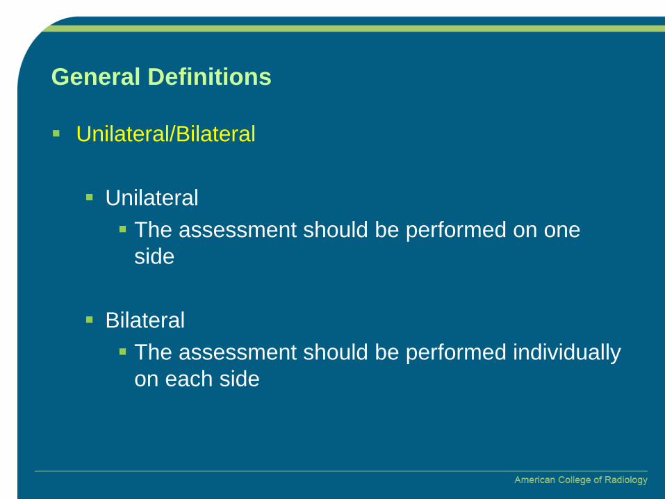
Unilateral/Bilateral
Unilateral The assessment should be performed on one
side
Bilateral The assessment should be performed individually
on each side
General Definitions
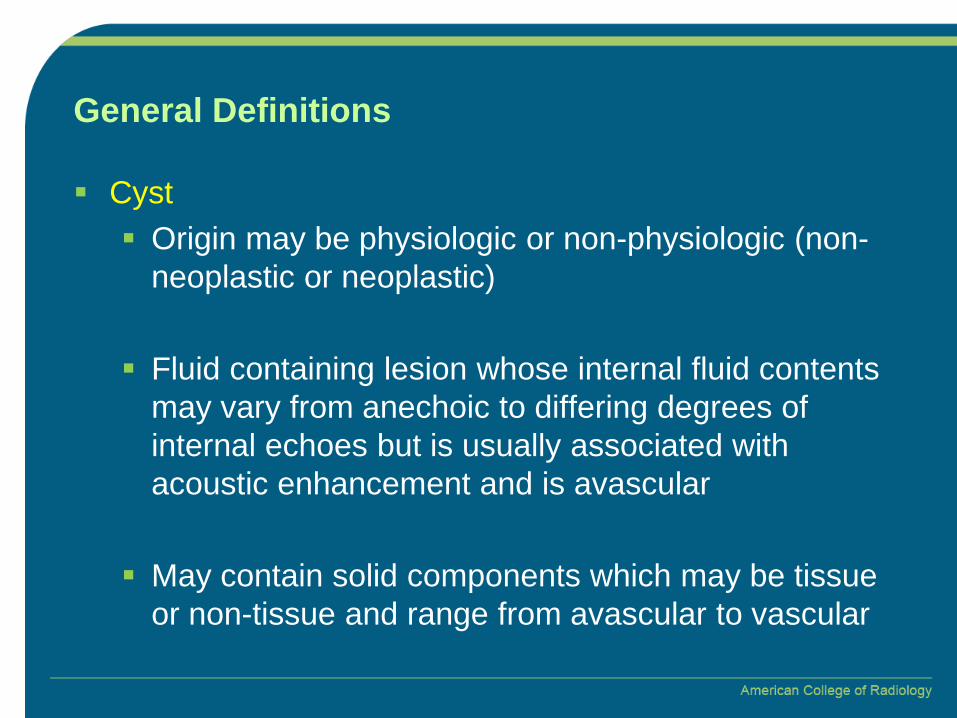
Cyst Origin may be physiologic or non-physiologic (non-
neoplastic or neoplastic)
Fluid containing lesion whose internal fluid contents may vary from anechoic to differing degrees of internal echoes but is usually associated with acoustic enhancement and is avascular
May contain solid components which may be tissue or non-tissue and range from avascular to vascular

General Definitions
Solid/solid-appearing (lesion or component) A structure that has echogenicity suggestive of
tissue (e.g. myometrium or ovarian stroma)
Note, the presence of flow (that can be confirmed with spectral Doppler if necessary) is diagnostic of solid tissue; the absence of flow is less helpful and the lesion may then be considered solid-appearing, depending on other features
Also judged by its echogenicity, size, and by the absence of internal movement which may be elicited when moving the transducer

Physiologic That which is consistent with normal ovarian
physiology (i.e. follicle and corpus luteum) Lesion That part of an ovary (or adnexa) judged by imaging
to not be consistent with normal physiology Not a stand alone term/descriptor; needs additional
descriptors to be appropriately defined More of a neutral term and should be used instead
of “mass” Can be divided into 5 major IOTA categories (to
follow)
General Definitions

Physiologic Category
Follicle: Simple (unilocular, anechoic) cyst, ≤ 3
cm in maximum dimension, in premenopausal group
Corpus luteum Thick-walled cyst ≤ 3 cm that may
have crenulated inner margins, internal echoes and (often intense) peripheral color Doppler flow
May sometimes appear as a hypoechoic region in the ovary with peripheral vascularity without a characteristic cystic component

Lesion Category (IOTA Classification)
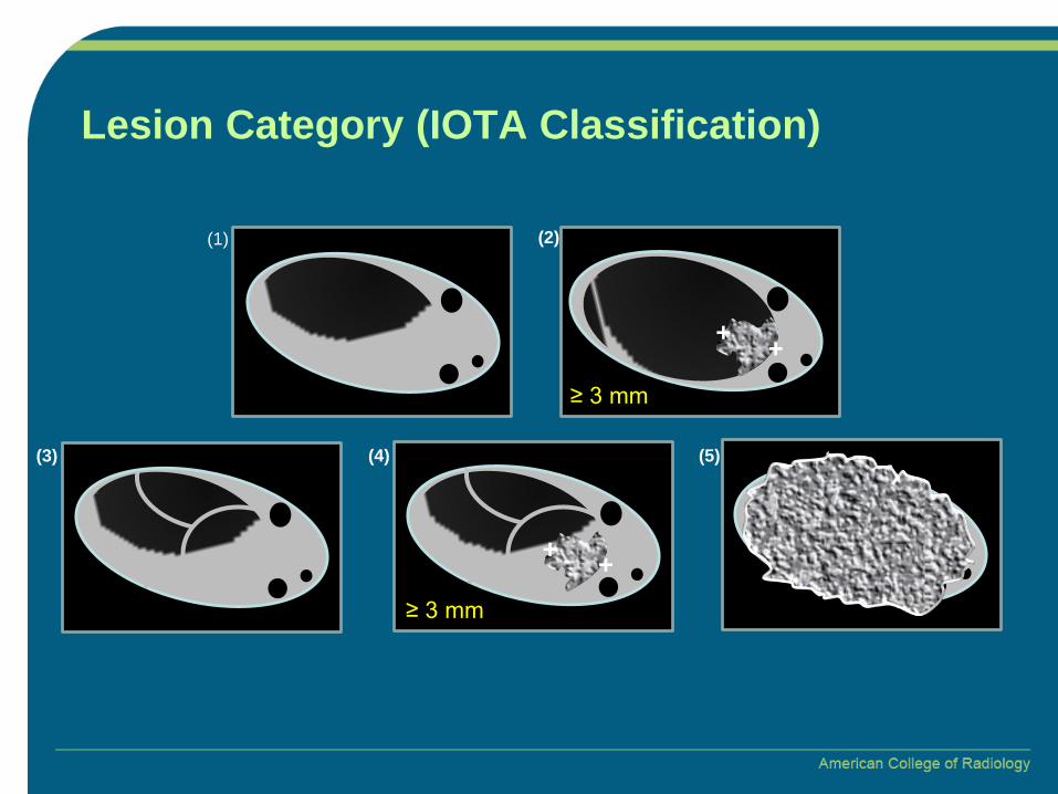
5 groups1. Unilocular cyst, no solid component(s) Simple cyst (subcategory of unilocular)
2. Unilocular cyst, with solid component(s)3. Multilocular cyst, no solid component(s)4. Multilocular cyst, with solid components5. Solid (≥ 80%) Purely solid (subcategory of solid; 100% solid)
(3) (4) (5)
(1) (2)
+ +
+ +
≥ 3 mm
≥ 3 mm
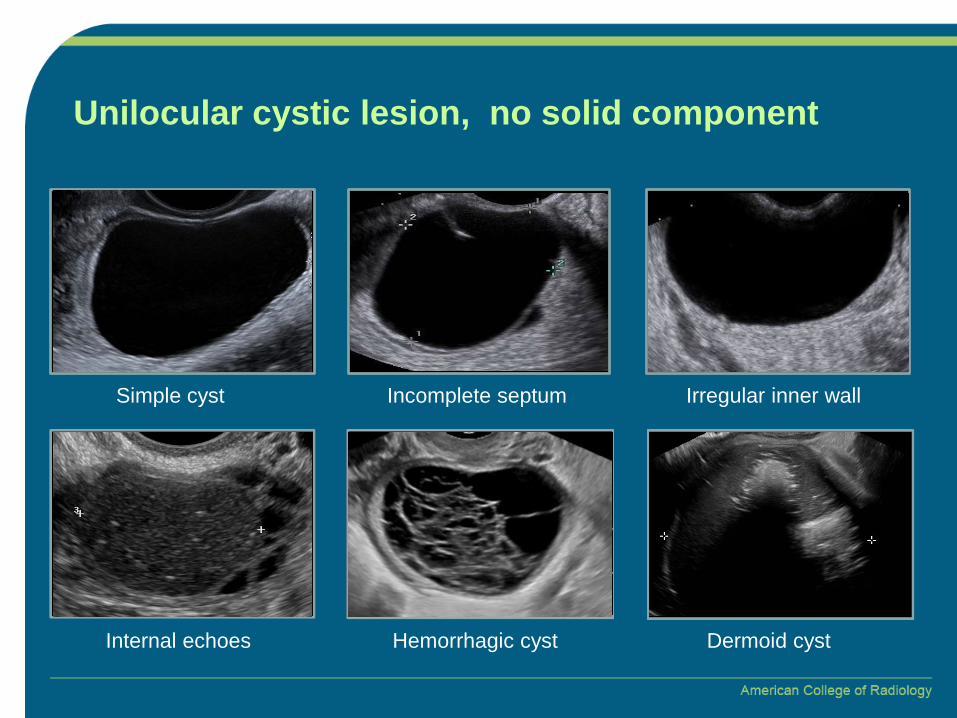
Lesion Category (IOTA Classification)

Cystic lesion with a single locule (contains no complete septa), and no solid/solid-appearing component
Incomplete septa (discontinuous), an irregular wall with focal thickening < 3 mm in height or internal echoes may be present
A simple cyst is a subset of a unilocular cyst, and has no internal components (thus anechoic), demonstrates acoustic enhancement, a smooth thin wall, and no internal septations (complete or incomplete)
Unilocular cystic lesion, no solid component

Unilocular cystic lesion, no solid component
The following are NOT considered solid/solid-appearing for the purposes of this lexicon:
Hemorrhagic products Mucinous or fat containing material Avascular hyperechoic structure with acoustic
shadowing (i.e. Rokitansky nodule) Normal ovarian tissue (e.g. Normal ovary within a
peritoneal inclusion cyst) Septa

< 3 mm
++
Unilocular cystic lesion, no solid component
Incomplete septumSimple cyst Irregular inner wall
Internal echoes Hemorrhagic cyst Dermoid cyst
< 3 mm
++
Unilocular cystic lesion, no solid component
Incomplete septumSimple cyst Irregular inner wall
Internal echoes Hemorrhagic cyst Dermoid cyst

Unilocular cyst, with solid component(s)
Cystic lesion with a single locule (i.e. contains no complete septa), which contains a solid/solid-appearing component ≥ 3 mm in height
≥ 3 mm
++
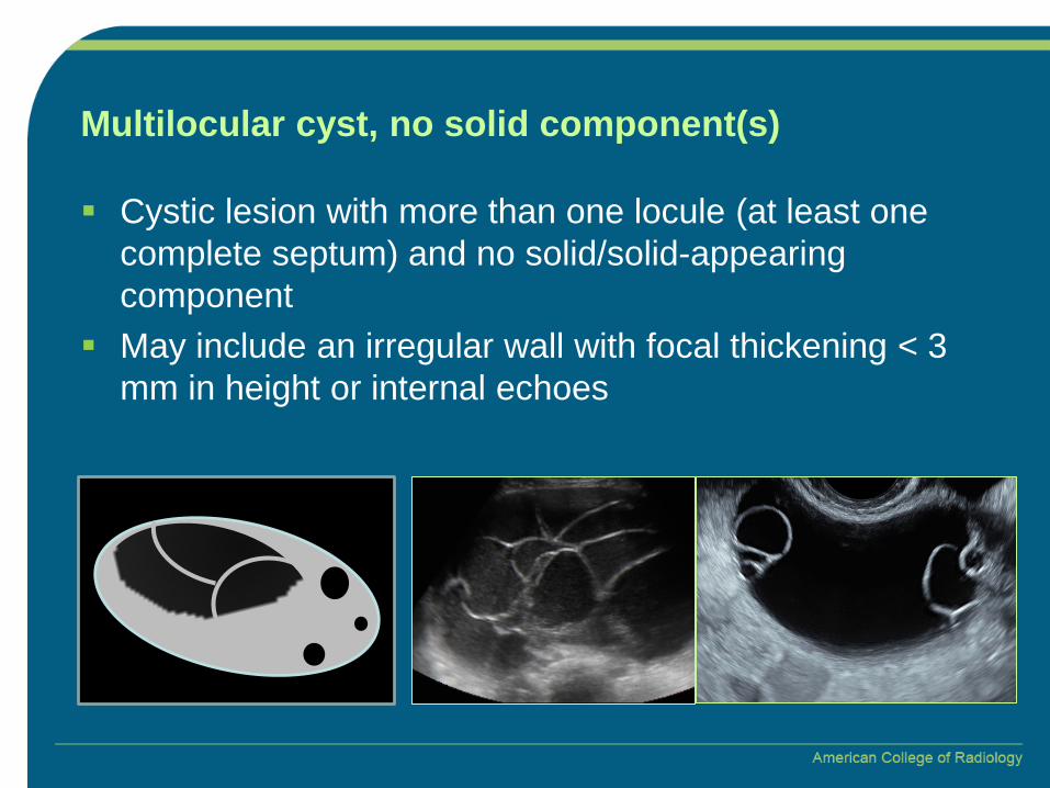
Multilocular cyst, no solid component(s)
Cystic lesion with more than one locule (at least one complete septum) and no solid/solid-appearing component
May include an irregular wall with focal thickening < 3 mm in height or internal echoes

Multilocular cyst, with solid component(s)
Cystic lesion with more than one locule (at least one complete septum), which also contains a solid/solid-appearing component ≥ 3 mm in height
≥ 3 mm
+ +
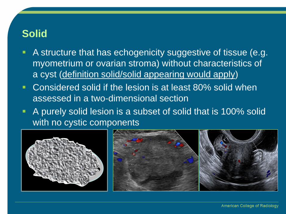
Solid A structure that has echogenicity suggestive of tissue (e.g.
myometrium or ovarian stroma) without characteristics of a cyst (definition solid/solid appearing would apply)
Considered solid if the lesion is at least 80% solid when assessed in a two-dimensional section
A purely solid lesion is a subset of solid that is 100% solid with no cystic components
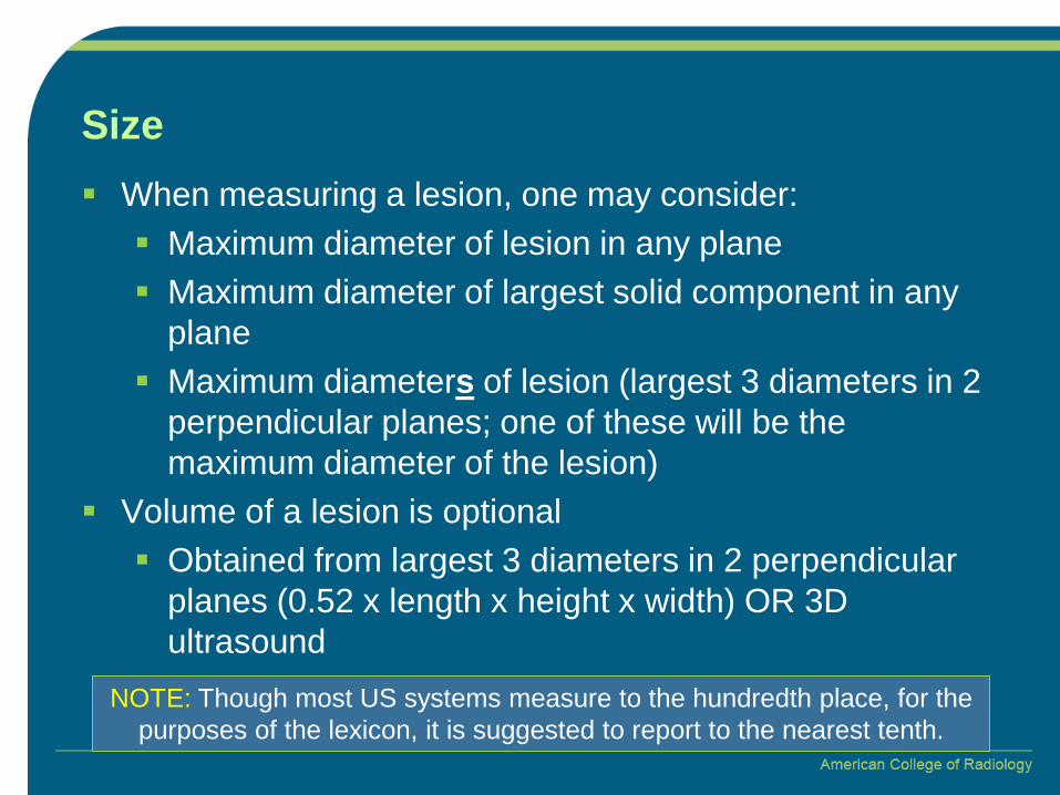
Size When measuring a lesion, one may consider: Maximum diameter of lesion in any plane Maximum diameter of largest solid component in any
plane Maximum diameters of lesion (largest 3 diameters in 2
perpendicular planes; one of these will be the maximum diameter of the lesion)
Volume of a lesion is optional Obtained from largest 3 diameters in 2 perpendicular
planes (0.52 x length x height x width) OR 3D ultrasound
NOTE: Though most US systems measure to the hundredth place, for the purposes of the lexicon, it is suggested to report to the nearest tenth.
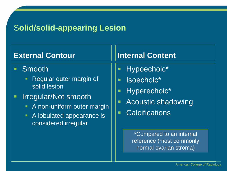
Solid/solid-appearing Lesion
External Contour Smooth
Regular outer margin of solid lesion
Irregular/Not smooth A non-uniform outer margin A lobulated appearance is
considered irregular
Internal Content Hypoechoic* Isoechoic* Hyperechoic* Acoustic shadowing Calcifications
*Compared to an internal reference (most commonly
normal ovarian stroma)

Cystic Lesion
Inner margin/Internal Wall Smooth
Regular, uniform throughout Calcified
High level echogenicity which is curvilinear or plaque-like
Associated with acoustic shadowing when dense or large enough
Irregular Non-uniform, focal thickening
of < 3 mm, papillary projections or mural nodules,irregular incomplete septa
Internal Content Fluid descriptors
Anechoic/simple fluid Hyperechoic components Ground glass or
homogeneous low-level echoes
Scattered low-level echoes Fluid/fluid level
Dermoid descriptors Hemorrhagic cyst descriptors Septations
Complete Thin: ≤ 3 mm Thick: > 3 mm
Solid/solid-appearing component

Cystic Lesion – Internal content, fluid descriptors Ground glass or homogeneous low-level echoes
Scattered low-level echoes
Typical for endometrioma
Typical for mucinous cystadenoma

Dermoid Descriptors
Hyperechoic lines and dots Bright linear echoes and foci (representing linear echoes seen
en face) Represents sections through hair within the liquefied
component Acoustic shadowing from a hyperechoic component
Attenuation of the acoustic beam distal to a hyperechoic component
Floating hyperechoic spherical structures Non-dependent hyperechoic spherules +/- acoustic shadowing Uncommon, but highly characteristic
NOTE: “tip of the iceberg”,“rokitansky nodule”, “dermoid mesh”, “dot-dash” and “dermoid balls” may be in common usage in some locations, but are not
preferred descriptors in this lexicon

Floating hyperechoic spherical structures
Acoustic shadowing from a hyperechoic component
v
Dermoid Descriptors Hyperechoic lines and dots

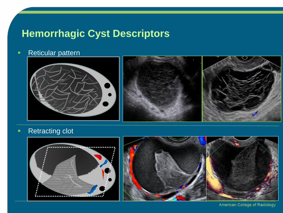
Hemorrhagic Cyst Descriptors
Reticular pattern Fine thin intersecting lines due to fibrin strands Not considered to be “septations”, which are usually thicker and more
continuous
Retracting clot Avascular echogenic component with angular, straight, or concave
margins
NOTE: “cobweb”, “fishnet”, “lacy”, “spider web” may be in common usage in some locations, but are not preferred descriptors in this lexicon
Reticular pattern
Retracting clot
Hemorrhagic Cyst Descriptors

Internal content, solid/solid-appearing component
Irregular External contour of the solid component within a cystic lesion is
nonuniform (i.e. spiky or lobular) Contour of any internal cystic area(s) is nonuniform (i.e. spiky or
angular rather than smooth) Smooth
No external or internal contour irregularities of the solid component Papillary projection(s) or nodule(s)
A solid component with height ≥ 3 mm that protrudes into the cyst cavity
External contour has an outwardly convex border and may be smooth or irregular
Can be mural or septal in origin Additional descriptors
Height Number

Vascularity
Circumferential Doppler flow in cyst wall Color Doppler flow is restricted to the wall and includes the majority of
the circumference of the wall This appearance, in the appropriate setting, may indicate a corpus
luteum
Internal color Doppler flow Color Doppler flow is detected internally within a solid
lesion/component or in a septation of the lesion, with or without peripheral (wall) flow
Color score 1- 4 (IOTA classification) Overall subjective assessment of entire lesion
NOTE: “Peripheral flow” and “Ring of fire” may be in common usage in some locations, but are not preferred descriptors in this lexicon

Color Score 1- 4 (IOTA Classification)
Color score 2 – minimal flow
Color score 3 – moderate flow Color score 4 – marked flow
Color score 1 – No blood flow
General and extra-ovarian findings
Paraovarian cyst Fallopian tube descriptors Peritoneal inclusion cyst Fluid descriptors Peritoneal thickening, nodules Adenopathy
Paraovarian cyst
Simple cyst existing separate from the ovary
Moves independent of the ovary when pressure is applied by the transducer
NOTE: Paraovarian and paratubal are used interchangeably as the origin (both Wolffian duct remnants) often cannot be determined by US alone.
Paraovarian will therefore be used to encompass both and there is no need to include paratubal in the differential.

Fallopian tube descriptors
Incomplete septation Appear as incomplete septations due to tubular nature of the lesion
when visualized along an oblique plane
Tubular Substantially longer in one dimension than in the two perpendicular
dimensions
Endosalpingeal folds Short round projections around the inner wall of tubular structure Best seen when orthogonal to the length (short axis) of a fluid-
filled tube Typically < 3mm in height
NOTE: “pseudoseptations”, “cogwheel” and “beads-on-a-string” may be in common usage in some locations, but is not a preferred descriptor in this lexicon
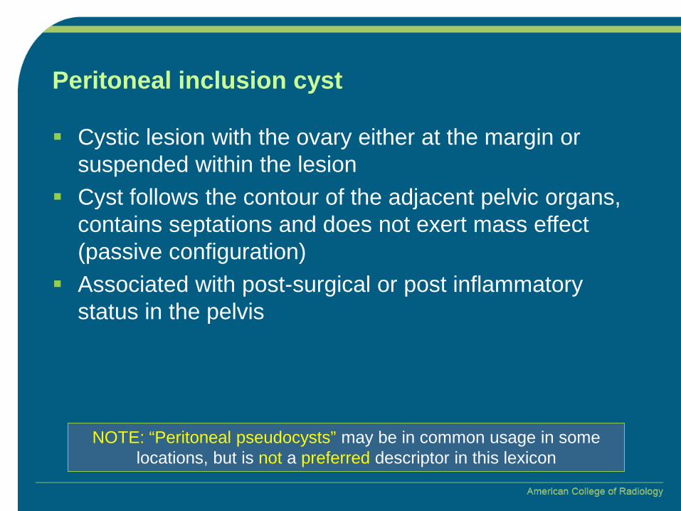
Peritoneal inclusion cyst
Cystic lesion with the ovary either at the margin or suspended within the lesion
Cyst follows the contour of the adjacent pelvic organs, contains septations and does not exert mass effect (passive configuration)
Associated with post-surgical or post inflammatory status in the pelvis
NOTE: “Peritoneal pseudocysts” may be in common usage in some locations, but is not a preferred descriptor in this lexicon

Fluid descriptors
Ascites If anteverted/anteflexed uterus, fluid extending beyond the pouch of
Douglas (cul-de-sac) and/or above uterine fundus If retroverted/retroflexed, fluid anterior to uterus (between uterus and
bladder)
Cul-de-sac fluid Confined to pouch of Douglas as defined by remaining below uterine
fundus or between uterus and bladder when uterus retroverted/retroflexed
In appropriate context (menstruating female), may be considered physiological fluid
Anechoic/simple fluid Fluid containing internal echoes (not simple)

Peritoneal thickening/nodules
Nodularity or diffuse thickening of the peritoneal lining(s) or along the bowel serosal surface or peritoneum
Associated with malignant etiologies and raises concern for peritoneal carcinomatosis
NOTE: Peritoneal implants/deposits may be in common usage in some locations, but is not a preferred descriptor in this lexicon
Adenopathy
Lymph nodes
Should be measured in short axis and location reported for management considerations
Test Your O-RADS Lexicon Knowledge
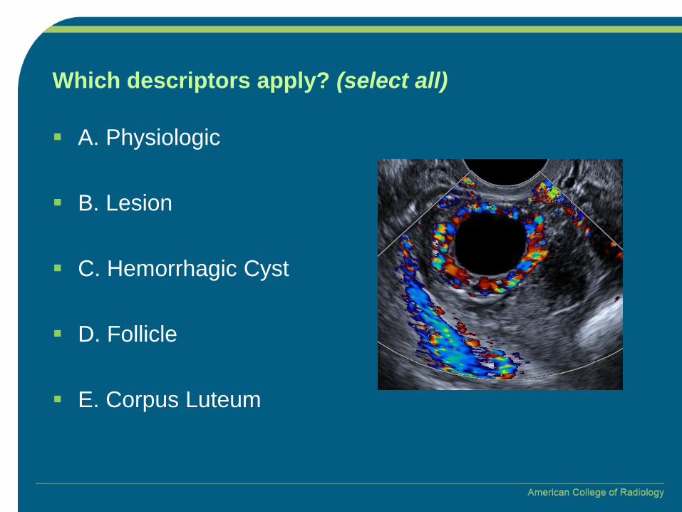
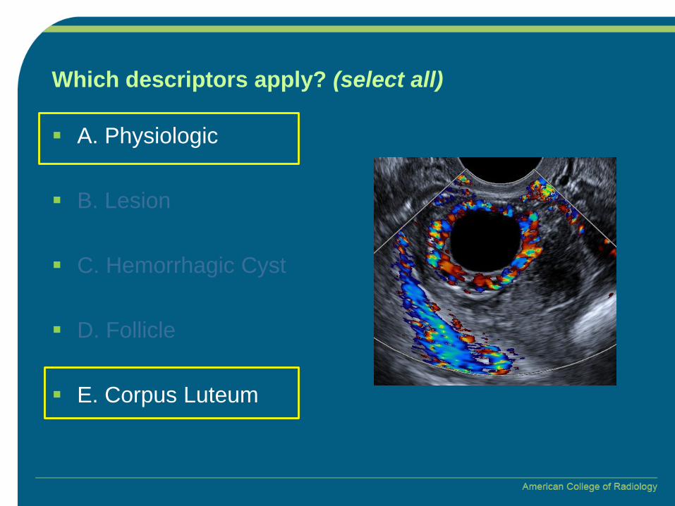
Which descriptors apply? (select all)
A. Physiologic
B. Lesion
C. Hemorrhagic Cyst
D. Follicle
E. Corpus Luteum
Which descriptors apply? (select all)
A. Physiologic
B. Lesion
C. Hemorrhagic Cyst
D. Follicle
E. Corpus Luteum
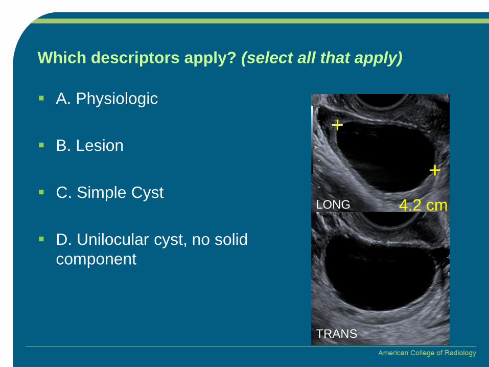
Which descriptors apply? (select all that apply)
+
+
4.2 cmLONG
TRANS
A. Physiologic
B. Lesion
C. Simple Cyst
D. Unilocular cyst, no solid component
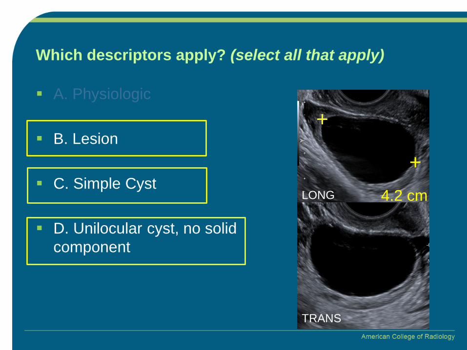
Which descriptors apply? (select all that apply)
+
+
4.2 cmLONG
TRANS
A. Physiologic
B. Lesion
C. Simple Cyst
D. Unilocular cyst, no solid component

Which descriptors apply? (select all that apply)
A. Homogenous low-level echoes
B. Scattered low-level echoes
C. Hyperechoic lines and dots
D. Papillary projections
E. Acoustic shadowing from a hyperechoic component

Which descriptors apply? (select all that apply)
A. Homogenous low-level echoes
B. Scattered low-level echoes
C. Hyperechoic lines and dots
D. Papillary projections
E. Acoustic shadowing from a hyperechoic component
Dx: Borderline mucinous cystadenoma
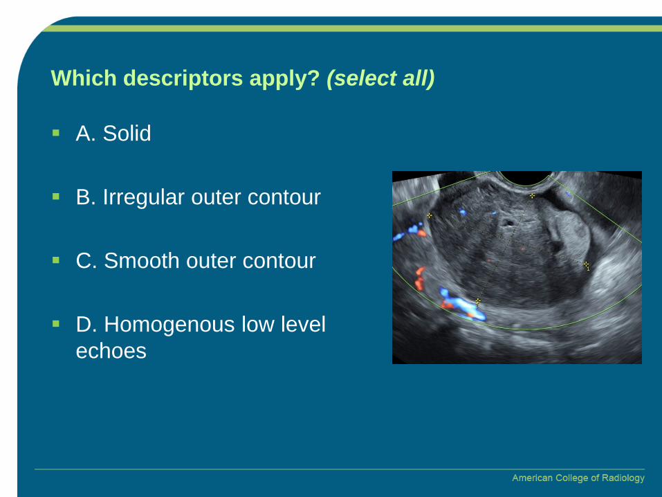
Which descriptors apply? (select all)
A. Solid
B. Irregular outer contour
C. Smooth outer contour
D. Homogenous low level echoes
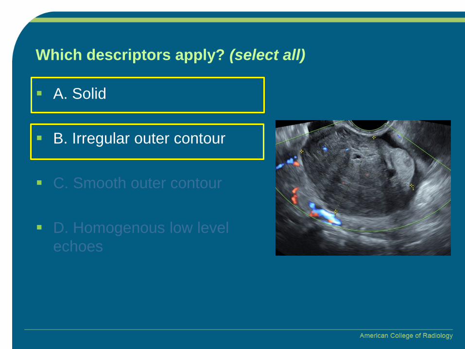
Which descriptors apply? (select all)
A. Solid
B. Irregular outer contour
C. Smooth outer contour
D. Homogenous low level echoes

What is the best color score for this lesion?
A. Color score 1
B. Color score 2
C. Color score 3
D. Color score 4

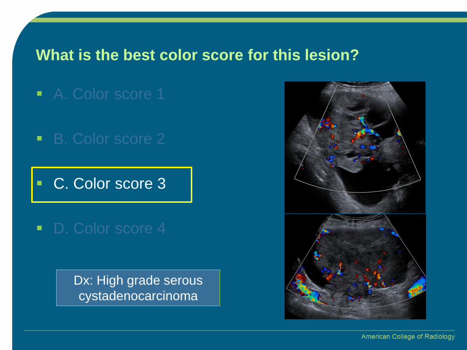
What is the best color score for this lesion?
A. Color score 1
B. Color score 2
C. Color score 3
D. Color score 4
Dx: Fibrothecoma

Which descriptors apply? (select all)
A. Cystic with irregular solid component
B. Solid
C. Smooth outer contour
D. Irregular outer contour
E. Internal vascularity
F. Peripheral vascularity

Which descriptors apply? (select all)
A. Cystic with irregular solid component
B. Solid
C. Smooth outer contour
D. Irregular outer contour
E. Internal vascularity
F. Peripheral vascularity

What is the best color score for this lesion?
A. Color score 1
B. Color score 2
C. Color score 3
D. Color score 4
What is the best color score for this lesion?
A. Color score 1
B. Color score 2
C. Color score 3
D. Color score 4
Dx: High grade serous cystadenocarcinoma

How is this lesion best characterized?
A. Corpus luteum
B. Dermoid
C. Mucinous cystadenoma
D. Hemorrhagic cyst

How is this lesion best characterized?
A. Corpus luteum
B. Dermoid
C. Mucinous cystadenoma
D. Hemorrhagic cyst
Reticular pattern