o microbial palaeontology and the origin of life: a...
TRANSCRIPT

Bollettino della Società Paleontologica Italiana, 55 (2), 2016, 85-103. Modena
ISSN 0375-7633 doi:10.4435/BSPI.2016.09
Microbial palaeontology and the origin of life: a personal approach
Frances Westall
F. Westall, CNRS-Centre de Biophysique Moléculaire, Rue Charles Sadron, CS 80054, Orléans cedex 2, France; [email protected]
Key words - Microbial palaeontology, Early Archaean, Origin of life, Extraterrestrial life.
ABSTRACT - Palaeontology is an essential tool for tracing the history of life in the geological record. However, access to the origin of life is blocked because of the lack of preservation of suitable rocks dating from the fi rst billion years of Earth’s history. Nevertheless, study of Early Archaean rocks (~4-3.3 Ga) indicates that the environmental conditions of the early Earth, upon which life emerged, were very different to those of today and provides essential information for guiding investigations into the origin of life in terms of realistic environmental scenarios and possible timing of the appearance of life. Microbial palaeontology investigations of well-preserved, Early Archaean rocks ~3.5 to 3.3 Ga show that the earliest preserved life was diverse and widespread and suggest that it probably appeared in the Hadean, as soon as the Earth’s surface was habitable. The extreme, anaerobic conditions characterising the early Earth, together with the ingredients of life, i.e. carbon molecules, liquid water and energy, were common on other planets and satellites in the early Solar System. Considering carbon and water-based life forms to be a cosmically frequent phenomenon, it is hypothesised that life could have emerged on some of these bodies and that traces of its appearance may still be preserved, for instance on Mars, Europa or Enceladus. Microbial palaeontology as well as information gleaned from extant extremophiles and experimental data provides us with essential information about what kinds of extant or fossilised life forms to look for on another planet or satellite. Moreover, the methods evolved to study and understand the remains of fossil traces of primitive microbial life will aid the search for life and its origins on Mars or other satellites. The perspective of returning to Earth rocks from Mars (or other samples from Europa or Enceladus?) containing potential traces of extraterrestrial life, most likely primitive anaerobic chemotrophs, will be a challenge for microbial palaeontology that we need to start addressing now. Most importantly, it will open up the possibility of establishing the universality of life.
RIASSUNTO - [L’origine della vita e la Paleontologia Microbica: il punto di vista dell’autore] - La Paleontologia è uno strumento essenziale per la ricostruzione della vtoria della vita sulla base di informazioni conservate nel record geologico e dell’evoluzione geo-biologica del nostro Pianeta. Lo studio dell’origine della vita e, quindi, delle prime forme in cui essa apparve, è reso inevitabilmente complicato dalla scarsa disponibilità di rocce terrestri più antiche di 3,5 miliardi di anni, e, in generale, dal loro stato di conservazione. Tuttavia, grazie allo studio delle rocce della prima parte dell’eone Archeano (da ~4,0 a ~3,2 Ga), ovvero fra le rocce più antiche del nostro Pianeta, oggi sappiamo non solo che le condizioni ambientali della Terra primitiva, sulla quale la vita si sviluppò, erano profondamente differenti da quelle attuali, ma abbiamo anche ottenuto informazioni (geologiche e ambientali) essenziali per indirizzare la ricerca sulla sua origine in termini di ricostruzione di scenari ambientali realistici e dei potenziali step evolutivi (almeno in termini di metabolismi possibili) da essa seguiti.
Studi di paleontologia microbica condotti negli ultimi anni in rocce sedimentarie e vulcaniche del Paleoarcheano (da ~3,6 a 3,2 miliardi di anni), hanno rivelato la presenza di abbondanti forme di vita (di tipo microbico) fra le più antiche oggi note, diversifi cate, abbastanza evolute e complesse, nonché diffuse in differenti habitat. Inoltre, è stato stabilito che molto probabilmente la vita popolò il nostro Pianeta già all’inizio dell’Archeano, e, forse, già durante l’eone Adeano (~4,6-4,0 Ga), quando il nostro Pianeta era già potenzialmente abitabile (~4,4 Ga). Le condizioni ambientali estreme e fortemente anaerobiche che caratterizzavano la Terra primitiva, e gli ingredienti per la vita così come noi la conosciamo (per es., molecole di carbonio, elementi bio-essenziali, acqua liquida e sorgenti di energia), erano aspetti che il nostro Pianeta aveva in comune con altri pianeti e satelliti del neonato sistema solare. Allora, pensando alla vita basata sul carbonio e acqua (come è il caso della vita terrestre che comunque ad oggi è l’unica conosciuta) come ad un fenomeno cosmico comune nell’Universo (carbonio e acqua nei differenti stati fi sici sono elementi comuni dell’Universo), è stato ipotizzato che essa potrebbe essersi originata anche su altri pianeti o satelliti dove, possibilmente, queste tracce (di tipo microbico) potrebbero ancora essere conservate nel relativo record roccioso, come per esempio potrebbe essere il caso di Marte, Europa o Encelado. Oggi la paleontologia microbica che si occupa di studiare fossili microbici di tipo procariotico nel record fossile dall’Archeano al Recente con una attenzione particolare allo studio di ambienti estremi moderni e fossili e allo studio in laboratorio di processi tafonomici e di fossilizzazione, di biomineralogia e geomicrobiologia (relazioni microbi-minerali), ci fornisce informazioni preziose circa quali forme di vita (microrganismi viventi o fossili) e quale strategia seguire nella ricerca della vita fuori dal nostro Pianeta. Così i metodi investigativi messi a punto per studiare, riconoscere e comprendere i resti fossilizzati di forme di vita primitiva (procarioti), potrebbero essere importanti sia per riconoscere tracce di vita terrestri sempre più antiche (per es., gechimica isotopica e tracce molecolari), sia per la ricerca di vita e delle sue origini su Marte o su altri pianeti o satelliti. La possibilità di riportare sulla Terra rocce da Marte (o da Europa o Encelado), che potrebbero contenere tracce di vita extraterrestre, molto probabilmente di tipo procariotico, anaerobico e chemiotrofi co, sono gli obiettivi delle imminenti missioni spaziali, e nello stesso tempo una grande sfi da per la paleontologia (microbica) e una opportunità per i paleontologi di mettere a punto strategie investigative (alla scala micro- e nano-metrica) nel tentativo di stabilire l’universalità della vita.
INTRODUCTION
Palaeontology is the most important tool for studying the history of life. Unfortunately, owing to the non-preservation of suitable rocks older than 3.5 Ga on Earth (Hadean crust having been recycled by tectonic activity
and large impacts; Kamber, 2015, Fig. 1), we have no direct evidence of the appearance of the fi rst cellular life and its earliest evolution. Nevertheless, well-preserved, Early Archaean rocks, 3.5 to ~3.3 Ga, provide essential information about the environment in which life appeared and evolved, documenting a planet that was habitable for
S. P. I.
SOC
IETA
' P
A
LEON TO L OGICA I T
AL
IANA
Invited Paper PA
LE
ONNN

Bollettino della Società Paleontologica Italiana, 55 (2), 201686
anaerobic microorganisms but completely different to the present day Earth. Thus, because of the lack of crust old enough to have preserved traces of the transition from prebiotic molecules to cellular life, study of the oldest preserved fossil remains and the environmental context in which they occur is an important means of approaching the emergence of life. Morphological and chemical microbial traces dating back to 3.5 Ga tell us that life was already evolved, phototrophs coexisting with chemotrophs in an anaerobic world and widely distributed (Walsh, 1992; Hofmann et al., 1999; Tice & Lowe, 2004; Westall et al., 2011a, b). But when did life emerge? Many hypotheses suggest that life could not have appeared until after the end of the hypothesised period of heavy bombardment (~4.0-3.85 Ga; Ryder et al., 2000) but the degree of evolution of life demonstrated already by 3.5 Ga, plus its global distribution, suggest that the emergence of life may have occurred during the Hadean and, theoretically, as soon as water had condensed on Earth’s surface and had reached a balmy temperature of about 100°C (e.g., Westall, 2012; Cavalazzi & Barbieri, 2016; Cavalazzi et al., 2016). Models indicate that water condensation occurred rapidly after the Moon-forming impact (~4.5 Ga) but the earliest direct evidence for its presence dates back to 4.35 Ga, the age of recycled zircon crystals formed by fractionation of hydrated crust (Mozjsis et al., 2001). The Early Archaean rocks, in which the earliest preserved traces of life occur, formed a billion years after the massive impact that formed the Moon (Kleine et al., 2009) and well after the appearance of habitable conditions. We do not know how long it took for life to appear (Orgel, 1998) but those working in the origins of life field consider that the process had to have been relatively rapid, i.e. probably less than a few million years rather than hundreds of millions of years. Thus, the precious traces in rocks 3.5-3.3 Ga are
all that remain of a history that was perhaps already many hundreds of millions of years old.
Nevertheless, the remains of early life, although relatively evolved compared to what must have been the first cellular life forms, provide precious indications concerning the kinds of life forms that first appeared and, especially, the environment in which the emergence of life likely took place. Moreover, the environmental context of the preserved biosignatures is important not only for aiding interpretation and identification of early life, indeed it is indispensable for understanding, but it is also of critical importance for understanding the early habitable conditions of the Earth. Thus, microbial palaeontology studies of early traces of life provide invaluable information related to the origin of life. However, the lack of Hadean crust is a huge limitation. Nevertheless, if life appeared as a natural evolution of chemical processes (de Duve, 1995), it could have appeared on any other extraterrestrial body having environmental conditions similar to those on the early Earth. This includes Venus and Mars during their early histories, as well as a number of other satellites that now have icy crusts, such as Europa and Calisto, orbiting Jupiter, and Enceladus and Titan around Saturn (e.g., Westall & Brack, 2016). All of them had water in contact with hot rock and a constant stream of prebiotic molecules arriving in meteorites, micrometeorites and cometary dust particles, as well as carbon molecules formed in situ on the planets.
One of the major objectives of the on-going mission Mars Science Laboratory and its rover Curiosity and the future missions ExoMars-2020 (Europe/Russia) and Mars 2020 (NASA) is the search for traces of past life on the surface of Mars. In this respect, microbial palaeontology investigations of the oldest traces of life on Earth are invaluable because they show us the nature of early life,
Fig. 1 - Distribution of Archaean cratons showing the locations of the Pilbara Craton in northwest Australia and the Barberton Greenstone Belt in South Africa (drawn from many sources). The latter two, dating from 3.5 Ga, are the only two cratons where Early Archaean sedimentary rocks are well preserved.
87F. Westall - Early life
how it is preserved, the types of signatures that can be used to identify it, the challenges involved in its identification, and the analytical methods needed to study it (Westall et al., 2015a). Without these studies it would not be possible to search for life on Mars or elsewhere.
In the following, I will set the scene by describing the environment of the early Earth in order to decipher and reconstruct the potential early habitat. This is important because the Hadean-Early Archaean Earth can be viewed environmentally as a totally different planet and therefore the early life forms living on it were very different from even the simplest of microbes living on Earth today - they were what would today be termed as extremophiles (e.g., Arndt & Nisbet, 2012). I will then describe the biosignatures preserved, and the diversity and distribution of early life. Finally, I will address the implications of early life for the search for life on Mars and on the icy satellites, Europa and Enceladus.
THE HADEAN-EARLY ARCHAEAN ENVIRONMENT
The Hadean/Early Archaean Earth was volcanic and hot. It was still cooling down after the magma ocean episode that followed the Moon-forming impact, as attested by a very specific type of lava, komatiites, formed from very hot magmas that only occurred on the early Earth (Arndt, 1994). While no Hadean crust has been preserved, there are some remnants of Early Archean crust. Most are highly metamorphosed but two enclaves of well-preserved volcanics and sedimentary deposits, dating from 3.5-3.3 Ga ago, occur in the Barberton Greenstone Belt in South Africa and the Pilbara Craton in Australia. These rocks have suffered burial metamorphism ranging from upper prehnite-pumpellyite to lower greenschist facies. However, we will see that, apart from a maturation of the carbon associated with microfossils, the metamorphism has not affected the biosignatures.
The volcanic and associated sedimentary rocks document an anaerobic Earth (Westall, 2012). Some suggestion has been made for local oxygenic conditions (Ohmoto et al., 2014) but there were numerous abiotic processes that could have resulted in the production of small amounts of oxygen: photolysis of water vapour in the atmosphere, radiolysis of water at the surface of the ocean (without significant oxygen in the atmosphere and a protective layer of ozone, UV radiation reached the surface of the Earth uninhibited [Lammer et al., 2008]), as well as dissociation of water by boiling of hydrothermal fluids as they reach the surface (in shallow water and subaerial environments). Today’s 21% O2 in the atmosphere is a consequence of interrelated biogenic and geological factors, the biological input owing to the evolution of oxygenic phototrophs. Had such organisms developed by the Early Archaean, with their vastly more efficient metabolisms they would have rapidly overrun the Earth. However, there is no indication that this happened (Westall, 2011).
Anaerobic conditions are mandatory for the emergence of life. No prebiotic reactions can take place in the presence of oxygen because of the immediate oxidation of the reduced carbon species. This means that the first cellular life was anaerobic and, indeed, life continued to be
largely anaerobic until the advent of the mutation that led to oxygenic photosynthesis sometime in the Late Archaean (Buick, 2008). At first a poison for all life forms except the oxygenic photosynthesisers, many microbes adapted to the presence of small amounts of oxygen, becoming micro-aerophilic to completely oxygenic. Nevertheless, more than two billion years were needed before the reduced surface of the Earth was oxidised sufficiently for oxygen to start building up in the atmosphere (Zahnle & Catling, 2016).
One corollary of the high radiation flux is that early life must have developed efficient strategies to deal with radiation damage. We will see below that there is plenty of evidence of life in early Archaean shallow water environments and even briefly exposed on the beach (cf. Westall et al., 2006a, 2011b). Perhaps early life used strategies similar to those used by microbes living today in environments exposed to high doses of radiation, such as UV-protective pigmentation, thick layers of polymer (extracellular polymeric substances, EPS), protection from layers of minerals, an efficient gene repair mechanism, rapid cell division, and rapid development of radiation-resistant mutants (e.g., Vítek et al., 2010; Moeller et al., 2012).
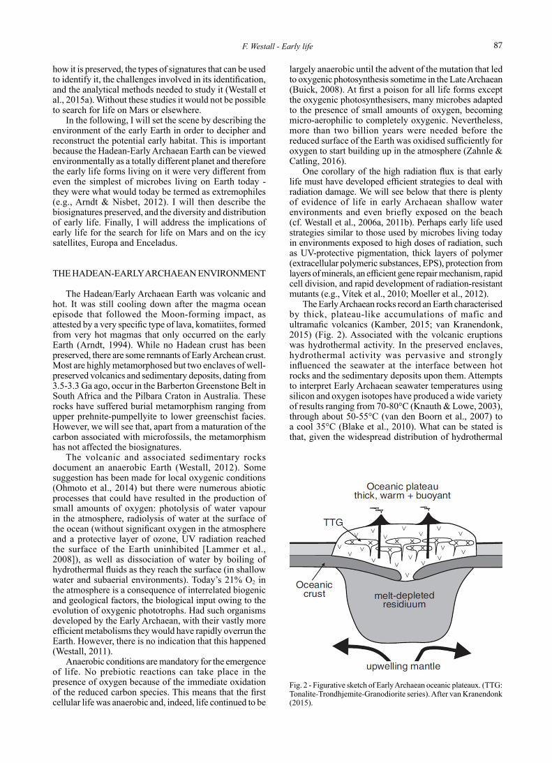
The Early Archaean rocks record an Earth characterised by thick, plateau-like accumulations of mafic and ultramafic volcanics (Kamber, 2015; van Kranendonk, 2015) (Fig. 2). Associated with the volcanic eruptions was hydrothermal activity. In the preserved enclaves, hydrothermal activity was pervasive and strongly influenced the seawater at the interface between hot rocks and the sedimentary deposits upon them. Attempts to interpret Early Archaean seawater temperatures using silicon and oxygen isotopes have produced a wide variety of results ranging from 70-80°C (Knauth & Lowe, 2003), through about 50-55°C (van den Boorn et al., 2007) to a cool 35°C (Blake et al., 2010). What can be stated is that, given the widespread distribution of hydrothermal
Fig. 2 - Figurative sketch of Early Archaean oceanic plateaux. (TTG: Tonalite-Trondhjemite-Granodiorite series). After van Kranendonk (2015).

Bollettino della Società Paleontologica Italiana, 55 (2), 201688
activity and fluids, temperatures at the rock/sediment/water interface must have been warm to hot. This means that any life living in these habitats was probably thermophilic. If the water column were cooler, especially in deeper water areas, early planktonic life forms could have been mesophilic.
There is much debate about the composition of the early oceans since compositional signatures of seawater are difficult to preserve. Studies of fluid inclusions in the Early Archaean rocks suggest that the seawater was perhaps twice as salty as it is today (de Ronde & Ebbersen, 1996). One major difference in the ion content of the early oceans compared to today would have been the lack of significant amounts of sulphate (SO4) owing to the anoxic conditions (e.g., Habicht et al., 2002; Lyons et al., 2014). Today’s oceans are sulphate-rich. Nevertheless, the small amounts of abiotic oxygen would have meant that some sulphate would have been available as a nutrient for microbes, such as sulphate- or sulphur-reducing bacteria. Moreover, given the prevalence of mafic and ultramafic lithologies being leached by hot hydrothermal fluids, iron and magnesium contents would probably have been higher than today. The significant hydrothermal influence, at least in the layers of seawater close to the sediment/rock interface, meant that that the seawater was saturated with respect to silica (e.g., Hofmann, 2011). This compositional information is important because it has a bearing on the solutes available to microbes and, especially, on the preservation of the biosignatures and the associated rocks.
There is also much debate about the pH of the early oceans. A priori alteration of mafic and ultramafic rocks
produces alkaline solutions (Kempe & Degens, 1985). On the other hand, an Earth characterised by a mainly CO2 atmosphere (Walker, 1985; Zahnle et al., 2010) would have acid rainfall and slightly acidic oceans. In addition, hydrothermal fluids may be acidic, especially if they are high temperature, or alkaline if they are lower temperature. The rock/sediment/seawater interface and the adjacent layers of water would have represented the mixing zone of fluids of different origins, compositions and pH, thus presenting a variety of habitats suitable for alkaliphilic to acidophilic microorganisms.
In conclusion, environmental conditions in the Early Archaean pose constraints on the type of life forms and their mode of life during that period. In the first place, all life was anaerobic. One could envisage the possibility of the development of tolerance of small amounts of oxygen through mutation for organisms living in environments where oxygen was produced by abiotic means, such as in the photic zone or close to shallow hydrothermal springs. If the oceans were more salty than today, they were also probably halophiles. Microorganisms living on or close to the sediment/rock/water interface were likely to have been thermophilic and, in terms of pH tolerance, ranged from acidophiles to alkaliphiles. Those living in the photic zone must have been radiation resistant.
From all points of view, the early Earth was an extreme environment - and its inhabitants were extremophiles (Fig. 3). Note that these extreme conditions were normal on habitable planets in the early history of the Solar System and will be far more common on habitable planets elsewhere in the Galaxy and Universe than the
Fig. 3 - Artist’s depiction of the early Earth. Note impact craters, hydrothermal activity and the closer moon. From http://www.cosmographica.com/.

89F. Westall - Early life
present day oxygenic environment of the Earth. In fact, the Earth today is rather unusual and should be therefore considered extreme, and the oxygenic life forms on it are the extremophiles.
THE NATURE OF EARLY LIFE AND ITS PRESERVATION
For long the subject of heated debate owing to the challenges involved in studying fossil microbes in extremely old rocks, it is now generally recognised that life was present by 3.5 Ga. Huge advances have been made over the last few decades in our understanding of the preservation potential of microbial life and the methods needed to study and identify its fossil traces in rocks. It is now generally recognised that context is as important as other biosignatures, such as morphological remains or organo-geochemical signatures. This was spectacularly underlined in the famous debate about the biogenicity of certain microstructures occurring in a black chert from the Apex Chert locality in the Pilbara, Australia. Being carbonaceous and having some morphological similarity with filamentous microfossils found elsewhere in the rock record (a typical example are the cyanophytes of the Early Devonian Rhynie Chert in Scotland; Preston & Genge, 2010), they were interpreted as the fossil traces of microbes, including cyanophytes (Schopf, 1993, 2006; Schopf et al., 2002) (Fig. 4a). However these features were found in a hydrothermal chert vein, a fact that had not previously been recognised (Brasier et al., 2002). While phototrophs are known from hydrothermal spring environments, they do not occur within the hydrothermal conduits themselves.
This example demonstrates the importance of direct context information and also the uncertainty of relying on only one kind of biosignature - morphology. Primitive microbial cellular morphologies are notoriously simple and many can be confused with abiotic features of similar shape and size (Westall & Cavalazzi, 2009). Another famous example may illustrate this point. A paradigm-changing discovery was made in 1996 when a group headed by David McKay at the Johnson Space Center in Houston announced that they had found fossil microbes in cracks in a meteorite from Mars (McKay et al., 1996) (Fig. 4b). The group had made very careful context, morphological and geochemical studies, concluding that microbes had been able to infiltrate water-filled fractures in what was basically a coarse-grained igneous lava or intrusion. One of their lines of evidence was the finding of small globular or elongated features up to a few hundreds of nanometres in size that were likened to “nanobacteria”. Nanobacteria, supposedly very small microorganisms, and their possible fossil signatures were much in vogue at that point in time (e.g., Folk & Linch, 1997). It was later determined that the “microfossils” in the meteorite were simply mineralogical artefacts produced during corrosion of carbonate deposits within the cracks (Gibson et al., 2001). We will come back to the interesting subject of traces of life on Mars below.
It is not the aim of this contribution to provide an exhaustive review of the traces of early life. I will highlight a few key studies to illustrate the diversity of
life on the early Earth and its implications for origins of life studies. In this respect I will first address the traces of phototrophic microorganisms for the reason that some of them reach macroscopic dimensions and their morphological remains are more readily observable and identification less contentious (but not always, as we have
Fig. 4 - Abiotic bacteriomorph structures. a) Carbonaceous structure in the Apex Chert, Pilbara craton, Australia, interpreted as a fossil cyanophyte (Schopf, 1993). b-c) Interpreted “nanobacteria” (black arrows) in the Martian meteorite ALH84001 (McKay et al., 1996).

Bollettino della Società Paleontologica Italiana, 55 (2), 201690
seen from the Apex Chert example cited above). I will then present some case studies of chemotrophic remains, microscopic, less readily identifiable and subject to more debate. However, chemotrophic microorganisms, with their more primitive metabolisms, appeared before the more sophisticated phototrophs and are the most likely elsewhere in the Solar System and in the Universe. It is these signatures that will be the object of search on Mars (Westall et al., 2015a).
Benthic phototrophic biosignaturesPhototrophic biosignatures are perhaps among the
most readily observable and interpretable of microbial traces. There are a number of reasons for this. In the first place, the later appearing, more sophisticated phototrophs have an extremely efficient metabolism compared to chemotrophs and, when in a suitable photic environment, are able to produce high biomass (des Marais, 2000). This ability was all the more important on the early Earth since there was no competition from other, more evolved life forms and no limitation due to grazing organisms, as is the case since at least the latest Proterozoic. Biofilms and mats formed by photosynthesisers can be thus relatively large (for example, modern phototrophic mats in the Baja California - e.g., Frank & Stolz, 2009 - cover huge areas up to hundreds of km2). Moreover, their sticky polymeric surfaces trap, bind and stabilise sediment surfaces, creating distinctive structures that are recognizable in the rock record back to 3.2 Ga, if not even 3.5 Ga (Krumbein, 1983; Hofmann et al., 1999; Allwood et al., 2006; Heubeck, 2009; Noffke, 2009; Noffke et al., 2013).
Early Archaean phototrophs produced macroscopically recognisable features in the form of tabular and domical to conical stromatolites (now silicified), as well as microbially induced sedimentary structures (MISS, cf. Noffke, 2009). They also produced microscopic biofilms and mats, whose spectacular degree of preservation was due to very early silicification.
Case study 1: the North Pole dome, Pilbara, australia (3.5-3.4 Ga) - A wide variety of macroscopic, stromatolite-like structures is preserved in the North Pole region of the Pilbara Craton. Investigators have been a pains to distinguish between those formed in association with hydrothermal fluids and those without this association in the belief that stromatolite-like features in the former category could not be phototrophic (Allwood et al., 2006; van Kranendonk, 2006). For instance, van Kranendonk (2006) described tabular to domical structures (up to some tens of cm in height) in the 3.49 Ga Dresser Formation where hydrothermal activity led to barite and chert precipitation, concluding therefore that the wrinkly laminites could not have been formed by phototrophic microorganisms. However, this supposition does not make sense because phototrophic mats and biofilms are common around hot springs today. Moreover, a domical structure of the order of 15 cm in diameter exhibiting plastically deformed laminae was observed in the direct vicinity of a shallow water hydrothermal vent (Westall, 2005) (Fig. 5). The deformation indicates that the dome consisted of laminae that were coherent at the same time as being soft and flexible, very similar to those formed by microbial phototrophic biofilms. Moreover, there is much
evidence for the presence of phototrophs in hydrothermal environments elsewhere at this period, as we will see below (cf. Westall et al., 2015b).
Also in the North Pole Dome region of the Pilbara, the Strelley Pool Chert contains stromatolites that developed on a carbonate platform in a restricted, hypersaline basin area with little clastic or (supposedly) hydrothermal input (Allwood et al., 2006, 2007; van Kranendonk, 2006). Morphological variations in the domical to conical stromatolites (up to about 10 cm in height) (Fig. 6) were interpreted to be biological responses to environmental factors, such as water depth, sediment influx and - hydrothermal activity (although Allwood et al., 2006, stipulate elsewhere that the stromatolites did not form in a hydrothermal environment and that any hydrothermal veins are later and cross-cut the strata). The environment, a tidal to supra-tidal rocky volcanic platform overlain by a chert boulder conglomerate and carbonate, underwent marine transgression with time. Stromatolites formed in relatively high-energy conditions on the rocky shoreline of the lower member are domical in shape and encrust the underlying breccia/conglomeritic fragments. With slightly increasing water depths (from supra-tidal to intertidal),
Fig. 5 - Domed stromatolite (15 cm in length) from the 3.48 Ga-old Dresser Formation, Pilbara craton, Australia. Note the plastically deformed internal laminae (white arrow) within the structure.
Fig. 6 - “Egg-carton” shaped stromatolites in plan view from the 3.4 Ga-old Strelley Pool Chert, Pilbara craton, Australia. Hammer for scale.

91F. Westall - Early life
these were followed by small crested/conical, wavy and “egg carton” laminites, as well as various large complex cones. Continued transgression of the carbonate platform was characterised by wavy laminites.
More enigmatic, potentially phototrophic features have been described as MISS from the 3.49 Ga Dresser Formation in environments characteristic of an ancient coastal sabkha (Noffke et al., 2013) (Fig. 7). First identified in recent sediments (Noffke & Krumbein, 1999), MISS comprise a series of fabrics and textures resulting from the interaction of sticky phototrophic microbial biofilms and mats with the underlying sandy sediment surfaces. The laminae are composed of primary carbonaceous matter and exhibit a range of textures including macroscopic polygonal oscillation cracks and gas domes, erosional remnants and pockets, mat chips, and microscopic tufts, sinoidal structures, and laminae fabrics. Note that MISS were also identified in sandy lithologies in 3.2 Ga rocks
from the Moodies Group in the Barberton Greenstone Belt, South Africa (Heubeck, 2009; Noffke, 2010).
Case study 2: the barbertoN GreeNstoNe belt, south afriCa (3.5-3.2 Ga) - The physical expression of stromatolites in the early Archaean formations (Onverwacht Group) of the Barberton Greenstone Belt, although silicified like those in the Pilbara, is different. Macroscopic expressions of stromatolites are rare and consist of low amplitude laminae (from mm to cm in height) formed on top of basaltic lavas in the 3.3-3.2 Ga Fig Tree Formation (Byerly et al., 1986) (Fig. 8). The laminae are not carbonaceous and were interpreted as stromatolites on the basis of their wavy, non-conformable morphology.
Other expressions of phototrophic biofilms and mats are, however, abundant and well preserved in abundance in various strata of the Onverwacht Group. These include
Fig. 7 - Microbially induced sedimentary structures (MISS) from the 3.48 Ga-old Dresser Formation, Pilbara craton, Australia. After Noffke et al. (2013). Insets a, b, and c show three different types of interpreted microbial mat structures observed in the Pilbara rocks: a) a fragament of mat on a bedding surface; b) a rolled up fragment of mat; c) a torn fragament of mat. The first column is an interpretive drawing of the “mat fragment” photograpahed in the central column. On the right are similar microbial mat features photographed in recent phototrophic mats.

Bollettino della Società Paleontologica Italiana, 55 (2), 201692
microscopically observable tabular laminae, perfectly preserved by rapid silicification and cementation. Microscopic remains of phototrophic biofilms in volcanic sediments in the Barberton greenstone Belt, were first observed by Walsh (1992). Tice & Lowe (2004) and Tice (2009) made a detailed investigation of the effects of light intensity and/or small variations in ambient current energy on the morphology of phototrophic biofilms in shallow-water deposits in the 3.42 Ga Buck Reef Chert at the base of the Kromberg Formation (Onverwacht Group, Barberton). However, in the following I will concentrate discussion on examples of phototrophic biofilms in the 3.33 Ga-old Josefsdal Chert because their exceptional state of preservation has allowed in situ analysis of a wide variety of associated organo-geochemical biosignatures. It is these kinds of analyses (and the more sophisticated methods that will be developed in the future) that will be necessary for studying more enigmatic bacteriomorph features in terrestrial rocks and, in the future, in rocks that will be brought back to Earth from other planets, such as Mars (Westall et al., 2011a, 2015a).
The 3.33 Ga Josefsdal Chert, the lateral equivalent of the Footbridge Chert in the Kromberg Formation (Lowe et al., 2012), consists of shallow water volcanic sediments deposited in water depths ranging from upper offshore to upper shore face (and partially exposed). Overprinted on these changing environmental conditions were localised hydrothermal effusions that left a clearly recognisable geochemical signature in all the sedimentary components and biosignatures, from alteration of the volcanic detrital clasts and physical disturbance of the sediments, elemental impregnation (e.g., Fe, Ni, Cu, As) of the inorganic detritus and organic remains, to control on the distribution of
chemotrophic colonies that relied on the hydrothermal fluids for their nutrients (Westall et al., 2015a).
The exceptional preservation of a robust, silicified and evaporite mineral-encrusted phototrophic biofilm in 3D on a sediment bedding surface (Westall et al., 2006a, 2011a) (Fig. 9a) has allowed a wide range of biosignature analyses to be undertaken, providing an unprecedented amount of structural and compositional detail of the kind that is usually only available when studying modern phototrophic mats, and this despite the great age of the rocks and evident maturity of the carbon (i.e., degraded kerogen consistent with the age of the rock matrix and lower greenschist metamorphism of the country rock). It is generally very unusual for individual organisms to be preserved within fossil stromatolitic laminae but the rapidly silicified, ~10µm thick biofilm was formed by very thin, long filaments < 0.5µm in diameter and up to tens of microns in length (Fig. 9b), much smaller than those forming phototrophic mats today. The biofilm was coated by a thick layer of EPS (Fig. 9b), a characteristic typical of phototrophic films formed in environments prone to desiccation and a protective mechanism against UV radiation and low water activity. Of interest as additional biosignatures is the fact that this 10µm thick biofilm had started to be calcified by microcrystalline aragonite (5-10nm in size) before it was silicified (Fig. 9c). This is the oldest evidence for in situ calcification in a phototrophic biofilm. As noted above, the carbon (kerogen) is highly mature, as is to be expected, and consists mainly of a relatively restricted range of aromatic molecules, including the sulphur containing molecule thiophene. Nevertheless it is still characterised by an elemental signature (presence of C, N, and S), as well as in situ carbon isotope values ~30‰, consistent with microbial fractionation. These are all organo-geochemical signatures of carbonaceous matter that are characteristic of life (Summons et al., 2011; Westall & Cavalazzi, 2011). Other examples of well-preserved, hydrothermally silicified phototrophic biofilms occur in the Josefsdal Chert (Westall et al., 2015b) (Fig. 9d).
Planktonic phototrophic biosignaturesApart from phototrophic benthic biofilms and mats, it is
possible that planktonic forms of phototrophs lived in the Early Archaean water column. Rather large carbonaceous structures with spindle shapes were first identified in the deposits of the Onverwacht Group by Walsh (1992). Similar, enigmatic, spherical to lenticular features up to > 50 µm in size were subsequently described from a number of Early to Mid Archaean strata from the Pilbara by Sugitani and colleagues (Sugitani et al., 2007, 2009, 2015) (Fig. 10). Some of the spindle-like forms at times appear to contain spheroidal clumps of carbon and many
Fig. 8 - Low amplitude domal stromatolite from the 3.2 Ga-old Fig Tree Formation, Barberton Greenstone Belt, South Africa. Photograph from A. Hofmann.
Fig. 9 - Silicified phototrophic microbial biofilm from the 3.33 Ga-old Josefsdal Chert, Barberton Greenstone Belt, South Africa (after Westall et al., 2011b). a) SEM view of the surface of an exquisitely preserved biofilm that was partially exposed and therefore exhibits desiccation cracks as well as evaporite mineral encrustation. Note the oriented filamentous texture, desiccation cracks and trapped volcanic clast in the middle. b) Note fine filament (< 1 µm in thickness) embedded in a thick coating of EPS. c-d) TEM micrographs of the calcified 10 µm thick biofilm. The STEM micrograph on the left of Fig. c shows a granular texture due to the precipitation of nanocrystallites of aragonite within the degraded kerogen matrix. Domains of thiophene molecules are dotted in the right HRTEM micrograph d. e) Optical micrograph of delicately preserved packet of phototrophic biofilms within pure hydrothermal silica. Note the pull-apart deformation caused by syngenetic infiltration of the hydrothermal fluids. Micrograph 8 mm long.

93F. Westall - Early life

Bollettino della Società Paleontologica Italiana, 55 (2), 201694
appear to have complex wall structures. Interpreted as the remains of planktonic microorganisms, the complexity of morphologies exhibited by these features suggests that they may represent different life cycle stages, as known from cyanobacteria, including reproductive cells, vegetative cells or endospores. There is no known affinity with modern photautotrophs, however, and the affiliation of these carbonaceous structures, if they indeed represent the remains of microorganisms, remains enigmatic. Certainly the carbonaceous matter of which they are made has elemental and carbon isotope signatures indicative of biogenic carbon. Javaux et al. (2010) also noted the presence of large, spherical (~100 µm) carbonaceous cell-like structures in deep water sediments of the 3.2 Ga Fig Tree Group in the Barberton Greenstone Belt. Their affinities are also uncertain but they may represent planktonic cells.
Chemotrophic biosignaturesThe signatures of chemotrophic microorganisms are
particularly difficult to identify and study, mainly because of the small size and simple structures of the chemotrophs. Although macroscopic chemotrophic mats have been observed around hydrothermal vents in modern oceans (e.g., Corliss et al., 1979; Sievert & Vetriani, 2012), these structures are formed by anaerobic sulphur reducing bacteria (SRBs) that require vast amounts of sulphate (SO4). Sulphate in the anoxic early Archaean oceans was a rare nutrient and its limited availability could not have supported significant SRB activity and huge mat development as in the modern oceans. Nevertheless, as we will see below, hydrothermal fluids in the Early Archaean oceans locally supported a sufficient chemotroph development to the extent that mats up to the centimetre scale could develop.
Because of the importance of chemotrophy with respect to extraterrestrial life, I will describe the chemotrophic biosignatures in some detail. The 3.33 Ga Josefsdal Chert in the Barberton Greenstone Belt presents a fine example of chemotrophic colonisation in the vicinity of hydrothermal activity (Westall et al., 2015a, b) - and is also an example of the challenges involved in the study of such traces. Volcanic clasts in these shallow water sediments are invariably coated to some extent by a very thin layer of carbon, even in the oligotrophic settings away from venting. Knowing the early oceans contained dissolved carbon (Lyons et al., 2014), as they do today, it is likely that a thin “conditioning film” of organic carbon formed around the particles as they settled down through the water column. But detrital grains originating from mafic and ultramafic lithologies are ideal substrates for lithoautotrophs, i.e. microbes obtaining their energy from oxidation/reduction reactions of elements occurring in the minerals exposed at the surfaces of the clasts (e.g., Stevens & McKinley, 1995; Parkes et al., 2014). So how is it possible to distinguish between an abiotic conditioning layer and colonisation of a particle by micro-organisms? Wacey et al. (2011) used detailed in situ sulphur isotope analysis (δ34S and δ32S) of micron-sized pyrite crystals associated with carbonaceous matter coating a 3.4 Ga quartz sandstone from the Strelley Pool Chert in the Pilbara to demonstrate microbial reprocessing of organic carbon through both sulphate reduction and sulphur disproportionation. The association of a carbon coating on the quartz crystals, however, is rather strange since quartz offers no nutrition for lithoautotrophic microorganisms - there are no elements that can be oxidised or reduced to provide energy in quartz.
In the Josefsdal Chert example, apart from the fine carbon coating that is generalised around the volcanic
Fig. 10 - Large spherical and spindle-shaped carbonaceous structures from Mid Archaean cherts, Pilbara Craton, Western Australia. After Sugitani et al. (2010). Figures a-h show different shapes and sizes of the spindle features with corresponding details.

95F. Westall - Early life
clasts, clasts deposited in the vicinity of hydrothermal activity are characterised by thick, irregular coats of carbon, at times up to 25-30 µm thick (Westall et al., 2015a, b) (Fig. 11a). What is striking about these coats is their apparently spiky vertical formation. While abiotic conditioning coats form angstrom-thin, surface conformable layers around objects in seawater, microbial colonies develop in a haphazard manner on the surfaces of particles. In situ analysis of δ13C of heavily carbon-coated clasts documents average values of ~ -31‰ (new data), consistent with microbial fractionation, while bulk analyses of dark, carbon-rich layers have slightly heavier values of ~ -27.6‰ (Westall et al., 2011a), probably indicative of a mixed biogenic signatures, as well as admixed abiotic carbon of various origins (Westall et al., 2015b). Concentrations of the hydrothermal tracer elements, Fe, Ni, Cu, and As, directly measured on the carbon-coated clasts, as well as rare earth element indications of an Eu anomaly, confirm the field indications of deposition of these sediments close to hydrothermal vents and, thus the importance of hydrothermal activity for the development of significant biomass. Hydrothermal fluids are an important source of nutrients, such as H2, CH4, and other small carbon molecules (Shock et al., 1998), that are essential for chemotrophs (both lithoautotrophs as well as organotrophs). Given the thick, organic encrustation of the volcanic clasts, it can also be assumed that the initial lithoautotrophic colonisers were themselves subsequently colonized by organotrophs that oxidised the carbon produced by dead colonies of the former.
F u r t h e r d o c u m e n t a t i o n o f p r o b a b l e chemolithoautotrophic colonisation of volcanic clasts comes from in situ observation of silicified, < 1 µm-sized, carbonaceous coccoidal bacteriomorphs, occurring in colony formations up to several hundreds of µm2 in size, directly on the surfaces of altered volcanic clasts in the 3.45 Ga Kitty’s Gap Chert, Pilbara (Fig. 11b), a volcanic sediment deposited in a littoral environment similar to that of the Josefsdal Chert (Westall et al., 2006b, 2011b, 2015b; Foucher et al., 2010). These colonies consist of two size classes of coccoids, interpreted as representing two different species. The individual cell-like structures are coated with a thin film-like coating possibly representing EPS. Detailed evaluation of such features in individual 1mm thick layers of sediment documented different stages of degradation before silicification, as well as the presence of sedimented fragments of phototrophic biofilms. Bulk, carbon isotopic analysis of the individual layers showed values varying around -27.7 to -25.9‰ (Westall et al., 2006b, 2011b). Apart from the direct attachment of the coccoidal colonies on the surfaces of individual volcanic clasts, observation of small corrosion tunnels filled with a film-like substance, similar to EPS, reinforces the interpretation of the coccoidal colonies as chemolithotrophs who obtained their energy from oxidation/reduction reactions on the surfaces of volcanic particles (Foucher et al., 2010).
Coccoidal microbial morphologies are, however, notoriously easily confused with abiotic spherical features (Westall & Cavalazzi, 2011), as we saw above with the example from the Martian meteorite (McKay et al., 1996; Gibson et al., 2001). The in situ observations from the Kitty’s Gap Chert have been challenged (Wacey, 2014) but
careful re-evaluation by Westall et al. (2015a) found that an abiotic explanation for the totality of observations was not tenable and that the Kitty’s Gap Chert coccoidal features were best explained by microbial formation. Nevertheless, this kind of discussion is useful because it demonstrates the difficulties in interpreting simple microbial structures and the care needed in their interpretation. Obviously, more detailed in situ geochemical studies, such as in situ carbon isotope analysis or study of the molecular and elementary composition of the features would greatly help. Given the extremely small size of these interpreted microfossils, these kinds of studies are technologically challenging. However, such methods need to be developed in view of investigations on rocks containing potential traces of life from Mars since, if life appeared on the red planet, it is more likely to have been primitive chemotroph-like life forms than phototrophs (Westall et al., 2015b).
Another study by Kiyokawa et al. (2006, 2012) documented the presence of chemotrophic activity, including methanogenesis, on the basis of bulk carbon isotope analysis in hydrothermal sediments deposited in an
Fig. 11 - Early Archaean volcanic clasts colonised by chemotrophic communities. a) Optical microscope view of a volcanic clast in cross section (now thoroughly silicified) showing an up to 30 µm thick, irregular carbonaceous coating interpreted as the remains of chemotrophic organisms. This clast (300 µm long) comes from a hydrothermally-influenced facies in the 3.33 Ga Josefsdal Chert, Barberton (Westall et al., 2015b). b) Scanning electron microscope view of a monolayer of bimodal coccoidal structures (~0.8 and 0.5 µm diameter) at the edge of a volcanic clast (now replaced by muscovite and silica that can be seen in the background. After Westall et al., 2011a). These features are interpreted as silicifed chemotrophs and the light veil coating them as EPS.
Bollettino della Società Paleontologica Italiana, 55 (2), 201696
island arc setting in the 3.2-3.1 Ga Dixon Island Formation off the NW coast of Australia (in the Pilbara Craton). The carbon-rich sediments exhibit a partially layered and partially clotted fabric, very similar to those of the hydrothermal carbonaceous cherts of the Josefsdal Chert, although their environment of formation is interpreted to be deep water. Identification of environment of deposition of these ancient cherts is often not evident because of the high degree of silicification (the rocks consist sometimes of > 99% silica), which makes observation of sedimentary structures difficult. However, from the published data, the sedimentary structures appear to be similar to those of the Josefsdal Chert and, therefore, the depth of deposition may be shallow as the one there interpreted (Kiyokawa et al., 2006, 2012).
PRESERVATION AND ANALYSIS OF BIOSIGNATURES IN THE EARLY ARCHAEAN CHERTS
One of the remarkable aspects of biosignatures in the Early Archaean cherts is often their exquisite degree of preservation. This was due to the very rapid silicification of the organisms, usually as they were living, because the early oceans were saturated in silica compared to today’s oceans. The silica originated from dissolution and alteration of the largely volcanic rocks and sediments, as well as high input into the oceans from hydrothermal fluids. As noted above, hydrothermal activity on the Early Archaean Earth was prevalent (Hofmann & Harris, 2008; Westall et al., 2015b). There is some debate in the literature as to whether silicification of not only the microbial features but also the sediments and tops of lava flows was due simply to silica saturated seawater (e.g., Lowe, 1999; Tice & Lowe, 2006; Ledevin et al., 2014) or to mainly hydrothermal input (Hofmann & Harris, 2008). The fact is that both situations existed and played a role in rapid silicification. In the Josefsdal Chert, we see:
1. facies of alternating black and white chert consisting of > 99% SiO2 in which the sedimentary structures are almost, although not entirely, invisible;
2. facies where the chert (90-96% SiO2) consists of visibly layered sedimentary structures; and
3. facies of purely hydrothermal silica, all facies containing microbial features (Westall et al., 2015a).
In all likelihood, there was a delicate interplay between the degree of hydrothermal influence on the seawater and pore water compositions of these shallow water interface sediments, albeit with a preponderance of hydrothermal input.
Experimental fossilisation of microbes using silica can inform us about how microbial silicification occurs (e.g., Westall, 1997; Toporski et al., 2002; Orange et al., 2009, 2011). In order to be fossilised, encapsulation and impregnation by the mineralising solutions needs to be fast (Fig. 12). Microbes and microbial communities generally have a relatively short life span of days to weeks. The individual microbial features described from the Early Archaean cherts range from extremely well preserved to, at times, partially degraded (see discussion
in Westall et al., 2015b). In a solution of silica (or any other dissolved species), the microbial structures serve as templates for passive fixation of silica to functional groups (carboxyls, phosphoryls, hydroxyls) in the organic surfaces, whether microbial cells themselves or their EPS, the more functional groups, the better the silica encrustation (Westall, 1997). In an anaerobic environment, such as that on the early Earth, the organic matter comprising the silicified organism remains entrapped within the structure, degrading to mature kerogen with geological time (and metamorphism). Apart from natural degradation, the degree of preservation of the organic carbon within the early Archaean fossilised microbial features is good enough to enable study its elemental, molecular, and isotopic compositions in situ (Westall et al., 2011a, 2015a, b).
Note that not all microbial forms inhabiting a particular environment are preserved. Experimentation shows that species that are very similar to each other may react very differently to the silicification process. Orange et al. (2009) showed that of two hyperthermophilic methanogens, Pyrococcus abyssi and Methanocaldococcus janischii, the former fossilised well with good cellular preservation whereas the cells of the latter lysed while their associated EPS fossilised very well. Cation bridging, for instance with iron (Fe), facilitates attachment of silica to Methanocaldococcus janischii cells, thus aiding their preservation (Orange et al., 2011). Given the commonality of mafic and ultramafic lithologies on the early Earth, and the anoxic conditions, Fe was common in seawater and may have acted as the cation bridge of silicification.
Other factors also influence the degree of preservation of microbial structures. Microbial colonies and biofilms are delicate features. Thus, biofilms formed on coarse-grained sediments are morphologically less well preserved than those formed on finer grained or hydrothermal sediments (Westall et al., 2015a). After silicification of the delicate microbial structures, it is also necessary for the sediments to be rapidly cemented in order to protect the delicate features and this was the situation with the Early Archaean cherts.
In terms of preservation, silica is an ideal medium for a number of reasons. It has a very small crystalline matrix that can reproduce fine-scale detail. This can be seen in the replication of the nanometre-scale reticulate structure of the kerogen making up the 3D-preserved microbial biofilm in the Josefsdal Chert in which even nanometre-sized crystallites of aragonite are preserved (Westall et al., 2006a, 2011a). Silica is also particularly impervious and resistant and is less subject to dissolution than other minerals, such as carbonate. Indeed, the silicified Early Archaean sediments form resistant ridges. For these reasons, the microbial biosignatures of the Early Archaean cherts are particularly well-preserved.
However, the heavy silicification of the Early Archaean sediments also causes analytical problems. The rocks are very hard; fresh fracture surfaces consist of quartz crystals and it is rare to observe fossil features on a freshly prepared surface. While study of the carbonaceous microbial biosignatures in thin section is essential for context information, at the same time as providing other essential morphological and compositional information, much detailed information can also be obtained from

97F. Westall - Early life
scanning electron microscope study of the morphological features revealed through delicate acid etching of the rock fracture surfaces or thin section surfaces. The procedure is, however, arduous and often frustratingly unsuccessful - too much etching and the biosignatures are lost, too little and
they are not revealed. As noted above, the individual cells comprising colonies and biofilms are tiny, < 1µm in size, and thus not visible individually in this section. On the other hand, the colonies encrusting volcanic grains, where well-developed, can be seen in this section, as are the biofilms.
Fig. 12 - Experimentally silicified, early Earth and early Mars analogue microorganism, Pyrococcus abyssi (after Orange et al., 2009). a) Unsilicified microorganism; b-c) SEM micrographs after 24 hours in silica; d) EDS spectrum showing presence of Si in the mineral precipitate; e-g) TEM micrographs showing delicate encapsulation of P. abyssii cells in a silica crust.

Bollettino della Società Paleontologica Italiana, 55 (2), 201698
For more detailed analyses, such as the elemental, molecular or isotopic composition of the organic matter from the microfossils, two approaches are commonly used. Bulk analysis of organic carbon, i.e. its isotopic and molecular composition (e.g., Derenne et al., 2008; Tice, 2009), obtained from dissolution of the fossil-containing rock, can provide very accurate measurements. However, what is eventually measured is a mixture of preserved organic carbon from all the carbonaceous remains in the rock, whether microbial or abiotic. Study of modern microbial communities using either traditional observational methods or the more sophisticated gene analysis techniques available document a huge array of species within one micro-ecosystem. Thus, the bulk analyses are just that, a general analysis that rarely reflects the variable species composition of a microbial community.
Given the small sizes and low biomass of the anaerobic microbial colonies in the Early Archaean cherts, and hence the low carbon concentrations associated with individual features, in situ analysis of individual features is challenging. The 3D-preserved biofilm in the Josefsdal Chert is just such a case where the structure was sufficiently large and the carbon content sufficiently high as to allow detailed in situ investigations (Westall et al., 2006a, 2011a, 2015a). Here it was possible to take ultrathin sections (90 nm to 3 µm) through the ~10µm thick biofilm for high-resolution transmission and scanning and electron microscope observation and elemental analysis. Detailed elemental analyses were made using NanoSIMS for the distribution of the bioessential elements, C, N, and S, synchrotron µ-XRF analysis and PIXE (proton-induced X-ray spectroscopy) for the trace-element composition, and synchrotron NEXAFS (S at the k-edge) and ToF-SIMs (Time-of Flight secondary ion spectroscopy) for molecular studies. The necessity for sophisticated, high-resolution techniques is due to the very high degree of silicification - while preserving the biosignatures perfectly in the original state in which they were silicified, i.e. either while living or already partially degraded - which also considerably dilutes any organo-geochemical signatures. Thus, elemental concentrations that were originally at the trace level, are now even lower, hence necessitating high energy techniques to detect them.
In light of the above discussion regarding bulk carbon isotope measurements, it is interesting to evaluate in situ measurements made on the 3D phototrophic biofilm
described above. Average in situ δ13C values within the biofilm range from -29.2 to -31.5‰, consistent with microbial fractionation. However, although these values were obtained in situ in a 10 µm thick biofilm, nevertheless, they are likely to represent a mixed signature. Phototrophic biofilms and mats are complex communities in which phototrophs and heterotrophs co-exist, the latter degrading the primary biomass produced by the former, as documented by evidence of the activity of sulphate reducing bacteria (heterotrophs) found in this 3D biofilm in the form of carbonate precipitation and enhanced sulphur concentrations (up to 1%) (Westall et al., 2011b).
PERSPECTIVES: MICROBIAL PALAEONTOLOGY AND THE APPEARANCE OF LIFE ELSEWHERE
IN THE SOLAR SYSTEM
It was noted above that extreme environmental conditions characterised the early Earth and were the scene for the emergence of life on Earth. These conditions were very likely similar to those of early Venus, probably also an ocean planet before the CO2 atmosphere and proximity to the Sun induced a “runaway greenhouse effect” that made the surface of the planet inhospitable (Kasting, 1988), and before the global volcanic resurfacing event that occurred about 500 million years ago (Strom et al., 1994).
Mars in its early history was also habitable - but in a slightly different manner. While the Earth has always been an ocean planet, Mars was always a land-locked planet with aqueous basins of various sizes distributed in different locations and at different times, a situation that is termed “punctuated habitability” (Westall et al., 2015a) (Fig. 13). This scenario has a number of consequences for potential life. In the first place, Mars had the same ingredients of life as the early Earth, i.e. the combination of liquid water and carbon molecules in contact with hot rock in an anaerobic environment. Of critical importance for the emergence of life is the length of time that these ingredients could coexist. As noted above, it is not known how long it took for the first cells to appear on Earth, hundreds of thousands of years or a couple of million years? For life to have emerged on Mars, the impact crater lakes or northern ocean (if it existed) needed to have been
Fig. 13 - Changes in the habitability of Mars with time (after Westall et al., 2015a): Stage 1: before the planet became habitable. Stage 2: beginnings of habitability during the preNoachian-Noachian (4.4-4.2 Ga) with life emerging (red star) in two different locations. Stage 3: Late Noachian (~3.8 Ga) with life emerging in yet another location while a potentially habitable lake remains uninhabited because viable cells cannot reach it; life has reached the subsurface aquifer (black dots) through fractures in the crust. Stage 4: Hesperion/Amazonian (< 3.6 Ga), the surface of Mars is uninhabitable due to degraded environmental conditions but there are still viable cells (red dots) in subsurface aquifers. Stage 5: Hesperion/Amazonian (< 3.6 Ga), a particularly large impact causes crustal fractures to reach an aquifer containing viable cells (red dots), which can briefly colonise a surficial crater lake.
99F. Westall - Early life
large enough to have sustained liquid water in contact with hot rock, i.e. hydrothermal systems in continuation and for up to perhaps a couple of million years. At a minimum, the craters needed to have dimensions of at least 100 km and probably more. During the habitable Pre-Noachian and Noachian periods of Mars (4.5 to ~3.8 Ga), there may have been many craters with these conditions and life could have appeared in more than one place and at more than one time. Once cellular life appeared and flourished, it could have survived as the habitable conditions of an individual crater slowly disappeared. Friedmann & Koriem (1989) first suggested the possibility that Martian life forms could have survived in endolithic habitats, as in desert environments on Earth, such as the Dry Valleys of Antarctica. Knowing that life can exist in the most extreme environments on Earth today - and that early life emerged in an extreme environment - it is likely that early Martian life forms managed to colonise all possible habitable niches, from lake bed floors and fissures and fractures beneath them, to emerged, protected cracks in rocks and evaporite deposits in littoral environments. While specific conditions are needed for life to emerge, once established, flourishing life can rapidly colonise even ephemerally habitable environments - if viable cells can reach the environment. This is an important point. On a water covered world, viable cells can always be transported from one habitat to another by ocean currents. This is not the case on a land-locked world. As noted by Cockell et al. (2012), this means that certain locations on Mars could theoretically have been habitable but not inhabited because no viable organism could reach the location. Various means of cell transport can be envisaged, such as through aquifers in the subsurface. Wind-blown transport is not an option because of damage from ionising radiation (Norman et al., 2014). Microorganisms
having infiltrated fractures in the crust could have been explosively transported in crustal fragments into another habitable environment through impact transfer, especially during the pre-Noachian and Noachian when large impacts were more frequent.
The implication of this punctuated habitability is that Mars never had the kind of continuous habitability that the Earth had and, thus, the possibilities for Martian life to evolve were limited. It is therefore unlikely that Martian life could have evolved much beyond the chemotrophic life style before habitable conditions at the surface of the planet degraded to the extent that the surface of the planet was no longer habitable (~3.8-3.6 Ga). As seen above, the study of primitive chemotrophic biosignatures is particularly arduous and, even on Earth where extremely sophisticated technology is available, still subject to controversy. On the other hand, the crust of Mars has not been destroyed by tectonic forces and, if life emerged on the red planet, it is possible that the precious trace of the passage from inert substances to living cells is still preserved, somewhere, on its surface.
There are further implications with respect to the search for traces of life on Mars. The present NASA mission, Mars Science Laboratory, and the future missions ExoMars 2020 of Europe/Russia and Mars 2020 of NASA, all aim at searching for biosignatures - of course, taking into account the all-important context information. However, the instrumentation developed for space missions, while highly miniaturized and as efficient as possible, is limited. No electronic microscopes, NanoSIMS or synchrotrons can be sent to Mars. It is not even possible to study the rocks in thin section in transmitted light, a simple method of observation that is the basis of all microbial palaeontology observations. Primitive, anaerobic chemotrophic life forms do not leave
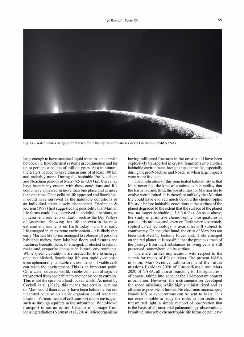
Fig. 14 - Water plumes rising up from fractures in the icy crust of Saturn’s moon Enceladus (credit NASA).

Bollettino della Società Paleontologica Italiana, 55 (2), 2016100
macroscopic signatures, such as MISS or stromatolites. They will therefore be difficult to observe in situ with space instrumentation. The ExoMars rover has Raman, infrared (IR) spectrometers, and laser desorption-gas chromatography-mass spectrometry (GC-MS) that should be able to detect the presence of organic molecules, although the experience with the SAM (Sample Analysis at Mars) spectrometer on Curiosity has shown just how difficult this can be (Freissinet et al., 2015). The Mars 2020 rover will also have a Raman spectrometer and will also be able to take samples with the aim of caching them for future sample return to Earth. It is evident that in situ identification of biosignatures on Mars with space instrumentation will be very difficult and so sample return is a must. The biosignatures in the Early Archaean rocks and the techniques used to study them provide us with a good idea as to what might be found in the returned Martian rocks and how to identify any potential biosignatures.
Mars could still possibly host extant life in the subsurface, but deep drilling to search for extant life forms is not possible in the immediate future. However, other satellites in the Solar System could host extant life in oceans beneath their icy crusts, such as Enceladus (e.g., McKay et al., 2014) around Saturn and Europa (e.g., Hand & Carlson, 2015) around Jupiter (Fig. 14). Indeed, there are a number of missions planned to explore the surface of Europa in the next decade. All that could be observed is the icy surface upon which possible salty deposits from the underlying ocean have been deposited around cracks in the ice crust. If there is extant life in the ocean, perhaps its traces could be found in the salt deposits. However, the extreme radiation conditions due to the vicinity to Jupiter would rapidly destroy organic molecules.
Enceladus is perhaps even more interesting for the fact that pressurised salt water plumes have been observed rising from its icy crust. Fly-by missions through the plumes could collect samples of the water for determination of its possible biosignature content (McKay et al., 2014).
With respect to the search for life on exo-planets, i.e. planets outside the Solar System but in our galaxy (e.g., Scharf, 2009), the Milky Way, since most rocky planets will probably be basltic in composition, the most common forms of life are likely to be chemotrophic, i.e. microorganisms that do not produce an O2 signature in the atmosphere. This has consequences for the detection of traces of life in the atmospheres of exoplanets. Methane would be a more relavent atmospheric biosignature, but also has significant abiogenic origins.
Despite the challenges involved in the search for and identifying extraterrestrial biosignatures, i.e. microfossil biosignatures, the very fact of finding a trace of life on another planet or satellite has enormous consequences for the origin of life (Brack, 2007). It would mean that life on Earth is not unique and that the emergence of life is, in fact, a chemical continuum (de Duve, 1995).
ACKNOWLEDGMENTS
This personal review is the fruit of a long history during which my career has taken me on a wonderful meander through marine geology, biogeology, planetary sciences to prebiotic chemistry and,
of course astrobiology (which is almost everything). I have been favoured by inspiration and support from many people. Claude Monty first introduced me to geobiology, and while in Italy I was lucky to have the support of Elisabetta Guerzoni, Laurita Boni, Paola Giordani, Roberto Barbieri, and Gian Gabriele Ori. At the Johnson Space Center, David Mckay and Everett Gibson accepted my “devil’s advocate” stance on the traces of life in the meteorite ALH84001. I also enjoyed fierce battles with Robert Folk over the existence or not of “nanobacteria” (the battle is still ongoing, both of us being very stubborn). I am happy to acknowledge my colleagues “in (astrobiological) crime” Barbara Cavalazzi and Annalisa Ferretti. André Brack took a momentous step in asking me to take over his prebiotic and origins of life group at the CNRS in Orléans, thus opening up my horizons challengingly. The possibility of working with the ExoMars mission is allowing me to dream of other worlds and other life, aided and abetted by many close friends and colleagues, especially Jorge Vago the project scientist of ExoMars, my research group in Orléans (Frédéric Foucher, Frédéric Gaboyer, Avinash Dass, Keyron Hickman-Lewis, Axelle Hubert, Marylène Bertrand, Annie Chabin), and CNRS friends and colleagues Philippe Martin and Pascale Gautret. Finally, where would we be without the support of the French Space Agency (CNES) and our astrobiological thematic leader, Michel Viso. And many, many others have had a lasting input into my research.
REFERENCES
Allwood A.C., Walter M.R., Burch I.W. & Kamberc Balz S. (2007). 3.43 billion-year-old stromatolite reef from the Pilbara Craton of Western Australia: Ecosystem-scale insights to early life on Earth. Precambrian Research, 158: 198-227.
Allwood A.C., Walter M.R., Kamber B.S., Marshall C.P. & Burch I.W. (2006). Stromatolite reef from the Early Archaean era of Australia. Nature, 441: 714-718.
Arndt N.T. (1994). Archean komatiites. In Condie K.C. (ed.), Archean Crustal Evolution. Elsevier, Amsterdam: 11-44.
Arndt N.T. & Nisbet E.G. (2012). Processes on the Young Earth and the Habitats of Early Life. Annual Review of Earth and Planetary Sciences, 40: 521-549.
Blake R.E., Chang S.J. & Lepland A. (2010). Phosphate oxygen isotopic evidence for a temperate and biologically active Archaean ocean. Nature, 464: 1029-1032.
Brack A. (2007). Astrobiology: from the origin of life on Earth to life in the Universe. In Horneck G. & Rettberg P. (eds), Complete Course in Astrobiology. Wiley-VCH Verlag, Weinheim, Allemagne: 1-22.
Brasier M.D., Green O.R., Jephcoat A.P., Kleppe A.K., Van Kranendonk M.J., Lindsay J.F., Steele A. & Grassineau N.V. (2002). Questioning the evidence of Earth’s oldest fossils. Nature, 416: 76-81.
Buick R. (2008). When did oxygenic photosynthesis evolve? Philosophical Transactions of the Royal Society B, 363: 2731-2743.
Byerly G.R., Lower D.R. & Walsh M.M. (1986). Stromatolites from the 3300–3500-Myr Swaziland Supergroup, Barberton Mountain Land, South Africa. Nature, 319: 489-491.
Cavalazzi B. & Barbieri R. (2016). Emergence and Evolution of Early Life in the Geological Environment. In Goffredo S. & Dubinsky Z. (eds), The Cnidaria, Past, Present and Future. Springer International Publishing Switzerland: 3-13.
Cavalazzi B., Glamoclija M., Brack, A., Orosei, R., Cady S.L. (2016). Astrobiology, Life Formation and Planetary Exploration. In Rossi A.P. & van Gasselt S. (eds), Planetary Geology. Springer-Berlin: in Press.
Cockell C.S., Balme M., Bridges J.C., Davila A. & Schwenzer S.P. (2012) Uninhabited habitats on Mars. Icarus, 217: 184-193.
Corliss J.B., Dymond J., Gordon L.I., Edmond J.M., von Herzen R.P., Ballard R.D., Green K., Williams D., Bainbridge A., Crane K. & van Andel T.H. (1979). Submarine Thermal Springs on the Galápagos Rift. Science, 203: 1073-1083.

101F. Westall - Early life
de Duve C. (1995). Vital Dust: Life as a Cosmic Imperative. Basic Books, New York.
de Ronde C.E.J. & Ebbesen T.W. (1996). 3.2 “b.y” of organic compound formation near sea-floor hot springs. Geology, 24: 791-794.
Derenne S., Robert F., Skryzpczak-Bonduelle A., Gourier D., Binet L. & Rouzaud J.-N. (2008). Molecular evidence for life in the 3.5 billion-year old Warrawoona Chert. Earth and Planetary Science Letters, 272: 476-480.
Des Marais D.J. (2000). When did photosynthesis emerge on Earth? Science, 289: 1703-1705.
Folk R.L. & Lynch F.L. (1997). Nanobacteria are alive on Earth as well as Mars. Proc. SPIE 3111, Instruments, Methods, and Missions for the Investigation of Extraterrestrial Microorganisms, doi:10.1117/12.278795.
Foucher F., Westall F., Brandstaetter F., Demets R., Parnell J., Cockell C.S., Edwards H.G.M., Beny J.-M. & Brack A. (2010). Testing the survival of microfossils in artificial martian sedimentary meteorites during entry into Earth’s atmosphere: The STONE 6 experiment. Icarus, 207: 616-630.
Franks J. & Stolz J.F. (2009). Flat laminated microbial mat communities. Earth-Science Reviews, 96: 163-172.
Freissinet C., Glavin D.P., Mahaffy P.R., Miller K.E., Eigenbrode J.L., Summons R.E., Brunner A. E., Buch A., Szopa C., Archer Jr. P.D., Franz H.B., Atreya S.K., Brinckerhoff W.B., Cabane M., Coll P., Conrad P.G., Des Marais D.J., Dworkin J.P., Fairén A.G., François P., Grotzinger J.P., Kashyap S., ten Kate I.L., Leshin L.A., Malespin C.A., Martin M.G., Martin-Torres F.J., McAdam A.C., Ming D.W., Navarro-González R., Pavlov A.A., Prats B.D., Squyres S.W., Steele A., Stern J.C., Sumner D.Y., Sutter B., Zorzano M.-P. & the MSL Science Team (2015). Organic molecules in the Sheepbed Mudstone, Gale Crater, Mars. Journal of Geophysical Research: Planets, 120: 495-514.
Friedmann E.I. & Koriem A.M. (1989). Life on Mars: how it disappeared (if it was ever there). Advances Space Research, 9: 167-172.
Gibson E.K., McKay D.S., Thomas-Keprta K.L., Wentworth S.J., Westall F., Steele A., Romanek C.S., Bell M.S. & Toporski J. (2001). Life on Mars: Evaluation of the evidence within Martian meteorites ALH84001, Nakhla, and Shergotty. Precambrian Research, 106: 15-34.
Habicht K.S., Gade M., Thamdrup B., Berg P. & Canfield D.E. (2002). Calibration of sulfate levels in the Archean ocean. Science, 298: 2372-2374.
Hand K.P. & Carlson R.W. (2015). Europa’s surface color suggests an ocean rich with sodium chloride: Sodium chloride on Europa’s surface. Geophysical Research Letters, 42: 3174-3178.
Heubeck C. (2009). An early ecosystem of Archean tidal microbial mats (Moodies Group, South Africa, ca. 3.2 Ga). Geology, 37: 931-934.
Hofmann A. (2011). Archaean hydrothermal systems in the Barberton greenstone belt and their significance as a habitat for early life. In Golding S.D. & Glikson M. (eds): Earliest Life on Earth: Habitats, Environments and Methods of Detection. Springer, Netherlands: 51-78.
Hofmann A. & Harris C. (2008). Stratiform alteration zones in the Barberton greenstone belt: a window into subseafloor processes 3.5 to 3.3 Ga ago. Chemical Geology, 257: 224-242.
Hofmann H.J., Grey K., Hickman A.H. & Thorpe R.I. (1999). Origin of 3.45 Ga coniform stromatolites inWarrawoona Group,Western Australia. Geological Society of America Bulletin, 111: 1256-1262.
Javaux E.J., Marshall C.P. & Bekker A. (2010). Organic-walled microfossils in 3.2-billion-year-old shallow-marine siliciclastic deposits. Nature, 463: 934-938.
Kamber B.S. (2015). The evolving nature of terrestrial crust from the Hadean, through the Archaean, into the Proterozoic. Precambrian Research, 258: 48-82.
Kasting J.F. (1988). Runaway and moist greenhouse atmospheres and the evolution of earth and Venus. Icarus, 74: 472-494.
Kempe S. & Degens E.T. (1985). An early soda ocean? Chemical Geology, 53: 95-108.
Kiyokawa S., Ito T., Ikehara M. & Kitajima F. (2006). Middle Archean volcano-hydrothermal sequence: Bacterial microfossil-bearing 3.2 Ga Dixon Island Formation, coastal Pilbara terrane, Australia. Geological Society of America Bulletin, 118: 3-22.
Kiyokawa S., Ito T., Ikehara M., Yamaguchi K.E., Koge S. & Sakamoto R. (2012). Lateral variations in the lithology and organic chemistry of a black shale sequence on the Mesoarchean seafloor affected by hydrothermal processes: The Dixon Island Formation of the coastal Pilbara Terrane, Western Australia. Island Arc, 21: 118-147.
Kleine T., Touboul M., Bourdon B., Nimmo F., Mezger K., Palme H., Jacobsen S.B., Yin Q.-Z. & Halliday A.N. (2009). Hf-W chronology of the accretion and early evolution of asteroids and terrestrial planets. Geochimica et Cosmochimica Acta, 73: 5150-5188.
Knauth L.P. & Lowe D.R. (2003). High Archean climatic temperature inferred from oxygen isotope geochemistry of cherts in the 3.5 Ga Swaziland Supergroup, South Africa. Geological Society of America Bulletin, 115: 566-580.
Krumbein W.E. (1983). Stromatolites – the challenge of a term in space and time. Precambrian Research, 20: 493-531.
Lammer H., Kasting J.F., Chassefière E., Johnson R.E., Kulikov Y.N. & Tian F. (2008). Atmospheric Escape and Evolution of Terrestrial Planets and Satellites. Space Science Reviews, 139: 399-436.
Ledevin M., Arndt N., Davaille A., Ledevin R. & Simionovici A. (2015). The rheological behaviour of fracture-filling cherts: example of Barite Valley dikes, Barberton Greenstone Belt, South Africa. Solid Earth, 6: 253-269.
Lowe B.R., Byerly G.R. & Heubeck C. (2012). Geologic map of the west-central Barberton Greenstone Belt. Geological Society of America Map and Chart Series MCH103, 1 sheet, scale 1:25,000.
Lowe D.R. (1999). Petrology and sedimentology of cherts and related silicified sedimentary rocks in the Swaziland Supergroup. Geological Society of America Special Paper, 329: 83-114.
Lyons T.W., Reinhard C.T. & Planavsky N.J. (2014). The rise of oxygen in Earth’s early ocean and atmosphere. Nature, 506: 307-315.
McKay C.P., Anbar A.D., Porco C. & Tsou P. (2014). Follow the Plume: The Habitability of Enceladus. Astrobiology, 14: 352-355.
McKay D.S., Gibson E.K., Thomas-Keprta K.L., Vali H., Romanek C., Clemett S.J., Chiller X.D.F., Maechling C.R. & Zare R.N. (1996). Search for past life on Mars: possible relic biogenic activity in Martian meteorite ALH84001. Science, 273: 24-930.
Moeller R., Reitz G., Li Z., Klein S. & Nicholson W.L. (2012). Multifactorial resistance of Bacillus subtilis spores to high-energy proton radiation: role of spore structural components and the homologous recombination and non-homologous end joining DNA repair pathways. Astrobiology, 12: 1069-1077.
Mozjsis S.J., Harrison T.M. & Pidgeon R.T. (2001). Oxygen-isotope evidence from ancient zircons for liquid water at the earth’s surface 4,3000 Myr ago. Nature, 409: 178-181.
Noffke N. (2009). The criteria for the biogeneicity of microbially induced sedimentary structures (MISS) in Archean and younger, sandy deposits. Earth-Science Reviews, 96: 173-180.
Noffke N. (2010). Microbial Mats in Sandy Deposits from the Archean Era to Today, Springer, New York.
Noffke N., Christian D., Wacey D. & Hazen R.M. (2013). Microbially Induced Sedimentary Structures Recording an Ancient Ecosystem in the ca. 3.48 Billion-Year-Old Dresser Formation, Pilbara, Western Australia. Astrobiology, 13: 1103-1124.
Noffke N. & Krumbein W.E. (1999). A quantitative approach to sedimentary surface structures contoured by the interplay of microbial colonization and physical dynamics. Sedimentology, 46: 417-426.

Bollettino della Società Paleontologica Italiana, 55 (2), 2016102
Norman R.B., Gronoff G. & Mertens C. (2014). Influence of dust loading on atmospheric ionizing radiation on Mars. Journal of Geophysical Research: Space Physics, 119: 452-461.
Ohmoto H., Watanabe Y., Lasaga A.C., Naraoka H., Johnson I., Brainard J. & Chorney A. (2014). Oxygen, iron, and sulphur geochemical cycles on early Earth: Paradigms and contradictions. Geological Society of America Special Paper, 504: 55-96.
Orange F., Chabin A., Gorlas A., Lucas-Staat S., Geslin C., Leromancer M., Prangishvili D., Forterre P. & Westall F. (2011). Experimental fossilisation of viruses from extremophilic Archaea. Biogeosci Discuss, 8: 2235-2257.
Orange F., Westall F., Disnar J.-R., Prieur D., Bienvenu N., Le Romancer M. & Défarge C. (2009). Experimental silicification of the extremophilic Archaea Pyroccus abyssi and Methanocaldococcus jannaschii. Applications in the search for evidence of life in early Earth and extraterrestrial rocks. Geobiology, 7: 403-418.
Orgel L.E. (1998). The origin of life – a review of facts and speculations. Trends in Biochemical Sciences, 23: 491-495.
Parkes R.J., Cragg B., Roussel E., Webster G., Weightman A. & Sass H. (2014). A review of prokaryotic populations and processes in sub-seafloor sediments, including biosphere: geosphere interactions. Marine Geology, 352: 409-425.
Preston L.J. & Genge M.J. (2010). The Rhynie Chert, Scotland, and the search for life on Mars. Astrobiology, 10: 549-560.
Ryder G., Koeberl C. & Mojzsis S.J. (2000). Heavy bombardment on the Earth at ~3.85Ga. The search for petrographic and geochemical evidence. In Canup R.M. & Righter K. (eds), Origin of the Earth and Moon. University of Arizona Press, Arizona: 475-492.
Scharf C.A. (2009). Extrasolar Planets and Astrobiology. 470 pp. University Science Books, Sausalito.
Schopf J.W. (1993). Microfossils of the Early Archean Apex Chert – New evidence of the antiquity of life. Science, 260: 640-646.
Schopf J.W. (2006). Fossil evidence of Archaean life. Philosophical Transactions of the Royal Society B, 361: 869-885.
Schopf J.W., Kudryavtsev A.B., Agresti D.G., Wdowiak T.J. & Czaja A.D. (2002). Laser-Raman imagery of Earth’s earliest fossils. Nature, 416: 73-76.
Shock E.L., McCollom T.M. & Schulte M.D. (1998). The emergence of metabolism from within hydrothermal systems. In Wiegel J. & Adams M.W.W. (eds), Thermophiles: The Keys to Molecular Evolution and the Origin of Life. Taylor & Francis, London: 59-76.
Sievert S.M. & Vetriani C. (2012). Chemoautotrophy at deep-sea vents: Past, present, and future. Oceanography, 25: 218-233.
Stevens T.O. & McKinley J.P. (1995). Lithoautotrophic microbial ecosystems in deep basalt acquifers. Science, 270: 450-455.
Strom R.G., Schaber G.G. & Dawson D.D. (1994). The global resurfacing of Venus. Journal of Geophysical Research, 99: 10,899-10,926.
Sugitani K., Grey K., Allwood A., Nagaoka T., Mimura K., Minami M., Marshall C.P., Van Kranendonk M.J. & Walter M.R. (2007). Diverse microstructures from Archaean chert from the Mount Goldsworthy–Mount Grant area, Pilbara Craton, Western Australia: microfossils, dubiofossils, or pseudofossils? Precambrian Research, 158: 228-262.
Sugitani K., Grey K., Nagaoka T. & Mimura K. (2009). Three-dimensional morphological and textural complexity of Archean putative microfossils from the northeastern Pilbara Craton: indications of biogenicity of large (> 15 μm) spheroidal and spindle-like structures. Astrobiology, 9: 603-615.
Sugitani K., Mimura K., Takeuchi M., Yamaguchi T., Suzuki K., Senda R., Asahara Y., Wallis S. & Van Kranendonk M.J. (2015). A Paleoarchean coastal hydrothermal field inhabited by diverse microbial communities: the Strelley Pool Formation, Pilbara Craton, Western Australia. Geobiology, 13: 522-545.
Summons R.E., Amend J.P., Bish D., Buick R., Cody G.D., Des Marais D.J., Dromart G., Eigenbrode J.L., Knoll A.H. & Sumner
D.Y. (2011). Preservation of martian organic and environmental records: final report of the Mars Biosignature Working Group. Astrobiology, 11: 157-181.
Tice M.D. & Lowe D.R. (2004). Photosynthetic microbial mats in the 3416-Myr-old ocean. Nature, 431: 549-552.
Tice M.M. (2009). Environmental Controls on Photosynthetic Microbial Mat Distribution and Morphogenesis on a 3.42 Ga Clastic-Starved Platform. Astrobiology, 9: 989-1000.
Tice M.M. & Lowe D.R. (2006). The origin of carbonaceous matter in pre-3.0 Ga greenstone terrains: a review and new evidence from the 3.42 Ga Buck Reef chert. Earth-Science Reviews, 76: 259-300.
Toporski J., Steele A., Westall F., Avci R., Martill D.M. & McKay D.S. (2002). Morphologic and spectral investigation of exceptionally well-preserved bacterial biofilms from the Oligocene Enspel Formation, Germany. Geochimica et Cosmochimica Acta, 66: 1773-1791.
van den Boorn S., van Bergen M., Nijman W. & Vroon P. (2007). Dual role of seawater and hydrothermal fluids in Early Archean chert formation: evidence from silicon isotopes. Geology, 35: 939-942.
van Kranendonk M.J. (2006). Volcanic degassing, hydrothermal circulation and the flourishing of early life on Earth: a review of the evidence from c. 3490–3240 Ma rocks of the Pilbara Supergroup, Pilbara Craton, Western Australia. Earth-Science Reviews, 74: 197-240.
van Kranendonk M.J. (2015). Making it thick: a volcanic plateau origin of Palaeoarchean continental lithosphere of the Pilbara and Kaapvaal cratons. Geological Society of London Special Publication, 389: 83-111.
Vítek P., Edwards H.G.M., Jehlička J., Ascaso C., De Los Ríos A., Valea S., Jorge-Villar S.E., Davila A.F. & Wierzchos J. (2010). Microbial colonization of halite from the hyper-arid Atacama Desert studied by Raman spectroscopy. Philosophical Transactions of the Royal Society A, 368: 3205-3221.
Wacey D. (2014). In situ Morphologic, Elemental and Isotopic Analysis of Archean Life. In Dilek Y. & Furnes H. (eds), Evolution of Archean Crust and Early Life, Modern Approaches in Solid Earth. Springer Science, Dordrecht: 361-365.
Wacey D., Saunders M., Cliff J. & Brasier M.D. (2011). Microfossils of sulphur-metabolizing cells in 3.4-billion-year-old rocks of Western Australia. Nature, 4: 698-702.
Walker J.C.G. (1985). Carbon dioxide on the early Earth. Origins of Life and Evolution of Biosphere, 16: 117-127.
Walsh M.M. (1992). Microfossils and possible microfossils from the early Archean Onverwacht Group, Barberton Mountain Land, South Africa. Precambrian Research, 54: 271-293.
Westall F. (1997). The influence of cell wall composition on the fossilisation of bacteria and the implications for the search for early life forms. In Cosmovici C.B., Bowyer S. & Werthimer D. (eds), Astronomical and biochemical origins and the search for life in the universe. Editrici Compositori, Bologna: 491-504.
Westall F. (2005) Life on the early earth: A sedimentary view. Science, 308: 366-367.
Westall F. (2011). Early life. In Gargaud M., López-Garcìa P. & Martin H. (eds), Origins of Life, An Astrobiology Perspective. Cambridge University Press, Cambridge: 391-413.
Westall F. (2012). Early Earth. In Impey C., Lunine J. & Funes J. (eds), Astrobiology. Cambridge University Press, Cambridge: 89-114.
Westall F. & Brack A. (2016). The importance of water for life. In Blanc M. (ed.), Water and Planets. Space Science Reviews, Springer, In Press.
Westall F. & Cavalazzi B. (2011). Biosignatures in rocks. In Thiel V. (ed.), Encyclopedia of Geobiology. Springer, Berlin: 189-201.
Westall F., Campbell K.A., Bréhéret F.G., Foucher F., Gautret P., Hubert A., Sorieul S., Grassineau N. & Guido D.M. (2015b). Complex microbe-sediment systems are ancient (3.33 Ga) and flourished in a hydrothermal context. Geology, 43: 615-618.
Westall F., Cavalazzi B., Lemelle L., Marrocchi Y., Rouzaud J.N., Simionovici A., Salomé M., Mostefaoui S., Andreazza
103F. Westall - Early life
C., Foucher F., Toporski J., Jauss A., Thiel V., Southam G., MacLean L., Wirick S., Hofmann A., Meibom A., Robert F. & Défarge C. (2011b). Implications of in situ calcification for photosynthesis in a ∼3.3 Ga-old microbial biofilm from the Barberton Greenstone Belt, South Africa. Earth and Planetary Science Letters, 310: 468-479.
Westall F., de Ronde C.E.J., Southam G., Grassineau N., Colas M., Cockell C.S. & Lammer H. (2006a). Implications of a 3.472–3.333 Gyr-old subaerial microbial mat from the Barberton Greenstone Belt, South Africa for the UV environmental conditions on the early Earth. Philosophical Transactions of the Royal Society B, 361: 1857-1875.
Westall F., de Vries S.T., Nijman W., Rouchon V., Orberger B., Pearson V., Watson J., Verchovsky A., Wright I., Rouzaud J.-N., Marchesini D. & Anne S. (2006b). The 3.466 Ga Kitty’s Gap Chert, an Early Archean microbial ecosystem. Geological Society of America Special Paper, 405: 105-131.
Westall F., Foucher F., Bost N., Bertrand M., Loizeau D., Vago J.L., Kminek G., Gaboyer F., Campbell K.A., Bréhéret J.-G., Gautret P. & Cockell C.S. (2015a). Biosignatures on Mars: What, Where, and How? Implications for the Search for Martian Life. Astrobiology, 15: 998-1029.
Westall F., Foucher F., Cavalazzi B., de Vries S.T., Nijman W., Pearson V., Watson J., Verchovsky A., Wright I., Rouzaud J.N., Marchesini D. & Anne S. (2011a). Early life on Earth and Mars: a case study from ∼3.5 Ga-old rocks from the Pilbara, Australia. Planetary Space Science, 59: 1093-1106.
Zahnle K., Schaefer L. & Fegley B. (2010). Earth’s earliest atmospheres. In Deamer D. & Szostak J.W. (eds): Additional Perspectives on The Origins of Life. Cold Spring Harb Perspect Biol 2010;2:a004895.
Zahnle K.J. & Catling D.C. (2016). Waiting for oxygen. Geological Society of America Special Paper, 504: 37-48.
Manuscript received 15 July 2016Revised manuscript accepted 28 August 2016Published online 30 September 2016Editor Barbara Cavalazzi
Frances Westall is a geologist now working in the very broad discipline of astrobiology, the study of the origins of life and its destiny in the Universe. After a PhD in marine geology at the University of Cape Town in South Africa, she worked in various research institutes around the world: the Alfred Wegener Institute for Polar and Marine Research in Bremehaven in Germany, the University of Nantes in France, the University of Bologna in Italy, the Johnson Space Center and Lunar and Planetary Institute in Houston, USA and, for the last 15 years, she is Director of Research at the CNRS-Orléans in France where she heads the astrobiology group. Her research interests and activities cover study of the earliest traces of life on Earth and the geological environment in which they lived, the artifical fossilisation of bacteria, the search for life on Mars, especially with the ESA/Russian ExoMars 2020 mission, and prebiotic chemistry and the origin of life.