o f t l ab n r u o siol journal of diabetes and metabolism ... · pheochromocytoma: a case report...
TRANSCRIPT
Pheochromocytoma: A Case Report and Literature ReviewXiaoyan Chen1*, Xuelian Deng1, Shunyou Deng1, Dongling Li1, Wenzhong Chen2, Wanling Chen3, Xia Gu4 and Yubao Guan4
1Department of Endocrinology, the First Affiliated Hospital of Guangzhou Medical University, China2Department of Urology, the First Affiliated Hospital of Guangzhou Medical University, China3Department of Pathology, the First Affiliated Hospital of Guangzhou Medical University, China4Department of Radiology, the First Affiliated Hospital of Guangzhou Medical University, China*Correspondence author: Xiaoyan Chen, Department of Endocrinology, The First Affiliated Hospital of Guangzhou Medical University, China, Tel: 13926096343; Fax:862083395651; E-mail: [email protected]
Received date: February 20, 2017; Accepted date: March 02, 2017; Published date: March 08, 2017
Copyright: © 2017 Xiaoyan Chen, et al. This is an open-access article distributed under the terms of the Creative Commons Attribution License, which permitsunrestricted use, distribution, and reproduction in any medium, provided the original author and source are credited.
Abstract
Objective: We present a case of pheochromocytoma with fatal paroxysmal dyspnea hemoptysis and shock. Asystematic review of literature on pheochromocytoma was performed to improve the understanding of the clinicalmanifestation, diagnosis and treatment of a typical pheochromocytoma.
Methods: The clinical manifestation, diagnostic examination, operation mode and pathological characteristics ofthe typical pheochromocytoma, were analyzed and summarized. Simultaneously relevant literature was reviewed.
Results: A 29-year-old male was diagnosed with pheochromocytoma with hemorrhagic cysts though without anytypical clinical manifestation such as paroxysmal hypertension headache diaphoresis and palpitation. We treatedthis case as functional pheochromocytoma during the perioperative preparation although there were not enoughevidence supporting that the tumor was pheochromocytoma. After sufficient preoperative preparation one of theadrenal grands was surgical resected and the diagnosis was confirmed on histopathology. Postoperation follow-upshowed that the patient had a good prognosis after six months.
Conclusion: The manifestations as we reported in this article fatal paroxysmal dyspnea hemoptysis and shockare quite rare as initial manifestations in pheochromocytoma. It showed that the initial manifestations ofpheochromocytoma are complicated and deserve an appropriate examination and careful handling, which mightreduce the misdiagnosis and surgical risk.
Keywords: Pheochromocytoma; Atypical; Diagnosis
IntroductionThe typical manifestations of pheochromocytoma, are the
paroxysmal hypertension, headache, palpitation and diaphoresis. Thispaper is reporting a case of pheochromocytoma with paroxysmaldyspnea hemoptysis and shock as the initial symptoms.
Case ReportA 29-year-old male was admitted to the emergency department with
sudden difficulty in breathing coughing up pink frothy sputum andvomiting stomach contents for One day and in a mental confusion forhalf an hour. He was previously healthy with normal blood pressure.
On physical examination, his blood pressure was 70/50 mmHg withobvious hypoxemia. The X-ray showed a butterfly shaped effusion withshadows in the bilateral pulmonary hilum. Hence, he was diagnosed ashaving acute pulmonary edema and shock. He was in remission, &after 4 days of assisting ventilation by endotracheal intubation and abreathing machine & anti-shock treatment with supporting treatment.The dynamic monitoring of his blood pressure, had shown the systolicblood pressure was ranging from 98 to 125 mmHg and the diastolicblood pressure ranging from 54 to 78 mmHg; and the heart rate
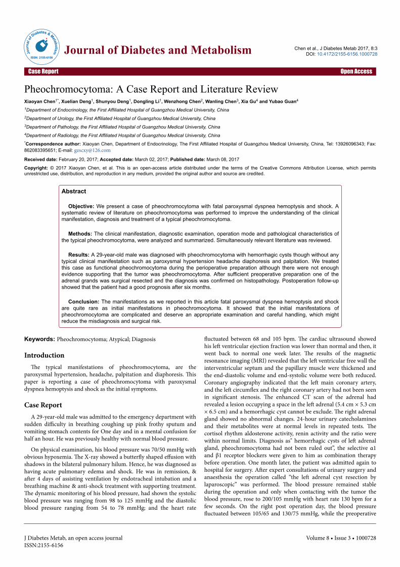
fluctuated between 68 and 105 bpm. The cardiac ultrasound showedhis left ventricular ejection fraction was lower than normal and then, itwent back to normal one week later. The results of the magneticresonance imaging (MRI) revealed that the left ventricular free wall theinterventricular septum and the papillary muscle were thickened andthe end-diastolic volume and end-systolic volume were both reduced.Coronary angiography indicated that the left main coronary artery,and the left circumflex and the right coronary artery had not been seenin significant stenosis. The enhanced CT scan of the adrenal hadrevealed a lesion occupying a space in the left adrenal (5.4 cm × 5.3 cm× 6.5 cm) and a hemorrhagic cyst cannot be exclude. The right adrenalgland showed no abnormal changes. 24-hour urinary catecholaminesand their metabolites were at normal levels in repeated tests. Thecortisol rhythm aldosterone activity, renin activity and the ratio werewithin normal limits. Diagnosis as" hemorrhagic cysts of left adrenalgland, pheochromocytoma had not been ruled out”, the selective α1and β1 receptor blockers were given to him as combination therapybefore operation. One month later, the patient was admitted again tohospital for surgery. After expert consultations of urinary surgery andanaesthesia the operation called “the left adrenal cyst resection bylaparoscopic” was performed. The blood pressure remained stableduring the operation and only when contacting with the tumor theblood pressure, rose to 200/105 mmHg with heart rate 130 bpm for afew seconds. On the right post operation day, the blood pressurefluctuated between 105/65 and 130/75 mmHg, while the preoperative
Chen et al., J Diabetes Metab 2017, 8:3 DOI: 10.4172/2155-6156.1000728
Case Report Open Access
J Diabetes Metab, an open access journalISSN:2155-6156
Volume 8 • Issue 3 • 1000728
Jour
nal o
f Diabetes & Metabolism
ISSN: 2155-6156Journal of Diabetes and Metabolism
blood pressure fluctuated between 87/47 and 130/76 mmHg. The heartrate changed from 63-116 bpm before the operation to 77-90 bpm afterthe operation. The α1 receptor blocker was stopped on the right postoperation day and the β1 receptor blocker was stopped on the 3rd dayafter operation. And his blood pressure and heart rate were stable. Thefinal diagnosis was confirmed by pathology as adrenalpheochromocytoma with bleeding and cystic change. The macroscopicfindings showed: that the tumor measured 6 cm × 5 cm × 2 cm, andthe thickness of cystic wall was about 0.2-0.4 cm, and there were sometaupe substances in the cyst. Immunohistochemical results showed:CgA/Syn (+), s100 (+), Ki67 (< 5%), P53 (partly +), CK/EMA (-).During the regular follow-up period for half a year, the blood pressurefluctuated between 110/70 and 115/75 mmHg and the heart ratefluctuated between 70 and 75 bpm. The patient had no difficulty inbreathing and hemoptysis.
DiscussionThis paper reported a case of pheochromocytoma which developed
in a young age with uncommon clinical manifestations. Without anysymptoms before the onset however the patient had a sudden acutepulmonary edema haemoptysis and shock. Pheochromocytoma arisesfrom the chromaffin cell which is the location where the generationstorage metabolism and secretion of catecholamine occur [1-3]. It’salso called a neuroendocrine tumor. Large amounts of catecholaminesincluding norepinephrine epinephrine and dopamine are released bypheochromocytoma continuously or discontinuously which causepersistent or paroxysmal hypertension and the functional andmetabolic disorders of multiple organs. The clinical manifestationsdepend on the quantity scale and release of epinephrine andnorepinephrine which are secreted by the tumor. Pheochromocytomais typically characterized by high blood catecholamine, which causespersistent or paroxysmal hypertension with throbbing headache,palpitation and sweating as the main clinical symptoms. Howeverabout 15 percent of patients never suffer from the clinicalmanifestation of high blood pressure. They may suffer fromuncommon clinical manifestations such as acute coronary syndrome[4,5] abnormal ventricular wall activity and ventricular dilatation [6]acute abdominal pain [7,8] limb weakness [8] lumbago [9] orthostatichypotension upper gastrointestinal hemorrhage [10] and diabetes[5,10] or even have no symptoms. There are some reports aboutHeterologous Cushing's syndrome [11,12] (centripetal obesity) andectopic adrenocorticotropic hormone (ACTH) syndrome [13,14] butrarely about non cardiogenic pulmonary edema [15]. Typicalpheochromocytoma can usually find adrenal occupying lesions byultrasound and CT, MRI and other imaging examination. Higher levelsof catecholamines and their metabolites can be detected in blood orurine. However the value of the examinations above, in the diagnosisof atypical pheochromocytoma is limited.
These less common clinical presentations might be related to thenecrosis hemorrhage cystic change of pheochromocytoma. There werecase reports about the hematoma and hemorrhagic shock caused byspontaneous rupture and traumatic rupture of the tumor [16,17]. Astudy found that there was some degree of hemorrhage with 48 in 70cases of pheochromocytoma patients detected by using spiralcomputed tomography scan [18]. The case that we reported startedwith a sudden fatal acute pulmonary edema and hypovolemic shockwithout typical manifestations, such as high blood pressurepalpitations headache and hyperhidrosis previously. The bigger size ofthe tumor, might lead to spontaneous rupture and hemorrhoea which
might be related to the imageological change of the adrenal cyst andhemorrhage. Reportedly at least one-third of the life-threateningbleeding tumors have atypical symptoms [19]. The most commonsymptoms are acute abdominal pain tachycardia, and peripheralvascular contraction and other signs include unstable blood pressurehyperhidrosis, vomiting leukocytosis and fever etc. Clinicians shouldbe alert to pheochromocytoma with internal bleeding, when theyencounter severe vasoconstriction, such as chills, sweating and acrapale associated with hypotension. These might be the body’s earlyresponses to a large number of secretions of catecholamines.
In the preoperative preparation for suspected pheochromocytoma ithas been found that even in the clearly diagnosed patient the deathrate of the emergency adrenal resection is far higher than the one ofthe scheduled surgery after conservative treatment [20]. We treatedthis case, as functional pheochromocytoma during the perioperativepreparation, although there were not enough evidence supporting thatthe tumor was pheochromocytoma, before the operation. We usedselective alpha 1 and beta 1 blockers as the patient's blood pressure wasmore than one month before the left adrenalectomy and the bloodcapacity was supplemented actively one week before the surgery. Thesurgery was performed using a retroperitoneal approach with thepatient in the lateral position which could avoid the abdominal organswithout affecting the full exposure of the left adrenal. Simultaneouslywe were prepared, for the possibility that the blood pressure would besharply fluctuated during the contact and removal of the tumor in theoperation. The blood pressure had risen not too badly for once duringthe surgery and it restored to a steady state soon after givingphentolamine. The patient was well followed-up for 10 months and hisblood pressure and heart rate were relatively stable in the normal rangeand the related symptoms had disappeared (Figures 1-4).
Figure 1: The scanning of transected T1 weighted images showed a5.4 × 5.3 × 6.5 cm heterogeneous mass in the left adrenal with highsignal on the edge and low in the center (arrow) (2014.5.26).
Clinicians should improve their understanding of atypicalpheochromocytoma in order to reduce the misdiagnosis and misseddiagnosis. At the same time, every patient with suspected adrenalpheochromocytoma should be prepared as a functional tumorwhatever the result of the preoperative examination is functional ornot.
Citation: Chen X, Deng X, Deng S, Li D, Chen W, et al. (2017) Pheochromocytoma: A Case Report and Literature Review . J Diabetes Metab 8:728. doi:10.4172/2155-6156.1000728
Page 2 of 4
J Diabetes Metab, an open access journalISSN:2155-6156
Volume 8 • Issue 3 • 1000728

Figure 2: The scanning of transected T2 weighted images showedthere was a boundary clearly round abnormal signal in the leftadrenal, with high signal intensity in the center and low on the edge(arrow) (2014.5.26).
Figure 3: The macroscopic findings: the left adrenal tumormeasured 6cm × 5cm × 2cm. The cutting surface was soft and darktan with focal gray-yellow, and the thickness of cystic wall wasabout 0.2-0.4 cm. In the cystic there were some taupe substances.2014.7.1.
Figure 4: Tumor cells formed like a nest and were made up of largecells which were polygonal (arrow) and a small number of smallcells, which were cylindrical and oval. The cells contain abundantcytoplasm with basophilic or eosinophilic granules. The boundaryof cells is not clear (H&E: × 400) (2014.7.3).
The blood capacity should be supplemented actively beforeoperation, and invasive arterial blood pressure monitoring is veryimportant during surgery. Try not to squeeze the tumor.Norepinephrine or dopamine should be transfused appropriately sothat we can cope with the situation that the intraoperative bloodpressure might fluctuate sharply. Finally, the postoperative patientsshould be regularly followed-up in order to timely avoid relapse whichmay be due to the incomplete resection of the tumor.
Compliance with ethics guidelinesAuthors declare that they have no conflict of interest. This
manuscript is a review article and does not involve a research protocolrequiring approval by the relevant institutional review board or ethicscommittee. The authors have no multiplicity of interest to disclose.
Reference1. Calhoun DA, Jones D, Textor S, Goff DC, Murphy TP, et al.
(2008) Resistant hypertension: diagnosis, evaluation, andtreatment: a scientific statement from the American HeartAssociation Professional Education Committee of the Council forHigh Blood Pressure Research. Circulation 117: e510-e526.
2. Cook LK ( 2009) Pheochromocytoma. The American journal ofnursing 109: 50-53.
3. Hodin R, Lubitz C, Phitayakorn R, Stephen A (2014) Diagnosisand management of pheochromocytoma. Current problems insurgery 51: 151-187.
4. Subramanyam S, Kreisberg RA (2012) Pheochromocytoma acause of ST-segment elevation myocardial infarction, transientleft ventricular dysfunction, and takotsubo cardiomyopathy.Endocrine practice: official journal of the American College ofEndocrinology and the American Association of ClinicalEndocrinologists 18: e77-e80.
5. Guo H, Shi BY, He H (2006) Ten cases of pheochromocytomawith special clinical manifestations at onset and a review of theliterature. Chinese Journal of Practical Internal Medicine 26:1817-1818.
6. Takizawa M, Kobayakawa N, Uozumi H, Yonemura S, Kodama T,et al. (2007) A case of transient left ventricular ballooning withpheochromocytoma, supporting pathogenetic role ofcatecholamines in stress-induced cardiomyopathy or takotsubocadiomyopathy. International Journal of Cardiology 114: e15-e17.
7. Marti JL, Millet J, Sosa JA, Roman SA, Carling T, et al. (2012)Spontaneous adrenal hemorrhage with associated masses:etiology and management in 6 cases and a review of 133 reportedcases. World journal of surgery 36: 75-82.
8. Zhang JP, Chen WX, Li LM (2012) Clinical analysis of 42 cases ofatypical pheochromocytoma. Chinese Journal of Postgraduates ofMedicine 35: 66-68.
9. Du HQ, Jia AH, Liu Y, Zhang J, Bai J (2013) Clinical pathologyanalysis of 60 cases of Pheochromocytoma. Chinese Journal ofInternal Medicine 52: 46-47.
10. Guo YQ, Xia DS (2007) A case of pheochromocytomamisdiagnosed as primary hypertension caused by uppergastrointestinal bleeding. Journal of Clinical Cardiology 23:231-232.
11. Sun X, Wang DW, Wang WQ, Su TW, Jin XL, et al. (2012)Approach to the patient with ectopic ACTH syndrome caused by
Citation: Chen X, Deng X, Deng S, Li D, Chen W, et al. (2017) Pheochromocytoma: A Case Report and Literature Review . J Diabetes Metab 8:728. doi:10.4172/2155-6156.1000728
Page 3 of 4
J Diabetes Metab, an open access journalISSN:2155-6156
Volume 8 • Issue 3 • 1000728

adrenal pheochromocytoma: diagnosis and treatment. ChineseJournal of Endocrinology and Metabolism: 28: 512-515.
12. Yu Q, Li ZZ, Qin GJ, Yang JJ, Yang H, et al. (2010) A case reportof ectopic ACTH syndrome caused by adrenalpheochromocytoma. Chinese Journal of Internal Medicine 49:161-162.
13. LI XG, Zhang DX, Li X, Cui XG, Xu DF, et al. (2012)Adrenocorticotropic hormone-producing pheochromocytoma: acase report and review of the literature. Chinese Medical Journal125: 1193-1196.
14. Nijhoff MF, Dekkers OM, Vleming LJ, Smit JW, Romijn JA, et al.(2009) ACTH-producing pheochromocytoma: clinicalconsiderations and concise review of the literature. Europeanjournal of internal medicine 20: 682-685.
15. Sun XF, Zhong X, Liu T, Wang MZ, Xu WB, et al. (2012) A casesof huge cystic pheochromocytoma with the pulmonary edemas atonset. Chinese Journal of Internal Medicine 51: 314-315.
16. Han XN, Chen B, Ye XD, Wang J, Liu GH (2009) Clinicalmanifestational pheochromocytoma: report of 70 case [inChinese]. Chinese Journal of Oncology 31:139-142.
17. Marti JL, Millet J, Sosa JA, Roman SA, Carling T, et al. (2012)Spontaneous adrenal hemorrhage with associated masses:etiology and management in 6 cases and a review of 133 reportedcases. World journal of surgery 36: 75-82.
18. Moazzam MS, Ahmed SM, Bano S (2010) Traumatic hemorrhageof occult phaeochromocytoma in a patient with septic shock.Journal of emergencies, trauma, and shock 3: 300.
19. O'Neal P, Francis Moore J, Gawande A, Cho N, Moalem J, et al.(2012) Hemorrhagic Shock as the Initial Manifestation ofPheochromocytoma: Report of a Sequential ManagementStrategy. Endocrine Practice 18: e81-e84.
20. Kobayashi T, Iwai A, Takahashi R, Ide Y, Nishizawa K, et al.(2005) Spontaneous rupture of adrenal pheochromocytoma:review and analysis of prognostic factors. Journal of surgicaloncology 90: 31-35.
Citation: Chen X, Deng X, Deng S, Li D, Chen W, et al. (2017) Pheochromocytoma: A Case Report and Literature Review . J Diabetes Metab 8:728. doi:10.4172/2155-6156.1000728
Page 4 of 4
J Diabetes Metab, an open access journalISSN:2155-6156
Volume 8 • Issue 3 • 1000728