number of structures available in the pdb per year
TRANSCRIPT

11/15/16
1
Number of structures available in the PDB per year
By Nov. 14th, 2016, total structures deposited in PDB: 124,286 X-ray Crystallography: 111,122 (89%) Solution NMR: 11,545 (9%) Electron Microscopy: 1,232 (1%)
0!
20000!
40000!
60000!
80000!
100000!
120000!
140000!
1975!
1977!
1979!
1981!
1983!
1985!
1987!
1989!
1991!
1993!
1995!
1997!
1999!
2001!
2003!
2005!
2007!
2009!
2011!
2013!
2015!
X-ray diffraction data collection
Crystals Electron Density Map
Structural Model Diffraction Image
Model building and refinement Data analysis and phase determination

11/15/16
2
X-ray Diffraction
Each diffraction spot represents a wave.
The diffraction pattern (the position and intensity of each diffraction spot) gives information on the arrangement of the atoms in the crystal.
X-ray
Crystals
Detector
Crystal!
Only the size and contents of one unit cell are necessary to describe the entire crystal. !
Molecule!
Symmetry!operation!
Three-dimensional periodic arrangement of a molecule in a repeating unit (so called the unit cell) into a lattice. !

11/15/16
3
Unit Cell!
a
b
c
β γ
α
O
7 Crystal Systems !!14 Bravais Lattices!!65 Space Groups !

11/15/16
4
From Crystal to Diffraction
Waves add in some directions!(Constructive)!
Waves subtract in other directions!
(Destructive)!
Wave interference
What are the conditions that produce diffraction? - Bragg’s Law
Scattered waves

11/15/16
5
f(x) = F sin 2π (hx + α)
f(x) à vertical height of the wave at any horizontal position x along the wave, measured in wavelengths, where x=1 implies one full wavelength.
F à amplitude (half its height from peak to valley)
h à frequency (number of wavelengths per radian)
α à phase (position of the wave, in radian, with respect to the origin)
A simple wave can be described by a periodic function
λ = 1/h
F x
Origin
1 2
α = 0
f(x)
x
f(x) = F sin 2π(hx+α)
F = 1 h = 1 α = 0
f(x) = sin 2πx
x = 1
λ = 1/h = 1
y = 1
y = -1

11/15/16
6
f1 (x) = sin 2πx
f2 (x) = sin 2πx
x = 1/4
x = 1/4
y = 1
y = -1
y = 1
y = -1
Constructive interference
f1 + f2 = 2sin 2πx
x = 1/4
y = 2
y = -2
F = 2
f1 (x) = sin 2πx
f2 (x) = sin (2πx + π)
x = 1/4
x = 1/4
Destructive interference
f1 + f2 = 0
x = 1/4
F = 0
Phase differences determine the interference.

11/15/16
7
X
x = λ/2 x = λ
O
Wave scattered from origin
Wave scattered from point at distance x
What is the relationship between wave phase differences and distance between scattering atoms?
X O
Wave scattered from point at distance x
For constructive interference: Path distance must be an integral number of wavelengths
Δx = nλ
What is the relationship between wave phase differences and distance between scattering atoms?
This is the central idea embodied by Bragg’s Law
x = 3/2 λ x = 2 λ

11/15/16
8
Think of a crystal as a set(s) of equivalent parallel planes of atoms.!
d
θ
d
Δx = nλ
θ
θ
a a
2a = nλ
2dsinθ = nλ
What is the relationship between d and θ such that these waves add constructively?
- Bragg’s Law
How to describe the planes in a crystal?

11/15/16
9
a
b
c
Lattice indices (h k l) of the atomic planes in a crystal
• Three indices hkl identify a particular set of equivalent, parallel planes.
• The index h gives the number of planes in the set per unit cell in the a direction or, equivalently, the number of parts into which the set of planes cut the a edge of each cell.
• The indices k and l specify how many such planes exist per unit cell in the b and c directions.
(1 0 0)
(0 0 1)
(0 1 0)
Planes apply to the whole lattice
a
b
(1 0 0) planes
(0 1 0) planes
x
y
Lattice point

11/15/16
10
Planes apply to the whole lattice
a
b
(1 0 0) planes
(0 1 0) planes
(1 1 0) planes
x
y
a
b
(1 2 0) planes
x
y

11/15/16
11
How to describe the diffraction pattern?
Construction of a reciprocal lattice
1) Choose coordinate axes for reciprocal space that are identical to real space, and origin can be arbitrary.
2) For each set of lattice planes (h k l), draw a line from the origin that is orthogonal to the planes.
3) Place reciprocal lattice point (h k l) at a distance 1/d from origin, where d is the interplane spacing.
A reciprocal lattice: • Its lattice dimensions are reciprocal to the original cell
(and correspond to the reflection positions);
• Its ‘size’ (the intensity of the reflection) corresponds to the contents of the unit cell.

11/15/16
12
a
b
O x a*
b*
O
(1 0 0) planes
d
x
1/d
(1 0 0)
a
b
O x a*
b*
O
(2 0 0) planes
d
x
1/d
x(2 0 0) (1 0 0)

11/15/16
13
a
b
O x a*
b*
O
(0 1 0) planes
d
x1/d
xx(0 1 0)
a
b
O x a*
b*
O
(1 1 0) planes
d
x
1/d
xx (1 1 0) x

11/15/16
14
a
b
O x a*
b*
O
(1 1 0) planes
x xx
(1 1 0)
x
= (-1 -1 0) planes
x(-1 -1 0)
a
b
O x a*
b*
Ox
x x
x xx
x
x
Reciprocal lattice is symmetric about origin

11/15/16
15
Crystal lattice
(in real space)
Reciprocal lattice
(in diffraction space)
• An entire set of parallel planes (h k l), not just one plane, acts as a single diffractor and produces one reciprocal lattice point.
The reciprocal lattice
The reciprocal lattice has the same symmetry as the crystal lattice.
a* = b x c
a (b x c)
b* = c x a
b (c x a)
c* = a x b
c (a x b)
V = unit cell volume = a (b x c) = b (c x a) = c (a x b)
The larger the crystal unit cell, the smaller the reciprocal lattice.
The mathematical definition of the reciprocal lattice constants is:

11/15/16
16
In orthogonal crystal lattices (α = β = γ = 90°)
a* = 1 a
Real unit cell
b*
c* a*
a
b
c
Reciprocal unit cell
b* = 1 b
c* = 1 c
= 1
d100
= 1
d010
= 1
d001
The reciprocal lattice is spatially linked to the crystal because of the way the lattice points are defined, so if we rotate the crystal, the reciprocal lattice rotates with it.
How to link the reciprocal lattice with the diffraction pattern?
O*
O
real lattice
Bragg’s law in reciprocal space - Ewald Sphere
1/λ
x
x
x
x
x
x
x
x
θ
θ x
reciprocal lattice
IncomingX-ray
1/d
P
A
- To show how each reciprocal-lattice point must be arranged with respect to the X-ray beam in order to satisfy Bragg’s law and produce a reflection from the crystal.
d
Ewald Sphere
TransmittedX-ray

11/15/16
17
O*
O
real lattice
1/λ
x
x
x
x
x
x
x
x
θ
θ x
reciprocal lattice
When is Bragg’s law (nλ = 2dsinθ) satisfied?
θ
P
A
1/d
sin θ = PO*/AO* = 1/d à 2dsinθ = λ
= PO* AO*
= 1/d 2/λ
= λ 2d
O*
O
real lattice
1/λ
x
x
x
x
x
x
x
x
θ
θ x
reciprocal lattice
θ
P
A
Any reciprocal lattice point on the Ewald sphere follows the Bragg’s law and produces diffraction!
detector

11/15/16
18
• An entire set of parallel planes (h k l), not just one plane, acts as a single diffractor and produces one reflection hkl.
• Whenever crystal is rotated so that a reciprocal lattice point comes in contact with the Ewald sphere, Bragg’s law is satisfied and a reflection occurs.
• At a certain orientation of the crystal, only a small number of reflections are in “diffraction condition”. Therefore, the crystal has to be rotated to collect all the reflections.
• The directions of the reflections, as well as number of the reflections, depend only on the unit-cell dimensions and not upon the contents of the unit cell.
• The intensity of that reflection depends upon the electron distribution and density along the planes that produce the reflection.
Diffraction pattern is an image of the reciprocal lattice
|S| = 1/d
S
1) d is spacing between real crystal lattice planes ~ Small d means high resolution.
2) Diffraction position reflects only geometry of crystal and experiment, not contents of unit cell.
3) The relative intensities of the diffractions contain information about the structure of the molecule.
2d sinθ = λ d = λ/2sinθ
when θ = 90°,
sinθ = 1
dmin= λ/2
Higher resolution
Low resolution

11/15/16
19
In reciprocal space, the lattice spacing is inversely proportional to the interplanar spacing within the crystal. The larger the crystal unit cell, the smaller the reciprocal lattice.
Sea salt Protein
From diffraction data to electron density
The Fourier transform describes precisely the mathematical relationship between an object and its diffraction pattern.
The Fourier transform allows us to convert a Fourier-series description of the reflections to a Fourier-series description of the electron density map.
A reflection can be described by a structure-factor equation, containing one term for each atom (or each volume element) in the unit cell. In turn, the electron density of each atom is described by a Fourier series in which each term is a structure factor.
We use the Fourier transform to convert the structure factors F(h, k, l) to ρ(x, y, z), the desired electron density equation.
How to construct a Fourier series?

11/15/16
20
f (x) = F cos 2π(hx + α) Fourier term
Fourier series f (x) = F0 cos 2π(0x + α0) + F1 cos 2π(1x + α1) + … + Fn cos 2π(nx + αn)
∑ h=0
n = Fh cos 2π(hx + αh)
∑ h
f (x)= Fh[cos 2π(hx) + i sin 2π(hx)] ∑ h
= Fhe2πi(hx)
One-dimensional waves
f (x) = F [cos 2π(hx) + i sin 2π(hx)], cosθ + i sinθ = eiθ, θ = 2π(hx), As
f (x) the vertical height of the wave at any horizontal position x;
F the amplitude of the wave; h frequency; α phase.
Three-dimensional waves
∑ h
f (x, y, z) = Fhkle2πi(hx + ky +lz) ∑ k ∑ l
f (x) = F sin 2π(hx + α) Or
(Sum of simple waves)
(a simple wave)
The Fourier transform:
F (h, k, l) = f (x, y, z)e2πi(hx + ky +lz)dxdydz
x y z ∫ ∫ ∫
f (x, y, z) = F (h, k, l)e-2πi(hx + ky +lz)dhdkdl h k l ∫ ∫ ∫
Inverse Fourier transform
• Is an operation that transforms a function containing variables of one type (say time or a length in Å) into a function whose variables are reciprocals of the original type (in this case, 1/time or frequency, or reciprocal length in Å-1).
• Is a precise mathematical description of diffraction. The diffraction patterns (in terms of structure factor) are Fourier transforms of the corresponding objects and arrays (in terms of electron density map).
Unit cell content (Electron density)
in real space
Object Diffraction
Diffraction pattern (Structure factor) in reciprocal space
Inverse Fourier transform

11/15/16
21
A structure factor describes one diffracted x-ray, which produces one reflection received at the detector. It can be written as a Fourier series in which each term gives the contribution of one atom to the reflection hkl.
Structure factor as a Fourier series
Fourier term (One atom)
fhkl = fj e2πi(hxj + kyj +lzj)
fhkl atomic structure factor, the contribution of the single atom j to reflection hkl;
fj scattering factor of atom j - a function that amounts to treating the atom as a simple sphere of electron density.
xj, yj, zj the coordinates of atom j in the unit cell (real space).
h, k, l the indices of a specific reflection in the reciprocal lattice satisfying Bragg’s law.
phase 2π(hxj + kyj +lzj) for atom j.
Fourier series (Sum of all atoms)
Fhkl = fje2πi(hxj + kyj +lzj) ∑ j=0
n
• Each diffraction ray is the sum of diffractive contributions from all atoms in unit cell.
• The contribution of each atom j to Fhkl depends on (1) what element it is, which determines fj, the amplitude of the contribution, and (2) its position in the unit cell (xj, yj, zj), which establishes the phase of its contribution.
Fhkl is a complex number!
Alternatively, Fhkl can be written as the sum of contributions from each volume element of electron density in the unit cell.
Structure factor as a Fourier series
Fhkl = ρ(x,y,z)e2πi(hx + ky +lz)dxdydz
x y z ∫ ∫ ∫
= ρ(x,y,z)e2πi(hx + ky +lz)dV
cell V ∫
• Each volume element contributes to Fhkl with a phase determined by its coordinates (x, y, z).
• Fhkl is the Fourier transform of ρ(x,y,z) on the set of real-lattice planes (hkl). All of the Fhkl s together compose the transform of ρ(x,y,z) on all sets of equivalent, parallel planes throughout the unit cell.
ρ(x,y,z): the three-dimensional electron density of the molecules that give the diffraction.
V: the unit-cell volume

11/15/16
22
Electron density as a Fourier series
If we measure F(h,k,l), we can calculate ρ(x,y,z). !
F(h,k,l) is a wave that has amplitude, frequency and phase.
I (h,k,l) = |F(h,k,l)|2
We can effectively measure |F(h,k,l)|, not F(h,k,l).
I (h,k,l): the measured reflection intensity of the reflection hkl.
Frequency: determined by the X-ray source.
The three-dimensional electron density of the molecules is a wave equation or periodic function because it repeats itself in every unit cell.
What we measure in the experiments: indices of each reflection and its intensity.
∑ h
ρ(x,y,z) = Fhkl e-2πi(hx + ky +lz) ∑ k ∑ l
1 V
Inverse Fourier transform
Phase problem
∑ h
ρ(x,y,z) = Fhkl e-2πi(hx + ky +lz) ∑ k ∑ l
1 V
F
α |F| cos α
i |F| sin α
Real
Imaginary
F = |F| cos α + i |F| sin α = |F| eiα = |F| ei2πα'
∑ h
ρ(x,y,z) = |Fhkl |ei2πα'hkle-2πi(hx + ky +lz) ∑ k ∑ l
1 V
F: the structure factor as a vector |F|: the length/amplitude of F, which is proportional to I1/2
α: the phase angle, α = 2πα'
∑ h
= |Fhkl |e-2πi(hx + ky +lz -α'hkl) ∑ k ∑ l
1 V

11/15/16
23
Electron Density Map Diffraction Image
I(h k l)
α(h k l)
1) Isomorphous Replacement
2) Anomalous Scattering
3) Molecular Replacement
FP FPH
Derivative Data Native Data
Isomorphous Replacement
11/15/16
24
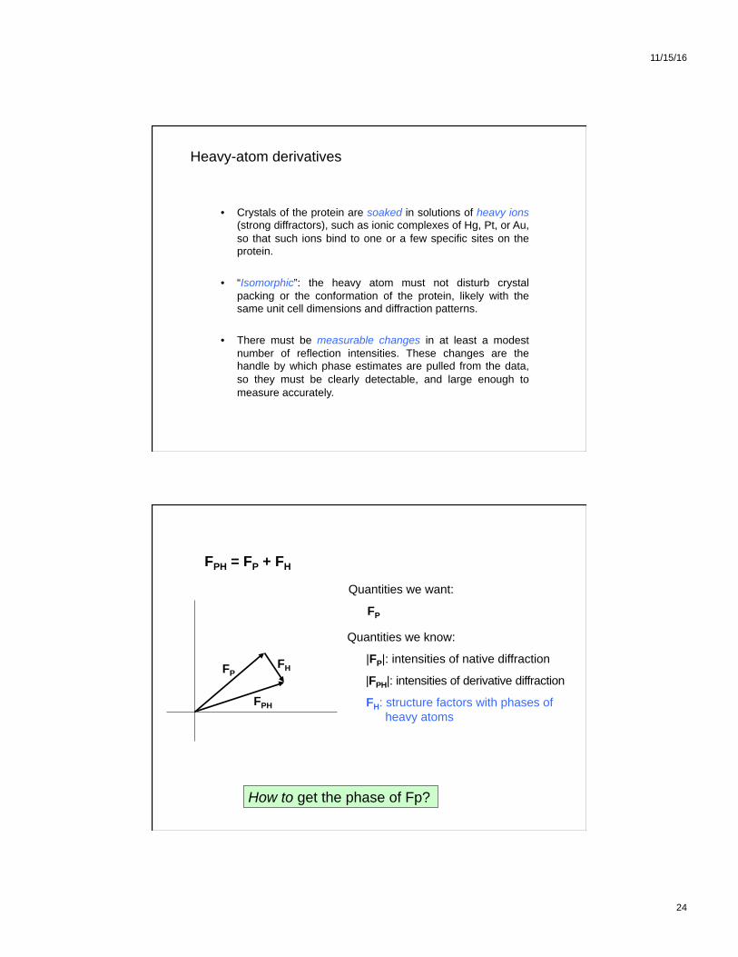
Heavy-atom derivatives
• Crystals of the protein are soaked in solutions of heavy ions (strong diffractors), such as ionic complexes of Hg, Pt, or Au, so that such ions bind to one or a few specific sites on the protein. !
• “Isomorphic”: the heavy atom must not disturb crystal packing or the conformation of the protein, likely with the same unit cell dimensions and diffraction patterns. !
• There must be measurable changes in at least a modest number of reflection intensities. These changes are the handle by which phase estimates are pulled from the data, so they must be clearly detectable, and large enough to measure accurately. !!!
FPH = FP + FH
FP FH
FPH
Quantities we want:
FP
Quantities we know:
|FP|: intensities of native diffraction
|FPH|: intensities of derivative diffraction
FH: structure factors with phases of heavy atoms
How to get the phase of Fp?

11/15/16
25
FPa
-FH
FPH
FPb
FP
How do we choose between FPa and FP
b?
FP = FPH – FH FP FH
FPH -FH
FPH
FPa
-FH’
FPH’
FPb
FP
Make a second derivative!
FP = FPH’ – FH’
∑ h
ρ(x, y, z) = FPb
e-2πi(hx + ky +lz) ∑ k ∑ l
1 V
α

11/15/16
26
FPH = FP + FH
FP FH
FPH
Quantities we want:
FP
Quantities we know:
|FP|: intensities of native diffraction
|FPH|: intensities of derivative diffraction
FH: structure factors with phases of heavy atoms
How to get FH?
ρH(x,y,z) requires knowing the position of heavy atoms in the unit cell.
Locating heavy atoms in the unit cell
FH(h,k,l) = ρH(x,y,z) e2πi(hx + ky +lz)dV
cell V ∫
The Patterson Function
By definition: P(u,v,w) = ρ(x,y,z)�ρ(x+u,y+v,z+w)dV
cell V ∫
x y (u v w)=(x1-x2,y1-y2,z1-z2) u v
(u v w) (x1 y1 z1)
(x2 y2 z2)

11/15/16
27
Patterson Function
x y u v
Patterson peaks will contain points corresponding to vectors between atoms in the real cell, i.e. inter-atomic distances (not atomic positions though).
Real Cell Patterson Cell
P(u,v,w) = ρ(x,y,z)�ρ(x+u,y+v,z+w)dV
cell V ∫
Patterson Function
Real Cell Patterson Cell
1) Patterson is symmetric about origin (centrosymmetry).
3) Contains N(N-1) non-origin peaks (not counting origin) à gets complicated!
2) Can see pattern of real cell in patterson cell repeated.

11/15/16
28
Patterson Function
Key point: can calculate P(u,v,w) from experimental data
P(u,v,w) = ρ(x,y,z) ρ(x+u,y+v,z+w)dV
cell V ∫
∑ h
ρ(x) = Fh e-2πihx 1 V
∑ h’
ρ(x+u) = Fh’ e-2πih’ (x+u) 1 V
∑ h
P(u) = Fh Fh’ e-2πihu ∑ h’
1 V2
e-2πi(h+h’)xdV
cell V ∫
The integration is equal to zero, unless h=h’ when it is equal to V,
By Friedel’s Law Fh=F-h,
∑ h
Fh2 e-2πihu 1
V P(u) =
Patterson Function
P(u,v,w) = |F(h,k,l)|2 cos 2π(hu + kv + lw) ∑h k l ∑∑
Patterson analysis is simplified for heavy atoms:
1) Use (|FPH(h k l)|- |FP(h k l)|)2 as coefficients
à “difference map” reflects heavy atom contribution
2) If atom i contains Zi electrons and atom j contains Zj electrons, the corresponding vector rij will have a weight proportional to ZiZj.
àheavy atoms, high Z, strong peaks 3) Calculate (x,y,z) of heavy atoms directly from Harker section peaks
1 V
It is a Fourier summation with intensities as coefficients and phase angles equal to zero.

11/15/16
29
Harker Peaks Symmetry related atoms give rise to peaks in Patterson map in specific locations. Each space group has its own Harker planes.
x
y
z
(x, y, z)
(-x, -y, z)
2 fold
(u,v,w) = (x-[-x], y-[-y], z-z)
(u,v,w) = (2x,2y, 0)
w=0
(2x,2y, 0)
u
v w=0 called Harker plane (section)
Native
A Hg derivative
Difference map

11/15/16
30