translation from the 5′ untranslated region shapes the...
TRANSCRIPT
RESEARCH ARTICLE SUMMARY◥
STRESS RESPONSE
Translation from the 5′ untranslatedregion shapes the integratedstress responseShelley R. Starck,* Jordan C. Tsai, Keling Chen, Michael Shodiya, Lei Wang,Kinnosuke Yahiro, Manuela Martins-Green, Nilabh Shastri,* Peter Walter*
INTRODUCTION: Protein synthesis is con-trolled by a plethora of developmental andenvironmental conditions. One intracellularsignaling network, the integrated stress re-sponse (ISR), activates one of four kinases inresponse to a variety of distinct stress stimuli:the endoplasmic reticulum (ER)–resident kinase(PERK), the interferon-induced double-strandedRNA–dependent eIF2a kinase (PKR), the gen-eral control nonderepressible 2 (GCN2), or theheme-regulated inhibitor kinase (HRI). Thesefour kinases recognize a central target andphosphorylate a single residue, Ser51, on thea subunit of the eukaryotic initiation factor 2(eIF2a), which is a component of the trimericinitiation factor eIF2 that catalyzes translationinitiation at AUG start codons. Phosphoryl-ation of eIF2a down-regulates eIF2-dependentprotein synthesis, which is important in devel-opment and immunity but also is implicated inneurodegeneration, cancer, and autoimmunity.However, protein synthesis does not cease on allmRNAs during the ISR. Rather, eIF2a phos-phorylation is required for expression of select
mRNAs, such as ATF4 and CHOP, that harborsmall upstream open reading frames (uORFs)in their 5′ untranslated regions (5′ UTRs). Stillother mRNAs sustain translation despite ISRactivation. We developed tracing translationby T cells (3T) as an exquisitely sensitive tech-nique to probe the translational dynamics ofuORFs directly during the ISR. With 3T, wemeasured the peptide products of uORFspresent in the 5′ UTR of the essential ER-resident chaperone, binding immunoglobulinprotein (BiP), also known as heat shock pro-tein family A member 5 (HSPA5), and charac-terized their requirement for BiP expressionduring the ISR.
RATIONALE:We repurposed the sensitivityand specificity of T cells to interrogate thetranslational capacity of RNA outside of an-notated protein coding sequences (CDSs). 3Trelies on insertion of a tracer peptide codingsequence into a candidate DNA sequence. Theresulting mRNAs harboring the nested tracerpeptide coding sequence are translated to pro-
duce tracer peptides. These translation prod-ucts are processed and loaded onto majorhistocompatibility complex class I (MHC I)molecules in the ER and transit to the cellsurface, where they can be detected by specificT cell hybridomas that are activated and quan-tified using a colorimetric reagent. 3T providesan approach to interrogate the thousands ofpredicteduORFs inmammaliangenomes, char-acterize the importance of uORF biology forregulation, and generate fundamental insightsinto uORF mutation-based diseases.
RESULTS: 3T proved to be a sensitive androbust indicator of uORF expression. We mea-sured uORF expression in the 5′ UTR ofmRNAs at multiple distinct regions, while
simultaneously detectingexpression of the CDS.Wedirectly measured uORFpeptide expression fromATF4mRNA and showedthat its translation per-sisted during the ISR.We
applied 3T to study BiP expression, an ERchaperone stably synthesized during the ISR.We showed that the BiP 5′ UTR harborsuORFs that are exclusively initiated by UUGand CUG start codons. BiP uORF expressionbypassed a requirement for eIF2 and was de-pendent on the alternative initiation factoreIF2A. Both translation of the UUG-initiateduORF and eIF2A were necessary for BiP ex-pression during the ISR. Unexpectedly, theproducts of uORF translation are predictedto generate MHC I peptides active in adaptiveimmunity. We propose that this phenomenonpresents an extracellular signature duringthe ISR.
CONCLUSION: Our findings introduce thenotion that cells harbor a distinct translationinitiation pathway to respond to a variety ofenvironmental conditions and cellular dys-function. We showed that cells utilize a dis-tinct, eIF2A-mediated initiation pathway, whichincludes uORF translation, to sustain expres-sion of particular proteins during the ISR. 3Toffers a valuable method to characterize thethousands of predicted translation eventsin 5′ UTRs and other noncoding RNAs and,expanded to a genome-wide scale, can com-plement ribosome profiling and mass spec-trometry in uORF and short ORF discovery.Our observations underscore the importanceof translation outside of annotated CDSsand challenge the very definition of the U in5′ UTR.▪
RESEARCH
SCIENCE sciencemag.org 29 JANUARY 2016 • VOL 351 ISSUE 6272 465
The list of author affiliations is available in the full article online.*Corresponding authors. E-mail: [email protected](S.R.S.); [email protected] (N.S.); [email protected] (P.W.) Cite this article as S. R. Starck et al., Science 351,aad3867 (2016). DOI: 10.1126/science.aad3867
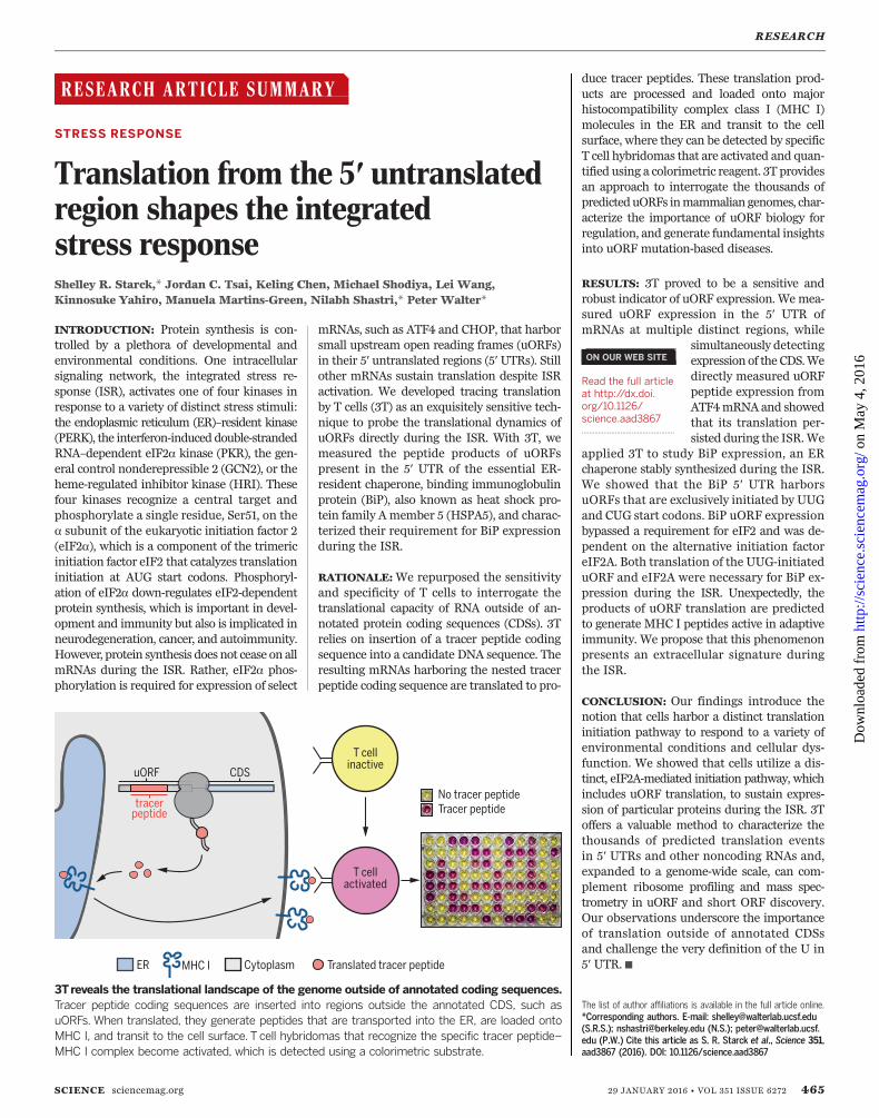
3Treveals the translational landscape of the genome outside of annotated coding sequences.Tracer peptide coding sequences are inserted into regions outside the annotated CDS, such asuORFs. When translated, they generate peptides that are transported into the ER, are loaded ontoMHC I, and transit to the cell surface. T cell hybridomas that recognize the specific tracer peptide–MHC I complex become activated, which is detected using a colorimetric substrate.
ON OUR WEB SITE◥
Read the full articleat http://dx.doi.org/10.1126/science.aad3867..................................................
on
May
4, 2
016
http
://sc
ienc
e.sc
ienc
emag
.org
/D
ownl
oade
d fr
om
RESEARCH ARTICLE◥
STRESS RESPONSE
Translation from the 5′ untranslatedregion shapes the integratedstress responseShelley R. Starck,1,2† Jordan C. Tsai,1 Keling Chen,2 Michael Shodiya,2 Lei Wang,3
Kinnosuke Yahiro,4 Manuela Martins-Green,3 Nilabh Shastri,2*† Peter Walter1*†
Translated regions distinct from annotated coding sequences have emerged as essentialelements of the proteome.This includes upstream open reading frames (uORFs) present inmRNAs controlled by the integrated stress response (ISR) that show “privileged” translationdespite inhibited eukaryotic initiation factor 2–guanosine triphosphate–initiator methionyltransfer RNA (eIF2·GTP·Met-tRNAi
Met).We developed tracing translation by Tcells to directlymeasure the translation products of uORFs during the ISR.We identified signature translationevents from uORFs in the 5′ untranslated region of binding immunoglobulin protein (BiP)mRNA(also called heat shock 70-kilodalton protein 5 mRNA) that were not initiated at the startcodon AUG. BiP expression during the ISR required both the alternative initiation factor eIF2Aand non–AUG-initiated uORFs.We propose that persistent uORF translation, for a variety ofchaperones, shelters selectmRNAs fromthe ISR,while simultaneously generatingpeptides thatcould serve asmajor histocompatibility complex class I ligands, marking cells for recognition bythe adaptive immune system.
Homeostatic mechanisms facilitate adapta-tion to a variety of environmental condi-tions and cellular dysfunction. The integratedstress response (ISR) is one such mecha-nism, triggered when cells encounter an
array of stress stimuli. These stimuli includemisfolded proteins, which elicit the unfolded pro-tein response (UPR) and thereby activate the endo-plasmic reticulum (ER)–resident kinase (PERK)(1–3). In addition, three related kinases are ac-tivated by other stimuli, such as the interferon-induced double-strandedRNA (dsRNA)–dependenteIF2a kinase (PKR) (by viral infection) (4, 5);the general control nonderepressible 2 (GCN2)(by amino acid deprivation) (6); and the heme-regulated inhibitor kinase (HRI) (by heme de-ficiency, oxidative stress, heat shock, or osmoticshock) (7). Each of these conserved kinases ini-tiate the ISR by phosphorylating the samesingle residue (Ser51) on the a subunit of eukary-otic initiation factor 2a (eIF2a) anddown-regulatetranslation initiation at AUG start codons bythe eukaryotic initiation factor 2–guanosinetriphosphate (GTP)–initiator methionyl trans-
ferRNA (tRNA) (eIF2·GTP·Met-tRNAiMet) ternary
complex. Phosphorylation of eIF2a (eIF2a-P) in-hibits exchange of guanosine diphosphate forGTP by eIF2B, the dedicated eIF2 guanine nu-cleotide exchange factor, which causes inhibitionof total protein synthesis (8). The blockade intranslation is important for cell survival and theeventual switch into apoptosis if homeostasiscannot be reestablished.Although eIF2a-P limits global translation, it
is required for the regulated expression of severalproteins, such as activating transcription factor 4(ATF4 or CREB-2) (9–11) and C/EBP homologousprotein (12, 13), that finely tune cell survival (14).These ISR-induced proteins are translated frommRNAs and harbor a series of upstream openreading frames (uORFs) in the 5′ untranslatedregion (5′ UTR) that limit ribosome access tothe main coding sequence (CDS), as first char-acterized in the budding yeast Saccharomycescerevisiae (15). According to the prevailing mod-el, under normal growth conditions, ribosome ini-tiation occurs predominantly at uORFs, whichprevents access to the downstream CDS. By con-trast, when the ISR is induced and eIF2a-P levelsrise, stochastic ribosome bypass of the uORFs al-lows access to the downstream CDS AUG startcodon.Another subset of mRNAs remains efficiently
translated during the ISR. These includemRNAsencoding heat shock and UPR proteins (1, 16–18)and a variety of inflammatory cytokines in re-sponse to viral (19, 20) and bacterial (21) patho-gens. In the context of the UPR, for example,translation of mRNAs encoding ER chaperones
is imperative to alleviate ER stress. BiP [im-munoglobulin heavy chain–binding protein, alsoknown as heat shock 70 kD protein (HSP70),heat shock protein family A member 5 (HSPA5),or glucose-regulated protein 78] is an essentialHSP70-type chaperone in the ER and is ex-pressed persistently during ER stress (22–24).It plays a role in cancer progression (25) and isa therapeutic target for a variety of diseases(26, 27). Yet, it has remained amystery how BiPand other stress-response mRNAs escape trans-lational down-regulation imposed by the ISR.Elements in the 5′ UTRs, including internal ri-bosome entry sites (IRESs), uORFs, and nucle-otide modifications, have all been suggested toconfer translational privilege to these mRNAs(28, 29).Recent genome-wide approaches predict that
nearly half of all mammalian mRNAs harboruORFs in their 5′UTRs, and many are initiatedwith non-AUG start codons (30–34). The pres-ence of uORFs in 5′ UTRs may reflect a generalmechanism to regulate downstream CDS expres-sion, such as proto-oncogenes and growth factors(30), as well as other disease-causing proteins(35), including hereditary thrombocythemia (36–38).Given the abundance of uORFs and their po-tential for regulatory roles, as well as the emer-ging plethora of short open reading frames(sORFs) (39–41) with bioactive properties (42),we developed a method to measure translationfrom RNA regions outside of annotated CDSssystematically.
Development of tracing translationby T cells (3T) to measure translationoutside of annotated coding sequences
Ribosome-profiling experiments reveal thatmRNAs encoding stress-response proteins har-bor a particularly high abundance of uORFs(43, 44). Yet, despite the thousands of peptidespredicted by ribosome profiling to be translatedfrom uORFs, very few uORF peptides have beenidentified by mass spectrometry (45). Currently,there is considerable effort to improve proteomicapproaches for the detection of peptides fromuORFs and other sORFs (39). Here, we exploitedthe exquisite sensitivity and specificity of T cellsto detect such translation products.We developed an approach, termed 3T, where
cells are supplied with DNA vectors containingnoncoding RNA elements, such as 5′ UTRs, har-boring sequences that encode tracer peptides(Fig. 1A). If the resulting RNA is translated, cellsproteolytically process the translated polypeptides.The resulting peptides are then transported bythe transporter associated with antigen process-ing (TAP) into the ER. In the ER, the peptides(also called antigens) are loaded onto majorhistocompatibility complex class I (MHC I) mol-ecules and displayed on the cell surface. Weassessed presentation of the translated tracerpeptide byMHC I by addition of a peptide–MHCI–cognate T cell hybridoma, engineered to ex-pressLacZ from the interleukin 2 (IL-2) promoterunder control of the N-FAT enhancer (46). En-gagement with tracer peptide–loaded MHC I
RESEARCH
SCIENCE sciencemag.org 29 JANUARY 2016 • VOL 351 ISSUE 6272 aad3867-1
1Department of Biochemistry and Biophysics, HowardHughes Medical Institute, University of California, SanFrancisco, CA 94143, USA. 2Division of Immunology andPathogenesis, Department of Molecular and Cell Biology,University of California, Berkeley, CA 94720, USA.3Department of Cell Biology and Neuroscience, University ofCalifornia, Riverside, CA 92521, USA. 4Departments ofMolecular Infectiology, Graduate School of Medicine, ChibaUniversity, Chiba, Japan.*These authors contributed equally to this work. †Correspondingauthors. E-mail: [email protected] (S.R.S.); [email protected] (N.S.); [email protected] (P.W.)
on
May
4, 2
016
http
://sc
ienc
e.sc
ienc
emag
.org
/D
ownl
oade
d fr
om
cells triggered LacZ expression, which resulted inproductionof its translationproduct,b-galactosidase.We detected b-galactosidase using its substrate,chlorophenol red–b-D-galactopyranoside (CPRG),
which yielded a red cleavage product. In theabsence of tracer peptide, the T cell hybridomadid not engage cells, and LacZ was transcrip-tionally inactive (Fig. 1A) (47).
To validate 3T, we tested the coding capacityof the activating transcription factor 4 (ATF4) 5′UTR, which contains well-characterized uORFs(fig. S1). To this end, we inserted tracer peptides
aad3867-2 29 JANUARY 2016 • VOL 351 ISSUE 6272 sciencemag.org SCIENCE
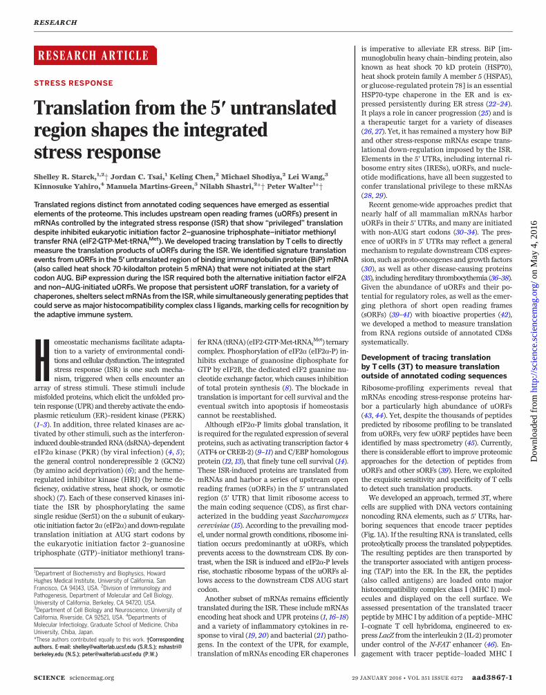
Fig. 1. 3T reveals the translational capacity of uORFs in 5′ UTRs. (A) Sche-matic of 3T: A tracer peptide inserted into noncoding regions of the 5′ UTR, suchas uORFs, followed by translation, peptide processing, and transport by TAP intothe ER for loading onto MHC I molecules. The tracer peptide–MHC I complextransits to the cell surface and is recognized by a Tcell hybridoma. Recognitionof the tracer peptide–MHC I complex on the cell surface activates the Tcellhybridoma, which is measured by a colorimetric assay and indicates thepresence of a uORF translation product. Schematics of the ATF4 5′ UTR withnested tracer peptides. Plotted Tcell responses and colorimetric readout of the3Tassay from tracer peptides generated from transfection of HeLa-Kb cells with
(B) uORF1(MYL8)–ATF4-luciferase detectedwith the BCZ103 Tcell hybridomaand (C) uORF2(KOVAK)–ATF4-luciferase detected with the B3Z Tcell hybridomawith simultaneous detection of ATF4-luciferase expression (D).Tcell responsesfrom tracer peptides generated from uORF1(WI9)–ATF4-luciferase, uORF1(WI9)-uORF2(KOVAK)–ATF4-luciferase,or uORF2(KOVAK)–ATF4-luciferase trans-fected (E) Db-L cells detected with the 11p9Z Tcell hybridoma or (F) Kb-L cellsdetected with the B3Z Tcell hybridoma. 3T responses from uORF2(KOVAK)–ATF4-luciferase (detected with the B3Z Tcell hybridoma) generated frommRNAtransfection of HeLa-Kb cells (G) and primary bone marrow–derived dendriticcells (H).Tcell responses are representative of n≥ 3. A595, absorption at 595 nm.
RESEARCH | RESEARCH ARTICLE
on
May
4, 2
016
http
://sc
ienc
e.sc
ienc
emag
.org
/D
ownl
oade
d fr
om
into two highly conserved uORFs that were pre-viously shown to regulate ATF4 expression inresponse to eIF2a phosphorylation (11, 48) (Fig.1, B and C, and fig. S1). To assess the levels ofuORF translation, we transfected cells harbor-ing the appropriate MHC I with uORF (tracerpeptide)–ATF4–luciferase constructs. We firstnested the coding sequence for the peptideMTFNYRNL (MYL8) (fig. S1) into uORF1 ofATF4-luciferase and the peptide KSIINFEHLK(KOVAK) (fig. S1) into uORF2. Both tracer pep-tides are presented byMHC I H-2Kb (KbMHC I).We then added, as a titration of increasing cellnumbers, an equal number of T cell hybridomasspecific for the tracer peptides displayed [BCZ103for uORF1(MYL8) or B3Z for uORF2(KOVAK)],followed by CPRG. As expected, T cell responseswere only observed when the tracer peptide waspresent in either uORF (Fig. 1, B and C).The tracer peptide constructs allowed robust
detection of translation from multiple different
regions of the mRNA. For example, we mea-sured ATF4-luciferase activity resulting fromCDS translation (Fig. 1D) from cells expressingtracer peptides from either uORF1 or uORF2(Fig. 1, B and C, and fig. S2). Similarly, using thetracer peptide WMHHNMDLI (WI9), presentedby MHC I H-2Db (Db MHC I) and detected by adifferent T cell hybridoma, wemeasured uORF1expression from either the uORF1(WI9) or theuORF1(WI9)-uORF2(KOVAK) constructs (Fig. 1E).As expected, uORF2(KOVAK) generated a pep-tide that was only detected by the appropriateT cell hybridoma (compare Fig. 1, E and F). Sim-ilarly, the KOVAK tracer peptide was detectedindependently of its placement in uORF1 oruORF2 (Fig. 1F).3T can be adapted for detection of tracer pep-
tides frommRNAs directly transfected into cells.In particular, we observed uORF2 tracer peptideexpression fromuORF2(KOVAK)–ATF4-luciferasemRNA (Fig. 1G). Furthermore, we readily ob-
served tracer peptide expression from uORF2from primary bone marrow–derived dendriticcells after only a 3-hour mRNA transfection(Fig. 1H). These observations underscore thepossibility that any mRNA and a diverse rangeof cell types can be used to measure uORF ex-pression, as long as the MHC I peptide presen-tation pathway is constitutively expressed inthese cells.For 3T to reflect expression from distinct
regions of the mRNA reliably, tracer peptideplacement should not deregulate the expressionof the main CDS during steady-state and stressconditions. Therefore, we tested CDS expression(ATF4-luciferase activity) from constructs bear-ing various uORF tracer peptide insertions. In-deed, insertion of tracer peptides into eitheruORF1 or uORF2 or simultaneously into bothuORFs did not substantially impair the inhibitoryfunction of the uORFs, as assessed by luciferaseactivity compared with wild type–ATF4-luciferase
SCIENCE sciencemag.org 29 JANUARY 2016 • VOL 351 ISSUE 6272 aad3867-3
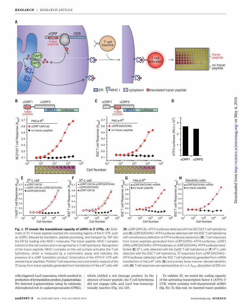
Fig. 2. uORFs are expressed during stress-induced expression of ATF4. (A) ATF4-luciferase levels during normal growth conditions were measured from HeLa-Kb
cells transfected with wild type–ATF4-luciferase, ATF4-luciferase constructs withtracer peptide insertions [uORF1(MYL8) or uORF2(KOVAK) tracer peptides], or aconstitutively active ATF4-luciferase variant (means ± SEM; n = 3 to 4). (B) Stress-induced ATF4-luciferase levels were measured from HeLa-Kb cells transfected withwild type–ATF4-luciferase and ATF4-luciferase constructs with tracer peptideinsertions [uORF1(MYL8) or uORF2(KOVAK) tracer peptides] and after treatmentswith tunicamycin (Tm) (1 mg/ml), Tg (1 mM), or Mutant SubAB or SubAB (0.2 mg/ml)for 6 hours (means ±SEM; n=3 to 4). HeLa-Kb cells from independent tracer peptide
DNA transfections were treated with NaAsO2 (10 mM) for 1 to 3 hours and analyzed by immunoblot (n = 3; *nonspecific ATF4-specific antibody signal) (C) or forluciferase expression from uORF2(KOVAK)–ATF4-luciferase–transfected cells (means ± SD; n = 3 to 4) (D).Tracer peptide expression wasmeasured fromHeLa-Kb
cells transfected with either uORF1(MYL8)–ATF4 luciferase (E) or uORF2(KOVAK)–ATF4 luciferase (F) after treatment with NaAsO2 (10 mM) (means ± SD fromtwo biological replicates are representative of n = 3). Stable uORF2(KOVAK)–ATF4-luciferase HeLa-Kb cells were treated for 3 hours with NaAsO2 (10 mM) andassayed for uORF2(KOVAK) tracer peptide expression (G) (means ± SD from two biological replicates are representative of n = 3) or for ATF4-luciferaseexpression (H) (means ± SEM; n = 3). Statistical significance was evaluated with the unpaired t test (NS, not significant; *P < 0.05; **P < 0.01; ***P < 0.001).
RESEARCH | RESEARCH ARTICLE
on
May
4, 2
016
http
://sc
ienc
e.sc
ienc
emag
.org
/D
ownl
oade
d fr
om
and a constitutively active variant of ATF4-luciferase (49) (Fig. 2A and fig. S3).To test whether control of ATF4 induction
by the ISR was likewise intact in the reportercell lines bearing the tracer peptide insertions,we exposed cells to subtilase cytotoxin (SubAB), abacterial AB toxin that is endocytosed by cellsand retrotransported to the ER lumen, where itdestroys BiP by proteolysis (50). BiP destructioninduced proteinmisfolding in the ER and PERK-
catalyzed phosphorylation of eIF2a, which inhib-ited cellular translation (fig. S4A). The translationblock was readily reversed by ISRIB, a small mol-ecule that overcomes the effects of eIF2a phos-phorylation (51–53) (fig. S4A). SubAB triggeredPERK activation, expression of endogenous ATF4,eIF2a phosphorylation (fig. S4B), andup-regulationof the plasmid-borne ATF4-luciferase reporter(fig. S4C). ISRIB inhibited endogenous ATF4expression and ATF4-luciferase expression, which
indicated that the ATF4 transgene behaved likeendogenous ATF4 (fig. S4). Furthermore,multipleUPR inducers stimulated ATF4-luciferase ex-pression equivalently from constructs harboringeither a uORF1 or uORF2 tracer peptide (Fig.2B). These results indicate that tracer peptideplacement does not compromise the control ofATF4 expression during ER stress.3T relies on proper processing of tracer pep-
tides, including import into the ER and loading
aad3867-4 29 JANUARY 2016 • VOL 351 ISSUE 6272 sciencemag.org SCIENCE
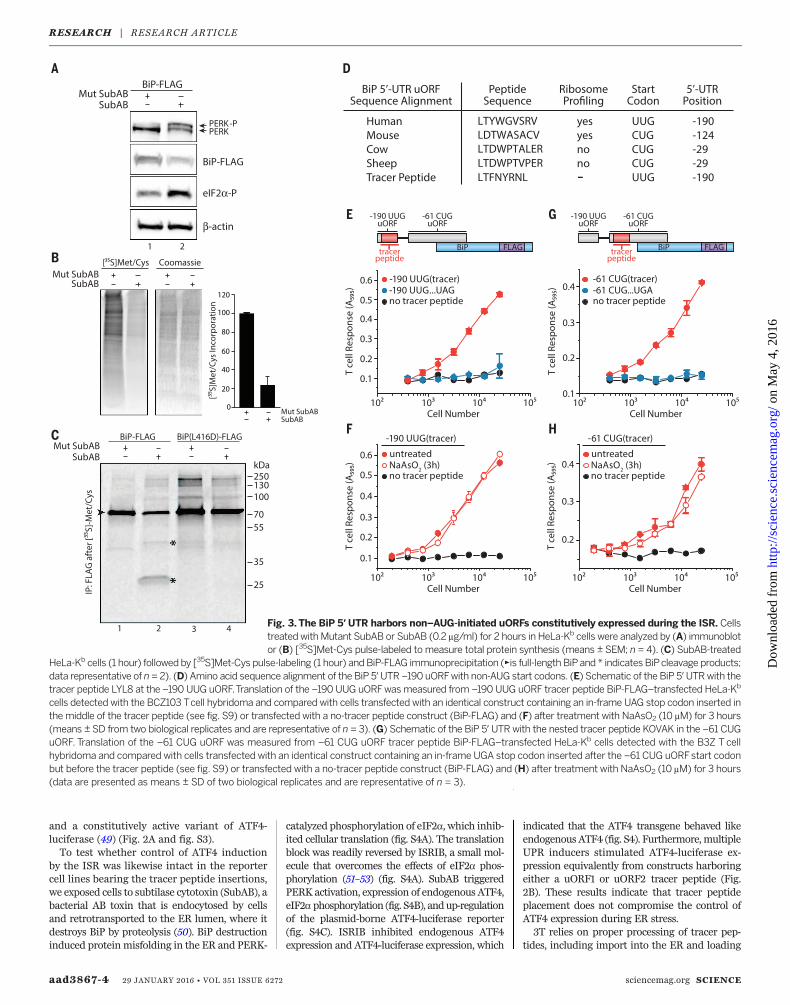
Fig. 3. The BiP 5′ UTR harbors non–AUG-initiated uORFs constitutively expressed during the ISR. Cellstreated withMutant SubAB or SubAB (0.2 mg/ml) for 2 hours in HeLa-Kb cells were analyzed by (A) immunoblotor (B) [35S]Met-Cys pulse-labeled to measure total protein synthesis (means ± SEM; n = 4). (C) SubAB-treated
HeLa-Kb cells (1 hour) followed by [35S]Met-Cys pulse-labeling (1 hour) and BiP-FLAG immunoprecipitation (►is full-length BiPand * indicates BiPcleavage products;data representative of n= 2). (D) Amino acid sequence alignment of the BiP 5′UTR –190 uORFwith non-AUG start codons. (E) Schematic of the BiP 5′UTRwith thetracer peptide LYL8 at the –190 UUG uORF.Translation of the –190 UUG uORFwasmeasured from –190 UUG uORF tracer peptide BiP-FLAG–transfected HeLa-Kb
cells detected with the BCZ103 Tcell hybridoma and compared with cells transfected with an identical construct containing an in-frame UAG stop codon inserted inthe middle of the tracer peptide (see fig. S9) or transfected with a no-tracer peptide construct (BiP-FLAG) and (F) after treatment with NaAsO2 (10 mM) for 3 hours(means ± SD from two biological replicates and are representative of n = 3). (G) Schematic of the BiP 5′ UTRwith the nested tracer peptide KOVAK in the –61 CUGuORF. Translation of the –61 CUG uORF was measured from –61 CUG uORF tracer peptide BiP-FLAG–transfected HeLa-Kb cells detected with the B3Z Tcellhybridoma and compared with cells transfected with an identical construct containing an in-frame UGA stop codon inserted after the –61 CUG uORFstart codonbut before the tracer peptide (see fig. S9) or transfected with a no-tracer peptide construct (BiP-FLAG) and (H) after treatment with NaAsO2 (10 mM) for 3 hours(data are presented as means ± SD of two biological replicates and are representative of n = 3).
RESEARCH | RESEARCH ARTICLE
on
May
4, 2
016
http
://sc
ienc
e.sc
ienc
emag
.org
/D
ownl
oade
d fr
om
onto MHC I molecules. To induce eIF2a phospho-rylationwithout disturbing the normal processesin the ER lumen, such as protein folding (com-promised by SubAB cleavage of BiP), or N-linkedglycosylation (inhibited by tunicamycin), or di-rectly activating the T cell hybridomas [calciumfluxes with thapsigargin (Tg)], we treated cellswithNaAsO2, an inducer of oxidative stress,whichrapidly induces eIF2a phosphorylation andATF4induction by activating cytosolic HRI (54). We ob-served rapid and robust expression of endogenousATF4 without activation of PERK or altered ex-pression of BiP (Fig. 2C and fig. S5). Pertinent totheutility of 3T,ATF4-luciferaseharboring anesteduORF2 tracer peptide showed the expected in-duction with NaAsO2 treatment (Fig. 2D). Pep-tide expression from uORF1 (Fig. 2E) and uORF2(Fig. 2, F to H, and fig. S6) largely persisted whenATF4-luciferase was induced, a finding that dif-fers from studies with yeast GCN4 (55). However,the pervasive uORF2peptide expressionmeasuredhere is consistent with recent ribosome-profilingstudies on ATF4 mRNA upon ISR induction(43, 44). These results validate 3T as a sensitiveand robust indicator of uORF expression. Assuch, we next applied this method to ask wheth-er uORF translation is a general strategy thatcells use to ensure privileged protein expressionduring the ISR.
3T reveals a novel regulatory elementin the 5′ UTR of BiP mRNA
BiP synthesis, which is initiated at a standardAUG start codon (fig. S7), persists during ERstress, when the ISR is induced (56). To explorethis phenomenon, we treated cells with SubAB,resulting in PERK activation, eIF2a phosphoryl-ation, and a massive reduction in protein syn-thesis (Fig. 3, A and B). As expected, treatmentwith a catalytically inactive form of SubAB (MutSubAB) did not result in ISR induction (Fig. 3, AandB, and fig. S8). To assess directly whether BiPis synthesized after onset of the ISR, we trans-fected cells with DNA encoding FLAG-taggedBiP [or BiP(L416D)-FLAG, a SubAB-resistantmutant] and treated them with Mut SubAB orSubAB. We next pulse-labeled cells with [35S]Met-Cys to label newly synthesized proteins,followed by immunoprecipitation of BiP. Inagreement with previous results, BiP from bothconstructs was readily synthesized despite ISRinduction (Fig. 3C). Thus, elements other thanconventional initiation at the annotated BiPAUG start codon or its CDS may be required toensure privileged expression during the UPR.Recent ribosome profiling measurements in-
dicate that BiPmRNA shows substantial levels ofribosome occupancy in the 5′UTR (33). Themostprominent ribosome initiation signal was de-tected at anUUG codon at position –190 (relativeto BiP coding sequence +1 AUG), encoding a pu-tative nine–amino acid peptide (Fig. 3D and fig.S9A). To test whether this predicted uORF istranslated, we nested a tracer peptide [LTFNYRNL(LYL8)] in the –190 UUG uORF (Fig. 3D), whichdid not alter the expression of BiP during basalconditions or its targeting to the ER as assayed
by SubAB sensitivity (fig. S10A). When assessedby 3T, we readily detected the –190 UUG leucinecodon–initiated expression of the tracer peptide(Fig. 3E). Translation of the –190 UUG uORFpersisted upon eIF2a phosphorylation inducedbyNaAsO2 treatment (Fig. 3F). Given that anUUGcodon codes for leucine and that leucine initiationis resistant to NaAsO2 treatment and reducedeIF2·GTP·Met-tRNAi
Met levels (57, 58), a non-canonical UUG or CUG initiation mechanismmay function in the BiP 5′ UTR.We similarly tested expression of the nested
KOVAK tracer peptide in the predicted –61 leu-cine CUG-initiated uORF in the BiP 5′ UTR (33)(Fig. 3G and fig. S9B). This uORF is out of framewith the BiP AUG-initiated CDS. Indeed, the –61CUG-initiated uORF supports expression of apeptide in cells when present out of frame (Fig.3G) or in frame with the BiP coding sequence(fig. S10, A and B). Expression of the –61 CUGuORF was not markedly reduced in the pres-ence of NaAsO2 (Fig. 3H), which suggests thatmultiple noncanonical initiation events are in-volved in sustaining BiP expression during stress.
BiP uORF expression is regulatedby eIF2A
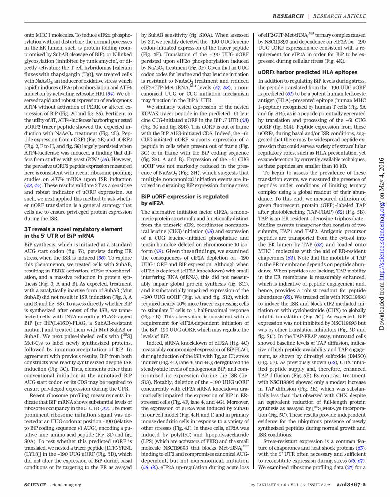
The alternative initiation factor eIF2A, a mono-meric protein structurally and functionally distinctfrom the trimeric eIF2, coordinates noncanon-ical leucine (CUG) initiation (58) and expressionof a CUG leucine–initiated phosphatase andtensin homolog deleted on chromosome 10 iso-form (59). Given these findings, we examinedthe consequences of eIF2A depletion on –190UUG uORF and BiP expression. Although wheneIF2A is depleted (eIF2A knockdown) with smallinterfering RNA (siRNA), this did not measur-ably impair global protein synthesis (fig. S11),and it substantially impaired expression of the–190 UUG uORF (Fig. 4A and fig. S12), whichrequired nearly 40%more tracer-expressing cellsto stimulate T cells to a half-maximal response(Fig. 4B). This observation is consistent with arequirement for eIF2A-dependent initiation ofthe BiP –190 UUGuORF, whichmay regulate thelevels of BiP.Indeed, siRNA knockdown of eIF2A (Fig. 4C)
measurably compromised expression of BiP-FLAG,during induction of the ISRwith Tg, an ER stressinducer (Fig. 4D, lane 4, and 4E); deregulated thesteady-state levels of endogenous BiP; and com-promised its expression during the ISR (fig.S13). Notably, deletion of the –190 UUG uORFconcurrently with eIF2A siRNA knockdown dra-matically impaired the expression of BiP in ER-stressed cells (Fig. 4F, lane 4, and 4G). Moreover,the expression of eIF2A was induced by SubABin our cell model (Fig. 4, H and I) and in primarymouse dendritic cells in response to a variety ofother stresses (Fig. 4J). In these cells, eIF2A wasinduced by poly(I:C) and lipopolysaccharide(LPS) (which are activators of PKR) and the smallmolecule NSC119893 that blocks Met-tRNAi
Met
binding to eIF2 and compromises canonical AUG-dependent, but not noncanonical, initiation(58, 60). eIF2A up-regulation during acute loss
of eIF2·GTP·Met-tRNAiMet ternary complex caused
by NSC119893 and dependence on eIF2A for –190UUG uORF expression are consistent with a re-quirement for eIF2A in order for BiP to be ex-pressed during cellular stress (Fig. 4K).
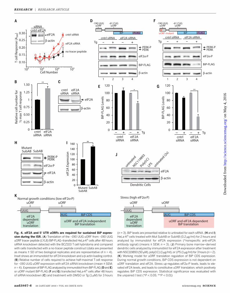
uORFs harbor predicted HLA epitopes
In addition to regulating BiP levels during stress,the peptide translated from the –190 UUG uORFis predicted (61) to be a potent human leukocyteantigen (HLA)–presented epitope (human MHCI–peptide) recognized by human T cells (Fig. 5Aand fig. S14), as is a peptide potentially generatedby translation and processing of the –61 CUGuORF (fig. S14). Peptide expression from theseuORFs, during basal and/or ISR conditions, sug-gested that there may be widespread peptide ex-pression that could serve a variety of extracellularregulatory roles, such as HLA presentation, yetescape detection by currently available techniques,as these peptides are smaller than 10 kD.To begin to assess the prevalence of these
translation events, we measured the presence ofpeptides under conditions of limiting ternarycomplex using a global readout of their abun-dance. To this end, we measured diffusion ofgreen fluorescent protein (GFP)–labeled TAPafter photobleaching (TAP-FRAP) (62) (Fig. 5B).TAP is an ER-resident adenosine triphosphate–binding cassette transporter that consists of twosubunits, TAP1 and TAP2. Antigenic precursorpeptides are transported from the cytosol intothe ER lumen by TAP (63) and loaded ontoMHC I molecules with the aid of ER-residentchaperones (64). Note that the mobility of TAPin the ERmembrane depends on peptide abun-dance. When peptides are lacking, TAP mobilityin the ER membrane is measurably enhanced,which is indicative of peptide engagement and,hence, provides a robust readout for peptideabundance (62). We treated cells with NSC119893to induce the ISR and block eIF2-mediated ini-tiation or with cycloheximide (CHX) to globallyinhibit translation (Fig. 5C). As expected, BiPexpression was not inhibited by NSC119893 butwas by other translation inhibitors (Fig. 5D andfig. S15). In the TAP-FRAP assay, untreated cellsshowed baseline levels of TAP diffusion, indica-tive of high peptide availability and TAP engage-ment, as shown by dimethyl sulfoxide (DMSO)(Fig. 5E). As previously shown (62), CHX inhib-ited peptide supply and, therefore, enhancedTAP diffusion (Fig. 5E). By contrast, treatmentwith NSC119893 showed only a modest increasein TAP diffusion (Fig. 5E), which was substan-tially less than that observed with CHX, despitean equivalent reduction of full-length proteinsynthesis as assayed by [35S]Met-Cys incorpora-tion (Fig. 5C). These results provide independentevidence for the ubiquitous presence of newlysynthesized peptides during normal growth andISR conditions.Stress-resistant expression is a common fea-
ture of chaperones and heat shock proteins (65),with the 5′ UTR often necessary and sufficientto reconstitute expression during stress (66, 67).We examined ribosome profiling data (33) for a
SCIENCE sciencemag.org 29 JANUARY 2016 • VOL 351 ISSUE 6272 aad3867-5
RESEARCH | RESEARCH ARTICLE
on
May
4, 2
016
http
://sc
ienc
e.sc
ienc
emag
.org
/D
ownl
oade
d fr
om
aad3867-6 29 JANUARY 2016 • VOL 351 ISSUE 6272 sciencemag.org SCIENCE
Fig. 4. eIF2A and 5′ UTR uORFs are required for sustained BiP expres-sion during the ISR. (A) Translation of the –190 UUG uORF from –190 UUGuORF tracer peptide (LYL8) BiP-FLAG–transfected HeLa-Kb cells after 48 hourssiRNA knockdown detected with the BCZ103 Tcell hybridoma and comparedwith cells transfected with a no-tracer peptide construct (data are presentedas means ± SD of two biological replicates and are representative of n = 4).Inset shows an immunoblot for eIF2A knockdown and a b-actin loading control.(B) Relative number of cells required to achieve half-maximal T cell responsefor –190 UUG uORFexpression with eIF2A siRNA knockdown (mean ± SEM;n = 4). ExpressionofBiP-FLAGanalyzedby immunoblot fromBiP-FLAG(DandE)or uORFmutant BiP-FLAG (F and G) transfected HeLa-Kb cells after 48 hoursof siRNA knockdown (C) and treatment with DMSO or Tg (1 mM) for 3 hours
(n = 3). BiP levels are presented relative to untreated for each siRNA. (H and I)HeLa-Kb cells treated with Mut SubAB or SubAB (0.2 mg/ml) for 2 hours andanalyzed by immunoblot for eIF2A expression (*nonspecific anti-eIF2Aantibody signal) (means ± SEM; n = 3). (J) Primary bone marrow–deriveddendritic cells analyzed by immunoblot for eIF2A expression after treatmentwithNSC119893 (50 mM), poly(I:C) (1 mg/ml), or LPS (1 mg/ml) for 3 hours (n=2).(K) Working model for uORF translation regulation of BiP CDS expression.During normal growth conditions, BiP CDS expression is not dependent onuORF translation and eIF2A. Stress up-regulates eIF2a-P levels, leads to ele-vated eIF2A levels, and leads to constitutive uORF translation, which positivelyregulates BiP CDS expression. Statistical significance was evaluated withthe unpaired t test (*P < 0.05; **P < 0.01)
RESEARCH | RESEARCH ARTICLE
on
May
4, 2
016
http
://sc
ienc
e.sc
ienc
emag
.org
/D
ownl
oade
d fr
om
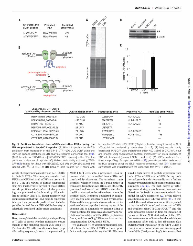
variety of chaperones to identify non-AUG uORFsin their 5′ UTRs. This analysis revealed thatUUG- and CUG-initiated uORFs are present inthe 5′ UTR of a variety of human chaperones(Fig. 5F). Furthermore, several of these uORFsencode peptides, which, after cellular process-ing, are predicted to be bound by HLA withstrong affinity (Fig. 5F). Taken together, ourresults suggest that the HLA peptide repertoireis larger than previously predicted and includespeptides derived from 5′UTRuORFs thatmay beselectively translated under stress conditions.
Discussion
Here, we exploited the sensitivity and specificityof T cells to assess whether translation occursoutside of the standard protein CDS using 3T.The basis for 3T is the insertion of a tracer pep-tide coding sequence, known to be presented by
MHC I to T cells, into a predefined DNA se-quence, which is transcribed into mRNA andtranslated by ribosomes. The translated tracerpeptides, whether nested in a polypeptide ortranslated from their own ORFs, are efficientlyprocessed and loaded onto MHC Imolecules inthe ER and travel to the cell surface, where thepeptide–MHC I complex is detected by exquis-itely specific and sensitive T cell hybridomas.This candidate approach allows customized in-sertion of tracer peptides into any region of thegenome and is amenable to genome tiling oftracer peptides for large-scale discovery and val-idation of translated uORFs, sORFs, protein iso-forms, and “noncoding” RNAs, such as introns,long noncoding RNAs, and 3′ UTRs.We validated 3T by detecting translated pep-
tides from the uORFs of ATF4, a transcriptionfactor only expressed during the ISR. We mea-
sured a high degree of peptide expression fromboth ATF4 uORF1 and uORF2 during bothnormal growth and stress conditions, a findingrecently predicted from ribosome profiling mea-surements (43, 44). The high degree of uORFexpression during stress, however, was not pre-dicted on the basis of the current reinitiationmodel for ATF4 expression (11) or the relatedyeast homolog GCN4 during stress (55). In thismodel, the small ribosomal subunit is expectedto remain mRNA bound and scans past uORF2and acquires eIF2·GTP·Met-tRNAi
Met and thelarge ribosomal subunit in time for initiation atthe conventional AUG start codon of the CDS.Ourmeasurements indicate either that reinitiationcommences after only a few ribosomes skipuORF2 or that translation of the ATF4 CDS is acombination of reinitiation and scanning pastthe uORFs (“leaky scanning”), two events that
SCIENCE sciencemag.org 29 JANUARY 2016 • VOL 351 ISSUE 6272 aad3867-7
Fig. 5. Peptides translated from uORFs and other RNAs during theISR are predicted to be MHC I peptides. (A) HLA epitope (human MHC I)prediction from translation of the BiP 5′ UTR –190 UUG uORF using theimmune epitope database (IEDB) analysis resource consensus tool (94).(B) Schematic for TAP diffusion [TAP1(GFP)-TAP2 complex] in the ER in thepresence or absence of peptides. (C) Meljuso cells stably expressing TAP1-GFP (62) treated for 1 hour with NSC119893 (25 mM) or CHX (50 mg/ml) andlabeled with 35S (n = 3) or (D) HeLa-Kb cells treated for 8 hours with
bruceantin (100 nM); NSC119893 (50 mM, replenished every 2 hours); or CHX100 mg/ml and analyzed by immunoblot (n = 3). (E) Meljuso cells stablyexpressing TAP1-GFP were treated with either NSC119893 or CHX for 1 hourand imaged using fluorescence confocal microscopy for lateral mobility ofTAP with treatment (means ± SEM; n = 4 to 7). (F) uORFs predicted fromribosome profiling of chaperone mRNAs (33) generate peptides predicted tobe HLA epitopes using the IEDB resource consensus tool (94). Statisticalsignificance was evaluated with the unpaired t test (***P < 0.001).
RESEARCH | RESEARCH ARTICLE
on
May
4, 2
016
http
://sc
ienc
e.sc
ienc
emag
.org
/D
ownl
oade
d fr
om
are technically challenging to distinguish (45).Our finding that uORFs are constitutively ex-pressed during stress and limiting eIF2·GTP·Met-tRNAi
Met levels prompts us to speculate thatuORF translation might serve an additional roleduring the ISR, which provides an explanationfor the high level of ribosome occupancy withinthe 5′ UTRs of mRNAs expressed during stress(43). Constitutive uORF expression could thus pro-tect mRNAs from translational down-regulationduring stress and thereby serve to control geneexpression.Using 3T, we showed that BiP harbors uORFs
that are constitutively translated during stress.By contrast to the AUG-initiated uORFs of ATF4,BiP 5′UTR uORFs are exclusively initiated by thenon-AUG leucine codons UUG and CUG. Expres-sion of the –190 UUG uORF from the BiP 5′UTRrequires the alternative initiation factor eIF2A.By contrast to initiation with the conventionaleIF2·GTP·Met-tRNAi
Met ternary complex, eIF2Ais a monomer and does not bind GTP (68). Inaddition to initiation at CUG start codons (58, 59),eIF2A is required for initiation at the AUG startcodon of a hepatitis C virus internal ribosomeentry site (IRES) reporter (69) and viral proteinsfrom the Sindbis alphavirus (70), during stressconditions and high levels of eIF2a phosphoryl-ation. Stress-induced eIF2A could substitute foreIF2 during initiation at the BiP AUG start codon.Our data support a model where eIF2A and acombination of uORFs maintain the levels ofBiP synergistically during translational attenua-tion accompanying cellular stress. We proposethat elevated levels of eIF2A, as we observed uponISR induction, protect BiP mRNA from trans-lational shutdown upon eIF2a phosphorylation.It was suggested previously that BiP mRNA
harbors an IRES in its 5′ UTR, required for BiPexpression during continuous heat shock (71, 72).Our data reveal an added level of regulationwithin the BiP 5′ UTR wherein persistent, non-canonical uORF translation and eIF2A functionas a cis-acting regulatory element necessary forprivileged BiP expression during stress. The rolefor eIF2A and noncanonical initiation in the 5′UTR during ribosome scanning of cellular mRNAsis an emerging property of translational regu-lation (59), which highlights a new translationalmechanism used during the integrated stress re-sponse. These findings add to a growing list ofalternative initiation events, such as initiation fromstructured 5′ UTRs with DEAD/DExH-box pro-teins (73, 74) or eIF2-independent recruitmentof Met-tRNAi
Met on viral mRNAs by Ligatin andMCT-1 or DENR (75). The observation that otherchaperones, also stably expressed during stress,harbor non–AUG-initiated uORFs in the 5′ UTRindicates that uORF expressionmay be a generalmechanism used to regulate the synthesis of pro-teins necessary during the ISR. Thus, substantialevidencenow suggests that uORFs are required forregulation of CDSs and that uORF mutations arelinked to a number of diseases [reviewed in (45)].The notion that some uORF peptides fall out-
side the detectable range of mass spectrometryanalysis because of their short length or selective
proteolysis (76) presents an advantage for 3T.The TAP channel readily transports short pep-tides into the ER for the peptide-loading complex,which is poised to efficiently facilitate loadingonto MHC I molecules. Therefore, the detectionof translated products from very short uORFs,such as ATF4 uORF1 (three amino acids), is idealfor 3T, because the entirety of the translateduORF serves to generate the mature peptidefor MHC I loading and subsequent detection byT cells. Additionally, the uORF products withshort half-lives that are efficiently processedserve to enhance the sensitivity of 3T, becauseproteolysis is required before TAP transport andMHC I loading. Last, the 3T assay is exquisitelysensitive because of the inherent sensitivity of Tcells that can detect even a few copies of thepeptide–MHC I and trigger the LacZ response(46, 77).The apparent disconnect between the number
of translated uORFs predicted by ribosome pro-filing (32, 33) and the number of translateduORFs identified by mass spectrometry is like-ly explained by the challenges encountered byextensive mass spectrometry analysis of HLA-bound peptides over the years (78, 79). TheseHLA-associated peptide analyses have revealedthat mass spectrometry analysis is unlikely tocapture all the different peptides presented byMHC I molecules for a variety of reasons. Earlyanalyses predicted that 10,000 to 20,000 dif-ferent peptides are presented on the cell sur-face by both MHC I and MHC II molecules,although recent estimates suggest that MHC Ialone could display between 30,000 and 120,000peptides on each cell surface (80). An analogousdisconnect is observed between the 42,271 humansORFs (40 to 100 amino acids long) predicted byribosome profiling (41) and the 1259 alternativeproteins detected by mass spectrometry (81). Ab-solute peptide abundance may limit the recoveryof some peptides if they are present at low copynumbers, such as 1 to 1000 molecules per cell(39, 78, 82). Furthermore, assignment of pep-tides from low-molecular-weight precursors (lessthan 100 amino acids) by mass spectrometry islimited by the presence of many unidentified ionpeaks and the lackof comprehensive low-molecular-weight reference databases (83), which is currentlybeing addressed (81, 83). In addition, standardassignment of recovered HLA-bound peptides istypically performed by alignmentwith the CDS ofthe mRNA (78, 79) not to regions outside theannotated open reading frames. Examples ofMHC I peptides generated from translation ofnoncoding RNA—such as 5′UTRs, introns, andintron-exon junctions (84)—highlight the needto explore regions outside of the CDS for sourcesof MHC I peptides and other bioactive pep-tides. The recent reports of non-AUG start codoninitiation (58, 59, 67) suggest that standard pep-tide alignments should also include ORFs ini-tiated with CUG, UUG, and other non-AUGstart codons, whether inside or outside of theannotated CDS (81).Discovery of uORF translation from the 5′
UTR of BiP and prediction of uORF translation
in other stress-related chaperones could gener-ate a pool of peptides that serve a variety of bio-logical functions. For example, uORF peptidesmay shape the immune response as self-antigensduring cancer progression and autoimmune dis-ease (80). Indeed, nonmutant tumor antigensgenerate physiologically relevant immune re-sponses for cancer immunotherapy (85). There-fore, a reexamination of HLA-associated peptidelibraries (78) promises to uncover peptides thatare generated from translation outside of anno-tated CDS, such as uORFs initiated with non-AUG start codons. Translated uORFs could alsogenerate bioactive peptides that directly or in-directly regulate expression of the main CDS, asseen with the peptide translated from the uORFin S-adenosylmethionine decarboxylase mRNA(86), or function in signaling pathways, such asToddler—a short, secreted peptide essential forembryogenesis (42).3T offers a robust approach for characteri-
zation of the numerous predicted translationevents in 5′ UTRs and other noncoding RNA(32, 33). These results emphasize the impor-tance of translated regulatory features in 5′ UTRs,which regulate translational control of the down-stream CDS, as indicated by a variety of disease-causing 5′ UTR uORF mutations (35). We proposethat this phenomenon presents an extracellularsignature during the ISR to modulate T cell im-mune responses (84).
REFERENCES AND NOTES
1. P. Walter, D. Ron, The unfolded protein response: From stresspathway to homeostatic regulation. Science 334, 1081–1086(2011). doi: 10.1126/science.1209038; pmid: 22116877
2. Y. Shi et al., Identification and characterization of pancreaticeukaryotic initiation factor 2 alpha-subunit kinase, PEK,involved in translational control. Mol. Cell. Biol. 18, 7499–7509(1998). doi: 10.1128/MCB.18.12.7499; pmid: 9819435
3. H. P. Harding, Y. Zhang, D. Ron, Protein translation and foldingare coupled by an endoplasmic-reticulum-resident kinase.Nature 397, 271–274 (1999). doi: 10.1038/16729;pmid: 9930704
4. J. Galabru, M. G. Katze, N. Robert, A. G. Hovanessian, Thebinding of double-stranded RNA and adenovirus VAI RNA tothe interferon-induced protein kinase. Eur. J. Biochem. 178,581–589 (1989). doi: 10.1111/j.1432-1033.1989.tb14485.x;pmid: 2912723
5. E. Meurs et al., Molecular cloning and characterization of thehuman double-stranded RNA-activated protein kinase inducedby interferon. Cell 62, 379–390 (1990). doi: 10.1016/0092-8674(90)90374-N; pmid: 1695551
6. J. J. Berlanga, J. Santoyo, C. De Haro, Characterization of amammalian homolog of the GCN2 eukaryotic initiation factor2alpha kinase. Eur. J. Biochem. 265, 754–762 (1999).doi: 10.1046/j.1432-1327.1999.00780.x; pmid: 10504407
7. L. Lu, A. P. Han, J. J. Chen, Translation initiation control byheme-regulated eukaryotic initiation factor 2alpha kinase inerythroid cells under cytoplasmic stresses. Mol. Cell. Biol. 21,7971–7980 (2001). doi: 10.1128/MCB.21.23.7971-7980.2001;pmid: 11689689
8. R. J. Jackson, C. U. Hellen, T. V. Pestova, The mechanism ofeukaryotic translation initiation and principles of its regulation.Nat. Rev. Mol. Cell Biol. 11, 113–127 (2010). doi: 10.1038/nrm2838; pmid: 20094052
9. T. W. Hai, F. Liu, W. J. Coukos, M. R. Green, Transcriptionfactor ATF cDNA clones: An extensive family of leucine zipperproteins able to selectively form DNA-binding heterodimers.Genes Dev. 3 (12B), 2083–2090 (1989). doi: 10.1101/gad.3.12b.2083; pmid: 2516827
10. P. D. Lu, H. P. Harding, D. Ron, Translation reinitiation atalternative open reading frames regulates gene expression inan integrated stress response. J. Cell Biol. 167, 27–33 (2004).doi: 10.1083/jcb.200408003; pmid: 15479734
aad3867-8 29 JANUARY 2016 • VOL 351 ISSUE 6272 sciencemag.org SCIENCE
RESEARCH | RESEARCH ARTICLE
on
May
4, 2
016
http
://sc
ienc
e.sc
ienc
emag
.org
/D
ownl
oade
d fr
om
11. K. M. Vattem, R. C. Wek, Reinitiation involving upstream ORFsregulates ATF4 mRNA translation in mammalian cells. Proc.Natl. Acad. Sci. U.S.A. 101, 11269–11274 (2004). doi: 10.1073/pnas.0400541101; pmid: 15277680
12. A. J. Fornace Jr., I. Alamo Jr., M. C. Hollander, DNA damage-inducible transcripts in mammalian cells. Proc. Natl. Acad. Sci.U.S.A. 85, 8800–8804 (1988). doi: 10.1073/pnas.85.23.8800;pmid: 3194391
13. D. Ron, J. F. Habener, CHOP, a novel developmentally regulatednuclear protein that dimerizes with transcription factors C/EBPand LAP and functions as a dominant-negative inhibitor ofgene transcription. Genes Dev. 6, 439–453 (1992).doi: 10.1101/gad.6.3.439; pmid: 1547942
14. J. Han et al., ER-stress-induced transcriptional regulationincreases protein synthesis leading to cell death. Nat. Cell Biol.15, 481–490 (2013). doi: 10.1038/ncb2738; pmid: 23624402
15. A. G. Hinnebusch, The scanning mechanism of eukaryotictranslation initiation. Annu. Rev. Biochem. 83, 779–812 (2014).doi: 10.1146/annurev-biochem-060713-035802;pmid: 24499181
16. G. Joslin, W. Hafeez, D. H. Perlmutter, Expression of stressproteins in human mononuclear phagocytes. J. Immunol. 147,1614–1620 (1991). pmid: 1880418
17. R. Panniers, Translational control during heat shock. Biochimie76, 737–747 (1994). doi: 10.1016/0300-9084(94)90078-7;pmid: 7893824
18. K. Richter, M. Haslbeck, J. Buchner, The heat shock response:Life on the verge of death. Mol. Cell 40, 253–266 (2010).doi: 10.1016/j.molcel.2010.10.006; pmid: 20965420
19. S. D. Der, A. S. Lau, Involvement of the double-stranded-RNA-dependent kinase PKR in interferon expression and interferon-mediated antiviral activity. Proc. Natl. Acad. Sci. U.S.A. 92,8841–8845 (1995). doi: 10.1073/pnas.92.19.8841;pmid: 7568028
20. F. D. Gilfoy, P. W. Mason, West Nile virus-induced interferonproduction is mediated by the double-stranded RNA-dependent protein kinase PKR. J. Virol. 81, 11148–11158(2007). doi: 10.1128/JVI.00446-07; pmid: 17686861
21. L.-C. Hsu et al., The protein kinase PKR is required formacrophage apoptosis after activation of Toll-like receptor 4.Nature 428, 341–345 (2004). doi: 10.1038/nature02405;pmid: 15029200
22. R. P. Shiu, J. Pouyssegur, I. Pastan, Glucose depletion accountsfor the induction of two transformation-sensitive membraneproteins[ ]in Rous sarcoma virus-transformed chick embryofibroblasts. Proc. Natl. Acad. Sci. U.S.A. 74, 3840–3844 (1977).doi: 10.1073/pnas.74.9.3840; pmid: 198809
23. I. G. Haas, M. Wabl, Immunoglobulin heavy chain bindingprotein. Nature 306, 387–389 (1983). doi: 10.1038/306387a0;pmid: 6417546
24. S. Munro, H. R. Pelham, An Hsp70-like protein in the ER:Identity with the 78 kd glucose-regulated protein andimmunoglobulin heavy chain binding protein. Cell 46, 291–300(1986). doi: 10.1016/0092-8674(86)90746-4; pmid: 3087629
25. J. Li, A. S. Lee, Stress induction of GRP78/BiP and its role incancer. Curr. Mol. Med. 6, 45–54 (2006). doi: 10.2174/156652406775574523; pmid: 16472112
26. M. S. Gorbatyuk, O. S. Gorbatyuk, The molecular chaperoneGRP78/BiP as a therapeutic target for neurodegenerativedisorders: A mini review. J. Genet. Syndr. Gene Ther. 4, 128(2013). doi: 10.4172/2157-7412.1000128; pmid: 23750325
27. L. Booth et al., GRP78/BiP/HSPA5/Dna K is a universaltherapeutic target for human disease. J. Cell. Physiol. 230,1661–1676 (2015). doi: 10.1002/jcp.24919; pmid: 25546329
28. C. U. Hellen, P. Sarnow, Internal ribosome entry sites ineukaryotic mRNA molecules. Genes Dev. 15, 1593–1612 (2001).doi: 10.1101/gad.891101; pmid: 11445534
29. J. Zhou et al., Dynamic m(6)A mRNA methylation directstranslational control of heat shock response. Nature 526,591–594 (2015). doi: 10.1038/nature15377; pmid: 26458103
30. S. E. Calvo, D. J. Pagliarini, V. K. Mootha, Upstream openreading frames cause widespread reduction of proteinexpression and are polymorphic among humans. Proc. Natl.Acad. Sci. U.S.A. 106, 7507–7512 (2009). doi: 10.1073/pnas.0810916106; pmid: 19372376
31. A. M. Resch, A. Y. Ogurtsov, I. B. Rogozin, S. A. Shabalina,E. V. Koonin, Evolution of alternative and constitutive regions ofmammalian 5′UTRs. BMC Genomics 10, 162 (2009).doi: 10.1186/1471-2164-10-162; pmid: 19371439
32. N. T. Ingolia, L. F. Lareau, J. S. Weissman, Ribosome profilingof mouse embryonic stem cells reveals the complexity anddynamics of mammalian proteomes. Cell 147, 789–802 (2011).doi: 10.1016/j.cell.2011.10.002; pmid: 22056041
33. S. Lee et al., Global mapping of translation initiation sites inmammalian cells at single-nucleotide resolution. Proc. Natl.Acad. Sci. U.S.A. 109, E2424–E2432 (2012). doi: 10.1073/pnas.1207846109; pmid: 22927429
34. N. T. Ingolia et al., Ribosome profiling reveals pervasivetranslation outside of annotated protein-coding genes. CellReports 8, 1365–1379 (2014). doi: 10.1016/j.celrep.2014.07.045; pmid: 25159147
35. M. Cazzola, R. C. Skoda, Translational pathophysiology: A novelmolecular mechanism of human disease. Blood 95,3280–3288 (2000).pmid: 10828006
36. T. Kondo et al., Familial essential thrombocythemia associatedwith one-base deletion in the 5′-untranslated region of thethrombopoietin gene. Blood 92, 1091–1096 (1998).pmid: 9694695
37. A. Wiestner, R. J. Schlemper, A. P. van der Maas, R. C. Skoda,An activating splice donor mutation in the thrombopoietin genecauses hereditary thrombocythaemia. Nat. Genet. 18, 49–52(1998). doi: 10.1038/ng0198-49; pmid: 9425899
38. N. Ghilardi, R. C. Skoda, A single-base deletion in thethrombopoietin (TPO) gene causes familial essentialthrombocythemia through a mechanism of more efficienttranslation of TPO mRNA. Blood 94, 1480–1482 (1999).pmid: 10484635
39. S. A. Slavoff et al., Peptidomic discovery of short open readingframe-encoded peptides in human cells. Nat. Chem. Biol. 9,59–64 (2013). doi: 10.1038/nchembio.1120; pmid: 23160002
40. S. J. Andrews, J. A. Rothnagel, Emerging evidence forfunctional peptides encoded by short open reading frames.Nat. Rev. Genet. 15, 193–204 (2014). doi: 10.1038/nrg3520;pmid: 24514441
41. V. Olexiouk et al., sORFs.org: A repository of small ORFsidentified by ribosome profiling. Nucleic Acids Res. gkv1175(2015). doi: 10.1093/nar/gkv1175; pmid: 26527729
42. A. Pauli et al., Toddler: An embryonic signal that promotes cellmovement via Apelin receptors. Science 343, 1248636 (2014).doi: 10.1126/science.1248636; pmid: 24407481
43. D. E. Andreev et al., Translation of 5′ leaders is pervasive ingenes resistant to eIF2 repression. eLife 4, e03971 (2015).doi: 10.7554/eLife.03971; pmid: 25621764
44. C. Sidrauski, A. M. McGeachy, N. T. Ingolia, P. Walter, The smallmolecule ISRIB reverses the effects of eIF2a phosphorylationon translation and stress granule assembly. eLife 4, (2015).doi: 10.7554/eLife.05033; pmid: 25719440
45. J. Somers, T. Pöyry, A. E. Willis, A perspective on mammalianupstream open reading frame function. Int. J. Biochem. CellBiol. 45, 1690–1700 (2013). doi: 10.1016/j.biocel.2013.04.020;pmid: 23624144
46. J. Karttunen, S. Sanderson, N. Shastri, Detection of rareantigen-presenting cells by the lacZ T-cell activation assaysuggests an expression cloning strategy for T-cell antigens.Proc. Natl. Acad. Sci. U.S.A. 89, 6020–6024 (1992).doi: 10.1073/pnas.89.13.6020; pmid: 1378619
47. Materials and methods are available as supplementarymaterials on Science Online.
48. H. P. Harding et al., An integrated stress response regulatesamino acid metabolism and resistance to oxidative stress. Mol.Cell 11, 619–633 (2003). doi: 10.1016/S1097-2765(03)00105-9;pmid: 12667446
49. L. M. Mielnicki, R. G. Hughes, P. M. Chevray, S. C. Pruitt,Mutated Atf4 suppresses c-Ha-ras oncogene transcript levelsand cellular transformation in NIH3T3 fibroblasts. Biochem.Biophys. Res. Commun. 228, 586–595 (1996). doi: 10.1006/bbrc.1996.1702; pmid: 8920955
50. A. W. Paton et al., AB5 subtilase cytotoxin inactivates theendoplasmic reticulum chaperone BiP. Nature 443, 548–552(2006). doi: 10.1038/nature05124; pmid: 17024087
51. C. Sidrauski et al., Pharmacological brake-release of mRNAtranslation enhances cognitive memory. eLife 2, e00498(2013). doi: 10.7554/eLife.00498; pmid: 23741617
52. C. Sidrauski et al., Pharmacological dimerization and activationof the exchange factor eIF2B antagonizes the integrated stressresponse. eLife 4, e07314 (2015). doi: 10.7554/eLife.07314;pmid: 25875391
53. Y. Sekine et al., Mutations in a translation initiation factoridentify the target of a memory-enhancing compound. Science348, 1027–1030 (2015). doi: 10.1126/science.aaa6986;pmid: 25858979
54. K. Zhan et al., Phosphorylation of eukaryotic initiation factor 2by heme-regulated inhibitor kinase-related protein kinases inSchizosaccharomyces pombe is important for resistance toenvironmental stresses. Mol. Cell. Biol. 22, 7134–7146 (2002).doi: 10.1128/MCB.22.20.7134-7146.2002; pmid: 12242291
55. J. P. Abastado, P. F. Miller, B. M. Jackson, A. G. Hinnebusch,Suppression of ribosomal reinitiation at upstream open readingframes in amino acid-starved cells forms the basis for GCN4translational control. Mol. Cell. Biol. 11, 486–496 (1991).pmid: 1986242
56. K. Gülow, D. Bienert, I. G. Haas, BiP is feed-back regulated bycontrol of protein translation efficiency. J. Cell Sci. 115,2443–2452 (2002). pmid: 12006628
57. S. R. Schwab, J. A. Shugart, T. Horng, S. Malarkannan,N. Shastri, Unanticipated antigens: Translation initiation at CUGwith leucine. PLOS Biol. 2, e366 (2004). doi: 10.1371/journal.pbio.0020366; pmid: 15510226
58. S. R. Starck et al., Leucine-tRNA initiates at CUG start codonsfor protein synthesis and presentation by MHC class I. Science336, 1719–1723 (2012). doi: 10.1126/science.1220270;pmid: 22745432
59. H. Liang et al., PTENa, a PTEN isoform translated throughalternative initiation, regulates mitochondrial function andenergy metabolism. Cell Metab. 19, 836–848 (2014).pmid: 24768297
60. F. Robert et al., Initiation of protein synthesis by hepatitis Cvirus is refractory to reduced eIF2·GTP·Met-tRNAi
Met ternarycomplex availability. Mol. Biol. Cell 17, 4632–4644 (2006).doi: 10.1091/mbc.E06-06-0478; pmid: 16928960
61. H. Rammensee, J. Bachmann, N. P. Emmerich, O. A. Bachor,S. Stevanović, SYFPEITHI: Database for MHC ligands andpeptide motifs. Immunogenetics 50, 213–219 (1999).doi: 10.1007/s002510050595; pmid: 10602881
62. E. A. Reits, J. C. Vos, M. Grommé, J. Neefjes, The majorsubstrates for TAP in vivo are derived from newly synthesizedproteins. Nature 404, 774–778 (2000). doi: 10.1038/35008103; pmid: 10783892
63. M. J. Androlewicz, P. Cresswell, How selective is thetransporter associated with antigen processing? Immunity 5,1–5 (1996). doi: 10.1016/S1074-7613(00)80304-0;pmid: 8758889
64. G. E. Hammer, T. Kanaseki, N. Shastri, The final touches makeperfect the peptide-MHC class I repertoire. Immunity 26,397–406 (2007). doi: 10.1016/j.immuni.2007.04.003;pmid: 17459809
65. K. R. Brandvold, R. I. Morimoto, The chemical biology ofmolecular chaperones—Implications for modulation ofproteostasis. J. Mol. Biol. 427, 2931–2947 (2015). doi: 10.1016/j.jmb.2015.05.010; pmid: 26003923
66. B. Wu, C. Hunt, R. Morimoto, Structure and expression of thehuman gene encoding major heat shock protein HSP70. Mol.Cell. Biol. 5, 330–341 (1985). doi: 10.1128/MCB.5.2.330;pmid: 2858050
67. X. Zhang et al., Translational control of the cytosolic stressresponse by mitochondrial ribosomal protein L18. Nat. Struct.Mol. Biol. 22, 404–410 (2015). pmid: 25866880
68. W. L. Zoll, L. E. Horton, A. A. Komar, J. O. Hensold,W. C. Merrick, Characterization of mammalian eIF2A andidentification of the yeast homolog. J. Biol. Chem. 277,37079–37087 (2002). doi: 10.1074/jbc.M207109200;pmid: 12133843
69. J. H. Kim, S. M. Park, J. H. Park, S. J. Keum, S. K. Jang, eIF2Amediates translation of hepatitis C viral mRNA under stressconditions. EMBO J. 30, 2454–2464 (2011). doi: 10.1038/emboj.2011.146; pmid: 21556050
70. I. Ventoso et al., Translational resistance of late alphavirusmRNA to eIF2alpha phosphorylation: A strategy to overcomethe antiviral effect of protein kinase PKR. Genes Dev. 20,87–100 (2006). doi: 10.1101/gad.357006; pmid: 16391235
71. D. G. Macejak, P. Sarnow, Internal initiation of translationmediated by the 5′ leader of a cellular mRNA. Nature 353,90–94 (1991). doi: 10.1038/353090a0; pmid: 1652694
72. Y. K. Kim, S. K. Jang, Continuous heat shock enhancestranslational initiation directed by internal ribosomal entry site.Biochem. Biophys. Res. Commun. 297, 224–231 (2002).doi: 10.1016/S0006-291X(02)02154-X; pmid: 12237106
73. M. C. Lai, Y. H. Lee, W. Y. Tarn, The DEAD-box RNA helicaseDDX3 associates with export messenger ribonucleoproteins aswell as tip-associated protein and participates in translationalcontrol. Mol. Biol. Cell 19, 3847–3858 (2008). doi: 10.1091/mbc.E07-12-1264; pmid: 18596238
74. V. P. Pisareva, A. V. Pisarev, A. A. Komar, C. U. Hellen,T. V. Pestova, Translation initiation on mammalian mRNAs withstructured 5′UTRs requires DExH-box protein DHX29. Cell 135,1237–1250 (2008). doi: 10.1016/j.cell.2008.10.037;pmid: 19109895
75. M. A. Skabkin et al., Activities of Ligatin and MCT-1/DENR ineukaryotic translation initiation and ribosomal recycling. Genes
SCIENCE sciencemag.org 29 JANUARY 2016 • VOL 351 ISSUE 6272 aad3867-9
RESEARCH | RESEARCH ARTICLE
on
May
4, 2
016
http
://sc
ienc
e.sc
ienc
emag
.org
/D
ownl
oade
d fr
om
Dev. 24, 1787–1801 (2010). doi: 10.1101/gad.1957510;pmid: 20713520
76. M. Oyama et al., Analysis of small human proteins reveals thetranslation of upstream open reading frames of mRNAs.Genome Res. 14 (10B), 2048–2052 (2004). doi: 10.1101/gr.2384604; pmid: 15489325
77. M. A. Purbhoo, D. J. Irvine, J. B. Huppa, M. M. Davis, T cellkilling does not require the formation of a stable matureimmunological synapse. Nat. Immunol. 5, 524–530 (2004).doi: 10.1038/ni1058; pmid: 15048111
78. D. F. Hunt et al., Characterization of peptides bound to the class IMHC molecule HLA-A2.1 by mass spectrometry. Science 255,1261–1263 (1992). doi: 10.1126/science.1546328;pmid: 1546328
79. H. Escobar et al., Large scale mass spectrometric profiling ofpeptides eluted from HLA molecules reveals N-terminal-extended peptide motifs. J. Immunol. 181, 4874–4882 (2008).doi: 10.4049/jimmunol.181.7.4874;pmid: 18802091
80. V. H. Engelhard, The contributions of mass spectrometry tounderstanding of immune recognition by T lymphocytes. Int. J.Mass Spectrom. 259, 32–39 (2007). doi: 10.1016/j.ijms.2006.08.009; pmid: 18167512
81. B. Vanderperre et al., Direct detection of alternative openreading frames translation products in human significantlyexpands the proteome. PLOS ONE 8, e70698 (2013).doi: 10.1371/journal.pone.0070698; pmid: 23950983
82. V. L. Crotzer et al., Immunodominance among EBV-derivedepitopes restricted by HLA-B27 does not correlate with epitopeabundance in EBV-transformed B-lymphoblastoid cell lines.J. Immunol. 164, 6120–6129 (2000). doi: 10.4049/jimmunol.164.12.6120; pmid: 10843661
83. G. Lubec, L. Afjehi-Sadat, Limitations and pitfalls in proteinidentification by mass spectrometry. Chem. Rev. 107,3568–3584 (2007). doi: 10.1021/cr068213f;pmid: 17645314
84. S. R. Starck, N. Shastri, Non-conventional sources of peptidespresented by MHC class I. Cell. Mol. Life Sci. 68, 1471–1479(2011). doi: 10.1007/s00018-011-0655-0;pmid: 21390547
85. D. J. Kowalewski et al., HLA ligandome analysis identifies theunderlying specificities of spontaneous antileukemia immuneresponses in chronic lymphocytic leukemia (CLL). Proc. Natl.Acad. Sci. U.S.A. 112, E166–E175 (2015). pmid: 25548167
86. G. L. Law, A. Raney, C. Heusner, D. R. Morris, Polyamineregulation of ribosome pausing at the upstream open reading
frame of S-adenosylmethionine decarboxylase. J. Biol. Chem.276, 38036–38043 (2001). pmid: 11489903
ACKNOWLEDGMENTS
We are grateful for technical help from K. Banta and S. J. Yang(University of California, Berkeley) and K. Crotty (University ofCalifornia, San Francisco). We thank M. Elvekrog, E. Costa, M. Lam,and W. Merrick for critical reading of the manuscript and C. Sidrauski,S. Ramundo, H. Tran, N. Ingolia, and J. Weissman for helpfuldiscussions and advice. The human retinal pigment epithelial cell line(RPE-19) was a gift from X. Gong (School of Optometry, University ofCalifornia, Berkeley). This research was supported by grants from theNIH to N.S. and P.W. P.W. is an Investigator of the Howard HughesMedical Institute.
SUPPLEMENTARY MATERIALS
www.sciencemag.org/content/351/6272/aad3867/suppl/DC1Material and MethodsFigs. S1 to S15References (87–98)
5 September 2015; accepted 3 December 201510.1126/science.aad3867
aad3867-10 29 JANUARY 2016 • VOL 351 ISSUE 6272 sciencemag.org SCIENCE
RESEARCH | RESEARCH ARTICLE
on
May
4, 2
016
http
://sc
ienc
e.sc
ienc
emag
.org
/D
ownl
oade
d fr
om
(6272), . [doi: 10.1126/science.aad3867]351Science Shastri and Peter Walter (January 28, 2016) Lei Wang, Kinnosuke Yahiro, Manuela Martins-Green, Nilabh Shelley R. Starck, Jordan C. Tsai, Keling Chen, Michael Shodiya,integrated stress response
untranslated region shapes the′Translation from the 5
Editor's Summary
, this issue p. 10.1126/science.aad3867Sciencegoing.were not down-regulated by stress. They identified a motif that helped keep chaperone protein synthesisdeveloped an approach that allowed them to look at the translation of specific messenger RNAs that
et al.down translation in general, but maintain or increase translation of specific proteins? Starck proteins that will help them cope with the misfolded proteins generated during stress. How do they turnturn down their rates of translation to help them survive the stress. They also turn on the translation of
When cells experience stresses that affect their ability to process newly synthesized proteins, theyHow cells keep going in the face of adversity
This copy is for your personal, non-commercial use only.
Article Tools
http://science.sciencemag.org/content/351/6272/aad3867article tools: Visit the online version of this article to access the personalization and
Permissionshttp://www.sciencemag.org/about/permissions.dtlObtain information about reproducing this article:
is a registered trademark of AAAS. ScienceAdvancement of Science; all rights reserved. The title Avenue NW, Washington, DC 20005. Copyright 2016 by the American Association for thein December, by the American Association for the Advancement of Science, 1200 New York
(print ISSN 0036-8075; online ISSN 1095-9203) is published weekly, except the last weekScience
on
May
4, 2
016
http
://sc
ienc
e.sc
ienc
emag
.org
/D
ownl
oade
d fr
om