normal spermatogenesis in fank1 (fibronectin type 3 and ... · normal spermatogenesis in fank1...
TRANSCRIPT

Normal spermatogenesis in Fank1(fibronectin type 3 and ankyrin repeatdomains 1) mutant miceJintao Zhang1,*, Xin Zhang1,*, Yue Zhang1, Wentao Zeng2,Shuqin Zhao2 and Mingxi Liu1
1 Department of Histology and Embryology, Nanjing Medical University, Nanjing, China2 Animal Core Facility of Nanjing Medical University, Nanjing, China* These authors contributed equally to this work.
ABSTRACTBackground: The fibronectin type 3 and ankyrin repeat domains 1 gene, Fank1, is anancient, evolutionarily conserved gene present in vertebrates. Short-hairpin RNA(shRNA)-based knockdown transgenic mice have oligospermia caused by an increase inapoptotic germ cells. In this study, we investigated the in vivo function of Fank1.Methods: In this study, we generated Fank1-knockout mice using the CRISPR/Cas9system. We then investigated the phenotype and in vivo function of Fank1.Testes and epididymis tissues were analyzed by histological and immunofluorescencestaining. Apoptotic cells were analyzed in terminal deoxynucleotidyl transferasedUTP nick end-labeling assays. Fertility and sperm counts were also evaluated.The GTEx database were used to assess gene expression quantitative trait loci andmRNA expression of candidate genes and genes neighboring single nucleotidepolymorphisms was analyzed by quantitative RT-PCR.Results: In contrast to the Fank1-knockdown model, no significant changesin epididymal sperm content and the number of apoptotic cells were observed inFank1-/- homozygotes. In addition, a different pattern of Dusp1, Klk1b21 andKlk1b27 mRNA expression was detected in Fank1-knockout testis. These results revealdifferences in the molecular changes between Fank1-knockdown mice andFank1-knockout mice and provide a basic resource for population genetics studies.
Subjects Andrology, HistologyKeywords Fank1, Male infertility, Gene knockout, Spermatogenesis
INTRODUCTIONGenetic studies are widely used for identification of susceptibility loci in human disease(Johnson & O’Donnell, 2009). Mouse models of gene editing are indispensable forinvestigations of gene function in vivo. However, the development of genetic research isrestricted by the lack of progress in our understanding of gene function. Thus, large-scaleknockout programs have been initiated to mutate all protein-encoding genes in themouse (Collins, Rossant &Wurst, 2007; Skarnes et al., 2011). The CRISPR/Cas9 system hasbeen used to target genomic loci in mammalian studies (Li et al., 2013; Mali, Esvelt &Church, 2013; Shen et al., 2013; Wang et al., 2013), and gene knockout mice have becomemore commonly used in genetic studies in mice. To date, 1,503 human diseases with one or
How to cite this article Zhang J, Zhang X, Zhang Y, Zeng W, Zhao S, Liu M. 2019. Normal spermatogenesis in Fank1 (fibronectin type 3and ankyrin repeat domains 1) mutant mice. PeerJ 7:e6827 DOI 10.7717/peerj.6827
Submitted 13 November 2018Accepted 21 March 2019Published 24 April 2019
Corresponding authorMingxi Liu, [email protected]
Academic editorPedro Silva
Additional Information andDeclarations can be found onpage 11
DOI 10.7717/peerj.6827
Copyright2019 Zhang et al.
Distributed underCreative Commons CC-BY 4.0

more mouse models have been recorded in the Mouse Genome Informatics database(Smith et al., 2018).
The fibronectin type 3 and ankyrin repeat domains 1 gene (Fank1) is an ancient,evolutionarily conserved gene present in vertebrates and expressed from the meiosisphase to the haploid phase of spermatogenesis in the testis (Zheng, Zheng & Yan, 2007).As a DNA binding protein, FANK1 recognizes the DNA sequence AAAAAG, and isimplicated as a transcription factor during spermatogenesis (Dong et al., 2014). In a studyof short-hairpin RNA (shRNA)-based knockdown transgenic mouse model, areduction in sperm number and an increase in apoptotic germ cells were observed(Dong et al., 2014).
In recent years, gene editing mouse models have played an indispensable role inelucidating gene function in vivo. A number of studies have revealed phenotypicdifferences between knockout (i.e., mutants) and knockdown (i.e., RNA interference)models (El-Brolosy & Stainier, 2017). These phenotypic differences could be caused bygene expression compensation in mutants or off-target effects of the knockdownreagents (El-Brolosy & Stainier, 2017). Both models have distinct advantages andlimitations for the elucidation of gene function. Gene knockdown is usually achievedwith exogenous reagents, for example morpholino, siRNA, shRNA and antisensemorpholino oligos (El-Brolosy & Stainier, 2017; Nasevicius & Ekker, 2000). Thesereagents usually induced sequence-dependent repression of the target gene in severaldifferent ways, such as mRNA degradation, translational inhibition or transcriptionalinhibition, which do not involve genomic mutation. However, gene knockouts aregenerated directly by genome editing and may be a better model of human geneticmutations.
Thus, in this study, we have generated a Fank1-mutant model using the CRISPR/Cas9system to investigate the phenotype and in vivo function of Fank1.
MATERIALS AND METHODSGene expression quantitative trait loci analysisThe publicly available RNA-seq and genotyping data of human samples from theGenotype-Tissue Expression project (GTEx, http://commonfund.nih.gov/GTEx/index)were used to assess gene expression quantitative trait loci (eQTL) for mRNA expression ofcandidate genes and genes neighboring single nucleotide polymorphisms (SNP).Statistical analysis was performed using The GTEx Consortium (2013).
Generation of Fank1-knockout mice by CRISPR/Cas9The mice were maintained and used in experiments according to the guidelines of theInstitutional Animal Care and Use Committee of Nanjing Medical University (China).The animal use protocol has been reviewed and approved by the Animal Ethical andWelfare Committee (Approval No. IACUC-1601117 and IACUC-1811001). Cas9 mRNAand single guide RNAs (sgRNAs) were produced and purified as previously described(Zhang et al., 2017). In brief, the Cas9 plasmid (Addgene, Watertown, MA, USA) was
Zhang et al. (2019), PeerJ, DOI 10.7717/peerj.6827 2/13

linearized by restriction enzyme digestion with AgeI and then purified using a MinElutePCR Purification Kit (Qiagen, Duesseldorf, Germany). Cas9 mRNA was produced byin vitro transcription using a mMESSAGE mMACHINE T7 Ultra Kit (Ambion, Austin,TX, USA) and purified using a RNeasy Mini Kit (Qiagen, Duesseldorf, Germany) accordingto the manufacturer’s instructions. The sgRNAs were designed on the basis of exon2of Fank1. The target sequence of sgRNA was 5′-GTGGCTTCGGTTCTCCATTGAGG-3′and 5′-GTCACCTTGCCCACAACAGGAGG-3′, respectively. The sgRNA plasmid waslinearized with DraI and then purified using a MinElute PCR Purification Kit(Qiagen, Duesseldorf, Germany). sgRNA was produced using the MEGA shortscript Kit(Ambion, Austin, TX, USA) and purified using the MEGA clear Kit (Ambion, Austin,TX, USA) according to the manufacturer’s instructions. Cas9 mRNA and sgRNAwere injected into mouse zygotes obtained by mating of wild-type C57BL/6 males withC57BL/6 superovulated females.
GenotypingEdited founders of Fank1 were identified by PCR amplification (Rapid Taq mastermix, Vazyme) with primers (Primer F 5′-GGTCCACAGTTGTTGTTGCT-3′ andR 5′-ATTCCAAGAGTCCATCGGTTCA-3′) and subcloned into pMD19-T (TaKaRa)followed by standard Sanger sequencing. The length of the corresponding wild-type andmutant alleles were 403 and 333 bp, respectively. The selected founder was crossedwith wild-type C57BL/6J to eliminate possible unwanted off-targets and to generate pureheterozygous mice. Fank1-/- mice were resequenced by Sanger sequencing and theresults were plotted using SnapGene (version 1.1.3). Genotyping was performedby agarose gel electrophoresis analysis of PCR products produced from DNA isolatedfrom tail biopsy specimens.
Western blot analysisTesticular protein extracts were prepared using lysis buffer [8 M urea, 75 mM NaCl,50 mM Tris-HCl, PH8.2] containing 1� cOmpleteTM EDTA-free Protease InhibitorCocktail (Roche, Basel, Switzerland). The proteins were separated by SDS-PAGE andtransferred onto a polyvinylidene difluoride membrane. The membranes were blockedwith 5% non-fat milk in TBS-T (20 mM Tris, 150 mM NaCl, 0.1% Tween 20) for 2 h atroom temperature and incubated overnight at 4 �C with the following primary detectionantibodies: anti-FANK1 (sc-398026; Santa Cruz Biotechnology, Santa Cruz, CA, USA)at a dilution of 1:1,000 and anti-β-actin (ac026, ABclonal, Wuhan, China) at a dilution of1:10,000. The membranes were washed with TBS-T buffer three times and incubatedat room temperature for 2 h with secondary detection antibodies at a dilution of 1:2,000.The signals from the detected proteins were visualized using High-sig ECL WesternBlotting Substrate (Tanon, Shanghai, China).
Histological analysisMouse testes or epididymis were collected from at least three mice for each genotype.The tissues were fixed in modified Davidson’s fluid for up to 24 h and stored in 70%
Zhang et al. (2019), PeerJ, DOI 10.7717/peerj.6827 3/13

ethanol. The samples were then dehydrated through a graded ethanol series and embeddedin paraffin. Tissue sections (thickness five mm) were prepared and mounted on glass slides.After deparaffinization, slides were stained with periodic acid Schiff for histologicalanalysis. Apoptotic cells in testis were detected using the terminal deoxynucleotidyltransferase dUTP nick end-labeling (TUNEL) assay (Vazyme, Nanjing, China) accordingto the manufacturer’s instructions.
Immunofluorescence analysisTestis sections were deparaffinized, rehydrated and boiled for 15 min in sodium citratebuffer for antigen retrieval. Sections were blocked in antibody dilution buffer (5% bovineserum albumin in phosphate-buffered saline (PBS) 137 mM NaCl; 2.7 mM KCl;10 mM Na2HPO4 and 2 mM KH2PO4) for 2 h at room temperature, followed by anovernight incubation at 4 �C with primary antibodies (list in Table S2). Three washeswith PBST (0.05% Tween 20 in PBS) were performed prior to incubation withsecondary antibody (list in Table S2) for 2 h at room temperature. Finally, sectionswere incubated with Hoechst 33342 (Invitrogen, Carlsbad, CA, USA) for 5 min andthen mounted. Images were captured using an LSM800 confocal microscope(Carl Zeiss AG, Jena, Germany).
Fertility testAdult males of each genotype were subjected to fertility tests. Each male was mated withthree wild-type C57BL/6 females, and the vaginal plug was checked every morning.The dates of birth and number of pups in each litter were recorded.
Computer-assisted sperm analysisMature sperm were obtained by making small incisions throughout the caudaepididymis, followed by extrusion and suspension in human tubal fluid culturemedium (In Vitro Care, Frederick, MD, USA). Sperm samples (10 ml) were used forcomputer-assisted semen analysis (Hamilton-Thorne Research, Inc., Beverly, MA, USA).Motile sperm number, progressive sperm number and sperm concentration for theexperimental and control groups were measured and analyzed.
Quantitative RT-PCR assayTotal RNA was extracted from the samples using TRIzol reagent (Invitrogen, Carlsbad,CA, USA). The concentration and purity of RNA were determined using a NanoDrop2000C (Thermo,Waltham, MA, USA) absorbance at 260/280 nm. Total RNA (one mg) wasreverse transcribed using a HiScript II Q RT SuperMix (Vazyme, R222, Nanjing,China) according to the manufacturer’s instructions. The cDNA (dilution 1:4) was thenanalyzed by quantitative RT-PCR in a typical reaction of 20 ml containing 250 nmol/l offorward and reverse primers, one ml cDNA and AceQ qPCR SYBR Green Master Mix(Vazyme, R222, Nanjing, China). The reaction was initiated by preheating at 50 �C for2 min, followed by 95 �C for 5 min and 40 amplification cycles of 10 s denaturationat 95 �C and 30 s annealing and extension at 60 �C. Gene expression was normalized to
Zhang et al. (2019), PeerJ, DOI 10.7717/peerj.6827 4/13

18 s within the log phase of the amplification curve. The primer sequences are listed inSupplementary Table S3 and Fig. S1.
Statistical analysisAll experiments were repeated at least three times. The differences between treatment andcontrol groups were analyzed using one-way ANOVA or unpaired two-tailed t-tests.P-values < 0.05 were considered to indicate statistical significance. All data represent themean ± the standard error of the mean. Analyses were performed using the MicrosoftExcel and GraphPad Prism 6.0.
RESULTSAssociation of 54 SNPs with Fank1 expression in humansGenome variants including common SNPs contribute to gene expression changes andare associated with human disease. To investigate the association of the genotypesof the SNPs with Fank1 mRNA expression, eQTL of Fank1 and relative SNPs.
Figure 1 The association of the genotypes of the SNPs with Fank1 mRNA expression. eQTL analysisof Fank1 mRNA expression level for genotypes Homo Ref, Het and Homo Alt at (A) rs3812681,(B) rs12770063, (C) rs35267061 and (D) rs61863578. Full-size DOI: 10.7717/peerj.6827/fig-1
Zhang et al. (2019), PeerJ, DOI 10.7717/peerj.6827 5/13
The eQTL data revealed lower Fank1 mRNA expression levels in testicular subsetswith homozygous genotypes of 54 SNPs compared with that of the homozygousreference (Table S1; Fig. 1). This result revealed the diversity of testicular Fank1expression levels in the population. Therefore, it is particularly important to study thephenotype of Fank1-deficient mice.
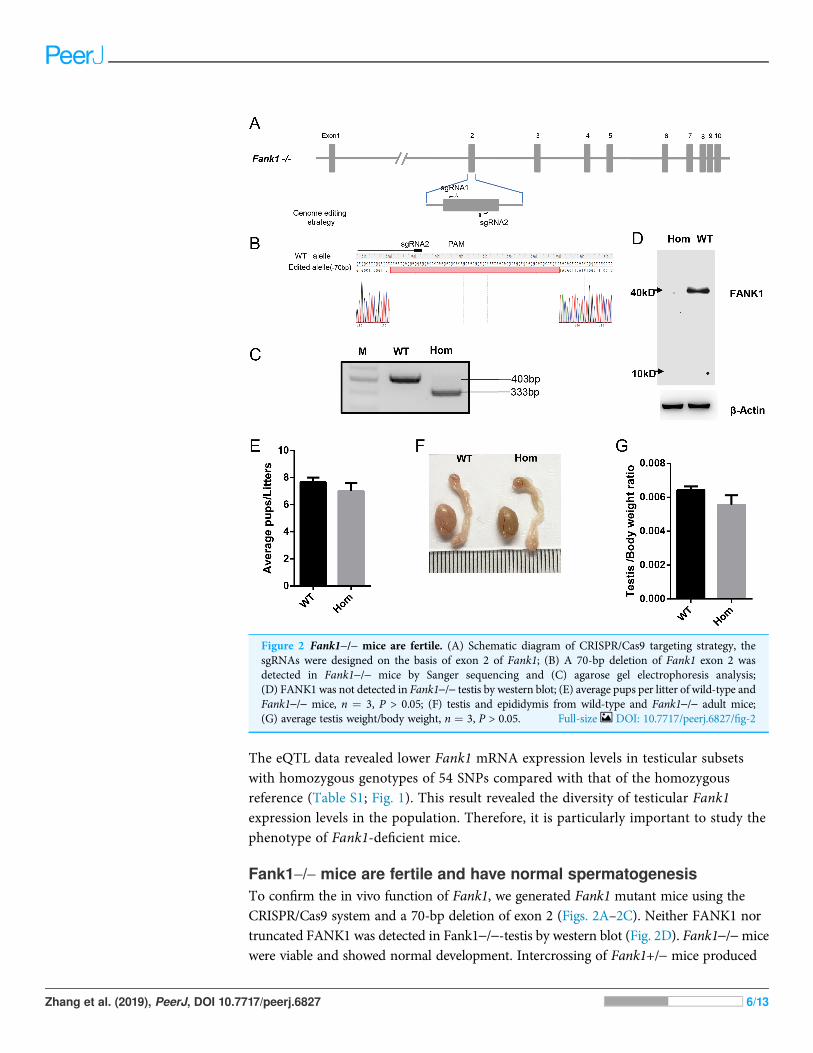
Fank1-/- mice are fertile and have normal spermatogenesisTo confirm the in vivo function of Fank1, we generated Fank1 mutant mice using theCRISPR/Cas9 system and a 70-bp deletion of exon 2 (Figs. 2A–2C). Neither FANK1 nortruncated FANK1 was detected in Fank1-/--testis by western blot (Fig. 2D). Fank1-/-micewere viable and showed normal development. Intercrossing of Fank1+/- mice produced
Figure 2 Fank1-/- mice are fertile. (A) Schematic diagram of CRISPR/Cas9 targeting strategy, thesgRNAs were designed on the basis of exon 2 of Fank1; (B) A 70-bp deletion of Fank1 exon 2 wasdetected in Fank1-/- mice by Sanger sequencing and (C) agarose gel electrophoresis analysis;(D) FANK1 was not detected in Fank1-/- testis by western blot; (E) average pups per litter of wild-type andFank1-/- mice, n ¼ 3, P > 0.05; (F) testis and epididymis from wild-type and Fank1-/- adult mice;(G) average testis weight/body weight, n ¼ 3, P > 0.05. Full-size DOI: 10.7717/peerj.6827/fig-2
Zhang et al. (2019), PeerJ, DOI 10.7717/peerj.6827 6/13

offspring of normal litter size at the predicted Mendelian and sex ratios. Similar to theFank1-knockdown model, Fank1-/- males were fertile (Fig. 2E). In adult Fank1-/- mice,the testes and epididymis were similar in size to those of the wild-type mice (Figs. 2Fand 2G). However, in contrast to the Fank1-knockdownmodel, histological analysis revealedthe presence of spermatogenic cells in the seminiferous tubules of adult Fank1-/- mice(Figs. 3 and 4). Furthermore, compared with the wild-type mice, there were no significantdifferences in the morphology of Fank1-/- spermatozoa found in the caudaepididymides (Figs. 5A–5C). The whole epididymal sperm content and the averagenumbers of motile sperm were unaffected in homozygotic male mice (Figs. 5D–5F).TUNEL analysis of testicular sections revealed that both the number of apoptotic cells pertubule and the number of tubules containing apoptotic cells were unaffected inhomozygotes (Fig. 6).
Figure 3 Spermatogenesis appears normal in Fank1-/- mice. Sections of periodic acid Schiff-stainedtestis from (A) wild-type and (B) Fank1-/- mice; Sections of hematoxylin and eosin-stained caudaepididymis from (C) wild-type and (D) Fank1-/- mice. Full-size DOI: 10.7717/peerj.6827/fig-3
Zhang et al. (2019), PeerJ, DOI 10.7717/peerj.6827 7/13

Expression changes in Fank1-/- testis are not consistent with those ofFank1-knockdown miceIt was reported that Dusp1, Klk1b21 and Klk1b27 were overexpressed in Fank1-knockdownmice and may be direct targets of Fank1 (Dong et al., 2014). However, in Fank1-/- testis,a reduction of Klk1b21 and Klk1b27 mRNA was detected but no increase in Dusp1transcripts (Fig. 7). These results reveal differences in the molecular changes ofFank1-knockdown and Fank1-knockout mice.
DISCUSSIONIn this study, we found that Fank1 mRNA expression levels correlated negatively withthe homozygous SNPs genotypes based on comparison with the GTEx database.This result showed the diversity of testicular Fank1 expression levels in the population
Figure 4 Spermatogenic markers appear normal in Fank1-/- mice. The spermatogonia (PLZF),spermatocytes (γ-H2AX) are comparable in testis sections from both (A) wild-type and (B) Fank1-/-mice; Spermatids (PNA) and Sertoli cells (Sox9) are comparable in testis sections from both (C) d-typeand (D) Fank1-/- mice. Full-size DOI: 10.7717/peerj.6827/fig-4
Zhang et al. (2019), PeerJ, DOI 10.7717/peerj.6827 8/13

and prompted us to study Fank1-knockout mice. This phenomenon was not detected instudies of another testicular-specific gene Pnldc1, which is an evolutionarily conservedgene and essential for male fertility (Zhang et al., 2017). One explanation for thisresult may be that Fank1 is dispensable for human reproduction. Thus, these geneticvariants were retained during evolution.
Unlike shRNA-based Fank1-knockdown mice, we generated Fank1 mutant miceusing the CRISPR/Cas9 system. We found that the expression of Fank1 was reduced byhalf in Fank1 mutant mice (Fig. 7; Fig. S1), which is possibly caused by nonsense-mediated mRNA decay. Neither FANK1 nor truncated FANK1 was detected inFank1 mutant testis. Similar to the Fank1-knockdown model, Fank1 mutant mice werefertile. Systematic studies have shown that neither testicular morphology nor spermfunction is affected in Fank1 mutant mice. In particular, the number of apoptotic cellswere unaffected in Fank1 mutant mice, while the number is markedly increased inFank1-knockdown model mice (Dong et al., 2014). Although knockdown modelsare known to differ from knockout models, more in-depth studies are required to clarifythese differences and their underlying mechanisms. The amino terminus of FANK1contains a fibronectin type III domain and the carboxyl terminus includes fiveankyrin repeats, which contain binding sites for DNA, heparin and the cell surface
Figure 5 Spermatozoa appear normal in Fank1-/- mice. (A) Hematoxylin and eosin-stained spermatozoa from wild-type and Fank1-/- mice;(B) fluorescence detection of AC-tubulin, PNA from wild-type and Fank1-/- spermatozoa; (C) cauda epididymal sperm contents from wild-typeand Fank1-/-mice, n¼ 3, P > 0.05; (D) average rate of motile sperm and (E) progressive sperm from wild-type and Fank1-/-mice, n¼ 3, P > 0.05;(F) abnormal epididymal sperm count from wild-type and Fank1-/- mice, n ¼ 3, P < 0.05. Full-size DOI: 10.7717/peerj.6827/fig-5
Zhang et al. (2019), PeerJ, DOI 10.7717/peerj.6827 9/13

(Skorstengaard et al., 1986). Ankyrin repeats have been found in proteins of diversefunction, such as transcriptional initiators and cell-cycle regulators (Skorstengaardet al., 1986). Lack of Fank1 leads to a reduction in Klk1b21 and Klk1b27 transcripts via amechanism that is unclear; however, transcriptional changes may also be inducedas a compensatory mechanism, thus accounting for the absence of fertility changes inFank1-/- males. In this study, we found no paralog of Fank1 which may compensatefor the Fank1 mutation. Thus, we cannot explain the mechanisms underlying thephenotypic differences between the Fank1 knockout and Fank1 knockdown mousemodels. Nevertheless, the Fank1 knockout mouse model generated in this studyprovides a basic resource for studies of population genetics, and also expandsour understanding of the differences in animal models established using differentapproaches.
Figure 6 Apoptotic cells are not increased in Fank1-/- testes. (A) TUNEL assay of wild-type andFank1-/- testes; (B) average apoptotic cells per seminiferous tubule; (C) average apoptotic cells perseminiferous tubules, n ¼ 3, P > 0.05. Full-size DOI: 10.7717/peerj.6827/fig-6
Zhang et al. (2019), PeerJ, DOI 10.7717/peerj.6827 10/13

CONCLUSIONSAlthough the diversity of testicular Fank1 expression levels was detected in the population,no significant changes in epididymal sperm content and the number of apoptotic cells wereobserved in Fank1-/- male mice. In addition, a different pattern of Dusp1, Klk1b21and Klk1b27 mRNA expression was detected in Fank1-knockout testis. These resultsreveal differences in the molecular changes between Fank1-knockdown mice andFank1-knockout mice and provide a basic resource for population genetics studies.
ACKNOWLEDGEMENTSWe thank Yiqiang Cui and Hao Wu for sgRNA design.
ADDITIONAL INFORMATION AND DECLARATIONS
FundingThis work was funded by the Natural Science Foundation of China (31571536 and31771654), The Natural Science Foundation of the Jiangsu Higher Education Institutionsof China (16KJA310003), the Natural Science Foundation of Jiangsu Province(No. BK20150990) and the Qing Lan Project. The funders had no role in study design,data collection and analysis, decision to publish, or preparation of the manuscript.
Grant DisclosuresThe following grant information was disclosed by the authors:Natural Science Foundation of China: 31571536 and 31771654.The Natural Science Foundation of the Jiangsu Higher Education Institutions of China:16KJA310003.Natural Science Foundation of Jiangsu Province: BK20150990.Qing Lan Project.
Klkb21
Klkb27
Dusp1
Fank1
0 .0
0 .5
1 .0
1 .5 W T
Hom
**
*****
Figure 7 Expression changes in Fank1-/- testis. Quantitative RT-PCR analysis of Dusp1, Klk1b21,Klk1b27 and Fank1 in testis, n ¼ 3, ��P < 0.01; ���P < 0.001. Full-size DOI: 10.7717/peerj.6827/fig-7
Zhang et al. (2019), PeerJ, DOI 10.7717/peerj.6827 11/13

Competing InterestsThe authors declare that they have no competing interests.
Author Contributions� Jintao Zhang performed the experiments, analyzed the data, contributed reagents/materials/analysis tools, prepared figures and/or tables, authored or reviewed drafts ofthe paper.
� Xin Zhang performed the experiments, analyzed the data, contributed reagents/materials/analysis tools, prepared figures and/or tables.
� Yue Zhang performed the experiments, analyzed the data.� Wentao Zeng performed the experiments.� Shuqin Zhao performed the experiments.� Mingxi Liu conceived and designed the experiments, prepared figures and/or tables,authored or reviewed drafts of the paper, approved the final draft.
Animal EthicsThe following information was supplied relating to ethical approvals (i.e., approving bodyand any reference numbers):
The mice were maintained and used in experiments according to the guidelines of theInstitutional Animal Care and Use Committee of Nanjing Medical University (China).The animal use protocol has been reviewed and approved by the Animal Ethical andWelfare Committee (Approval No. IACUC-1601117 and IACUC-1811001).
Data AvailabilityThe following information was supplied regarding data availability:
The raw measurements are available in the Supplemental Files.
Supplemental InformationSupplemental information for this article can be found online at http://dx.doi.org/10.7717/peerj.6827#supplemental-information.
REFERENCESCollins FS, Rossant J, Wurst W. 2007. A mouse for all reasons. Cell 128(1):9–13
DOI 10.1016/j.cell.2006.12.018.
Dong W-W, Huang H-L, Yang W, Liu J, Yu Y, Zhou S-L, Wang W, Lv X-C, Li Z-Y,Zhang M-Y, Zheng Z-H, Yan W. 2014. Testis-specific Fank1 gene in knockdown miceproduces oligospermia via apoptosis. Asian Journal of Andrology 16(1):124–130DOI 10.4103/1008-682X.122592.
El-Brolosy MA, Stainier DYR. 2017. Genetic compensation: a phenomenon in search ofmechanisms. PLOS Genetics 13(7):e1006780 DOI 10.1371/journal.pgen.1006780.
Johnson AD, O’Donnell CJ. 2009. An open access database of genome-wide association results.BMC Medical Genetics 10(1):6 DOI 10.1186/1471-2350-10-6.
Li D, Qiu Z, Shao Y, Chen Y, Guan Y, Liu M, Li Y, Gao N, Wang L, Lu X, Zhao Y, Liu M. 2013.Heritable gene targeting in the mouse and rat using a CRISPR-Cas system. Nature Biotechnology31(8):681–683 DOI 10.1038/nbt.2661.
Zhang et al. (2019), PeerJ, DOI 10.7717/peerj.6827 12/13

Mali P, Esvelt KM, Church GM. 2013. Cas9 as a versatile tool for engineering biology.Nature Methods 10(10):957–963 DOI 10.1038/nmeth.2649.
Nasevicius A, Ekker SC. 2000. Effective targeted gene ‘knockdown’ in zebrafish. Nature Genetics26(2):216–220 DOI 10.1038/79951.
Shen B, Zhang J, Wu H, Wang J, Ma K, Li Z, Zhang X, Zhang P, Huang X. 2013. Generation ofgene-modified mice via Cas9/RNA-mediated gene targeting. Cell Research 23(5):720–723DOI 10.1038/cr.2013.46.
Skarnes WC, Rosen B, West AP, Koutsourakis M, Bushell W, Iyer V, Mujica AO, Thomas M,Harrow J, Cox T, Jackson D, Severin J, Biggs P, Fu J, Nefedov M, de Jong PJ, Stewart AF,Bradley A. 2011. A conditional knockout resource for the genome-wide study of mousegene function. Nature 474(7351):337–342 DOI 10.1038/nature10163.
Skorstengaard K, Jensen MS, Sahl P, Petersen TE, Magnusson S. 1986. Complete primarystructure of bovine plasma fibronectin. European Journal of Biochemistry 161:441–453DOI 10.1111/j.1432-1033.1986.tb10464.x.
Smith CL, Blake JA, Kadin JA, Richardson JE, Bult CJ, the Mouse Genome Database Group.2018. Mouse Genome Database (MGD)-2018: knowledgebase for the laboratory mouse.Nucleic Acids Research 46(D1):D836–D842 DOI 10.1093/nar/gkx1006.
The GTEx Consortium. 2013. The Genotype-Tissue Expression (GTEx) project. Nature Genetics45(6):580–585 DOI 10.1038/ng.2653.
Wang H, Yang H, Shivalila CS, Dawlaty MM, Cheng AW, Zhang F, Jaenisch R. 2013. One-stepgeneration of mice carrying mutations in multiple genes by CRISPR/Cas-mediated genomeengineering. Cell 153(4):910–918 DOI 10.1016/j.cell.2013.04.025.
Zhang Y, Guo R, Cui Y, Zhu Z, Zhang Y, Wu H, Zheng B, Yue Q, Bai S, Zeng W, Guo X,Zhou Z, Shen B, Zheng K, Liu M, Ye L, Sha J. 2017. An essential role for PNLDC1 inpiRNA 3’ end trimming and male fertility in mice. Cell Research 27(11):1392–1396DOI 10.1038/cr.2017.125.
Zheng Z, Zheng H, Yan W. 2007. Fank1 is a testis-specific gene encoding a nuclear proteinexclusively expressed during the transition from the meiotic to the haploid phase ofspermatogenesis. Gene Expression Patterns 7(7):777–783 DOI 10.1016/j.modgep.2007.05.005.
Zhang et al. (2019), PeerJ, DOI 10.7717/peerj.6827 13/13