normal e na hipertensÃo portal - sociedade catarinense de...
TRANSCRIPT

Túlio A. A. Macedo
DOPPLER HEPÁTICONORMAL E NA
HIPERTENSÃO PORTAL
Universidade Federal de Uberlândia


HIPERTENSÃO PORTAL
TÚLIO MACEDO - [email protected]
• Aumento da pressão no sistema portal hepático (≥ 12 mmHg)
• Tipos
• Pré-sinusoidal
• Trombose da veia porta
• Esquistosomose
• Sinusoidal
• Cirrosis
• Pós-sinusoidal
• Budd-Chiari
• D. veno-oclusiva, ICC

SINAIS DOPPLER DE HIPERTENSÃO PORTAL
• Veia porta
• VP dilatada
• Diminuição da velocidade média (<15 cm/s)
• Fluxo "vai e vem"/ hepatofugal
• Aumento da pulsatilidade (VPI)
• Fístula arterio-portal
• Veia hepática
• Fluxo pseudoportal
• Arteria hepática
• Ampliação e tortuosidade
• Aumento de IR e IP
• Colaterais portossistêmicas
Harkanyi Z. Ultrasound Clin 2006 ; 1 : 443 – 455.

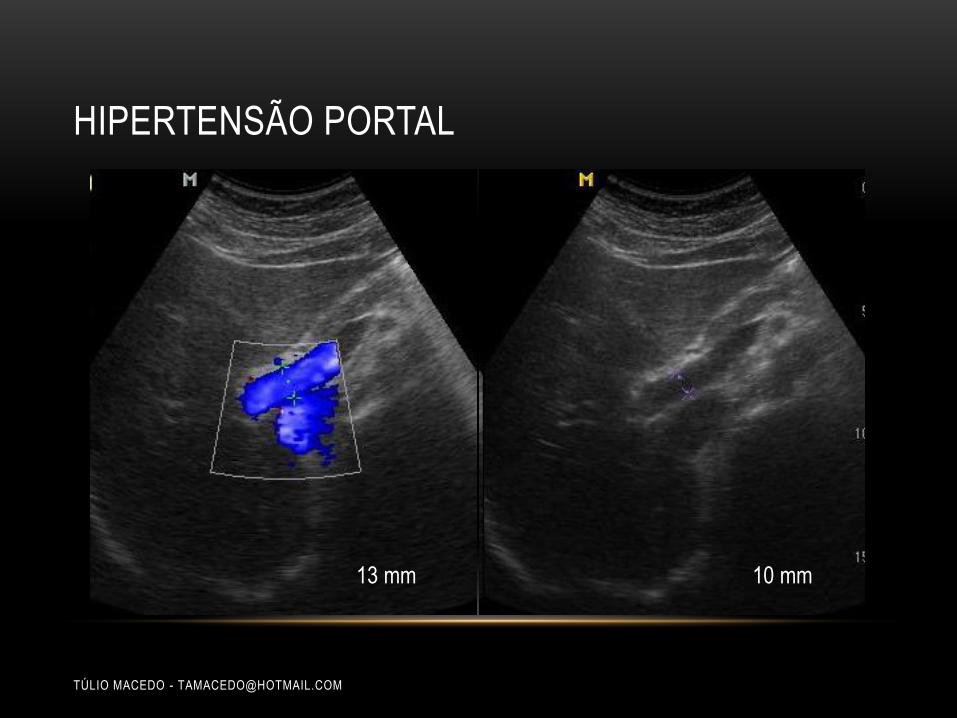
DIÂMETRO DA VEIA PORTA
1 Chammas et al. Ultrassonografia abdominal, 2009.2 Goyal AK et al. J Ultrasound Med 1990 ; 9 : 45 – 48.
Robinson KA et al. Ultrasound Quarterly 2009 ; 25 : 3 – 13.
Diâmetro: 17 mm
Sinal de hipertensão portal
Contovérsia no diâmetro normal da VP
Até 12 mm1
Até 16 mm2
Tamanho normal da VP: não exclui HPP


VELOCIDADE DA VEIA PORTA
Velocidade baixa: bom indicador da
hipertensão portal
Velocidade normal: não exclui a HP
Controvérsia sobre a velocidade da VP
Reprodutibilidade prejudicada
Velocidade média normal: 15 a 18 cm / seg
Swart J et al. Ultrasound Clin 2007 ; 2 : 355 – 375.
Fígado reduzida e contornos
lobulados
Vmed: 10 cm / seg

HIPERTENSÃO PORTAL E O FLUXO DA VEIA PORTA
Fluxo normal
Kok Th et al. Scand J Gastroenterol 1999 ; 34 (Suppl 230) : 82 – 88.
Fluxo invertido
HP avançada
Shunt porto-sistêmico
Fluxo “To and fro”
Hipertensão portal
avançada
Insuficiência cardíaca
Fístula arterio-portal

“TO-AND-FRO FLOW” - FLUXO TIPO "VAI E VEM"
Fluxo hepatopetal e hepatofugal a cada batimento cardíaco
Estágio prévio ao fluxo hepatofugal
Wachsberg RH et al. RadioGraphics 2002 ; 22 : 123 – 140.
Estudo Doppler realizado em apneia
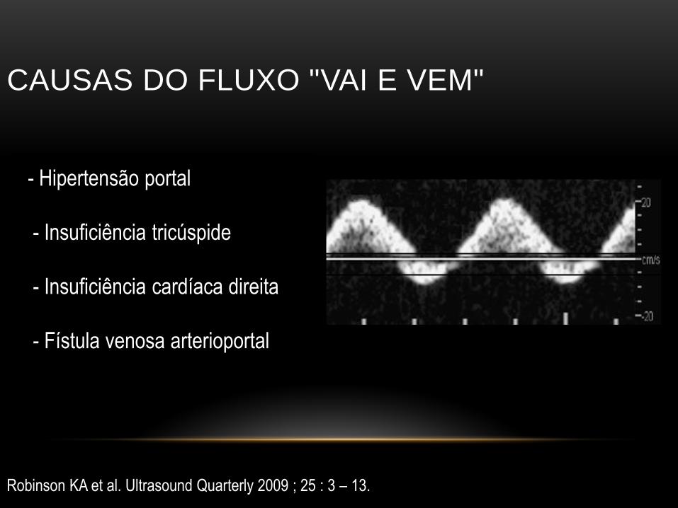
CAUSAS DO FLUXO "VAI E VEM"
Robinson KA et al. Ultrasound Quarterly 2009 ; 25 : 3 – 13.
- Hipertensão portal
- Insuficiência tricúspide
- Insuficiência cardíaca direita
- Fístula venosa arterioportal

FLUXO HEPATOFUGAL
Robinson KA et al. Ultrasound Quarterly 2009 ; 25 : 3 – 13.
Não é patognomônico de cirrose
Hipertensão portal grave - raro

PSEUDOCOÁGULO DA VEIA PORTA
Robinson KA et al. Ultrasound Quarterly 2009 ; 25 : 3 – 13.
At rest
No detectable flow
Depois da compressão da parte
inferior do abdome

SINAIS DOPPLER DE HIPERTENSÃO PORTAL
• Veia porta
• VP dilatada
• Diminuição da velocidade média (<15 cm/s)
• Fluxo "vai e vem"/ hepatofugal
• Aumento da pulsatilidade (VPI)
• Fístula arterio-portal
• Veia hepática
• Fluxo pseudoportal
• Arteria hepática
• Ampliação e tortuosidade
• Aumento de IR e IP
• Colaterais portossistêmicas
Harkanyi Z. Ultrasound Clin 2006 ; 1 : 443 – 455.

OS DIFERENTES TIPOS DE FLUXO NA VEIA HEPÁTICA
Kok Th et al. Scand J Gastroenterol 1999 ; 34 (Suppl 230) : 82 – 88.
Multifásico Bifásico
Cirrose
Síndrome Budd-Chiari
Metástases
Ascite
Monofásico
Cirrosis
Síndrome Budd-Chiari
Metástases
Ascite

DAMPING INDEX
DI: 0.26
HVPG: 7 mmHg
DI: 0.72
HVPG: 15 mmHg
Kim MY et al. Liver International 2007 ; 27 : 1103 – 1110.
Sens. 76%
Spec. 82%

ÍNDICE DE “PORTALIZAÇÃO” DA VEIA HEPÁTICA (DAMPING INDEX)
Kim MY et al. Liver International 2007 ; 27 : 1103 – 1110.
Minimum velocity of downward HV
Maximum velocity of downward HVDamping index =
Valor normal: < 0,6
Hipertensão portal grave: ≥ 0,6


SINAIS DOPPLER DE HIPERTENSÃO PORTAL
• Veia porta
• VP dilatada
• Diminuição da velocidade média (<15 cm/s)
• Fluxo "vai e vem"/ hepatofugal
• Aumento da pulsatilidade (VPI)
• Fístula arterio-portal
• Veia hepática
• Fluxo pseudoportal
• Arteria hepática
• Ampliação e tortuosidade
• Aumento de IR e IP
• Colaterais portossistêmicas
Harkanyi Z. Ultrasound Clin 2006 ; 1 : 443 – 455.

ARTERIA HEPÁTICA PROEMINENTE
AH proeminente com aspecto tortuoso ou sacarrolhas
O aumento do fluxo da AH para compensar a diminuição de perfusão da VP
Swart J et al. Ultrasound Clin 2007 ; 2 : 355 – 375.

CAUSAS DE AUMENTO DA ARTÉRIA HEPÁTICA
Cirrose
Fibrose hepática congênita
Tumores vasculares
Telangiectasia hemorrágica hereditaria
Buscarini E et al. Ultraschall Med 2004 ; 25 : 348 – 55.
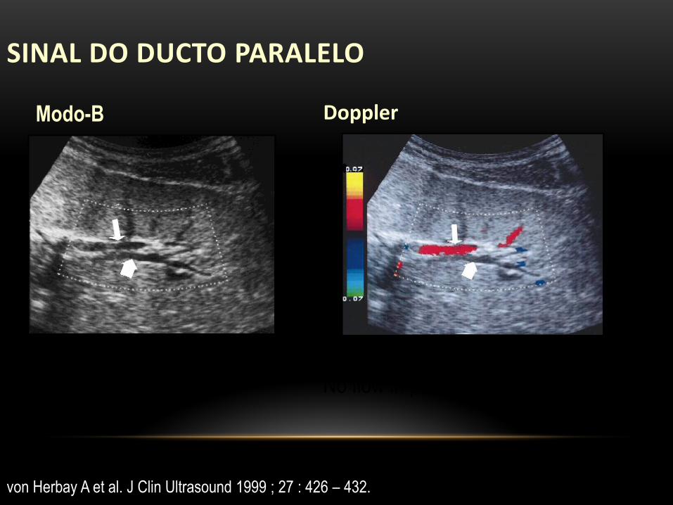
SINAL DO DUCTO PARALELO
von Herbay A et al. J Clin Ultrasound 1999 ; 27 : 426 – 432.
Modo-B
IH parallel channel sign
Suspicious of dilated IHBD
Doppler
Blood flow in anterior structure
No flow in posterior structure

AS VARIAÇÕES DE FLUXO DA ARTÉRIA HEPÁTICA
Kok Th et al. Scand J Gastroenterol 1999 ; 34 (Suppl 230) : 82 – 88.
Diminuição de fluxo diastólicoFluxo normal
patients
Fluxo diastólico invertido

ÍNDICE DE PULSATILIDADE (PULSATILITY INDEX)
CONTROLE X PACIENTES CIRRÓTICOS
Schneider AW et al. J Hepatol 1999 ; 30 : 876 – 881.
PI: 0.85
20 controles
0.92 ± 0.1
PI: 1.22
50 patientes cirróticos
1.14 ± 0.18
P< 0.001
Diretamente correlacionado com a hipertensão portal

SINAIS DOPPLER DE HIPERTENSÃO PORTAL
• Veia porta
• VP dilatada
• Diminuição da velocidade média (<15 cm/s)
• Fluxo "vai e vem"/ hepatofugal
• Aumento da pulsatilidade (VPI)
• Fístula arterio-portal
• Veia hepática
• Fluxo pseudoportal
• Arteria hepática
• Ampliação e tortuosidade
• Aumento de IR e IP
• Colaterais portossistêmicas
Harkanyi Z. Ultrasound Clin 2006 ; 1 : 443 – 455.

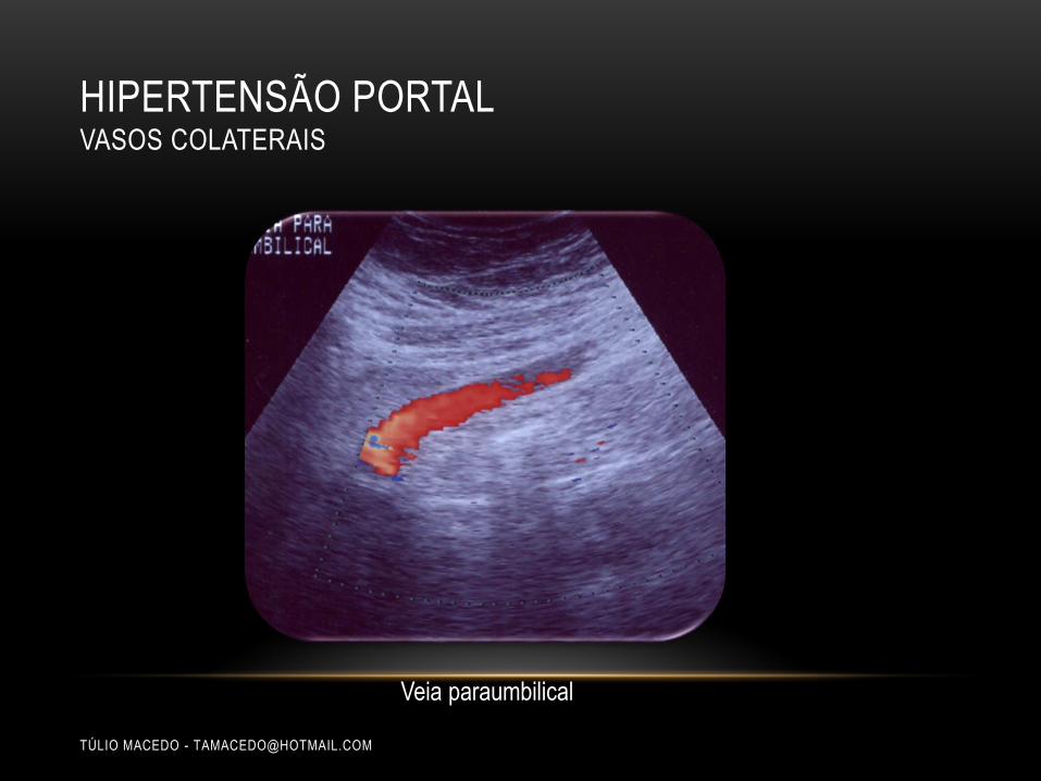
COLATERAIS PORTO-SISTÊMICASALTA SENSIBILIDADE E ESPECIFICIDADE
• Coleterais anatômicasJá existentes
Robinson KA et al. Ultrasound Quarterly 2009 ; 25 : 3 – 13.
Veia coronária (gástrica esquerda)
Veias gástricas curtas
Ramos da VMS e VMI
• Colaterais neoformadas
“Novas formações ou recanalizadas"
•
Veia umbilical recanalizada
Colaterais esplenorrenais
Colaterais gastrorrenais
Colaterais retroperitoneais

Robinson KA et al. Ultrasound Quarterly 2009 ; 25 : 3 – 13.
N Engl J Med 2005 ; 353 : e19.
Cabeça de medusa – recanalização da veia umbilical

ANASTOMOSE ESPLENORRENAL
Yamada M et al. Abdom Imaging 2006 ; 31:701 – 705.
Mansour MA et al. Vascular Diagnosis. Elsevier-Saunders, Philadelphia, 1st edition, 2005.

ANASTOMOSE ESPLENORRENAL – FLUXO INVERSO NA VEIA ESPLÊNICA
Mansour MA et al. Vascular Diagnosis. Elsevier-Saunders, Philadelphia, 1st edition, 2005

VEIAS GÁSTRICAS CURTAS
Sato T et al. J Gastroenterol 2002 ; 37 : 604 – 610.

GASTRORRENAL COLATERAL
Yamada M et al. Abdom Imaging 2006 ; 31 : 701 – 705.
Maruyama H et al. Acad Radiol 2008 ; 15 : 1148 – 1154.
GRS LRV

VASOS ESPLENOLOMBARES
Mansour MA et al. Vascular Diagnosis. Elsevier-Saunders, Philadelphia, 1st edition, 2005

VEIA MESENTÉRICA INFERIOR – INVERSÃO DO FLUXO
Mansour MA et al. Vascular Diagnosis. Elsevier-Saunders, Philadelphia, 1st edition, 2005.

VASOS COLATERAIS RETROPERITONEAIS
Wachsberg RH. Am J Roentgenol 2005 ; 184 : 481 – 486.

INVERSÃO DE FLUXO NA VEIA MESENTÉRICA INFERIOR E VARIZES PERIRRETAIS
Wachsberg RH. Am J Roentgenol 2005 ; 184 : 481 – 486.

VASOS GASTROESOFÁGICOS
McGahan J et al. Diagnostic ultrasound, Informa Healthcare, 2nd edition, 2008.
Veia gástrica esquerda (coronária)


VARIZES PERICOLECÍSTICAS
Harkanyi Z. Ultrasound Clin 2006 ; 1 : 443 – 455.
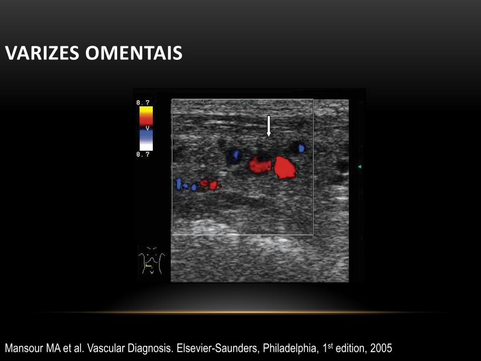
Mansour MA et al. Vascular Diagnosis. Elsevier-Saunders, Philadelphia, 1st edition, 2005
VARIZES OMENTAIS
