nitric oxide synthase expression in macrophages of histoplasma capsulatum-infected mice is
TRANSCRIPT

INFECTION AND IMMUNITY,0019-9567/98/$04.0010
Nov. 1998, p. 5520–5526 Vol. 66, No. 11
Copyright © 1998, American Society for Microbiology. All Rights Reserved.
Nitric Oxide Synthase Expression in Macrophages of Histoplasmacapsulatum-Infected Mice Is Associated with Splenocyte
Apoptosis and UnresponsivenessBETTY A. WU-HSIEH,* WEN CHEN, AND HSIN-JU LEE
Graduate Institute of Immunology, College of Medicine, NationalTaiwan University, Taipei, Taiwan, Republic of China
Received 23 February 1998/Returned for modification 9 April 1998/Accepted 11 August 1998
Splenic macrophages from Histoplasma capsulatum-infected mice express inducible nitric oxide synthase(iNOS), and the iNOS expression correlates with severity of the infection. We examined whether production ofNO is responsible for apoptosis and the anti-lymphoproliferative response of splenocytes from mice infectedwith H. capsulatum. In situ terminal deoxynucleotidyl transferase nick end labeling revealed apoptotic nucleiin cryosections of spleen from infected but not normal mice. Splenocytes of infected mice were unresponsive tostimulation by either concanavalin A or heat-killed H. capsulatum yeast cells. Splenocyte responsiveness wasrestored by addition to the medium of NG-monomethyl-L-arginine, a known inhibitor of NO production. Theproliferative response of splenocytes from infected mice was also restored by depletion of macrophages or byreplacement with macrophages from normal mice. In addition, expression of iNOS returned to its basal levelwhen the animals had recovered from infection. These results suggest that suppressor cell activity of macro-phages is associated with production of NO, which also appears to be an effector molecule for apoptosis ofcultured splenocytes from infected mice.
Nitric oxide (NO) has been reported to induce apoptosis inmany cells including smooth muscle cells (20), oligodendro-cytes (27), pancreatic b cells (11), melanoma cells (35), thy-mocytes (7), B lymphocytes (4), and macrophages (2). Fehselet al. recently demonstrated apoptosis in freshly isolated thy-mocytes after exposure to NO (7). In the same report, they alsoshowed apoptotic foci in close proximity to blood vessels afterlipopolysaccharide treatment. Capillary endothelial and den-dritic cells adjacent to apoptotic foci stained strongly for in-ducible nitric oxide synthase (iNOS), suggesting that NO maybe the mediator for thymic apoptosis (7). Data from anotherlaboratory also showed that cloned thymic stromal cell mono-layers eliminate thymocytes in vitro through production of NO(26). Furthermore, apoptosis has been suggested as a mecha-nism by which the immune system replenishes itself and main-tains homeostasis (30).
The dimorphic fungus Histoplasma capsulatum is a faculta-tive intracellular pathogen of the macrophage (32). Although itis not an obligate intracellular pathogen, the organism is foundalmost exclusively inside host cells during histoplasmosis (5).In our in vitro studies, H. capsulatum exhibits uninhibitedgrowth in normal unstimulated murine macrophages (32). Inactivated macrophages, either peritoneal macrophages andcells from the Raw 264.7 line stimulated by gamma interferon(IFN-g) or splenic macrophages stimulated by IFN-g and li-popolysaccharide, growth of the fungus is inhibited (13, 18, 32).Furthermore, the anti-histoplasma activity of macrophages isdependent on the expression of iNOS and the production ofNO (14, 18). However, the significance of NO production inimmunoregulation of histoplasmosis is not clearly defined.
In this study, we examined whether NO can act as a regu-
lator of apoptosis in lymphoproliferative responses of spleno-cytes from H. capsulatum-infected mice. We showed that iNOSwas induced in splenic macrophages during active infectionand the expression of iNOS coincided with active infection. Wealso observed by in situ terminal deoxynucleotidyl transferase(TdT) nick end labeling (TUNEL) of spleen sections that ap-optosis occurred in immune cells in the spleens of infectedmice but was minimal in control mice. The link between apo-ptosis and NO production was established by inclusion of NG-monomethyl-L-arginine (NMMA) in the culture medium. In-hibition of NO production reduced the amount of apoptosis insplenocyte culture. Thereby, we also confirmed the findings ofZhou et al. (36) that production of NO by splenocytes ofH. capsulatum-infected mice suppressed the splenic lympho-cyte proliferative response. In addition, we showed that mac-rophages were mediators of splenocyte unresponsivenessthrough the NO that they produced and that NO productionwas associated with apoptotic changes in cultured splenocytesfrom infected mice.
MATERIALS AND METHODS
Animals. C57BL/6 mice at 6 to 8 weeks of age were obtained from JacksonLaboratory (Bar Harbor, Maine) and were housed in sterilized plastic cagesfitted with filter cage tops. The animals were fed with sterilized food and water.
Animal model. H. capsulatum 505 was grown at 37°C for 72 h on brain heartinfusion agar supplemented with cysteine and glucose. A suspension of washedyeast cells was prepared. Mice were injected intravenously with 2 3 105 H. cap-sulatum yeast cells (sublethal dose) as described previously (33).
Reagents. RPMI 1640 culture medium (GIBCO-BRL, Grand Island, N.Y.)was supplemented with 10% heat-inactivated fetal bovine serum (HyClone,Logan, Utah), 1 mM sodium pyruvate, 2 mM L-glutamine, 0.1 mM nonessentialamino acids, 5 3 1025 M 2-mercaptoethanol, and 50 mM HEPES buffer. All thesupplements were purchased from GIBCO-BRL. Tritiated thymidine (1 mCi/ml)was obtained from Du Pont-NEN (Boston, Mass.); propidium iodide andNMMA were obtained from Calbiochem (San Diego, Calif.); and RNase A,Triton X-100, sodium citrate, and concanavalin A (ConA) were obtained fromSigma (St. Louis, Mo.). The TUNEL reaction mixture was purchased fromBoehringer GmbH, Mannheim, Germany.
Nitrite assay. Splenocytes from normal and infected mice were cultured (107
per ml) for 24 h in modified Eagle’s medium (GIBCO) containing 5% heat-inactivated fetal bovine serum. Culture supernatant fluids were collected and
* Corresponding author. Mailing address: Graduate Institute of Im-munology, College of Medicine, National Taiwan University, No. 1Jen-Ai Rd., Section 1, Taipei 100, Taiwan, Republic of China. Phone:886-2-321-7510. Fax: 886-2-2321-7921. E-mail: [email protected].
5520
on April 9, 2019 by guest
http://iai.asm.org/
Dow
nloaded from

analyzed for nitrite levels. The nitrite concentration in the supernatant fluids wasdetermined by a colorimetric assay with Griess reagents as described previously(14).
Semiquantitative reverse transcription-PCR. Under RNase-free conditions,total RNA was extracted from freshly harvested mouse spleen. The spleen washomogenized in TRIzol reagent (GIBCO-BRL), containing phenol and guani-dine thiocyanate, as specified by the manufacturer. The homogenate was thencentrifuged for 10 min at 12,000 3 g at 4°C. The supernatant fluid was collectedand mixed with chloroform. After settling at room temperature for a few min-utes, the mixture was centrifuged at 12,000 3 g for 15 min and total RNA wasrecovered from the water-soluble layer. RNA was precipitated by addition of anequal volume of isopropanol. After centrifugation at 12,000 3 g for 10 min, theprecipitate was recovered and washed repeatedly with 75% alcohol, and theRNA was dissolved in 20 to 40 ml of diethypyrocarbonate-H2O. The quality andquantity of extracted RNA were assessed by examining the ratio of spectropho-tometric readings at optical densities of 260 and 280 as well as by 28S and 18SRNA banding in a 1% agarose gel. Extracted RNA was stored at 280°C beforebeing subjected to reverse transcription.
A 2-mg quantity of RNA (1 mg/ml) was reverse transcribed with 200 U ofSuperscript TM II RNase H2 reverse transcriptase (GIBCO-BRL) and 1 ml ofNotI-(dT)18 primer (0.48 mg/ml) in a total volume of 12 ml. After addition ofprimer, the mixture was incubated at 70°C for 10 min and then at 0°C for 1 to 2min. Deoxynucleoside triphosphate, dithiothreitol, and first-strand buffer wereadded before the mixture was heated to 37°C for 5 min. Reverse transcriptasewas then added. The reaction mixture was incubated at 37°C for 50 min, and thereaction was stopped by incubation at 70°C for 20 min. cDNA was stored at220°C.
iNOS-specific cDNA was amplified by the use of paired primers: 59-TGGGAATGGAGACTGTCCCAG and 39-GGGATCTGAATGTGATGTTTG (22).Hypoxanthine phosphoribosyltransferase (HPRT)-specific primers were 59-GTTGGATACAGGCCAGACTTTGTTG and 39-GAGGGTAGGCTGGCCTATAGGCT (22). A semiquantitative PCR was carried out with the following reactionmixture: 1 ml of sample RNA; 2.5 ml each of HPRT 39 primer, HPRT 59 primer,iNOS 39 primer, and iNOS 59 primer (each at 2 mM); 0.5 ml of 10 mM de-oxynucleoside triphosphate; 0.25 ml of Taq polymerase (5 U/ml) and 10.25 ml ofdiethylpyrocarbonate-H2O. The thermal cycles were 1 cycle of 90°C for 3 minfollowed by 35 cycles of 94°C for 40 s, 60°C for 60 s, and 72°C for 60 s. Our pilotruns confirmed that under these conditions, PCR products of each primer set fellwithin the linear range. The reaction was terminated by incubation at 72°C for 8min and then stored at 4°C.
PCR products were separated by electrophoresis on a 2% agarose gel(NuSieve 3:1 agarose; FMC BioProducts, Rockland, Maine). The bands werestained in ethidium bromide for 10 min. The differential quantity of each bandwas analyzed with a densitometer (UltraScanXL laser densitometer; Pharmacia).The ratio of iNOS and HPRT PCR products was derived from densitometerreadings.
In situ iNOS staining. Spleens removed from mice were immediately fixedovernight in 4% paraformaldehyde before being serially immersed in 10, 20, and30% sucrose solutions. The spleens were then embedded in O.C.T. (OptimalControl Temperature Compound; Miles Incorp., Elkhart, Ind.) in liquid nitrogenand stored at 280°C before being subjected to cryosection. The frozen spleenswere warmed to 220°C and sectioned at a thickness of ,5 mm. The sections weredried, fixed in 4% paraformaldehyde for 10 min, and rinsed in phosphate-buffered saline (PBS). Fluorescein isothiocyanate (FITC)-conjugated rabbit anti-mouse iNOS polyclonal antibody (Transduction Laboratories, Lexington, Ky.) ata 1:100 dilution was applied to the section. After a 2-h incubation at 37°C, theslides were washed in PBS and mounted in 1:1 glycerol-PBS mounting fluid.Immunofluorescence staining was viewed under a Olympus fluorescence micro-scope, and photographs were taken with Fuji color film (ASA 400).
Nuclear staining with propidium iodide. Splenocytes in single-cell suspensionwere isolated from normal and infected mice. At 2, 22, and 44 h of incubation inRPMI medium containing supplements, the cells were centrifuged and the su-pernatant fluids were discarded. The cell pellets were resuspended in 1 ml ofhypotonic DNA staining buffer that contained sodium citrate (3.4 mM), TritonX-100 (0.3%), propidium iodide (0.15 mM), and RNase A (1.5 U/ml). Sampleswere kept at 4°C and protected from light until analyzed by flow cytometry at anexcitation wavelength of 488 nm. The fluorescence intensity of degraded DNAdebris fell below 101. Intact DNA in cells fluoresced at an intensity above 103
(12). Nuclei with fragmented DNA fluoresced at an intensity between 101 and103. The percentage of nuclei with fragmented DNA was recorded as percentapoptotic cells.
Splenocyte proliferation assay. Splenocytes (5 3 105/100 ml) from normal andinfected mice were added to separate wells in a 96-well flat-bottom plate. A100-ml volume of supplemented medium or medium containing ConA (2 mg/ml)or heat-killed (60°C for 1 h) H. capsulatum yeast cells at a 1:40 splenocyte-to-yeast ratio was added to triplicate wells in the presence or absence of NMMA(1.2 mM). The cells were pulsed with 1 mCi of [3H]thymidine from 0 to 24 h. AFiltermate 196 harvester (Packard Instrument Co., Meridien, Conn.) was used towash and to lyse the cells. Cell lysate was collected onto a 24-well microplate, andthe radioisotope uptake was determined in a Topcount Microplate scintillationand luminescence counter (Packard Instrument).
Macrophage depletion by plastic adherence and nylon wool. Splenocytes wereisolated from normal or infected mice. Single-cell suspensions were adjusted to5 3 107 to 10 3 107 cells per 10 ml of complete RPMI 1640 medium and platedin heat-inactivated fetal bovine serum-precoated tissue culture dishes. Afterincubation for 1 h at 37°C, nonadherent cells in the suspension were collectedand the plate was washed with warm medium. The cells were centrifuged andresuspended in 10 ml of medium for another cycle of adherence. Nonadherentcells were collected as above. After centrifugation, the cell pellet was resus-pended in 2 ml of medium and loaded onto a prewashed nylon wool column.After a 45-min incubation at 37°C, the cells were eluted with 15 ml of warmmedium.
Nylon wool-nonadherent cells (5 3 105) alone or cocultured with 105 perito-neal macrophages from either normal or infected animals were stimulated withConA (2 mg/ml) for 48 h. At 16 h before harvest, the cells were pulsed with 1 mCiof [3H]thymidine per well. The cells were harvested and the radioisotope uptakewas determined as described above.
TUNEL staining for detection of apoptosis. We used the TUNEL method todetect DNA fragmentation in cells of spleen sections (8, 9). Spleen sections wereprepared and cryosectioned as described above for in situ iNOS staining. Thecryosections were then dried, fixed again in paraformaldehyde, and washedbefore treatment with 3% H2O2. After thorough washing with PBS, TdT-medi-ated, FITC-conjugated dUTP was applied to the sections. The reaction wasallowed to take place at 37°C for 90 min. The sections were then washed in PBSfor 15 min at room temperature before avidin-biotin-peroxidase-conjugated anti-FITC antibody was added. After a 30-min incubation at room temperature andwashing, diaminobenzidine tetrahydrochloride (DAB) was added for color de-velopment. The sections were then counterstained with hematoxylin, and micro-photographs were taken with a Fuji film (ASA 400).
To determine the phenotype of apoptotic splenocytes, freshly harvested spleencells were prepared as single-cell suspensions and delivered to V-bottom 96-wellplates at 106 per 100 ml. In separate wells, monoclonal phycoerythrin (PE)-conjugated hamster anti-mouse CD3, PE-conjugated rat anti-mouse B220, orPE-conjugated rat anti-mouse Mac-1 antibody at 0.1 mg in 100 ml was added.After a 30-min staining on ice with constant shaking, the cells were centrifuged,washed, and resuspended in 2% paraformaldehyde solution. After 30 min offixation, the cells were washed and permeabilized with 0.1% Triton X-100 in0.1% sodium citrate for 2 min on ice. After they were washed, TUNEL reactionmixture was added to each well. The culture plates were then wrapped inaluminum foil and placed in a 37°C water bath for 1 h. The cells were thenwashed again and analyzed by flow cytometry.
Trypan blue exclusion. Splenocytes from normal or infected animals wereadded (5 3 105 per 100 ml) in triplicate to flat-bottom 96-well culture plates andcultured in supplemented RPMI 1640 medium with or without NMMA. Imme-diately after plating (0 h) and at 20 and 46 h of incubation, the cell suspensionin each well was thoroughly mixed and the numbers of viable and dead cells weredetermined by the use of 0.1% trypan blue. To minimize errors introduced byplating, the percentages of viable cells at 20 or 46 h were calculated by dividingthe number of viable cells at 20 or 46 h by that at 0 h.
RESULTS
Splenic macrophages are activated to express iNOS in re-sponse to H. capsulatum infection. Intravenous inoculation ofmice with yeast cells of H. capsulatum results in disseminatedinfection (3, 33). By semiquantitative reverse transcription-PCR methods, we found that iNOS was expressed in spleens ofinfected mice during the course of active infection (Fig. 1).Between 1 and 2 weeks of infection, when mouse spleen wasenlarged with the highest fungal burden (33), iNOS mRNAwas induced. At this time, iNOS mRNA expression was about0.77 times that of a housekeeping gene. In contrast, iNOSmRNA in normal spleen was only minimally detectable (0.12iNOS/HPRT ratio). At 6 to 8 weeks of infection, when thefungus was cleared and the spleen had returned to its normalsize, iNOS expression was at a basal level (0.21 iNOS/HPRTratio). It is apparent that iNOS expression coincided with ac-tive infection. Furthermore, we observed by immunohisto-chemical staining (Fig. 2) that the most prominent iNOS-pro-ducing cells found in the spleen of an infected mouse were cellswith extensive cytoplasm, morphologically similar to macro-phages and giant cells. Taken together, we conclude thatsplenic macrophages and giant cells were activated to produceiNOS during the active inflammatory response to H. capsula-tum. However, we are unable to rule out the additional in-
VOL. 66, 1998 NO, APOPTOSIS, AND SPLENOCYTE UNRESPONSIVENESS 5521
on April 9, 2019 by guest
http://iai.asm.org/
Dow
nloaded from

volvement of granulocytes and endothelial cells, which are alsoknown to produce iNOS.
Apoptosis of splenocytes in response to H. capsulatum infec-tion. In view of the known toxicity of NO to mammalian cells,we examined whether iNOS expression in the spleen is asso-ciated with the death of splenocytes. By use of TUNEL re-agents, we examined spleen sections of infected and normalmice for the presence of apoptotic nuclei. Apoptotic nucleiwere detected in the spleen of an infected mouse (Fig. 3A),whereas they were minimally detectable in the spleen of anormal mouse (Fig. 3B). Furthermore, double staining withanti-CD3, anti-B220, or anti-Mac-1 antibodies and TUNEL re-agents and analysis by flow cytometry demonstrated that apo-ptotic cells in the spleens of infected mice included T and Blymphocytes and macrophages. Apoptotic cells in the freshly
harvested splenocyte population of infected mice consisted of4.0 3 106 T cells, 3.9 3 106 B cells, and 0.6 3 106 macrophages.In contrast, only 0.84 3 106 T cells, 0.84 3 106 B cells, and0.04 3 106 macrophages were found to be apoptotic in freshlyharvested normal splenocytes.
Splenocyte apoptosis is reduced by inhibition of NO pro-duction. To determine if NO production was the cause ofsplenocyte apoptosis, spleen cells were cultured in mediumwith or without NMMA. The percentage of apoptotic cells inthe splenocyte population was assessed by propidium iodidestaining of the nuclei (Fig. 4). While the total number of nucleiremained constant over the observed period, at 2 h of incuba-tion 5% of the nuclei in spleen cells from infected mice wereapoptotic, compared with ,2% for normal mice. Apoptoticcells in cultures of spleen cells from infected mice increasedfrom 5 to 43% by 22 h and to 65% by 44 h, compared with 24and 38% apoptosis at 22 and 44 h, respectively, in normalsplenocyte cultures. Addition of NMMA reduced the numberof apoptotic cells in cultures of infected splenocytes from in-fected mice to a level comparable to that for normal spleno-cytes. Since NMMA competitively inhibits NO production, weconclude that while spontaneous apoptosis occurred in normalsplenocytes in culture, NO induced significantly higher apo-ptosis in splenocytes of infected mice (P , 0.05).
Analysis of cell viability by the trypan blue exclusion assayshowed that 77% 6 14% and 70% 6 10% of normal spleno-cytes were viable at 20 and 46 h, respectively. In contrast, only55% 6 13% (P , 0.01) and 22% 6 10% (P , 0.01) of spleno-cytes from infected mice survived after similar incubation pe-riods. Moreover, addition of NMMA increased the percentageof viable cells in cultured splenocytes of infected mice to 75%6 14% (P , 0.05) and 59% 6 8% (P , 0.01) at 20 and 46 h,respectively. However, the addition of NMMA did not changethe viability of normal splenocytes in culture. The viability was82% 6 10% (P . 0.05) and 68% 6 1% (P . 0.05) at the tworespective time points. These results, together with those in
FIG. 1. iNOS mRNA expression in spleens of H. capsulatum-infected micecoincides with active infection. Total RNA extracted from normal (lane 1) andinfected mice on days 10 (lane 2), 35 (lane 3), and 55 (lane 4) after infection.RNA was reverse transcribed and cDNA was amplified by PCR with pairediNOS primers and HPRT primers simultaneously. The arrowhead points toHPRT PCR products (352 bp), and the arrow points to iNOS PCR products (306bp). The ratios of iNOS to HPRT are 0.12, 0.77, 0.39, and 0.21 for lanes 1, 2, 3,and 4, respectively.
FIG. 2. Splenic macrophages express iNOS protein in H. capsulatum-infected mice. Spleen cryosections from H. capsulatum-infected (2 to 3 weeks postinfection)(A) and normal (B) mice were stained with FITC-conjugated polyclonal rabbit anti-mouse iNOS antibody. (A) The arrow points to a cell, most probably a macrophageor giant cell by morphology, which stains positive for iNOS. (B) The spleen section was stained with ethidium bromide as a counterstain. Magnification, 3400.
5522 WU-HSIEH ET AL. INFECT. IMMUN.
on April 9, 2019 by guest
http://iai.asm.org/
Dow
nloaded from
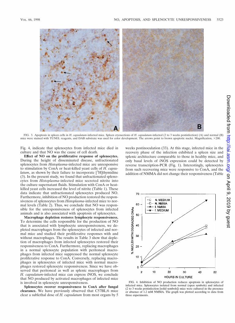
Fig. 4, indicate that splenocytes from infected mice died inculture and that NO was the cause of cell death.
Effect of NO on the proliferative response of splenocytes.During the height of disseminated disease, unfractionatedsplenocytes from Histoplasma-infected mice are unresponsiveto stimulation by ConA or heat-killed yeast cells of H. capsu-latum, as shown by their failure to incorporate [3H]thymidine(3). In the present study, we found that unfractionated spleno-cytes from Histoplasma-infected mice secreted nitrite intothe culture supernatant fluids. Stimulation with ConA or heat-killed yeast cells increased the level of nitrite (Table 1). Thesedata indicate that unfractionated splenocytes produced NO.Furthermore, inhibition of NO production restored the respon-siveness of splenocytes from Histoplasma-infected mice to nor-mal levels (Table 2). Thus, we conclude that NO was respon-sible for the unresponsiveness of splenocytes from infectedanimals and is also associated with apoptosis of splenocytes.
Macrophage depletion restores lymphocyte responsiveness.To determine the cells responsible for the production of NOthat is associated with lymphocyte unresponsiveness, we de-pleted macrophages from the splenocytes of infected and nor-mal mice and studied their proliferative responses with andwithout macrophages. The results in Table 3 show that deple-tion of macrophages from infected splenocytes restored theirresponsiveness to ConA. Furthermore, replacing macrophagesin a normal splenocyte population with peritoneal macro-phages from infected mice suppressed the normal splenocyteproliferative response to ConA. Conversely, replacing macro-phages in splenocytes of infected mice with normal macro-phages restored splenocyte responsiveness. Since we have ob-served that peritoneal as well as splenic macrophages fromH. capsulatum-infected mice can express iNOS, we concludethat NO produced by activated macrophages of infected miceis involved in splenocyte unresponsiveness.
Splenocytes recover responsiveness to ConA after fungalclearance. We have previously observed that C57BL/6 miceclear a sublethal dose of H. capsulatum from most organs by 5
weeks postinoculation (33). At this stage, infected mice in therecovery phase of the infection exhibited a spleen size andsplenic architecture comparable to those in healthy mice, andonly basal levels of iNOS expression could be detected byreverse transcription-PCR (Fig. 1). Interestingly, splenocytesfrom such recovering mice were responsive to ConA, and theaddition of NMMA did not change their responsiveness (Table
FIG. 4. Inhibition of NO production reduces apoptosis in splenocytes ofinfected mice. Splenocytes isolated from normal (open symbols) and infected(2 to 3 weeks postinfection [solid symbols]) mice were cultured in the presenceor absence of 1.2 mM NMMA. The graph was plotted according to data fromthree experiments.
FIG. 3. Apoptosis in spleen cells in H. capsulatum-infected mice. Spleen cryosections of H. capsulatum-infected (2 to 3 weeks postinfection) (A) and normal (B)mice were stained with TUNEL reagents, and DAB substrate was used for color development. The arrows point to brown apoptotic nuclei. Magnification, 3200.
VOL. 66, 1998 NO, APOPTOSIS, AND SPLENOCYTE UNRESPONSIVENESS 5523
on April 9, 2019 by guest
http://iai.asm.org/
Dow
nloaded from

4), showing a direct correlation between splenic macrophageiNOS expression and splenocyte unresponsiveness.
DISCUSSION
NO production by macrophages via iNOS (NOS 2) is de-scribed as a high-output NO pathway, in contrast to the low-output pathway via nNOS (neuronal NOS, NOS 1) and eNOS(endothelial NOS, NOS 3) (16). The high-output NO pathwayhas been shown to be crucial in antimicrobial functions ofmacrophages (16). NO is also known to be anti-proliferative.With its anti-proliferative property, NO has been described asan immunosuppressant (16). However, the question of howNO functions as an immunsuppressant has not been resolved.
In murine models of experimental histoplasmosis, the anti-histoplasma activity of macrophages is dependent on the ex-pression of iNOS and the production of NO (14, 18). It hasbeen shown that spleen cells from H. capsulatum-infected micedo not respond to antigenic or mitogenic stimulation during
the active phase of the infection (3). Macrophages and/or somefactors produced by macrophages were described as the medi-ator for immune suppression in murine histoplasmosis (19).
In this study, we have demonstrated that splenic macro-phages in H. capsulatum-infected animals expressed iNOSmRNA and protein and that the expression of iNOS coincidedwith the acute inflammatory response. Spleen cells isolated fromanimals during the course of active infection died in culturemore rapidly than did normal splenocytes, and the increaseddeath rate was directly related to the production of NO. Wealso observed that splenocytes from infected mice were unre-sponsive to specific antigenic or mitogenic stimulation and thatresponsiveness was restored by inhibition of NO. These resultsare consistent with results reported previously in experimentalhistoplasmosis (36). Furthermore, we showed that the unre-sponsiveness could be corrected by removal of macrophagesfrom infected splenocytes. It is apparent that the production ofNO is associated with splenocyte unresponsiveness in dissem-inated histoplasmosis, which is similar to the immunosuppres-sion described for other disseminated infections by intracellu-
TABLE 1. Presence of nitrite in splenocyte culturesupernatant fluids
Source ofsplenocytesa Stimulant Level of nitrite
(mM)d
Normal mice None 2.3ConAb 0.5H. capsulatumc 1.1
Infected mice None 13.6ConA 21.1H. capsulatum 18.4
a Splenocytes from normal and H. capsulatum-infected mice at 2 to 3 weeksafter infection were cultured at 107 cells per ml. Supernatant fluids were col-lected 24 h after incubation.
b ConA was added at 2 mg/ml.c Heat-killed H. capsulatum yeasts were added at a 1:40 splenocyte-to-yeast
ratio.d These data are representative of three separate experiments.
TABLE 2. Effect of NO on the proliferative response ofsplenocytes from H. capsulatum-infected mice
Source ofsplenocytea NMMAb ConAb Heat-killed
H. capsulatumc SId
Normal mice 2 2 2 1.02 1 2 7.32 2 1 1.21 2 2 ,1.01 1 2 7.31 2 1 1.3
Infected mice 2 2 2 1.02 1 2 ,1.02 2 1 1.71 2 2 7.31 1 2 21.51 2 1 9.0
a Splenocytes from normal and H. capsulatum-infected mice at 2 to 3 weeksafter infection were cultured at 5 3 105 cells in a 96-well plate.
b NMMA was added at 1.2 mM. Con A was added at 2 mg/ml.c Heat-killed H. capsulatum yeasts were added at a 1:40 splenocyte-to-yeast
ratio.d The stimulation index (SI) is the counts per minute (cpm) of splenocytes
from either normal or infected mice stimulated with ConA or heat-killed yeastcells divided by the cpm of splenocytes from either normal or infected micewithout stimulation. Background thymidine uptakes were 2,934 6 580 for normalsplenocytes and 2,250 6 982 for splenocytes from infected mice.
TABLE 3. Effect of macrophages from H. capsulatum-infectedmice on the lymphocyte response to ConAa
Source of wholesplenocytes
Macrophagedepletionb
Macrophageadditionc SId
Normal mice 2 2 8.5Infected mice 2 2 1.2
Normal mice 1 2 4.7Infected mice 1 2 5.5
Normal mice 1 1 (normal mice) 6.9Normal mice 1 1 (infected mice) ,1.0Infected mice 1 1 (normal mice) 4.1Infected mice 1 1 (infected mice) 1.9
a The effect of macrophages on the splenocyte response to ConA (2 mg/ml)was assessed by the depletion of macrophages from splenocytes with or withoutthe addition of peritoneal macrophages from normal or H. capsulatum-infectedmice. Data represent the results of three separate experiments.
b Splenocytes from normal mice and mice at 2 to 3 weeks after infection weredepleted of macrophages by adherence to plastic and nylon wool.
c Peritoneal macrophages (3 3 105) from either normal or infected mice wereadded to macrophage-depleted splenocyte culture.
d Stimulation index (SI) is defined as the cpm of splenocyte culture in thepresence of ConA divided by the cpm of splenocyte culture without ConA.Background thymidine uptakes were 201.9 6 25.0 for normal splenocytes and140.7 6 16.3 for splenocytes from infected mice. Addition of macrophageswithout ConA did not change the background counts.
TABLE 4. Splenocytes recover from unresponsivenessto ConA after fungal clearance
Source ofsplenocytesa
Culture medium containing:SIb
NMMA ConA
Normal mice 2 1 10.71 1 10.5
Infected mice 2 1 6.01 1 7.2
a Splenocytes were isolated from normal mice and mice inoculated 6 weekspreviously with H. capsulatum. No fungus was recovered from the spleen at thetime of the experiment. The size of the spleen had returned to a size comparableto that of a normal spleen.
b The stimulation index (SI) is defined as the cpm of splenocytes from normalor infected mice stimulated with ConA divided by the cpm of splenocytes fromnormal or infected mice without ConA.
5524 WU-HSIEH ET AL. INFECT. IMMUN.
on April 9, 2019 by guest
http://iai.asm.org/
Dow
nloaded from

lar pathogens (6, 10, 23–25). One common feature of theseinfections is macrophage activation, and the suppression is dueat least in part to “suppressor macrophages,” which downregulate the T-cell proliferative response to specific antigens ormitogens. In some cases, macrophage production of NO is thecause of the observed suppression (15, 36). It was recentlyshown that after infection with Leishmania major, spleen cellsfrom homozygous mice lacking the iNOS gene had significantlyhigher levels of T-cell proliferation when stimulated by leish-manial antigen or ConA than did spleen cells from heterozy-gous or wild-type controls (29). The results of these experi-ments support the notion that NO is anti-proliferative (1,17). However, the conclusion that NO suppresses lymphocyteproliferation was drawn from experiments based on the thy-midine uptake assay, which did not differentiate active sup-pression and cell death. Based on our findings, we propose thatthrough production of NO, there is an association betweenmacrophage-mediated splenocyte unresponsiveness and apo-ptotic cell death. However, the direct causal link between un-responsiveness and apoptosis still awaits clarification. It will bepossible when the mechanism of NO-induced apoptosis is bet-ter understood.
In situ staining with TUNEL reagents showed that thespleens of infected mice exhibited apoptotic nuclei (Fig. 3),indicating that apoptosis occurred in the spleens of infectedmice. The apoptotic population included T and B lymphocytesas well as macrophages. Interestingly, the appearance and dis-appearance of apoptotic nuclei in infected mice coincided withthe kinetics of iNOS expression (31). Although direct evidenceshowing apoptosis in the vicinity of iNOS expression is lacking,our data point to the possibility that during an acute inflam-matory response, high-output NO causes the death of spleencells in vivo (7).
Splenomegaly is a hallmark of disseminated histoplasmosis(34). Analysis of single-cell suspensions of splenocytes by flowcytometry revealed an increase in the number of T and Blymphocytes and macrophages in infected mice (28). Due tothe adherent nature of macrophages, they are not readily iso-lated from the spleen, and hence their number is often under-represented in single-cell suspensions of splenocytes (21). Inan earlier study of H. capsulatum-infected mice, we found thatin cryosectioned spleen, infiltrating macrophages account forthe majority of cells in the enlarged spleen (34). While infil-trating macrophages did not remain in the marginal zones butinvaded the follicles, T lymphocytes migrated from the folliclesinto the marginal zones and intermingled with macrophages.Furthermore, the number of T lymphocytes was comparativelysmall and both the CD4 and CD8 cells are sparsely distributedin the spleen. Taken together, these findings indicate that dur-ing the inflammatory response to Histoplasma infection, mac-rophages are activated to produce NO, which in turn negativelyregulates T-lymphocyte expansion by inducing their apoptosis.
Based on these and our previous findings, we propose a work-ing model for the role of NO in the clearance of H. capsulatumand the maintenance of homeostasis of cells in the immunesystem in the course of acute infection. During primary H. cap-sulatum infection, the spleen is enlarged due to massive mac-rophage infiltration and lymphocyte expansion. Upon acti-vation, T lymphocytes produce IFN-g and other cytokines,which in turn activate macrophages. The high-output path-way of NOS is induced in activated macrophages, which arearmed to control the growth of the fungus (14, 18). Due to abystander effect, cells in the vicinity of NO producers and theproducers themselves are also killed. Through this mechanism,splenocytes are reduced in number and the spleen returns toits normal size after recovery from infection. After all, upon
clearance of the pathogen, recruited inflammatory cells are nolonger needed in the microenvironment. It is both economicaland efficient for the immune system to use the high-outputNO pathway as a double-edged sword not only to kill thepathogen but also to maintain homeostasis.
Recently, NO was shown not to be important in secondaryhistoplasmosis and its role in primary infection was confirmed(37). It will be of interest to investigate splenocyte responsive-ness in relation to NO and macrophages in animals with sec-ondary histoplasmosis.
ACKNOWLEDGMENTS
This study was supported in part by R.O.C. National Science Coun-cil grants NSC 86-2314-B-002-125 and 87-2314-B-002-257 and in partby U.S. Public Health Service grant R01 AI-32630 from the NationalInstitutes of Health.
We gratefully acknowledge John Kung and Ping-Ning Hsu for theircritical reading of the manuscript. We thank Sylvia Odesa for herexcellent technical assistance.
REFERENCES1. Albina, J. E., J. A. Abate, and W. L. Henry, Jr. 1991. Nitric oxide production
is required for murine resident peritoneal macrophages to suppress mitogen-stimulated T-cell proliferation. Role of IFNg in the induction of the nitricoxide-synthesizing pathway. J. Immunol. 147:144–148.
2. Albina, J. E., S. Cui, R. B. Mateo, and J. S. Reichner. 1993. Nitric oxide-mediated apoptosis in murine peritoneal macrophages. J. Immunol. 150:5080–5085.
3. Artz, R. P., and W. E. Bullock. 1979. Immunoregulatory responses in exper-imental disseminated histoplasmosis: depression of T-cell-dependent andT-effector responses by activation of splenic suppressor cells. Infect. Immun.23:884–892.
4. Bosca, L., C. Stauber, S. Hortelano, E. Baixeras, and C. Martinez. 1995.Characterization of signals leading to clonal expansion or cell death duringlymphocyte B activation. Curr. Top. Microbiol. Immunol. 200:39–50.
5. Deepe, G. S., and W. E. Bullock. 1988. Histoplasmosis: a granulomatousinflammatory response, p. 733. In J. J. Gallin, I. M. Goldstein, and R. Snyder(ed.), Inflammation: basic principles and clinical correlates. Raven Press,New York, N.Y.
6. Eisenstein, T. K., D. Huang, J. Meissler, and B. Al-Ramadi. 1994. Macro-phage nitric oxide mediates immunosuppression in infectious inflammation.Immunobiology 191:493–502.
7. Fehsel, K., K.-D. Kroncke, K. L. Meyer, H. Huber, V. Wahn, and V. Kolb-Bachofen. 1995. Nitric oxide induces apoptosis in mouse thymocytes. J. Im-munol. 155:2858–2865.
8. Gavrieli, Y., Y. Sherman, and S. A. Ben-Sasson. 1992. Identification ofprogrammed cell death in situ via specific labeling of nuclear DNA fragmen-tation. J. Cell Biol. 119:493–501.
9. Gold, R., M. Shmied, G. Rothe, H. Zischler, H. Breitschope, H. Wekerle, andH. Lassmann. 1993. Detection of DNA fragmentation in apoptosis: appli-cation of in situ nick translation to cell culture system and tissue sections.J. Histochem. Cytochem. 41:1023–1030.
10. Gregory, S. H., E. J. Wing, R. A. Hoffman, and R. L. Simons. 1993. Reactivenitrogen intermediates suppress the primary immune response to listeria.J. Immunol. 150:2901–2909.
11. Kaneto, H., I. Fuji, H. J. Seo, K. Suzuki, T. Matsuoka, M. Nakamura,H. Tatsumi, Y. Yamasaki, T. Kamada, and N. Taniguchi. 1995. Apoptoticcell death triggered by NO in pancreatic b cells. Diabetes 44:733–738.
12. Krishan, A. 1975. Rapid flow cytofluorometric analysis of mammalian cellcycle by propidium iodide staining. J. Cell Biol. 66:188–193.
13. Lane, T. E., B. A. Wu-Hsieh, and D. H. Howard. 1993. Gamma interferoncooperates with lipopolysaccharide to activate mouse splenic macrophagesto an antihistoplasma state. Infect. Immun. 61:1468–1473.
14. Lane, T. E., G. C. Otero, B. Wu-Hsieh, and D. H. Howard. 1994. Expressionof inducible nitric oxide synthase by activated macrophages correlates withtheir antihistoplasma activity. Infect. Immun. 62:1940–1945.
15. Mabbott, N. A., I. A. Sutherland, and J. M. Sternberg. 1995. Suppressormacrophages in Trypanosoma brucei infection: nitric oxide is related to bothsuppressive activity and life span in vivo. Parasite Immunol. 17:143–150.
16. Macmicking, J., Q.-W. Xie, and C. Nathan. 1997. Nitric oxide and macro-phage function. Annu. Rev. Immunol. 15:323–350.
17. Mills, C. D. 1991. Molecular basis of ‘suppressor’ macrophages: argininemetabolism via the nitric oxide synthetase pathway. J. Immunol. 146:2719–2723.
18. Nakamura, T., B. Wu-Hsieh, and D. H. Howard. 1994. Recombinant murinegamma interferon stimulates macrophages of the RAW cell line to inhibitthe intracellular growth of Histoplasma capsulatum. Infect. Immun. 62:680–684.
VOL. 66, 1998 NO, APOPTOSIS, AND SPLENOCYTE UNRESPONSIVENESS 5525
on April 9, 2019 by guest
http://iai.asm.org/
Dow
nloaded from

19. Nickerson, D. A., R. A. Havens, and W. E. Bullock. 1981. Immunoregulationin disseminated histoplasmosis: characterization of splenic suppressor cellpopulations. Cell. Immunol. 60:287–297.
20. Nishio, E., K. Fukushima, M. Shiozaki, and Y. Watanabe. 1996. Nitric oxidedonor SNAP induces apoptosis in smooth muscle cells through cGMP-independent mechanism. Biochem. Biophys. Res. Commun. 221:163–168.
21. Nusrat, A. R., S. D. Wright, A. A. Aderam, R. M. Steinman, and Z. A. Cohn.1988. Properties of isolated red pulp macrophages from mouse spleen. J.Exp. Med. 168:1505–1510.
22. Reiner, S. L., S. Zheng, D. B. Corry, and R. M. Locksley. 1994. Constructingpolycompetitor cDNAs for quantitative PCR. J. Immunol. Methods 165:37–46.
23. Rockett, K. A., M. M. Awburn, E. J. Rockett, W. B. Cowden, and I. A. Clark.1994. Possible role of nitric oxide in malarial immunosuppression. ParasiteImmunol. 16:243–249.
24. Schleifer, K. W., and J. M. Mansfield. 1993. Suppressor macrophages inAfrican trypanosomiasis inhibit T cell proliferative responses by nitric oxideand prostaglandins. J. Immunol. 151:5492–5503.
25. Sternberg, J., and F. McGuigan. 1992. Nitric oxide mediates suppression ofT cell responses in murine Trypanosoma brucei infection. Eur. J. Immunol.22:2741–2744.
26. Tai, X. G., Y. Saitoh, T. Satoh, N. Yamamoto, Y. Kita, G. Takenaka, W. G.Yu, J. P. Zou, T. Hamaoka, and H. Fujiwara. 1994–1995. Thymic stromalcells eliminate T cells stimulated with antigen plus stromal Ia moleculesthrough their cross-talk involving the production of interferon-gamma andnitric oxide. Thymus 24:41–56.
27. Vartanian, T., Y. Li, M. Zhao, and K. Stefansson. 1995. Interferon-gamma-induced oligodendrocyte cell death; implications for the pathogenesis ofmultiple sclerosis. Mol. Med. 1:732–743.
28. Watson, S., T. B. Miller, T. J. Redington, and W. E. Bullock. 1983. Immu-
noregulation in experimental disseminated histoplasmosis: flow microflu-orometry (FMF) studies of the Thy and Lyt phenotypes of T lymphocytesfrom infected mice. J. Immunol. 131:984–990.
29. Wei, X.-Q., I. G. Charles, A. Smith, J. Ure, G.-J. Feng, F.-P. Huang, D. Xu,W. Muller, S. Monocada, and F. Y. Liew. 1995. Altered immune responses inmice lacking inducible nitric oxide synthase. Nature 375:408–411.
30. Williams, G. T. 1994. Apoptosis in the immune system. J. Pathol. 173:1–4.31. Wu-Hsieh, B. Unpublished data.32. Wu-Hsieh, B., and D. H. Howard. 1987. Inhibition of intracellular growth of
Histoplasma capsulatum by recombinant murine gamma interferon. Infect.Immun. 55:1014–1016.
33. Wu-Hsieh, B. 1989. Relative susceptibility of inbred mouse strains C57BL/6and A/J to infection with Histoplasma capsulatum. Infect. Immun. 57:3788–3792.
34. Wu-Hsieh, B., G.-S. Lee, M. Franco, and F. Hofman. 1992. Early activationof splenic macrophages by tumor necrosis factor alpha is important in de-termining the outcome of experimental histoplasmosis in mice. Infect. Im-mun. 60:4230–4238.
35. Xie, K., S. Huang, Z. Dong, S. H. Juang, M. Gutman, Q. W. Xie, C. Nathan,and I. J. Fidler. 1995. Transfection with the iNOS gene suppresses tumori-genicity and abrogates metastasis by K-1735 melanoma cells. J. Exp. Med.181:1333–1343.
36. Zhou, P., M. C. Sieve, J. Bennett, K. J. Kwon-Chung, R. P. Tewari, R. T.Gazzinelli, A. Sher, and R. A. Seder. 1995. IL-12 prevents mortality of miceinfected with Histoplasma capsulatum through induction of IFN-gamma.J. Immunol. 155:785–795.
37. Zhou, P., G. Miller, and R. A. Seder. 1998. Factors involved in regulatingprimary and secondary immunity to infection with Histoplasma capsulatum:TNF-a plays a critical role in maintaining secondary immunity in the absenceof IFN-g. J. Immunol. 160:1359–1368.
Editor: T. R. Kozel
5526 WU-HSIEH ET AL. INFECT. IMMUN.
on April 9, 2019 by guest
http://iai.asm.org/
Dow
nloaded from