nicstar 2015 - iiaglobal.comiiaglobal.com/uploads/documents/2 manuscripts nicstar 2015.pdf ·...
TRANSCRIPT

NAARRI International Conference
State
[Radiation Processing in a changing world
March 4
PAPER No.
1 Electron beamD. Chmielewska, A. Marek
2 Gamma Radiation Technology for Hygienisation of Municipal Dry Sewage Sludge Lalit Varshney and Naresh Kumar Garg
3 A novel method to make Cesium radiation source pencils from Cesium extfrom Nuclear Waste using advanced polymer compositeLalit Varshney and Amar Kumar
4 Radiation synthesis of gold nanoparticles having plate like morphology Sanju Francis and Lalit Varshney
5 Studies on effects of gamma irradiation on deodorizatlives of tuberose and marigold cut flowers Probir Kumar Ghosh, Sayani Pal and Paramita Bhattacharjee*
6 Gamma radiation induced modification of cotton waste with MPTAC for textile dye effluent treatmentAtanu Jha, N. K. Goel
7 γ - RIB (Gamma
solution for the qualitative segregation of products in gamma radiation processing facilities Arjun Kunnoar, Amit Shrivastava
8 Radiation ProRita Singh, Antaryami Singh and Durgeshwer Singh
9 Gamma irradiation effects on Polysulfone and its NanocompositesTapan Kumar Chaki
10 Integrity testing of poton irradiator by radiometry techniqueLaxmidhar Sahoo, A S Tapase ,V.K. Sharma, Suri M.M.K,
NAARRI International Conference on
State-of-the-Art Radiation processing
NICSTAR 2015 Radiation Processing in a changing world]
March 4-6, 2015, Hotel Renaissance, Powai, Mumbai
TITLE
Electron beam-tool for silver nanoparticles synthesis in different matrixesD. Chmielewska, A. Marek
Gamma Radiation Technology for Hygienisation of Municipal Dry Sewage
Lalit Varshney and Naresh Kumar Garg
A novel method to make Cesium radiation source pencils from Cesium extfrom Nuclear Waste using advanced polymer composite Lalit Varshney and Amar Kumar
Radiation synthesis of gold nanoparticles having plate like morphologySanju Francis and Lalit Varshney
Studies on effects of gamma irradiation on deodorization of coconut oil and shelf lives of tuberose and marigold cut flowers Probir Kumar Ghosh, Sayani Pal and Paramita Bhattacharjee*
Gamma radiation induced modification of cotton waste with MPTAC for textile dye effluent treatment Atanu Jha, N. K. Goel*, N. Misra, V. Kumar, L. Varshney
RIB (Gamma – Radiation Indicator Button): A cost effective indigenous solution for the qualitative segregation of products in gamma radiation processing
Arjun Kunnoar, Amit Shrivastava
Radiation Processing of Biological Tissues – Tissue Banking Rita Singh, Antaryami Singh and Durgeshwer Singh
Gamma irradiation effects on Polysulfone and its NanocompositesTapan Kumar Chaki
Integrity testing of poton irradiator by radiometry technique ar Sahoo, A S Tapase ,V.K. Sharma, Suri M.M.K, Murali S
1
6, 2015, Hotel Renaissance, Powai, Mumbai
rticles synthesis in different matrixes
Gamma Radiation Technology for Hygienisation of Municipal Dry Sewage
A novel method to make Cesium radiation source pencils from Cesium extracted
Radiation synthesis of gold nanoparticles having plate like morphology
ion of coconut oil and shelf
Gamma radiation induced modification of cotton waste with MPTAC for textile
A cost effective indigenous solution for the qualitative segregation of products in gamma radiation processing
Gamma irradiation effects on Polysulfone and its Nanocomposites
Murali S

2
11 Application of electron beam irradiation in modification of thermal stability of lignocellulose U. Gryczka, W. Migdał, D. Chmielewska, M. Walo
12 The innovative application of electron beam in disinfection process W. Migdal, U. Gryczka, D. Chmielewska, M.Ptaszek, L. B. Orlikowski
13 Status of electron beam irradiation facility under development at RRCAT Jishnu Dwivedi
14 Electron beam irradiation of various samples for agricultural and material research V.C. Petwal, V. P. Verma, S. Yadav, R. Pramod, J. Dwivedi, A.C. Thakurta
15 Gamma radiation induced synthesis of 2,3-Epoxy propyl methacrylate stabilized Gold Nanoparticles for catalytic application Nilanjal Misra, Virendra Kumar, N.K. Goel, Lalit Varshney
16 Radiation induced immobilization of Gold nanoparticles on AA-g-Polyurethane foam for catalytic property N. K. Goel, N. Misra, Virendra Kumar, L. Varshney
17 Product Dose Mapping of Co-60 Gamma Irradiation Facility Based on Monte Carlo GUO Quan ZENG Minsheng Zhongjin Irradiation Incorporated Company, Shenzhen 518019, China
18 Controlled release through radiation crosslinked matrices: Mechanical integrity, antibiotic efficacy and drug release Jhimli Paul Guin, K.A. Dubey, Y. K. Bhardwaj and Lalit Varshney
19 Indigenous development of a low-cost EPR dosimeter K. A. Dubey, S. K. Suman, R. M. Kadam, Y. K. Bhardwaj, L. Varshney
20 Radiation effects on fluorocarbon-elastomeric nanocomposites R. K. Mondal, K. A. Dubey, Y. K. Bhardwaj, L. Varshney
21 Development of a radiation grafted functional adsorbents based process for dye waste water treatment Virendra Kumar, N. K. Goel, N. Misra, L. Varshney
22 Self-shielded e-beam for polymer industries Seung Tae Jung, Sung-myun Kim, Bum-Soo Han* EB TECH Co., Ltd.
23 Design, Development and Commercialization of ISOCAD (Integrated System Of Computer Aided Dosimetry) for Gamma Irradiators Amit Shrivastava, ISOMED

3
24 Sterilization of Bacteriological Culture Media by High Energy Electron Beam Machine K P Rawat, Chanda Arjun, S A Khader, D Padmanabham, Grace Samuel and K S S Sarma
25 Upgradation of 2MeV- EB Accelerator to 5MeV for its utilization to process packaged products and for waste water treatment S A Khader, Ravindra K Patkari, and K S S Sarma
26 Prospects of Sewage Sludge Disinfection using recently upgraded 5MeV ILU-type Electron Beam Accelerator K P Rawat, S A Khader and KSS Sarma
27 New developments in the Dosimetry for industrial EB processing applications PG Benny, S A Khader and K S S Sarma
28 On the crystallinity and mechanical properties of an industrially important thermoplastic-elastomer blend based composite: effect of composition, MgO and electron beam crosslinking Bhuwanesh Kumar Sharma, Subhendu Ray Chowdhury, P.A. Mahanwar, K.S.S. Sarma
29 Radiation processing of polyamide by electron beam for development of high performance engineering material Dr. R.S. Haldar
30 Radiation grafting of an Industrially important Polyolefin Elastomer Atanu Jha, Subhendu Ray Chowdhury and K.S.S. Sarma
31 Cobalt-60 sealed sources for radiation processing-The fascinating journey from reactor to customer S. A Tariq, T. M Ashraf, S.P Gupta, B. Pintu, D. Paul & K.V.S Sastri
32 Initial Experimental Results of Flue Gas Treatment by a DC Electron Accelerator S. Acharya, et al.
33 Simulation Studies on Flue Gas Treatment by Electron Beam S.K. Majji & S. Acharya
34 Combined effect of gamma irradiation and frozen storage on the microbiological ppts and shelf life of shrimps Manjanaik Bojayanaik, Kavya Narot, Veena Shetty, Somashekarappa Hiriyur , Rajashekar Patil
35 GAMMA RAYS ACCELERATED SEED GERMINATION AND PHYSIOLO GICAL ATTRIBUTES IN CANARIUM STRICTUM ROXB . Akshatha and K.R.Chandrashekar
36 Optimization of activity distribution for radiation sterilization of health care products in NIPRO irradiator Jain Regi George, P. Srivastava, B K Pathak

4
37 Effect of radiation processing in elimination of Klebsiella pneumoniae from food. Gautam, R. K. Nagar, V. and Shashidhar, R.
38 Utilization of Irradiated Onion Scales as a Potential Source of Bioactive Natural Food Colour Sweetie R Kanatt, Snehal Tari & S. P Chawla
39 Thermoluminescence studies on Lithium and Calcium Borophosphate glass systems for radiation dosimetry in food irradiation Bhaskar Sanyal, Madhumita Goswami, V. Prakasan, Aparna Patil, S. P. Chawla
40 Shelf life extension of Ready-To-Eat Sugarcane cubes using Gamma Radiation Processing and low temperature storage Bibhuti Bhusan Mishra, Satyendra Gautam, and Arun Sharma
41 Effect of electron beam and gamma-irradiation on breathable MAP for fresh produces Ramakrishna A, Ravi N, Petwal VC and Raju PS
42 Low Dose Go/No Go Indicators
V. Prakasan, S. P. Chawla & Arun Sharma

5
ELECTRON BEAM -TOOL FOR SILVER NANOPARTICLES SYNTHESIS IN DIFFEREN T MATRIXES
D. Chmielewska 1, A. Marek 2
1 Institute of Nuclear Chemistry and Technology, Dorodna 16, 03-195 Warsaw, Poland 2 Warsaw University of Technology, Faculty of Chemical and Process Engineering, Warynskiego 1, 00-645
Warsaw, Poland
Abstract: Silver nanoparticles stabilized in different matrixes (cotton, cellulose, silica powder material) were obtained with application of electron beam to direct reduction of silver bound in the materials. As silver nanoparticles precursor AgNO3 solution of different concentration was used. Application of dose of 25 kGy ensures simultaneous sterilization of the products. SEM investigation confirmed uniform distribution of silver nanoparticles within materials and dependence of silver nanoparticles size on initial concentration of AgNO3 solution. All obtained silver nanocomposites exhibit antibacterial and antifungal activity. Significant differences in cellulose-silver nanocomposites antibacterial activity against Gram-negative and Gram-positive bacteria can be explained by the difference in the cell membrane structure of the Gram-negative and the Gram-positive bacteria.
1. Introduction
Application of the ionizing radiation as a tool for the synthesis of silver nanoparticles stabilized by different agents has been extensively studied [1,2]. Silver is one of the most common and best known antibacterial agent that has been used for the wounds treatment and disinfection for centuries. Silver characterizes broad spectrum of antibacterial activity, even against antibiotic-resistant bacteria and low toxicity to mammalian cells [3]. Therefore many materials have been functionalized with silver nanoparticles in order to develop their antibacterial and antifungal properties [4,5].
In this work method for nanosilver composites synthesis using 10 MeV-8 kW linear electron accelerator is presented. Pieces of cotton fabric, cellulose membrane and powders based on water glass (WG), TiO2 and quaternary ammonium compound (QAC) were immersed in AgNO3 solution of different concentration and exposed to the dose of 25 kGy in order to ensure simultaneous sterilization of the products. Most of the energy is absorbed by aqueous solution of AgNO3 and in the result of water radiolysis the formation of very powerful reducing radicals-the solvated electron (eaq
-) and hydrogen atom(H.) takes place [6]. Silver nanoparticles were formed in the different materials matrixes by direct reduction of AgNO3 with electron beam irradiation. Silver nanocomposites with antibacterial and antifungal activity were obtained. 2. Materials and methods
Morphology of the samples, size and distribution of silver nanoparticles within different matrixes were examined with Scanning Electron Microscope (SEM) equipped with the Energy selective Backscattered (EsB) and backscattered electrons (BSE) detectors. Investigation of antibacterial activity of the obtained materials against different bacteria and fungi has been carried out. A 1ml suspension of each microorganisms was spread onto the surface of an agar medium containing cotton and cellulose samples modified with silver. The Petri plates were incubated at 37oC for 48h for the bacteria and at 25oC for 72h for the fungi. The same procedure was carried out for the control samples unmodified with silver. The percentage of microbe reduction (Table 1) was calculated according to the formula:
%100]/)[( ⋅−= ACAR (1)
where R is the percentage reduction of microbe; A is the number of microbes recovered from the inoculated control sample after incubation and C the number of microbes recovered from the inoculated test specimen after incubation.
Control sample of powder material WG(TiO2)-QAC and sample of this material doped with silver were suspended in deionised water to obtain 1-5% suspensions. Cellulose membranes were impregnated with obtained suspensions placed on the Perti plates and incubated at 25oC for 24h for the E.coli and Saccharomyces cerevisiae and at 37oC for 120h for the Penicillium spp. and Aspergillus niger. Then growth inhibition zones around cellulose membranes were measured in mm.
3. Results and discussion
Fig. 1 shows the SEM-EsB images of Ag-cellulose nanocomposites obtained with application of AgNO3 solutions of three different concentrations. The homogeneity of silver nanoparticles within cellulose matrix as well as size of these particles depend on concentration of the AgNO3 solution. For the highest concentration of the AgNO3

6
solution except small silver nanoparticles covering whole surface of the cellulose we can observe big agglomerates of small silver nanoparticles as well (fig. 1A). These aggregates consist of silver nanoparticles of size ~ 100 nm (Fig. 2). If a lower concentration of Ag+ ions (concentration of AgNO3 solution 4.3.10-4 mol dm-3) is applied we obtain homogeneous distribution of small silver nanoparticles within cellulose matrix (fig. 1B). For the lowest concentration of AgNO3 solution only several bigger silver particles are formed (fig. 1C).
Fig.1 SEM-EsB image of silver-cellulose nanocomposite for different concentration of AgNO3 solution: 4.3.10-3 mol dm-3 (A), 4.3.10-4 mol dm-3 (B) , 4.3.10-5 mol dm-3 (C).
Fig. 2 presents SEM image of the WG(TiO2)-QAC powder doped with silver. SEM image obtained with BSE detector application confirms uniform silver distribution within the material matrix (white spots). Size of the most of the silver nanoparticles doesn’t exceed 50 nm.
Fig. 2 SEM (A) and SEM-BSE (B) images of silver doped WG(TiO2)-QAC powder.
All obtained material with silver exhibit very good antibacterial and antifungal properties. Antimicrobial activity of cellulose and cotton fabrics modified with silver is summed up in Table 1. Significant differences in silver -cellulose nanocomposites antibacterial activity against Gram-negative and Gram-positive bacteria can be explained by the difference in the cell membrane structure of the Gram-negative and the Gram-positive bacteria. Smaller silver particles have a larger specific surface area that makes them more prone to the interactions with microbial cells that result in their higher antimicrobial activity. Smaller nanoparticles of silver also more easer penetrate inside the bacteria and cause further damage by possibly interacting with sulphur- and phosphorus-containing compounds such as DNA [7].This may suggest the presence of very small silver nanoparticles that are not visible in the SEM images of Ag-cotton nanocomposites with the lowest silver content.
Table 1 Antimicrobial activity of the silver-cellulose and silver-cotton nanocomposites
Initial concentration
of AgNO3 solution
[mol/dm3]
Microbe reduction [%] Cellulose Cotton
Bacteria Fungi Bacteria Fungi Staphylococcus
aureus (Gram +)
ATCC 6538
Escherichia coli
(Gram -) ATCC 10536
Aspergillus niger
ATCC 16404
Rhodotorula mucilaginosa
DSM 70403
Staphylococcus aureus
(Gram +) ATCC 6538
Escherichia coli
(Gram -) ATCC 10536
Aspergillus niger
ATCC 16404
Rhodotorula mucilaginosa
DSM 70403
0 0 0 0 0 0 0 0 0
4×10⁻³ 2 99 90 99 90 60 0 30 4×10⁻⁴ 10 75 80 60 70 50 0 10 4×10⁻⁵ 80 70 0 20 80 60 10 5

7
Although QAC is very active antimicrobial agent (fig. 3A) addition of the silver to the material results in
synergistic effect in the antimicrobial activity of the material and noticeable growth of inhibition zone (fig. 3B). Ag NPs embedded in the different matrixes were successfully synthesized by electron beam irradiation. It was confirmed that the initial concentration of the silver salt solution affected the size and distribution of the silver nanoparticles in the materials. All obtained silver nanocomposites exhibit antibacterial and antifungal activity.
Fig. 3. Growth inhibition zone for the WG(TiO2)-QAC powder (A) and WG(TiO2)-QAC powder doped with silver (B).
Acknowledgements
This work was supported by the Ministry of Science and Higher Education, project “The industrial and environmental application of electron beams”
4. References
[1] Y. Liu, S. Chen, L. Zhong, G. Wu, Preparation of high-stable silver nanoparticle dispersion by using sodium alginate as a stabilizer under gamma radiation. Radiation Physics and Chemistry 78 (2009) 251–255 [2] S.P. Ramnani, J. Biswal, S. Sabharwal, Synthesis of silver nanoparticles supported on silica aerogel using gamma radiolysis. Radiation Physics and Chemistry 76 (2007) 1290–1294 [3] M. Rai, A. Yadav, A. Gade, Silver nanoparticles as a new generation of antimicrobials. Biotechnology Advances 27(2009) 76-83 [4] Y.H. Ngo, D. Li, G.P. Simon, G. Garnier, Paper surfaces functionalized by nanoparticles. Advances in Colloid and Interface Science 163 (2011) 23–38 [5] S.-H. Min, J.-H. Yang, J.Y. Kim, Y.-U. Kwon, Development of white antibacterial pigment based on silver chloride nanoparticles and mesoporous silica and its polymer composite. Microporous and Mesoporous Materials 128 (2010) 19–25 [6] J. Belloni, M. Mostafavi, H. Remita, J.-L. Marignier, M.-O. Delcourt, Radiation-induced synthesis of mono- and multi-metallic clusters and nanocolloids. New J. Chem. 22 (1998) 1239-1255 [7] R.R. Khaydarov, R.A. Khaydarov, S. Evgrafova, S. Wagner, S.Y. Cho, Environmental and Human Health Issues of Silver Nanoparticles Applications, In: Environmental Security and Ecoterrorism, NATO Science for Peace and Security Series C: Environmental Security (2011)117–127

8
GAMMA RADIATION TECHNOLOGY FOR HYGIENISATION OF M UNICIPAL DRY SEWAGE SLUDGE
Lalit Varshney* and Naresh Kumar Garg Radiation Technology Development Division, Bhabha Atomic Research Centre,
Mumbai 400085. India *Corresponding author, email : [email protected], Phone-91-22-25593745
Abstract: Sewage is the wastewater discharged from domestic premises consisting mainly of human waste. Sewage typically contains more than 99.9% water and about 0.05% solid. The solid part results in the formation of sludge. Largely, sludge is disposed in unorganized manner resulting in environmental pollution and spread of diseases. The sludge produced carries a heavy microbiological load including pathogens and therefore its disposal has been a challenge to the urban development authorities. Indian cities and towns together are generating an estimated sewage load of 38,254 million liters per day (MLD). Out of which 11,787 MLD is treated at sewage treatment plants (STP) with a capacity gap of 26737 MLD [1]. Considering 0.05 % solid content, the total potential of sludge generation from the sewage is 19127 tons per day. High energy ionizing radiation technology has a great potential for hygienising municipal sewage sludge and make it safer for use or disposal. In the present study, dry sludge hygienisation process was studied and compared with wet sludge hygienisation. Dry sludge in plastic bags was hygienised in a terminal process using standard fully automatic irradiation plant without manual intervention. Dry sludge containing 75-80 % solid, irradiated at an average dose of 8 kGy, showed absence of indicator organism, E.Coli/total coliforms even after 10 months of study period. Heavy metal concentrations in the domestic sewage sludge was observed to be much below the United States Environmental Protection Agency (US EPA) and Ministry of Urban Development, New Delhi (MOU) norms [1,2]. Inoculation of the hygienised sludge with Rhizobium, Azotobacter and Phosphate solubilizing bacteria showed 100-1000 times higher growth in comparison to growth in unhygienised sludge. A comparison between dry and wet sludge (4-6% solid) irradiation shows that it is more practical to hygienise dry sludge which is economical, reliable and scalable to treat 100-500 tons of city dry sludge. Use of radiation technology for sludge hygienisation can significantly contribute to “Clean India Mission”. The technology is available with the department.
Keywords: Sewage sludge, dry sludge, wet sludge, Gamma radiation, radiation hygienisation, pathogens, microorganisms, radiation dose, over dose ratio.
1. Introduction
STP’s generated dry sludge contains heavy load of bacterial counts including pathogens which could vary between 105 to 109 per gram. Sludge carries other life forms like worms, ova, viruses, helminthes, weeds etc. It contains toxic heavy metals and organic pollutants like pesticides, polyaromatic hydrocarbons, drugs and other persistent pollutants. Sludge is also a rich source of many macro (Nitrogen, Phosphorous, Potassium), micro nutrients (Zinc, Iron, Copper, Manganese) and organic carbon essential for soil. Hygienised sludge can be gainfully used for agriculture, road side plantation, forestry, city home plants etc. Ionizing radiation like gamma rays from cobalt-60 and electron beam from accelerator can be employed to deliver sufficient radiation dose to sludge to inactivate all life forms in the sludge and hygienise it [3].
The sewage sludge can be treated by two methods: (1) Wet sludge Irradiation (4-10% solid content, 96-90% water) and (2) Dry Sludge Irradiation (~75-80% solid, 25-20% water). End product of both the process is dry solid cake.
2. Experimental
Plastic bags were filled with dry sludge (25 kg each) from drying beds of the STP and irradiated in a standard irradiator with conveyor system having about 700 kCi Cobalt-60 source . Ceric-cerous sulphate dosimeters and microbiological dosimeters were placed at minimum and maximum dose positions inside the bags and were sealed. Electro chemical method was used to evaluate absorbed radiation dose. Absorbed dose measurement and microbiological studies were carried out as per standard procedures[4]. The process of wet sludge hygienisation is described elsewhere[4]. Reported values are average of three readings. Microbiological indicator disks containing 1 x 10⁶ spores of Bacillus Pumilus ATCC14884 and Ceric Cerous Sulphate Dosimeters were obtained from Board of Radiation and Isotope Technology (BRIT).
3. Results and discussions
The quality of sludge ( dry, semi dry or wet sludge) is determined by three factors namely (a) the presence of pollutants (Arsenic, cadmium, chromium, copper, lead, mercury, nickel, selenium and zinc.(b)The presence of pathogens ( e.g. bacteria, viruses, parasites) and (c) The sewage sludge attractiveness to vectors e.g. rodents, flies,

9
mosquitoes, birds etc[2]. The process followed by STPs and determination of heavy metals could help in checking the suitability of sludge for use. In wet sludge irradiation, at 3-5 kGy dose, a larger portion of energy is consumed by water (96%) and output of dry sludge is only 4%, as well as the irradiator would require large quantity of Cobalt-60 and thus making economics unfavorable. Irradiation of Dry sludge containing about 80% solid at an average dose of 8-10 kGy gave 20 times more output and lower radiation source requirement per kg of hygienised sludge produced. The results of microbiological investigations on irradiated dry sludge are given in Table 1. US EPA recommends use of treated sludge within eight hours of pathogen reduction process to overcome regrowth issue[2]. Hygienised dry sludge in plastic bags did not show increase in E.Coli/total coliforms even after 10 months of study period. Reduction by about 4 log cycles of highly radiation resistant Bacillus Pumilus in the microbiological indicator (Table1.) demonstrates efficacy and reliability of radiation treatment for hygienisation of dry sludge.
The results of inoculation studies showed 100-1000 times more growth of useful bacteria in the hygienised sludge in comparison to unhygienised sludge. The US/EPA requirements of heavy metals and microorganisms are given in Table 2. The heavy metal concentration in domestic sewage( Collected at SHRI facility, Vadodara) was observed to be much lower than the norms and therefore can be applied to land. Irradiation degrades toxic chemicals and does not alter the heavy metal concentration in the sludge [3]. On meeting the norms, the hygienised sludge can be safely applied on land applications. The radiation technology for dry sludge hygienisation is available with the department.
Acknowledgement
The authors acknowledge the support and encouragement given by Dr. Sekhar Basu, Director BARC, Dr. K.L. Ramakumar, Director RC&IG and Dr. A.K. Kohli, CE BRIT for developing Radiation Technologies for societal benefits. 4. References
1. Manual on sewerage and sewage treatment system, Ministry of Urban Development, Part A, Page nos.- November 2013, New Delhi (http://moud.gov.in)
2. United States Environmental Protection Agency(US,EPA), 40 CFR Part 503, Biosolid Rules 3. Irradiated sewage sludge for application to cropland, IAEA-TECDOC – 1317, International Atomic Energy Agency
(October 2002) page 1-3. 4. Satyendra Gautam, M.R. Shah, Sunil Sabharwal and Arun Sharma, Gamma Irradiation of Municipal Sludge for Safe
Disposal and Agricultural Use, Water Environment Research, volume 77,number 5, Sep./Oct. 2005.472-478
Table 1. Microbiological investigations on Dry Sludge
Sr No Sample No Average dose (kGy)
Total *Coliform /g
Bacillus Pumilus spores after irradiation
Total Coliform /g after 10 months
1 Control 11.5 x 10⁶ 1x 10⁶ 8.7 x 10³
2 A1(minimum)# 6.85 NIL 0.96 x10² NIL
3 A2(maximum)# 9.03 NIL 0.30 x10² NIL
4 B1(minimum) 7.07 NIL 0.42 x10² NIL
5 B2(maximum) 9.15 NIL 0.16 x10² NIL
6 C1(minimum) 6.81 NIL 0.85 x10² NIL
7 C2(maximum) 9.04 NIL 0.10 x10² NIL

10
*US, EPA recommends less than 1000 CFU/MPN per gram at the time of disposal.
# Minimum and maximum dose positions in the bag containing dry sludge
Table 2. Toxic metal limits* in sludge
Pollutant Ceiling* Concentration
limits for All Biosolids
Applied to lands (mg/kg)
Typical Conc. In SHRI#
Sludge
Dry basis (mg/kg)
Annual pollutant loading rate
Limits for APLR Biosolids
(kg/hectare per 365 day period)
Aresenic 75 - 20
Cadmium 85 <10 1.9
Chromium 3000 - 150
Copper 4300 190 75
Lead 840 125 15
Mercury 57 Not detected 0.85
Molybdenum 75 - --
Nickel 420 65 21
Selenium 100 - 5.0
Zinc 7500 620 140
Applies to: All biosolids that are land
applied
- Bagged biosolids
* US EPA 40 CFR 503
# SHRI is a wet sludge hygienisation research irradiator at Vadodara, India.

11
A NOVEL METHOD TO MAKE CESIUM RADIATION SOURCE PENCILS FROM CESIUM EXTRACTED FROM NUCLEAR
WASTE USING ADVANCED POLYMER COMPOSITE .
Lalit Varshney and Amar Kumar# Radiation Technology Development Division, Waste Management Division#
Bhabha Atomic Research Centre, Mumbai 400085, India. Corresponding author, email : [email protected], Phone-91-22-25593745
Abstract: 137Cs (T1/2 = 30 years) is one of the principal radionuclides produced during the fission of 235U in nuclear reactors. It is a major source of radiation in high level liquid nuclear waste (HLLW) generated after the reprocessing of spent nuclear fuel. Separation of Cs from HLLW, reduces radiation exposure during the vitrification process and prevents thermal deformation of conditioned waste matrix during storage. Separation of 137Cs would also markedly reduce it’s volatility during vitrification, extent of migration from the vitrified mass in repository, operational, transportation and storage cost. In addition, 137Cs finds applications as a radiation source for food preservation, sterilization of medical products, brachytherapy, blood irradiation, hygienization of sewage sludge etc. The use of 137Cs in place of 60Co (T1/2 = 5.2 years) will also reduce the shielding requirement, frequency of source replenishment and ease the handling / transportation of radioactive source. Cesium can be separated from HLLW using ion exchanger process using recently patented PES-AMP Polymer composite ALIX or solvent extraction based process using 1,3-Di-n-OctyloxyCalix[4]arene-Crown-6 (CC6) as an extractant [1-4]. In India, Cs recovered using these processes will be immobilized in alkali borosilicate glass matrix for its application as a radiation source. This process is highly complex and generates large volume of secondary waste. In the present study, ALIX loaded with Cs was directly compressed at 7-10 tons/cm2 to get the pallet of desired size which can be directly encapsulated in the SS container for its application as a radiation source. The density of the pallets was found to be 3.0 g/cm3. Experimental Results show that 6.0Ci of 137Cs can be loaded in 1.0 cm3 of the pallet. Neither mechanical nor chemical degradation was observed after irradiating the pallet up to 100 MRads and heating up to 4000C. Major advantage of this process is very simple to execute, pallet can be made of any desired size and it is highly economical. Also, unspent Cesium after use can be recovered in a simple extraction process using ALIX and converted back into pellets.
Keywords: Cesium, polymer extractant, ALIX, radiation source, Radiation source pencil, Radiation sterilization
1. Introduction
Cesium is present in High level liquid nuclear waste (HLLW) in significant quantity( ~ 9Ci/L) It is desirable to remove this Cesium not only to reduce radiation exposure but also the same can be utilized as a radiation source for medical and industrial applications. The processes generally employed for separation of Cesium from HLLW include use of ion exchangers specially Ammonium Molybdophosphate and its polymer composites (AMP) designated as ALIX and Crown ethers. Both the materials are specific to Cesium. Cesium is loaded on these substrates from HLLW and stripped using solvents in complex processes. The separated Cesium is mixed in specific glass formulation at about 850-900 OC and poured into SS pencils which gets vitrified on cooling resulting in source pencil. The activity of such source pencil is about 4 Ci/cm3. These processes use expensive materials, generate secondary waste and are complicated. In the present study, Cesium loaded an advanced polymer composite [2] was directly compressed to give pellets which could be directly loaded into SS pencils without vitrification process.
2. Experimental
The advanced polymer composite in glass column was used for loading inactive Cesium from simulated HLLW. After loading, the polymer composite was washed with water to remove acid and dried at 1500C to remove all the water present in the composite material. Cesium loaded composite was then put into hydrolic press and compressed at 7-10 ton/cm2 to form pellets of desired size and dimension. The radiation stability of pellets were tested using Gamma chamber, GC-5000 from BRIT, India. Thermal stability of the material was evaluated using Thermogravimetric analyser TG-DSC-1 of Mettler.
3. Results and discussions

12
Figure 1 shows the equilibrium capacity of ALIX. It shows that 2.2Ci of 137Cs can be loaded in one gram of the material. Figure 2 shows the compressed pellets produced from Cesium loaded polymer composite. The density of these pellets is ~ 3.0 g/ cm3 and carries about 6.6 Ci/cm3 of Cesium activity more than the glass source. Cesium in this form is water insoluble. The pellets have high temperature stability of more than 400 OC as shown by TGA thermoanalytical curve of the composite in Figure 3. The first peak is due to loss of water. The pellet did not show any significant change in its mechanical strength even at 1000 kGy. Figure 4 shows a typical glass source pencil. The over all process of producing these pellets is simple and could be made in remote operation without generating secondary waste. The process has potential for making radiation sources for medical and industrial applications. Unspent Cesium after use can be recovered using ALIX in a simple extraction process and converted back into pellets.
0.00 0.01 0.02 0.03 0.04 0.050.0
0.1
0.2
0.3
0.4
0.5
0.6
0.7
[Cs+ ] S
orbe
nt ,
(meq
/gm
)
[Cs +]solution
, (M)
Fig. 2. Cesium Loaded polymer composite pellets Fig. 3. TGA Thermaanalytical curve of AlIX
Concentration of Cs: 5 x 10⁻⁴ M to 5 x 10⁻² M
Aqueous phase: 3.0 M HNO3
CEC of ALIX: 0.63 meq/gm (2.42 Ci of 137Cs /gm)
Fig 1; Equilibrium sorption isotherm for Cs+ on ALIX in 3.0 M HNO3
0 200 400 600 800 1000
0
20
40
60
80
100
Tem p.(0C)
Wei
ght (
%)
-0.30
-0.25
-0.20
-0.15
-0.10
-0.05
0.00
0.05
Polym er decom position
W ater loss
AM P-Bead ---TGA
(g/s)

13
Fig. 4. A typical Cesium loaded glass source in SS pencil.
4. References
1. Recovery of Cesium from high level liquid nuclear waste by an advanced polymer composite, Lalit Varshney, Amar kumar, Tessy V., V. Venugopal and S.D. Mishra, BARC News Letter,26/Issue no. 327/July-August, 2012
2. Polymer composite for extracting Cesium from nuclear waste and/or other inorganic waste solutions, Lalit Varshney and Amar Kumar. United State Patent No. US 8828532 B2, Sep. 9 2014.
3. Preparation of PAN based absorber for separation of Cesium and Cobalt from radioactive wastes. A Nilchi, H.
Atashi, A.H. Javid, R. Saberi, Applied Radiation and Isotopes 65(2007)482-487.
4. Selective extraction of cesium ion with calix[4]arene crown ether through thin sheet supported liquid
membranes. Journal of Membrane Science, Volume 187, Issues 1-2, 15 June 2001, Pages 3-11J. K. Kim, J. S.
Kim, Y. G. Shul, K. W. Lee, W. Z. Oh.

14
RADIATION SYNTHESIS OF GOLD NANOPARTICLES HAVING PLA TE LIKE MORPHOLOGY
Sanju Francis a and Lalit Varshney b [email protected]; [email protected]
Radiation Technology Development Division, Bhabha Atomic Research Centre, Trombay, Mumbai 400085, India.
Abstract: Gold nanoparticals have attracted immense interest owing to their unique physical, chemical, and biocompatible properties which has resulted in promising applications in catalysis, bioimaging, photothermal therapy etc [1, 2]. Majority of research so far has focused on the synthesis of spherical nanoparticles and now we have gained a reasonable understanding about the relation between size and properties of nanoparticles [3]. However, for many applications it is desirable or sometimes necessary to have good control of the morphology of nanoparticles. In this study we report a gamma irradiation strategy for the synthesis of gold nanoparticles with plate like morphology. These nanoplates have triangular and hexagonal shapes and their size could be varied from 500 nm to 5 µm by tuning the experimental conditions. X-ray diffraction and selected area electron diffraction investigations proved that the nanoplates are single crystals bound by the 111 planes on the surfaces. 1. Introduction
Gamma radiation has been utilized for the synthesis of different types of nanoparticles employing a variety of capping agents [4- 6]. The synthesis of nanoparticles by gamma irradiation is a convenient process because external reducing agents are not required and the reduction rate of metal ions in solution can be easily controlled by proper selection of dose rate. However, it is difficult to control the shape of the nanoparticles and mostly spherical or irregular particles are produced. Here, we report a method to produce gold nanoplates by employing polydiallyldimethylammonium chloride (PDDA) as the capping agent.
2. Experimental
Gold (III) chloride hydrate (Purity 99.999%) and polydiallyldimethylammonium chloride (PDDA, Mv ~ 100,000 – 200,000) were purchased from Aldrich. The concentration of Au3+ in the solution was fixed at 100 µM and the concentration of PDDA was varied to get different ratios (R) of Au3+/ PDDA. Different ratios were studied to determine the influence of PDDA on the shape of the nanoparticles produced. To synthesize the nanoparticles, the prepared solution was exposed to gamma radiation in a 60Co gamma chamber (GC 5000, BRIT, India) at a dose rate of 2.2 kGyh-1. UV-Visible spectra of the solution were recorded using Thermo, Evolution 300 spectrophotometer. Transmission electron microscopy (TEM) imaging was done using Zeiss Libra 120 electron microscope operating at an accelerating voltage of 120 keV and selected area electron diffraction (SAED) pattern was recorded using JEOL (Model: 200 FX) electron microscope. Scanning electron microscopy (SEM) images were recorded using SERON Technologies Inc., (Model: AIS 2100) operating at an accelerating voltage of 20 keV. X-ray powder diffraction studies were carried out with Philips X-ray diffractometer (Model PW 1729) using Cu Kα radiation and X-ray photoelectron spectroscopy (XPS) investigations were carried with VG (CLAM-2) spectrometer, using Mg Kα radiation.
3. Results and discussion
Radiolytic reduction is an efficient method to produce metallic nanoparticles from the respective metal ions [4, 6]. When a dilute aqueous solution is irradiated, the direct effect of radiation is negligible and the effect mainly occurs via the important primary species produced as a result of water radiolysis (1).
H2O ·OH, H·, eaq (1)
The most important reducing species produced during radiolysis is the hydrated electron, with a standard reduction potential of -2.87 V NHE and H· with a standard reduction potential of -2.3 V NHE [7]. Both these species are strong reducing agents which can easily reduce Au3+ to Au0. It was found that a dose of 2.5 kGy was sufficient to completely reduce Au3+ to Au0 in presence of PDDA under the conditions of our experiments. SEM/ TEM images of the samples were recorded to determine the size and shape of the nanoparticles produced. Fig. 1a shows hexagonal and triangular nanoplates produced at R=1:3. These nanoplates have an average edge length of about 5 µm. When the

15
concentration of PDDA in the solution is increased to R=1:5, smaller nanoplates with an average edge length of 500 nm are obtained as shown in Fig. 1b.
Fig.1 (a) SEM image of gold nanoparticles synthesized at R=1:3; (b) TEM of gold nanoparticles synthesized image at R=1:5
Fig. 2 shows the UV-Vis spectra of the nanoparticle solutions synthesized at the two different ratios. At R=1:5, the solution appeared reddish-purple in color and absorption peak is observed at 530 nm. The spectrum shows only a single absorption peak, but there is appreciable absorption from about 600 nm to 1100 nm. At R=1:3, when the solution was exposed to gamma radiation, the faint yellow color of the solution disappears and shiny particles start to appear in the solution. The spectrum shows a continuous featureless absorption in the entire UV-Visible region which further extends into near-infrared region.
200 300 400 500 600 700 800 900 1000 11000.00
0.02
0.04
0.06
0.08
0.10
0.12
0.14
0.16
0.18
0.20
R=1:3
R=1:5
Abs
orba
nce
Wavelength, nm
20 30 40 50 60 70
R=1:5
R=1:3
(111)
(111)
2θθθθ (degree)
Inte
nsity
(a.
u.)
The XRD pattern of the nanoplates synthesized at R=1:5 and R=1:3 shows an extremely strong diffraction peak at 38.2 degrees (Fig. 3). This peak obtained at R=1:5 is broader than that obtained at R=1:3 because the nanoplates formed are smaller. No other peaks are observed in the entire scanning range suggesting that the nanoplates are single crystal fcc gold [8, JCPDS Card No. 04-0784]. SAED analysis of the samples (not shown) produced bright hexagonal pattern of spots. This observation agrees well with the XRD results and confirms that the gold nanoplates are single crystals with fcc structure, bound by 111 lattice planes on the top and bottom [9]. Although nanoplates are bound by lowest energy 111 plane on the surface, their formation is less favoured thermodynamically compared to other polyhedral structures due to the large surface area. The formation of Au nanoplates in our study may be explained by the facet blocking mechanism, wherein the capping agent plays a key role by adsorbing preferentially or selectively on a particular facet. This selectivity arises due to the difference in coordination number of atoms on a crystallographic plane
Fig.2 UV-Visible spectrum of the gold nanoparticles synthesized at different Au3+/PDDA ratios.
Fig.3 XRD pattern of gold nanoparticles synthesized at different Au3+/PDDA ratios

16
and the ligand or capping agent will interact with the surface or plane which results in the overall lowering of energy in the system. Once a particular surface is passivated or blocked by the adherence of capping agent, further growth on this surface becomes difficult and growth takes place along other directions resulting in the formation of anisotropic nanoparticles. For high yield synthesis of nanoplates the 111 planes needs to be blocked by proper choice of the capping agent. The capping agent, PDDA, used in our study is reported to preferentially adsorb on the 111 surface of gold with its quaternary ammonium head group [11-13]. It binds to the 111 plane of the radiolytically generated seeds and blocks access to this surface and hence, further reduction of Au3+ or growth has to take place along other directions, mainly <110>, resulting in the formation of nanoplates.
4. Conclusions
In summary, we have developed a gamma irradiation strategy for synthesis of gold nanoplates. In this method, gamma radiation brings about the reduction of Au3+ through the mediation of transients species, obviating the need of any chemical reducing agent, while PDDA acts as a shape-directing stabilizing agent. The size of the nanoplates could be easily controlled by varying the ratio of Au3+/ PDDA in the solution. These nanoplates could find applications in catalysis and biomedical fields.
5. References
[1] A.J. Haes and R.P. Van Duyne, A nanoscale optical biosensor: Sensitivity and selectivity of an approach based on the localized surface plasmon resonance spectroscopy of triangular silver nanoparticles, J. Am. Chem. Soc. 124 (2002) 10596-10604. [2] K. Imura, T. Nagahara and H. Okamoto, Photoluminescence from gold nanoplates induced by near-field two-photon absorption, Appl. Phys. Lett. 88 (2006) 23104-23106. [3] X. Sun, S. Dong and E. Wang, Large-scale synthesis of micrometer-scale single-crystalline Au plates of nanometer thickness by a wet-chemical route, Angew. Chem. Int. Ed. 43 (2004) 6360-6363. [4] J. Belloni, M. Mostafavi, H. Remita, J.L. Marignier and M.O. Delcourt, Radiation-induced synthesis of mono and multi-metallic clusters and nanocolloids, New J. Chem. 22 (1998) 1239-1255. [5] J. Belloni, Nucleation, growth and properties of nanoclusters studied by radiation chemistry - Application to catalysis. Catal. Today 113 (2006) 141-156. [6] A. Henglein and D. Meisel, Radiolytic control of the size of colloidal gold nanoparticles, Langmuir 14 (1998) 7392-7396. [7] A.J. Swallow, Radiation chemistry: An introduction, Wiley, New York. 1973. [8] D.A. Porter and K.E. Easterling, Phase transformation in metals and alloys, Chapman and Hall: New York, 1981. [9] C. Kan, X. Zhu and G. Wang, Single-crystalline gold microplates: synthesis, characterization, and thermal stability, J. Phys. Chem. B 110 (2009) 4651-4656. [10] C. Li, W. Cai, B. Cao, F. Sun, Y. Li, C. Kan and L. Zhang, Mass synthesis of large, single-crystal Au nanosheets based on a polyol process, Adv. Funct. Mater. 16 (2006) 83-90. [11] A.J. Swallow, Radiation chemistry: An introduction, Wiley, New York. 1973. [12] S. Link and M.A. El-Sayed, Spectral properties and relaxation dynamics of surface plasmon electronic oscillations in gold and silver nanodots and nanorods, J. Phys. Chem. B. 40 (1999) 4212-4217.

17
STUDIES ON EFFECTS OF GAMMA IRRADIATION ON DEODORIZATION OF COCONUT OIL AND
SHELF LIVES OF TUBEROSE AND MARIGOLD CUT FLOWERS
Probir Kumar Ghosh, Sayani Pal and Paramita Bhattacharjee* Department of Food Technology and Biochemical Engineering, Jadavpur University, Kolkata 700 032, India
(*Corresponding author: [email protected]) Abstract: Among oil crops, coconuts occupy a prime niché in south India and are also extensively cultivated in West Bengal. Coconut oil is a rich source of lauric acid, besides mother’s milk and is a rich house of phytochemicals (such as phenols, alkaloids and glycosides); however, its usage as an edible oil in eastern India is limited owing to its unpleasant lactonic odor. Among the horticultural produce, tuberose and marigold flowers are extensively cultivated in West Bengal. These flowers are highly valued for their ornamental appeal and reportedly have therapeutic potency. However, their short shelf lives limit their commercial utilization for export and therapeutic applications. Therefore, there is huge wastage of these oilcrops and horticultural products. In our study, low to medium dose irradiation has been employed for reduction of off-odor of coconut oil (for wider acceptability as frying oil) and for enhancement in shelf lives of tuberose and marigolds cut flowers (for preserving their therapeutic potential). The effects of irradiation on these commodities were analyzed by hedonic sensory evaluation and assays of their physiochemical and phytochemical properties. Our studies showed promising results in reduction of obnoxious odor in coconut oil and extension of shelf lives of these flowers with concomitant preservation of their physiochemical and phytochemical properties.
Keywords: coconut oil, tuberose, marigold, irradiation, shelf life. 1. Introduction
Agro commodities and horticultural products such as nuts and flowers are produced in bounty in India. In West Bengal, there is a huge cultivation of coconuts (Cocos nucifera L.) and ornamental flowers such as tuberose (Polianthes tuberosa L.) and marigold (Tagetes erecta L.). All of these have high commercial value but are wasted owing to improper processing, post harvest.
Coconut oil, most commonly obtained from the dried coconut kernel (copra), is rich in phytochemicals (such as phenols, alkaloids and glycosides) [1, 2] and is the second best source of lauric acid besides mother’s milk [3]. However, the oil has a characteristic obnoxious odor owing to presence of lactones and certain fatty acids [4] which limit its use as an edible oil. The conventional physical refining processes cause thermal degradation of phytochemicals, necessitating an alternate ‘non-thermal’ technology for reduction of the obnoxious odor in coconut oil. The current work envisages reduction of the obnoxious odor of coconut oil by gamma irradiation. The assessment has been made by evaluation of sensory attributes, and physicochemical and phytochemical properties of the oil with storage.
Tuberose flowers, commonly known as Rajnigandha in India, have a sweet characteristic fragrance. Marigold flowers are well-known for its bright yellow to orange color. Both these flowers have short shelf lives, which limit their commercial value. Common procedures for preservation of flowers include chemical preservatives and/or CAP/MAP packaging. However, chemicals leave harmful residues in the flowers and CAP/MAP packaging has difficulties with in controlling O2 and CO2 levels in the package [5]. Our aim was therefore to preserve tuberose and marigold cut flowers by γ-irradiation and determination of shelf lives of these irradiated flowers by evaluating their sensory, physiochemical and phytochemical properties, with storage. 2. Materials and methods
2.1. Materials
Commercial coconut oil (expeller pressed copra oil from West coast tall variety, M/s KPL Oil Mills, Kerala, India) was purchased from a local supermarket of Kolkata. GC-5000 unit (of Jadavpur University, Kolkata) was employed for γ-irradiation. Fresh tuberose (Calcutta single variety) and marigold cut flowers were procured from the farmers of Barasat, West Bengal. Specialty chemicals were procured from M/s Sigma, USA and AR grade chemicals from M/s Merck, Germany. PET bottles and LDPE packets (both authenticated by National Test House, Kolkata) were procured form a local supermarket. 2.2. Methods
2.2.1. γ-irradiation of coconut oil for reduction of obnoxious odor

18
Coconut oil was filled in screw capped bottles, pre-flushed with nitrogen and irradiated at doses of 2,4,6,8 and10 kGy. Post irradiation, the oil samples were stored at room temperature (23±2°C) in the dark. The samples were withdrawn at an interval of 30 days(for three months) and analyzed for sensory attributes and physicochemical and phytochemical properties. All hedonic sensory evaluation experiments (for oil and flower samples) were conducted by a semi-trained panel constituted by faculty and research scholars of our University. 2.2.2. Sensory, physicochemical and phytochemical analyses of irradiated coconut oil
The irradiated coconut oil samples were subjected to sensory evaluation for attributes of overall appearance, color, odor, taste and after taste; physiochemical properties such as specific gravity, color (by Lovibond Tinctometer), FFA (% oleic acid [6]); phytochemical properties: antioxidant activity (DPPH assay IC50 value (mg/mL), in accordance to Karkaya and Şimşek [7]) and total phenol content (mg gallic acid eq./g dry copra) by the method reported by Spanos and Wrolstad [8]. 2.2.3. γ-irradiation of tuberose cut flowers for improvement in shelf stability
Tuberose cut flowers were stored in LDPE packets (6 flowers per packet) and subjected to γ-irradiation at doses 0.05 and 1.0 kGy (dose determined from previous trials). The irradiated flowers were stored at 15±1°C, 70% RH (in a controlled environmental chamber) and withdrawn at an interval of 2 days for analyses. 2.2.4. γ-irradiation of marigold cut flowers for improvement in shelf stability
Marigold cut flowers were similarly stored in LDPE packets (6 flowers per packet) and subjected to γ-irradiation in the dose range of 0.02 to 2.5 kGy. The irradiated flowers were stored in the above-mentioned chamber at 23±2°C, 80% RH, and withdrawn at an interval of 2-3 days for analyses. 2.2.5. Sensory, physicochemical and phytochemical analyses of irradiated tuberose cut flowers
The irradiated tuberose cut flowers were characterized by sensory evaluation by the semi-trained panel. The attributes evaluated for tuberose cut flowers were overall appearance, color, aroma, firmness and shrinkage of petals. The physicochemical parameter assessed was % moisture of flowers. The phytochemical properties [DPPH and total phenol content (mg gallic acid eq./100 g fresh flowers)] were assayed using solvent extracts of these flowers. 2.2.6. Morphological, physicochemical and phytochemical analyses of irradiated marigold cut flowers
The irradiated marigold cut flowers along with non-irradiated/LDPE packaged (experimental control) and non-irradiated/non-packaged (negative control) flowers were assayed by evaluation of their morphological characteristics such as overall appearance, color, petal shrinkage, firmness and visual fungal infestation similar to the method described in 2.2.1. The flowers possessing acceptable morphological characteristics were further evaluated for their physicochemical parameters (% moisture of flowers) and phytochemical properties (DPPH assay and total phenol content) in accordance to procedures as described in 2.2.5. 3. Results and discussion
3.1 Sensory, physicochemical and phytochemical analyses of irradiated coconut oil Sensory evaluation of the oils revealed highest acceptability by the panel for the samples irradiated at 4 and 6 kGy, wherein panelists’ reported minimum intensity of characteristic obnoxious odor and rancid acid aroma (Fig. 1a, 3 months). For these two samples, the physicochemical and phytochemical parameters remained unchanged with storage (data not shown). These observations suggest that γ-irradiation did not affect the physicochemical and phytochemical properties of the coconut oil. 3.2. Sensory, physiochemical and phytochemical analyses of irradiated tuberose cut flowers
From sensory evaluation, the shelf lives of irradiated flowers obtained were: 12 days (0.05 kGy) > 9 days (0 kGy) > 5 days (1.0 kGy) (Fig. 1b, day 5). The hierarchy in irradiation doses causing decrease in % moisture of the flowers was in the order: 1.0 kGy>0 kGy> 0.05 kGy (Fig. 1d). A similar hierarchy in irradiation doses was observed when antioxidant activity and total phenol content of the irradiated flowers were assayed (Figs. 1f and 1g).
3.3. Morphological, physiochemical and phytochemical analyses of irradiated and non-irradiated marigold cut flowers Flowers irradiated with 2.3 kGy showed highest shelf-life of 11 days followed by those irradiated at 2.5 kGy
(10 days) and 1.0-2.0 kGy (9 days). The morphological characteristics of the flowers (by the panel) also showed highest acceptability for the samples irradiated at 2.3 and 2.5 kGy (Fig. 1c, day 5). Hence these two samples were further

19
evaluated for their physicochemical (% moisture content) and phytochemical properties w.r.t their control. The hierarchy irradiation doses causing loss in % moisture of the flowers was 0 kGy (negative control) > 0 kGy (experimental control) > 2.5 kGy > 2.3 kGy (Fig. 1e). Similar hierarchy for the irradiation dose was also observed for phytochemical assays (Figs. 1h and 1i), attesting 2.3 kGy to be the optimized dose of irradiation for marigold flowers, at which all characteristics including sensory, physicochemical and phytochemical properties were least affected. 4. Conclusion
This study established novel applications of gamma irradiation technology for reduction of obnoxious odor of coconut oil and improvement of shelf life of tuberose and marigold cut flowers, without affecting their physiochemical and phytochemical properties.
Acknowledgement
Probir Kumar Ghosh thanks DST-INSPIRE (IF: 131031) and Sayani Pal thanks UGC (Ref. No.: 1575/(NET-JUNE 2012) for providing financial assistance for the study. 5. References
1. Dayrit C. 1997. Medicinal aspects of coconut oil, Coconuts Today, 14(1), 5-19. 2. Ghosh P.K., Bhattacharjee P., Mitra S. and Poddar-Sarkar M. 2014. Physicochemical and Phytochemical Analyses of
Copra and Oil of Cocos nucifera L. (West Coast Tall Variety), Int. J. Food Sci. http://dx.doi.org/10.1155/2014/310852.
3. Hegde B.M. 2006. Coconut Oil – Ideal fat next only to mother’s milk (scanning coconut’s horoscope), J. Indian Acad. Clinical Med., 7(1), 16-19.
4. Santos JER, Villarino BJ, Zosa AR and Dayrit FM (2011), Analysis of volatile organic compounds in virgin coconut oil and their sensory attributes, Phillipine J. Sci., 140(2), 161-171.
5. Halevy A.H. and Mayak S. 1981. Senescence and post harvest biology of cut flowers II. Hort. Rev., 3, 59-143. 6. A.O.A.C. 2006. Official Methods of Analysis, Gaithersburg, MD, USA. 7. Karakaya S. and Şimşek S. 2011. Changes in total polar compounds, peroxide value, total phenols and antioxidant
activity of various oils used in deep fat frying. J. Am. Oil Chem. Soc., 88(9), 1361–1366. 8. Spanos G.A. and Wrolstad RE. 1990. Influence of processing and storage on the phenolic composition of Thompson
seedless grape juice. J. Agri. Food Chem., 38(7), 1565–1571.

20
Figure 1. Sensory, physiochemical and phytochemical analyses of irradiated tuberose and marigold cut flowers

21
Gamma radiation induced modification of cotton waste with MPTAC for Textile dye effluent treatment
Atanu Jha, N. K. Goel*, N. Misra, V. Kumar, L. Varshney
Radiation Technology Development Division
Bhabha Atomic Research Centre, Trombay, Mumbai 400 085, INDIA *[E-mail:[email protected], Ph: 022-25594763/5689/]
Abstract In the present work, cotton waste is surface modified with [(Methacryloylamino)-propyl] trimethyl
ammonium chloride (MPTAC), a strong anion exchanger, by using gamma radiation induced grafting method. Different grafting parameters, such as total absorbed dose, monomer concentration, solvents, etc., have been investigated to obtain maximum grafting yield. The PMPTAC-g-Cotton samples were characterized using FTIR, elemental analysis and SEM techniques. Surface modified PMPTAC-g-Cotton samples were investigated as a strong anion exchanger for waste water treatment, particularly for textile dyes effluents. The performance of the grafted anion exchanger was evaluated by adsorption of anionic dyes namely, Acid blue 25 (AB25) under batch process in aqueous solutions. Langmuir isotherm model was utilized to analyze the equilibrium adsorption data of the dye. Higher correlation coefficient (>0.99) for these dyes suggested that adsorbent follow Langmuir adsorption isotherm and there is a good agreement between the experimental and calculated values of adsorption capacity. Maximum adsorption capacity was found almost linear function of the extent of grafting. The maximum adsorption capacities of grafted adsorbents (with 20% G.Y.) were found to be 250 mg/g for AB25 at room temperature. The excellent uptake of organic dyes by the grafted anion exchanger suggested its promising application for industrial wastewater treatment.
1. Introduction
Surface-modified functional polymers have gained great importance in many diverse applications, such as functional adsorbents, antimicrobial and anti-fouling surfaces, support for enzyme immobilization, etc. Surface-modified polymers are of substantial importance in many diverse aspects of modern technology, and whilst there are a number of existing physical and chemical methods like UV, plasma, conventional chemical methods, enzymatic [1] and most recently laser surface modification for surface modification of polymers, the frequent requirement for significant infrastructure, harsh reaction conditions and limitation to specific polymer types led to explore high energy radiation (gamma-ray, electrons beams etc.) based technology known for convenience, high efficiency, high purity, easy and environmental friendly process for such modifications. Radiation grafting method has been widely adopted to modify different polymers to improve their desired physicochemical properties for different applications due to its distinctive advantages over conventional chemical grafting [2]. Cotton is one of the most popular and abundant natural fibres, made of mainly cellulose. Cotton widely used as clothing materials for its natural abundance and posses good qualities as good heat conductor, excellent moisture absorption, good dyeability and biodegradable [3,4]. Thus, radiation grafting method has been adopted to modify cellulose based fiber to enhance its physicochemical properties. It is highly interesting material for research and industrial purposes because of its renewable, biodegradable and biocompatible characteristics and easy fabrication.
2. Experimental Locally available cotton fiber waste was used as a substrate. MPTAC monomer (purity >97%) 50% (v/v)
from Sigma Aldrich was used as received. Mohr’s salt and other chemicals were of AR (Purity >99%) grade. Ultra-pure water was used for preparation of all the solutions. Gamma chamber (GC-5000, BRIT) of effective dose rate 1.7kGy/h was used as a radiation source for radiation grafting purpose.,
3. Result and discussions Various experimental parameters, such as total absorbed dose, monomer concentration, solvent effect and
acid concentration were optimized for achieving maximum grafting levels. The developed grafted matrix was further tested for the Acid Blue 25 (AB25) model anionic dye for the textile effluents treatment. Grafting yield was found as a function of total absorbed dose and saturated beyond 2kGy (Fig.1). Grafting yield was also found to be near to linear function of monomer concentration up to 50% as shown in fig. 2. Further, effect of additives like Mohr’s salt

22
and acid was not found prominent which is generally used for homo-polymer inhibition and accelerating in grafting yield simultaneously.
0 1 2 3 4 5
0
2
4
6
8
10
12
Gra
fting
Yie
ld,%
Dose (kGy)
0 10 20 30 40 50
0
10
20
30
40
50
Gra
fting
Yie
ld, %
Monomer Concentration, %
Characterization Surface modified samples were characterized with various techniques, such as gravimetrically, FTIR, SEM,
TGA, Water Uptake and elemental analysis. Polymer grafted on to cotton fibrils have a nitogen per unit monomer and hence garfted was confirmed by elemental analysis for nitrogen. As shown in figure 3, nitrogen contents were found propotional to the grafting extent. Further, grafted samples were also characterised with TGA and SEM to investigate the thermal stablity and morphological simultaneously after surface modification (Fig. 4 and Fig.5).
Dye uptake study
Batch equilibrium adsorption studied was carried out with an anionic dye AB25 as a model dye for the textile effluent treatment. The surface modified samples with different extent of grafting yield were dipped for 24hrs under stirring conditions in the known concentration of the dye solution. Concentration of dye was monitored spectrophotometrically at equilibrium. Dye uptake was found to increase almost linearly with the grafting yield (Fig. 6). MPTAC-g-Cotton adsorbent with 20% GY was used for dye adsorption study. Maximum adsorption capacity of the adsorbent was found ~250mg/g.
Fig.1. Effect of Dose on grafting yield Fig. 2. Effect of Monomer Conc. on grafting
0 100 200 300 400 500 600 700
0
20
40
60
80
100
Wei
ght L
oss
(%)
Temperature (0C)
5 10 15 20 25 30 35 40
1.0
1.5
2.0
2.5
3.0
3.5
% N
itrog
en
Grafting Yield (%)
Fig.3. Nitrogen content (%) with Grafting Fig.4. TGA thermograms of (a) Cotton (b) PMPTAC-g-Cotton and (c) PMPTCA
(a)
(b) (c)

23
Fig.5. SEM images of (a) control and (b) MPTAC-g-Cotton grafted sample
0 5 10 15 20 25 30 35 40 45
0
50
100
150
200
250
300
Upt
ake
capc
ity, m
g/g
G rafting Yie ld , %
4. Conclusion
Radiation induced grafting is environmental friendly process for modifying the cotton fibrils. Optimized experimental parameters showed the desired extent of grafting can be obtained. Various characterization techniques confirmed the grafting reaction and also provided the insight of the morphological and thermal stability of the grafted samples. Radiation grafted MPTAC-g-Cotton was found be an efficient adsorbent for acid dye. 5. References 1. A. Bhattacharya, B. N. Misra, Prog. Polym. Sci. 29, 767 (2004) 2. N. K. Goel, V. Kumar, Y. K. Bhardwaj, S. Pahan, S. Sabharwal, J. Hazard. Mater. 193, 17 (2011) 3. Gowariker, V.R., Viswanathan, N.V., Sreedhar, J., 1999. Individual polymer. In: Gowariker, V.R. (Ed.), Polymer
Science. p. 258. 4. Flaque, C., Rodrigo, L.C., Ribes-Greus, A., 2000. J. Appl. Polym. Sci. 76 (3), 326–335.
Fig.6. Effect of grafting % on dye uptake
(a) (b)

24
γ - RIB (GAMMA – RADIATION I NDICATOR BUTTON ): A COST EFFECTIVE INDIGENOUS SOLUTION FOR THE
QUALITATIVE SEGREGATION OF PRODUCTS IN GAMMA RADIAT ION PROCESSING FACILITIES
Arjun Kunnoar, Amit Shrivastava ISOMED, BRIT South Site BARC, Trombay, Mumbai, India
[email protected] [email protected] Abstract: Gamma Radiation processing facilities extensively use radiation indicator buttons (currently imported) for the qualitative indication and segregation of the products in the processing areas of the facilities. The qualitative indicators of gamma radiation processing facilitate easily recognizable, concrete objective evidence to the stake holders of the facilities with respect to the processing of the products. The γ - RIBs have been indigenously developed, test validated at ISOMED facility and being used conveniently.They were prepared by thin films made of Poly Vinyl Alcohol (PVA) and NitroBlueTetrazolium (NBT) chloride salt dissolved in single distilled water. The color of γ-RIB changes from transparent to deep blue on irradiation. Color change is radiation dose dependent. The pre irradiation stability and post irradiation stability of γ-RIB have been investigated. The feasibility study and cost effectiveness of γ -RIBs have been evaluated at ISOMED Trombay. It is believed that γ – RIBs would provide an impeccable , cost effective indigenous solution to all the operators of the gamma radiation processing facilities along with the primary manufacturers of the medical and food / allied products.
Key words: Radiation processing, Radiation Indicator Button (RIB), NBT, PVA, MSDS. 1. Introduction
Gamma Radiation processing facilities for food and medical products are increasing day by day. These facilities use radiation indicator buttons as preliminary marker for identifying radiation processed product from unprocessed product.Radiation indicator buttons undergo color change after irradiation. Many dyes used in the preparation of gamma radiation indicator buttons contain volatile organic compounds which is a major issue of health concern. It is reported by many users that irradiated RIB color fades after some time if kept in ambient conditions leading to misinterpretation. ISOMED is accredited with ISO 9001:2008, ISO 13485:2003, ISO 22000:2005,ISO 14001:2004, ISO 11137-1 and OHSAS. These certification agencies during the certification/surveillance audit raised the point regarding the specifications and MSDS of dye present in the Radiation Indicator buttons pasted on product boxes. To comply with ISO requirement and to have a user friendly alternative for imported RIB, the NBT based films are prepared to study the efficacy of films as gamma radiation indicator buttons(γ-RIB).Thedosimetricpropertiesof Aqueous-ethanol NBT solution has been studied by many authors[1, 2,3]. NBT atetrazolium salt on irradiation yield highly coloured water insoluble formazans [4]due to radiolytic reduction. Formazans are non-volatile and non-hazardous in nature[5,6]. The γ-RIB which contains NBT dye in PVA undergoes colour change from transparent to deep blue depending upon the dose. Theproperty of colourchanges due to radiation and formation of non-volatile formazanhave led us to develop films of NBT-PVA as Gamma Radiation indicator buttons to use at SPICE Irradiator Vashi and ISOMED Trombay. 2. Materials and method
Nitro Blue Tetrazolium chloride (NBT) was procured from Himedia and PVA(Poly Vinyl Alcohol) from Local supplier. Solution containing 0.5mM NBT dye was prepared by dissolving 0.408g of NBT in 1000ml single distilled water. 10% polymeric solution is made by dissolving 10g of PVA in 100ml of 0.5mM NBTin a conical flask of volume 250ml kept on a magnetic stirrer for six hour at 50oC. A homogeneous solution of NBT-PVA (20ml) was poured onto each horizontally leveled polystyrene plates of 15cmX15cm and dried at room temperature in dark for 72hr. The films were peeled off and cut into 10mmx10mm pieces, dried, stored and pasted on the labels which are to be used on the customer boxes.
3. Experimental
The control films were kept at ambient temperature and free from ionizing radiation for 2 yearsto verify the pre-irradiation stability. The films exposed to different doses in GIC 5000 at ISOMED were also kept for 2 years at ambient environmental conditions for checking the post irradiation stability of films.The color change with incremental dose is shown in figure1. The efficacy of the γ-RIBis studied in routine radiation processing at

25
ISOMED. About 500 γ-RIBs were prepared and used for routine radiation processing of the customers boxes along with imported RIBs at ISOMED (figure 2 & 3).
Figure 1 color change with radiation dose
Figure 2 γ-RIB with imported RIB (26 kGy)
Figure 3 γ-RIB with imported RIB (Un-irradiated)
Technical Specification:
Color: Transparent (Pre-irradiation) Color: pink (6kGy-12kGy) Color: deep blue (15-25kGy) Size: 10mmX10mm Thickness: 0.5mm (approx) Storage temperature: 20-35ᵒC Pre-irradiation stability: 2 years (preferably keep in dark and away from ionizing radiation) Post-Irradiation stability: 2 years (ambient environmental condition) Range: 5kGy-50kGy
Production cost:
EstimatedProduction cost of 15000 Numbers of γ-RIBs is given below: Cost of Raw Materials: Rs 5500/- (approx) Adhesive cost Rs 400/- (approx) Cutting cost (10mmx10mm) Rs 2000/- (approx)

26
Total Rs 7900/- (approx) Note: Procuring cost of 15000/- Nos of imported RIBs of size 12mm diameter is Rs 16000/- (approx) where as the γ –RIB cost for the same quantity will be Rs7900/- i.e., about 50% lessof the imported RIB. 4. Results and discussion
The color of the γ-RIBs at 6kGy is pink which deepens up to 12kGy then changes to blue at 15kGy onwards. This color change is due to conversion of monofarmazans to diformazans at higher doses [2].Pre- irradiation stability is 2years ifγ-RIBs were kept in ambient temperature (20-35ᵒC) and radiation free environment. Post irradiation stability is 2years at ambient environmental conditions. 5. Conclusion
γ-RIBs have good pre and post irradiation stability, free from volatile organic compounds,economical and easy to prepare.The expenditure on purchasing RIB will reduce tohalf to that of the imported RIBs. Acknowledgement: Authors express their sincere thanks to Dr A K Kohli, CE, BRIT, P SrivastavSGM,Engg/CP,and L N Bandi DGM, GRPS, BRIT for their encouragement in this work. 6. References
1. Kovacs, A., Wojnarovits, L., Baranyai, M., Mousa, A., Othman, I., Mclaughlin, W. L., 1999.Aqueous-ethanol nitro blue tetrazolium solutions for high dose dosimetry.Radiat.Phys. Chem. 9, 737-740.
2. Mousa, A., Baranyai, M., Wojnarovits, I.,Kovacs, A., McLaughlin, W. L., 2003. Dosimetry characteristics of the nitro blue tetrazolium polyvinyl alcohol film for high dose applications. Radiat. Phys. Chem. 68, 1011-1015.
3. Ahmed A. Basfar., Khalid A. Rabaeh., Akram A. Mousa., Rashed I. Msalam., 2011. Dosimetry characterization of nitro-blue tetrazolium polyvinyl butryral films for radiation processing. Radiat. Phys. Chem. 80, 763-766.
4. Auclair, C., Voisin, E., 1985. Nitrobluetetrazolium reduction. In: Greenwald, R. A. (Ed.), CRC Handbook of Methods for Oxygen Radical Research. CRC Press inc, Boca Raton, pp. 122-132.
5. GurusamyMariappan, RejaulKorim, NandMadhwa Joshi, FarukAlam, RajibHazarika, Deepak Kumar, Tiewlasubon Uriah, 2010. Synthesis and biological evaluation of formazan derivatives. J Adv Pharm Technol Res. Oct-Dec;1(4):396-400.
6. MSDS for Nitro Blue Tetrazolium chloride.

27
RADIATION PROCESSING OF BIOLOGICAL T ISSUES – T ISSUE BANKING
Rita Singh, Antaryami Singh and Durgeshwer Singh Radiation Processing Group, Defence Laboratory,
Defence Research & Development Organization, Jodhpur, India
Abstract: Radiation processed biological tissues can be provided by tissue banks for the management of injuries due to trauma or disease. Allograft tissues from human donor like bone, skin, amniotic membrane and other soft tissues can be used for repair or reconstruction of the injured part of the body and provide an excellent alternative to autografts. However, tissue allografts should be sterilized to make them safe for clinical use. Radiation processing has well appreciated technological advantages and is the most suitable method for sterilization of biological tissues. Sterility assurance level (SAL) defines the probability of a viable microorganism being present on an individual product unit after sterilization. The tissue allografts must receive a sterilization dose high enough to ensure that the probability of an organism surviving the dosage is no greater than one in one million units tested (10-6). Conventionally, a radiation dose of 25 kGy is the generally accepted dose for sterilization. Generally, doses between 25 and 35 kGy induce no significant damaging effect on tissues and their constituents. 1. Introduction
A number of surgical procedures require tissue substitutes to repair or replace damaged or diseased tissues. Biological tissues from human donor like bone, skin, amniotic membrane and other soft tissues can be used for repair or reconstruction of the injured part of the body. The use of allograft tissue avoids the donor site morbidity and reduces the operating time, expense and trauma associated with the acquisition of autografts. Further, allografts have the added advantage of being available in large quantities. This has led to a global increase in allogeneic transplantation and development of tissue banking. However, the risk of infectious disease transmission via tissue allografts is a major concern. Therefore, tissue allografts should be sterilized to make them safe for clinical use. Radiation processing has well appreciated technological advantages and is the most suitable method for sterilization of biological tissues. Radiation sterilized allograft tissues have helped millions of injured and disabled people around the world [1].
2. History of Tissue Banking
The concept of storing and transplanting human tissue is centuries old. Indeed references to bone and limb transplantation can be found in Bible, mythology, medical records of the ancient Middle East, medieval art and church commentaries. The art of tissue grafting was also known in ancient India. It appears in Sanskrit text authored by Sushruta. Skin grafting was attempted for the repair of noses lost in battle or mutilated for the punishment of crimes. However, widespread interest did not develop until the 19th century. World War II resulted in large numbers of casualties. Losses of bone, fractures or burn wounds in victims of the War compelled surgeons to come up with methods of tissue-grafting and tissue-transplantation to repair the defects. The establishment of the US Navy Tissue Bank in Bethesda, Maryland in 1949 by George Hyatt, an orthopaedic surgeon marked the emergence of the modern tissue bank. Hyatt developed a system for procuring tissue, with a focus on bones, from cadavers in operating theatres, and employed freeze drying to store bones. This helped to increase the availability of allogenic bones [2].
3. Collection of Tissues
Tissues can be obtained from a living donor or a cadaveric donor. Tissues obtained from living donors are amniotic membranes and bones removed during surgical procedures like excised femoral head in fracture neck of femur, resected bone slices from total knee replacement operation and bones from corrective osteotomy. Bones can also be obtained from primary traumatic limb amputation. Tissues procured from cadavers or dead bodies are allograft skin, cancellous and cortico-cancellous bone, cortical bone, osteochondral bone and bones with attached ligaments and tendons.
Tissue safety is a major concern in transplantation. The transmission of infectious agents from donor to recipient with allografts is their major risk and disadvantage. The presence of virulent microorganisms in the allografts can lead to potentially serious complications for tissue recipients [3]. Safety issues regarding the transmission of biological infections via allograft transplantation are of critical concern to both surgeons and tissue

28
recipients. Therefore, adequate donor screening coupled with appropriate processing are employed to reduce the risk of disease transmission.
Each donor is screened based on the medical history, physical examination and laboratory examination. Information regarding age, past medical history, medications taken, family medical history, exposure to toxic materials and cause of death are obtained for donor screening. Any adverse information conforming to ‘Donor Exclusion Criteria’ leads to refusal of the donation in the interest of the recipient. The minimum serological testing accepted universally includes Human immunodeficiency virus antibody (HIV I and II), Hepatitis B virus surface antigen (HBSAg), Hepatitis C Virus antibody (HCV) and Syphilis. Other tests can be done selectively when indicated such as CMV test, Hepatitis B core antibody, Histopathological examination, Antigen testing for HIV and HTLV I + II.
4. Processing of Tissues
Processing is an important step to assure quality, safety and effectiveness of tissue. Ideally tissue should be processed immediately after procurement. Tissues from more than one donor are never pooled during procurement and processing to avoid cross-contamination. Tissues are aseptically processed in the controlled environment primarily to maintain or enhance the biological integrity. There are various methods used for processing by individual tissue banks [4]. In general, processing consists of washing, decontamination, deproteinisation, pasteurization, freezing or freeze drying, packaging, labelling and sterilization.
There are four general methods of tissue preservation: simple hypothermia, freezing, freeze drying and cryopreservation. Simple hypothermia is mainly used for transport of fresh tissues and maintenance of skin and osteoarticular grafts through refrigeration at 1-10o C. Freezing is mechanically achieved via storage at -400C or lower. Freezing is especially useful for large bone grafts and tendons that need mechanical strength. In freeze drying or lyophilization, the grafts are frozen and then bound water is sublimed under vacuum. It prevents oxidative reactions, lends stability to the product and allows storage at room temperature. Cryopreservation or controlled rate freezing between –1000C to –1960C is useful for preservation of structural integrity and mechanical properties of the tissue.
5. Radiation Sterilization of Tissues
Extensive screening of donor medical and social history combined with vigorous serological and bacterial screening improves the safety of tissue allografts [5]. In addition, techniques to further process bone and soft tissue minimize the risk of transmitting infectious diseases with the allograft [6]. Screening of donor for disease, bacterial testing and aseptic processing, substantially reduce risk, but do not completely eliminate the possibility of allograft associated infections. Allogenic transplants have been associated with transmission of viruses, bacteria and fungi [7]. Transplants are contaminated primarily by pathogens originating from the donor. Secondary contamination can take place during the removal and/or subsequent processing of the transplant or during implantation. Viral transmission may also come from infected donor such as HIV and Hepatitis. Hepatitis B virus (HBV), hepatitis C virus (HCV), human immunodeficiency virus (HIV), and human T-lymphotropic virus (HTLV) have all been transmitted by tissue transplantation [8].
Sterilization is a definitive method for eliminating microorganisms and can prevent life-threatening allograft associated infections. Various sterilization techniques have been developed to prevent infection through allografts. These include gamma irradiation, ethylene oxide gas chemical processing, and antibiotic soaks. However, most current sterilization procedures have inherent disadvantages affecting biological properties and mechanical function of the graft. Ionizing radiation offers a better alternative for sterilising tissues. The use of ionizing radiations to sterilize non-viable tissue allografts is an extension of their utilization for the production of sterile single use disposable medical products [9]. Gamma radiation has been established as a technique for inactivating bacteria, fungal spores, and viruses [10]. The virucidal and bactericidal effects of gamma irradiation are created by two mechanisms [11]. The primary mechanism is direct alteration of nucleic acids leading to genome dysfunction and destruction. A secondary mechanism is the generation of free radicals. Gamma radiation can affect DNA directly, by energy deposition in this critical target, or indirectly, by the interaction of radiation with other atoms or molecules in the cell or surrounding the cell. In particular, radiation interacts with water, leading to the formation of free radicals (hydrogen atoms, hydroxyl radical and solvated electron) that can diffuse far enough to reach and damage DNA.
The tissue allografts must receive a sterilization dose high enough to ensure that the probability of an organism surviving the dosage is no greater than one in one million units tested (10-6). Conventionally, a radiation dose of 25 kGy is the generally accepted dose for sterilization. Generally, doses between 25 and 35 kGy induce no significant damaging effect on tissues and their constituents [12]. For frozen tissues, higher doses, 30 to 40 kGy,

29
may be required to inactivate HIV [13] since fewer free radicals are produced in the frozen state due to less radical mobility. There are several advantages of radiation sterilization. The high penetration of gamma radiation enables the bulk of the hard and soft tissues to be sterilized in their final packaged form. The effect is instantaneous and simultaneous for the whole target. The process control is precise and can be applied accurately to achieve sterility. Irradiation time is the only parameter which needs to be controlled.
6. Summary
Tissue bank can supply radiation processed biological tissues free from any transmissible disease help to restore the growth and function of the damaged tissues due to disease or trauma. Defence Laboratory, Jodhpur has Multipurpose Gamma Irradiation Facility, RAVI (Raksha Anusandhan and Vikas Irradiator) and is involved in the processing of allograft tissues. Amniotic tissues collected from the placentae of screened donors are processed as biological dressings for use in burn injuries and non-healing ulcers. Femoral heads excised during surgery are collected from screened donors and are processed as deep frozen and freeze-dried bone allografts. Sterilization of allograft tissues is carried out by gamma irradiation at 25 kGy. Processed amniotic membrane and bone grafts have been successfully used in clinical cases.
Acknowledgements
Authors express sincere gratitude to Dr. SR Vadera, Director, Defence Laboratory, Jodhpur for the support.
7. References
1. Phillips, G.O. and Morales, J. (2002): Catalysts for better health care. Medical tissue banks bring multiple benefits to countries. IAEA Bulletin 44(1): 17-20.
2. Tomford, W.W. (2000): Bone allografts: Past, present and future. Cell Tissue Bank. 1: 105-109. 3. Mankin, H.J., Hornicek, F.J. and Raskin, K.A. (2005): Infection in massive bone allografts. Clin. Orthop.
Relat. Res. 432: 210-216. 4. Vaishnav, S. and Vangsness, Jr. T. (2009): New technique in allograft tissue processing. Clin. Sports Med.
28: 127-141. 5. Tomford, W.W. (1995): Transmission of disease through transplantation of musculoskeletal allografts. J.
Bone Joint Surg. Am. 77: 1742-1754. 6. Asselmeier, M.A., Caspari, R.B. and Bottenfield, S. (1993): A review of allograft processing and
sterilization techniques and their role in transmission of the human immunodeficiency virus. Am. J. Sports Med. 21: 170-175.
7. Kakaiya, R., Miller, W.V. and Gudino, M.D. (1991): Tissue transplant transmitted infections. Transfusion 31(3): 277–284.
8. Eastlund, T. and Strong, D.M. (2004): Infectious disease transmission through tissue transplantation. In: Advances in tissue banking. Phillips, G.O. ed., World Scientific, Singapore, pp 51-131.
9. Phillips, G.O. (1994): Radiation technology in surgery and the pharmaceutical industry: an overview of applications. IAEA Bulletin, Vienna 36(1): 19-23.
10. Grieb, T.A., Forng, R., Stafford, R.E., Lin, J., Almeida, J., Bogdansky, S., Ronholdt, C., Drohan, W.N. and Burgess, W.H. (2005): Effective use of optimized, high-dose (50 kGy) gamma irradiation for pathogen inactivation of human bone allografts. Biomaterials 26(14): 2033-2042.
11. Hansen, J.M. and Shaffer, H.L. (2001): Sterilization and preservation by radiation sterilization. In: Disinfection, sterilization, and preservation. Block, S.S. ed., Lippincott, Williams & Wilkins, Philadelphia, pp 729-746.
12. Singh, R., Purohit, S. and Chacharkar, M.P. (2007): Effect of high doses of gamma radiation on the functional characteristics of amniotic membrane. Radiat. Phys. Chem. 76: 1026-1030.
13. Fideler, B.M., Vangsness, C.T., Moore, T., Li, Z. and Rasheed, S. (1994): Effects of gamma irradiation on the human immunodeficiency virus. J. Bone Joint Surg. 76A: 1032-1035.
30
INTEGRITY TESTING OF POTON IRRADIATOR BY RADIOMETRY TECHNIQUE Laxmidhar Sahoo1, A S Tapase2 ,V.K. Sharma2, Suri M.M.K1, Murali S1,
Rajvir Singh1, Pradeepkumar K.S1, Ashutosh Dash2 Radiation Safety Systems Division1,
Isotope Production & Applications Division2, Bhabha Atomic Research Centre, Trombay, Mumbai - 400085, INDIA
Email: [email protected]; Fax: +91-22-25505313; Phone: +91-22-25595424 Abstract: Radiation processing of food items is a useful application of Nuclear Ionizing radiation. All the processing of food product is carried out by gamma irradiation inside an irradiator. The food items are exposed to a prescribed quantity of radiation to improve shelf life and quality of the product by reducing microbial load. Some of the food items employing radiation processing technique are spices, condiments, mangoes, fruits, fishery products etc. Developed countries accept only such radiation processed food for import. The shielded irradiator contains 60Co source of multi kCi strength used for radiation processing. It is extremely important to find out the defects if any, before actual operation of the facility, since presence of any defect in radiation shield could result in radiation leakage and potential radiation hazards. The objective of radiometry testing of POTON (Potato and Onion) irradiator was to rule out presence of any defects in the biological shielding of the cell. This paper discusses about the radiometry work carried out on the radiation shield of POTON irradiator to test shielding integrity of cell walls, plug doors and potential leak points. The present name of POTON irradiator is Krushak (Krushi Utpadan Sanrakshan Kendra). This plant was set up by BARC at Lasalgaon, district Nashik, Maharashtra, for demonstration of low dose applications of radiation for preservation of agricultural commodities. The plant is dedicated to the nation by the then Prime Minister Atal Bihari Vajpayee. Keywords: 60Co sealed source, Tech ops radiography camera, teletector, µR survey meter, DRD, TLD 1. Introduction:
The Radiation processing facility was created to demonstrate the feasibility of increasing the shelf life of potatoes and onions on commercial scale at Lasalgaon, Maharastra. The facility is having a wet source storage pit, categorized under panoramic class IV type of irradiator, designed to house of maximum of 300 kCi of 60Co. It was expected to save onions worth Rs 50 million/annum (~ 20 % of total onion production). The defects in shield could be due to faulty design, voids, cracks, mismatching of door surfaces, etc. On request to test the integrity of radiation shielding of irradiator, radiometry technique was used to rule out any defects. Radiometry was carried out to identify and plug radiation leaks to reduce the radiation hazards to personnel, while working in vicinity of the irradiator. Tech ops camera was used for the radiometry of POTON irradiator (fig.-1) because its driving unit (fig.-3) could be extended up to 8 m so that personal exposure can be minimized during the radiometry testing. A manipulator and a trolly was specially designed/fabricated to position the source at desired place and for source movement at different location of cell respectively. Radiometry was carried out at height of 490 mm & 1240 mm and horizontally at every 50 cm of the cell. Thickness of various concrete walls was 700 mm, 1000 mm & 1700 mm. The area covered under radiometry testing was radiation shielding windows, embedment plugs, wall joints etc . A number of test points were decided to evaluate the effectiveness of the shielding system, for the purpose for which it was designed. Total of about 160 points were covered during the radiometry inspection. The transmitted radiation was measured using suitable instruments. The shield source 60Co with average gamma photon energy of 1.25 MeV was chosen considering its high penetration power in concrete. The half value layer (HVL) of 60Co works out to be 60.5 mm for concrete and 21.6 mm for steel. The source strength was estimated considering the cell wall thickness and the minimum transmitted dose rate. The requirement was to have the transmitted dose rate much above the background level. 160 Ci 60Co source was chosen to get a contact dose rate of ~100 µSv/h. This work of radiometry testing was jointly carried out by IAD & RSSD using 60Co gamma camera remotely operated IGRED (Industrial Gamma Radiography Exposure Device) (fig-.2) and radiation monitoring instruments teletector & µ R survey meter were used. Principle of Radiometry: (Direct transmission mode) The transmission of γ radiation through any material is governed by the following exponential relationship;
I = β I0 e –(µt) (1) Where

31
I = Intensity of radiation transmitted through the shielding material at a given distance. Io = Initial radiation intensity measured at the same distance without shielding material. µ = Linear attenuation coefficient t = Shielding material thickness β = Build up factor
The Build up factor is computed using the Taylor formula as
β = A e( -α1µ t ) + (1-A) e( -α2 µ t ) (2) Where A, α1 and α2 are Taylor’s parameters.
2. Methodology Radiometry testing essentially consists of placing the source and the detector on the opposite sides of the
concrete wall and measuring the transmitted gamma radiation through the shield. If the transmitted radiation level is more than the expected radiation level then it is an indication of weak shield / leak at that point. The selection of test points is based on considering the type of items (Ducts, embedment plugs, penetration points etc) to be checked. Any defect will be reflected in terms of increased dose rates on the other side. Thickness of various concrete walls of POTON irradiator was 700 mm, 1000 mm & 1700 mm. First of all the whole concrete cell wall of the irradiator was marked at a height of 490 mm & 1240 mm and at horizontally at every 50 cm distance of the cell. As the driving unit of the tech ops camera was extended up to 8 meters, it was easy and convenient to operate it from remote places at the desired locations without the operator getting exposed to radiation. By using the driving unit of the radiography camera the sealed source was positioned at the expected place. Radiation field were measured by teletector and µR survey meter at opposite side of the source. Total of about 160 points were tested during the radiometry testing. Teletector is the preferred device for radiation measurement as it provides lot of safety because of distance from the radiation source. Its detector probe can be extended up to 3 m from the operator to measure the radiation field from a safe distance without exposing the operator. Strict radiological protection was observed to avoid any untoward event. During the radiometry process the whole area was ‘cordoned off’ and caution boards like ‘No Entry’, ‘Keep Away’were displayed at different places to restrict the movement of persons closer to the source. In addition to the display of caution boards, persons were standing at different locations during the process to restrict personnel movement towards the restricted area. Persons involved in the radiometry were using TLD (Thermo Luminescence Dosimeter) and DRD (Direct Reading Dosimeter) to estimate the individual exposure.
fig-1.View of POTON fig.-2. Tech ops radiography fig.-3. Driving unit of radiography camera camera 3. Results and Discussion
Radiometry of Shielding integrity of POTON irradiator unit was carried out at height of 490 mm & 1240
mm at every 50 cm horizontally on the cell wall. No major defect was found in the structure & thickness uniformity was also found satisfactory. Radiometry of plug door showed radiation leakage of maximum about 45 mSv/hr (4.5 R/hr) at a few locations (shown in table-1). Subsequently after the repairs radiometry testing was again carried out and no major radiation leakage was observed then and results indicated the structure & thickness uniformity.

32
Table-1: Showing the observed unacceptable high Dose rates (in mSv/h) at the bottom portion of the plug door.
Bottom portion of the plug door
Position Left side At the Centre Right side
35 25 45
4. Conclusions
Total of about 160 points were covered during the radiometry inspection. No major defect was observed in the structure & thickness uniformity was satisfactory. Radiometry of plug door showed radiation leakage of about 45mSv/hr (4.5 R/hr) at a few locations. Subsequent to the repair of door, radiometry testing was again carried out and then door shielding was found satisfactory. From this technique it is observed that radiometry technique is very useful technique to find any radiation leakage and rule out flaws in shielding at joints / crevices and defects of the cell so that better shielding mechanism can be achieved in the shielding system. 5. References [1] A. S. Tapase1, Umesh Kumar2 and Ashutosh Dash3, Controlling Sensitivity in Gamma Radiometry Testing, Proceeding: National seminar on non destructive testing NDE 2014.

33
APPLICATION OF ELECTRON BEAM IRRADIATION IN MODIFICA TION OF THERMAL STABILITY OF LIGNOCELLULOSE
U. Gryczka*, W. Migdał, D. Chmielewska, M. Walo 1-Institute of Nuclear Chemistry and Technology, Dorodna 16, 03-195 Warsaw, Poland
(*)Email: [email protected] Abstract: This work investigated the effects induced in natural lignocellulosic material caused by irradiation with electron beam. The thermal properties of investigated samples of willow were tested using thermogravimetric analysis (TG). Irradiation of willow biomass samples with doses from 100 to 900 kGy resulted in degradation of its structure observed as decrease of degradation temperature. 1. Introduction
Wood biomass is the most abundant source of the renewable feedstock for biofuels production. Lignocellulosics are natural composite materials having in their structure cellulose, lignin, hemicelluloses and extractives. Lignocellulosic-based materials have significant importance due to their industrial applications. New directions and applications are associated with the formation of wood and synthetic polymers composites. There are numbers of methods of wood processing which can be categorized as microbiological, chemical or physical processes. Since the modification process should have a low environmental impact, the ionizing radiation is proposed to induce the physical, chemical or morphological changes in lignocellulose. Absorption of radiation energy in raw wood material resulted in changes both in chemical and morphological structure of lignocellulose materials [1].
In presented work the radiolytic transformation of willow biomass induced by e-beam irradiation was observed using thermogravimetric analysis (TG). The irradiation was carried out using electron accelerator Elektronika, in the dose range 100–900 kGy [2]. 2. Results and discussion
To evaluate the degradation temperature of the main composites of willow biomass TG analysis were performed for microcrystalline cellulose (MCC) and lignin. The TG and the firs derivative (DTG) curves in the Fig.1. shows the degradation temperature of cellulose and lignin: 295 ⁰C and 425 ⁰C respectively On the DTG curve of willow three decomposition peaks are observed.
Fig.1. Thermal decomposition of microcrystalline cellulose, lignin and lignocellulose (TG and DTG curves).

34
As reported by Serapiglia [3] the composition of lignocellulosic biomass can be predicted by TGA measurements in an air atmosphere. K. Cheng [4] proved that for the thermal decomposition of Sugar Maple wood in air, the mass loss in the temperature range from 170°C to 235°C is mainly arising from hemicellulose; the mass loss in the temperature range from 235°C to 325°C primarily results from cellulose degradation; the mass loss in the temperature range from 325°C to 410°C is mainly contributed by lignin degradation.
For thermal decomposition of willow biomass (Fig.1.) the regions of lignocellulose components degradation are: for hemicellulose from 175°C to 265°C, for cellulose form 265°C to 350°C and for lignin from 350°C to 450°C.
Fig.2. Dependence of thermal stability of willow biomass on dose of irradiation.
The changes of thermal properties of willow biomass observed after irradiation with e-beam in the dose
range from 100 to 900 kGy are shown in the Fig.2. Irradiation of the biomass with 100⁰C resulted mailny in degradation of hemicellulose, the less thermally stable component of wood. Decrease in thermal stability of cellulose can be observed for does higher than 100 kGy due to stability of its crystalline part. With increasing dose of irradiation the degradation temperature of each component decrease. 3. Summary
Irradiation of willow biomass resulted in degradation of its chemical structure detectable by different analytical techniques. Decreasing polymerization degree of lignocellulose components resulted in decreasing its thermal stability. TGA analysis has shown that this technique can be used to identify the changes in structure of biomass induced under irradiation. Acknowledgements
This work was supported by the Ministry of Science and Higher Education, project “The industrial and environmental application of electron beams”.
4. References [1] Cheng, K., An Investigation of Lignocellulosic Biomass Including: Compositional Modeling, Novel Pretreatments and Extraction of Cellulose Nanocrystals, 2010, (Ph.D. Thesis) [2] Gryczka U, Migdał W, Chmielewska D, Antoniak M, Kaszuwara W, Jastrzębska A, Olszyna A. Examination of changes in the morphology of ligocellulosic fibers treated with e-beam irradiation. Radiation Physics and Chemistry. 2014, 94, p. 226-230

35
[3] Owczarczyk HB, Migdal W, Stachowicz W. EB dose calibration for 10 MeV linear accelerator. 2002, 63 (3-6), pp. 803-805 [4] Michelle J. Serapiglia, Kimberly D. Cameron, Arthur J. Stipanovic ,Lawrence B. Smart. High-resolution Thermogravimetric Analysis For Rapid Characterization of Biomass Composition and Selection of Shrub Willow Varieties. Appl Biochem Biotechnol (2008) 145:3–11
36
THE INNOVATIVE APPLICATION OF ELECTRON BEAM IN DISIN FECTION PROCESS W. Migdal1, U. Gryczka1*, D. Chmielewska1, M.Ptaszek2, L. B. Orlikowski2
1-Institute of Nuclear Chemistry and Technology, Dorodna 16, 03-195 Warsaw, Poland 2- Research Institute of Horticulture, Konstytucji 3 Maja 1/3, 96-100 Skierniewice, Poland
(*)Email: [email protected] Abstract: The interest in application of e-beam accelerators to eliminate microbiological contamination is increasing worldwide. The advantages of irradiation process using machine sources (e-beams or X-rays) make it applicable to disinfect medical devices, food products, cultural heritage object, plant growth substrates etc. This work demonstrates the evolving application of e-beam disinfection process. 1. Introduction
Machine source facilities (e-beams or X-rays) use electricity to produce ionizing radiation and therefore one
of their advantages over radioisotope facilities is that they can be switched off when not needed. E-beams machines deliver the dose at a high rate. An additional benefit is the fact that the machines do not involve a nuclear connotation and so mitigate negative consumer perceptions.The effectiveness of disinfection process with use of ionizing radiation is well known but there is still the need to investigated the effect of irradiation induced in treated materials.
2. E-beam disinfection process Food irradiation
Growing number of food borne diseases shows that current methods of food products hygienization are insufficient. Three types of ionizing radiation have been approved for food irradiation — gamma rays from cobalt -60, high-energy electrons (up to 10 MeV), and X-rays (up to 5 MeV) [1]. At present most irradiated food is treated using gamma radiation. In European Union, according to EC Reports, there are 25 facility approved for food irradiation but only 6 of them is equipped in e-beam machines [2,3].
Food irradiation is the process of exposing food to controlled levels of ionizing radiation in order to reduce food looses by delay ripening, to control spoilage and food-borne pathogenic microorganisms as well as harmful insect pests. The novel application is to proceed foods for immuno-compromised patients [4]. Cultural Heritage Disinfection
The interest of the application of ionizing radiation to paper treatment dates from the early 60'. In the present, large volumes of books are affected by bio-burden because of improper storage conditions or accidents such as floods. The microbiological degradation of archives and book collections can be efficiently inhibited with radiation processing. Protection of books, archives and artifacts from destruction caused by insects and microorganisms is one of the main aims of the cultural heritage objects conservation. Moreover the microbiological burden is harmful for the librarians’ and archivists’ health as well. Currently the most common method used for decontamination of library and archival collections is ethylene oxide treatment, which is toxic to human and natural environment. The promising alternative for this technique can be ionizing radiation. Gamma irradiation has already been applied to the treatment of large volumes of books and archives as well as wooden sculptures.
However, to gain public acceptance for radiation methods in large-scale the possible changes of mechanical, chemical and physical properties of treated objects must be determine.
The technology of paper production has been changing over time. Paper is very complex material which structure depends on the method of manufacture. Paper, besides the cellulose, contains also hemicelluloses, lignin and additives (binding materials, inorganic fillers, dyes, pigments, metal ions, etc.) in different amounts, depending on the source of cellulose and on the purpose of use of the material, respectively. Changes in the properties of the different types of paper induced during e-beam microbiological decontamination process with dose of 12 kGy were observed by pH measurements of paper, thermogravimetric analysis (TGA) and Fourier transform infrared spectroscopy (FTIR).

37
Table 1. Changes in the thermal and pH of paper samples after irradiation.
Sample Temperature of maximum weight
loss[⁰C ]
Residues at 600 C⁰ [%]
Ph
Control
Handmade paper 354.18 15.87 5,77 Machine paper 358.41 31.44 4,45 Copy paper 363.69 22.06 7,31 Newsprint paper 315.27 16.56 6,75
Irradiation with 12 kGy
Handmade paper 355.39 18.28 5,73 Machine paper 356.60 29.96 4,41 Copy paper 360.46 16.78 7,35 Newsprint paper 315.44 19.16 6,43
Fig.1. ATR-FTIR spectra of different type of paper samples.
Differences in composition were observed in FTIR spectra: signal in the range of 1600-1750 cm-1, related
to the presence of cellulose oxidation products (C=O) [5], is the strongest in the spectrum of newsprint paper. The band related to C-O-C stretching in cellulose structure is observed at 1100 cm-1. The band at 1505 cm-1, attributed to C=C in phenyl ring in lignin structure, is stronger in copy and newsprint paper than in machine and handmade paper. Also band at 870 cm-1 in related to lignin content due to C-H in aromatic ring [6]. Plant Growth substrates
Disinfection of infected soils and substrates is very important in the production of high quality plant material free of soil-borne pathogens. Application of pesticides containing dazomet or metam sodium, which are commonly used for the eradication of soil-borne pathogens, is effective at a rate of about 60–80%. Moreover, in the case of peat, composted pine bark, or a mixture of the two, at least 4 weeks are necessary for the residue of dazomet to decompose. Steaming of substrates is expensive and destroys the structure of peat. Short intervals between production cycles and apprehension about possible toxic residues have been a driving force behind the search for new, fast and environmentally friendly methods of disinfecting horticultural substrates. In that respect, E-beam irradiation could be a more effective and faster method of disinfecting substrates, which can also open a new area of application for ionising radiation. The experiments shown the high effectiveness of e-beam irradiation against

38
different plant pathogens (Fig.3.) [7,8]. The experiments shown that growing plants in irradiated substrates didn’t influenced negatively its growth.
Acknowledgements
This work was supported by the Ministry of Science and Higher Education, project “The industrial and environmental application of electron beams”.
3. References
[1] Directive 1999/2/EC of the European Parliament and of the Council of 22 February on the approximation of the laws of the Member States concerning foods and food ingredients [2] Food Irradiation Technology. Nuclear Technology Review. IAEA Vienna 2013 [3] http://eur-lex.europa.eu/LexUriServ/LexUriServ.do?uri=OJ:C:2012:265:0003:0006:EN:PDF [4] IAEA Report of the Second Research Coordination Meeting on the Development of Irradiated Foods for Immuno-Compromised Patients and other Potential Target Groups, Manila, Philippines, 21-25 November 2011 http://www-naweb.iaea.org/nafa/fep/crp/fep-irradiated-foods-for-ICP-2RCM.pdf [5] J. Łojewska, P. Miśkowiec, T. Łojewski, L.M. Proniewicz. Cellulose oxidative and hydrolitic degradation: In situ FTIR approach. Polymer Degradation and Stability 88 (2005) 512-520 [6] K.K. Pandey. A study of chemical structure of soft and hardwood and wood polymers by FTIR Spectroscopy. Journal of Apliec Polymer Science 71 (1999) 1969-1975 [7] Wojciech Migdał, Leszek B. Orlikowski, Magdalena Ptaszek, Urszula Gryczka. Influence of electron beam irradiation on growth of Phytophthora cinnamomi and its control in substrates. Radiation Physics and Chemistry. 81 (2012) 1012-1016 [8] Leszek B. Orlikowski, Wojciech Migdał, Magdalena Ptaszek, Urszula Gryczka. Effectiveness of electron beam irradiation in the control of some soilborne pathogens. NUKLEONIKA 2011;56(4):357−362
0
10
20
30
40
50
60
70
80
90
100
Dia
met
er o
f col
ony
[mm
]
0 1.5 3 6
Dose [kGy]
Fusarium oxysporum Phytophtora cinnamoni Rhizoctonia solani Sclerotinia sclerotiorum

39
STATUS OF ELECTRON BEAM IRRADIATION FACILITY UNDER D EVELOPMENT AT RRCAT
JishnuDwivedi*, RS Sandha, VikashPetwal, Pankaj Kumar, RK Soni, H Kumar, SG Goswami, S Dutta, RS Choudhary, Arihant Jain, Pramod R, Ajay Kumar, Vijay Pal Verma, AC Thakurta, PD Gupta
*- [email protected] Ramanna Centre for Advanced Technology, Raja Indore, India.
Abstract: An electron linac based demonstration radiation processing facility is under development at RRCAT. The facility is located in the premises of Devi AhilyabaiHolkar Fruit and Vegetable Market, Indore. The building for the facility has a linac vault with radiological shielding, a product hall and laboratory space for operation, testing and maintenance activities. The linac would be in horizontal orientation with beam scanning in vertical plane. A roller conveyor would transports the product in front of the linac for the irradiation. The conveyor is designed, at present, for batch mode processing and can process the product in one sided or two sided irradiation. For achieving two sided irradiation the product is turned using a simple Y-turn manoeuvre. The vault is planned to have sufficient number of air changes to remove ozone through underground ducts. The product hall can store the irradiated as well as un-irradiated products and has no pillars for ease of material handling and conveyor re-design possibilities. As an important criteria towards achieving reliability of operations and to have a real possibility of multiplication of such electron linear accelerator based facilities, indigenous development of travelling wave (TW) linac technology has been undertaken. The development of TWlinac has been pursued for last three years and encouraging beam test results have been achieved. The paper will present salient points undertaken regarding the development of the facility. 1. Introduction
An electron linac based demonstration radiation processing facility is under development at RRCAT. The facility is located in the premises of Devi AhilyabaiHolkar Fruit and Vegetable Market, Indore with excellent road connectivity. The facility is located on a plot of approximately9000 m2 and the building area is about 1800 m2. The layout of the facility is shown in Fig.1.
It is planned to install two linacs in side by side configuration. However, only one of the linacs will be used for irradiation and the second one will be kept as standby to increase availability of thelinac, which would be in its infancy of deployment.
Fig.1. Layout of the radiation processing facility showing the two linacs and the product conveyor.
2. Linacs The facility will use two travelling wave linacs. The main specifications of the linac are given below.
Beam energy 10 MeV

40
Pulse width 10 microsecond PRR 300 Hz Frequency 2856 MHz Beam power 5 kW
The accelerating structure of electron linac consists of input and output RF couplers, buncher section and regular accelerating section, Fig.2. The buncher section has fivebuncher cavities and the regular section has 45 accelerating cavities of similar shape. The length of accelerating structure is about 1.8 m. The accelerating structure cavities are made ofoxygen free copper and have dimensional tolerances better than 10 microns and surface finish better than 0.4 microns. Geometricquality control was done using coordinate measuring machine and characterization for RF properties was done using a large number of specially developed RF measurement setups.
The accelerating structure was fabricated in-house by vacuum brazing the cavities and couplers. Copper-copper and copper-stainless steel braze joints were developed with controlled joint thickness (to control accelerating gap length), good RF contact at cavities interface, vacuum leak tightness. Also, care was taken to ensure that braze filler does not runaway in cavity volume during brazing. The joints are qualified, inspected and tested using metallographic examination by means of optical microscopy, tensile testing andhelium leak testing. The accelerating structure in use was fabricated using multi-stage brazing using vacuum grade brazing fillers, Cusil (BVAg-8) and Palcusil-5 (BVAg-30).
Jacket based scheme has been usedfor accelerator structure cooling. Separate cooling jackets are provided for accelerating structure and couplers. A precision chiller is used for maintain the operating temperature of linac.
The assembly of accelerating structure with electron gun, microwave system, vacuum system and beam diagnostics is shown in Fig.2.
A beam of 10 MeV has been achieved with beam current of 140 mA and beam power of2 kW. Further developments and testing are in progress.
Fig.2. Linac under testing at RRCAT Fig.3. Product handling system installed inside linac vault
3. Product handling system
The facility is planned with a batch mode processing product handling systemhaving features for double-side and multi-passirradiation. The product handling system has been installed in the facility. It consists of a roller conveyor of length more than 100 meters and has eighteen segments and two electro-pneumatically actuated merging stations. It has a wide speed range of 0.5 m/min to 10 m/min. It has a PLC based control system which integrates several motor-drives, sensors and switches to execute defined logic of process cycles. Each conveyor segment has inlet & exit sensors for sensing carrier position, optical encoder for conveyor speed measurement and safety features like emergency switch and pull-rope switch. The carrier jamming scenario and carrier actual speed in front of linac is sensed using a defined logic based on limit switches. The conveyor has a product handling capacity of 2 ton/hour. The schematic of product handling system is shown in Fig.1 and the system installed in irradiation room is shown in Fig.3.
4. Dosimetryset-up

41
A calibrated dosimetry laboratory traceable to national standard has been setup for facility qualification, process validation and routine dosimetry. ISO/ASTM 51261 (Standard Guide for Selection and Calibration of Dosimeters for Radiation Processing) has recommended alanine EPR system as reference standard dosimetry system and for routine measurements. The two types of dosimetry systems established for the facility are described below.
4.1. Alanine EPR dosimetry system Stable free radicals are formed in alanine (a tissue equivalent material) when exposed to ionizing radiation.
Concentration of the free radicals is proportional to the radiation dose received by the alanine, which is measured (off-line) by Electron Paramagnetic Resonance (EPR) spectrometer as the peak to peak height of the central line in the spectrum. The Alanine dosimeters are less sensitive to environmental condition, and their lower fading rate (< 1% per annum) make them suitable for calibration of radiation field and routine dosimeters. The wide dynamic range 50 -106Gy make the system versatile for dose measurement for e-beam radiation processing.
4.2. Radiochromic film dosimetry system
The radiochromic film dosimeters are considered as routine dosimeters for e-beam radiation processing. The B3 and GafChromic film dosimeters together with Genysis spectrophotometer and heat treatment system procured from GEX Corporation, USA are used for off-line dosimetry. The radiochromic films which are initially colorless, progressively changes to deep pink/blue upon exposure to ionizing radiation. The change in coloration (optical density), which is proportional to the absorbed dose is measured with spectrophotometer at 554/550 nm wavelength. The films are hermitically sealed in air tight packets to provide protection against humidity and scratches during handling/storage. Batch calibration of the films have been carried out in dose range 0.5 -50 kGy (B3 film) and 0.05-1 kGy (Gaf film) at Bhabha Atomic Research Centre, Mumbai using the standard Co-60 source.
5. Ozone exhaust system
Ozone gas is formed with the interaction of high energy electron beam or x-rays with oxygen present in air. Permissible threshold limit value for ozone is 0.1 ppm. An enclosure surrounding the beam extraction system is contemplated to minimise the air volume available for ozone formation. Stainless steel piping has been laid underground to transfer the ozone mixed air from the irradiation room to blower room for proper handling. Dust filtered air will be supplied in the labyrinth of the accelerator vault in order to maintain dust free air in the irradiation room.
6. Conclusion
A demonstration electron accelerator based irradiation facility based on 10 MeV TW electronlinac is under development at Indore. Dosimetry systems have been established for the facility. Work on the linac development is well underway with plans for achieving reliable operation of linacs at the facility.
Acknowledgement Authors are thankful to the colleagues from Pulse High Power Microwave Section, Ultra High Vacuum Section, Accelerator Controls Section, Accelerator Beam Physics Lab and various other expert groups and servicesof RRCAT for their valuable and crucial support in the facility development. 7. References 1. Electronic Irradiation of Foods:An Introduction to the Technology, by R.B.Miller, Springer Science+Business
Media (2005).

42
ELECTRON BEAM IRRADIATION OF VARIOUS SAMPLES FOR AGR ICULTURAL AND MATERIAL RESEARCH
V.C. Petwal*, V. P. Verma, S. Yadav, R. Pramod, J. Dwivedi, A.C. Thakurta Raja Ramanna Centre for Advanced Technology, Indore- 452013, India
*Email: [email protected] Abstract: An experimental irradiation facility based on radio frequency linear electron accelerator is operational at RRCAT. The accelerator is operated in the energy range of 7 MeV - 10 MeV at controllable power level up to 3 kW. The process parameters of the facility have been optimized to meet the low-dose, medium-dose and high-dose requirements for radiation processing of food, agricultural and medical products in small batches. A large number of seed samples have been irradiated with electron beam in the dose range of 0.1-1.5 kGy forthe purpose of shelf life extension andnew variety development experiments. Research samples such as technological glasses, plastic materials, breathable films, acrylic denture material, and sensors for space application have been irradiated in dose range of 1-500 kGy to study modification in the properties. An online dosimetry system has been developed for precise dose delivery to the samples. This paper describes dosimetric measurements performed for standardization of the irradiation process and results of the irradiation experiments carried-out. Keywords:Electron accelerator, online dosimetry, radiochromic film, electron paramagnetic resonance 1. Introduction
Electron accelerators are diversely used for various applications such asshelf life extension of food & agricultural commodities, development of new crop varieties, quarantine treatment to meet sanitary and phytosanitary (SPS) requirement of international trade for agricultural produce, medical sterilization, property modification of industrial and research material etc.Electron accelerator are becoming favorable choice over the conventional isotope based sources due to many inherent advantages such as tunability of beam energy, wide range of the dose rate, availability of radiation both in electron mode and X-ray mode, control of radiation from an ON-OFF switch and other safety & security related merits(1-2). Recently there has been considerable interest in using the EB for mutation breeding experiments, as it has been reported that electron beam possess lower damage, higher mutagen frequency, and wider mutagen spectrum than Co-60 gamma-radiation(3). In our country most of the irradiation studies on agricultural commodities have been carried-out with Co-60 radioisotope and exploration using electron accelerator is limited due to un-availability of suitable accelerators for such applications. A prototype facility based on linear electron accelerator is operational at RRCAT. The accelerator can be operate in the energy range of 7 MeV - 10 MeV at controllable power level up to 3 kW. The process parameters of the facility have been optimized to cater the requirements of low-dose, medium-dose and high-dose irradiation experiments in small batches. A large number of irradiation experiments in wide dose range of 0.1-500 kGyhave been carried in collaboration of various research institutes and universities. Dosimetric measurements for characterization of EB radiation field and standardization of irradiation process andvarious irradiation experiments carried out are presented in the paper. 2. Material and Methods
2.1 Irradiation set-up
The experimental irradiation facility based on 10 MeV electron linac can be operated in electron mode in energy range 7 MeV to 10 MeV and in X-ray mode at 7.5 MeVwith varying power levels up to 3 kW.The linac is mounted in horizontal configurationwith a fast current transformer at the exit to monitor the accelerated beam current coming out from the linac. A vacuum chamber (scanner) is installed downstream.A thin titanium foil (thickness ~ 50 µm) at exit end of the scanner serves as the beam window for transporting the electrons from vacuum to atmosphere.The accelerated electron beam has a diameter of ~ 20 mm at the exit of the beam window. A pilot material handling system has been installed to transport the product in front of the scanner for irradiation purpose. Depending on the sample size and dose requirements, product irradiation is carried out either in stationary condition (the product is placed in front of the scanner and remains stationary during irradiation) or moving condition (the product batch moves at a predetermined constant speed of conveyorin front of the scanner). 2.2 Dosimetric set-up

43
To cater the wide dose range of industrial irradiation with electron beam, a calibrated dosimetry laboratory has been set up, equipped with following systems for off-line and on-line dosimetric measurements:
(a) Radiochromic film dosimetry system The radiochromic film dosimeters arerosaniline-cyano dye derivatives coated on transparent polyester base.The B3 and GafChromic filmdosimeters together with Genysis spectrophotometer and heat treatment system procured from GEX Corporation, USA are used for off-line dosimetry.The films are hermitically sealed in air tight packets to provide protection against humidity and scratches during handling/storage.The radiochromic films which are initially colorless,progressively changes to deep pink/blue upon exposure to ionizing radiation. The change in coloration (optical density), which is proportional to the absorbed dose is measured with spectrophotometer at 554/550 nm wavelength.Batch calibration of the films have been carried out in dose range 0.5 -50 kGy (B3 film) and 0.05-1 kGy (Gaf film) at Bhabha Atomic Research Centre, Mumbai using the standard Co-60 source. Figure 1& 2shows the calibration plot for B3 & GafChromic films respectively
. (b) Alanine EPR dosimetry systems
Stable free radicals are formed in alanine (a tissue equivalent material) when exposed to ionizing radiation. Concentration of the free radicals is proportional to the radiation dose received by the alanine,whichis measured (off-line) by Electron Paramagnetic Resonance (EPR) spectrometer as the peak to peak height of the central line in the spectrum. The Alanine dosimeters are less sensitive to environmental condition, possess wide dynamic range 50 -106 Gy and less fading rate (< 1% per annum) make these detectors versatile for dose measurements.Bruker Germany make EPR dosimetry system having calibration traceability to NPL, UK is used for precise dose measurements
(c) Online dose monitoring& controlling system
An online dose monitoring system has been developed and installed to deliver precise dose to the samples under stationary state. The ACCT (AC current transformer) installed at the exit of linac precisely measures the current signal corresponding to each beam pulse. The downstream electronics and software integrate the current signals pulse by pulse and monitor the real time integrated charge delivered at the exit of Linac. The integrated value of the charge over a time period is termed as Monitor Units (MU’s).The monitor units are further calibrated in terms of the dose delivered at a reference plane, using the alanine EPR dosimetry system. Figure-3 shows the plot of the MU’s for optimized operation parameters as function of central
axis dose in a reference plane. The dosimetry system calculates the number of monitor units corresponding to the set value of dose and terminates the beam delivery as the required dose is delivered.As the dose monitoring system measures real time dose from each pulse, it eliminates the uncertainty in dose delivery that may otherwise arise due to time dependent fluctuations in beam current from the accelerator. 2.3 Radiation Field Characterization:
Fig.4 Calibration of B3 film at Co-60
Fig. 2 Calibration of Gaf film at Co-60 energy
Fig. 3 Calibration of On-line dosimetry system

44
(a) Beam energy measurements: Electron beam energy is measured in accordance with the ISO/ASTM 21549(4), using the Al wedge and energy cards procured from GEX, USA. Figure 4 shows the depth dose profile of electron beam measured with Al wedge for typical two set of optimized parameters. The practical range of electron beam is measured to be 1.6 cm and 2.0 cm, which corresponds to beam energy of 7.5 MeV and 10 MeV respectively. Operating parameters of accelerator are optimized to obtain electron beam of desired energy in the range 7-10 MeV. (b) Uniform radiation field determination As mentioned above, the electron beam coming out from accelerator window has diameter 20 mm and possess Gaussian distribution of electrons within the envelope. In general, to irradiate the products, the electron beam is scanned inside a vacuum chamber with a time varying magnetic field and the product to be irradiated is transported in-front of the scanner at a constant speed. Dose uniformity over the product surface is achieved by proper harmonization of beam parameters, scanning frequency and conveyor speed. The uniform dose distribution over the surface requires proper overlapping of beam spots along the scanning and product movement direction. Under such situation, the uniform dose is achieved relatively at higher dose of few kGy. So the conventional beam scanning with conveyor movement was not found suitable technique to deliver low dose in the range of 0.1-1 kGy. For samples packed in small packets, dual scattering technique with un-scanned beam was used to get wide uniform radiation field. The two dimensional dose mapping on a reference plane located 80 cm away from window foil was carried out in a perspex phantom with radiochromic films and uniformed radiation field with 5% uniformity in a region of 15 cm x 15 cm was achieved. For irradiation purpose samples are mounted in the uniform radiation field. Figure-5 compares the dose profiles of un-scattered and dual scattered beam. 3. Various irradiation experiments
The recent irradiation experiments carried-out with the EB facility at RRCATare summarized as follows:
(a) Irradiation for new crop variety development A large number of seeds varieties such as wheat (UC 306, NIAW 917, MP 3054, HD 2189, HI 1500, HI 1531), soya, lentils, moong beans (TM 96-2), black gram (TU 94-2), cowpea and chickpea seeds were irradiated by 7.5 MeV electron beam using linear accelerator. The precise dose in the range of 100- 700 Gy in steps of 50 Gy has been delivered to seeds to determine the GD50 and LD50 doses. After irradiation, the wheat seeds were grown in hydroponic condition and a large number of changes are observed in seedling. The irradiated seeds are under field trials for observation of useful traits.These experiments have been carrier-out in collaboration with Nuclear Agriculture & Biotechnology Division (NA&BTD), BARC.
(b) Irradiation for shelf life extension and reduction of ANF’s factors: The minor millets such as Kodou, Kutkyetc traditionally grown in tribal areas of MP, are well known for excellent nutritional and medicinal values. Due to their anti-diabetic and anti- rheumatic attribute theyare considered as healthy food supplements. These millets are sensitive for insect infestation and need special care during storage. Electron beam treatment of these millets has been taken-up in collaboration with JNKV, Jabalpur, where apart from the efficacy of electron beam to extend the shelf life, effect on the nutritional values is being studied. More than 50 samples of different variety have been irradiated in the dose range of 500 Gy to 1.5 kGy. Also the electron beam has been utilized to study the reduction in Anti-Nutritional Factors (ANFs) and enhancement of nutritional & oil quality of the Soybean. More than 15 samples of four variety of soybean have been irradiated in the dose range of 5-50 kGy. This work is being carried-out in collaboration with Directorate of Soybean Research, Indore.
(c) Irradiation of dental implants Acrylic based denture resins (Trevalon, Lucitone and DPI) were irradiated to develop bio-compatible material for dental implants. The study focuses on modification of physical, mechanical and bio-compatible properties using
Fig. 4 Measurement of practical range for beam energy determination
Fig. 5 Uniform radiation field produced by dual scattering of beam

45
electron beam. More than 20 samples of different resin combination were irradiated in dose range of 5 kGy to 50 kGy. This work is being carried-out in collaboration of ABS Memorial Institute of Dental Sciences, Mangalore.
(d) Technological glass samples Europium doped lead fluoroborate glasses find wide uses for space applications (making window panes of space crafts to provide safe environment to astronauts in space), military applications. More than 50 samples of different composition and thickness of these technological glassesdeveloped by KMCIC Manipal University were irradiated in dose range of 50 -250 kGy, to study the changes in physical, mechanical and optical properties and their degradation under radiation environment.
(e) Sensors for space applications Response of the electronic sensors used by ISRO for space applications were to be studied in radiation environment before deployment in the space. The micro sun sensors and optically immersed thermistor bolometers were exposed to 8 MeV electron beam to nominal dose of 5 kGy. The study is being carried out in collaboration of Laboratory for Electro- Optics Bangalore and Department of Physics Mysore University. 4. Conclusion
Electron accelerator based facility based on 10 MeV linac is operational at RRCAT. Well calibrated dosimetry laboratory has been set-up to precisely deliver the required dose to the sample batches. The facility is being used by various research groups form the country and more researchers are welcome to use the facility.
Acknowledgement
Authors are thankful to the colleagues from Pulse High Power Microwave Section, Accelerator Control Section and Ultra High Vacuum Section for their continuous support in linac operation. Also we are thankful to the collaborative institution, who have provided opportunity to us to work for these applications.
5. References
[1] National Research Council of the National Academies. Radiation Source Use and Replacement.The National Academies Press, Washington, DC (2008).
[2] Meissner, J., et al. X-ray treatment at 5 MeV and above. Radiation Physics andChemistry, 57, nos. 3-6 (2000) 647-651.
[3] Guo BJ, Wu YY, Ruan JH (1982). Studies on the mutagenic effect of 5MeV electron irradiation on rice. ActaGeneticaSinica 9(6):461- 467.
[4] ISO/ASTM 51649:2005 Practice for Dosimetry in an Electron Beam Facility for Radiation Processing at Energies between 300 keV and25 MeV.

46
GAMMA RADIATION INDUCED SYNTHESIS OF 2,3-EPOXY PROPYL METHACRYLATE STABILIZED GOLD NANOPARTICLES FOR CATALYTIC APPLICATION
Nilanjal Misra*, Virendra Kumar, N.K. Goel, Lalit Varshney
Radiation Technology Development Division Bhabha Atomic Research Centre, Mumbai-400085
*Email: [email protected], [email protected]; Ph: 022-25594763 Abstract: Noble metal nanoparticles are widely employed as catalysts for carrying out a vast array of organic reactions. The primary challenge involved in the synthesis of noble metal nanoparticles is the stabilization of these particles using a suitable capping/ stabilizing agent. The use of ionizing radiation for the synthesis of metal nanoparticles is clean, room temperature process devoid of use of any external chemical reducing agents. In this work, we report the 60Co-Gamma radiation induced synthesis of 2,3-Epoxy propyl methacrylate stabilized gold nanoparticles (EPMA-Au-NPs) and their application as a catalyst for reduction of p-nitrophenol to p-aminophenol in presence of NaBH4. The system was tested for catalytic application by spectrophotometrically monitoring the reduction of p-nitrophenol (PNP) to p-aminophenol (PAP) in presence of NaBH4. The intensity of p-nitrophenol, which absorbs at 400 nm, was observed to decrease with time in presence of NaBH4 and EPMA-Au NPs catalytic system, whereas no change was observed in absence of the catalyst. The reaction was found to proceed rapidly to completion in presence of EPMA-Au-NPs catalytic system within 60 minutes. 1. Introduction Metal nanoparticles are widely used as catalysts [1], chemical and bio sensors [2] antibacterial substances [3] and drug delivery systems [4]. The high surface area provided by these nanoparticles make them extremely efficient as catalytic materials to carry out reactions that are otherwise difficult to initiate [5]. Although numerous chemical methods are available for fabrication of noble metal nanoparticles, most of these methods involve the use of toxic chemical reducing agents or harsh reaction conditions. Therefore, in recent years, radiolytic reduction route for generation of metal nanoparticles has emerged as a clean, environment friendly, room temperature technique for designing nanoparticles of desired morphology [6]. Another important parameter that requires to be optimized during nanoparticle synthesis is the choice of a suitable capping agent/ stabilizer for providing optimum stability to the metal nanoparticles. The aggregation of nanoparticles would significantly lower their catalytic efficiency by reducing their specific surface area. In this work, we report the 60Co gamma radiation induced fabrication of stable, uniformly dispersed, spherical Gold nanoparticles (Au NPs) using, for the first time, 2, 3-Epoxy propyl methacrylate (EPMA) as an effective stabilizer/ capping agent. The catalytic efficiency of the EPMA-Au NPs system was tested by taking the catalytic reduction of p-nitrophenol (PNP) to p-aminophenol (PAP) in presence of NaBH4 as a model reaction. 2. Materials and methods 2,3-Epoxy propyl methacrylate (≥97.0%), p-nitrophenol (99.5%) and NaBH4 (98%) were procured from Sigma Aldrich and used as received. Hydrogen tetrachloroaurate trihydrate (99.99%) was procured from MV laboratories, India. All aqueous solutions were prepared in ultra pure water with resistivity=18MΩ.cm produced in water purification system ‘Ultraclear TWF UV’ (SG Wasseraufbereitung & Regenerierstation GmbH, Germany). For fabrication of EPMA-Au NPs, An aqueous solution containing 1 ×10⁻⁴ mol dm-3 Au3+, 0.1% EPMA (w/v), 0.5 mol dm-3 2-propanol was purged with N2 and irradiated for an absorbed dose of 1.0 kGy. The formation of Au NPs was indicated by development of pink color and the saturation dose was determined by spectroscopic monitoring. In order to determine the catalytic efficiency of EPMA-Au NPs, Aqueous solutions of p-nitrophenol and NaBH4 were mixed and the solution diluted with nanopure water to maintain a final molar ratio of 1:100. To the solution was added an optimized concentration of radiolytically synthesized EPMA-Au NPs. The reaction mixture was monitored using a spectrophotometer within the wavelength range of 290-450nm. 3. Results and discussions The EPMA-Au NPs were characterized by TEM analysis. It was observed that the nanoparticles formed were uniformly dispersed and spherical in shape with average particle size in the range of 8-10 nm (fig. 1). These nanoparticles were subsequently employed as a catalytic system for carrying out the reduction of para-nitrophenol (PNP). PNP solution exhibits a strong absorption peak at 317 nm which is instantaneously red-shifted to 400 nm when treated with an aqueous solution of NaBH4. The absorption at 400 nm arises due to the formation of p-

47
nitrophenolate ions in presence of NaBH4 owing to an increase in solution alkalinity. The intense yellow colour of p-nitrophenolate ions (400 nm) remains unchanged in the absence of any catalysts. However, addition of EPMA-Au NPs catalytic solution to the p-nitrophenolate solution results in a gradual decrease in the peak intensity with time. The probable mechanism of the catalytic reaction is that the EPMA-Au NPs catalyst acts as a hydrogen carrier between NaBH4 and p-nitrophenol. NaBH4, in presence of H2O, generates H2 gas which is responsible for carrying out the reduction reaction. The inherent adsorption-desorrption behaviour of H2 on the surface of Au NPs facilitates the transportation of the H2 generated to the vicinity of the substrate site which subsequently carries out the reduction
Figure 1: TEM image of EPMA-Au NPs
Figure 2: UV-Visible spectra of 100µM PNP in presence of NaBH4 and 10µM EPMA-Au NPs after (a) 0 min (b) 5 min (c) 10 min (d) 15 min (e) 20 min and (f) 25 min 4. Conclusion
300 320 340 360 380 400 420 440
0.0
0.5
1.0
1.5
2.0
(f)
(e)
(d)
(c)
(b)
OD
Wavelength (nm)
(a)

48
The present work highlights the first reported use of EPMA as a stabilizer for gamma radiation induced fabrication of Au nanoparticles in the absence of any external reducing agent. These EPMA-Au NPs were effectively employed as catalysts for carrying out the catalytic reduction of p-nitrophenol to p-aminophenol in presence of NaBH4. The reaction was observed to show maximum activity when catalyst concentration was optimized at 10µM. 5. References 1. Crooks R. M., Zhao M., Sun L., Chechik V., Yeung L. K., Characterization, and Applications to Catalysis. Acc. Chem. Res. 2001, 34, 181–190. 2. Liu. J., Lu. Y., J. Am. Chem. Soc. 2003, 125, 6642–6643.
3. Zhao Y., Tian, Y. Cui, Y. Liu, W. Ma, W. Jiang, X., J Am Chem Soc. 2010, 132, 12349-56. 4. Brown S. D., Nativo P., Smith J., Stirling D., Edwards P. R. Venugopal, B. Flint, D. J. Plumb J. A., Graham D.,
Wheate N. J., J. Am. Chem. Soc. 2010, 132, 4678–84. 5. Britt H., Janssens T. V. W., Clausen B. S., Falsig H., Christensen C. H., Nørskov J. K., Nano Today. 2007, 2, 14-
18. 6. Misra N., Kumar V., Borde L., Varshney L., Sens. Actuat. B. 2013, 178, 371– 378.

49
RADIATION INDUCED IMMOBILIZATION OF GOLD NANOPARTICLES ON AA- G-POLYURETHANE FOAM FOR
CATALYTIC PROPERTY
N. K. Goel*, N. Misra, Virendra Kumar, L. Varshney Radiation Technology Development Division
Bhabha Atomic Research Centre, Trombay, Mumbai 400 085, INDIA
* [E-mail: [email protected], Ph: 022-25594763/5689/] Abstract: Gold nanoparticles (Au NPs) find wide applicability as a highly efficient catalyst for carrying out a gamut of organic reactions. However, the primary challenge involved in the use of Au NPs as catalysts is to ensure reusability and stability of the system so as to make the entire process economically viable. In this work, an attempt has been made to immobilize Au NPs onto carboxylic acid functionalized-Polyurethane foam (PUF) matrix, developed via mutual irradiation grafting of Acrylic Acid (AA). Gamma irradiation technique was employed for in-situ generation and immobilization of Au NPs onto the functionalized PAA-g-PUF matrix. Catalytic activity of the AuNPs-immobilized-PAA-g-PUF system was tested by monitoring the reduction of p-nitrophenol to p-aminophenol, spectrophotometrically. The acidic environment required for the reaction was provided by the poly(acrylic acid) grafted onto the PUF surface. The reaction was monitored by recording the decrease in the peak intensity of p-nitrophenol, which absorbs at 400nm. 1. Introduction Noble metal nanoparticles, such as those of gold and silver are widely employed as catalytic materials because of their exceptionally high surface areas [1]. However, the high cost involved and the relative scarcity of these noble metals act as limitations in the widespread use of these materials as commercially viable catalysts. Therefore, the solution lies in the immobilization of these nanoparticles onto suitable matrices to facilitate their reuse and to improve their thermal and mechanical stabilities. Of the numerous methods developed for immobilization of metal nanoparticles, radiation induced immobilization stands out as a highly efficient, clean and environment friendly technique to achieve a high degree of immobilization. In this paper, we report the fabrication of a PUF based matrix for immobilization of Au nanoparticles via radiation induced grafting of acrylic acid onto the PUF surface. Au NPs were generated in-situ via gamma radiolysis and simultaneously immobilized onto the PAA-g-PUF surface. The catalytic activity of immobilized Au NPs was tested by spectrophotometrically by monitoring the reduction of p-nitrophenol (PNP) to p-aminophenol (PAP) under acidic conditions. 2. Materials and Methods Acrylic acid monomer (purity >99%) from Merck was used as received and Polyurethane foam, yellowish in color, bulk density (0.023 g/cc) was procured from local supplier. p-Nitrophenol (99.5%, Sigma Aldrich) and Hydrogen tetrachloroaurate trihydrate (99.99%, MV laboratories, India) were used as received. All aqueous solutions were prepared in ultra pure water with resistivity=18MΩ.cm produced in water purification system ‘Ultraclear TWF UV’ (SG Wasseraufbereitung & Regenerierstation GmbH, Germany). 3. Results and discussions
Surface modification of polyurethane (PUF) foam was carried out by grafting of acrylic acid (AA) using mutual irradiation grafting technique. PUF sample of known weight was immersed in AA monomer solution of known composition in glass-stoppered bottles for an hour prior to irradiation. The bottles containing PU, immersed in grafting solution, were then irradiated in gamma chambers for required radiation doses [2]. The homo-polymer was removed from the grafted samples using water as an extractant by Soxhlet extraction for 8h. The Poly(acrylic acid) grafted polyurethane (PAA-g-PU) samples were dried at 50oC under vacuum and stored in desiccators for further use. Grafting yield (G.Y.) was determined gravimetrically using Equation (1):
Weight after grafting - Initial weight Grafting yield (%) = -------------------------------------------------- x 100 (1) Initial weight

50
Fig.1 represents the variation of grafting yield as a function of total absorbed dose. PAA-g-PUF sample with G.Y. of 100% was employed for the immobilization of Au NPs. Briefly; a predetermined weight of PAA-g-PUF matrix (100% G.Y.) was thoroughly washed with ultra-pure water. The sample was dipped in a 400µmM Au3+ precursor ion solution prepared in ultra-pure water. After 3 hrs, 0.1ml isopropanol was added to the solution, followed by N2 purging for 5 minutes. The reaction system was subjected to a gamma irradiation dose of 2 kGy at a dose rate of 1.6kGy/h. Au NPs were generated in situ via gamma radiolytic process and simultaneously immobilized onto the matrix surface through the carboxylic acid functional groups present on PAA grafted chain.
For testing of the catalytic activity of Au NPs immobilized-PAA-g-PUF system, 0.5 ml each of an aqueous solution of p-nitrophenol (5mM stock solution) was diluted to 10 ml using ultra-pure water. To this solution was added a known weight of AuNPs immobilized-PAA-g-PUF sample. The reaction mixture was monitored spectrophotometrically within a wavelength range of 350-460 nm. Figure 2 highlights the UV-visible spectra of the reaction system in presence of PAA-g-PUF. It was observed that with increase in the incubation time, there was a steady decrease in the intensity of the peak at 400nm. No change was observed in the absence of the catalytic system even after incubation at room temperature for 48 hours.
4. Conclusion
Radiation Induced grafting method is environmental friendly and efficient technique for the modification of polymer surfaces. Extent of grafting can be varied as a function of absorbed dose. Gold nanoparticles were immobilized on to acrylic acid modified PUF radiolytically. Au immobilized AA-g-PUF system was successfully employed for catalytic conversion of p-nitrophenol to p-aminophenol. 5. References 1. N. K. Goel, V. Kumar, K. A. Dubey, Y. K. Bhardwaj, L. Varshney; Development of functional adsorbent from PU foam waste via radiation induced grafting I: Process parameter standardization. Rad. Phys. Chem. 82(2013), 85–91. 2. R. Javaid, S. Kawasaki, A. Suzuki,T.M. Suzuki Beilstein; Simple and rapid hydrogenation of p-nitrophenol with aqueous formic acid in catalytic flow reactors. J. Org. Chem. 9(2013), 1156–1163.
300 320 340 360 380 400 420 440
0.5
1.0
OD
wavelength (nm)
(a)
(g)
0 2 4 6 8 10 12
0
20
40
60
80
100
120
0 1 2 3 4 560
70
80
90
100
110
120
(4789)
(4813)
(4683)
(4647)(4638)
(4527)
(4290)
(4226)(4220)
G.Y
.(%
)
Dose (kGy)
G.Y
.(%
)
Dose rate (kGy.hr -1)
Fig. 1: Effect of dose on grafting of AA on PU foam at dose rate of 2.2 kGy.hr-1 [AA] =15%, [MS] =1% [H2SO4] =0.5M. Inset: Effect of dose rate (Total dose= 4.4 kGy) [AA] =15%, [MS] =1% [H2SO4] =0.5M
Fig.2: UV-visible spectra of PNP in presence of Au-AA-g-PUF catalytic system after (a) 0 min (b) 5 min (c) 10 min (d) 15 min (e) 20 min (f) 45 min and (g) 60 min

51
PRODUCT DOSE M APPING OF CO-60 GAMMA I RRADIATION FACILITY BASE ON M ONTE CARLO
GUO Quan and ZENG Minsheng Zhongjin Irradiation Incorporated Company, Shenzhen 518019, China
Abstract: Measuring the radiation dose mapping of the ZhongJin irradiation incorporated company BFT-IV Co-60 γ-ray irradiation facility based on MCNP-4C program. Processing the results with the integral method,we can calculate the products dose mapping of the moving continue irradiation. Comparing with the actual measurement results, the deviation are between ±6%. We can make sure that the mathematical model is logical. Keywords: MCNP program, BFT-IV irradiation facility, Dose mapping 1. Introduction
Radiation processing technology is a modern high-technology. At present, it is mostly used in the area of radiation chemical, medicine and medical instrument antisepsis, food preservation and so on in China. In irradiation process, the irradiated material gets physical, chemical or biological effects after absorbing γ-ray. We can achieve the expected goal. The effect of radiation is related to the absorbed dose. We usually ensure the product quality of radiation processing by controlling the irradiation dose.
We mainly control the radiation dose to ensure the irradiation processing quality. 60-Co irradiation dose mapping is one of the important technique data. At present in the world, it has been widely used in the irradiation facility installation and operation of the product identification and dose mapping which using a mathematical model to calculate the dose mapping in theoretically. It is one of the mostly used methods that using the MCNP program to calculating the absorbed dose. The measurement results based on MCNP program are accord with the actual measure. According to the irradiation goods and loading pattern, we can change the program input file to do the dose mapping of many different goods. That is useful for reducing the risk and guiding the production.
2. MCNP Program
MCNP program is a large multifunctional general Monte Carlo procedure that is developed by the Los Alamos National Laboratory in USA. It can study the neutron, photon and electron combined transport problems and critical issue. The energy of the neutron can be from 10-11MeV to 20MeV, and the energy of the photon and electron can be from 1KeV to 1000MeV. We use the unique combination of surface geometry in the MCNP program. The program is universal.
Establishing and improving the MCNP program can avoid repetitive works. We can study the Monte Carlo method based on MCNP program. In turn, the research results can helpfully for us to improve the MCNP program.
The input of the MCNP-4C includes several files. The main is a file named inp that is prepared by the user. The inp includes all input information.
3. Input file
Writing the input file is the main work of Monte Carlo simulation. The input file has three main parts: cell card, surface card and data card. The cell is made up of several surfaces. We use many cells to simulate the structure of the irradiation room. 3.1. Radioactive source
BFT-IV Co-60 irradiation facility is a single plate irradiation facility. The design activity is 1.48 × 10¹⁷Bq. The facility has two parallel source boards. Each source board is divided into 12 regions.
Each region can be placed 50 cobalt source rods (see Fig. 1). In April 2012, the facility was performed fifth times plus source, the radioactivity was 8.61 ×10¹⁶Bq after adding the radioactive sources. There are in total of 304 cobalt rods. The other position placed 896 stainless steel rods on the source board that’s size is similar as the radioactive source.
The internal of the cobalt source rod is 60Co; the outside is the stainless steel cladding (see Fig. 2). Purple part is 60Co, blue part is a stainless steel cladding, and the green part is air.

52
Figure.1 Source module Figure.2. Cobalt source Pencil
3.2 Irradiation container
Each irradiation container of BFT-IV 60Co irradiation facility has the same material and structure. The size is 120cm× 60cm× 150cm. The main material is the stainless steel support structures and aluminum alloy plates.
There are three planes named plane 0, plane 1 and plane 2 that are parallel to the source boards. Plane 1 is in the middle of plane 0 and plane 2 (see Fig. 3).
Fig.3 Irradiation container
The container is equally divided into ten layers from the bottom to the top in vertical direction. Each layer is
16.6cm apart. In horizontal direction, the container is equally divided into five parts from left to right. The dosimeters are set in the points that the horizontal lines and the vertical lines are placed. We set each point one dosimeter. There are 180 dosimeters in one Irradiation container. 3.3 Irradiation facility
We design two layers, six channels and 66 irradiation stations in BFT-IV 60Co irradiation facility. Fig 4 is for the operation condition of irradiation container in the irradiation chamber. Each box represents a stations, each station can stay an irradiation container.
Stainless steel cladding
60Co
Air

53
Fig.4. The carrier row preface of irradiation process
In the processing, the facility can make the irradiation containers going to anther layer. Each one of the irradiation containers will follow the same pattern to go through the 66 stations. In theory, the absorbed doses of all irradiation containers with the same density products completed and the same irradiation cycles are similar. The test calculates the absorbed dose of 33 containers that are in the same side of the source rack. Integrating with the absorbed dose, we can know the dose mapping of the half cycle. As the irradiation containers to both sides of the source board are symmetric, the dose mappings of each side are also symmetric. Adding together the data, we can get the final dose mapping about the products. We do not consider the absorbed doses in the labyrinth because of that are little. 4. Results and discussion
We compare the simulation with the actual measurement. Simulation use MCNP 4C software, the actual measurement by dose mapping. We use Harwell red perspex dosimeter to measure the dose. The calibration is traceable to the National Institute of Metrology P.R.China’s NDAS. The simulated products are corrugated cardboards that density is 0.1g/cm3. Simulation calculation and actual measurement use the same conditions, including the source activity, source array, irradiation time, simulation material and monitoring points of dose and so on. The data which are gray are points about the max and min absorbed doses in a irradiation container.

54
0 10 20 30 40 50 602223242526272829303132333435
Dos
e(kG
y)
Point
Dose mapping of lateral surface MNCP results of lateral surface Dose mapping of median surface MNCP results of median surface
Fig.1 Data comparison of MCNP and measurement for the lateral and median surface
0 5 10 15 20 25 30 35 40 45 50 55
-0.06
-0.04
-0.02
0.00
0.02
0.04
0.06
0.08
0.10
Dev
iatio
n
Point
the deviation of lateral surface the deviation of median surface
Fig.2 The deviation of lateral and median surface
0 10 20 30 40 50 600
10
20
30
40
50
60
70
80
90
100
110
120
130
140
150
X(cm)
Y (
cm)
26.50
27.19
27.88
28.56
29.25
29.94
30.63
31.31
32.00
0 10 20 30 40 50 60
0102030405060708090
100110120130140150
X (cm)
Y (
cm)
26.5027.1927.8828.5629.2529.9430.6331.3132.00
Fig.3 lateral surface dose by measurement Fig.4 lateral surface dose by MCNP

55
0 10 20 30 40 50 600
102030405060708090
100110120130140150
X (cm)
Y (
cm)
23.5024.1924.8825.5626.2526.9427.6328.3129.00
0 10 20 30 40 50 600
102030405060708090
100110120130140150
X (cm)
Y (
cm)
23.5024.0024.5025.0025.5026.0026.5027.0027.50
Fig.5 median surface dose by measurement Fig.6 median surface dose by MCNP
5. Conclusions
From the comparisons of the above charts, the max and min dose areas of the simulation and the actual measurement are same. The deviation is between ±6%. The MCNP simulation calculation results are correct and reliable. References [1] AAMI TIR29:2002 Guide for process control in radiation sterilization [2] ASTM E2232-02, ‘Standard Guide for Selection and Use of Mathematical Methods for Calculating Absorbed Dose in Radiation Processing Applications’. [3] Radiation Safety Information Computational Center. Rsicc computer code collection MCNP4C[R].Los Alamos, New Mexico: Los Alamos National Laboratory,2000. [4] Monte Carlo simulation for the dose mapping of products irradiated by 60Co γ-ray, LIU Jiangping, 2010 [5] Radiation safety information computational center. Rsicc computer code collection MCNP4C[R]. Los Alamos, New Mexico: Los Alamos National Laboratory, 2000
56
CONTROLLED RELEASE THROUGH RADIATION CROSSLINKED MAT RICES: M ECHANICAL INTEGRITY , ANTIBIOTIC EFFICACY AND DRUG RELEASE
Jhimli Paul Guin, K.A. Dubey, *Y. K. Bhardwaj and Lalit Varshney Radiation Technology Development Division
[*Corresponding author e-mail: [email protected]; Fax: 91-022-25505151] Abstract: The study reports synthesis of novel polymer blends matrices for controlled drug release. Carboxymethyl cellulose blended polyvinyl matrices were crosslinked to different extent by high energy radiation. Carboxymethyl cellulose (CMC) content was found to have profound impact on equilibrium degree of swelling and mechanical properties of the matrices, leading to around 5 times increase in equilibrium degree of swelling (EDS) and 3 times increase in young’s modulus. Mesh size decreased from 637 nm to 98 nm with the increase in absorbed dose. The drug release kinetics changed significantly with the dose imparted. The drug loaded matrices were tested for sustained antibiotic effect against E Coli. The results suggest that high energy radiation and CMC can be effective in tailoring kinetics and physic-mechanical properties of PVA based drug release formulations. 1. Introduction
Doxycycline is an antibiotic drug having efficacy against a variety of bacteria, protozoa and anaerobes [1]. It has been envisaged to be highly effective in treating chronic inflammation associated with urinary tract infections, intestinal infections, eye infections, gonorrhea, chlamydia and periodontitis [2]. In most of the chronic infections, however, the treatment regime demands repeated local drug administration, which has been reported to be associated with drug overdose and poor patient compliance. Considerable emphasis is therefore being given on designing matrices that assure controlled and localized release of doxycycline and enables the administration of right doses over an extended period of time according to the severity and nature of infection/inflammation [3]. Crosslinked biodegradable polymers are among the most promising materials for drug delivery applications [4]. They offer good physicomechanical characteristics along with cytocompatibility and biodegradablablity. They can be made stimuli sensitive and can be easily crosslinked by chemical, photo or using high energy radiation. Carboxymethyl cellulose (CMC) is a biodegradable polymer with excellent mucoadhesive and cytocompatible behavior. However, its mechanical properties and swelling behavior limits its use in drug release and tissue culture applications. Polyvinyl alcohol (PVA) is another polymer widely used for pharmaceutical applications. Its mucoadhesive and biodegradation behavior is not as good as of CMC, but it has excellent flexibility and film forming properties. Moreover, its efficacy for crosslinking by high energy radiation is considerable higher than that of CMC, as a result, blends of PVA and CMC are frequently explored for a variety of applications such as drug delivery, effluent treatment and tissue culture. High energy radiation is an additive-free process to crosslink polymer matrices and both CMC and PVA can be crosslinked by high energy radiation. Different crosslinking densities can be achieved by varying radiation dose and since crosslinking density affects the mesh size, such crosslinked matrices can be used for drug delivery application. This study reports synthesis of crosslinked CMC/PVA blends and their application in controlled release of doxycycline. Efforts were made to model the mechanical properties as well as doxycycline release kinetics. Finally, in vitro antibiotic efficacy of doxycycline loaded matrices was tested against E coli. 2. Materials
Carboxymethyl cellulose and polyvinyl alcohol was procured from M/s SD fine India. Double distilled water was used for swelling and crosslinking density determination. Doxycycline was procured from Aldrich and used as such without further purification. 2.1. Synthesis of drug delivery matrices
Synthesis of matrices was carried out by sonication assisted solvent blending followed by free radical induced crosslinking. Irradiation was carried out under aerated condition using a gamma chamber 5000 (GC-5000) having Co-60 gamma source supplied by M/s BRIT, India. The dose rate of the gamma chamber was ascertained to be 1.2 kGy/h by Fricke dosimetry prior to irradiation of samples. 2.2. Mechanical properties
The tensile strength and elongation at break were measured using a universal testing machine supplied by M/s HEMETEK, Mumbai, INDIA at crosshead speed of 10 mm/min at room temperature.

57
0 20000 40000 60000 80000 1000000.0
0.2
0.4
0.6
0.8
1.0
Time (sec)
3.0 3.2 3.4 3.6 3.8-1.0
-0.8
-0.6
-0.4
log
(Ct/C
∞)
log (time)
Ct/C
οο
2.3. Crosslinking density, mesh size and sorption studies.
For sorption studies, radiation cross-linked blends were washed repeatedly with hot water till the sol part was removed completely. The insoluble gel part was dried initially, under room conditions and later under vacuum at 40°C. The dried blend thus obtained was cut into uniform square pieces (1 cm2) using a sharp edged die and used for swelling and drug release studies. Measurements were taken until the samples attained constant weight. The swollen samples were periodically removed, blotted free of surface solvent using laboratory tissue paper, weighed on an analytical balance (accuracy 0.00001 g) from M/s Citizen, Japan, in stopper bottles. Mesh size in angstrom was calculated using the following relation
1
2 1/23 ( )p orξ φ−
= (1)
Where, ϕp is volume fraction of filler in the swollen state and (
) is the root mean squared end to end distance 2.4. Drug release studies
Drug was loaded in the matrices by swelling them in the 10 mM aqueous solution of the drug till equilibrium swelling. The samples were dried and drug release kinetics from them was followed spectrophotometrically using a calibration curve for doxycycline. Available standard procedure was followed for buffer solution, calibration curves, pH balance, drug loading and drug release kinetics measurements. 3. Result and discussion 3.1. Effect of CMC and organoclay on the mechanical properties
CMC had a profound effect on the mechanical properties of PVA/CMC blends. About 150% increase in the modulus was observed at 0.6 weight fraction of CMC. There was however significant decrease in tensile strength and elongation at break with the addition of CMC. Elongation at break decreased to less than 5% at 0.6 wt% of CMC, suggesting that beyond 0.4 weight fractions, blends are very brittle, and might not be suitable for drug release application. Tensile strength, on the other hand, showed a minima; it decreases from 45 to 12 MPa and then increases again to 40 MPa. These results indicated, 0.2 weight fraction of CMC is optimal as it led to 66% increase in elastic modulus with respect to PVA and 100% increase in elongation at break with respect to CMC. 3.2. Equilibrium swelling, mesh size and crosslinking density
It was observed that CMC content has a profound effect on the swelling behavior and crosslinking density of blends (figure 1). There was almost three times enhancement in the swelling extent just with addition of 0.2 weight fraction of CMC. With the increase in dose, swelling decreased for all formulations which were attributed to increased crosslinking density. The porosity of the matrix, varies with increase in crosslinking density and therefore it is expected to affect both chain-relaxation as well as diffusion kinetics. The porosity of the matrix was assessed by determining mesh size between consecutive crosslinking points. The mesh size depends on the root mean squared end to end distance of polymer chain in its unperturbed state and crosslinking density. It was observed that CMC-PVA
blends predominantly undergo crosslinking on irradiation as was reflected in increase in crosslinking density and decrease in mesh size with increase in radiation dose. The mean dissolution time (MDT) increased by about 1.2 hrs though the kinetics for the initial 60% drug release remains almost unchanged with the increase in dose. It suggested the role of polymer relaxation is important in highly crosslinked samples. In-vitro antibiotic effect of the drug loaded matrices was tested against E coli. It was found that with proper optimization of composition, sustained antibiotic effect over a period of four days could be achieved.
Figure 1: Drug release kinetics of the matrix crosslinked at 150 kGy

58
Table 1: Effect of radiation dose on drug release kinetics and pore size (k and n are parameters related with drug transport).
Dose Log (k) n MDT Pore size (A) Crosslinking density 25 -2.56 0.56 3.72 6370 4.59x 10⁻⁶ 75 -2.54 0.54 4.92 4300 8.61x10⁻⁵
150 -2.63 0.56 4.96 984 9.36x10⁻⁵
4. Conclusion
The study suggest that high energy radiation can be effectively used to modulate doxycycline release kinetics from CMC blended PVA matrices. High energy radiation provides an easy control over mesh size and CMC concentration increased the EDS significantly. Most of the matrices deviated from Fickian transport, suggesting both polymer relaxation and drug diffusion affect the drug release kinetics. 5. References
[1] K. Feng, H. Sun, M.A. Bradley, E.J. Dupler, W. V Giannobile, P.X. Ma, Novel antibacterial nanofibrous PLLA scaffolds., J. Control. Release. 146 (2010) 363-369.
[2] F.Y.S. Kong, S.N. Tabrizi, M. Law, L.A. Vodstrcil, M. Chen, C.K. Fairley, et al., Azithromycin versus doxycycline for the treatment of genital chlamydia infection: a meta-analysis of randomized controlled trials., Clin. Infect. Dis. 59 (2014) 193-205.
[3] B. Sivaraman, A. Ramamurthi, Multifunctional nanoparticles for doxycycline delivery towards localized elastic matrix stabilization and regenerative repair., Acta Biomater. 9 (2013) 6511–25.
[4] J.P. Karam, C. Muscari, L. Sindji, G. Bastiat, F. Bonafè, M.C. Venier-Julienne, Pharmacologically active microcarriers associated with thermosensitive hydrogel as a growth factor releasing biomimetic 3D scaffold for cardiac tissue-engineering., J. Control. Release. 192 (2014) 82–94.

59
I NDIGENOUS DEVELOPMENT OF A LOW -COST EPR DOSIMETER
K. A. Dubey1, S. K. Suman2, R. M. Kadam3, *Y. K. Bhardwaj1, L. Varshney1 1Radiation Technology Development Division
2Radiation Safety Systems Division 3Radiochemistry Division
Bhabha Atomic Research Centre, Trombay, Mumbai-400 085, India *Corresponding author: [email protected]
Abstract: This paper presents development of EPR based dosimeter for routine dosimetry. The developed dosimeter is sensitive, precise and stable. The dosimeter is independent of dose rate, temperature and humidity. The dosimeter was calibrated against Fricke in gamma chamber as well as under plant conditions, and the values estimated by dosimeter showed good agreement with those observed by Fricke. The dosimeter is low cost and was produced using a solvent free continuous process. It is flexible, easy to use and has a linear range from 100 Gy to 20 kGy and an extended range up to 60 kGy. 1. Introduction
Accurate assessment of the imparted radiation dose is a major aspect of radiation processing and radiotherapy[1]. A lower dose will yield sub-optimal effect, whereas high dose might adversely affect the properties and may even damage the product. In industrial processing plants, mainly for food irradiation and sterilization, the need for precise estimation of radiation dose is also governed by legal and economic factors. Radiation dosimeters measure directly or indirectly the absorbed dose, exposure or their derivatives. There are a variety of dosimetric systems available based on different principles such as ionization chambers, radiographic films, radiochromic films, optically stimulated luminescence, diodes and radical intensity[2]. Alanine, an amino acid, is extensively used for radiation dosimetry. This dosimeter can be used over a broad range with good precision and sensitivity. High energy radiation leads to the formation of free radicals which are stable and little affected by ambience. The radical intensity is dependent on dose but independent of dose rate and the concentration of radical is measured by electron paramagnetic resonance. Due to these characteristics along with ease of use, alanine EPR dosimetry is considered now as a “Gold standard”. Though alanine has significant advantages, the linear range of alanine based dosimetry is quite low and its sensitivity in the lower dose range is not good. In addition, alanine based dosimeters are expensive and the dosimeter is synthesized in batches. The present study reports development of a new dosimeter through a solvent free continuous procedure. The dosimeter shows quite promising results in terms of linearity, sensitivity, precision, temperature and humidity, dose rate dependence. 2. Materials and methods
Dosimeter was developed by melt compounding using a twin screw extruder. Detailed compositional characteristics are not presented as the process is going to be patent protected. Irradiation was carried out under aerated condition using a gamma chamber 5000 (GC-5000) having Co-60 gamma source supplied by M/s BRIT, India. The dose rate of the gamma chamber was ascertained to be 1.2 kGy/h using Fricke dosimetry prior to irradiation of samples. For tensile strength measurements at least five dumbbell shaped specimens were cut from composite sheets using a sharp edged steel die of standard dimensions. Physicomechanical characteristics of the samples also checked to assure that the material has desirable physical, mechanical and processing properties. 3. Result and discussion
Figure 1 shows the calibration profile of the new dosimeter against Fricke. Five pieces of dosimeter were put along with standard Fricke solution to get the desired doses. It can be seen from the profile that the response of new dosimeter is linear and matches well with the response obtained from Fricke. The regression coefficient was ~0.99. Figure 2 (a-c) shows the effect of dose rate, temperature and humidity on the dosimetric response. Dose rate is an important parameter and varies from plant to plant; it is expected to affect the radical recombination process and hence the dosimetric response. However, it can be seen from the profile that the response is almost independent of the dose rate. The temperature during irradiation is another important parameter that can affect the EPR signal. The samples were therefore irradiated to different temperatures for a fixed dose. The dosimetric response obtained demonstrated that EPR signal remain unaffected in the range 20-60oC. Humidity might also affect the number of

60
radicals generated in the matrix, due to contribution from the radiolysis of absorbed water. It can be seen from the figure 2 (c) that, humidity does not have any significant effect on the radical intensity.
Figure 1: Calibration of a new dosimeter against Fricke
Figure 2: Effect of dose rate (a), temperature (b) and humidity (c) on dosimetric response
Figure 3: (a) Effect of time on signal (b) processing parameters (c) plant validation of dosimeter (rice box irradiation)
0.851.7
3.1
0.0 0.5 1.0 1.5 2.0 2.5 3.0 3.5 4.00
20000
40000
60000
80000
100000
ES
R (
norm
aliz
ed in
tens
ity)
Dose Rate (KGy/Hr)
10 kGy
2030
4060
20 30 40 50 600
20000
40000
60000
ES
R (
norm
aliz
ed in
tens
ity)
Temperature (oC)
5 kGy0
13
33 7580
94
0 20 40 60 80 1000
10000
20000
30000
40000
50000E
SR
(no
rmal
ized
inte
nsity
)
Humidity (%)
5 kGy
4 6 8 10 12 140
2000
4000
6000
8000
10000
Time (minutes)E
SR
sig
nal
Time (minutes)
4 6 8 10 12 14
0.0
0.2
0.4
0.6
0.8
1.0
1.2
1.4
282 Gy
217 Gy
174 Gy Abs
orbe
nce
Fricke
New dosimeter
86.8 Gy
0 10 20 30 40-50
-40
-30
-20
-10
0
10
20
30
40
50
1000 Gy 2000 Gy
% d
ecay
in s
igna
l
Days
-5 0 5 10 15 20 25 30 35 40 45 50 550.2
0.4
0.6
0.8
mfi torq
MGL (%)
MF
I (g/
10m
in)
10
12
14
16
Tor
que
(Nm
)
0 5 10 15 20 25150
200
250
300
BottomMiddle
Dos
e (G
y)
Position
New Dosimeter Fricke
Top

61
Figure 3 (a) shows the fading characteristics of the samples. It is evident from the figure that up to 30 days, the deviation in radical sensitivity was within 5 %. Processability of the formulations was gauged by torque and melt flow measurements; these parameters increased with the increase in the fraction of active phase (EPR active); however all composites could be easily extruded and the %variance of dosimetric response was less than 5% at all doses, highlighting homogeneity of dispersion. Figure 3 (c), showed field trials of the dosimeter at a food processing facility.
The irradiation was carried out in a rice box, and at 27 positions (in different planes) new dosimeter was placed along with Fricke. It can be seen that at each measurement point there was a close agreement between the dose estimated by Fricke and “developed dosimeter”, reflecting the suitability of the dosimeter for radiation processing. 4. Conclusion
These results suggest that new dosimeter has an excellent linearity, stability and sensitivity and could be a promising choice for routine dosimetry. 5. References
[1] H. Tuner, M. Oktay Bal, M. Polat, Radiation sensitivity and EPR dosimetric potential of gallic acid and its esters, Radiat. Phys. Chem. 107 (2015) 115–120. doi:10.1016/j.radphyschem.2014.10.004.
[2] K.S. Alzimami, A.M. Maghraby, D.A. Bradley, Comparative study of some new EPR dosimeters, Radiat. Phys. Chem. 95 (2014) 109–112. doi:10.1016/j.radphyschem.2012.12.039.

62
RADIATION EFFECTS ON FLUOROCARBON -ELASTOMERIC NANOCOMPOSITES
R. K. Mondal, K. A. Dubey, *Y. K. Bhardwaj, L. Varshney Radiation Technology Development Division
Bhabha Atomic Research Centre, Trombay, Mumbai-400 085, India [*Corresponding author e-mail: [email protected]; Fax: 91-022-25505151]
Abstract: Effect of gamma radiation on the physicomechanical and crosslinking behvaiour of a novel fluorocarbon polymer nanocomposite was investigated. Fluorocarbon-elastomeric (FCE) nanocomposite was prepared by melt compounding and crosslinked by high energy gamma radiation. The gel fraction, crosslinking density and elastic modulus increased with increase in absorbed dose for pristine FCE, whereas, for nano-composites, the gel fraction and the relative change in elastic modulus were less than observed for pristine FCE. Interestingly, overall crosslink density and elastic modulus increased significantly, both with the absorbed dose as well as with nano carbon black (NCB) content. This peculiar behavior was attributed to contribution from physical crosslinking of the FCE matrix by NCB, which was also established by good polymer-filler interaction.
1. Introduction
In this era of green technologies, there is an ever increasing demand to develop or modify polymers through environment friendly routes [1-3]. Radiation processing is an additive free environment friendly process to synthesize and modify polymers; however during high energy radiation vulcanization, crosslinking and degradation processes compete with each other and predominance of crosslinking over degradation depends on the macromolecular characteristics as well as on the irradiation conditions. Most importantly, if large amount of additives are used the observed radiation effect on parent polymer matrix may be totally different. Not much information is available on the effect of high energy radiation on highly filled fluroelastomers. In present study, effects of high energy radiation on FCE/ NCB nanocomposites have been reported with an emphasis on gel fraction and elastic modulus. 2. Materials and methods
Flurocarbon elastomer [(66% fluorine; copolymer of vinylidene fluoride (VF2) and hexafluoropropylene (HFP)] supplied by a local supplier was used. Nano carbon black (NCB) (size 50 nm, surface area 70 m2/g, density 1.8 g/cc) was procured from M/s TA Corporation, MUMBAI, India. Acetone of AR grade (purity > 99.9%) from a local supplier M/s SD Fine chemicals, Mumbai was used. A series of FCE/NCB nanocomposites were prepared by initially mixing the components homogeneously in Brabender plasticordar at 100oC, 30 rpm for 20 minutes. The homogeneous mix was cut to small pieces and compressed into sheets of size 12x12 cm2 of different thicknesses in range 1-4 mm using compression-molding machine at 150 kg/cm2 pressure for 30 minutes at 120oC. Irradiation was carried out under aerated condition using a gamma chamber 5000 (GC-5000) having Co-60 gamma source supplied by M/s BRIT, India. The dose rate of the gamma chamber was ascertained to be 1.2 kGy/h using Fricke dosimetry prior to irradiation of samples. For tensile strength measurements at least five dumbbell shaped specimens were cut from composite sheets using a sharp edged steel die of standard dimensions. The tensile strength and elongation at break were measured by using a universal testing machine supplied by M/s Hemetek at crosshead speed of 100 mm/min at room temperature. 3. Results and discussion
Elastic modulus is one of the most important properties of crosslinked polymers; it has direct correlation with crosslinking density and provides an estimate of the resistance to elastic deformation [3]. Elastic modulus of FCE/NCB nanocomposites increased significantly with increase in the radiation dose as well as with increase in NCB fraction (Figure 1). However, pristine and 30% NCB loaded nanocomposites, both showed drastic decrease in elongation at break (EB) with absorbed dose. The EB was expected to decrease with increase in filler fraction as the polymer-filler interface act as crack propagation site; moreover at higher filler loading filler agglomeration can also accentuate the failure. On the other hand, exposure to high energy radiation can crosslink or degrade the polymer

63
matrix. As reflected from the modulus values (figure 1) FCE, predominantly undergoes rapid crosslinking on irradiation. Increase in crosslinking density can account for the observed precipitous fall in the EB.
Figure 1: Effect of absorbed dose and NCB incorporation on modulus of FCE (a) 30% loading (b) 0% loading
Higher the crosslinking density lesser will be inter and intra chain slippage since polymer chains are interconnected through covalent bonding. A similar trend has been reported earlier for other filled elastomeric and polymeric systems[1-3]. Changes in the gel fraction of filled and unfilled FCE have been shown in figure 2 & its inset. The gel fraction increased with dose, both for filled and unfilled FCE. Maximum gel fraction was around 0.89 for unfilled FCE and around 0.80 for FCE containing 35% NCB. For different filler loading for a total absorbed doses of 50 and 100 kGy the reduction in gel fraction is in the range 7-10% .
Figure 2: Effect of absorbed dose on gel fraction (a) FCE (b) 35% loaded nanocomposite. Inset: Gel fraction for FCE containing 35% NCB at different doses (a) 50 kGy (b) 100 kGy
50 100 150 200 250 3000
1
2
3
4
40
60
80
100
120E
last
ic m
odul
us (
MP
a)
Dose (kGy)
(a)
(b)
0 50 100 150 200 250 3000.0
0.2
0.4
0.6
0.8
1.0
Gel
frac
tion
Dose (kGy)
(a)
(b)
0.0 0.1 0.2 0.3 0.4 0.50.0
0.2
0.4
0.6
0.8
1.0
Gel
frac
tion
NCB (wt. fraction)
(a)(b)

64
4. Conclusion
The study indicates that FCE/NCB system can be effectively crosslinked by gamma radiation and gel fraction up to 0.8 was achieved at 100 kGy. Elastic modulus of the nanocomposites increased with increase in radiation dose. Polymer-filler interaction analysis revealed that NCB acts as reinforcing filler for FCE. These results suggest that high energy radiation can be an effective additive free tool to crosslink FCE and improve nanocomposites’ functional properties.
5. References
[1] Dubey KA, Bhardwaj YK, Chaudhari CV, Bhattacharya S, Gupta SK, Sabharwal S. Radiation effects on SBR-EPDM blends: A correlation with blend morphology. Journal of Polymer Science, Part B: Polymer Physics. 2006; 44(12): 1676-1689. [2] Dubey KA, Bhardwaj YK, Chaudhari CV, Sabharwal S, Mohan H. Structure-reactivity studies on the polymerization and crosslinking behavior of tri(propylene glycol) diacrylate in aqueous solutions. Reactive and Functional Polymers. 2007; 67(4): 282-293. [3] Dubey KA, Majji S, Sinha SK, Bhardwaj YK, Acharya S, Chaudhari CV, et al. Synergetic effects of radiolytically degraded PTFE microparticles and organoclay in PTFE-reinforced ethylene vinyl acetate composites. Materials Chemistry and Physics. 2013; 143(1): 149-154.

65
DEVELOPMENT OF A RADIATION GRAFTED FUNCTIONAL ADSORB ENTS BASED PROCESS FOR DYE WASTE
WATER TREATMENT
Virendra Kumar*, N. K. Goel, N. Misra, L. Varshney Radiation Technology Development Division
Bhabha Atomic Research Centre, Trombay, Mumbai 400 085, INDIA *E-mail: [email protected],[email protected]; Ph: 022-25595689/4763
Abstract: An anionic adsorbent was prepared from cellulosic cotton waste via a single step-green-radiation grafting process using 60Co-gamma radiation source, wherein poly(4-vinylbenzenesulfonic acid sodium salt) (PVBSA) was covalently attached to cotton cellulose substrate. Effect of different experimental parameters, such as radiation dose, monomer concentration, volume of monomer solution to weight of substrate ratio, etc., on the grafting yield was studied. Maximum grafting extent of ~30% (GY) could be achieved under optimized conditions. The grafted samples were characterized by TGA, SEM and water uptake analysis. After optimizing the grafting yield, the adsorbent was employed for removal of a model dye Basic Red 29 (BR29) from manufactured aqueous solutions under batch and column process. PVBSA-g-Cellulose adsorbent with G.Y.~25% exhibited adsorption capacity ~300mg/g. The radiation grafted adsorbent was regenerated and reused for multiple cycles and dye was recovered using a suitable eluent system. 1. Introduction
A large amount of cellulose based waste generated from agriculture, food processing and cotton based industries has no commercial value. The consumption of cotton fibers in India is ~26 million tons per annum, of which approximately ~0.21 million tons of cotton waste is generated during yarn manufacture (1). The waste, because of its high volume, occupies lot of space, attracts pests, spread foul odor and can lead to un-healthy surroundings. The utilization of textile cotton waste for production of value added product, such as functional adsorbent for treatment of textile dye effluent, offers an attractive and low cost option.
Various kinds of synthetic dyestuffs appear in the effluents of textiles, leather, paper, printing and plastic industries (2). Release of these dyes in water stream has serious environmental impact. It deteriorates the ground water quality, which causes serious health risk to human population. Dyes reduce sunlight transmission into water, affecting photosynthesis phenomenon and the dissolved oxygen concentration, which ultimately disturbs aquatic ecosystem, aquatic life and food chain (3). Increased public concern and strict regulation enforcement have forced industry to pre-treat effluents before discharging and to look for economically viable ways to treat effluents. Physico-chemical methods, such as adsorption, coagulation, precipitation, filteration, ozone treatment, chemical oxidation, photo-catalytic processes, biological treatment and ionizing radiation degradation have been proposed for dye effluent treatment (4). The adsorption process has been found to be comparatively simple, effective and efficient process for separation and removal of dyes from the effluent. Adsorbents such as activated carbon, peat, chitin, coir pith, silica etc. have been tried for the purpose. The need is for cost-effective, recyclable adsorbent system which can treat high concentration of dyes usually found in industrial wastewater effluents (5).
The objective of the present work is to develop a low cost functionalized adsorbent from a textile cotton waste using single step-green-radiation grafting process and to examine the possibility of its use for the treatment of another textile industrial waste, i.e., dye effluent. Radiation induced grafting has proven to be an easy and highly efficient process for incorporating desired chemical groups onto various existing polymer substrates (6). Moreover, brush like cationic grafted polymer chains offer three dimensional spaces for adsorption of dye molecules resulting in the high adsorption capacity and fast adsorption kinetics (7). In present work, the cotton cellulose based cationic adsorbent, synthesized by radiation induced grafting of PVBSA, and its characterization has been reported. The adsorbent was finally employed for removal of a model dye Basic Red 29 (BR29) from manufactured aqueous solutions under batch and column process
2. Experimental
Grafting of 4-vinylbenzenesulfonic acid sodium salt (VBSA) (Sigma Aldrich) on to cotton cellulose backbone was carried out by mutual- irradiation grafting process. Briefly, cotton fabric samples of known weight were immersed in the aqueous solution of VBSA in stoppered glass tubes and left for an hour. The glass tubes were irradiated in gamma chamber (GC 5000, BRIT) for various absorbed radiation doses at known dose rates. After irradiation, the grafted samples were removed from the glass tubes and extracted in soxhlet extraction assembly, using water as an extractant, in order to remove entrapped PVBSA homopolymer in grafted samples. The washed

66
grafted samples were dried under vacuum at 50oC and stored in desiccator. The PVBSA-g-Cellulose adsorbent was tested for removal of Basic Red 29 (BR29) from manufactured aqueous solutions under batch and column process using UV/Vis spectrophotometer (Evolution-300, Thermoelectron Corporation, Ltd., UK).
Radiation grafting extent of PVBSA on cotton cellulose substrate was ascertained by grafting yield (G.Y.) measurement determined gravimetrically using relation (1)
G.Y (%) = [(Weight after grafting - Initial weight)/Initial weight] x 100 (1) The grafted samples were characterized by Fourier transformed infrared (FTIR) spectroscopy.
Thermogravimetric analysis (TGA and Scanning Electron Microscopy (SEM). 3. Results and Discussion
Figure1: TGA plots and their first derivative plots of (a) Control Cellulose (b) PVBSA-g-Cellulose (GY=25%) (c) PVBSA.
Thermal stabilities of the samples were investigated by thermogravimetric analysis. Figure 1 presents the TGA plots of cellulose, PVBSA-g-Cellulose and PVBSA. The first weight loss for all the three samples represented the water loss. Cellulose shows a major degradation peak in the range 300-400oC, whereas, PVBSA shows sharp loss at ~468oC followed by gradual weight loss. PVBSA-g-Cellulose sample showed signature of both cellulose and PVBSA, which was on the expected line. Moreover, TGA plot of PVBSA-g-Cellulose also revealed the radiation induced degradation of cellulose backbone, which was reflected by the thermal degradation of cellulose backbone in grafted sample at lower temperature (Figure 1: derivative plot). Water absorption study showed that the water uptake capacity of PVBSA-g-Cellulose sample increases linearly with the increase in grafting yield.
The morphology of control cellulose and PBVSA-g-cellulose fibril was investigated by SEM and SEM images are shown in figure 2. The smooth surface of control cellulose fibrils transformed in to rough surface after grafting of PVBSA. Furthermore, the thickness of the cellulose fibril also increased by ~135% after grafting of PVBSA.
200 400 600 800 1000
0
20
40
60
80
100
(c)
(b)
(a)
Wei
ght (
%)
Temperature (oC)
200 400 600 800 1000
-2.0
-1.5
-1.0
-0.5
0.0
(b)(c)
468oC
375oCFirs
t der
itive
of w
eigh
t (dW
/dT
)
Temperature (oC)
335oC
(a)

67
Figure 2: SEM micrographs of (a) Control Cellulose (b) PVBSA-g-Cellulose (GY=25%)
PVBSA-g-Cellulose adsorbent with G.Y.~25% exhibited adsorption capacity of ~300mg/g. The
equilibrium adsorption data obtained from batch process was analyzed by using different adsorption isotherms, namely, Langmuir and Freundlich isotherm. In the column process, the breakthrough curve was established for the system, which is very important for operation and dynamic response of an adsorption column process. For 1.0 g of the adsorbent packed column and flow rate of ~50mL/h, 10% breakthrough point was obtained at 1500 mL volume of 225 ppm dye solution. The radiation grafted adsorbent was regenerated and reused for multiple cycles and dye was recovered back by eluting it out in a very small volume of an optimized elutent with elution percentage of ~95%. The important feature of the process is that there is no generation of secondary waste and all the components can be reused including water. 4. Conclusion
A textile cotton waste based anionic adsorbent was successfully developed by a single step-environment benign-aqueous solvent based-radiation induced grafting process for removal of basic dyes from aqueous solutions. Use of a radiation grafted PVBSA-g-Cellulose adsorbent, synthesized from one textile cotton waste, offers an attractive alternative for treatment of another textile waste, i.e., dye effluent lines, without generating any secondary pollution. 5. References 1. Sundar Raj, C.; Arul, S.; Sendilvelan, S.; Saravanan, C.G. (2009) The Open Waste Management Journal, 2, 1. 2. Zollinger, H. Color Chemistry-Synthesis, Properties and Application of Organic Dyes and Pigments, VCH Publishers, New York, 1987. 3. Figueiredo, S. A.; Boaventura, R. A.; Loureoro, J. M. (2000), Sep. Purif. Technol., 20, 129. 4. Rai, H. S.; Bhattacharyya, M. S.; Singh, J.; Bansal, T. K.; Vats, P.; Banerjee, U.C. (2005), Crit. Rev. Env. Sci. Tec., 35(3), 219. 5. Vandevivere, P. C.; Bianchi, R.; Verstraete, W. (1998), J. Chem. Technol. Biot., 72, 289. 6. Virendra Kumar, N. K. Goel, Y. K. Bhardwaj, S. Sabharwal, L. Varshney, (2012) Sep. Sci. Technol., 47(13), 1937. 7. Virendra Kumar, Y.K. Bhardwaj, S.N. Jamdar, N.K. Goel, S. Sabharwal, (2006) J. Appl. Polym. Sci., 102, 5512.

68
SELF-SHIELD ED E-BEAM FOR POLYMER I NDUSTRIES
Seung Tae Jung, Sung-myun Kim, Bum-Soo Han* EB TECH Co., Ltd.
170-9 Techno 2-ro, Yuseong-gu, Daejeon, 305-550, Republic of Korea *e-mail address : [email protected]
Industrial electron accelerators have been used for cross-linking and modification of polymeric products
and such demand for highly-reliable, compact equipment has promoted the design of self-shielded system. The self-shielded accelerators, with stainless steel and lead, make it possible to avoid construction of special buildings, as well as to make effective use of space. Depending on the applications, the energy of accelerated electrons is in the range of 0.05 ~ 0.7 MeV and the beam power up to 20 kW. The self-shield structure also varies with electron energy and applications, such as processing of polymer film, sheet and other materials. And the self-shield structures are designed to fit the guideline of International Council for Radiation Protection (ICRP) and the regulation of Korean Government with Monte Carlo simulation methods (MCNP).
(a) 50~100keV e-beam (b) 100~200keV e-beam (c) 0.5~0.7MeV e-beam

69
DESIGN, DEVELOPMENT AND COMMERCIALIZATION OF ISOCAD (I NTEGRATED SYSTEM OF COMPUTER A IDED DOSIMETRY ) FOR GAMMA IRRADIATORS
(A NOVEL , BAR CODE BASED COMPUTERIZED APPLICATION PACKAGE FR OM BRIT FOR THE CERRIC CERROUS POTENTIOMETRIC DOSE M EASUREMENT SYSTEM FOR GAMMA I RRADIATORS )
Amit Shrivastava Facility-Incharge, ISOMED, BRIT
BARC, South Site, Trombay Mumbai - 85
The present generation of gamma irradiators operating worldwide and catering to the radiation processing
requirements of medium and high dose products from the healthcare and food processing industry invariably resort to Cerric Cerric Potentiometric Dose Measurement Systems (CCPDMs).
BRIT, being one of the major suppliers of this dose measurement system in the country in particular and having expanded its clientele to the Asian continent as well in the recent past, had been endeavoring to provide its valued clients this internationally acclaimed dose measurement system at competitive pricing. The perpetually changing quality conscious business environment with demanding cGMP requirements particularly from the healthcare and the food sector propelled the development of the ISOCAD.
This system is essentially the computerized automated variant of the present CCPDMs with impeccable operational features that substantially minimizes the potential occurrences of error events due to extensive manual data entry provisions available in the current variants.
The ubiquitous cerric cerrus potentiometric dose measurement systems in the gamma irradiators operating in the country also deploy sealed 2 ml boro silicate glass ampoules. In order to facilitate electrochemical cell of the system to yield the radiation dose output, the glass ampoules require a circumferential cut across the neck. The current practices resort to an emery stone hammer blow for snapping across the neck which bears the potential risk to the operator.
The internationally available portable tools with built-in improved safety features for this specific application are rather costlier and a monopolistic Minimum Order Quantity demand on the supplier’s side deters this procurement mode.
It was therefore felt that an indigenous design and development of this tool and incorporating in the ISOCAD package would be beneficial to all the radiation processing facilities operational in the country. A portable, risk free snapping tool for the cerric cerrus dosimeter glass ampoule thus developed facilitates a quicker and safe mode of the snapping of the glass ampoules and safe disposal of the snapped glass waste.
ISOCAD has been successfully operating in ISOMED and the techno commercial viability has been convincingly demonstrated to the operators of the gamma irradiators from the country. ISOCAD is now available as one of the commercial product packages from BRIT and is being procured by different gamma irradiators in the country.
• ISOCAD - key features
• An integrated Cerric Cerrus Potentiometric Dose Measurement System with automatic data entry for key quality parameters
• Unique DDD (Direct Dose Display) feature allows direct display of Gamma Radiation Dose on the Computer Screen
• No need to read voltage and then feed into computer • Automatically Transfers the voltage generated in ECC to the Software • Directly displays the dose in kGy once the ECC gives voltage output • Dose is directly printed into Customer Order Processing Forms • Bar code based read out for Dosimeter vials with improved traceability • Dose Trending with multiple filter options • With minimal manual data entry – System is counterfeit proof • Complies in full to International cGMP requirements • Minimal technical skills required to operate the system • High reliability and precision of the dose read out • Quick dosimetry read outs • Low operating and maintenance cost

• Enhanced traceability in dosimetry• Password protection for user authentication and data security• Electronic form of data storage facilitates hassle free quality critical data arch• Ease of data portability across various platforms• The portable risk free snapping tool for the dosimeter glass ampule conforms to OHSAS 18001 requirements
System includes a state of the art, CE certified electronic data transfer device (between ECC ancomputer), a back end custom made versatile proprietary software for providing a complete e dosimetry platform to the user containing features such as bar code generator, tool for generating graphs for dose trending with multiple filter options. System also includes the bar code scanner and the printer.
The following images display the screen shots of the operperipheral instrumentation used for DDD feature.
Main page of the software
Bar Coded Dosimetry Vials DDD feature through RS
70
Enhanced traceability in dosimetry Password protection for user authentication and data security Electronic form of data storage facilitates hassle free quality critical data archival Ease of data portability across various platforms The portable risk free snapping tool for the dosimeter glass ampule conforms to OHSAS 18001 requirements
System includes a state of the art, CE certified electronic data transfer device (between ECC ancomputer), a back end custom made versatile proprietary software for providing a complete e dosimetry platform to the user containing features such as bar code generator, tool for generating graphs for dose trending with
em also includes the bar code scanner and the printer.
The following images display the screen shots of the operating environment of the ISOCAD along with the peripheral instrumentation used for DDD feature.
Main page of the software ISOCAD in Operation (depicting DDD) feature
Bar Coded Dosimetry Vials DDD feature through RS -32 compatible data converter
The portable risk free snapping tool for the dosimeter glass ampule conforms to OHSAS 18001 requirements
System includes a state of the art, CE certified electronic data transfer device (between ECC and the computer), a back end custom made versatile proprietary software for providing a complete e dosimetry platform to the user containing features such as bar code generator, tool for generating graphs for dose trending with
ating environment of the ISOCAD along with the
ISOCAD in Operation (depicting DDD) feature
32 compatible data converter

71
PORTABLE RISK FREE SNAPPING TOOL FOR DOSIMETER GLAS S AMPOULE
Actual Image Tool – Ampoule alignment
Snapping the ampule Ejecting out the snapped glass
It is believed that deployment of the ISOCAD in the routine dosimetric applications in the commercial irradiators would bolster the quality and occupational safety infrastructure available with the facilities thus meeting the requirements of the internationally acclaimed cGMP requirements. The impeccable DDD feature of the system is expected to ensure quality critical dosimetry data security in a counterfeit proof manner. The irradiator operators would get extensively benefited resorting to this system as it would effectively address the quality concerns of their stake holders.

72
STERILIZATION OF BACTERIOLOGICAL CULTURE M EDIA BY H IGH ENERGY ELECTRON BEAM M ACHINE
K P Rawat1, Chanda Arjun2, S A Khader1, D Padmanabham2, Grace Samuel2 and K S S Sarma1 1Electron Beam Processing Section, RTDD, BARC, Vashi, Navi Mumbai 400703. India
2Radiopharmaceutical-Quality Control Program, Board of Radiation and Isotope Technology, Navi Mumbai, India Abstract: The demand for ready to use bacteriological media is increasing day by day. Currently manufacturers are relying on conventional autoclaving process (Steam under pressure) for manufacturing. This requires stringent maintenance of sterile atmosphere in the manufacturing area. With availability of industrial EB accelerator in near future, in India, the manufacturer has a choice of using more efficient and environmental friendly technology for manufacturing ready to use bacteriological media. We have studied the sterilization of two widely used media viz. Plate Count Agar (PCA) and MacConkey agar. Complete sterility was achieved when theses plates were treated with EB dose of 25 kGy. Inoculation of test organism on EB sterilized media plates showed no difference in colony count or colony characteristic as compared to growth of test organism in autoclaved plates. The growth of these test organism on EB sterilized did not alter any of the bio-chemical reaction when subsequently inoculated on various bio-chemical medium. These results suggest that the EB sterilization of bacteriological media can be done at commercial level without compromising on its quality.
Keywords: Electron Bream, Sterilization, Bacteriological Media, autoclave, kGy. 1. Introduction
Radiation technology is very widely used for sterilization of medical and pharmaceutical products (1,2,3). Most of the radiation sterilization is done by using isotopic source of radiation. The increase in R & D activities created a demand for ready to use bacteriological culture media in order to save on time and man power. Currently manufacturers of ready to use media, are relying on conventional autoclaving process (Steam under pressure) for manufacturing. This requires stringent maintenance of sterile atmosphere in the manufacturing area. Some manufacturers have also tried the gamma radiation for sterilization of ready to use media. The gamma sterilization process takes more time to sterilize and may lead spoilage of some media components. With availability of industrial EB accelerator in near future, in India, the manufacturer has a choice to use more energy efficient and environmental friendly technology.
The EB sterilization offers various advantages over autoclaving and Gamma sterilization, such as fast delivery of sterilization doses, easy to integrate with existing manufacturing line. This technology has a better public acceptance as it is non nuclear. Being the /OFF type less radiation safety hazards are associated with. As it is not isotopic source, radioactivity disposal problems are not associated with it. The ability to deliver fast dose rates gives EB technology a distinctive advantage over the gamma radiation. The higher throughputs mean less turnaround time and better process economy. 2. Materials and Methods
The dehydrated bacteriological cultural media and biochemical test kits were procured from HiMedia Ltd, India. The media used were Plate Count Agar (M091), MacConkey Agar (M081A). The dehydrated medias were reconstituted with distilled water and boiled to dissolve its contents. They were then poured in non-sterile plastic petriplates to a thickness of not greater than 3 mm. These plates were then allowed to cool for 30 minutes without lid cover. After solidification of the agar, the lid is closed and these petriplates were sealed in polyethylene bags. Irradiation in EB was carried out after one hour. No special precautions as to sterile handling were taken during the process. These plates were sterilized by EB with 25 kGy in one pass. Controls were run by simultaneously exposing the biological indicator disc containing Bacillus pumilus spores during this process. Biological indicator, Biological indicator spores were procured from Board of Radiation and Isotope Technology (BRIT), Vashi.
The sterility of the plates were checked by keeping them in room temperature for 14 days. Growth promotion studies were done using pure culture of E. coli ATCC 25922 and Staphylococcus aureus ATCC 25923 which were grown overnight in Nutrient broth (M002) and spread plated on respective media. For E. coli ATCC 25922, growth characteristics were further investigated by growing them on Endos (M029), EMB (M317) and HiChrome Coliform agar (M1300) and for Staphylococcus aureus ATCC 25923 it was done by growing them on Baird Parkar agar (M043).
The EB treatment was carried out in 2-MeV electron beam machine ILU-6. Radiation doses were measured with a graphite calorimeter at the machine parameters 2 MeV and 10 Hz; the conveyor speed was 0.9 m/min. An

73
optical film dosimetry system (Aer’ODE, FRANCE) was used for depth-dose studies and routine dosimetry. In these studies, the machine parameters were 1.6 MeV, 10 Hz and conveyer speed 0.9 m/min (4).
For biochemical analysis of E. coli ATCC 25922, HiMedia, HiE.coli test kit ( KB010) was used. The parameters studied were Indole production, Methyl Red Test, Voges Prosekaure’s Test, Citrate utilization, Utilization of various sugars viz., Glucose, Adonitol, Arabinose, Lactose, Sorbitol, Mannitol, Rhanose and Sucrose. For bio-chemical analysis of S. aureus ATCC 25923, HiStaph Identification kit (KB004) was used. The pure culture of these organism isolated from EB treated media was re-inoculated in these bio-chemical media test kit and incubated at 37OC for 24 hours and then checked for particular biochemical reaction as instructed in instruction sheet. The parameter studied were Voges Proskauer’s Test, Alkaline Phosphatase, ONPG, Urease, Arginine Utilization, Utilization of various sugars viz. Mannitol, Sucrose, Lactose, Arabinase, Raffinose, Trehalose and Maltose. 3. Results and Discussions
3.1 Electron Beam Sterilization No visible bacterial colonies were noticed in the EB treated ( 25 kGy) Plate Count Agar and MacConkey
agar, when these plates were stored at RT (~30OC) for 14 days. No growth was seen in Bacillus pumilus spore strips irradiated along with media plates when inoculated in Soyabean Caesin digest medium (M011) and incubated at 37 OC for 14 days. No colour change was noticed in Plate Count Agar, but MacConkey’s media showed slight loss of colour after EB sterilization. This may be due to the damage to the chromophore group of the dye neutral red. To study the effect of EB sterilization on growth of test organism, EB sterilized MacConkey agar plates were inoculated with the pure culture of E. coli ATCC 25922 . The E. coli cells grew well in these plates suggesting no alteration /damage to the nutrient contents of the culture media. There was no change in colony characteristics of EB sterilized media plates as compared to autoclaved MacConkey plates except for slight discoloration towards a pale pink colour. For EB sterilized Plates Count Agar, S. aureus ATCC 25923was used as test organism. These media plates showed normal growth of the test organism. Sterilization of bacteriological culture media by gamma rays was also reported by other workers (5,6,7), but no work has been done on EB sterilization of bacteriological culture media. 3.2 Biochemical analysis:
Biochemical tests were conducted on E. coli ATCC 25922 culture, grown on EB treated and autoclaved MacConkey agar, using HiE.coli test kit(KB010). No change was observed in these tests (Table1). This indicates that EB treatment does not alter the biochemical characteristics of organism grown on it. Similarly, Biochemical tests were carried out on S. aureus ATCC 25923 cells (grown on EB sterilized Plate Count Agar) by using HiStaph Identification kit (KB0001). No change in any of the biochemical reactions (Table2) were noticed. Qualitative checking of hardness, made by passing an inoculation needle over the surface, showed no practical difference in comparison to autoclaved media. These results indicate that growth characteristics of EB sterilized media plates and autoclaved media plates is the same. The EB sterilization does not alter the nutrients contents of the culture media and can be a good alternative to autoclaving process. 4. Conclusions
Electron beam machine can be used successfully for sterilization of the bacteriological media. The EB sterilization process does not affect the nutrient contents of the bacteriological culture media. No change in color of Plate count agar was seen but slight discoloration of MacConkey agar was noticed. The test organism grown on EB sterilized culture media plates does not show any change in growth characteristics when compared with test organism grown on autoclaved media. The EB sterilization does not alter any of the biochemical functioning of the test organism. Our result shows that EB sterilization of ready to use bacteriological media is a viable alternative. The electron beam machines can be easily integrated with production lines with ease. The higher throughput will assure its cost effectiveness. Acknowledgements: We are thankful to Head, RTDD and Director, RC & I Group, BARC for his encouragement and support. We are also thankful to all the staff members of the ILU-6 facility for providing necessary support.

74
5. References: (1)International Organization for Standardization, Sterilization of Health care Products, Radiation sterilization, ISO 11137:1995, 1995, ISO, Geneva. (2)Gopal N. G. S., 1978, Radiat. Phys. Chem., Vol.12, 35-50 (3)Sadat T., Morisseau D. and Ross A., 1993,Radiat. Phys Chem, Vol.42, 491-494 (4)Benny P. G., Khader S. A. and Sarma K. S. S., 2014. Nuclear Instruments and methods in Physics research A 751, 88 – 94 (5)Altmann G., Eisenberg E. , Bogokowsky B.,1979, International Journal of Applied Radiation and Isotopes ,Vol.30, 527-529. (6)Eisenberg E., Bogokowsky B. , Altmann G. , 1981, International Journal of Applied Radiation and Isotopes ,Vol.32 ,891-893. (7)Bogokowsky B., Eisenberg E.,and Altmann G, 1983, International Journal of Applied Radiation and Isotopes, Vol.34,1441-1443.
Table 1: Biochemical tests results of E coli ATCC25922 grown on HiE.coli™ Identification Kit (HiMedia KB010).
Sr. no.
Test
Standard Results
Autoclaved media
Electron Beam treated media
1 Voges Proskauer’s + + +
2 Alkaline Phosphatase +
+
+
3
ONPG
4
Urease
+w
+w
+w
5
Arginine utilization
+
+
+
6
Mannitol
+
+
+
7
Sucrose
+
+
+
8
Lactose
+
+
+
9
Arabinose
10
Raffinose
11
Trehalose
+
+
+
12
Maltose
+
+
+
+ = Positive (more than 90%) = Negative (more than 90%) +w = Positive to weak reaction

75
Table 2: Biochemical tests results of S. aureus ATCC25923 grown on HiStaph™ Identification kit (HiMedia KB004)
+ = Positive (more than 90%)
= Negative (more than 90%) V = Variable (usually positive 11-89%)
Sr. no.
Test
Standard Results
Autoclaved media
Electron Beam treated
media 1
Methyl red
+
+
+
2
Voges Proskauer’s
3
Citrate
4
Indole
+
+
+
5
Glucorinidase
+
+
+
6
Nitrate reduction
V
+
V
7
ONPG
+
+
+
8
Lysine utilization
+
+
+
9
Lactose
+
+
+
10
Glucose
+
+
+
11
Sucrose
12
Sorbitol
+
+
+

76
UPGRADATION OF 2MEV EB ACCELERATOR TO 5MEV FOR ITS UTILIZATION TO PROCESS
THICK POLYMERS, PACKAGED PRODUCTS AND FOR WASTE WATER TREATMENT
S A Khader, Ravindra K Patkari, P G Benny and K S S Sarma Electron Beam Processing Section
Radiation Technology Development Division Bhabha Atomic Research Centre
e-mail: [email protected]
Abstract: The 2MeV/20kW industrial electron beam accelerator (EBA) which was in operation for the last two decades at BARC - BRIT premises has been recently upgraded to 5MeV energy. The original machine is a unique pulsed accelerator, comprising of single toroidal RF cavity supplied by Budker Institute of Nuclear Physics BINP, Novosibirsk, Russia in 1988. Higher energy upto 5 MeV is achieved by replacing the old RF cavity with similar type of new RF cavity and with modifications incorporated in appropriate places. During a decade long operation of the old 2 MeV EB accelerator, the facility has been developed into a demonstration & service providing centre that carried out irradiation of poly gaskets, polymer bushes, wire & cable insulations, heat shrinkable products, diamonds on commercial scale; and automobile tyres, special polymer composites, vacuum forming electrical insulation sheets etc. on developmental scale. The new accelerator, after approval from the BARC Safety regulatory authority, shall be put into operation for carrying out applications related to crosslinking of thick polymer composites/components; to treat packaged products for sterilization and food irradiation and also to hygieneize waste water. This paper reports about the work carried out in the facility during the process of upgrading the energy from 2.0 to 5.0MeV. 1. Introduction
It is well known that free radical induced modifications in polymers brought out a new era in producing polymer products specific to the industrial needs especially in the field of crosslinking and degradation. A whole lot of crosslinked wire &cables, heat shrinkable components and other products are being manufactured for variety of applications for their good performance in terms of improved shelf life, high temp and high radiation resistance etc. Last two decades, EB accelerators have been significantly contributing to the growth of this industry by way of processing polymer products without the much use of chemical additives. The 2 MeV/20kW EB accelerator at vashi complex, has been developed into a demonstration & service providing facility that carried out irradiation of poly gaskets, polymer bushes, wire & cable insulations, heat shrinkable products, diamonds on commercial scale; and automobile tyres, special polymer composites, vacuum forming electrical insulation sheets etc. on developmental scale. It catered to many industrial scale developments which have been realized into successful commercial applications by installing suitable material handling conveyors. Thus, the facility played significant role in propagation of EB processing technology in setting up six EB irradiator plants exclusively by the private cable industry[1]. After more than two decades operation, the same accelerator has been modified for its operation at increased energies from 3.5 to 5.0MeV and is planned to utilize to widen the applications- especially for treating packaging products, thick polymer composites and waste water treatment.
• Accelerator modifications
The basic model of the accelerator is ILU-6 type of make INP, Novosibirsk. The construction of the EB accelerator is based on a single toroidal RF cavity made from OFSC copper located inside a S S vacuum tank maintaining 10-6 torr. The design of the cylindrical shaped cavity of dia.1230mm and the height 1030mm is such that with a single accelerating gap of 120mm to achieve the desired energy when tuned and excited at the frequency of 120MHz [2]. To achieve the increase in energy upto 5MeV, a new RF cavity designed by INP has been installed in place of the old one. While the diameter of the cavity remained same, the height has an increase of 450mm. Major modifications incorporated in the design are mentioned in table 1 with the cavity photograph shown in fig. 1.

77
Table. 1 Modified systems
RF cavity Vacuum system and change in vacuum line Modifications in Pusle power transformer ,, ,, High Voltage Rectifier plate Change in DM water circulation loop Beam scanning circuit Beam window with linear scanning
Fig. 1 New RF cavity
• Facility modifications
a) Biological shielding: The EB facility earlier was built with 1.3m thick concrete wall as biological shielding and the labyrinth 1200mm corresponding to the energy of 2MeV (as shown in the fig. 2). For the upgraded energy of Max. 5 MeV/15kW the shielding has been evaluated as Sidewards : 1.8m; topisde: 1.6m; Forward: 3.0 m and the modified facility with additional shielding (shaded portion) layout is shown in fig.2
Fig.2 Modified layout of the ILU-EBA cell area

b) Search and secure controls: A sequential search and secure operation system is installed before stconsisting of pushbuttons/emergency scram/ door interlocks to ensure prohibition of presence and entry of personnel into the radiation zone. c) Operations and controls: Necessary modificbeen incorporated. The new featuressystem incorporated is shown in fig.
Table 2. ILU-EBA Features
Energy : 3.5 to 5.0 MeV (max) Current: 3mA (max) Pulse width : ~ 500µS Pulse frequency: 2 Hz to 50 Hz RF frequency: 120MHz Scan width: 90x100cm Vacuum System: Four Sputter ion pumps
symmetrically connected to the lower portion of the RF cavity (vacuum better than 10
• Accelerator installation and initial evaluation of operating parameters
During the initial evaluation, the accelerator has been cordoned off for the public and all the working personnel are provided with the TLD badges and alsomodel no.)------ while monitoring the radiation levels continuously, the beam energy has been increased without the beam current. The vacuum cavity needs to be properly tuned so that the maintained with minimum HV/HF breakdowns. The process took over one week to achieve so that the vacuum levels are well within the limit and cooling of the various components has been found adequate.
Following parameters are presented in
photographs.
HV pulse and beam current pulse Beam energy Beam current Beam uniformity (2 probe on Oscilloscope) Beam profile on glass plate Beam scanning curve –Axial and lateral
• Utilization
78
A sequential search and secure operation system is installed before starting the operation of the accelerator consisting of pushbuttons/emergency scram/ door interlocks to ensure prohibition of presence and entry of
Necessary modifications in the software and hardware for operations & controls have features of the accelerator are tabulated in Table 2 and A new PC display and control in fig. 3
EBA Features
Energy : 3.5 to 5.0 MeV (max)
Pulse frequency: 2 Hz to 50 Hz
Vacuum System: Four Sputter ion pumps symmetrically connected to the lower portion of the RF cavity (vacuum better than 10-6Torr)
Fig.3 Operation parameter display
Accelerator installation and initial evaluation of operating parameters
During the initial evaluation, the accelerator has been cordoned off for the public and all the working personnel are provided with the TLD badges and also carried out radiation monitoring with the
while monitoring the radiation levels continuously, the beam energy has been increased without the beam current. The vacuum cavity needs to be properly tuned so that the desired energy levels are reached and maintained with minimum HV/HF breakdowns. The process took over one week to achieve so that the vacuum levels are well within the limit and cooling of the various components has been found adequate.
ters are presented in
Axial and lateral
Fig.4 (above) Voltage and current probe pulseFig.4 (below) Beam impression a glass plat
arting the operation of the accelerator consisting of pushbuttons/emergency scram/ door interlocks to ensure prohibition of presence and entry of
ware for operations & controls have A new PC display and control
eration parameter display
During the initial evaluation, the accelerator has been cordoned off for the public and all the working carried out radiation monitoring with the Survey meter (make/
while monitoring the radiation levels continuously, the beam energy has been increased without desired energy levels are reached and
maintained with minimum HV/HF breakdowns. The process took over one week to achieve so that the vacuum levels are well within the limit and cooling of the various components has been found adequate.
Fig.4 (above) Voltage and current probe pulse

79
To start with, after obtaining approval from the regulatory authority, it is proposed to operate the machine at 4.5 MeV and around 10kW power in order to estimate the performance of various components on continuous regular operation. Installation &commissioning dosimetry shall be carried out using CTA film dosimeters. Major products for irradiation shall be Sterilization of retail packets of. medical devices, hygienization of food & agro products. Optimization studies of package dimensions to treat on industry level processing shall be carried out using proper dosimetry /microbiological experiments. Dosimetry shall be carried out using thin CTA films calibrated in gamma chamber available in BARC-BRIT Complex.
2. Conclusions
The 2MeV EBA, having successfully utilized to demonstrate the industrial scale processing for polymer modification applications, diamond irradiation etc, is now ready for operation at upgraded energy 5MeV and shall be of help to optimize parameters for carrying out package level irradiation of food and medical products. Acknowledgements
Authors thank Director, RC&IG, BARC; CE, BRIT; and Head, RTDD for their constant encouragement. References
[1] Developments in Electron Beam Processing Technology, K S S Sarma, , K P Rawat, PG Benny, S A Khader BARC News Letter, Nov.-Dec.2011, ISSN 0976-2108. [2] Technical Description manual no. ILU10.TD , Accelerator ILU-10, Budker Institute of nuclear Physics, Siberian Branch of Russian Academy of sciences, Novosibirsk, Russia, 2008

80
PROSPECTS OF SEWAGE SLUDGE DISINFECTION USING RECENTLY UPGRADED 5MEV ILU- TYPE ELECTRON BEAM ACCELERATOR
K P Rawat, S A Khader and KSS Sarma Electron Beam Processing Section
Radiation Technology Development Division, BARC BRIT-BARC Complex, Vashi, Navi Mumbai
e-mail : [email protected]
Abstract: We have earlier carried out studies using 2 MeV electron beam machine for estimating the efficacy in disinfection of the domestic sewage sludge. The EB dose of 1.5 kGy was found to be sufficient for complete elimination of total coliforms from 30% and 42% solids sludge at thickness of 6 mm. As the accelerator has been recently completed its upgradation to operate at 5MeV, process thickness and volumes will get more than doubled and the operating parameters will be suitable for carrying out the semi-industrial scale treatment of the sewage sludge. This paper gives an account of the prospects of application of the upgraded 5MeV EB accelerator for sewage sludge hygienization. Key words: wastewater, disinfection, electron beam, kGy, coliform, total bacteria 1. Introduction
Urban population has increased manifold in recent decades, this has increased the pressure on various existing infrastructures including waste disposal. The sewage sludge is good source of Nitrogen, Potassium, Phosphate and various trace elements essential for plant growth. The sewage sludge can be gainfully reused in agriculture in order to conserve the natural resources and decreased the dependency on chemical fertilizers. The presence of pathogenic bacteria (1,2,3) in it makes it unfit for reuse in agriculture. It has to be properly hygienized before reuse, to prevent infection cycle (Fig.1) in the society. There are various methods of sludge dis-infection, such as liming, heat pasteurization, and chlorination etc. but each method has some drawbacks such as liming increases the pH to 14 thus makes it unfit for agricultural reuse. Heat pasteurization is energy intensive and pollutes the air. Chlorination produces the carcinogenic compounds after reacting with organic compound present in sludge. In recent years, use of ionizing radiation to treat sewage sludge is fast emerging as alternative disinfecting technology. The effectiveness of ionizing radiation is established by various researchers (4,5,6,7,8,9). There are various plants in the world utilizing this technology to treat different types of sewage components. In India we have sludge hygienization plant SHRI at Baroda (10) using Co-60 as source of treatment. However, in recent past the focus is shifting from gamma based technology to Electron beam based technology (11,12,13) as it has certain advantage over gamma technology. Firstly being non nuclear gives it wide public acceptance. Secondly as it is machine source, on/off type, less safety problems as compared to gamma irradiation. The high dose rate of EB machines makes it economically viable at higher throughputs. Earlier we have used 2Mev, 20 kW machine (16). This papers elaborate the prospects of sewage sludge dis-infection using recently upgraded 5MeV electron beam machine. 2. Material and Methods
The dewatered sewage sludge samples (~30% solids) can be collected Municipal Corporation’s sewage treatment plants. The dewatered sludge then sun dried to get 42% solid sludge. We will be using recently refurbished 5 MeV electron beam machine (% MeV, 15kW). Depth dose and routine dosimetry will be carried out using optical film dosimetry system (Aer’ODE, France). The microbiological analysis of sludge will be carried out by using standard methods. 3. Results and discussions
Our earlier results with 2MeV machine (16) has shown that EB dose of 1.5 kGy was sufficient to eliminate total coliform count from 30% and 42% solid sludge in 6 mm thick sheet ( Table 1). The same treatment dose will be used in our upgraded machine. Figure 2 shows the difference in penetration depth by various electron beam energies. This means the effective process thickness of the sludge will be more than double by using 5 MeV machine. Based on this we are planning to fabricate a suitable underbeam device for 5MeV machine to extrude continuous sheet of dewatered sludge (30%). Once fabricated it can treat ~3 cubic meters of dewatered sludge (30% solid) per hour with treatment dose of 1.5 kGy. Similarly 42% sludge can be obtained from sand drying beds. The sand dried sludge can be pulverized, spread on trays and send for EB treatment. The large scale studies were also carried out in South Korea to treat semi solid sludge (17) using continuous sludge sheet. They have also

81
demonstrated the EB treatment of textile dye waste from textile dye industry on large scale. The biodegradability of textile dye waste was enhanced by EB pre-treated. 4. Conclusion
The recently upgraded EB machine will double the quantity of the sewage sludge treated as compared to 2 MeV machine. This increased treatment capacity can meet the vast quantity of waste generated by mega cities. Higher treatment capacity will ensure better economy of treatment and give the municipalities a viable option.
Acknowledgements
The authors wish to thank Head, RTDD and Director, RC & I Group, BARC for their constant encouragement to pursue technology demonstration of EB Processing in various fields using ILU-6 EB Accelerator. 5. References (1) C V Ramakrishnan, J. Sci. Ind. Res.,30,228(1971) (2) E B Pike, Wat. Pollut. Control, 85,472(1986) (3) Al-Nakshbandi, G A Saqqar, M M Shatanawi, M R Fayyed, M and Al-Horani, Jordan Agric. Water Manag, 34(1), (1997) (4) H N Lowe, W G Lacy, B F Surkiewich and R F Yaegar, 48, 1363 (1956) (5) G M Ridenour and E H Armbruster, J Am. Wat. Work. Assoc., 48,671 (1956) (6) T Lessel and A. Suess, , Radiat. Phys. Chem., 24, 3 (1984) (7) J G Trump, E W Merill and K A Wright, Radiat. Phys. Chem., 24,55 (1984) (8) A K Pikaev and V N Shubin, Radiat. Phys. Chem., 24, 77,(1984) (9) J F Swinwood and F M Fraser, , Radiat. Phys. Chem.,42, 683 (1993) (10) K P Rawat, A. Sharma and S M Rao, Wat. Res.,32, 737 (1998) (11) Sampa M H O, S I Borrely, Morita, D M, Electron beam treatment of industrial waste water,IAEA-SM325, (1992) (12) S Farooq, C N Kurucz, T D Waite and W J Cooper, Wat. Res.,27,1177 (1993) (13) A K Pikaev, Radt. Phys. Chem., 65, 515 (2002) (14) K.P. Rawat, S.A. Khader, M.Assadullah, K.S.S. Sarma, Proc. NAC-2004, April 15-16, 2004, MUMBAI (16) K.P. Rawat, S.A. Khader, M.Assadullah, K.S.S. Sarma, Proc. NIC-2010, April 15-16, 2010, MUMBAI (17) Y Kim, B. Han, J K Kim,N Ben Yaacov and K Y Jeong, Proceedings of International Topical Meeting on Nuclear Research Applications and Utilization of Accelerators,SM/EB-25,2009, Vienna
Table 1. Total Coliforms in 6 mm thick Sewage Sludge
Total Coliform Count/dry gm
E B Doses
Control
1.5 kGy
4.5 kGy
7.5 kGy
10.5 kGy
30 % solid sludge
3.2 x 10⁵
bdl
bdl
bdl
bdl
42 % solid sludge
1.7 x 10⁵
bdl
bdl
bdl
Bdl

Figure1: Infection cycle spread in society by untreated sludge applied on soil
Figure 2. EB energy Vs treatment depth
82
Figure1: Infection cycle spread in society by untreated sludge applied on soil
EB energy Vs treatment depth. Figure 3. Continuous sludge treatment
Figure1: Infection cycle spread in society by untreated sludge applied on soil
Continuous sludge treatment

83
NEW DEVELOPMENTS IN THE DOSIMETRY FOR INDUSTRIAL ELECTRON BEAM PROCESSING APPLICATIONS
P. G. Benny, S. A. Khader, K. S. S. Sarma Radiation Technology Development Division
Bhabha Atomic Research Centre Trombay, Mumbai – 400 085, India
E mail: [email protected] Abstract: An industrial type 2MeV/20kW Electron beam (EB) accelerator is operational in India for development of applications and technology demonstration to the Indian industry in the field of polymer modifications and for processing of various other products. We have developed a method of dosimetry using graphite calorimeters of different thicknesses for estimating various irradiation parameters such as average absorbed dose, energy fluence, absorbed dose as a function of depth in graphite and practical range. Calorimeters with suitable thickness can be used either as absorbed dose calorimeters or as total energy absorption calorimeters and a method of selection is explained. For routine monitoring of beam current during radiation processing of products at the facility, a current density measurement system was fabricated using high quality graphite material. Also, this paper discusses the dosimetry mapping for delivering a uniform dose to cylindrical objects and homogenous materials of different densities for multi-sided irradiation under the beam. 1. Introduction
In India, an industrial electron beam accelerator ILU-6 of Radiation Technology Development Division (RTDD) of Bhabha Atomic Research Centre (BARC) is currently utilized as demonstration and research facility for radiation processing applications such as cross linking of Polyethylene (PE), degradation of Polytetrafluoroethylene (PTFE) and color enhancement of diamonds [1]. This accelerator is capable of delivering powered electron beams up to 20 kW average beam power at energy of 2 MeV. The output beam is made available in air using a beam scanning mechanism so that industrial scale processing of materials is carried out. In the facility a product travels under the scanning- type beam to ensure uniform dose to the product.
At industrial electron accelerators, high absorbed doses (1-100 kGy) are delivered at high dose rates of 102 to 105 Gy/s. Calorimeters are convenient dosimeters to measure absorbed doses at electron accelerators. Graphite calorimeters of different thickness have been designed and fabricated for the dosimetry of 2 MeV electron beam accelerator. Using calorimeters of different thicknesses, various parameters such as energy fluence, average absorbed dose, absorbed dose at any depth in the medium and practical range were estimated. We have fabricated a current density measurement system using high quality graphite material for routine monitoring of beam at the facility and detail of the charge collector is explained in the experimental section. Quality of the radiation process depends on radiation dose and homogeneity of the dose distribution over the entire product, which becomes a major concern for treating products with a 2 MeV electron beam. The relatively low penetration of the 2 MeV electrons necessitates a limited thickness of the product for radiation processing. Also, this paper discusses the dosimetry mapping for delivering a uniform dose to cylindrical objects and homogenous materials of different densities for multi-sided irradiation under the beam.
2. Experimental
The calorimeter consists of a graphite disc of diameter 30 mm. The disc is surrounded by another graphite ring (90 mm outer diameter and 31 mm inner diameter) leaving 0.5 mm gap between them. Calorimeter discs and surrounding rings of different thicknesses have been fabricated in the range of 0.6 mm to 10 mm using high quality graphite material having density 1.7 g/cc. The temperature of the graphite calorimeter was measured by small K type thermocouple located at the side of the disc. A current density measurement system was fabricated using high quality graphite material (fig.3). It consist of a graphite cup that collects the electrons entering through an exposure aperture and the signal is acquired using a KEITHLEY electrometer (model no. 614). The graphite cup is enclosed by an aluminium container (10 mm thickness) with an exposure aperture of 1 cm2 to receive normal incident electrons. Boxes of homogenous material of different density with different thicknesses were irradiated from opposite sides to arrive at optimum irradiation condition. Similarly, circumferential dose distribution in the pipes induced by the scanned electron beam of 2 MeV energy has been studied for a set of HDPE pipes for irradiations from different sides. All the above experiments were carried out at EB accelerator of 2 MeV, 20 kW. 3. Results and Discussion
Average energy absorbed (in Joule) has been estimated for the calorimeters and plotted as a function of thickness (shown in fig. 1). It is seen that energy absorbed in the calorimeters is linear function of thickness up to 3

mm (shown in inset of the fig.1). In this linear range, measurement is related to the absorbed dose in the calorimeter. When the thickness is increased beyond 3 mm of the graphite disc, the energy absorption saturated at about 5 mm. Therefore a calorimeter with thickness more than 5 mm can be used as total energy absorption calorimeter for the electron beam energy of 1.6 MeV. As shown in fig. 2, calibration of CTA films was carried out directly under electron beam by simultaneous irradiation of CTA fil
Fig.1. Energy absorbed in Joule as a function of graphite thickness Fig.2. Experimental set Linear function of energy absorption up to 3 mm is shown in inset.
Fig.3 Schematic sketch of the current density measurement system. Fig.4. Current density as a function of average pulse current.
For routine monitoring of beam current during radiatio
density measurement system was fabricated using high quality graphite material (shown in fig.3). The current density measurement system was exposed to a beam current of different intensity and the responshown in fig.4. The response was found to be linear in the range of our present application. Three dimensional dose distributions under the beam window has been measured using CTA strips and it is shown in fig.5.
0.00
10.00
20.00
30.00
40.00
50.00
60.00
70.00
0.0 2.0 4.0
En
ergy
(J)
Graphite Thickness (mm)
84
mm (shown in inset of the fig.1). In this linear range, measurement is related to the absorbed dose in the calorimeter. When the thickness is increased beyond 3 mm of the graphite disc, the energy absorption saturated at about 5 mm.
meter with thickness more than 5 mm can be used as total energy absorption calorimeter for the electron beam energy of 1.6 MeV. As shown in fig. 2, calibration of CTA films was carried out directly under electron beam by simultaneous irradiation of CTA film stack and total absorption graphite calorimeters.
Fig.1. Energy absorbed in Joule as a function of graphite thickness Fig.2. Experimental set-up for the calibration of CTA films Linear function of energy absorption up to 3 mm is shown in inset. under electron beam using total absorption calorimeter
Fig.3 Schematic sketch of the current density measurement system. Fig.4. Current density as a function of
For routine monitoring of beam current during radiation processing of products at the facility, a current density measurement system was fabricated using high quality graphite material (shown in fig.3). The current density measurement system was exposed to a beam current of different intensity and the responshown in fig.4. The response was found to be linear in the range of our present application. Three dimensional dose distributions under the beam window has been measured using CTA strips and it is shown in fig.5.
6.0 8.0 10.0
Graphite Thickness (mm)
mm (shown in inset of the fig.1). In this linear range, measurement is related to the absorbed dose in the calorimeter. When the thickness is increased beyond 3 mm of the graphite disc, the energy absorption saturated at about 5 mm.
meter with thickness more than 5 mm can be used as total energy absorption calorimeter for the electron beam energy of 1.6 MeV. As shown in fig. 2, calibration of CTA films was carried out directly under
m stack and total absorption graphite calorimeters.
up for the calibration of CTA films
under electron beam using total absorption calorimeter
Fig.3 Schematic sketch of the current density measurement system. Fig.4. Current density as a function of
n processing of products at the facility, a current density measurement system was fabricated using high quality graphite material (shown in fig.3). The current density measurement system was exposed to a beam current of different intensity and the response of the system is shown in fig.4. The response was found to be linear in the range of our present application. Three dimensional dose distributions under the beam window has been measured using CTA strips and it is shown in fig.5.

85
Fig.5. Three dimensional dose distribution under beam window. Fig. 6. Dose distribution in saw dust (0.30 g/cm2 density) of different thicknesses for double sided irradiation.
Fig.6 shows absorbed dose distribution for two-sided irradiation with 2 MeV electron beam in saw dust by
varying thickness in the range of 10 mm to 45 mm at the interval of 5 mm. Similar studies were carried out for higher density materials and it has been found that the process thickness varies approximately in inverse proportion to the density. A comprehensive dosimetric evaluation of multi-sided irradiations on the HDPE pipes was carried out and the results for three and four sided irradiations are shown in fig.7 and fig.8 respectively.
Fig.7. Circumferential dose distribution of the pipe irradiated at Fig.8. Circumferential dose distribution of pipe irradiated at
one, two and three sides at 120° angle. one, two, three and four sides at 90° angle. 4. Conclusion
Various parameters such as average absorbed dose, energy fluence, absorbed dose at any depth in the medium, practical range (Rp) were estimated using graphite calorimeters of different thickness. A current density measurement system was fabricated using graphite for monitoring beam current during radiation processing of products. Dosimetry evaluation for delivering uniform dose to cylindrical objects and homogenous materials of different densities was carried out. 5. Reference 1. Sarma K. S. S., Benny P. G., Khader S. A., Patkari R. K. and Soman Nair. (2012) Safety Aspects of a Medium
Energy industrial Electron Beam Accelerator being utilized for Technology Demonstration and Commercial Operations. Indian Journal of Pure and Applied Physics 50, 805 – 807.

86
ON THE CRYSTALLINITY AND MECHANICAL PROPERTIES OF AN INDUSTRIALLY IMPORTANT
THERMOPLASTIC -ELASTOMER BLEND BASED COMPOSITE : EFFECT OF COMPOSITION , M GO AND ELECTRON BEAM CROSSLINKING
Subhendu Ray Chowdhury*a, Bhuwanesh Kumar Sharmab, P.A. Mahanwarb, K.S.S. Sarmaa
aRadiation Technology Development Division, Bhabha Atomic Research Centre, Mumbai-400 085, India bInstitute of Chemical Technology, Matunga, Mumbai, India
* Tel: +91-22-27887347, e-mail: [email protected]; [email protected]
Abstract: The effect of blend compositions, magnesium oxide (MgO) concentration and electron beam crosslinking on the crystallization and mechanical behavior of high-density polyethylene (HDPE) and ethylene-propylene-diene elastomer (EPDM) blends based composites (HE 73, HDPE 70% and EPDM 30%, and HE 37) have been studied. HDPE/EPDM/MgO composites have been prepared by melt mixing followed by compression molding. The compression-molded specimens are irradiated by electron beam radiation at different dose of 50, 100 and 150 kGy. Samples are studied by gel content measurement, UTM and differential scanning calorimetry (DSC). Gel content i.e. degree of crosslinking keeps on increasing with increase of EPDM, MgO concentration and electron beam radiation dose. It is noticed that magnesium oxide speeds up the degree of crosslinking for both composites (10 and 20% MgO). For both compositions (73 and 37), tensile strength and Young’s modulii are increased with increase in MgO concentration as well as electron beam radiation dose but it is reverse for percentage of elongation. From DSC analysis, there is no significant effect of magnesium oxide loading and electron beam crosslinking on melting temperature (Tm) of both (HE73 and 37) blend based composites. Degree of crystallinity of HDPE is reduced with increase in magnesium oxide concentration but for EPDM it is found to be increased slightly. Electron beam crosslinking diminishes the crystallinity of both HDPE and EPDM rich blends but interestingly, it is not true for blend based composites, where percentage of crystallinity goes up for both HDPE and EPDM at higher electron beam radiation dose(150 kGy). Key Words: HDPE, EPDM, MgO, EB crosslinking, thermoplastic elastomer 1. Introduction
Thermoplastic elastomer blends and composites have increased interest in academics and industry for development of materials with synergistic and modified properties. Electron beam irradiation, one type of high-energy radiation technique, is a popular method instead of chemical crosslinking, as it is simple, fast, environmental friendly and room temperature process that enhances the mechanical, thermal and environmental properties of thermoplastic elastomer blends and composites [1-2]. High-density polyethylene (HDPE) has good mechanical, thermal properties while ethylene propylene diene elastomer (EPDM) shows good radiation, thermal resistance and environmental properties [3-4]. Incorporation of metal oxide filler into polymer blends adds the mechanical and thermal properties of composites. In our study, we have reported the effect of blend composition, MgO concentration and EB crosslinking density on the crystallinity and mechanical properties of HDPE/EPDM/MgO systems.
2. Experimental
HDPE (density = 0.960gm/cc, MFI= 8.0gm/10min.) and EPDM (density= 0.88 gm/cc, Ethylene= 70%, ENB= 0.5%) are mixed in 70/30 and 30/70 proportions with 10 and 20 wt% of magnesium oxide (MgO, density= 3.5gm/cc) in brabender type sigma mixer at 180°C and 70 rpm for 5 min. Compression molded dumble shaped specimens (HE 73, HE 37, HE 73M10 and HE 73M20, HE 37 M10 and HE 37M20) are irradiated by electron beam at various doses of 50, 100 and 150 kGy in inert atmosphere. Mechanical and thermal properties are determined by tensile test and DSC analysis, respectively. 3. Result and discussion
EPDM and EPDM rich blends and composites show higher gel fractions compared to HDPE and HDPE rich blends and composites [2]. Gel fraction is found to be increased with increase in electron beam dose (Fig.1). MgO filled blends show higher gel fractions compared to unfilled one. HDPE rich blend and composites show higher tensile strength and modulus rather than EPDM rich blend and composite. Both tensile strength and modulus are increased with increase of both MgO concentration as well as electron beam radiation dose [1] (Fig.2 and Fig.3). Crystallinity of HDPE is reduced with increase of MgO concentration while crystallinity of EPDM remains almost unchanged (Fig. 4and Fig. 5). Electron beam irradiation reduces the crystallinity of HDPE and EPDM in HE 73 and HE 37. However, EB irradiation of MgO filled composites increases crystallinity of HDPE in both HE73M20 and 37M20 (Fig. 4 and Fig.5).

87
4. Conclusion
Magnesium oxide (MgO) plays important role in electron beam assisted crosslinking, tensile properties and crystallization of HDPE/EPDM blends. Both tensile strength and modulus are increased with MgO loading and electron beam dose. Crystallinity of HDPE in HDPE/EPDM/MgO composites is reduced with increase of MgO concentration but interestingly it is increased after electron beam irradiation. 5. References 1. S.T. Bee, A Hassan, CT Ratnam, Tiam-Ting Tee and Tin Sin Lee. Interactions of montmorillonite and electron beam irradiation in enhancing the properties of alumina trihydrate– added polyethylene and ethylene vinyl acetate blends. Journal of CompositeMaterials, , 48(10), 2014; 1155–1171. 2. S. Chattopadhyay. K. Chaki and A. K. Bhowmick. Electron beam modification of thermoplastic elastomeric blends made from polyolefins. Journal of Materials Science, 36, 2001; 4323-4330. 3. B.S. Shin, D.K. Seo, H. B. Kim, J. P. Jeun, P. H. Kang. A study of the thermal and mechanical properties of electron beam irradiated HDPE/EPDM blends in the presence of triallyl cyanurate. Journal of Industrial and Engineering Chemistry, 18, 2012; 526–531. 4. M. M. Abou Zeid, S. T. Rabie, A. A. Nada, A. M. Khalil, and R. H. Hilal. Effect of Gamma and UV Radiation on Properties of EPDM/ GTR/HDPE Blends. Polymer-Plastics Technology and Engineering, 47, 2008; 567–575.
0 20 40 60 80 100 120 140 160 180-3.0
-2.5
-2.0
-1.5
-1.0
-0.5
0.0
Hea
t Flo
w(m
W)
Temperature(oC)
H100 E100 HE73 HE37 HE73-150 HE37-150
0
10
20
30
40
50
60
70
80
90
100
0 50 100 150
Gel
Fra
ctio
ns(%
)
Radiation Dose(kGy)
H 100
E 100
HE 73
HE 37
HE 73 M20
HE 37M20
0
2
4
6
8
10
12
14
16
0 100 150
Ten
sile
str
eng
th (
MP
a)
Radiation dose(kGy)
H 100
E 100
HE 73
HE 37
73 M10
73 M20
37 M10
37 M20
Fig.1. Variation of gel fractions with radiation dose
Fig.2. Change of tensile strength with radiation dose Fig.4. DSC graphs of neat HDPE, EPDM and their irradiated and unirradiated blends

88
050
100150200250300350
0 100 150
Ten
sile
Mod
ulus
(M
Pa)
Radiation dose (kGy)
H 100E 100HE 73HE 73M10HE 73 M20HE 37 M10HE 37 M20HE 37
0 20 40 60 80 100 120 140 160 180
-2.5
-2.0
-1.5
-1.0
-0.5
0.0
Hea
t Flo
w(m
W)
Temperature(oC)
HE73M10 HE37M10 HE73M20 HE37M20 HE73M20-150 HE37M20-150
Fig.3. Change of tensile modulus with radiation dose Fig.5. DSC graphs of irradiated and unirradiated HDPE/EPDM/MgO composites.

89
RADIATION PROCESSING OF POLYAMIDE BY ELECTRON BEAM FOR DEVELOPMENT OF HIGH PERFORMANCE ENGINEERING MATERIAL
Nilay K Pramanik and Ramsankar Haldar
Shriram Institute for Industrial Research, 19 University Road, Delhi-110 007, India
Abstract: Nylon 66 was irradiated by e- beam in presence of triallyl isocyanurate as cross-linker, when superior performance of irradiated nylon 66 was observed by optimizing the dose of e-beam. Significant improvement of hardness, tensile strength, flexural modulus and impact resistance was obtained on radiation processing of nylon 66 by e-beam. Percent water absorption of nylon 66 was reduced substantially on irradiation. Improvement of mechanical properties and reduction of water absorption of irradiated nylon 66 were due to the cross-linking of polyamide molecules. Increase of cross-linking with dose of e-beam was verified by the increased gel content at higher doses. Irradiated nylon 66 showed better dimensional stability than those achieved with pristine nylon 66. The increase in dimensional stability may be attributed to reduction in crystallinity with increasing dose of e-beam as revealed by DSC studies.
1. Introduction
Nylon 66 being a versatile engineering polymer is widely used in various engineering applications. But due to its hygroscopic nature nylon 66 is not very successful in outdoor uses, especially in places where humidity, high temperature and repeated impact are encountered.1 It absorbs moisture from atmosphere, which affects badly on a range of its important properties resulting in poor processibility, dimensional instability, weak mechanical and chemical properties and finally on the performance of products made out of it.2 Apart from this drawback, nylon 66 possesses superior physico-mechanical properties and could be a potential material of choice in many critical applications, if the above shortcomings are suitably overcome. Several efforts have been put forth to minimize the limitations of nylon 66 and the most convenient way is crosslinking of the polyamide chains brought out by using high energy ionizing radiation like gamma rays or electron beam.3-4 The feasibility of crosslinking in nylon 66 have been studied by number of researchers starting with Charlesby, who for the first time successfully crosslinked nylon 66 with high energy radiation coming from an atomic piles.5 Later, Lawton et al. and Valentine and coworkers on irradiation of nylon 66 using high-energy pile radiation showed that the primary effect was cross-linking accompanied by considerable degradation and loss of crystallinity.6-8 Radiation cross-linking of nylons reported recently by several other researchers,9-11 but in most of the cases they used either extruded films or yarns or fibers. Only a few work reported the effect of e-beam irradiation on injection molded specimens.12-13 In this study we reported quite extensively the investigation of the effect of e-beam irradiation on nylon 66 blended with triallyl isocyanurate as crosslinker towards the improvement of its physico-mechanical properties and modification of morphological parameters.
2. Experimental
Injection molding grade Nylon 66, Zytel 101L from DuPont, USA was used as base polymer in this study. LR grade Triallyl isocyanurate from Acros Organics, Belgium was used as crosslinker. Nylon 66 granules were dried in an air circulated oven at 800 C for 4 hours before injection molding. The dried granules were mixed with 2 phr of Triallyl isocyanurate (TAIC) in a rotating tumbler at room temperature. The materials were injection molded at 2700 C on a microprocessor-based reciprocating screw horizontal injection molding machine of clamping force 40 MT and shot capacity 25 g of Joy D’zine, India, into various test specimens. The molded specimens were packed in polyethylene pouch and sealed immediately after molding in order to prevent moisture absorption. Test specimens of different compositions were irradiated by e-beam at BRIT, Mumbai using 2 MeV E-beam Accelerator in air at ambient temperature. The specimens were arranged in arrays in stainless steel trays attached to the conveyor system running at a speed of 3 cm/s and receiving 10 kGy dose of e- beam per pass. For nylon 66 test specimens single-side radiation was enough for complete penetration. The values of radiation doses were 50,100,150,200,250 and 300 kGy. As soon as the irradiation was over, the specimens were repacked in polyethylene (PE) zipper bags to minimize moisture absorption.
Tensile strength, percent elongation and tensile modulus were determined following ASTM D 638-94 using type IV specimens on a Universal Testing Machine, Model 4302, Instron. Flexural strength and flexural modulus were determined on the same Universal Testing Machine as per ASTM D 790-92. Rockwell hardness was determined as per ASTM D785-93 following procedure ‘B’ using ‘R’ scale on a Rockwell Hardness Tester, Model

90
RAB 250, Saroj Engg. Udyog, India. Izod impact strength was measured at room temperature on notched specimens according to ASTM D256-93 on Ceast Universal Pendulum, Model 6545.Ten specimens were tested for each mechanical property and the average of all values was reported. Standard deviation was calculated over the entire range of data against each of the above properties at every dose of radiation and in every case the error was found to be less than 1% of the value reported. Water absorption was determined following ASTM D570-98 on molded sample pieces using a Mettler balance, Model AG 204, where the conditioned specimens were dipped in a container of distilled water maintained at a temperature of 23 ± 2°C for 24 hours. Percent gel content was determined by using about 0.5 g sample pieces collected from the molded specimens and dipped in 85 % formic acid.14 Extract at room temperature after three days produced insoluble gels, which were collected by filtering through a fritted glass crucible and weighed followed by determining their percentages in the respective samples. Radiation chemical yield of crosslinking and chain scission were calculated by using Charlesby-Pinner equation.15 Crystallization study was carried out on the samples of nylon 66 before and after irradiation under nitrogen atmosphere with a Differential Scanning Calorimeter, model Q200 of TA instruments. 3. Results and discussion
The results reveal that the composition where 2% TAIC was added to nylon 66 generates important results only. This composition undergoes an improvement of 21% in tensile strength and 28% in modulus and a decrease of percent elongation by 28% at 300 kGy of e-beam when compared with the corresponding values of pristine nylon 66. Such a significant increase in tensile strength and modulus of nylon 66 when irradiated by e-beam at 300 kGy in presence of TAIC as cross-linker as compared to un-irradiated virgin nylon 66 can be considered as remarkable. It is evident from these results that the irradiation by e-beam can be used for modifications in the tensile properties of nylon 66. The incorporation of cross-linker leading to further improvement of the tensile properties of nylon 66 can be considered as an achievement in this respect. Necking was observed in all the specimens before the rupture. This implies that the mode of deformation of all the compositions were of yielding type, which is indicative of ductile failure and the extent of yielding decreased with the increasing dose of e-beam. The drop in percent elongation and the rise in tensile strength and modulus certainly signify that nylon 66 became stronger and more rigid on irradiation by e-beam in presence of cross-linker. The increase of tensile strength and modulus on irradiation is due to the inter-chain cross-linking of poly(hexamethylene adipamide) molecules, while the chain scission plays important role in the fall of tensile properties during irradiation. Interestingly at 0 kGy, tensile strength of nylon 66/TAIC composition is 6.6 % higher than that of virgin nylon 66, which may be due to the thermal crosslinking of the polyamide molecules by isocyanurate crosslinker during molding. On the other hand, TAIC mixed nylon 66 showed more significant improvement in flexural properties as compared to the pristine compositions; 21 % improvement in flexural modulus of nylon66/TAIC was observed, while there was only 4.4 % increase for virgin nylon 66 at 300 kGy e-beam. Flexural strength of TAIC mixed nylon 66 was also superior to that of pristine composition; nylon 66/TAIC composition showed an improvement of flexural strength of 3% even at 0 kGy and 35% at 300 kGy of e-beam when compared with the value of pristine nylon 66. The observations of flexural strength and flexural modulus of nylon 66/TAIC are in line with the results of its tensile strength and tensile modulus and are due to the thermal crosslinking of polyamide chains before irradiation, while e-beam induced inter-chain crosslinking after irradiation. Mode of deformation in the flexure of all the compositions was yielding-type, which is indicative of ductile failure, and the extent of yielding decreased with the increasing dose of e-beam.
Results of Rockwell hardness indicate that surface hardness increases in every composition as the dose of e-beam increases. Maximum improvement in Rockwell hardness was observed for TAIC mixed nylon 66 composition and it was 9% more at 300 kGy e-beam than unirradiated pristine nylon 66. On the other hand, only 4% increase of Rockwell hardness for pristine nylon 66 was observed at 300 kGy compared to the unirradiated nylon 66. The rise in hardness being in line with the rise in tensile and flexural modulus and drop in percent elongation as mentioned earlier clearly indicates that the polymer has become more rigid on irradiation. Increased rigidity improves the machinability of nylon 66, which is one of the most desirable characteristics for a plastic material to be used successfully in engineering applications. For all compositions Izod impact strength were found to decrease with increasing dose of e-beam. However the impact strengths of all TAIC mixed nylon 66 remained much higher compared to virgin nylon 66 in the entire range of e-beam irradiation. The increase of impact strength of nylon 66 in presence of TAIC may be attributed to the absorption of impact energy by the three dimensional crosslinked network of polyamide chains developed by isocyanurate crosslinker. On the other hand, at higher doses of e-beam, polyamide chain scission may play an important role in the deterioration of impact strength. Fracture surfaces of impact specimens for all compositions of nylon 66 were observed to be smooth over the entire doses of irradiation till 300 kGy, which is indicative of ductile failure. There were no cavities observed at the broken surfaces indicating

91
that the specimens were free of porosity. It may be pointed out in this context that the absolute value of impact strength of nylon 66/TAIC at 300 kGy is 126 J/m, which is much higher than the impact strength value of 59 J/m of pristine nylon 66. Hence nylon 66/TAIC irradiated by 300 kGy e-beam may be chosen as a suitablel material for many important industrial applications where rigidity, tensile and flexural strength and impact resistance are the critical requirements.
The most significant observation in this study is the decrease of water absorption of nylon 66 with increasing dose of e-beam for all the compositions, which is very much desired as far as the performance of the components made out of this materials is concerned. Percent water absorption was decreased by 30 % at 300 kGy for nylon 66 /TAIC as compared to only 14 % decrease for virgin nylon 66. Even at 0 kGy irradiation (i.e., before irradiation) the percent water absorption value of TAIC mixed nylon 66 was 6 % less than that of the pristine nylon 66. In the study of e-beam irradiation of nylon 66 film, Sengupta and coworkers [16] also found that water uptake was less for the films that received a radiation dose of 200 and 500 kGy than the un-irradiated film. Reduction of water absorption may be attributed to the crosslinking of polyamide molecules in nylon 66. This is further evident from the fact that melt mixing of TAIC with nylon 66 lead to its thermal crosslinking resulting a 6% reduction of water absorption, even when it was not irradiated by e-beam.
In case of virgin nylon 66, there was no gel formation till 50 kGy, followed by 48 % gel at 100 kGy and thereafter gradual increase to 70% gel at 300 kGy was observed. TAIC mixed nylon 66, on the other hand, underwent gel formation from the beginning of irradiation and showed 76% gel at 50 kGy followed by gradual increase of gel content to 80 % at 300 kGy e-beam. Gels are insoluble and infusible fractions constituted of three dimensional crosslink network structures and generated by crosslinking of linear polymer molecules. Post polymerization crosslinking in nylon 66 is possible by high energy irradiation when crosslinking of polyamide molecules may proceed through free radical reactions in the solid phase resulting in the formation of gels. Improvement of mechanical properties and reduction of water absorption of nylon 66, when irradiated by e-beam may be due to the crosslinking of polyamide molecules in presence of high energy radiation. Increase of crosslinking of nylon 66 with increasing dose of e-beam has been verified by the increase of gel content in nylon 66 as the dose of e-beam increases. Formation of gel on irradiation has been enhanced further in presence of TAIC as crosslinker as was seen in case of TAIC mixed nylon 66 composition where all the mechanical properties were improved more than the improvement observed in virgin nylon 66. G(s)/G(x) value of virgin nylon 66 as determined from Charlesby-Pinner plot was 1.40, whereas it was decreased to 1.32 for nylon 66/ TAIC. This decrease in G(s)/G(x) value is because of the occurrence of more cross-linking in nylon 66 when TAIC was added as cross-linker into this composition. Cross-linker played very important role by accelerating the process of cross-linking through the generation of free radicals during irradiation by e-beam. The more the decrease of G(s)/G(x) value, the better is the efficiency of the cross-linker to enhance the properties of nylon 66 through more intermolecular cross-linking of the polyamide chains. It is interesting to note that when TAIC was blended with nylon 66 by melt mixing, the mechanical properties of nylon 66 were much affected, even when it was not irradiated by e-beam. The improvement in mechanical properties and decrease of water absorption at zero irradiation when cross-linker was incorporated into nylon 66 by melt mixing might be attributed to the post polymerization cross-linking of polyamide molecules occurred thermally during melt mixing. However, the extent of thermal cross-linking of polyamide molecules during melt mixing is not enough to form insoluble and infusible nylon 66 gel. This may be advantageous as far as the processibility of nylon 66/TAIC is concerned since thermal cross-linking will not affect the melt processing of this material for making products.
DSC thermograms of TAIC mixed nylon 66 reveal that crystalline melting temperature (Tm) and crystallization temperature (Tc) of nylon 66 in presence of TAIC have decreased considerably with the increase of dose of e-beam irradiation. Tm of nylon 66 was reduced from 263 0C to 250 0C while its Tc was decreased from 234 0C to 219 0C, when irradiated by 0 to 300 kGy e-beam in presence of TAIC. The decrease of melting and crystallization temperatures of nylon 66 with increasing dose of e-beam may be due to the chain scission by e-beam at higher doses causing reduction in size of molecular chains of nylon 66, which melt faster and crystallize slowly at comparatively lower temperature. Virgin nylon 66 undergoes a reduction of 14.2 % crystallinity at 300 kGy irradiation, while crystallinity of nylon 66 mixed with TAIC has decreased by 26.4 % at 300 kGy e-beam and by 7.6 % at zero irradiation (i.e., before irradiation). The decrease of percent crystallinity of nylon 66 with increasing dose of e-beam may be due to the fact that the growth of crystalline structure in the crosslinked mass of nylon 66 due to e–beam irradiation is not favorable. Sengupta and coworkers10 studied on injection molded nylon 66, where specimens were dipped in triallyl cyanutate solution before irradiation by e-beam at ambient temperature and found that crystallinity decreased with increasing radiation. The decrease of percent crystallinity is indicative of the development of more amorphous nature in nylon 66 when irradiated by e-beam. The improvement of dimensional stability in irradiated nylon 66 may be due to the increase of its amorphous nature. In previous sections we have

92
already discussed that water absorption of nylon 66 decreases with the increasing dose of irradiation. Reduction of percent crystallinity of nylon 66 with increasing dose of e-beam can thus be correlated with the decrease of water absorption. Similarly, the reduction of percent crystallinity of nylon 66 may be correlated with the increase of its gel content with increasing dose of e-beam. This indicates that as the dose of e-beam increases, there occur more crosslinking resulting in increased gel formation, which is responsible for increase in amorphous nature and deccrease in hygroscopicity of irradiated nylon 66.
4. Conclusions
Electron beam radiation is proved to be a powerful means of improving the mechanical properties of nylon 66 and overcoming the inherent limitations of this polymer. Physical and mechanical properties improved significantly when nylon 66 irradiated by electron beam in presence of TAIC as crosslinker. This is because of the development of crosslinked structures in nylon 66 brought about by e-beam. The crosslinked Nylon 66 may be useful for preparing rigid and dimensionally stable articles with superior mechanical properties and improved serviceability. Decrease of water absorption by e- beam irradiation of nylon 66 in presence of crosslinker is another important achievement as it helps to reduce the inherent hygroscopic nature of nylon 66 resulting in improved dimensional stability and enhanced shelf life of the components made out of it. Development of cross-linked structure in irradiated nylon 66 has been evidenced by the formation of infusible and insoluble gel. It is further verified by the increase of gel content in irradiated nylon 66 with increasing dose of e-beam. Presence of crosslinker has decreased G(S)/G(X) value in irradiated nylon 66 indicating occurrence of more cross-linking than chain scission in irradiated sample than that occurred in virgin nylon 66.
5. References
1. Kohan M, Nylon Plastics Handbook, Hanser / Gardner Publications, Inc. New York, 1995, 631 2. ASM Engineered Materials Handbook, Vol. 2: Engineering plastics, ASM International, USA, 1988, 883 3. Sakurada I, Some remarks on applied radiation chemistry, Radiat Phys Chem., 1979, 14, 23–38 4. Bhattacharya A, Radiation and industrial polymers, Prog. Polym. Sci., 2000, 25, 371–401 5. Charlesby A, Effect of High-energy Radiation on Long-chain Polymers, Nature,1953, 171, 167 6. Lawton EJ, Bueche AM, Balwit JS, Irradiation of polymers by high-energy electrons, Nature, 1953, 172, 76 7. Zimmerman J, Degradation and crosslinking in irradiated polyamides J. Polym. Sci., 1960, 46, 151–162 8. Deeley CW, Woodward A E, Sauer JA, Effect of irradiation on dynamic mechanical properties of 6-6 nylon,
J. Appl. Phys., 1957, 28 (10), 1124–1130 9. Sengupta R, Tikku VK, Somani AK, Chaki TK, Bhowmick A.K, Electron beam irradiated polyamide-6,6
films—I: characterization by wide angel X-ray scattering and infrared spectroscopy. Radiat. Phys. Chem., 2005, 72, 625–633
10. Sengupta R, Sabharwal S, Tikku VK, Chaki TK, Bhowmick AK, Effect of ambient-temperature and high-temperature electron-beam radiation on the structural, thermal, mechanical, and dynamic mechanical properties of injection-molded polyamide-6,6. J. Appl. Polym. Sci., 2006, 99, 1633–1644
11. Jung C H, Choi JH, Lim Y M, Jeum JP, Kang PH, Nho YC, Preparation of TAIC- reinforced nylon 6 by e-beam irradiation-induced X-linked network formation. Applied chemistry, 2006, 10(2), 421-424
12. Dadbin S, Frounchi M D, Goudarzi D, Electron beam induced crosslinking of nylon 6 with and without the presence of TAC. Polymer degradation and stability, 2005, 89, 436 – 441
13. Pramanik N K, Haldar R S, Bhardwaj Y K, Sabharwal S, Khandal R K, Radiation processing of nylon 6 by e-beam for improved properties and performance. Radiat Phys Chem., 2009, 78, 199
14. Pramanik N K, Haldar R S, Bhardwaj Y K, Sabharwal S, Khandal R K, Modification of nylon 66 by electron beam irradiation for improved Properties and Superior Performances. J. Appl. Polym. Sci. 2011, 122, 193-202
15. Charlesby A, Pinner SH, Analysis of the solubility behavior of the irradiated polyethylene and other polymers. Proc. Roy. Soc., 1959, A249, 367-377

93
Radiation grafting of an Industrially Important Pol yolefin Elastomer
Atanu Jha, Subhendu Ray Chowdhury* and K.S.S. Sarma
Radiation Technology Development Division, Bhabha Atomic Research Centre, Trombay, Mumbai-400 085, India.
* To whom all correspondence should be addressed Tel: +91-22-27887347, e-mail: [email protected]; [email protected]
Abstract: Three different types of ENGAGEs were grafted with methacrylic acid in water as a solvent. The grafting was carried out using gamma radiation from Co 60 source of 4.2 KGy/hr dose rate. Monomer concentration, total dose, Mohr salt quantity, effect of solvent polarity etc have been thoroughly analyzed and optimized. Percentage of grafting was calculated gravimetrically. These grafted materials are characterized by FTIR, and crystallinity are measured by differential scanning calorimetry (DSC). Introduction: Polyolefin elastomer (POE) is the new class of polymer, with rapidly growing research and industrial impact. ENGAGE is an attractive PEO, a co-polymer of ethylene and octane developed by Dow chemicals [1].These POE’s are hydrophobic in nature due to their low surface energy. This has some disadvantages over its application. To make this POE’s more compatible, polar polymer grafting by chemical or in-situ grafting method is well known. But there are drawbacks in these well practiced methods. Here in this study we have used gamma radiation assisted grafting, which is easy, room temperature process, easily controllable and user friendly process. [2] [3]. Experimental: Material used : Ethelene-octene co-polymer (ENGAGE) of Dow Chemical was used as a base polymer. Mathycrylic acid of more than 99% (Sigma Aldrich) puritywas used as a grafting monomer without any further purification. Double distilled water was used as a grafting medium. Grafting method: ENGAGE pellets were dried and taken in beaker and the monomer of proper concentration in water medium was added into that.The set up was kept for 6 hr for soaking the monomer units unto the base polymer. Then it was irradiated by the Co 60 source gamma source at a dose rate of 4.2 KGy/hr up to a required time. After irradiation, it was kept for some time and then washed with water and hot water, followed by ultrasonic cleaning for 2 hrs. The grafted polymer then was dried in vacuum oven at a temperature of 70 °C to a constant weight.The degree of grafting (G%) was calculated using the equation-
% Grafting = (Wg −Wo/Wo) ∗ 100 where Wg and Wo are the weights of grafted and original ENGAGE , respectively. Characterization : The chemical structure of the pure polymer and grafted polymer were analyzed by FTIR spectroscopy with a Bruker- Alpha’s Platinum ATR model. Samples were characterized on Attenuated total reflection (ATR) mode in wave number ranging from 500-4000 cm-1The thermal stability and changes on the crystal structure were measured by analyzing the Differential Scanning Calorimetry (DSC). Results and discussion Effect of dose on grafting: It has been found that around 3 kGy dose the % grafting is maximum. This experiment was done with 10 % monomer concentration (Figure 1). Effect of monomer concentration on grafting: The effect of monomer concentration is almost linear with % grafting. The result is depicted in the following (Figure 2)
0.0 0.5 1.0 1.5 2.0 2.5 3.0 3.5 4.0 4.50.5
1.0
1.5
2.0
2.5
3.0
3.5
% g
raft
ing
Dose (kGy)
5 10 15 20 25 30
0
2
4
6
8
10
12
% g
raft
ing
Monomer concentration (%)
Figure 1. Variation of % grafting with dose Figure 2. Variation of % grafting with monomer conc.

94
Effect of Mohr salt concentration on grafting: The effect of Mohr salt has been observed by varying the conc. of Mohr salt and studying the % grafting keeping the other parameters constant. But the effect of Mohr salt is not very prominent. At the conc of 20 mM the best result has been found by gravimetric analysis (Figure 3).
0 5 1 0 1 5 2 0 2 5 3 0
9 .5
1 0 .0
1 0 .5
1 1 .0
1 1 .5
1 2 .0
% G
raft
ing
M o h r s a l t c o n c e n t r a t i o n ( m M )
FTIR analysis: FTIR spectra of pure and the grafted ENGAGE were recorded and analyzed (Fig 4 and Fig.5). In the grafted characteristics peaks for the methacrylic acid has been found and assigned. This result supports the grafting and from the intensity variation we can compare the grafting % further. For the grafted ENGAGE the characteristic peak of carbonyl group has been found at 1680 cmˉ1,which supports the incorporation of methacrylic acid onto the base polymer. Effect of the Mohr salt has been observed and supported by IR also, the relative intensity of carbonyl peak is maximum at 10 mM conc of Mohr salt,and at 30 mM conc the carbonyl peak vanishes. May be at 30 mM conc of Mohr salt it inhibited the radicals so much that no grafting is occurring. Differential scanning calorimetry (DSC) analysis: From Table 1, it is obvious that Tm( melting temperature), and Tc (crystallization temperature) do not change on grafting for all three ENGAGEs. But interestingly, the heat of enthalpy is found to be higher for grafted ENGAGEs compared to pure ENGAGEs. The changes of Mohr salt concentration (Table 2) does not show any impact on Tm but heat of enthalpy of ENGAGE keeps on decreasing with Mohr salt concentration. Conclusion: The concentration of monomer, Mohr salt and dose have been optimized to obtain up to 10% grafting of MAA onto ENGAGE., which was our target to achieve for developing like material compatibilizer.
0.5
0.6
0.7
0.8
0.9
1
1.1
500 1000 1500 2000 2500 3000 3500
ENGAGE pure
ENGAGE grafted
0.7
0.75
0.8
0.85
0.9
0.95
1
1000 1200 1400 1600 1800 2000
transmittance 0
mM mohr
transmittance 5
mM mohr
transmittance 10
mM mohr
transmittance 20
mM Mohr
transmittance 30
mM Mohr
pure
Figure 3. Variation of % grafting with Mohr salt co nc.
Figure 4. FTIR of pure and grafted ENGAGE Figure 5. FTIR: Effect of Mohr salt on grafting
1680cm-1

95
Table 1. DSC: parameters
Sample type Melting temp.(Tm) Enthalpy of melting(∆Hm)(J/g)
Crystallization temp(Tc)
EN 8003 pure 85°C 35.76 63°C EN 8003 grafted 84°C 45.33 62°C EN 8150 pure 48°C 13.79 40°C EN 8150 grafted 48°C 15.17 39.2°C EN 7447 pure 48°C 6.72 28°C EN 7447 grafted 42.19°C 11.17 36.54°C
Table 2. Effect of Mohr salt on the DSC data (EN 8150):
Mohr salt concentration
Melting temp.(Tm) Enthalpy of melting(∆Hm)(J/g)
Crystallization temp(Tc)
0 mM 43.69°C 14.55 41.88°C 20 mM 43.72°C 13.79 42.68°C 30 mM 43.03°C 12.96 43.08°C 50 mM 42.15°C 8.39 42.31°C
References
1. L. Peng, M. Jingxia, Z.Na, A.Peng and W. Jingan, J. Appl.Polym.Sci 101(5), (2006) 2847-2853 2. A. Bhattacharya , Prog. Polym. Sci. 25 (2000) 371–401. 3. M. Mahmoud Nasef, H. Saidi, A. Mokhtar Dessoukiand E. Mohamed EI-Nesr, Polym Int 49 (2000) 399-
406 .

96
COBALT-60 SEALED SOURCES FOR RADIATION PROCESSING, THE FASCINATING JOURNEY FROM REACTOR TO CUSTOMER
S. A Tariq, T. M Ashraf, S.P Gupta, B. Pintu, D. Paul & K.V.S Sastri
Board of Radiation and Isotope Technology (BRIT)-Regional Centre (RAPPCOF), Kota, India Corresponding Author: Email: [email protected]; Phone: +91-1475242183, Mob: +91-9413346696
Abstract: The application of ionizing radiation and radiation technology in healthcare industry, agriculture and research is one of the most significant peaceful uses of nuclear energy along with the nuclear power generation. Among these the radiation processing using gamma rays is the most popular and beneficial next to the teletherapy treatments of cancer. Ionizing radiations used for radiation processing are mainly, γ-rays from radioactive isotopes and electron beams/X-rays from accelerators. Among these the γ-rays from Cobalt-60 radioisotope is the most widely used considering the energy of emitted photons, half-life and means of production for developing countries. The Board of Radiation and Isotope Technology (BRIT) a unit of Department of Atomic Energy (DAE) is the only radiation source supplier in India. BRIT carries out fabrication, supply and post supply jobs in a systematic and safe manner. BRIT supplying sealed sources as per the demand in the country and abroad with the Co-60 produced in various Indian PHWR power reactors as a byproduct of neutron regulation. 1. Introduction
The application of ionizing radiation and radiation technology in healthcare industry, agriculture and research is one of the most significant peaceful uses of nuclear energy along with the nuclear power generation. Early realization of importance of radioisotopes and radiation technology for societal benefits and national development by the Department of Atomic Energy (DAE) resulted in development and setting up of infrastructural facilities in the country for harnessing the benefits of nuclear technology for the benefit of society. Accordingly, the Board of Radiation and Isotope Technology (BRIT) was carved out of Bhabha Atomic Research Centre (BARC) in the year 1989, as an independent unit under DAE with the intention of popularizing this technology for welfare of the people in the country.
Radiation processing using γ-rays from radioactive isotopes and electron beams/X-rays from accelerators has got wide range of applications which include the enhancement of polymers, sterilization of healthcare products, packaging sanitation, preservation of food, surface curing, environmental remediation. The radiation processing using gamma rays is the most popular and beneficial next to the teletherapy treatments of cancer. Among these the γ-rays from Cobalt-60 radioisotope is the most widely used considering the energy of emitted photons, half-life and means of production for developing countries.
Cobalt-60 is a synthetic radioactive isotope of cobalt with half life of 5.27 years. It is produced artificially by neutron activation of the naturally occurring isotope cobalt-59. Cobalt-60 decays by beta decay to Nickel-60. The activated Nickel-60 nucleus emits two gamma rays of energy 1.173 and 1.332 MeV. Isotope Group of BARC started its production in CIRUS in the early sixties. The Pressurized Heavy Water Reactors (PHWR) programme of Department of Atomic Energy of for power production created an opportunity to take up production of cobalt-60 on large scale. Rajasthan Atomic Power Project (RAPP) was the first unit in the series of PHWRs. RAPP Cobalt Facility (RAPPCOF) was established within the exclusive zone of RAPP in the early seventies to coincide with the commissioning of first unit with annual production capacity of 2-3 Million Curies of Co-60 activity. Cobalt facility engaged in cutting and recovery of Cobalt-60 activity from adjuster rods of various power reactors in the country since its inception and it started fabricating various sealed sources in 2006 when the facility was augmented. During last 35 years, RAPP Cobalt facility progresses in leaps and bound and today it is the only facility in the country with a complete production cycle, where all works from receiving of cobalt adjusters from reactors to fabrication, supply and post-supply jobs of sealed sources are carried out. This is also the nodal facility catering the demand of sealed sources for all industrial irradiators across the country. Now we started exporting the sealed sources which lead to recognition of BRIT in a global scenario. In the year of 2013 facility crosses the milestone by record processing of 2.7 MCi Co-60. In addition to the supply of industrial irradiator sources, facility is engaged in fabrication of Cobalt Teletherapy Sources (CTS) for the treatment of cancer. Facility successfully fabricated the CTS using indigenous pellets. This is the first step towards the self reliant of the facility in the CTS fabrication. This will help the country to fight against growing menace of cancer.
2. Methods and materials

97
Cobalt Production in Power Reactors: All the nuclear power reactors of PHWR type use adjuster rods and regulating rods for xenon override and control of reactivity in their operations. Two vertical locations in the centre of the core contain four regulating rods and four corner locations contain eight adjuster rods. All the twelve fresh rods can provide a change of 13milli-k reactivity. Stainless steel is one of the options for using as neutron absorbing material for these rods, but it has no end use. On the other hand, use of natural cobalt serves the purpose of providing reactivity changes and also produces a useful isotope of cobalt called Co-60. This way, all the neutrons otherwise wasted in the regulating and adjuster rods are used for production of cobalt-60 and will be used for the benefit of society.
Cobalt Adjuster Rods: Cobalt is produced in two forms, 6 mm dia, 25/212 mm long cobalt slugs and 1 mm dia, 1mm long nickel plated pieces called pellets as per the specific activity (Ci/g) requirement of sealed sources. Pellets used for higher specific activity requirements like CTS and slugs used for moderate specific activity requirements like irradiator sources. Both these forms of cobalt are encapsulated in zircalloy capsules. 02 slugs capsules are sealed in a zircalloy pencil and six such pencils form a slug sub-assembly when assembled together. 02 Pellet capsules each having pellets of approx 38 gm are sealed in zircalloy pencil. Eight zircalloy pencils containing pellet capsules form one pellet sub-assembly. Four sub-assemblies of slugs or pellets are mounted on a support tube of zircalloy attached to retention support (latch) at one end. Final assembly is 2005 mm long. Diameter of the latch is 73 mm for upper rod and 68 mm for lower rod. The rods are positioned in the flow tubes of the reactor. The flow tubes are further attached to the drive mechanism for moving the rods.
Production: Cobalt-59 adjuster rods are loaded in the power reactor for reactivity adjustment and xenon override.Co-59 reacts with the neutrons in the reactor core and become Co-60 by (n,γ) reaction as given below:
27Co59 + 0n1 →→→→ 27Co60 + Q (T1/2) : 5.27 years
Cobalt-60 decays to stable Ni-60 emitting 1β (0.332 MeV) & 2 γs (1.173 & 1.332 MeV). The activity of a
source is specified as Curie, which is the quantity of an isotope undergoing 3.7 x 10¹⁰ disintegrations per second. In SI units, it is measured as Becquerel (Bq), which is single disintegration per second. Specific Activity is curies per gram specifies the level of activation in a reactor. Specific activity of an isotope by irradiation with neutrons after time‘t’ is given by the equation:
( ) =
Nσφ1 − eλAx3.7x10
Where, Na - Avogadro number σ - is the activation cross section in barns (37 barns for Co-59) (1 barn = 10-24 cm2) φ - Thermal neutron flux in n/cm 2/sec λ - Decay constant of the isotope produced t - Irradiation time A - Atomic weight of the target. This is a not a comprehensive equation. After taking into account of all the factors such as depletion of
target nuclei as the activity builds up, loss of activity due to natural decay and loss of activity by further absorption of neutrons by the new isotope the Co-60 production in reactors is shown as graphical representation:
Technical Aspects during Cobalt Processing: The required form (pellets/ slugs) of natural Cobalt (Co-59) are used in adjuster rods and these rods are positioned in the flow tubes of the reactor. The natural cobalt will be converted into radioactive Cobalt (Co-60) by absorption of neutrons available inside the reactor core. These adjuster rods will be removed for processing after 3-4 full power years directly into an adjuster shielding flask of weighing up to 13000kg to avoid radiation exposure to the workers. This adjuster flask is transported into RAPPCOF for further processing of adjusters. The adjuster is stored in storage pool and cutting of the adjuster carried out under water. This result in separation of sub assemblies of adjuster, activity estimation of each sub assembly is then carried out. The separated sub assembly is then taken into Hot cell for recovering the activity. Hot Cell is a specially designed shielding enclosure having wall thickness of about 2m of concrete and radiation shielding glass windows of equivalent thickness, equipped with remote handling Master Slave Manipulators (MSM) and in-cell crane. The

98
recovered activity is used for various sealed sources fabrication after inspection of primary capsule for contamination and leak. The secondary encapsulation is done in SS pencils of standard size using Gas Tungsten Arc Welding (GTAW). The activity measured sealed source is transported to the end user in a type approved transportation flasks as Type B(U) package after final contamination and leak test.
Fig-1: Specific Activity build-up of Co-60 in core
Applications of Cobalt-60 Sealed Sources: The Co-60 sealed sources are used in a number of applications in agricultural industry as well medical. Radiation treatment of sewage sludge, sterilization of health care products, irradiation of various agricultural/food products for different purposes such as radiation preservation of fruits and vegetables, sprout inhibition in onion and potatoes, vulcanization of rubber latex, decontamination of spices etc are some of them. Teletherapy treatment of cancer, blood irradiation etc are some of the medical application of Cobalt-60 sources. 3. Conclusions
The successful application of radiation technology in the industry and health care products is the result of the contribution of a large number scientists and engineers of Bhabha Atomic Research Centre (BARC), Board of Radiation & Isotope Technology (BRIT) and Nuclear Power Corporation of India (NPCIL), who have worked tirelessly to master the technology of processing of large quantities of radiation sealed sources. Demand of Cot-60 increased notably during last 10 years, credit goes to Chief Executives-BRIT and their team for encouraging the private entrepreneurs to enter in the field of radiation processing. There are number of irradiators operational in the various parts of the country and this number is growing up. These plants which are coming up in different parts of the country with technical support from BRIT are offering an alternative to traditional methods of sterilization and food preservation. At the facility we are committed to timely supply of sealed sources to the end users in a safe manner & strictly in compliance with Atomic Energy Regulatory Board (AERB) regulations and will continue to contribute to the society.
Aknowledgement
The work presented is the result of contributions made by dedicated team of engineers and scientists of BRIT under able guidance of Dr A.K. Kohli, Chief Executive-BRIT.
4. Reference
[1] Jaspal Singh (2008), “Production and quality control of Sealed Sources”, ARRT-2008, Page No: 42

99
I NITIAL EXPERIMENTAL RESULTS OF FLUE GAS TREATMENT BY A DC ELECTRON ACCELERATOR
S. Acharya, R. I. Bakhtsingh, Sirisha K. Majji, S.K. Srivastava, Vijay Sharma, S.P. Dewangan, Rehim N Rajan, L.M. Gantayet(retd.), K.C. Mittal(retd), Seema Gond, S.R. Ghodke, Mahendra Kumar, Sushant Nayak, Rupesh
Patel, Srutarshi Banerjee, D.K. Sharma, Abhay Wagmare, Nitin Thakur, D.B. Bhattacharjee, Biswaranjan Nayak & Rajneesh Tiwari
Accelerator and Pulse Power Division, Bhabha Atomic Research Centre, Mumbai-400 085, India Sangita D. Kumar & Naina Raje
Analytical Chemistry Division, Bhabha Atomic Research Centre, Mumbai-400 085, India K. Rajavel, K.B Padhi, Brijnandan Chaudhary, Manoj Thakur, Sashi Kumar & K.M. Kabilash
Bharat Heavy Electricals Limited, Ranipet, Tamilnadu, India Abstract: An indigenously developed DC electron accelerator is currently operating at the Electron Beam Centre, Kharghar at 1MeV energy and 5 kW power. Experiments were conducted on simulated Flue Gas for treatment of SO2 and NO at a flow rate of 500 Nm3/hr. Flue gas is simulated by burning LPG in furnace and diluted air is mixed with the combustion air to get desired temperature and flow rate. This hot air stream is injected with NO and SO2 from cylinders by means of mass flow controllers to achieve the desired concentrations of the pollutants. This simulated flue gas passes through 80 meter long mild steel duct of cross-section 113 x 113 mm with a velocity of about 10 m/s. Water vapor is introduced in the flue gas by a sprayer arrangement. Temperature, humidity and pressure in the duct are continuously monitored by sensors installed at different locations along the duct. This gas is treated in the reaction vessel of dimension 1m x 1m x 0.3m fitted with a leak-tight 50 µm thick titanium window. The electron beam from accelerator emerges through a 25µm thick, 1 meter long titanium window and irradiates the flue gas in the reaction vessel. Ammonia is injected near the inlet of the reaction vessel. When flue gas passes through this vessel, electron beam irradiates this gas to produce oxidizing radicals which produce sulphuric and nitric acid mist which are neutralized by ammonia to yield ammonium sulphate and ammonium nitrate. The concentration of pollutant gases at the inlet and outlet duct are monitored by electrochemical sensors and chemiluminiscent sensors.
To begin with, experiments on electron beam treatment of SO2 and NO were carried out at room temperature. Beam current was varied from 0 to 5mA at a step of 0.5mA at 1 MeV energy. An initial concentration of 56 ppm of NO in the simulated Flue Gas was reduced to 22 ppm at a current of 4.5 mA at room temperature of 250 C at 65-70% RH. Without the introduction of ammonia, the initial concentration of 120 ppm of SO2 was reduced to 60 ppm at 5 mA current. The magnitude of reduction is same irrespective of initial concentration. When ammonia was used, significant reduction of SO2 occurred even without the electron beam. While thermal reaction between ammonia and SO2 is found to be the dominant mechanism in SO2 removal, electron beam was found to be instrumental in NO removal from flue gas. The products were collected from the Bag Filter Unit and chemical analysis revealed these to be a mixture of ammonium sulphate and ammonium nitrate. Some water-loading experiments were also tried by heating the duct with LPG. A relative humidity of 25% was achieved at 700C in the duct by injecting water at 13 lph. Further experiments are in progress to understand the process better and optimize the removal efficiency. 1. Experimental setup
Experiments on the treatment of Flue Gases have been conducted using 1 MeV DC electron beam accelerator developed by Accelerator & Pulse Power Division, Bhabha Atomic Research Centre. It is installed at Electron Beam Centre, Kharghar and currently operating at 1 MeV energy and 5 kW power. The high voltage column of the accelerator is located in SF6 environment at 6 kg/cm2 pressure and provides the accelerating voltage to ten NEC make accelerating tubes. Electron beam at 5 keV is generated in electron gun with LaB6 cathode and is injected into accelerating column at a vacuum of 10-7 torr. After acceleration, the beam is magnetically scanned and taken out in air through a 100 cm X 7 cm titanium window of 50 µm thickness and irradiates simulated flue gas in the reaction vessel. The voltage and current can be controlled from a user-friendly Touch panel with features of machine and human safety implemented in the control logic. A schematic of the accelerator is shown in Fig.1.
Fig 1. Schematic of DC Accelerator

100
Fig.2: Hot Air Generator Flue gas is generated by burning LPG at the rate of 1-2 kg/hr. The hot combustion air is diluted with air taken through a blower to result in a flow rate of 500 Nm3/hr. The LPG is ignited by a spark switch from a remote panelIt will produce spark by starting spark switch which is
ignition source for burning LPG. The furnace temperature in the Hot Air Generator (HAG) can go up to Furnace temperature will be 8500 C. The HAG is lined inside with fire crate super cement. The pressure at the outlet of HAG is 200mm of water column above the atmospheric pressure. A flow meter is arranged to monitor flow diluting the hot air. This hot air is mixed with NO and SO2 injected from cylinders by means of mass flow controllers to achieve the desired concentrations of the pollutants. This simulated flue gas passes through mild steel duct of cross-section 113x113 mm having length of 79mts. For introducing moisture into the duct, an ejector with a compressor at pressure varying from 2-5 kg/cm2 is used.. Temperature, humidity and pressure in the duct are continuously monitored by sensors installed at different locations along the duct. This gas is treated in the reaction vessel of dimension 1m x 1m x 0.3m fitted with a leak-tight 50 µm thick titanium window. The photograph of the vessel is shown in Fig.3.
The electron beam from accelerator emerges from vacuum through the Titanium window fitted with the scan-horn and has a 40mm passage through air before encountering the Titanium window of the reaction vessel.
Fig.3: Reaction vessel Fig. 4: Bag filter
Ammonia is injected near the inlet of the reaction vessel. When flue gas passes through this vessel, electron
beam irradiates this gas to produce oxidizing radicals which produce sulphuric and nitric acid mist which are neutralized by ammonia to yield ammonium sulphate and ammonium nitrate. The products are collected at bag filter which is situated near the stack. Pressure drop across the bag filter was 150mmWC. Bag filter unit shown in Fig.4 consists of 25 bags and they can be purged by air provided from a compressor. Outlet and Inlet of ducts are connected with sampling probes through which the sample is collected and it is sent to dilution probe which dilutes the sample according to set ratio. NO measurement was done using the dilution probe followed by chemiluminiscent sensor. Pollutant concentrations are recorded for every 15 min interval and stored in computer. Portable units KM900 of Kane International make provided with SO2 and NO electrochemical sensors were fitted at the outlet duct to measure their concentration with and without beam.con 2. Experimental Results:
Experiments on electron beam treatment of SO2 and NO were carried out at room temperature. Beam current was varied from 0 to 5mA at a step of 0.5mA at 1 MeV energy. Experiments were conducted by changing concentration of pollutants at different ammonia dosing and at different beam currents. An initial concentration of 56 ppm of NO in the simulated Flue Gas was introduced at 250C temperature and 80%RH, without any SO2 to observe the effectiveness of beam to treat NO. A beam current 4 mA reduces NO from 56ppm to 22ppm at temperature of 250C at 80% RH. Similar results are observed when NO initial concentration is 52ppm is irradiated at temperature of 380C and at RH: 36%..Without the introduction of ammonia,

101
20
25
30
35
40
45
50
55
60
0 1 2 3 4 5
NO
Co
nce
ntr
ati
on
(P
PM
)
Beam Current mA
NO reduction with 1MeV Beam
Temp:25degC, R
H: 80%
Temp:38DegC, R
H:36%
Fig: 5 Treatment of NO with beam
0
100
200
300
400
500
600
700
800
900
0 1 2 3 4 5 6
SO
2 C
on
cen
tra
tio
n (
PP
M)
Beam current mA
SO2 Reduction with Beam
Temp:45DegC, RH:
32%
Temp:34DegC, RH:
36
Fig .6: Treatment of SO2 with beam
0
500
1000
1500
2000
2500
0 5 10
SO
2 P
Pm
NH3 SLPM
Reduction of SO2 with NH3
5 SLPM SO2
6 SLPM SO2
Poly. (5 SLPM
SO2)
Fig: 7: Reduction of SO2 with NH3
0
10
20
30
40
50
60
70
80
90
0 20 40 60 80 100 120
tem
p in
de
g
time in min
Temperature trend with time
Water off
LPG offWater in 13LPH
Fig.8:Temperature trend With LPG burning
the initial concentration of 120 ppm of SO2 was reduced to 60 ppm at 5 mA current. Similar experiment was done at higher initial concentration of 800ppm of SO2 and it was observed that the magnitude of reduction is same irrespective of initial concentration. When ammonia was used, significant reduction of SO2 occurred even without the electron beam.
In Fig.7, the decline in the concentration of SO2 is shown by increasing the flow rate of ammonia in units of standard litres per minute (slpm) for two different initial concentrations. While thermal reaction between ammonia and SO2 is found to be the dominant mechanism in SO2 removal, electron beam was found to be instrumental in NO removal from flue gas. The products were collected from the Bag Filter Unit and chemical analysis revealed these to be a mixture of ammonium sulphate and ammonium nitrate. Thermal reactions of SO2 were done at room temperature to observe the role of NH3 in removal of SO2 without beam. It is observed that major reduction of SO2 is done by introduction of NH3. Some water-loading experiments were also tried by heating the duct with LPG. A relative humidity of 25% was achieved at 700C in the duct by injecting water at 13 lph. Arrows in Fig.8 & 9(to be seen together) indicate the water injection, stoppage and LPG on-off time
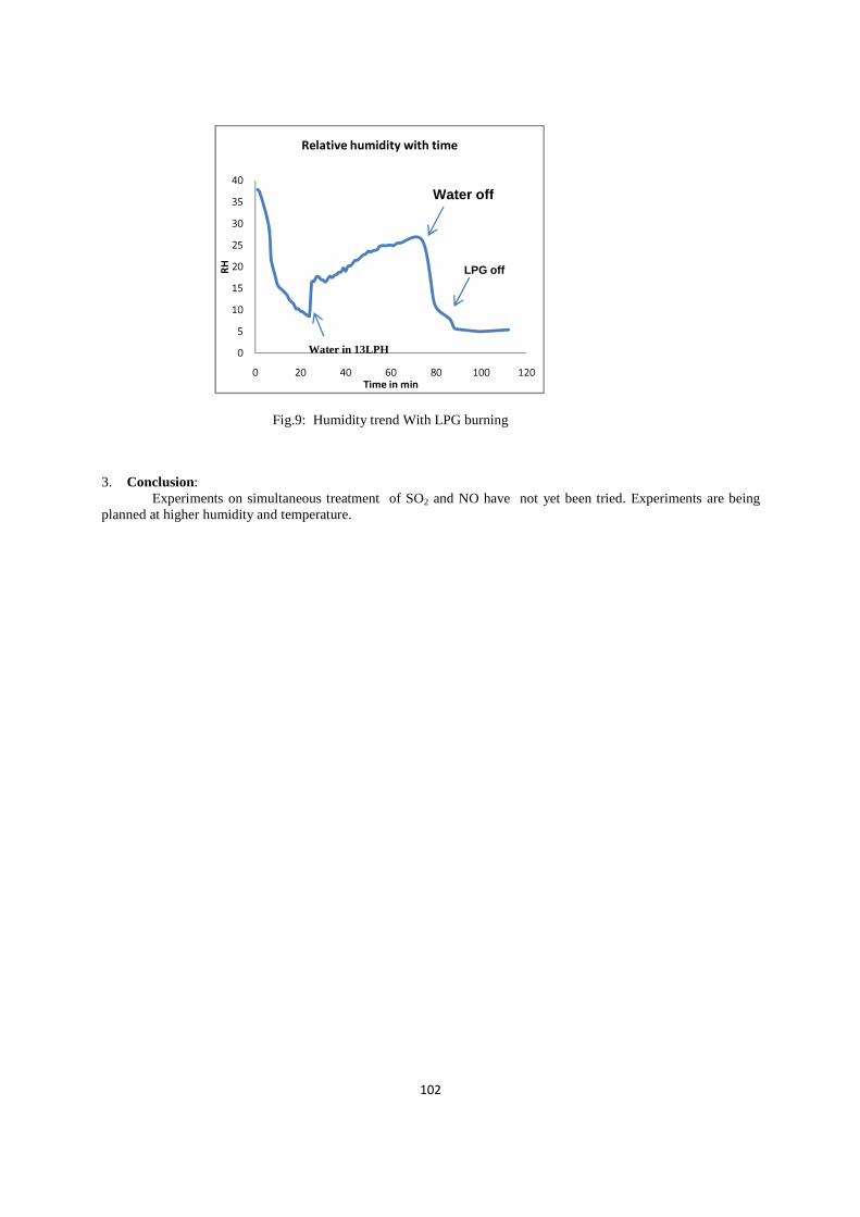
102
0
5
10
15
20
25
30
35
40
0 20 40 60 80 100 120
RH
Time in min
Relative humidity with time
Water in 13LPH
LPG off
Water off
Fig.9: Humidity trend With LPG burning
3. Conclusion: Experiments on simultaneous treatment of SO2 and NO have not yet been tried. Experiments are being
planned at higher humidity and temperature.

103
SIMULATION STUDIES ON FLUE GAS TREATMENT BY ELECTRON BEAM
S.K.Majji & S.Acharya Accelerator and Pulse Power Division, Bhabha Atomic Research Centre, Mumbai-400 085, India
Abstract: In the last two decades, experiments have been conducted on the treatment of flue gas with electron beam and it appears to be a promising technology. Our department has an ongoing plan of indigenous development of electron accelerators and a DC accelerator at 1 MeV energy, 5 kW power is already operational at Electron Beam Centre, Kharghar. Prior to carrying out demonstrative experiments on SOx and NOx treatment with this accelerator, simulation work was done using Geant4 for dose deposition in flue gas and chemical kinetics was studied using Chemical Kinetics Simulator (CKS). This paper reports the simulation results for the removal efficiencies of SOx and NOx considering 65 chemical reactions. The removal of SOx and NOx depends on the concentration OH radical produced by electron beam, initial concentrations of SOx and NOx and temperature of Flue Gas. The CKS code which runs the principle of stochastic algorithm converts the coupled differential rate equations to a random walk process for various species to obtain concentration of the pollutants as a function of time. In this code, most prominent reactions have been taken into account using their rate constants from literature. Parameters like initial concentration of SO2, NO2, temperature and dose were varied in these simulations. Based on the simulations, we can conclude that the dose rate required for the optimal removal of pollutants is 5kGy/sec for 1000 ppm of SO2 and 250 ppm of NOx, at 650C with 5% of water content by volume in the flue gas when the residence time is about 3 sec in the reaction vessel. It was found that electron beam dose significantly affects the removal efficiency of NOx while thermal reaction between SO2 and NH3 is dominant for SO2 removal. 1. Introduction
The Accelerator & Pulse Power Division of BARC is planning to conduct demonstrative experiments on the Flue Gas treatment by an indigenously developed DC accelerator currently operating at 1 MeV energy and 5 kW power. The paper presents some of the numerical computations to understand the radiation induced chemistry in the flue gas.
Simulation studies: The Depth Dose distribution in the Flue Gas was studied using the open source MonteCarlo software Geant4[1]. A 1 MeV, 5 mA electron beam was used in the simulation and we took into account photo-electric effect, ionisation, bremsstrahlung, Compton scattering and multiple scattering in the interactions. It was found that dose-rate of 5 kGy/s can be realised with these EB parameters. Homogenization of dose is expected to because of thermal motion of molecules in the Flue Gas. The simulation geometry with the scanning magnet, beam exit window and flue gas in reactor vessel is shown in Fig.1.
Chemical kinetics of flue gas during irradiation was studied using Chemical Kinetics Simulator(CKS) code[2]. The CKS code which runs on the principle of stochastic algorithm converts the coupled differential rate equations to a random walk process for various species. In stochastic formulation the reaction constants are regarded not as reaction “rates” but as reaction “probabilities per unit time”. The typical composition of a Flue gas mainly comprising Nitrogen, Oxygen and Carbon Dioxide was used in the simulation. Parametric studies were carried out by varying the concentrations of SO2, NO, NO2 , H2O and NH3 . If we consider a slice of the gas at the entry of the vessel and track its flow, it will be receiving EB dose during its motion from the entry of the reaction vessel to the exit. During this time oxidising radicals like OH, HO2, O3 etc. are generated and used up in the reactions occurring in the slice[3,4]. CKS allows us to calculate concentrations of all reactants and products in a chemical system as a function of time. In these simulations, the chemical reactions with their rate constants and initial chemical concentrations of
Fig. 2:Conc. vs time at 5kGy dose-rate
Fig: 1.Simulation Geometry in Geant4

104
species are taken as inputs. Simulation size is only limited by computer memory. This program terminates when all reaction probabilities are zero.
In present simulations pressure, temperature and volume are taken as constant. The reactions rates are given in the form of Arrhenius equation. The generation of secondary electrons from the primary beam cannot be simulated in CKS and as mentioned earlier, the dose deposition due to secondary particles is estimated using Geant4. The production rates for different species like N2
+, N+, O2+,O+ and excited states of atoms as well as
molecules per kGy dose can be calculated using their G-values. These production rates have been used in the simulations. It was noted from the reactions that OH radicals are produced dominantly by the interaction of the above ions with water rather than the direct dissociation of water by electrons.
2. Simulation results
In Fig.2, the results are presented for 1000 ppm of SO2, 200 ppm of NO, 50 ppm of NO2 1000 ppm of NH3 5% of water content by volume at 650C at dose rate of 5kGy/sec. After 3 seconds i.e 15 kGy dose, more than 80% of NOx is seen to be removed. Actually SO2 does not require much dose but 90% is removed in 4 seconds mainly due to the thermal reaction between it and ammonia. This was seen by running a simulation without ammonia for only SO2 with 20 kGy dose and getting removal efficiency of only 10-15%. Based on the simulations, we can conclude that the dose rate required for the optimal removal of pollutants is 5 kGy for the residence time of flue gas of about 3 sec in the reaction vessel.
The water content was varied from 5% to 15% by volume composition at 650C. Increased water content serves leads to larger production of OH radicals. Besides this, reaction rate of SO2 with OH radicals is enhanced when water molecule is the third body. It can be seen from Fig.3 and Fig.4 that marginal improvement in NO and SO2 treatment occurs in going from 5% to 15% humidity level. It is known that as the humidity is increased to 10-15% of water vapour by volume, number of acid molecules grow in the vessel, nucleation with water molecules, coagulation, condensation etc. will occur. The absorption of SO2 and NH3 in these aerosols and their subsequent conversion into ammonium sulphate in the liquid phase has not been considered in this part of the study.
The concentration of NO was varied from 100 ppm to 400 ppm, in the absence of SO2 and ammonia at a dose rate of 10kGy/sec. The change in concentration of NO, NO2 and HNO3 with time is shown in Fig.5 and Fig.6. NO is first converted to NO2 and so the rise of NO2 is seen for sometime. Afterwards, NO2 reacts with OH radicals to form nitric acid and then the concentration of HNO3 is seen to rise and saturate as no ammonia is present for neutralization. It is observed that more time and thus more dose is necessary to remove NO as its concentration is increased. One striking observation can be made from comparing Fig.1 and Fig.5. At a dose of 12 kGy, 200 ppm of NO becomes almost nil in Fig.2 whereas the same thing happens at 5kGy dose in Fig.6 in the presence of 1000 ppm of SO2. This indicates that the presence of SO2 helps in the removal of NO.
Fig. 3: Effect of 5% humidity Fig. 4: Effect of 15% humidity

105
Fig. 5: Decline of NO concentration from 100ppm Fig.6: Decline of NO concentration from 200ppm
Temperature was varied from 650 C to 950 C to study its effect on the process. It is seen that as the temperature rises, the removal efficiency of SO2 deteriorates. There is hardly any impact on the removal of NOx.
Fig. 7: Conc. vs time at 650 C Fig. 8:Conc. vs time at 950 C The initial SO2 concentration was varied from 250 ppm to 1750 ppm and accordingly ammonia concentration was varied from 250 ppm to 1750 ppm. The time taken for significant removal was found to be around 3 seconds as can be seen from Fig.9 and Fig.10. At higher dose rates the removal efficiency of SO2 is not increasing concluding that the SO2 removal is weakly dependent on Dose and thermal reactions of SO2 is dictating time for its treatment.
Fig. 9: initial concentration of SO2: 250ppm Fig. 10: initial concentration of SO2:1750ppm
106
3. Conclusion
A set of 65 reactions have been used with measured rate constants taken from publications for simulation of chemical kinetics induced by the interaction of an electron beam with flue gas. The simulation can be improved further by taking into account more reactions after consulting additional literature. The simulation indicates that efficient removal of SO2 and NOx is possible at 5% humidity at 65-750 C with radiation dose between 10-15 kGy[5,6]. It is found that NO and NO2 are more readily removed by radiation. The thermal reaction between ammonia and sulphur dioxide contributes significantly to removal of sulphur dioxide. The simulation results will be compared with experimental results when elaborate experiments with a 1 MeV, 5 kW will be carried out.
Acknowledgment
The authors would like to sincerely thank Dr. K.C. Mittal, former Head, Accelerator & Pulse Power Division, BARC for his encouragement during this work. 4. References: 1) Book for Application Development of GEANT4. http://geant4.web.cern.ch/geant4/G4UserDocuments/UsersGuides/ForApplicationDeveloper/html (2005). 2) Chemical Kinetics simulator 1.0 User Manual, IBM Almaden Research Center, IBM Corporation, 1995. 3) G.Y. Gerasimov tal Homogenous and Heterogenous Radiation induced NO and SO2 removal from power plants flue gases-Modelling Study, Radiation Phys. Chem 48, Vol 6, 763-769 4) Slides of Pawelec’s summer school downloaded from Internet 5) Chmielewski, A.G, et al “Radiation Processing: Environmental Applications”, IAEA -RPEA, 2007, ISBN 92-0-100507-5. 6) A. G. Chmielewski et al “Operational experience of the industrial plant for electron beam flue gas treatment”, Institute of Nuclear Chemistry and Technology, Dorodna 16, 03-195 Warsaw, Poland, Radiation Physics and Chemistry pp.439–442, (2004).

107
GAMMA RAYS ACCELERATED SEED GERMINATION AND PHYSIOLO GICAL ATTRIBUTES IN CANARIUM STRICTUM ROXB.
Akshatha and K.R.Chandrashekar* Department of Applied Botany, Mangalore University, Mangalagangothri, 574199, Karnataka, India.
*Corresponding author: Tel:0918242287271; Fax: 0918242287347 E-mail address: [email protected]
Abstract: Impact of presowing gamma irradiation on Canarium strictum Roxb. was studied by subjecting seeds to different doses of radiation (0 Gy to 200 Gy). Enhanced germination speed and percentage along with two fold increase in vigor was noticed at 50 Gy dose. An increase in total chlorophyll content might have helped in improving photosynthetic assimilation (6.30 umol CO2/m
2/s) leading to enhanced carbohydrate production. Increased phenolic, proline content and antioxidant activity was also noticed with increased dose.
1. Introduction
From past three decades applications of radioisotopes in scientific world has steadily increased starting from scientific research and further implements in agriculture and industrial production [1]. As an ionizing radiation gamma rays interact with molecules and produces free radicals which may modify morphology, anatomy, biochemistry and physiology of plants. These changes in-turn lead to the modification in the rate of photosynthesis, antioxidant system and accumulation of phenolic compounds [2]. In the present study effect of gamma irradiation on one of the endemic species of Western Ghats Canarium strictum Roxb is discussed where germination of these seeds is exceptionally difficult due to its hard seed coat and slow germination in the natural condition [3]. 2. Materials and Methods
Mature Seeds of Canarium strictum Roxb. were collected from the Guddekeri region (13°33'50.68", 75° 7'41.83"E and 2179 feet elevation) of Agumbe, Karnataka, India. Hundred seeds were taken in five polypropylene bags with 20 seeds each and exposed to gamma radiation. Likewise all the five groups were irradiated to six different doses (25 - 200 Gy) of gamma irradiation using Co60 gamma source at BARC (Bhabha Atomic Research Center, Mumbai).
All the seeds were kept for germination on sand bed at 2 cm depth in the green house of Applied Botany Department, Mangalore University. Speed of germination (S) was determined using the following equation (1)[4]. S= (N1x1)+(N2- N1)×1/2+(N3-N2) x1/3……..+(Nn-Nn-1)×1/n.
With N1, N2,,N3……… Nn-1, Nn.=Proportion of germinated seeds observed at 1,2,3……up-to n-1 and nth day.
Germination percentage (G) was calculated after completion of germination at 140 days. Germination was monitored daily.
Shoot length, root length and number of leaves were measured on the day of harvesting. Relative growth rate was measured in terms of dry weight by keeping the plants in an oven at 103oC for 18 hours. Vigor was determined using the following Equation, where V=Vigour index, %G=germination percentage, ASL=Average shoot length and ARL=Average root length.
V=%G× (ASL+ARL)
Total chlorophyll content [5], proline content [6], total carbohydrates [7] and phenolic content[8] of the plants were estimated. DPPH (2‑diphenyl‑1‑picrylhydrazyl) radical scavenging activity was determined using Mensor et al.[9] method. Data obtained were subjected to statistical analysis using IBM SPSS 20 statistical software (SPSS Inc., Chicago, IL, USA) in completely randomized block design.
3. Results and Discussion
Higher on par germination percentage was observed at 25, 50 and 100 Gy as illustrated in Table. 1. Compared to all other treatments 50 Gy showed highest percentage of germination (83%) whereas control ones scored only 50%. Lower doses of 25 Gy and 50 Gy significantly enhanced the germination speed. Kuzin et al. [10] opined that increase in germination percentage and speed at different doses of gamma irradiation might be due to their stimulating effects on activating RNA synthesis or protein synthesis. Substantial increase in shoot and root length was also evident. A two fold increase in vigor index was noticed with the exposure to 50 Gy compared to

108
control group. An on par increment in the total number of leaves per plant was noticed at different doses of irradiation, ranging from 50 Gy to 200 Gy compared to control plants. Similarly, increase in vigor index of gamma irradiated seeds were observed at 10 and 20 krad in tomato and okra seeds respectively [11-12]. The results obtained for C. strictum is almost similar to previous reports.
Table 1.Effect of different doses of gamma irradiation on germination and growth parameters of Canarium strictum Roxb. Means within a column followed by the same letter are not significantly different (P≤ 0.05). The data shown are means of five replicates ±SD.
Significantly highest amount of chlorophyll-a was observed at 50 Gy compared to control (Table.2). On par
higher concentrations of chlorophyll b was observed in control, 50 Gy and 100 Gy. On par total chlorophyll content was observed in control, 50 and 100 Gy, the highest being in 50 Gy (11.32 mg/g FW). Plants irradiated at 16 Gy showed a significant increase in their chlorophyll content which was correlated to the stimulated growth in red pepper [2]. Enhanced production of carbohydrate was noticed at two doses of gamma irradiation viz. 50 Gy and 100 Gy which was significantly higher than the control plants (Table. 2). Irradiation with 30 Gy showed highest value of total sugar content in snap beans. Significantly higher on par proline content of 0.93501 µ moles/g FW was observed in the seedlings exposed to 50 and 150 Gy followed by seedlings exposed to 100 (0.6926 µ moles/g FW) and 200 Gy (0.7237 µ moles/g FW) irradiation dose compared to control plants (0.6025 µ moles/g FW) (Fig.1). Present study revealed enhanced phenolics production in all the seedlings obtained from irradiated seeds compared to control. The highest on par phenolic content 0.567 mg/g and 0.547 mg/g fw at 25 and 150 Gy respectively (Tabe.2). On par DPPH radical scavenging activity was observed at 3 different doses of radiation viz 25 Gy (92.37%), 100 Gy (94.95%) and 150 Gy (95.9%) which was nearly 20 % higher than the control (67.9%). Plant phenolics are one of the potent scavengers of these free radicals and increment in antioxidant activity was compared with the total phenolic compounds. It has been found in the literature that there was an increase in the activity of peroxidase (potent antioxidant enzyme) at 40 Gy and 50 Gy in the plantlets of Citrus sinensis Kiong [13] irradiated with gamma rays. Table 2. Effect of different doses of gamma irradiation on total chlorophyll, carbohydrate, phenolics content and DPPH radical scavenging activity of C. strictum. Means within a column followed by the same superscript are not significantly different (P≤0.05).
SL. No
Dose Germination% Germination speed
Shoot length(cm)
Root length(cm)
Vigor index(V) No.of leaves
Dry weight(g)
1 Control 50±2.8bc 0.1487±0.02b 23.7±3.1b 11±3.0 b 1743.9±301cd 4.8±0.65b 0.96±0.178a 2 25Gy 72.5±4.9ab 0.237±0.05a 24.2±3.2b 14.2±2.59a
2767±422a
4.739±0.75b 0.996±0.096a 3 50Gy 83±7a 0.29±0.06a 27.9±6.1a 14.24±4a 3337±820a 5.3±1.1ab 1.06±0.15 a 4 100Gy 72.5±4.9abc 0.21±0.02b 22.9±9.1bc 14.8±3.8a 2133.6±711a 5.65±0.88a 1.01±0.264a 5 150Gy 59±1.4c 0.14±0.01b 15.8±4.3d 14±14a 1365.9±334ab 5.6±1.4a 0.36±.0.18 c 6 200Gy 48±5.6bc 0.16±0.005b 19.3±4.3cd 13.4±2.98a 1703±440a 5.59±1.2a 0.67±0.259b
Sl. No
Dose Chlorophyll content ( mg/g FW) a b Total
Total carbohydrate (mg/g DW)
Phenolic content mg/mg extract
DPPH scavenging activity(%)
1 Control 3.104±0.01bcd 7.259±0.56 a 10.36±0.554ab 102.1±4.949b 0.397±0.023d 67.9±2.96c 2 25Gy 3.746±0.73ab 5.269±0.23 c 9.011±0.49bc 89.75±2.33c 0.567±0.0128a 92.37±0.28ab 3 50Gy 4.035±.077 a 7.29±.0.43 a 11.32±.0.51 a 117.35±3.747a 0.47±0.014c 90.71±0.127b 4 100Gy 3.27±0.001abc 7.25±0.558 a 10.49±0.6ab 115.45±4.879a 0.52±0.01b 94.95±0.21a
5 150Gy 2.62±0.008cd 6.51±0.06ab 9.13±0.053bc 78.55±5.303d 0.547±0.0014ab 95.9±0.42a
6 200Gy 2.37±0.355 d 5.49±0.67bc 7.86±1.02c 50.25±2.89e 0.335±0.0089e 66.9±1.97c

c c
a
b
0
0.5
1
1.5
0 Gy 25 Gy 50 Gy 100 GyPro
lin
e c
on
ten
t in
µ
mo
les/
g F
W
Irradiation Dose
4. Conclusion
Conclusively, the present tolerance of C. strictum by producing protective metabolites and improvement of growth through increased production of carbohydrates by enhanced photosynthesis. Alleviation of phenolics and proline might help in overcoming the damage formed by free radicals produced during irradiation. Thus, it may be a useful management tool in afforestation projects in arid and semiarid areas as a promising technique for forestry improvement.
Acknowledgement
Authors are grateful to BRNS (Board of Research in Nuclear Sciences) for the financial support, Mangalore University for providing research facility. Authors also acknowledge BARC (Bhabha Research Center) for providing irradiation facility. 5. References
[1] Glubrecht. H., 1977. Future Trends in the Application of Isotopes and Radiation. IAEA Bulletin. 19 (6), 3847. [2] Kim, J.H., Baek, M.H., Chung, B.Y., Wi, S.G., Kim, J.S., 2004. Alterations in the photosynthetic pigments
and antioxidant machineries of red peJ Plant Biol. 47, 314-321.
[3] Iglesias-Andreu, L.G., Octavio-Low Doses of Gamma Radiation in Forest Species, Gamma
[4] Chiapuso, G., Sanchez, A.M., Reigosa, M.J., Gonzalez, L., Pellissieradequately reflect allelochemical effects on the germination process. J Chem Ecol. 23, 2445
[5] Arnon DL. Copper enzymes in isolated chloroplasts polyphenoloxidase in Beta Vulgaris. Plant Physiol 1949;24:1‑15. [6] Bates, L.S., 1973. Rapid determination of free proline for water stress studies. Plant and soil[7] Hegde, J.E., Hofreiter, B.T., 1962. In: Carbohydrate chemistry 17 (Eds.Whistler R.L. and Be Miller, J.N)
Academic Press, Newyork. [8] Taga, M.S., Miller, E.E., Pratt, D.E., 1984. Chia seeds as a source of natural lipid antioxidants. J Am. Oil
Chem Soc. 61, 928-931. [9] Mensor, L.I., Menezes, F.S., Leitao, G.G., Reis, A.S., Dos Santos, T., Coube, C.S., Leitao, S.G., 2001.
Screening of Brazillian plant extracts for antioxidant activity by the use of DPPH free radical method. Phytother Res.15, 127-130.
[10] Kuzin, A.M., Vagabova, M.E., Revin, A.F., 1976. Molecular mechanisms of the stimulating action of ionizing radiation on seeds. Activation of259-261.
[11] Nargis, S., Gunasekaran, M., Lakshmi, S., Selvakumar, P., 1998. Effect of gamma irradiation on seed germination and vigour of tomato (
[12] Arvind-Kumar., Mishra, M., 2004. Effect of gammapollen viability and plant survival in M1 and M2 generations of okra (Adv Plant Sci. 17(1), 295-297.
[13] Kiong, A.A., Ling Pick, S.H., Grace Lai., Harun, A.R., 2008. Physiological responses of stamineus L plantlets to gamma irradiation. Am
109
b
a
b
100 Gy150 Gy200 Gy
Irradiation Dose
Figure.1. Effect of different doses of gamma irradiation on proline content of Free radicals formed during irradiation, changes the plant cellular structure and metabolism like modulation of thylakoid membrane and antioxidative system [2]. Hence, enhancement in antioxidant activity and phenolics in be due to activation of defensive mechanism which could be correlated to earlier reports.
the present study confirmed the effective role of gamma-irradiation in increasing the by producing protective metabolites and improvement of growth through increased
production of carbohydrates by enhanced photosynthesis. Alleviation of phenolics and proline might help in ming the damage formed by free radicals produced during irradiation. Thus, it may be a useful
management tool in afforestation projects in arid and semiarid areas as a promising technique for forestry
to BRNS (Board of Research in Nuclear Sciences) for the financial support, Mangalore University for providing research facility. Authors also acknowledge BARC (Bhabha
esearch Center) for providing irradiation facility.
echt. H., 1977. Future Trends in the Application of Isotopes and Radiation. IAEA Bulletin. 19 (6), 38
Kim, J.H., Baek, M.H., Chung, B.Y., Wi, S.G., Kim, J.S., 2004. Alterations in the photosynthetic pigments antioxidant machineries of red pepper (Capsicum annuum L.) seedlings from gamma
-Aguilar, P., Bello-Bello, J., 2012. Current Importance and Potential Use of Low Doses of Gamma Radiation in Forest Species, Gamma Radiation. FerizAdrovic. 263Chiapuso, G., Sanchez, A.M., Reigosa, M.J., Gonzalez, L., Pellissier, F., 1997. Do germination indices
flect allelochemical effects on the germination process. J Chem Ecol. 23, 2445Arnon DL. Copper enzymes in isolated chloroplasts polyphenoloxidase in Beta Vulgaris. Plant Physiol
Bates, L.S., 1973. Rapid determination of free proline for water stress studies. Plant and soilHegde, J.E., Hofreiter, B.T., 1962. In: Carbohydrate chemistry 17 (Eds.Whistler R.L. and Be Miller, J.N)
M.S., Miller, E.E., Pratt, D.E., 1984. Chia seeds as a source of natural lipid antioxidants. J Am. Oil
Mensor, L.I., Menezes, F.S., Leitao, G.G., Reis, A.S., Dos Santos, T., Coube, C.S., Leitao, S.G., 2001. an plant extracts for antioxidant activity by the use of DPPH free radical method.
[10] Kuzin, A.M., Vagabova, M.E., Revin, A.F., 1976. Molecular mechanisms of the stimulating action of ionizing radiation on seeds. Activation of protein and high molecular RNA synthesis. Radiobiologiya
] Nargis, S., Gunasekaran, M., Lakshmi, S., Selvakumar, P., 1998. Effect of gamma irradiation on seed germination and vigour of tomato (Lycopersicon esculentum Mill). Orissa J Hortic. 26(2), 47
Kumar., Mishra, M., 2004. Effect of gamma-rays EMS and NMU on germination, seedling vigour, pollen viability and plant survival in M1 and M2 generations of okra (Abelmoschus esculentus
] Kiong, A.A., Ling Pick, S.H., Grace Lai., Harun, A.R., 2008. Physiological responses of
L plantlets to gamma irradiation. Am-Eurasian J Sustain Agric. 2(2), 135-149.
Effect of different doses of gamma irradiation on proline content of C. strictum.
formed during irradiation, changes the plant cellular structure and metabolism like modulation of thylakoid membrane and anti-
system [2]. Hence, enhancement in antioxidant activity and phenolics in C. strictum may be due to activation of defensive mechanism which could be correlated to earlier reports.
irradiation in increasing the by producing protective metabolites and improvement of growth through increased
production of carbohydrates by enhanced photosynthesis. Alleviation of phenolics and proline might help in ming the damage formed by free radicals produced during irradiation. Thus, it may be a useful
management tool in afforestation projects in arid and semiarid areas as a promising technique for forestry
to BRNS (Board of Research in Nuclear Sciences) for the financial support, CARRT, Mangalore University for providing research facility. Authors also acknowledge BARC (Bhabha Atomic
echt. H., 1977. Future Trends in the Application of Isotopes and Radiation. IAEA Bulletin. 19 (6), 38-
Kim, J.H., Baek, M.H., Chung, B.Y., Wi, S.G., Kim, J.S., 2004. Alterations in the photosynthetic pigments L.) seedlings from gamma-irradiated seeds.
Bello, J., 2012. Current Importance and Potential Use of 263-280.
, F., 1997. Do germination indices flect allelochemical effects on the germination process. J Chem Ecol. 23, 2445–2453.
Arnon DL. Copper enzymes in isolated chloroplasts polyphenoloxidase in Beta Vulgaris. Plant Physiol
Bates, L.S., 1973. Rapid determination of free proline for water stress studies. Plant and soil. 39, 205-207. Hegde, J.E., Hofreiter, B.T., 1962. In: Carbohydrate chemistry 17 (Eds.Whistler R.L. and Be Miller, J.N)
M.S., Miller, E.E., Pratt, D.E., 1984. Chia seeds as a source of natural lipid antioxidants. J Am. Oil
Mensor, L.I., Menezes, F.S., Leitao, G.G., Reis, A.S., Dos Santos, T., Coube, C.S., Leitao, S.G., 2001. an plant extracts for antioxidant activity by the use of DPPH free radical method.
[10] Kuzin, A.M., Vagabova, M.E., Revin, A.F., 1976. Molecular mechanisms of the stimulating action of protein and high molecular RNA synthesis. Radiobiologiya.16,
] Nargis, S., Gunasekaran, M., Lakshmi, S., Selvakumar, P., 1998. Effect of gamma irradiation on seed c. 26(2), 47-49.
rays EMS and NMU on germination, seedling vigour, Abelmoschus esculentus L. Moench).
] Kiong, A.A., Ling Pick, S.H., Grace Lai., Harun, A.R., 2008. Physiological responses of Orthosiphon 149.

110
Optimization of Activity Distribution for Radiation Sterilization of Healthcare Products in NIPRO Irradiator
Jain Reji George, P. Srivastava, B K Pathak Board of Radiation & Isotope Technology
Department of Atomic Energy BRIT/BARC Vashi Complex, Sector-20, Vashi, Navi Mumbai 400703, India
Email: [email protected]
ABSTRACT
Dose profile study was carried out for the NIPRO gamma irradiator before the source strength enhancement of the plant and on the basis of that, the design of activity distribution was optimized for improving the dose uniformity in healthcare products. The details of the study are discussed in this paper.
1. INTRODUCTION
The gamma irradiation plant of NIPRO India corporation pvt. ltd is situated in Satara district of Maharashtra. This plant is designed for maximum 5MCi Co60 source strength to be used for sterilization of healthcare products. The vertical rectangular source rack of dimensions 330 cm x 310 cm has 6 tiers for the arrangement of Co60 source pencils. Healthcare products in small cartons are to be loaded in Aluminum tote boxes of dimensions 105cm x 61cm x 183cm. There are two such totes in a carrier which moves in a shuffle - dwell fashion around the source rack along a 2+2 pass system. One tote occupies 16 dwell positions in lower level and another 16 dwell positions in the upper level during its movement around the source. The dwell positions are as shown in Fig.1.There were 34 source pencils of total activity 240.15 kCi loaded in the 6 tier source rack. This distribution of activity was giving a very high value of Dose Uniformity Ratio (DUR) [1]. Therefore there was a requirement of improving the DUR during the first source strength enhancement for which a dose profile study was done. Additional 882.5 kCi in 80 source pencils were to be added to the existing 240.15 kCi in 34 pencils.
2. MATERIALS AND METHODS FOR DOSE PROFILE STUDY
The 114 source pencils (34 old+ 80 new) of total activity 1122.65 kCi were of activity values varying from 5124 Ci to 14118 Ci. Dose profile was studied for various options of activity distribution. For this 27 target points in a tote box were chosen. For one target point in the product box, total dose received, from each source pencil, at all 32 positions of its movement in two levels were evaluated. The source pencils were BRIT made BC-188 type with 45.1 cm length and 1.11 cm diameter. These were assumed as line sources and analytical methodology [2] is used for evaluations. The exact geometries of the source-product system were measured from the AutoCAD drawings provided by NIPRO and the movement of totes was physically observed by visiting the plant before computation. Bulk density of healthcare products used in the plant was 0.103g/cc.
3. RESULTS
For the NIPRO gamma plant it was observed that if the activity is distributed in the middle 4 tiers out of the 6 tiers the DUR is 1.23, a better value than for a 6 tier distribution. A distribution in 6 tiers was giving a DUR equal to 1.43. Based on this, the loading pattern was optimized in such a way that the majority of activity is in middle 4 tiers (two lowest Ci pencils were kept in the upper and lower tiers, for identification when source rack is under water) as shown in Fig. 2 and the pencils were loaded accordingly. Later, the commissioning dosimetry conducted by BRIT [3] gave a DUR of 1.204 for healthcare products in NIPRO irradiator.
4. CONCLUSION
Distribution of activity has an important effect on the distribution of absorbed dose in materials undergoing radiation processing. The activity distribution design has to be optimized meticulously especially for sterilization of materials like healthcare products where the DUR requirement is very stringent.
ACKNOWLEDGEMENT

111
The authors wish to express their gratitude to the Staff of NIPRO for all the help provided during this work. Thanks are also due to Chief Executive, BRIT for the permission to present this work.
5. REFERENCES
[1]. IAEA TECHNICAL REPORTS SERIES No. 409, Dosimetry for food irradiation, 2002. [2]. Jain Reji George, Pradhan, A.S., Theoretical evaluation of dose distribution in product in radiation processing plants. Journal of Radiation Physics and Chemistry, 77, 2008. [3]. BRIT’s Dosimetry report of NIPRO Irradiator, 2013.

112

113
Effect of radiation processing in elimination of Klebsiella pneumoniae from food Raj Kamal Gautam, Vandan Nagar, Shashidhar R
Food Technology Division, Bhabha Atomic Research Centre, Mumbai 400 085 India.
Abstract
Klebsiella pneumoniae is an emerging opportunistic pathogen Foods such as fish, vegetables, infant feeds have been reported to be contaminated with Klebsiella spp. Radiation sensitivity of K. pneumoniae in different foods like sprouts, poultry and fish was carried out. The decimal reduction dose (D10) of Klebsiella pneumoniae MTCC 109 in saline, nutrient broth, mixed sprouts, poultry and fish samples were 0.11 kGy, 0.13 kGy, 0.14 kGy, 0.12 kGy, and 0.27 kGy, respectively. Radiation treatment with 1.5 kGy dose resulted in complete elimination of 105 CFU/g of Klebsiella pneumoniae MTCC 109. Even after enrichment and selective plating no recovery of K. pneumoniae MTCC 109 was observed in the 1.5 kGy treated samples stored at 4° C up to 12 days. The study shows that a 1.5 kGy dose of irradiation treatment is enough to eliminate 5 log CFU/g K. pneumoniae MTCC 109 from mixed sprouts, poultry and fish.
1. Introduction Klebsiella pneumoniae is well known as a cause of community-acquired bacterial pneumonia [1]. K. pneumoniae was included in the hazard identification category “B” by FAO and WHO on microorganisms, in powdered infant formula, which are considered to be an emerging public health issue [2]. Klebsiella species can cause severe infections that include meningitis, bronchitis, bacteremia, pneumonia, urinary tract infections in humans and animals [3]. Domestic animals such as cattle and horses are principal hosts for Klebsiella species. Improper farm management techniques and/or improper hygiene may facilitate contamination of water sources with Klebsiella species [4]. Most of the infections caused by Klebsiella resulted due to consumption of contaminated food such as fish and /or water [5]. Several physical and chemical treatment methods have been found to be ineffective in complete elimination of the food-borne pathogens under experimental conditions [6]. Radiation processing, a cold process, has been shown to be an effective method for eliminating food-borne pathogens in sprouts [7] and flesh products such as meat and fish [8]. Irradiation ensures the microbiological safety without compromising the sensory and nutritional properties of meat and poultry [9, 10]. The objectives of the current study were (i) to determine the D10 values of K. pneumoniae in saline. (ii) to determine the radiation dose for a 5-log elimination of K. pneumoniae from sprouts, poultry and fish samples and (iii) to study the survival and recovery of K. pneumoniae, if any, of K. pneumoniae in inoculated and radiation processed sprout, poultry and fish samples during storage at 4 °C for 12 days.
2. Material and methods
2.1. Decimal reduction dose (D10) in saline, nutrient broth, mixed sprouts, poultry and fish samples
To obtain desired cell density, overnight culture of K. pneumoniae MTCC 109 (≈ 109 CFU/ml) was centrifuged The harvested cells were re-suspended in 1.5 ml of sterile saline or nutrient broth and further diluted to obtain a cell concentration of 107 CFU/ml. In case of mixed sprouts, poultry and fish 105 cells were attached by separately dipping in sterile tap water (1.2 L) containing 107 CFU/ml of K. pneumoniae for 3 min and dried on sterile blotting paper to remove off excess water under aseptic conditions. These were irradiated to doses of 0, 0.1, 0.2, 0.3, and 0.4 kGy at 0-4 °C in a cobalt-60 irradiator (Gamma cell 220) at a dose rate of 4 Gy/min. Food samples were homogenized and total viable count was determined. The average number of surviving viable cells (CFU/ml) in the saline was plotted against the radiation dose. The slopes of the individual survivor curves were calculated by linear regression. The D10 value was calculated by taking the negative reciprocal of the survival curve slope.
2.2. Determination of the dose required to eliminate 5-log CFU/g of the inoculated cells
The decontaminated mixed sprouts, poultry and fish samples (10 g) were inoculated with K. pneumoniae as described earlier in section 2.7. The inoculated samples (105 CFU/g of K. pneumoniae) in triplicate were irradiated at 0, 1, 1.5 and 2 kGy in a cobalt-60 irradiator (GC-5000, BRIT, Mumbai), and the surviving population was determined by plating the serial dilutions with TSA after an incubation of 24 h at 37 °C.

114
2.3. Storage studies of gamma-irradiated mixed sprout, poultry and fish samples inoculated with K. pneumoniae
Decontaminated mixed sprouts, poultry and fish samples were inoculated with 105 CFU/g of K. pneumoniae, as described earlier. The inoculated packs were irradiated in melting ice (0-4 °C) with doses 0, 1, 1.5, and 2 kGy in a cobalt-60 irradiator (GC-5000, BRIT, Mumbai) and stored at 4 °C. The mixed sprouts, poultry and fish samples were screened for the presence of K. pneumoniae on the 0th, 4th, 8th, and 12th day. Enrichment and selective plating were carried out to confirm the complete elimination of the pathogens. Each experiment was repeated three times.
3. Results and discussion
3.1. D10 values of K. pneumoniae MTCC 109 in saline, nutrient broth, mixed sprout homogenate, fish homogenate, and poultry homogenate
K. pneumoniae MTCC 109 was found to be sensitive to gamma radiation, with mean D10 values in different medium ranging from 0.11 kGy to 0.27 kGy (Fig. 1). The mean D10 values in saline, nutrient broth, fish homogenate, sprout homogenate and poultry homogenate were 0.11, 0.13, 0.14, 0.12 and 0.27 kGy respectively. There are no reports available for the radiation sensitivity of an emerging food borne pathogen K. pneumoniae.
05
10
0 200 400
log
(CF
U/m
l)
Dose (Gy)
Saline
0
5
10
0 100 200 300 400 500
log
(CF
U/m
l)
Dose (Gy)
Nutrient Broth

115
Fig. 1. Survival of K. pneumoniae MTCC 109 in saline (117 Gy, -0.0086X+7.18, r2 = 0.99), Nutrient broth (136 Gy, -0.0073X+ 7.50; r2 = 0.95), Mixed sprout (142.8 Gy, -0.007X+7.43, r2 = 0.98), Poultry (119 Gy, -0.0084X+6.48, r2 = 0.99) and Fish homogenate (277 Gy, -0.0036X+5.95, r2 = 0.96) after gamma radiation. Each symbol represents plate counts at each dose. Average values of three experiments are plotted along with standard deviation.
Among different food homogenates, the maximum D10 value of K. pneumoniae was in fish homogenate (0.27 kGy). The D10 values in various food homogenates show higher radiation resistance in comparison to D10 values in saline and nutrient broth at 4°C. Venugopal [11] have reported difference in D10 values of different pathogens in different fish/shellfish medium at varied temperature and atmosphere. Dion [12] reported that at all dose rates; the bacteria were more radiosensitive when irradiated in a saline solution (0.85% NaCl) than in a poultry
0
5
10
0 200 400 600log
(CF
U/g
)
Dose (Gy)
Mixed Sprout Homogenate
02468
0 200 400 600
log
(CF
U/g
)
Dose (Gy)
Poultry Homogenate
01234567
0 100 200 300 400 500log
(CF
U/g
)
Dose (Gy)
Fish Homogenate

116
breast meat suspension. Intrinsic properties of the food products such as water activity, food composition, irradiation temperature, presence of oxygen, etc affect the D10 values of bacteria in food [6].
3.2. Radiation dose for 5-log elimination of K. pneumoniae from sprouts, poultry and fish samples
Treatment with 1kGy and above doses could eliminate 5-log CFU/g of K. pneumoniae from samples when survival of pathogen was checked immediately after irradiation. However, recovery of the pathogen in all the samples was observed in 1 kGy treated sample after enrichment and selective plating. This could be due to repair of radiation-induced damage during enrichment. No recovery of K. pneumoniae was observed in 1.5 kGy and 2 kGy treated mixed sprout samples after enrichment and selective plating.
3.3. Storage studies of irradiated sprout, poultry and fish samples inoculated with K. pneumoniae
K. pneumoniae counts were observed in control samples throughout the storage till 12 days at 4 °C. No viable counts were detected in 1 kGy treated samples up to 12 days of storage at 4 °C, whereas viable counts were observed in all of the above samples after enrichment. During storage of 1 kGy treated samples, variable recovery of Klebsiella pneumoniae was observed in some of the replicates, which may be due to uneven distribution of K. pneumoniae inocula. However, no viable counts and recovery of K. pneumoniae was observed in 1.5 kGy and 2 kGy irradiated samples during 12 days storage at 4 °C, even after enrichment and selective plating. Thus radiation treatment of mixed sprout, poultry and fish samples with 1.5 kGy can be sufficient to eliminate 105 CFU/g of K. pneumoniae.
4. Conclusions
K. pneumoniae in mixed sprouts, poultry and fish samples was found to be sensitive to gamma radiation. A radiation dose of 1.5 kGy is effective to eliminate 5-log reductions in K. pneumoniae. No growth was evident in all the 1.5 kGy radiation treated samples, even up to storage of 12 days at 4 °C. This is the first report of radiation sensitivity and inoculated pack study of one of emerging pathogen K. pneumoniae in food.
References 1. Puspanandan, S., Afsah-Hejri, L., Loo, Y.Y., Nillian, E., Kuan, C.H., Goh, S.G., Chang, W.S., Lye, Y.L., John, Y.H.T., Rukaryadi, Y., Yoshitsugu, N., Nishibuchi, M., Son, R. 2012. Detection of Klebsiella pneumoniae in raw vegetables using Most Probable Number-Polymerase Chain Reaction (MPN-PCR). Int. Food Res. J. 19(4), 1757-1762. 2. FAO-WHO (2004). Joint FAO/WHO workshop on Enterobacter sakazakii and other microorganisms in powdered infant formula, Geneva, 2-5 Feb, 2004. http://www.who.int/foodsafety/micro/meetings/en/report.pdf Last accessed on 30 July, 2014. 3. Siri, P.S., Sithebe, N.P., Ateba, C.N., 2011. Identification of Klebsiella species isolated from Modimola dam (Mafikeng) North West Province-South Africa. African j Microbiol Res. 5(23), 3958-3963. 4. Podschun R, Pietsch S, Höller C, Ullmann U., 2001. Incidence of Klebsiella species in surface waters and their expression of virulence factors. J. Appl. Environ. Microbiol., 67(7): 3325-3327. 5. Haryani, Y., Noorzaleha, A.S., Fatimah, A.B. , Noorjahan, B.A., Patrick, G.B., Shamsinar, A.T., Laila, R.A.S., Son, R., 2007. Incidence of Klebsiella pneumoniae in street foods sold in Malaysia and their characterization by antibiotic resistance, plasmid profiling, and RAPD–PCR analysis. Food Control. 18, 847–853. 6. Nagar, V., Bandekar, J.R., 2011. Effectiveness of radiation processing in elimination of Aeromonas from food. Radiat. Phys. Chem. 80, 911-916. 7. Saroj, S.D., Shashidhar, R., Panday, M., Dhokane, V., Hajare, S., Sharma, A., and Bandekar, J.R., 2006. Effectiveness of radiation processing in elimination of Salmonella Typhimurium and Listeria monocytogenes from sprouts. J Food Protect. 69(8): 1858-1864. 8. Meng, J.H., Doyle, M.P., 2002. Introduction: microbiological food safety. Microb. Infect. 4, 395–397. 9. AbuTarboush, H.M., AlKahtani, H.A., Atia, M., AbouArab, A.A., Bajaber, A.S., ElMojaddidi, M.A., 1997. Sensory and microbial quality of chicken as affected by irradiation and post-irradiation storage at 40 C. J. Food Prot. 60, 761–770. 10. Hashim, I.B., Resurreccion, A.V.A., McWaiters, K.H., 1995. Descriptive sensory analysis of irradiated frozen or refrigerated chicken. J. Food Sci. 60, 664–666. 11. Venugopal, V., Doke, S.N., Thomas, P., 1999. Radiation processing to improve the quality of fishery products. Crit. Rev. Food Sci. Nutr. 39(5), 391-440. 12. Dion, P., Charbonneau, R., and Thibault, C., 1994. Effect of ionizing dose rate on the radioresistance of some food pathogenic bacteria. Can J Microbiol. 40(5):369-74.

117
Utilization of Irradiated Onion Scales as a Potential Source of Bioactive Natural Food Colour
Sweetie R Kanatt1, Snehal Tari2 & S. P Chawla1
1 Food Technology Divsion Bhabha Atomic Research Centre
Mumbai 400085 2Department of Biotechnology
Ramnarain Ruia College of Arts and Science, Matunga, Mumbai
Abstract The use of onion in food industry introduces a large quantity of its scales as a waste product. Onion scales (OS) are prone to fungal contamination. The objective of this study was to evaluate the potential of using irradiated onion scales as a source of natural food color. Onion scales (OS) are prone to fungal contamination. The objective of this study was to evaluate the potential of using irradiated onion scales as a potential source of natural food colour. The extraction time, temperature and solvent were standardized for obtaining OS extract having maximum bioactivity and highest anthocyanin content. OS extracted in absolute ethanol containing 1M citric acid kept at 37⁰C for 48 hours was found to be optimum. The OS were irradiated at various doses (0-10 kGy) to prevent fungal contamination and dose of 6 kGy was found to be ideal. At this dose the OS extract had enhanced antioxidant activity and anthocyanin content and showed antibacterial activity against both gram positive and negative organisms. The total phenolic, flavanoid, carotenoid and anthocyanin content of irradiated OS extract was 40.20 mg/g, 9.72 mg/g, 181.73 mg/100g and 60.28 mg/100g respectively. The IC50 for 6 kGy irradiated OS extract for DPPH and superoxide radical scavenging activity was 462 µg and 886 µg respectively. The extract also had high Antioxidant Activity Coefficient (1014.9) and reducing power (0.74). Even at a concentration of 500 µg/ml irradiated OS extract had 81 % iron chelation capacity. Hence, OS can be used as a source of natural food colour due to its high anthocyanin content and excellent bioactive properties. i. Introduction In the food industry there has been an increasing trend towards the replacement of synthetic food colours by natural colours because of safety and health benefits. Onion scales (OS) are rich source of anthocyanins which could be tapped as a potential food colorant. India is the second largest onion (Allium cepa L.) growing country in the world and Maharashtra ranks first amongst the states in onion production. The aim of this study was extraction of anthocyanins from irradiated OS which could be used as a potential natural food colour with bioactive properties. ii. Experimental An ideal system was standardised for preparation of OS extract. Effect of irradiation on the phenolic, flavanoid and anthocyanin content was determined. The phenolic compounds present in OS extract were identified by HPLC. Antioxidant potential of irradiated OS extract was evaluated using various in vitro assays. Antibacterial potential of OS extract against common food pathogens/spoiler organisms namely E. coli, P. fluorescens, S. aureus, B. cereus was also studied. iii. Result & Discussion OS extracted in absolute ethanol containing 1M citric acid incubated at 37⁰C for 48 h had maximum phenolic and anthocyanin content. [A] Effect of irradiation on phenolic content OS were irradiated at various doses to prevent fungal contamination. From Table 1 it can be seen that extract prepared from OS irradiated at 6 kGy had maximum phenolic and anthocyanin content. Table 1. Effect of Irradiation on total phenolic, flavanoid and anthocyanin content RADIATION DOSE
0 kGy 2 kGy 4 kGy 6 kGy 8 kGy 10 kGy
Phenolic content (mg/g OS)
37.70 36.28 37.36 38.82 33.26 33.19
Flavanoids content (mg/g OS)
8.504 9.324 10.60 9.72 8.504 8.996
Anthocyanin content (mg/100g)
38.34 31.66 52.50 60.28 44.65 43.18

118
[B] Effect of irradiation on antioxidant potential of OS
Various in vitro assays were carried out to evaluate the antioxidant potential of OS extract. In case of DPPH radical scavenging activity irradiation did not affect the antioxidant potential of OS extract and incubating the OS in the extracting solvent for 48 hours improved its antioxidant potential (Fig. 1). Irradiated OS extract also showed excellent superoxide radical scavenging activity (Fig. 2) and high antioxidant activity potential as measured by beta carotene bleaching assay (Fig. 4). Iron chelation activity of OS extract decreased slightly on irradiation (Fig. 3).
0.0 0.5 1.0 1.5 2.0 2.5 3.0 3.5 4.0 4.5 5.0 5.50
20
40
60
80
10037oC 24 h
Concentration (mg/ml)
0 kGy 2 kGy 4 kGy 6 kGy 8 kGy 10 kGy
0.0 0.5 1.0 1.5 2.0 2.5 3.0 3.5 4.0 4.5 5.0 5.50
20
40
60
80
10037oC 48 h
% D
PP
H ra
dica
l sca
veng
ing
activ
ity
0.2 0.3 0.4 0.5 0.6 0.7 0.8 0.9 1.00
10
20
30
40
50
60
70
37oC 24 h
% S
uper
oxid
e ra
dica
l sca
veng
ing
activ
ity
Concentration (mg/ml)
0 kGy 2 kGy 4 kGy 6 kGy 8 kGy 10 kGy
0.2 0.3 0.4 0.5 0.6 0.7 0.8 0.9 1.00
10
20
30
40
50
60
70
8037oC 48 h
Fig.1. Effect of irradiation on DPPH radical scavenging activity of OS extract
Fig.2. Effect of irradiation on Superoxide radical scavenging activity of OS extract
0.2 0.4 0.6 0.8 1.0 1.2 1.440
50
60
70
80 37oC 24 h
% Ir
on c
hela
tion
Concentration (mg/ml)
0 kGy 2 kGy 4 kGy 6 kGy 8 kGy 10 kGy
0.2 0.4 0.6 0.8 1.0 1.2 1.4
40
50
60
70
80 37oC 48 h
1.0 1.5 2.0 2.5 3.0 3.5 4.0 4.5 5.0750
800
850
900
950
1000
1050
1100
1150
37oC 24 h
Antio
xida
nt A
ctiv
ity C
oeffe
cien
t (AA
C)
Concentration (mg/ml)
0 kGy 2 kGy 4 kGy 6 kGy 8 kGy 10 kGy
1.0 1.5 2.0 2.5 3.0 3.5 4.0 4.5 5.0700
750
800
850
900
950
1000
105037oC 48 h
Fig. 3. Effect of irradiation on Iron chelation activity of OS extract
Fig. 4. Antioxidant activity of OS extract estimated by β-carotene bleaching assay
[C] Identification of phenolics present in OS extract
The major phenolics present in the irradiated (6 kGy) OS extract was determined using HPLC and it was seen that all the major phenolics reported in OS such as Quercetin, Kaempferol and the major anthocyanin which is Cynidin-3-glucoside was present in the irradiated extract. Thus radiation processing of the OS did not result in any loss of the major phenolics present in it.

119
Fig. 5. HPLC profile of Onion Scale extract
[D] Antimicrobial activity of irradiated OS extract
The Minimum Inhibitory Concentration (MIC) of irradiated OS extract was only 2.5 µg/ml for B. cereus and 10 µg/ml for E. coli, P. fluorescens and S. aureus. It is noteworthy that OS which is a natural extract was also very effective against Gram negative organisms.
0 2 4 6 8 100
20
40
60
80
100% In
hibi
tion
Irradiation dose (kGy)
12.5 ugs 10 ugs 7.5 ugs 6.25 ugs 5 ugs 2.5 ugs 1.25 ugs
0 2 4 6 8 100
20
40
60
80
100
(MIC 10 ugs)
(MIC 10 ugs)
E coli
Pseudomonas
0 2 4 6 8 100
20
40
60
80
100S aureus
B cereus
% In
hibi
tion
Irradiation dose (kGy)
12.5 ugs 10 ugs 7.5 ugs 6.25 ugs 5 ugs 2.5 ugs 1.25 ugs
0 2 4 6 8 100
20
40
60
80
100
(MIC 10 ugs)
(MIC 2.5 ugs)
Fig.6. MIC of OS extract against Gram negative organisms
Fig.7. MIC of OS extract against Gram positive organisms
iv. Conclusion
Onion scales generated in large quantities as waste material in onion growing regions could be used as a potential natural food colorant with antioxidant and antimicrobial activity. Encapsulation of this extract could increase its stability and utility in food systems.
RReetteenntt iioonn tt iimmee PPhheennooll iicc ccoommppoouunndd
37.352 Quercitin
39.4 Kaempferol 38.785 Myrcetin
43.119 Cynidin-3-glucoside
40.619 Naringenic acid
35.352 Ferulic acid
34.185 Ellagic acid
5.9 Ascorbic acid

120
Thermoluminescence studies on Lithium and Calcium Borophosphate glass systems for radiation dosimetry in food irradiation
Bhaskar Sanyal1*, Madhumita Goswami2, V. Prakasan1, Aparna Patil2, S. P. Chawla1 1Food Technology Division, Bhabha Atomic Research Centre, Mumbai- 400 085, India
2Glass and Advanced Material Division, Bhabha Atomic Research Centre, Mumbai- 400 085, India *Email:[email protected]
Abstract Radiation processing of perishable food items is required to be carried out at sub-ambient temperatures (-10 to 4°C). Aqueous chemical dosimeters used in commercial food irradiation facilities are not suitable to measure absorbed dose at low temperatures. Solid state systems on the other hand could be a useful option to address this problem. Two glass systems namely Lithum Borophosphate (LBP:Dy) and Calcium Borophosphate (CaBP:Dy) doped with Dysprosium (1 wt%) were prepared by solid state route. Thermoluminescence (TL) properties of the systems were studied to assess the feasibility of the systems in measuring food irradiation doses. The glasses were exposed to gamma ray in the dose range of 0.5 to 4 kGy. Well defined TL glow with a linear dose response were observed for LBP system. However, a fast fading of TL output was found out as a limiting factor. In case of CaBP glass an enhanced TL output was observed with an improved TL stability. 1. Introduction
Study on radiation induced defect centres using thermoluminescence (TL) phenomenon in glasses has been an interesting subject of investigation in recent years, since such studies help for examining the suitability of the glasses for radiation dosimetry applications [1,2]. Radiation processing of perishable food items is required to be carried out at sub-ambient temperatures (-10 to 4°C) [3]. Aqueous chemical dosimeters used in commercial food irradiation facilities are not suitable to measure absorbed dose at low temperatures. Solid state systems on the other hand could be a useful option to address this problem. Lithium based glass system is a known and important starting material in this development for a long period, since its effective atomic number Zeff ~ 7.25 has the property of being nearly tissue equivalent that makes it as a very promising material in the field of radiation dosimetry [4, 5]. Recently TL studies were extended to several other amorphous materials which exhibit simple glow curve structure, a high gamma ray sensitivity, low fading of TL signal, linear dose-response relationship, simple annealing procedure for reuse, chemical stability and inertness to extreme climatic variations. Some of the recently investigated glass systems along these directions include phosphate, tellurite, SiO2 mixed bioactive glasses, alkali alumino silicate and borosilicate glasses [6, 7]. Several attempts were also made to enhance thermoluminescence (TL) sensitivity of these glass materials by adding different transition and rare earth metal ions to these glass samples [3, 7]. In the present study two glass systems namely Lithium Borophosphate (LBP) and Calcium Borophosphate (CaBP) doped with Dysprosium (1 wt %) were prepared. Thermoluminescence (TL) properties of the systems were studied to assess the feasibility of the systems in measuring food irradiation doses.
2. Experimental
Two glass systems namely Lithum Borophosphate (LBP) with a composition of 40 Li2O-20 B2O3-40 P2O5 doped with 1.0 wt% Dy2O3 and Calcium Borophosphate (CaBP) doped with Dy2O3 (1 wt %) were prepared by solid state route. The glass samples were distributed in 2 parts. One part was kept as control without any irradiation. Remaining part was again grouped into several aliquots to expose with gamma radiation in the dose range of 0.5 to 4 kGy using Gamma Chamber 5000 (Co 60, BRIT, India). TL for all the samples were measured immediately after irradiation and also during storage by increasing the temperature from ambient to 305 °C with a heating rate of 5 °C/s using a TL reader (Nucleonix Systems Pvt. Ltd, India).
3. Results and discussion
Fig 1a shows a well defined TL glow peak of irradiated (0.5 kGy) LBP:Dy glass sample. The glow curve was characterized by a dosimetric peak at around 134 C when recorded 1 hr after exposure to gamma radiation. The non-irradiated glasses did not show any measurable TL. Fig. 2 b exhibits the TL dose response of the sample after 1 hr and 4 days of irradiation. In both the cases linear dose responses of R = 0.988 and R = 0.998 were observed. In order to study the TL stability of the sample thermoluminescence measurements were carried out for a storage period of 8 days in dark at normal laboratory condition. Fig 2a shows the glow curve structures of the irradiated samples during storage. A considerable reduction in TL output of the order of 78 % was observed after 1 day of storage. The glow peak temperature at 136 °C was observed to be shifted towards higher temperature along with fast reduction of total integral TL. Similar observation has recently been reported in

121
Lithium based glass system doped with transition metal ion such as Mn2+ [8]. The fast detrapping of the charge carriers from the shallow traps corresponding to 136 C in amorphous matrix could be responsible for the observed TL behavior. Fig 2 b shows the complementary nature of the TL peak temperature and total TL output with time for this glass system. In radiation measurement the stability of the radiation induced signal is one of the important properties. But in LBP glass a fast fading of TL was observed as a limiting factor in spite of linear dose response behavior. In order to address this problem Calcium Borophosphate (CaBP:Dy) glass was synthesized and TL behavior was studied after irradiation as depicted in Fig 3. An enhanced TL output and improved dose response were recorded for this sample. After gamma irradiation the TL glow curve was characterized by two shallow peaks at around 117 and 188 °C. The peak intensity of the prominent high temperature peak (288 °C) exhibited linear dose response. The TL glows were recorded after 1 d storage and showed a slow fading of the high temperature peak of the order of 11 to 17%. The stable TL of the sample was possibly because of the deep trapped charges correlated to high temperature peak at around 288 °C.
4. Conclusion
This result suggested that the TL from irradiated amorphous systems has potential to measure radiation doses in food irradiation dose range where crystalline systems often exhibit TL saturation. However, optimization of glass composition, response to irradiation at sub-ambient temperature, and dosimetric characterizations would be necessary for practical applications.
4. Acknowledgements
The authors are thankful to Dr. S. P. Kale, Head, Food Technology Division & Associate Director (A), Bioscience group and Dr. K. Madangopal, Head, Glass and Advanced Material Division, Bhabha Atomic Research Centre, Mumbai, India.
5. References
[1] M.R. Chialanza, J. Castiglioni, L. Fornaro, J. Mater. Sci, 47 (2012) 2339. [2] E.L. Pires, S.H. Tatumi, J.C.R. Mittani, A. Kinoshita, O. Baffa, L.V.E. Caldas, Rad. Measurements 46
(2011) 1492. [3] B. Sanyal. V. Natarajan, S. P. Chawla, A. Sharma, Rad. Measurements 45 (2010), 899 – 905. [4] M. Szumera, I. Walawska, J. Thermal Analys. Calorimetry 108 (2012) 583. [5] M. Ignatovych, M. Fasoli, A. Kelemen, Rad. Phys. Chem. 81 (2012) 1528. [6] M. Kayhan, A. Yilmaz, J. Alloys Compd. 509 (2011) 7819. [7] A. K. Bakshi, B. Sanyal, V. J. Joshi, M. K. Bhide, V. Natarajan, A. Sharma, Appl. Radiat. Isotope, 69
(2011), 254 – 260. [8] B.J. R. Swamy, B. Sanyal, Y. Gandhi, R.M. Kadam, V. Natarajan, P. Raghava Rao, N. Veeraiah, J
Noncrystalline Solids 368 (2013), 40 – 44.
a) b)
50 100 150 200 250 300
0
50
100
150
200
250
300
350
400
132 C
LBP:Dy (1 wt %), 0.5 kGy, 1h
TL,
AU
Temperature, C
0 500 1000 1500 2000 2500 3000 3500 4000 45000
10000
20000
30000
40000
50000
4 days after irradiation
1 hr after irradiation
Inte
gral
TL,
AU
/mg
Dose, Gy
B Dose response after 1 h (0.99850) D Dose response after 4 d (0.99987)
Fig 1.a) TL glow curve after irradiation at 0.5 kGy, b) TL response with increasing dose of LBP:Dy glass sample

122
a) b)
50 100 150 200 250 3000
50
100
150
200
178 C
136 C
LBP:Dy (0.5 kGy) fading kinetics
TL,
AU
Temperature, C
0 day 1 day 2 day 4 day 7 day 8 day
0 2 4 6 8 101000
2000
3000
4000
5000
6000
7000
8000
LBP:Dy (1.0 wt%) TL & Peak temp with time
TL, AU/mg Peak Temp, C
Time, day
Int T
L, A
U/m
g
130
140
150
160
170
180
190
Peak T
emp, C
Fig 2. a) Behavior of TL glow curve during storage, and b) Integral TL and peak temperature with time for LBP:Dy glass
50 100 150 200 250 300
0
20
40
60
80
100
120
140
160Ca-Phosphate:Dy
0.0 0.5 1.0 1.5 2.0
0
5
10
15
20
25
30
Ca-Phosphate:Dy
TL
inte
nsi
ty, A
U/m
g
Dose, kGy
288 C
188 C
117 C
TL,
AU
Temperature, C
0 kGy 0.5 kGy 1 kGy 2 kGy
Fig. 3. Post irradiation TL glow and dose response of CaBP:Dy glass sample

123
Shelf Life Extension of Ready-To-Eat Sugarcane Cubes using Gamma Radiation Processing and Low Temperature Storage
Bibhuti Bhusan Mishra, Satyendra Gautam, and Arun Sharma Food Technology Division
Bhabha Atomic Research Centre Mumbai - 400085, INDIA
Abstract Sugarcane (Saccharum officinarum L.) stem is often eaten raw by chewing and liked by many because of its sweet taste and flavour. Due to poor shelf life caused by microbial growth and spoilage, sugarcane cubes cannot be marketed for fresh consumption. To address this issue a combination method including gamma radiation treatment (10 kGy) and low temperature (4°C) storage was found to extend the shelf life of packaged ready to eat sugarcane cubes up to 45 days, whereas, the control samples spoiled within 3 days. The total bacterial count, yeast and mold count, coliform and staphylococcus count were reduced below detection limit in the processed samples. At ambient and 10°C storage temperature, the shelf life of these processed sugarcane cubes were found to be about 4 and 16 days, respectively. The natural colour of the radiation processed samples was retained due to reduction in PPO and POD activity which leads to reduction in browning discolouration which was found to be radiation dose dependent. The sensory evaluation scores showed that the ready-to-eat sugarcane cubes were well acceptable during recommended storage period. Introduction Sugarcane (Saccharum officinarum L.) is one of the important commercial crops involved in the production of sugar crystals, jiggery, molasses, juices and its major center of production is Brazil followed by India (Mondal, 2014). Besides being an important source of various products of industrial importance, it is eaten raw by chewing due to high sucrose content and unique flavor. However, the peeling sugarcane stem with teeth and other manual modes are tedious. The peeled and sliced pieces of sugarcane are also very prone to microbial contamination and spoils with one day of processing. Due to limited shelf life caused by microbial growth and spoilage, sugarcane stem cubes cannot be marketed for fresh consumption. In current study, gamma radiation processing in combination with packaging and storage conditions were explored for shelf life extension of ready-to-eat sugarcane cubes. Experimental Sample preparation: The fresh sugarcane stem was cleaned, peeled and the internode parts were made into pieces (~3 cm length), packaged in radiation sterilized polyethylene packets. The gamma irradiation of samples performed in a cobalt-60 based package irradiator at FTD, BARC. The processed sugarcane cubes were stored at different temperatures (28 ±2, 10 ±2, and 4 ±2 ºC). Microbiological studies: Sugarcane samples (25 g) were aseptically homogenized for 2 min with 75 ml of sterile saline (0.85%) using Stomacher Blender (Stomacher Lab Blender, model 400, U.K). The homogenate was serially diluted using sterile saline and spread plated in duplicate. Total bacterial load (TBC) was determined by using plate count agar. The yeast and mold count (YMC) was determined using potato dextrose agar. Presumptive coliform were determined on violet red bile agar (VRBA) plates and Staphylococcus on Baird- Parker agar plates (Mishra et al., 2011). Sugar and Protein estimation: The total sugar including reducing sugars were estimated using dinitro-salicylic acid (DNSA) reagent and sucrose and glucose as standards, as detailed earlier (Mishra et al., 2015). The Bradford method was used for protein estimation (Mishra et al., 2012a). Browning discoloration measurement : Browning index was measured by reflectance measurement (Mishra et al., 2012a) using a Minolta CM-3600D spectrophotometer (Konica Minolta Sensing, Inc., Japan). The color parameters used were L* (Lightness). The extent of browning/darkening was calculated as 100-L (Lightness). Enzyme activities: Polyphenol oxidase (PPO) and Peroxidase (POD): The PPO activity was determined using 4-methylcatechol as substrate, where the oxidation of 4-methylcatechol catalyzed by PPO was measured at 410 nm using the method detailed earlier (Mishra et al., 2012b). The peroxidase activity was determined based on oxidation of guaiacol to tetraguaiacol polymer by the action of POD and measured at 470 nm as the method detailed earlier (Mishra et al 2012b). Texture analysis: The texture of the papaya cubes was analyzed using a Texture Analyser (TA.HD plus, Stable MicroSystems, UK) with a P/2N needle probe. The texture was expressed in terms of force (N) measuring resistance offered by the sample to the penetrating needle probe (Mishra et al 2015). Organoleptic evaluation: The evaluation was performed by a panel from Food Technology Division, BARC in a Taste Panel Laboratory (Mishra et al., 2015). The quality attributes including appearance, color, aroma, taste, and overall acceptance of juice were evaluated on a 9-point hedonic scale. (1-dislike extremely, 2-dislike very

124
much, 3-dislike moderately, 4-dislike slightly, 5-like slightly, 6-like fairly well, 7- like moderately, 8- like very well, 9-like extremely). Results & Discussion
The whole sugarcane stem used for eating was found to be contaminated with bacteria (~3 log cycle) including presumptive coliforms and Staphylococcus (> 2 log cycle) and also mold (~2 log cycle). The processing into cubes increased the counts by two fold. The gamma irradiation reduced the microbial counts in sugarcane cubes in dose dependent manner (Fig. 1). The absorbed dose of 10 kGy was found to be adequate for lowering microbial load below detection limit. During storage at 28 ±2, 10 ±2, and 4 ±2 ºC, the microbes were not observed in processed sample till 45, 16, and 4 days respectively.
The total and reducing sugar in sugarcane cubes were found to be in the range of 65-87 and 4.2-2.8 mg%, respectively. The radiation treatment of sugarcane cube did not show any significant effect on sugar content. The protein content was found to be in the range of 11-14 mg% in sugarcane cubes and was insignificantly affected by radiation treatment. The texture in terms of resistance Newton force (N) felt by the piercing needle probe of the processed cubes was found to be in the range of 10 to 14 N. The texture as shown in figure 2 was found to be insignificantly affected by radiation processing. The browning discoloration was observed in control stored sugarcane cubes. A radiation dose dependent reduction in browning was observed in processed samples (Fig. 2). The radiation treatment also resulted in reduction of PPO and POD activities in sugarcane cubes which probably helped in controlling post processing browning in the cubes (Fig 3).
0
1
2
3
4
5
6
whole 0 kGy 1 kGy 3 kGy 5 kGy 10 kGy
TBC
YMC
Coliform
Staphylococcus
Mic
rob
ial co
un
t (l
og
cfu
/g)
Figure 1. The effect of gamma radiation treatment on microbial count of ready-to-eat sugarcane cubes.

The organoleptic evaluation results showed that 10 kGy processed sugarcane cubes during storage at 4 ±2 ºC were acceptable till 45 days (Table 1). The scores foabove 6 (like fairly well) and above 7 (like moderately).
Table 1. Effect of gamma irradiation on organoleptic characteristics of radiation treatment (10 kGy) sugarcane cubes during storageDays of storage Appearance
1 7.5±1.0
10 7.2±0.8
20 7.4±0.6
30 6.8±1.1
45 6.7±0.8 Conclusion Gamma radiation treatment (10 kGy) and low temperature storage (4 ºC) significantly extended the shelf life of ready-to-eat sugarcane cubes. Without affectinmicrobial safety. Thus the radiation processing may provide a process to ensure the availability of the product in the market for fresh consumption. References [1]. Mondal, P. (2014). Sugarchttp://www.yourarticlelibrary.com/cultivation/sugarcanedistribution/20945/accessed December 08, 2014.[2]. Mishra, B.B., Gautam, S. Sharma, Agamma radiation processing. J. Food Sci. 76, M573[3]. Mishra, B. B., Gautam, S., & Sharma, A. (2012a). Browning of freshstorage. Postharvest Biology and Technology, 67, 44[4]. Mishra, B.B., Kumar, S., Wadhawan, S., Hajare, S.N., Saxena, S., More, V., Gautam, S., Sharma, A. (2012). Browning of litchi fruit pericarp: role of polyphenol oxidase, peroxidase, phenylalanine ammonia lyase and effect of gamma radiation. J. Food Biochem. [5]. Mishra, B.B., Gautam, S. Sharma, A. (2015). properties of intermediate moisture shelf stable ready
125
The organoleptic evaluation results showed that 10 kGy processed sugarcane cubes during storage at 4 ±2 ºC were acceptable till 45 days (Table 1). The scores for different quality attributes were found to be in the range of above 6 (like fairly well) and above 7 (like moderately).
Table 1. Effect of gamma irradiation on organoleptic characteristics of radiation treatment (10 kGy) sugarcane cubes during storage
Color Aroma Taste After taste
7.8±0.9 7.6±1.1 7.5±0.9 7.2±0.8
6.1±1.2 7.3±0.8 7.2±0.8 7.2±0.7
7.2±0.5 6.7±1.6 7.3±0.9 6.6±1.2
6.1±0.9 7.5±0.8 7.2±0.7 6.9±1.1
6.3±0.6 7.2±0.7 7.0±0.6 6.8±1.6
Gamma radiation treatment (10 kGy) and low temperature storage (4 ºC) significantly extended the shelf life of eat sugarcane cubes. Without affecting the overall quality attributes, the processing have ensured
microbial safety. Thus the radiation processing may provide a process to ensure the availability of the product in
Mondal, P. (2014). Sugarcane Cultivation in India: Conditions, Production and Distribution. http://www.yourarticlelibrary.com/cultivation/sugarcane-cultivation-in-india-conditions-productiondistribution/20945/accessed December 08, 2014. [2]. Mishra, B.B., Gautam, S. Sharma, A. (2011). Shelf life extension of sugarcane juice using preservatives and gamma radiation processing. J. Food Sci. 76, M573-78. [3]. Mishra, B. B., Gautam, S., & Sharma, A. (2012a). Browning of fresh-cut eggplant: Impact of cutting and
Biology and Technology, 67, 44–51. [4]. Mishra, B.B., Kumar, S., Wadhawan, S., Hajare, S.N., Saxena, S., More, V., Gautam, S., Sharma, A. (2012). Browning of litchi fruit pericarp: role of polyphenol oxidase, peroxidase, phenylalanine ammonia lyase
ffect of gamma radiation. J. Food Biochem. 36, 604-612. Mishra, B.B., Gautam, S. Sharma, A. (2015). Characterization of nutritional, organoleptic and functional
properties of intermediate moisture shelf stable ready-to-eat Carica papaya cubes. Food Bi
0
2
4
6
8
0 kGy 1 kGy 3 kGy
PPO sp …
Figure 3. The effect of radiation treatment on
The organoleptic evaluation results showed that 10 kGy processed sugarcane cubes during storage at 4 ±2 ºC r different quality attributes were found to be in the range of
Table 1. Effect of gamma irradiation on organoleptic characteristics of radiation treatment (10 kGy)
After taste Overall acceptability
7.1±0.7
7.6±0.5
6.5±0.8
6.8±0.6
6.4±0.9
Gamma radiation treatment (10 kGy) and low temperature storage (4 ºC) significantly extended the shelf life of g the overall quality attributes, the processing have ensured
microbial safety. Thus the radiation processing may provide a process to ensure the availability of the product in
ane Cultivation in India: Conditions, Production and Distribution. production-and-
helf life extension of sugarcane juice using preservatives and
cut eggplant: Impact of cutting and
[4]. Mishra, B.B., Kumar, S., Wadhawan, S., Hajare, S.N., Saxena, S., More, V., Gautam, S., Sharma, A. (2012). Browning of litchi fruit pericarp: role of polyphenol oxidase, peroxidase, phenylalanine ammonia lyase
Characterization of nutritional, organoleptic and functional cubes. Food Biosci. (in press).
5 kGy 10 kGyFigure 3. The effect of radiation treatment on

126
Low Dose Go/No Go Indicators
V. Prakasan, S. P. Chawla & Arun Sharma
Food Technology Division, Bhabha Atomic Research Centre,
Trombay, Mumbai 400085 Abstract Radiation processing is used for insect disinfestations, sprout inhibition, delaying ripening and shelf-life extension of agricultural commodities. In radiation processing facility the chances of inadvertent mixing of irradiated and un-irradiated products always exist. Visually noticeable radiation sensitive color indicator labels affixed on the product boxes can be used to identify processed commodities from unprocessed ones. For radiation processing that requires doses < 1 kGy no such radiation sensitive indicator labels are available. The objective of the present study was to develop colored agarose based gel system which operates at dose < 1 kGy. Colored agarose based gel system was prepared by incorporating methyl orange and chloral hydrate. The gel which was yellow colored initially undergoes progressive dose dependent discoloration. At a radiation dose of 400 Gy complete discoloration has been achieved. Result of pre-irradiation and post-irradiation storage stability studies showed that gel is stable when stored at 4°C and 25°C. The results of present study suggest agarose based indicator system have potential application in commercial low dose irradiation facility as go/no go indicator. Introduction In an irradiation facility, visually noticeable radiation sensitive indicator labels which undergo color change on irradiation are currently being used to identify processed commodities from unprocessed ones. Currently dye based indicator labels that work in the range of > 3 kGy are commercially used. For radiation processing that requires doses < 1 kGy, that include dose required for quarantine treatment, insect disinfestations, delaying ripening and sprout inhibition, no such radiation sensitive indicator labels are available. The aim of the present study was to develop indicator which works in the < 1 kGy radiation dose range. The study also investigates pre and post irradiation stability and utility of these indicators in radiation processing facility. Materials and Methods Preparation of agarose based radiation indicator: Agarose gel (1.5 % w/v) were prepared. Concentration of chloral hydrate and methyl orange indicator in gel were optimized. The resulting yellow colored gels were cut into rectangular shape (2.5 cm x 3.0 cm), sealed in polythene pouches and used in study. Dose response studies: Agarose indicators were exposed to different irradiation doses in 60Co Gamma cell 220 (AECL, Canada) having a dose rate of 4 Gy/min. The irradiation was carried out at the centre of the irradiation volume in air at ambient temperature. The indicators were sandwiched between two poly styrene plates having 3.5 mm thickness to have electronic equilibrium. Color changes were monitored visually as well as by spectrophotometric measurements. Pre and Post-Irradiation stability: Before and after irradiation the indicators were stored at different temperatures and absorbance was measured at regular intervals. Utility in radiation processing plant: To evaluate the performance of the system in radiation processing plant, the indicators were exposed to a radiation of 400 Gy in Food Package Irradiator at FIPLY. Results & Discussion Absorption spectra of indicators Absorption spectra of indicators exposed to gamma radiation is shown in Fig. 1. It can be seen that indicators had absorption maxima at 466 nm and there was gradual dose dependent discoloration as indicated by the gradual decrease in absorbance at 466 nm (Fig. 1, 2). The color of the indicators changed from yellow to colorless for a radiation dose of 400 Gy (Fig.3).

127
350 400 450 500 550 600 650-0.05
0.00
0.05
0.10
0.15
0.20
0.25
0.30
0.35A
bso
rban
ce
Wavelength (nm)
Control (0 Gy) 50 Gy 100 Gy 150 Gy 200 Gy 250 Gy 300 Gy
0 100 200 300 4000
20
40
60
80
100
% d
ecol
oura
tion
Absorbed dose (Gy)
[A0 -Ai / A0]*100
Fig. 1 Absorption spectra of indicator Fig. 2 Percentage reduction in OD at 466 nm
Fig. 3 Dose dependent color changes in agarose indicator Pre and post irradiation stability: The storage temperature may vary from place to place. To investigate effect of pre and post irradiation storage on color of indicator over a period of time stored at different temperatures, indicators were prepared and stored at different temperatures and checked for their absorbance. It was observed that the indicators retain their color when stored at 4˚C and 25˚C. The absorbance values remains stable over the period of time studied. But when stored at 37˚C, the indicators slightly change its color as indicated by the slight increase in absorbance after two weeks. There is no redevelopment of color at any of the temperature conditions studied. (Fig 3)
0 Day 1 wk 2 wk 3 wk 4 wk 6 wk 8 wk0.0
0.1
0.2
0.3
0.4
0.5
Abs
orba
ce a
t 466
nm
Time in week
control in dark control 4oC control 25oC control 37oC irradiated 4oC irradiated 25o C irradiated at 37o C
Fig 4 Pre and post irradiation stability of indicator Fig 5. Indicator response in Food Package Irradiator.

128
Utility in radiation processing plant To check the utility of the indicator in radiation processing plant, they were affixed on the periphery of the product boxes and exposed to a radiation of 400 Gy in Food Package Irradiator at FIPLY. The color change and absorbance readings were in agreements with the experiments conducted using Gamma cell (Fig. 4) Conclusions Low dose go/no go radiation indicator was developed which was inexpensive and could be easily prepared It had good pre and post irradiation stability and showed color change from yellow to colorless which can be commercially used in low dose irradiation facility being used for quarantine treatment of fruits.

Combined Effect of Gamma Irradiation and Frozen Storage on the Microbial, Bio- chemical Quality
and Shelf life of Shrimp
Manjanaik Bojayanaik a*, Kavya Naroth
a, Veena Shetty
b, Somashekarappa Hiriyur
c , Rajashekar Patil
c
a Department of Fish Processing Technology, Karnataka Veterinary, Animal and Fisheries Sciences
University, College of Fisheries, Mangalore – 575002, India. b Department of Microbiology, K S Hegde Medical Academy, Deralakatte, Mangalore- 575018, India.
c Centre for Application of Radioisotopes and Radiation Technology (CARRT),
Mangalore University, Mangalore-574199, India
E mail : [email protected]
Abstract Application of ionizing radiation in food is considered as one of the most efficient technological processes for the
reduction of microorganisms in food. It can be used to improve the safety and quality of the aquatic food products, and
to extend their shelf life to reduce post harvest losses. The aim of this study was to evaluate the combined effects of
gamma irradiation and frozen storage for improvement of microbial and bio- chemical qualityof fresh shrimp. Fresh
farm raised shrimp (Penaeus monodon) were procured and packed aseptically in a polyethylene bags, and exposed to
0.0 (un-irradiated), 1.0, 3.0, 5.0 kGy of gamma irradiation and held for refrigeration (4oC) and frozen storage (-18
oC)
temperatures. The control and irradiated shrimp samples were underwent periodically for microbial analysis (total
mesophilic counts, total coliforms, faecal coliforms, Salmonella, E. coli, Staphylococcus aureus) and chemical
characteristics (TMA-N, TVB-N, pH, TBARS) on different time intervals. Microbial analysis indicated that irradiation
and frozen storage had a significant effect (p< 0.05) on the reduction of microbial loads. The chemical parameters
(trimethylamine, total volatile base nitrogen values) for irradiated shrimp samples were significantly lower than the
non- irradiated samples at both storage temperatures and the rate of decrease was more pronounced in samples
irradiated at the higher dose (p<0.05). The results revealed that the combination of irradiation and frozen storage
resulted in overall reductions on microbial load and stabilized the biochemical characteristics of shrimp.
Key words : gamma irradiation, frozen storage, TBARS, TMA-N.
I. Material and methods
Fresh farm raised shrimp (Penaeus monodon) was procured from shrimp farm located near to Mangalore, India. The
sample was immediately packed in polyethylene bags with ice and brought to the laboratory and stored at 4oC until
irradiated. Each sample was divided into four packs with one of the pack being control (Non-irradiated) and the three
packs used for three levels of exposure to gamma irradiation of doses 1.0, 3.0 and 5 kGy. Samples were irradiated at the
Centre for Application of Radioisotope and Radiation Technology (CARRT), Mangalore University, India, using a 60
cobalt radiation source (Gamma Chamber 500, made in Board of Radiation and Isotope Technology, Mumbai, India).
The doses applied in this study were 1.0, 3.0 and 5.0 kGy at the dose rate of 6.947kGy/hour. The absorbed dose was
monitored by Freaky dosimeter (BRIT, Mumbai). After irradiation both the irradiated and non-irradiated (control)
samples were transported within 1hr to the laboratory with packed ice in isolated polystyrene ice boxes and held for
refrigeration (4oC) and frozen temperatures (-18
oC) until the last day of experiment. The frozen storage of the samples
lasted for 90 days.
Proximate Analysis
Moisture, crude protein, lipid and ash contents were measured according to the protocols recommended by AOAC
method (2005). The control and irradiated shrimp samples were underwent periodically for microbial (ICMSF, 1986;
APHA, 1998) and bio-chemical characteristics on different time intervals.
Microbial Analysis
Aerobic mesophile counts and pathogens were analyzed throughout the experiment for a period of 90 days. 25 grams of
sample were taken aseptically into a sterile blender containing 225 ml of sterilized physiological saline (0.85% NaCl)
and blended for 3 min at low speed. 0.1 ml of decimal dilutions were plated on to plate count agar and incubated at
35oC for 24-48hrs. Total plate counts were transformed into logarithms of the number of colony forming units per gram
of sample (CFU/g). For the enumeration of total coliforms 3 tube MPN method was followed and appropriate dilutions
were inoculated into LSTB, EC broth (Himedia, Mumbai) and incubated at 37 o
C and 44.5oC respectively. Pathogens
such as Escherichia coli using EMB agar (Himedia, Mumbai); Staphylococcus aureus using Baird parker agar
(Himedia, Mumbai); Salmonella using BSA agar (Himedia, Mumbai) and Vibrios using APW and TCBS (Himedia,
Mumbai).
Chemical analysis
Biochemical parameters such as pH (Vyncke, 1981), total volatile basic nitrogen (Beatty and Gibbons, 1937),
trimethylamine Nitrogen (Beatty and Gibbons, 1937), thiobarbituric acid reactive substance (Raghav and Multin, 2005)
were analyzed periodically until the last day of the experiment.

Diagramtic reperestnation followed for irradiation of shrimp
II. Results and discussion
Among the seafood consumed, shrimp being rich in proteins, free amino acids, minerals and other soluble nitrogenous
substances, demonstrates an exceptional nutritional value in the human diet (Konosu & Yamaguchi, 1982; Hocaglu et
al., 2012). The shrimp used in the study had 74.90% moisture; 22.43% protein; 1.609% crude lipids and 1.06% ash.
The combination of low dose gamma irradiation and refrigerated or frozen storage resulted in a significant reduction of
bacterial growth and irradiation at 3.0 and 5.0 kGy dose with frozen (-18oC) or refrigerated (4
oC) storage could inhibit
microbial growth significantly (Venugopal et al., 1999; Hocaglu et al., 2012). None of the human pathogens were
detected in both the samples except the faecal coliforms which were reduced completely upon exposed to irradiation
(Fig.1 & 2).
The employed radiation dose (1, 3 & 5 kGy) in conjunction with frozen and refrigerated storage extended the shelf-
life of shrimp 90 and 12 days respectively.
The levels of biochemical parameters such as pH, TBA, TVB-N and TMA-N in irradiated and non-irradiated shrimp
samples were also examined. Irradiated samples of shrimp had significantly lower concentrations of TVB-N and TMA-
N during their refrigerated and frozen storage (p < 0.05) as compared with the controls, which may be attributed to the
reduction of microbial population (Fig.3-10).
These parameters were within the acceptable limits until the end of both refrigerated and frozen storage in irradiated
samples except the value of TBARS were increased significantly in shrimp sample.
The irradiation at high dose (5 kGy) might enhance lipid oxidation in shrimp, although the growth of microorganism
was inhibited significantly.
Fig 1. Total plate count of shrimp sample
irradiated at 1,3, 5kGy and stored at 4oC
Fig 2. Total plate count of shrimp sample
irradiated at 1,3, 5kGy and stored at -18oC
0
2
4
6
8
1 4 7 12
CONTROL
1kGy
3kGy
5kGY
No. of storage days
log
cfu
/g
0
1
2
3
4
5
6
7
1 3 5 7 14 21 30 60 90
control
1kGy
3kGy
5kGy
No. of storage days
Exposed to Irradiation
Dose rate 6.947kGy/hr
0kGy 1kGy 3kGy 5kGy
Refrigeration (4oC) Frozen storage (-18oC)
No. of storage days
(upto 12 days)
No. of storage days
( Upto 90 days)
Shrimp
(P. monodon)
Microbial quality:
Mesophilic count,
E.coli, S. aureus,
Faecal coliforms
Salmonella, Vibrios
Biochemical quality
TVB-N, pH
TMA, TBARS
Packaged with
polyethylene bags

Fig. 3. TVB-N of shrimp samples
irradiated at 1, 3, 5kGy and stored at 4°C
Fig. 4. TVB-N of shrimp samples
irradiated at 1, 3, 5kGy and stored at -18°C
Fig. 5. TMA-N of shrimp samples
irradiated at 1, 3, 5kGy and stored at 4°C
Fig. 6. TMA-N of shrimp samples
irradiated at 1, 3, 5kGy and stored at -18°C
Fig. 7. pH of shrimp samples
irradiated at 1, 3, 5kGy and stored at 4°C
Fig. 8. pH of shrimp samples
irradiated at 1, 3, 5kGy and stored at -18°C
Fig. 9. TBARS of shrimp samples
irradiated at 1, 3, 5kGy and stored at 4°C
Fig. 10. TBARS of shrimp samples
irradiated at 1, 3, 5kGy and stored at -18°C
III. Acknowledgement Financial support for this work from the BRNS, Mumbai, Department of Atomic Energy, Government of India
is gratefully acknowledged.
IV. Literature cited
1. AOAC., 2005. Official Methods of Analysis AOAC International.Edt. Horwiz, W. Edn. 18,
Association of Official Analytical Chemist International, Gaithersburg, Maryland, USA.
2. APHA.,1998. Recommended Methods For The Microbial Examination of Foods, Broadway, American
Public Health Association, 19 :181-188.
3. Beatty, S. A. and Gibbons, N. E., 1937.The measurement of spoilage of fish.J. Biol. Bd. Can., 3: 77-91.
4. ICMSF, 1986. International commission on microbiological specifications for foods, sampling plans
for fish and shellfish. In: Microorganisms in Foods. Sampling for Microbiological Analysis: Principles
and Scientific Applications, second edn. University of Toronto Press, Toronto, pp. 181–196.
5. Raghavan, S. and Hultin, H. O., 2005. Model system for Testing the Efficacy of Antioxidants in Muscle
Foods.J. Of Agric. Food Chem., 53: 4572-4577.
6. Venugopal, V. Doke, S. N. and Thomas, P., 1999. Radiation processing to improve the quality of fishery
products. Critical Review In Food Science and Nutrition, 39(5): 391-440. and others.
0
10
20
30
40
50
60
1 4 7 12
CONTROL
1kGy
3kGy
5kGY
No. of storage days
mg
N/1
00
g
0
2
4
6
8
10
12
14
1 3 5 7 14 21 30 60 90
control
1kGy
3kGy
5kGy
No. of storage days
mg
N/1
00
g
0
5
10
15
20
1 4 7 12
CONTROL
1kGy
3kGy
5kGY
No. of storage days
mg
N/1
00
g
0
1
2
3
4
5
6
1 3 5 7 14 21 30 60 90
control1kGy3kGy5kGy
No. of storage days
mg
N/1
00
g
0
2
4
6
8
10
1 4 7 12
CONTROL
1kGy
3kGy
5kGY
No. of storage days
pH
6
6.5
7
7.5
8
8.5
1 3 5 7 14 21 30 60 90
control1kGy3kGy5kGy
No. of storage days
pH
0
0.5
1
1.5
1 4 7 12
CONTROL
1kGy
3kGy
5kGy
mg
MD
A/k
g
No. of storage days
0
1
2
3
4
5
1 3 5 7 14 21 30 60 90
control
1kGy
3kGy
5kGy
No. of storage days
mg
MD
A/k
g