new spectrophotometric method for the assessment of
TRANSCRIPT

1
5
6
789
101112
New Spectrophotometric Method for the Assessment of Catalase
Enzyme Activity in Biological Tissues
Thulfeqar A. Hamza1, Mahmoud Hussein Hadwan 2, ∗ 1 pathological analysis Dept., Al-Mustaqbal University College, Hilla city, Babylon Governorate, Iraq 2 Chemistry Dept., College of Science, University of Babylon, Hilla city, Babylon Governorate,
p.o. 51002, Iraq∗ Corresponding author.
Email address: [email protected]
14
15
16
17
18
19
20
21
22
23
24
25
26
27
28
29
30
31
32
33
34
35
36
Abstract:
Background: Catalase is a vital antioxidant enzyme that dismutates H2O2 into water and
molecular oxygen. Many protocols have been developed to measure catalase enzyme
activity. Spectrophotometric methods are the most common assays that used to
assess catalase enzyme activity.
Methods: Because the rate-limiting step during catalase enzyme activity depends upon
the dissociation of hydrogen peroxide, the developed assay measures the reaction
between a hydroquinone/anilinium sulfate/ammonium molybdate reagent and
Unreacted Hydrogen Peroxide, which results in the production of a purple, disubstituted
quinone compound with a maximum absorbance value at 550 nm.
Results: To clarify the precision of the developed method, the coefficients of variation
were determined to be 2.6% and 4.7% for within run measurements and between run
measurements, respectively. This method returned results that correlated well (r =
0.9982) with the results returned using the peroxovanadate method to assess catalase
enzyme activity. Additionally, we examined the use of the newly developed
hydroquinone assay to measure catalase enzyme activity in liver and bacterial
homogenate samples.
Conclusion: These results demonstrated that this assay can be used for scientific
research and routine health applications because it is inexpensive, simple, accurate, and
rapid. This method is suitable for use in clinical pathology laboratories because it is
simple and produces precise and reproducible results.
Keywords: catalase activity; hydrogen peroxide; hydroquinone; anilinium sulfate;
ammonium molybdate. 37
Preprints (www.preprints.org) | NOT PEER-REVIEWED | Posted: 29 October 2019 doi:10.20944/preprints201910.0323.v1
© 2019 by the author(s). Distributed under a Creative Commons CC BY license.
Peer-reviewed version available at Current Analytical Chemistry 2020, 16; doi:10.2174/1573411016666200116091238

2
1. Introduction38
Catalase is a vital antioxidant enzyme that dismutates H2O2 into water and molecular 39
oxygen [1]. Catalase can be found in animal cells, plant cells, and aerobic 40
microorganisms, and within the human body, catalase has been identified in 41
erythrocytes, kidney, and liver, where it is typically considered to be a component in 42
the major defense system against H2O2 formation [2]. Measuring changes in the 43
endogenous antioxidant activity of catalase enzyme has previously been used to 44
quantify reactive oxygen species [3]. Therefore, the assessment of catalase enzyme 45
activity is essential and vital to the evaluation of oxidative stress in biological tissues. 46
Many protocols have been developed to measure catalase enzyme activity. The 47
most common assay used to assess catalase enzyme activity is the ultraviolet (UV) 48
spectrophotometric protocol, which measures the catalase enzyme-mediated 49
dismutation of hydrogen peroxide into molecular oxygen and water as a change in 50
absorbance at 240 nm [4]. However, the application of the UV spectrophotometric 51
method is limited for the following reasons [5]. First, catalase enzyme activity can be 52
inhibited by alterations in the structures of catalase active sites that occur in the presence 53
of high concentrations of H2O2. Second, DNA and proteins absorb UV light, making 54
the UV spectrophotometric method unsuitable for the assessment catalase enzyme 55
activity in biological tissues [6]. 56
Spectrophotometric methods have widely been used to estimate catalase 57
enzyme activity, using diverse reagents to form colored complexes that absorb light 58
within the visible spectrum, such as the ferrous oxidation in xylenol orange (FOX) assay 59
[7] that absorbs at 560 nm, the carbonate cobaltate(III) complex that absorbs light at60
440 nm [8], the indamine dye with a water-soluble ironporphyrin [9] that absorb at 590 61
nm, and the peroxovanadate complex (NH4[VO(O2)SO4) that absorbs light at 452 nm 62
[10]. Shivakumar et al., [11] utilized an isonicotinic acid hydrazidepyrocatechol system 63
to examine the dissociation of hydrogen peroxide and assess catalase activity. 64
Optical sensors have also been used to assess catalase activity. For the 65
continuous determination of hydrogen peroxide concentrations, Posch and Wolfbeis 66
[12] described three types of sensors that measure the oxygen levels formed in response67
to the dissociation of hydrogen peroxide by a specific catalyst. The fluorescence 68
quenching of a silica gel-adsorbed dye entrapped in silicone rubber was utilized to 69
Preprints (www.preprints.org) | NOT PEER-REVIEWED | Posted: 29 October 2019 doi:10.20944/preprints201910.0323.v1
Peer-reviewed version available at Current Analytical Chemistry 2020, 16; doi:10.2174/1573411016666200116091238
3
quantify changes in oxygen levels and applied to the assessment of catalase enzyme 70
activity. Cohen and Weber [13] described a device for the detection and generation of 71
hydrogen peroxide, in situ, using gold-coated optical fiber. This device 72
photochemically produces and electrochemically senses H2O2 in aqueous buffered 73
samples and was utilized to assess catalase enzyme activity. Finally, Bekdeşer et al., 74
[14] developed a low-cost and rapid optical sensor based cupric reducing antioxidant75
capacity (CUPRAC) assay to assess catalase enzyme activity in liver and kidney tissue 76
homogenates. 77
Special methods have been developed based upon the estimation of liberated 78
oxygen concentrations, in response to the dissociation of H2O2. Oxygen generation can 79
be quantified using a low-flow gas meter [15] or oxygen electrodes [16]. 80
Another method utilizes different probes in the presence of peroxidase or 81
peroxidase mimics [17, 18, 19] to detect the residual H2O2 concentration. However, 82
peroxidase is expensive and very unstable in solution [20]. 83
In this manuscript, we report a simple spectrophotometric method for rapidly 84
measuring catalase activity, based on the measurement of H2O2. This method utilizes a 85
reaction between a hydroquinone/anilinium sulfate/ammonium molybdate reagent and 86
unreacted hydrogen peroxide, which yields a purple, disubstituted quinone compound 87
with a maximum absorbance at 550 nm. This new method is inexpensive, rapid, simple, 88
and accurate and can be applied to systematic research analyses and routine clinical 89
measurements. 90
2. Materials and Methods91
2.1 Chemicals 92
All chemical substances were attained from standard commercial suppliers. 93
2.2 Principle 94
The current method utilized the reaction between a hydroquinone/anilinium sulfate 95
/ammonium molybdate reagent and unreacted hydrogen peroxide, which forms a 96
purple, disubstituted quinone compound with a maximum absorbance at 550 nm. The 97
reaction between hydroquinone and aniline in the presence of H2O2 is shown in Scheme 98
1 [21]. 99
Preprints (www.preprints.org) | NOT PEER-REVIEWED | Posted: 29 October 2019 doi:10.20944/preprints201910.0323.v1
Peer-reviewed version available at Current Analytical Chemistry 2020, 16; doi:10.2174/1573411016666200116091238

4
100
Scheme 1. Proposed reaction for the formation of purple -colored product of the 101
disubstituted quinone compound. 102
The system, H2O2/ MoO4 2-, could act as a peroxidizing agent via the generation of 103
singlet oxygen (1O2) [22], as shown in equations 1 and 2 [23]. Subsequently, 1O2 104
molecules react with reduced hydroquinone to form the oxidized form. 105
MoO4 2- + nH2O2 MoO4-n(O2)n
2-+nH2O ---(1) 106
MoO4-n(O2)n2- n=2-4 1O2+MoO6-n(O2)n-2
2- ---(2)107
2.3 Reagents 108
109
(1) Hydroquinone solution (0.25 mol/L) was composed of 2.75 g hydroquinone in 100110
ml of distilled water (DW). 111
(2) Anilinium sulfate solution (0.125 mol/L) was composed of 3.554 g anilinium sulfate112
dissolved in 100 ml DW. 113
(3) Ammonium molybdate solution (0.05%) was composed of 0.5 g ammonium114
molybdate dissolved in 100 ml DW. 115
(4) Working reagent was prepared freshly by mixing anilinium sulfate (200 ml),116
hydroquinone (300 ml), and ammonium molybdate (100 ml). The sequence of addition 117
for the component solutions was very important to the achievement of accurate results. 118
(5) Phosphate buffer (pH 7.4, 50 mM): solution (a) was composed of 6.81 g KH2PO4119
dissolved in 1 l of DW, and solution (b) was composed 8.90 g Na2HPO4.2H2O dissolved120
in 1 l of DW; freshly prepared phosphate buffer was prepared by mixing (a):(b) at a121
1:1.5 ratio.122
Preprints (www.preprints.org) | NOT PEER-REVIEWED | Posted: 29 October 2019 doi:10.20944/preprints201910.0323.v1
Peer-reviewed version available at Current Analytical Chemistry 2020, 16; doi:10.2174/1573411016666200116091238

5
(6) Hydrogen peroxide solution (10 mM) was freshly prepared by mixing 114 µL H2O2123
(30%) with 100 ml phosphate buffer. 124
2.4 Erythrocyte samples 125
Three milliliters of whole blood were used to prepare erythrocyte lysates. The blood 126
was drawn from a researcher in the Department of Chemistry at the University of 127
Babylon (Iraq). The heparinized whole blood was centrifuged at 400 × g for 10 min, 128
and then the buffy coat and plasma were removed and disposed. Subsequently, aliquots 129
of the resulting red blood cells (500 µl) were washed three times with 5 ml NaCl 130
solution (0.9%). The samples were centrifuged after each wash at 500 g for 10 min. 131
The test tubes were vortexed for five seconds after the addition of 2 ml ice-cold DW. 132
Then, the test tubes were incubated at 4 °C, for 15 min in the dark. Stock hemolysates 133
were resuspended in phosphate buffer solution (0.05 M), at a dilution factor of 500. The 134
resulting erythrocyte lysates were utilized as a suitable source of catalase enzyme 135
activity. 136
2.5 Tissue preparations 137
The central animal house of the College of Science at the University of Babylon, Iraq 138
provided male albino mice and rats. Broiler chickens were obtained from the central 139
market (Hilla City, Iraq). Liver tissues were surgically excised immediately after the 140
animals were sacrificed. Livers were washed with 0.9% NaCl solution (w/v) to 141
eliminate blood and contaminates, and then the liver tissues were homogenized in 142
1.15% (w/v) cold KCl. After filtration, the resulting sample was diluted with 0.05 M 143
phosphate buffer solution, at a ratio of 1:500, and utilized as a suitable source of catalase 144
enzyme activity. 145
2.6 Instrument 146
A spectrophotometer (Shimadzu 1800) was utilized in this study. 147
2.7 Procedure 148
The procedure utilized to assess catalase activity is shown in Table 1. 149
Table 1. Details of the procedure utilized to assess catalase activity. 150
Reagents Test Standard Blank
Sample 1000 μl ----- -----
Distilled water ----- 1000 μl 3000 μl
Hydrogen peroxide 2000 μl 2000 μl -----
Test tubes were vortexed and incubated at 37 °C for 2 min and the following reagent
was added thereafter:
Working reagent 6 ml 6 ml 6 ml
Preprints (www.preprints.org) | NOT PEER-REVIEWED | Posted: 29 October 2019 doi:10.20944/preprints201910.0323.v1
Peer-reviewed version available at Current Analytical Chemistry 2020, 16; doi:10.2174/1573411016666200116091238

6
151
Test tubes were vortexed and incubated at room temperature for 10 min, and absorbance 152
was read at 550 nm. 153
2.8 Calculation 154
Catalase enzyme activity was determined according to the rate constant of a first-order 155
reaction equation (k): 156
S
Sºl
t
2.303kU test ofActivity Catalase og157
158
where t is time, S° is the absorbance of the standard test tube, and S is the absorbance 159
of the sample test tube. 160
161
3 Results and Discussion 162
The current method utilized the reaction between a hydroquinone/anilinium 163
sulfate/ammonium molybdate reagent and unreacted hydrogen peroxide, resulting in 164
the formation of a purple, disubstituted quinone compound with a maximum 165
absorbance at 550 nm [21]. 166
The method takes place in a neutral pH, and this is an additional positive feature. 167
In the present method, the hydroquinone/anilinium sulfate/ammonium molybdate 168
reagent acts as a catalase enzymatic reaction stop bath. Ammonium molybdate reagent 169
was used to halt the catalase reaction via consume all the hydrogen peroxide molecules. 170
Ammonium molybdate reacts with H2O2 molecule that is not dissociated by the catalase 171
enzyme to form singlet oxygen (1O2). Subsequently, 1O2 molecules react with reduced 172
hydroquinone to form the oxidized form in the presence of aniline and molybdate to 173
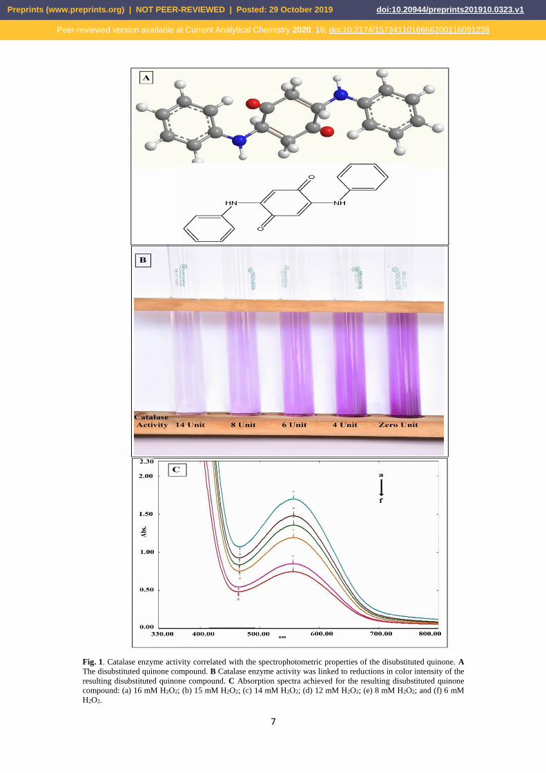
form a purple, disubstituted quinone compound (Fig. 1a). 174
A reduction in the intensity of the purple disubstituted quinone compound is 175
associated with increased catalase activity (Fig. 1b). The spectrum of the resulting 176
disubstituted quinone compound was scanned from 330 nm to 700 nm, and the 177
maximum absorbance was observed at 550 nm (Fig. 1c); therefore, this peak was used 178
to measure catalase activity. 179
180
Preprints (www.preprints.org) | NOT PEER-REVIEWED | Posted: 29 October 2019 doi:10.20944/preprints201910.0323.v1
Peer-reviewed version available at Current Analytical Chemistry 2020, 16; doi:10.2174/1573411016666200116091238
7
181
Fig. 1. Catalase enzyme activity correlated with the spectrophotometric properties of the disubstituted quinone. A 182The disubstituted quinone compound. B Catalase enzyme activity was linked to reductions in color intensity of the 183resulting disubstituted quinone compound. C Absorption spectra achieved for the resulting disubstituted quinone 184compound: (a) 16 mM H2O2; (b) 15 mM H2O2; (c) 14 mM H2O2; (d) 12 mM H2O2; (e) 8 mM H2O2; and (f) 6 mM 185H2O2. 186
Preprints (www.preprints.org) | NOT PEER-REVIEWED | Posted: 29 October 2019 doi:10.20944/preprints201910.0323.v1
Peer-reviewed version available at Current Analytical Chemistry 2020, 16; doi:10.2174/1573411016666200116091238
8
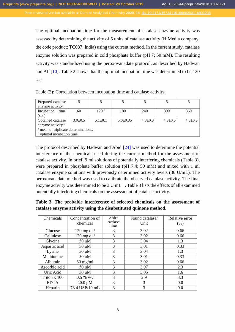
The optimal incubation time for the measurement of catalase enzyme activity was 187
assessed by determining the activity of 5 units of catalase activity (HiMedia company; 188
the code product: TC037, India) using the current method. In the current study, catalase 189
enzyme solution was prepared in cold phosphate buffer (pH 7; 50 mM). The resulting 190
activity was standardized using the peroxovanadate protocol, as described by Hadwan 191
and Ali [10]. Table 2 shows that the optimal incubation time was determined to be 120 192
sec. 193
Table (2): Correlation between incubation time and catalase activity. 194
Prepared catalase
enzyme activity
5 5 5 5 5 5
Incubation time
(sec)
60 120 b 180 240 300 360
Obtained catalase
enzyme activity a
3.0±0.5 5.1±0.1 5.0±0.35 4.8±0.3 4.8±0.5 4.8±0.3
a mean of triplicate determinations.b optimal incubation time.
195
The protocol described by Hadwan and Abid [24] was used to determine the potential 196
interference of the chemicals used during the current method for the assessment of 197
catalase activity. In brief, 9 ml solutions of potentially interfering chemicals (Table 3), 198
were prepared in phosphate buffer solution (pH 7.4; 50 mM) and mixed with 1 ml 199
catalase enzyme solutions with previously determined activity levels (30 U/mL). The 200
peroxovanadate method was used to calibrate the observed catalase activity. The final 201
enzyme activity was determined to be 3 UmL−1. Table 3 lists the effects of all examined 202
potentially interfering chemicals on the assessment of catalase activity. 203
Table 3. The probable interference of selected chemicals on the assessment of 204
catalase enzyme activity using the disubstituted quinone method. 205
Chemicals Concentration of
chemical
Added
catalase/
Unit
Found catalase/
Unit
Relative error
(%)
Glucose 120 mg dl-1 3 3.02 0.66
Cellulose 120 mg dl-1 3 3.02 0.66
Glycine 50 μM 3 3.04 1.3
Aspartic acid 50 μM 3 3.01 0.33
Lysine 50 μM 3 3.04 1.3
Methionine 50 μM 3 3.01 0.33
Albumin 50 mg/ml 3 3.02 0.66
Ascorbic acid 50 μM 3 3.07 2.3
Uric Acid 50 μM 3 3.05 1.6
Triton x 100 0.5 % v/v 3 2.9 3.3
EDTA 20.0 μM 3 3 0.0
Heparin 78.4 USP/10 mL 3 3 0.0
206
Preprints (www.preprints.org) | NOT PEER-REVIEWED | Posted: 29 October 2019 doi:10.20944/preprints201910.0323.v1
Peer-reviewed version available at Current Analytical Chemistry 2020, 16; doi:10.2174/1573411016666200116091238

9
The reliability of the disubstituted quinone method was also examined, using 207
homogenized red blood cells as a source of catalase activity. The disubstituted quinone 208
method was used to evaluate catalase enzyme activity, and the results were compared 209
with those using the peroxovanadate protocol, as described by Hadwan and Ali [10]. 210
Similar buffers, samples, and reagents were utilized during both methods. Table 4 211
demonstrates that the disubstituted quinone assay has good precision. In addition, the 212
data obtained from the disubstituted quinone method were significantly associated with 213
the data obtained from the peroxovanadate assay, as shown in Table 5. 214
Table 4. Precision of the disubstituted quinone method. 215
n. Mean (±SD):
U.mL-1
95% Confidence
Interval
CV%
Within-run 20 7.5 ± 0.2 (7.41 to 7.59) 2.6%
Between-run 20 7.38 ± 0.35 (7.15 to 7.45) 4.7%
216
Table 5. The statistical correlation between the catalase activity assessments obtained 217
from the disubstituted quinone method and those from the peroxovanadate method. 218
The numbers of measurements 20
Mean of catalase activity that assessed by the present method
U.mL-1.
5.74
Mean of catalase activity that assessed by the peroxovanadate
method U.mL-1.
5.67
Mean of catalase activity that assessed by both methods U.mL-1. 5.7
The regression coefficient B 0.9863
The regression coefficient A -0.101
The correlation coefficient 1.006
219
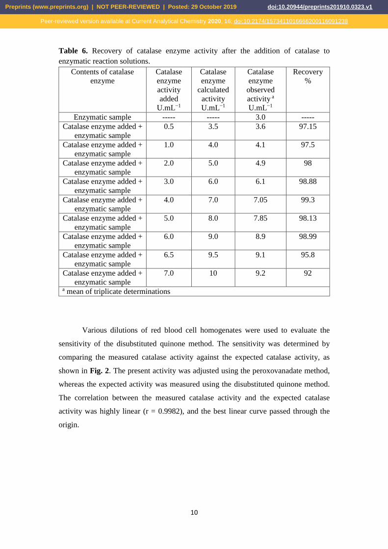
The recovery of added known catalase enzyme activities (HiMedia company; the code 220
product: TC037, India) was used to determine the accuracy of the current method. In 221
these experiments, catalase enzyme solution was prepared in cold phosphate buffer (pH 222
7; 50 mM), and the resulting activity was standardized using the peroxovanadate 223
method. The recovery of catalase enzyme activity was equal to 92% in the presence of 224
10 UmL−1 of enzyme and increased to 97.15% when the activity of the enzyme was 225
varied between 0.5 to 6.5 UmL−1 (Table 6). 226
Preprints (www.preprints.org) | NOT PEER-REVIEWED | Posted: 29 October 2019 doi:10.20944/preprints201910.0323.v1
Peer-reviewed version available at Current Analytical Chemistry 2020, 16; doi:10.2174/1573411016666200116091238
10
Table 6. Recovery of catalase enzyme activity after the addition of catalase to 227
enzymatic reaction solutions. 228
Contents of catalase
enzyme
Catalase
enzyme
activity
added
U.mL−1
Catalase
enzyme
calculated
activity
U.mL−1
Catalase
enzyme
observed
activity a
U.mL−1
Recovery
%
Enzymatic sample ----- ----- 3.0 -----
Catalase enzyme added +
enzymatic sample
0.5 3.5 3.6 97.15
Catalase enzyme added +
enzymatic sample
1.0 4.0 4.1 97.5
Catalase enzyme added +
enzymatic sample
2.0 5.0 4.9 98
Catalase enzyme added +
enzymatic sample
3.0 6.0 6.1 98.88
Catalase enzyme added +
enzymatic sample
4.0 7.0 7.05 99.3
Catalase enzyme added +
enzymatic sample
5.0 8.0 7.85 98.13
Catalase enzyme added +
enzymatic sample
6.0 9.0 8.9 98.99
Catalase enzyme added +
enzymatic sample
6.5 9.5 9.1 95.8
Catalase enzyme added +
enzymatic sample
7.0 10 9.2 92
a mean of triplicate determinations
229
230
Various dilutions of red blood cell homogenates were used to evaluate the 231
sensitivity of the disubstituted quinone method. The sensitivity was determined by 232
comparing the measured catalase activity against the expected catalase activity, as 233
shown in Fig. 2. The present activity was adjusted using the peroxovanadate method, 234
whereas the expected activity was measured using the disubstituted quinone method. 235
The correlation between the measured catalase activity and the expected catalase 236
activity was highly linear (r = 0.9982), and the best linear curve passed through the 237
origin. 238
Preprints (www.preprints.org) | NOT PEER-REVIEWED | Posted: 29 October 2019 doi:10.20944/preprints201910.0323.v1
Peer-reviewed version available at Current Analytical Chemistry 2020, 16; doi:10.2174/1573411016666200116091238

11
239
Figure 2. Comparison between the catalase enzyme activities of RBC homogenates 240
obtained by utilizing the disubstituted quinone method and peroxovanadate method. 241
Catalase activity assays were then performed using lysates from five different 242
bacterial laboratory strains to clarify the further potential applications of the 243
disubstituted quinone method. The results of these experiments indicated that similar 244
catalase enzyme activities were obtained using the disubstituted quinone method as 245
were obtained using the peroxovanadate method in bacterial strains, as described by 246
Hadwan and Ali [10]. In agreement with their findings, our results (Table 7) showed 247
that Staphylococcus aureus has higher catalase enzyme activity than other types of 248
bacteria. 249
Table 7. Comparison between the disubstituted quinone and peroxovanadate methods 250
for the assessment of catalase activities (KU) in different types of bacteria. 251
Name of bacteria Peroxovanadate
method
Disubstituted quinone
method
Staphylococcus aureus 15.5 15.2
pseudomonas aeruginosa 12.0 12.3
Escherichia coli 7.7 7.5
Klebsiella pneumonia 12.0 12.7
Enterococcus faecalis 0 0
252
The disubstituted quinone method was utilized to assess catalase enzyme activities in 253
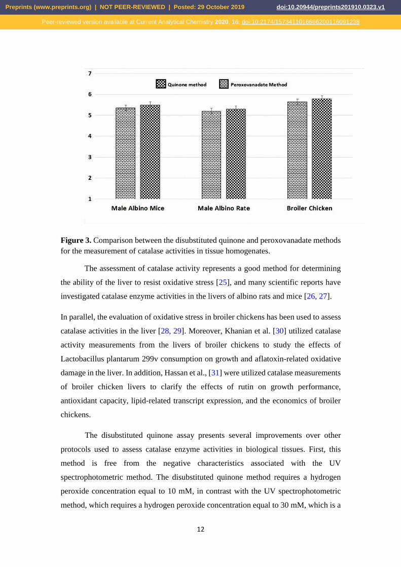
liver tissue homogenates from male albino mice, male albino rats, and broiler chickens. 254
Liver tissue homogenates exhibited high catalase enzyme activities (Fig. 3). 255
Preprints (www.preprints.org) | NOT PEER-REVIEWED | Posted: 29 October 2019 doi:10.20944/preprints201910.0323.v1
Peer-reviewed version available at Current Analytical Chemistry 2020, 16; doi:10.2174/1573411016666200116091238
12
256
257
Figure 3. Comparison between the disubstituted quinone and peroxovanadate methods 258
for the measurement of catalase activities in tissue homogenates. 259
The assessment of catalase activity represents a good method for determining 260
the ability of the liver to resist oxidative stress [25], and many scientific reports have 261
investigated catalase enzyme activities in the livers of albino rats and mice [26, 27]. 262
In parallel, the evaluation of oxidative stress in broiler chickens has been used to assess 263
catalase activities in the liver [28, 29]. Moreover, Khanian et al. [30] utilized catalase 264
activity measurements from the livers of broiler chickens to study the effects of 265
Lactobacillus plantarum 299v consumption on growth and aflatoxin-related oxidative 266
damage in the liver. In addition, Hassan et al., [31] were utilized catalase measurements 267
of broiler chicken livers to clarify the effects of rutin on growth performance, 268
antioxidant capacity, lipid-related transcript expression, and the economics of broiler 269
chickens. 270
The disubstituted quinone assay presents several improvements over other 271
protocols used to assess catalase enzyme activities in biological tissues. First, this 272
method is free from the negative characteristics associated with the UV 273
spectrophotometric method. The disubstituted quinone method requires a hydrogen 274
peroxide concentration equal to 10 mM, in contrast with the UV spectrophotometric 275
method, which requires a hydrogen peroxide concentration equal to 30 mM, which is a 276
Preprints (www.preprints.org) | NOT PEER-REVIEWED | Posted: 29 October 2019 doi:10.20944/preprints201910.0323.v1
Peer-reviewed version available at Current Analytical Chemistry 2020, 16; doi:10.2174/1573411016666200116091238

13
high enough concentration to inhibit catalase by modifying its active site structure [32]. 277
Additionally, proteins and DNA molecules can absorb UV light during the UV 278
spectrophotometric method, whereas the disubstituted quinone assay is based upon the 279
decreased absorbance of the characteristic disubstituted quinone band at 550 nm, which 280
is not in the UV spectrum. The disubstituted quinone protocol could be made accessible 281
as assay kits, does not require the use of cumbersome techniques, and is inexpensive. 282
Additionally, the method is simple, shows high precision, and can be applied at low 283
H2O2 concentrations and in the presence of significant quantities of several types of 284
biochemicals without interference. 285
Conclusion 286
A simple and precise assay for estimating catalase enzyme activity was developed and 287
described in this study. This is a simple and cost-effective method, with the advantages 288
of high precision and accuracy and requiring only instruments that are readily available 289
in most laboratories. 290
Competing financial interests 291
The author declares no competing financial interests. 292
Competing interests 293
The authors declare that they have no competing interests with the contents of this 294
paper. 295
Acknowledgements 296
We thank our colleagues in University of Babylon/College of Science for their technical 297
supporting, comments and help regarding our study, especially Dr. Noor S. K. 298
AlKhafaji. 299
300
References 301
1. Glorieux C, Zamocky M, Sandoval JM, Verrax J, Calderon PB. Regulation of302
catalase expression in healthy and cancerous cells. Free Radical Biology and303
Medicine. 2015 Oct 1;87:84-97.304
2. Rahman I, Biswas SK, Kode A. Oxidant and antioxidant balance in the airways305
and airway diseases. European journal of pharmacology. 2006 Mar 8;533(1-306
3):222-39.307
3. Zaidi SK, Ansari SA, Tabrez S, Naseer MI, Shahwan MJ, Banu N, Al-Qahtani308
MH. Antioxidant potential of Solanum nigrum aqueous leaves extract in309
Preprints (www.preprints.org) | NOT PEER-REVIEWED | Posted: 29 October 2019 doi:10.20944/preprints201910.0323.v1
Peer-reviewed version available at Current Analytical Chemistry 2020, 16; doi:10.2174/1573411016666200116091238

14
modulating restraint stress-induced changes in rat’s liver. Journal of pharmacy 310
& bioallied sciences. 2019 Jan;11(1):60. 311
4. Yao XH, Min H, Lü ZH, Yuan HP. Influence of acetamiprid on soil enzymatic312
activities and respiration. European Journal of Soil Biology. 2006 Apr313
1;42(2):120-6.314
5. Aebi H. [13] Catalase in vitro. InMethods in enzymology 1984 Jan 1 (Vol. 105,315
pp. 121-126). Academic Press.316
6. Mueller S, Riedel HD, Stremmel W. Determination of catalase activity at317
physiological hydrogen peroxide concentrations. Analytical biochemistry. 1997318
Feb 1;245(1):55-60.319
7. Ou P, Wolff SP. A discontinuous method for catalase determination at ‘near320
physiological’concentrations of H2O2 and its application to the study of H2O2321
fluxes within cells. Journal of biochemical and biophysical methods. 1996 Jan322
11;31(1-2):59-67.323
8. Hadwan MH. Simple spectrophotometric assay for measuring catalase activity324
in biological tissues. BMC biochemistry. 2018 Dec;19(1):7.325
9. Masuoka N, Wakimoto M, Ubuka T, Nakano T. Spectrophotometric326
determination of hydrogen peroxide: catalase activity and rates of hydrogen327
peroxide removal by erythrocytes. Clinica chimica acta. 1996 Oct328
29;254(2):101-12.329
10. Hadwan MH, kadhum Ali S. New spectrophotometric assay for assessments of330
catalase activity in biological samples. Analytical biochemistry. 2018 Feb331
1;542:29-33.332
11. Shivakumar A, Nagaraja P, Chamaraja NA, Krishna H, Avinash K.333
Determination of catalase activity using chromogenic probe involving iso-334
nicotinicacidhydrazide and pyrocatechol. Journal of biotechnology. 2011 Oct335
10;155(4):406-11.336
12. Posch HE, Wolfbeis OS. Optical sensor for hydrogen peroxide. Microchimica337
Acta. 1989 Jan 1;97(1-2):41-50.338
13. Cohen CB, Weber SG. Photoelectrochemical sensor for catalase activity based339
on the in situ generation and detection of substrate. Analytical Chemistry. 1993340
Jan 1;65(2):169-75.341
14. Bekdeşer B, Özyürek M, Güçlü K, Alkan FÜ, Apak R. Development of a new342
catalase activity assay for biological samples using optical CUPRAC sensor.343
Spectrochimica Acta Part A: Molecular and Biomolecular Spectroscopy. 2014344
Nov 11;132:485-90.345
15. Siqueira AJ, Remião JO, Azevedo AM, Azambuja CR. A gasometric method to346
determine erythrocyte catalase activity. Brazilian journal of medical and347
biological research. 1999 Sep;32(9):1089-94.348
16. Kroll RG, FREARS ER, BAYLISS A. An oxygen electrode‐based assay of349
catalase activity as a rapid method for estimating the bacterial content of foods.350
Journal of Applied Bacteriology. 1989 Mar;66(3):209-17.351
17. Slaughter MR, O’Brien PJ. Fully-automated spectrophotometric method for352
measurement of antioxidant activity of catalase. Clinical biochemistry. 2000353
Oct 1;33(7):525-34.354
Preprints (www.preprints.org) | NOT PEER-REVIEWED | Posted: 29 October 2019 doi:10.20944/preprints201910.0323.v1
Peer-reviewed version available at Current Analytical Chemistry 2020, 16; doi:10.2174/1573411016666200116091238

15
18. Abderrahim M, Arribas SM, Condezo-Hoyos L. A novel pyrogallol red-based355
assay to assess catalase activity: Optimization by response surface356
methodology. Talanta. 2017 May 1;166:349-56.357
19. Huang XM, Zhu M, Shen HX. N, N-diethylaniline as hydrogen donor for358
determination of hydrogen peroxide catalyzed by metalloporphyrins as enzyme359
mimetic of peroxidase. Microchimica Acta. 1998 Mar 1;128(1-2):87-91.360
20. Ci YX, Wang F. Spectrofluorimetric determination of hydrogen peroxide based361
on the catalytic effect of peroxidase-like manganese tetrakis (sulphophenyl)362
porphyrin on the oxidation of homovanillic acid. Analytica Chimica Acta. 1990363
Jan 1;233:299-302.364
21. Elnemma EM. Spectrophotometric determination of hydrogen peroxide by a365
hydroquinone-aniline system catalyzed by molybdate. Bulletin of the Korean366
Chemical Society. 2004;25(1):127-9.367
22. Nardello V, Bouttemy S, Aubry JM. Olefin oxidation by the system368
H2O2MoO2− 4: competition between epoxidation and peroxidation. Journal369
of Molecular Catalysis A: Chemical. 1997 Mar 14;117(1-3):439-47.370
23. Wahlen J, De Vos DE, Groothaert MH, Nardello V, Aubry JM, Alsters PL,371
Jacobs PA. Synergism between molybdenum and lanthanum in the372
disproportionation of hydrogen peroxide into singlet oxygen. Journal of the373
American Chemical Society. 2005 Dec 14;127(49):17166-7.374
24. Hadwan MH, Abed HN. Data supporting the spectrophotometric method for the375
estimation of catalase activity. Data in brief. 2016 Mar 1;6:194-9.376
25. Shin SK, Cho HW, Song SE, Bae JH, Im SS, Hwang I, Ha H, Song DK.377
Ablation of catalase promotes non-alcoholic fatty liver via oxidative stress and378
mitochondrial dysfunction in diet-induced obese mice. Pflügers Archiv-379
European Journal of Physiology. 2019 Jun 1;471(6):829-43.380
26. Adisa RA, Kolawole N, Sulaimon LA, Brai B, Ijaola A. Alterations of381
Antioxidant Status and Mitochondrial Succinate Dehydrogenase Activity in the382
Liver of Wistar Strain Albino Rats Treated with by Ethanol Extracts of Annona383
senegalensis Pers (Annonaceae) Stem Bark. Toxicological research. 2019384
Jan;35(1):13.385
27. Aldulaimi AM, Husain FF. Effect of Aqueous Extract Cyperus rotundus Tubers386
as Antioxidant on Liver and Kidney Functions in Albino Males Rats Exposed387
to Cadmium Chloride Toxic. Baghdad Science Journal. 2019;16(2):315-22.388
28. Qadri SS, Biswas A, Mir NA, Mandal AB, Biswas AK. Physico-biochemical389
and microbial characteristics of broiler chicken meat fed diet incorporated with390
Kappaphycus alvarezii. Journal of Applied Phycology. 2019:1-7.391
Preprints (www.preprints.org) | NOT PEER-REVIEWED | Posted: 29 October 2019 doi:10.20944/preprints201910.0323.v1
Peer-reviewed version available at Current Analytical Chemistry 2020, 16; doi:10.2174/1573411016666200116091238
16
29. Baradaran A, Samadi F, Ramezanpour SS, Yousefdoust S. Hepatoprotective392
effects of silymarin on CCl4-induced hepatic damage in broiler chickens model.393
Toxicology reports. 2019 Jan 1;6:788-94.394
30. Khanian M, Karimi-Torshizi MA, Allameh A. Alleviation of aflatoxin-related395
oxidative damage to liver and improvement of growth performance in broiler396
chickens consumed Lactobacillus plantarum 299v for entire growth period.397
Toxicon. 2019 Feb 1;158:57-62.398
31. Hassan F, Roushdy E, Kishawy A, Zaglool A, Tukur H, Saadeldin I. Growth399
Performance, Antioxidant Capacity, Lipid-Related Transcript Expression and400
the Economics of Broiler Chickens Fed Different Levels of Rutin. Animals.401
2019 Jan;9(1):7.402
32. Mueller S, Riedel HD, Stremmel W. Determination of catalase activity at403
physiological hydrogen peroxide concentrations. Analytical biochemistry. 1997404
Feb 1;245(1):55-60.405
406
407
408
409
410
411
412
413
414
415
416
417
418
419
420
421
422
423
Preprints (www.preprints.org) | NOT PEER-REVIEWED | Posted: 29 October 2019 doi:10.20944/preprints201910.0323.v1
Peer-reviewed version available at Current Analytical Chemistry 2020, 16; doi:10.2174/1573411016666200116091238