new: live comments - efort · 2018-06-25 · new: live comments 2011/2012 ... in class ii and iii...
TRANSCRIPT


info Information available on: http://efort.covr.be
New: Live comments
2011/2012
Join the Sessions online!
Given the limited time we have available for Q&A sessions during the course, we are introducing a new live comment facility:Give your comments about a lecture live -Interact directly with your peers in written comments -
It’s very simple - join in!
While attending the CRC, there is now an additional way in which you can interact with the speaker. With our live comment tool, you are able to post comments or ask questions by visiting a website on your mobile phone or your laptop. Your entry will then be displayed on the big screen so the speaker can respond if he or she finds it appropriate.
To enter a comment simply Start up your browser1. WLAN access: EFORT 20112. Go to: http://efort.covr.be3.
You will be directed to this page. Here, you can fill in your credentials and enter a comment. The session ID will be announced during the lecture.
After clicking the submit button on the page above, your entry will be shown on the big screen:

2
IntroductionWelcome
Welcome to this third edition of the EFORT Comprehensive Review Course. This course held during the Copenhagen congress will provide the basis of the core theoretical knowledge expected of all orthopaedic trainees at the end of their specialty curriculum. The course is an outline that cannot be exhaustive but certainly the essentials will be found in the lectures and the syllabus. It is also a convenient way for senior surgeons to obtain an update of current practices and state of the art information on the whole field of orthopaedics and traumatology. My thanks go to all the lecturers and writers of syllabus chapters. This collective effort will certainly be of benefit to our participants.Prof. Dr. Pierre Hoffmeyer, EFORT Vice-president
Experience more of the EFORT 2011 Congress with e-Science NEW! Introducing EFORT congress sessions online: A new initiative providing a comprehensive digital library of up to 270 hours of scientific ses-sions.
For a limited time, congress registrants will be able to order “e-Congress2011”. Order at the Congress and receive substantial savings!With EFORT e-Congress2011 you can:
Review up to 270 hours of content from the 12th EFORT Congress -Separate track for the CRC course only available! -Search and bookmark your favourite sessions -Watch sessions you were unable to attend in Copenhagen -Access sessions conveniently at anytime online and on DVD-ROM -Share your experiences with friends and colleagues -
Order today at the e-science booth located in the Exhibition hall or opposite the e-poster stations.www.efort.org/eCongress2011
The full attendance of the CRC course entitles to 6 European CME credits (ECMEC’s)The Certificate will be sent to each participants by PDF file to the registered e-mail address after the course. If you have not yet registered your e-mail account with us, please contact the “Scientific Information” desk in the registration hall.
EFORT does not in any way monitor or endorse the content of any given lecture by any of the speakers during the course. EFORT does not accept any responsibility for the content of any individual session as presented by the speakers nor printed in the syllabus and thus declines all liability of what-ever nature arising thereof.
© Copyright by EFORT 2011
Prof. Dr. Pierre Hoffmeyer
University HospitalGeneva, Switzerland
CRC Sessions for EUR 99 only!

3
EYE OPENEROrthopaedic experiences with the Afghan war Dr. Finn Warburg .................................................................................................................................................4
BASIC SCIENCEBiomechanics of musculoskeletal tissue - Biomaterials (trauma, prosthesis) Prof. Dr. Elizabeth Tanner ........................................................................5Metabolic bone disease Univ.-Prof. Dr. med. Karsten Dreinhöfer .....................................................................................................................................................9
PAEDIATRICSSpecific fractures in the childhood Dr. Geraldo de Coulon...............................................................................................................................................................15Neuroorthopaedics Dr. Erich Rutz .............................................................................................................................................................................................................18Abnormalities of newborn’s feet Dr. Dimitri Ceroni ............................................................................................................................................................................23Hip diseases in the childhood Bjarne Møller-Madsen MD.DMSCi ..................................................................................................................................................25
BASIC SCIENCEDiagnostic work up and recognition of primary bone tumours Mr. Stephen Cannon ...........................................................................................................27Diagnostic algorithm and treatments options in bone metastasis Prof. Dr. Miklòs Szendröi ...............................................................................................29
RECONSTRuCTIONHip reconstruction: Osteotomy and joint replacement Prof. Dr. Klaus-Peter Günther ...........................................................................................................32Knee: Osteotomy and arthroplasty Dr. Martin Pietsch ......................................................................................................................................................................34Reconstructive foot and ankle Dr . Marino Delmi ...............................................................................................................................................................................39Shoulder degenerative disorders Dr. Richard Wallensten ..................................................................................................................................................................45Elbow arthritis Dr. Domizio Suva ...............................................................................................................................................................................................................51
SPINE (INCL. TRAuMA)Spine deformities in the childhood Prof. Dr. Pierre Lascombes .......................................................................................................................................................53Management of spine fractures Prof. Dr. Benny Dahl ........................................................................................................................................................................56Degenerative spine disease Prof. Dr. Enric Caceres Palou ...................................................................................................................................................................59
SPORTS/ kNEE SOFT-TISSuEACL, PCL, collaterals, meniscus Prof. René Verdonk ............................................................................................................................................................................61
TRAuMAFractures: Pelvic ring und acetabular fractures Prof. Dr. Ulrich Stöckle ......................................................................................................................................66Fractures: Hip, femur, knee and tibia Dr. Olivier Borens, MER ........................................................................................................................................................69Fractures: Pilon, ankle, talus and calcaneus Dr. Mathieu Assal .....................................................................................................................................................73Fractures of distal radius and scaphoid and wrist instability Prof. Dr. Philippe Kopylov .......................................................................................................78Upper Limb Trauma: Shoulder girdle, proximal humerus, humeral shaft Prof. Dr. Pierre Hoffmeyer ................................................................................84Upper Limb Trauma: Elbow and forearm Prof. Dr. Pierre Hoffmeyer .............................................................................................................................................94
Index

4
In the period 2006 to 2010 Copenhagen university Hospital, ”Rigshos-pitalet” received 63 wounded soldiers. The cause of injury was gunshots, rocket propelled grenades and mines.The author was deployed to the NATO field hospital in kandahar in 2006 and 2007. In Rigshospitalet he was responsible for reception, orthopedic proce-dures and coordination of the wounded soldiers.After treatment and rehabilitation we succeded in discharging 61 of the patients to their own home, selfdependent. We lost one patient in the hospital after prolonged intensive care and one is still training to relieve himself of dependence. Today’s most prevalent war injuries are presented in cases from kanda-har as well as their initial treatment. The methods used in final repair and rehabilitation of injured soldiers in the university hospital is outlined. Interesting experience regarding sur-gery, reconstruction, infection control, pain relief and training has been gained and is now used in other patients as well.
Eye opener
Dr. Finn Warburg
The Danish Society of Military MedicineFarum, [email protected]
Orthopaedic experiences with the Afghan war

5
Basic Science
Prof. Dr. Elizabeth Tanner
University of GlasgowGlasgow, [email protected]
Biomechanics of musculoskeletal tissue - Biomaterials (trauma, prosthesis)
IntroductionThis lecture will consider biomechanics of bones and joints that are applied due to movement by people and then the biomaterial consider-ations relevant to orthopaedic implants. Biomechanics and biomateri-als are obviously both huge subjects but so only the areas of each of importance to orthopaedic surgeons will be considered.
BiomechanicsThe mechanics of moving objects, including the human body, are gov-erned by Newton’s Laws of Motion. The 1st Law states that “a body will remain in a state of rest, or move at constant velocity, unless acted upon by a force”. The 2nd Law states that “a body acted upon by a force will change its velocity in proportion to the applied force”. While the 3rd Law says that “when two bodies exert a force upon each other the force acts on the line connecting them and the two force vectors are equal and opposite”. What do these laws mean when applied to the human body? Firstly for anything to start moving a force has to act on it, secondly how fast it moves depends on the magnitude of the applied force. The appli-cations of these two laws to the human body are relatively obvious, mus-cles act by contracting and thus generating a force. What needs to be considered is that shortening a muscle against no resisting force requires no muscle force, what produces the force is the muscle shortening against some form of resistance. The third law is commonly restated as “every action has an equal and opposite reaction” and it is this law com-bined with the first law that is used in calculating forces generated in the body and how these effect the movement of parts of the body. The second basic element needing to be considered in biomechanics is the behaviour of levers. Archimedes (287-212BC) is quoted as having said “Give me a fulcrum and I will move the world”. We can analyse the behaviour of the human body as a mechanical system by modelling the bones as levers, the weight of components of the body as the loads which need to be moved and the muscles as the applying forces. Levers come in three classes, depending on the relative positions of the fulcrum, the pivot point about which the lever moves, and the load force which the force which needs to be moved and the effort force which is the force doing the moving, that is the muscle force in the body (Figure 1). An example of a Class I lever is the child’s seesaw, where the fulcrum is in
the centre and the two people are the load and effort forces. In the human body there are few Class I levers, one example is at the head where the C1 vertebra acts as the fulcrum, mass of the head is the load force and is anterior to this fulcrum, while the extensor muscles of the neck supply the effort force. In Class II and III levers the fulcrum is at one end of the lever and the load and effort forces are to the same side of the fulcrum. In Class II the load force is between the fulcrum and the effort force while in Class III the effort force is between the load force and the fulcrum and the Class III lever is the most common type of lever found in the body. As the forces multiplied by their distance from the fulcrum have to balance where the effort force is nearer the fulcrum than the load force the effort force has to be higher than the load force.
Force is measured in Newtons (N) in the SI (Système International) unit scheme. 1 Newton is the force exerted by 1 kg (kilogram) when accel-erated at 1ms-2, thus force exerted by 1kg on earth is 9.81N as the accel-eration due to gravity on earth is 9.81ms-2. One simple way to remem-ber the value of a Newton is that the force exerted on earth by a typical apple weighing about 100g is about 1N. In analysing the biomechanics of the body we can consider a simple action, holding a weight in the hand with the forearm held horizontal and the upper arm horizontal (Figure 2). The weight is acting downwards and to be held still the vertical forces in the arm through to the body must be equal and the moments about the elbow joint must be equal. If we assume the weight of the lower arm is 20N and the weight held in the hand is 10N (thus approximately 2kg and 1 kg mass respectively) and that the length from the elbow joint to the hand is 300mm and to the the centre of mass of the forearm is 130mm with the line of action of the biceps muscle being 50mm. We can calculate that the force in the biceps has to be 112N.
Fig. 1 Lever types
Class I Class II Class III
Basi
c Sc
ienc
e

6
Basic Science
Fig. 2 Forces involved in carrying a 1kg weight in the hand.
If we apply similar calculations to a person standing on one leg and mak-ing appropriate assumptions of distances in the body then we can cal-culate that the load on the femoral head is 2.58 times the subject’s body weight and that the forces in the abductor muscles is 1.77 times body weight. The calculations have to include that the distance between the hip joint and the line of action of the hip abductors must be taken as perpendicular to the line of the muscle (Figure 3). If these simple calcu-lations are compared with the data from an instrumented hip prosthe-sis (Bergmann, Graichen et al. 1993) then it can be seen that the forc-es calculated using a simple two dimensional analysis can give a good estimate of the actual forces occurring in vivo. These types of analysis can be applied throughout the body.
Fig. 3 Incorrect and correct methods of calculating the moment about a hip joint
The final factor to be considered is the number of load cycles applied during walking and other activities. (Wallbridge and Dowson 1982) found that the number of load cycles applied to the legs dropped from an aver-age of 2 million per year when people were in their 20s down to 0.5 mil-lion in their 80s. The interesting factor was that they also measured some joint replacement patients and found they these people were applying more load cycles than the average for their age group. In the hand Joyce and unsworth (2000) estimated similar number of load cycles for the fingers, but estimated that the loading the fingers considered of two groups, high movement with low loads interspersed with limited motion but high loads.
Biomaterials “A biomaterial is a non viable material used in a medical device, intend-ed to interact with biological systems” according to (Williams 1999) and to function successfully it needs to be biocompatible, that is it “has the ability to perform with an appropriate host response in a specific appli-cation” (Williams 1999). The behaviour of a material in the body depends on two factors: the effect the implant material has on the body and the effect the body has on the implant material. The reaction to an implant-ed material (and thus implant) can be divided into four types: Toxic, that is it kills cells in contact with or away from implant, Bioinert, that is produces no response by the body and which never truely occurs as there is always a response to implantation, but when the response is minimal the material is called bioinert. Bioactive, which is encourages an advan-tageous response from the body and this will depend on where the implant is placed in the body and thus the required bioactive response and finally Biodegradable where the implant breaks down in the body to non-toxic components which are excreted by the body. The effects the body has on an implant can be defined as the response of the mate-rial to the internal environment of the body from the physiological envi-ronment, protein absorption, which is a particular problem with poly-mers, degradation whether required or not and finally corrosion, which particularly applies to metal implants. When we are considering the mechanical properties of a material these are measured using stress, which is the force per unit area and strain which is a measure of the change in dimension and the ratio of these two is called Young’s Modulus or stiffness. Further important mechani-cal factors are the ultimate strength, that is how much force a materi-al can take before it breaks, the ductility, the amount a material deforms before it breaks and toughness which is a measure of how fast a crack progresses through a material once fracture starts. When choosing a material for use in the body one of the considerations is the mechani-cal properties of the material compared to those of the body component being replaced. Cortical bone has Young’s modulus of 7-25GPa, strength of 50-150MPa and a fracture toughness of 2-12 MN m-3/2, while cancellous bone has modulus of 0.1-1.0GPa and compressive strength of 1-10MPa (Currey 1998; Currey 2006). Cortical and cancellous bone are both brittle, but being able to react to their mechanical environment can be considered to be “smart” materials. Cancellous bone behaves as a typical foam, that is increasing the density (or decreasing the porosity) increases the stiff-ness and strength (Gibson and Ashby 1999). Ligaments and tendons have non-linear mechanical properties with the stiffness increasing as the load increases. Materials can be defined into four basic groups: metals, ceramics, pol-ymers and composites. Metals are normally used as alloys, that is small or larger amounts of other atoms are added to tailor the properties. Met-als are reasonably stiff, ductile, that is they deform before they fracture, they generally have good fatigue properties and can be plastically deformed, that is they can be bent into new shape and remain in that shape as is used in the moulding of fracture fixation devices. The major metals used in orthopaedics are the stainless steels, the cobalt chrome alloys, titanium and its alloys. Stainless steel used in medical applica-tions is usually 316 or 316L and consists of 18% chromium, 13% nick-el, 2.5% molybdenum, and the rest is iron. The presence of the chromi-um leads to the alloy being “stainless” as a chromium oxide layer is produced on the surface, which does not easily oxidise further. Stainless steel has a Young’s modulus of 210 GPa, is ductile, can be deformed (cold worked) and the fatigue properties are acceptable. Cobalt Chrome alloy consists of 27-30% chromium, 5-7% molybdenum with the rest cobalt.
7
Basic Science
This formulation means that there is no nickel which is important for those patients who are nickel sensitive. Nickel sensitivity rates are var-iable within Europe and can reach over 20% in the Scandinavian popu-lation. Cobalt chrome has a Young’s modulus of 230 GPa, a higher fatigue limit than Stainless Steel and has good wear properties. There are three major groups of titanium: commercially pure which is >99% titanium, Ti-6%Al-4%V which is therefore 90% titanium, 6% aluminium and 4% vanadium and finally the shape memory alloys which are approximate-ly 50:50 titanium:nickel, with the exact composition being used to con-trol the temperature at which the shape memory effect occurs. Most titanium alloys have a lower Young’s modulus of 106 GPa, the wear debris is black in body thus looks unsightly to the surgeon, but this wear debris is not known to produced significant extra problems compared to oth-er wear debris which may be as present in the body but is not as obvi-ous to the surgeon. Titanium is notch sensitive, that is any notches or other sharp corners lead to significant reductions in the fatigue life, and also is heat treatment sensitive. (Cook, Thongpreda et al. 1988) showed that with appropriate heat treatment the fatigue limit, which is the fatigue load at which the specimen does not break, was 625MPa, but if a porous coating was applied with an inappropriate heat treatment this fatigue limit was reduced to 200MPa. More recently newer titanium alloys are being developed which have yet lower Young’s moduli, at 42GPa, thus bringing their stiffness’s closer to those of cortical bone (Hao, Li et al. 2007). Bioceramics can be divided into 2 major groups, the bioinert which are principally zirconia (ZrO2) and alumina (Al2O3) and the bioactive main-ly hydroxyapatite (Ca10(PO4)6(OH)2) and tricalcium phosphate (Ca3(PO4)2). The bioinert ceramics are principally used for articulating surfaces as either ceramic-on-polymer or ceramic-on-ceramic. Initially Al2O3 was preferred as ZrO2 can be morphologically unstable but now PSZ (Partial-ly Stabilised Zirconia) is available. Al2O3 has been used by Sedel in Par-is for more than 30 years as ceramic-on-ceramic hip replacements (Nizard, Pourreyron et al. 2008). In the initial implants the individual grains in the ceramics components were large and failures occurred, now grain size is reduced and failures have reduced to >1:2000. However, very close tolerances on head-cup dimensions are needed so matched pairs are supplied to reduce the fracture risk. Bioactive ceramics are used in five major applications: bulk implants, that is space filling implants, porous when used as implants for ingrowth or scaffolds for tissue engineering, granules used to bulk out or to replace bone graft, coatings which are either plain HA or HA+TCP (also called biphasic CaP - BCP) and finally as injectable where the calcium phos-phate, with or without some calcium sulphate and other additives, is mixed in the operating theatre, injected into the body and sets in situ. Polymers used in orthopaedics are primarily ultrahigh molecular weight polyethylene (uHMWPE), polymethylmethacrylate (PMMA), other meth-acrylates, polyesters, poly(glycolic acid) and poly(lactic acid) and final-ly the hydrogels. Polyethylene was introduced by Sir John Charnley in 1960 as the first metal-on-polymer joint replacement. Charnley initial-ly used polytetrafluoroethylene (PTFE) as the bearing surface for his hip replacements and found such drastic wear that after 1 year joint motion was seriously reduced. He originally High Density Polyethylene (HDPE), which was replaced in 1970s with ultra High Molecular Weight Poly-ethylene (uHMWPE) and now a range of Enhanced Polyethylene (par-tially cross linked) or heavily irradiated PE are used to reduce the pro-duction of wear particles. PE is used as concave bearing surfaces against metal or ceramics such as acetabular cups, the tibial plateaux of knee replacements, patella buttons etc. PMMA bone cement is used to fix (grout) joint replacements in place thus is used to space fill. It is sup-
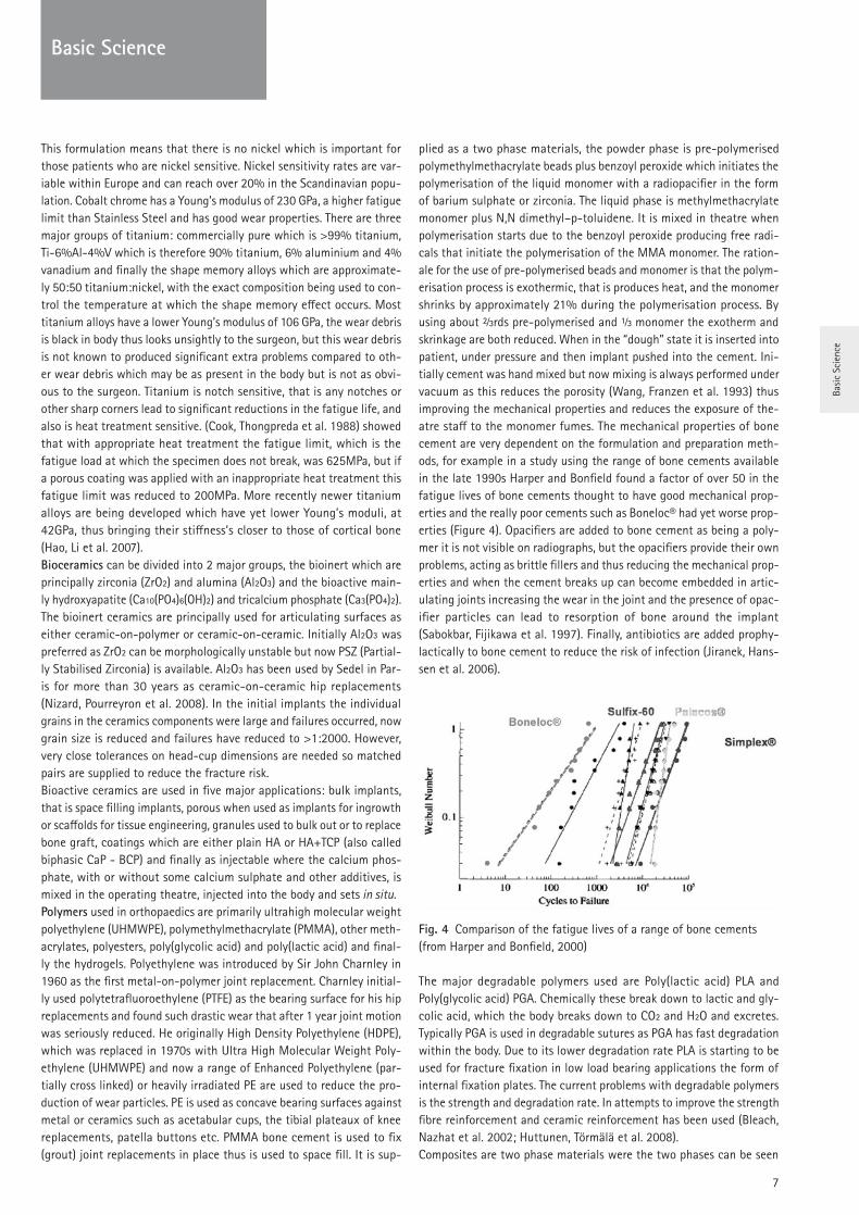
plied as a two phase materials, the powder phase is pre-polymerised polymethylmethacrylate beads plus benzoyl peroxide which initiates the polymerisation of the liquid monomer with a radiopacifier in the form of barium sulphate or zirconia. The liquid phase is methylmethacrylate monomer plus N,N dimethyl–p-toluidene. It is mixed in theatre when polymerisation starts due to the benzoyl peroxide producing free radi-cals that initiate the polymerisation of the MMA monomer. The ration-ale for the use of pre-polymerised beads and monomer is that the polym-erisation process is exothermic, that is produces heat, and the monomer shrinks by approximately 21% during the polymerisation process. By using about 2/3rds pre-polymerised and 1/3 monomer the exotherm and skrinkage are both reduced. When in the “dough” state it is inserted into patient, under pressure and then implant pushed into the cement. Ini-tially cement was hand mixed but now mixing is always performed under vacuum as this reduces the porosity (Wang, Franzen et al. 1993) thus improving the mechanical properties and reduces the exposure of the-atre staff to the monomer fumes. The mechanical properties of bone cement are very dependent on the formulation and preparation meth-ods, for example in a study using the range of bone cements available in the late 1990s Harper and Bonfield found a factor of over 50 in the fatigue lives of bone cements thought to have good mechanical prop-erties and the really poor cements such as Boneloc® had yet worse prop-erties (Figure 4). Opacifiers are added to bone cement as being a poly-mer it is not visible on radiographs, but the opacifiers provide their own problems, acting as brittle fillers and thus reducing the mechanical prop-erties and when the cement breaks up can become embedded in artic-ulating joints increasing the wear in the joint and the presence of opac-ifier particles can lead to resorption of bone around the implant (Sabokbar, Fijikawa et al. 1997). Finally, antibiotics are added prophy-lactically to bone cement to reduce the risk of infection (Jiranek, Hans-sen et al. 2006).
Fig. 4 Comparison of the fatigue lives of a range of bone cements(from Harper and Bonfield, 2000)
The major degradable polymers used are Poly(lactic acid) PLA and Poly(glycolic acid) PGA. Chemically these break down to lactic and gly-colic acid, which the body breaks down to CO2 and H2O and excretes. Typically PGA is used in degradable sutures as PGA has fast degradation within the body. Due to its lower degradation rate PLA is starting to be used for fracture fixation in low load bearing applications the form of internal fixation plates. The current problems with degradable polymers is the strength and degradation rate. In attempts to improve the strength fibre reinforcement and ceramic reinforcement has been used (Bleach, Nazhat et al. 2002; Huttunen, Törmälä et al. 2008). Composites are two phase materials were the two phases can be seen
Basi
c Sc
ienc
e
8
as separate either with the naked eye or using a microscope, which is the two phases can be differentiated on the micron scale. Artificial com-posites are generally used to optimise the properties of the two phases. The individual phases interact be it mechanically or functionally. The major groups of composites are polymer reinforced with ceramics/glass-es, polymers reinforced with different polymer or polymer form such as drawn fibres of a polymer in a amorphous matrix of the same polymer, an example is the PLLA in PLDLA used in some degradable fracture fix-ation plates. Ceramic metal composites, which are also known as met-al matrix composites a few of these have been developed for medical applications and finally ceramic-ceramic composites, but neither of these but have as yet reached clinical applications. In a composite there is nor-mally one continuous phase called the matrix and a second phase called the filler distributed in the matrix as particles, fibres or fabric. General-ly phases chosen as when specific properties of one phase are “good” in the other they are “bad”, but by getting right balance of phases can bal-ance the properties to optimise the material. Applications of biocom-posites in medical applications is beginning to increase (Tanner 2010). The earliest ones were bioinert, but now bioactive implants are benefi-cially interacting with the human body.
ConclusionsIn conclusion when placing implants in the body there are two major interacting factors that need to be considered for the survival of an implant in the body. The first is how heavily is it being loaded and the second is what is it made of. Without appropriate interactions between both of these factors an implant will not be successful
REFERENCESBergmann, G., F. Graichen, et al. (1993). “Hip-joint loading during walk-1.
ing and running, measured in 2 patients.” Journal of Biomechanics 26(8): 969-990.
Bleach, N. C., S. N. Nazhat, et al. (2002). “Effect of Filler Content on 2. Mechanical and Dynamic Mechanical Properties of Particulate Biphasic Calcium Phosphate Polylactide Composites”.” Biomaterials 23(7): 1579-1585.
Cook, S. D., N. Thongpreda, et al. (1988). “The effect of post-sintering 3. heat treatments on the fatigue properties of porous coated Ti-6Al-4V alloy.” Journal of Biomedical Materials Research 22(4): 287-302.
Currey, J. D. (1998). “Mechanical properties of vertebrate hard tissues.” 4. Proceedings of the Institution of Mechanical Engineers: Part H Engineer-ing in Medicine 212-H(6): 399-412.
Currey, J. D. (2006). Bones: Structure and Mechanics, Princeton Univer-5. sity Press.
Gibson, L. J. and M. F. Ashby (1999). Cellular Solids. Oxford, Pergamon 6. Press.
Hao, Y. L., S. J. Li, et al. (2007). “Elastic deformation behaviour of Ti-24-7. Nb-4Zr-7.9Sn for biomedical applications.” Acta Biomaterialia 3(2): 277-286.
Harper, E.J., Bonfield, W. (2000) “Tensile Characteristics of Ten Com-8. mercial Acrylic Bone Cements. Journal of Biomedical Materials Research 53-A(5): 605-616. Errata 2001;58-A(2):216.
Huttunen, M., P. Törmälä, et al. (2008). “Fiber-reinforced bioactive and 9. bioabsorbable hybrid composites.” Biomedical Materials 3(3).
Jiranek, W. A., A. D. Hanssen, et al. (2006). “Antibiotic-loaded bone 10. cement for infection prophylaxis in total joint replacement.” Journal of Bone and Joint Surgery 88-A(11): 2487-2500.
Joyce, T. J. and A. Unsworth (2000). “The design of a finger wear sim-11.
ulator and preliminary results.” Proceedings of the Institution of Mechan-ical Engineers: Part H Engineering in Medicine 214-H(5): 519-526.
Nizard, R. S., D. Pourreyron, et al. (2008). “Alumina-on-alumina hip 12. arthroplasty in patients younger than 30 years old.” Clinical Orthopaed-ics and Related Research 466: 317-323.
Sabokbar, A., Y. Fijikawa, et al. (1997). “Radio-opaque agents in bone 13. cement increase bone resorption.” Journal of Bone and Joint Surgery 79B(1): 129-134.
Tanner, K. E. (2010). Hard tissue applications of biocomposites. Bio-14. medical Composites. Ed L. Ambrosio. Cambridge, UK, Woodhead Publish-ers.
Wallbridge, N. and D. Dowson (1982). “The walking activity of patients 15. with artificial hip joints.” Engineering in Medicine 11(3): 95-96.
Wang, J. S., H. Franzen, et al. (1993). “Porosity of Bone Cement reduced 16. by mixing and collecting under vacuum “ Acta Orthopaedica Scandinavi-ca 64(2): 143-146.
Williams, D. F. (1999). The Williams Dictionary of Biomaterials. Liver-17. pool, Liverpool University Press.
Basic Science
9
Univ.-Prof. Dr. med. Karsten Dreinhöfer
Charité Universitätsmedizin BerlinBerlin, [email protected]
Metabolic bone disease
Metabolic bone disease is an umbrella term referring to abnormalities of bones caused by a broad spectrum of disorders. Most of these disor-ders are caused by abnormalities of minerals (e.g. calcium, phosphorus, magnesium, vitamin D) leading to dramatic clinical disorders that are commonly reversible once the underlying defect has been treated. A dif-ferent group comprises genetic bone disorders where there is a defect in a specific signaling system or cell type that causes the bone disor-der.
1. Bone structure and functionThe bony skeleton (206 bones in the adult) not only provides structural integrity and strength to the body, it protects vital organs and plays a very critical role in the hematological system in the body. In addition, it is responsible for the mineral homeostasis, mainly storage of essential minerals like calcium (1-2kg), phosphorus (1kg), magnesium, and sodi-um.
1.1. Cortical and trabecular boneThe hard outer layer of bones is composed of compact bone tissue (poros-ity 5-30%) and accounts for 80% of the total bone mass. The interior is filled with trabecular (cancellous) bone tissue, an porous network that make the overall organ lighter (porosity 30-90%) and contains blood vessels and marrow. Trabecular bone accounts for only 20% of total bone mass but has nearly ten times the surface area of cortical bone. Because osteoblasts and osteoclasts inhabit the surface of bones, trabecular bone is more active, more subject to bone turnover, to remodeling. The majority of bone is made of the bone matrix that has inorganic ele-ments (65%) and organic matrix (35%).
1.2.1. Inorganic componentThe bone mineral is formed from calcium hydroxyapatite (Ca10(PO4)6OH2) and provides bone strength and hardness. It acts as a storehouse for 99% of the body’s calcium, phosphate as well as sodium and magnesium.
1.2.2. Organic componentThe organic part of matrix consists mainly of Type I collagen (90%), syn-thesised intracellularly as tropocollagen and then exported, forming fibrils. According to the pattern of collagen forming the osteoid two types of bone can differentiate: the mechanically weak woven bone with collagen deposit in random weave or the mechanically strong lamellar bone with a regular parallel alignment of collagen.When osteoblasts produce osteoid rapidly woven bone occurs. This is the case in the fetal skeleton especially at growth plates, in the fracture healing process and with Paget´s Disease. Woven bone is weaker with a smaller number of randomly oriented collagen fibers, but resists forces equally from all directions. The presence of woven bone in the adult is always pathological. Lamellar bone gradually replaces woven bone during growth or after a
fracture (bone substitution). Lamellar bone formation is much slower (1-2µm per day) but leads to a much stronger consistence. It consists of many collagen fibers parallel to other fibers in the same layer, in alter-nating layers they run in opposite directions
1.3. Cellular StructureThe bone-forming cells constitute only 2% of bone weight but are responsible for formation and maintenance of bone [5].
1.3.1. Osteoprogenitor cellsOsteoprogenitor cells are pluripotential mesenchymal stem cells differ-entiating into osteoblast when stimulated
1.3.2. OsteoblastsOsteoblasts are mononucleate bone-forming cells located on the sur-face of bone. They synthesize, transport, and arrange matrix proteins (collagen type I, proteoglycans, glycoproteins) and initiate mineraliza-tion by producing osteoid, a protein mixture. They have receptors for parathyroid hormone, vitamin D, estrogen, cytokines, growth factors etc. Bone lining cells are essentially inactive osteoblasts. They cover all of the available bone surface and function as a barrier for certain ions. Osteoblasts are immature bone cells, and eventually become entrapped in the bone matrix to become osteocytes. Estrogen and PTH stimulate the activity of osteoblasts.
1.3.3. OsteocytesOsteocytes are terminally differentiated bone-forming cells forming a cellular network by connecting with each other and with osteoblasts on the bone surface through canaliculi. They are actively involved in bone turnover including formation of bone, matrix maintenance and calcium and phosphorus homeostasis. Osteocytes play also an important role in sensing extracellular mechanical stress loaded on the bone. These mechanical signals may regulate the overall metabolism of cells in bone tissue. Osteocytes are stimulated by calcitonin and inhibited by PTH.
1.3.4. Osteoclast Osteoclasts are responsible for bone resorption. They are large, multinu-cleated cells located on bone surfaces derived from a monocyte stem-cell. Because of their origin they are equipped with phagocytic-like mechanisms similar to circulating macrophages. They migrate to dis-crete bone surfaces and upon arrival, active enzymes, such as tartrate resistant acid phosphatase, are secreted against the mineral substrate and thus they break down bone to its elemental units.
1.4. RemodelingRemodeling or bone turnover is a constant process right from the embry-onic age to the end of life [5]. Each year 18% of the total skeletal cal-cium is deposited and removed. This cycle of bone resorption and for-
Basic Science
Basi
c Sc
ienc
e

10
mation is a process carried out by the basic multicellular unit (BMu), composed of a group of osteoclasts and osteoblasts, and coupled togeth-er via paracrine cell signalling. A micro-crack starts the process, the oste-ocytes sense damage and send signals into the marrow space. Preoste-oclasts turn into multi-nucleated osteoclasts and start resorption, meanwhile preosteoblasts turn into osteoblasts and start forming oste-oid which then mineralizes. The rate of mineralization varies, but there are normally 12 -to 15- days between formation of matrix and its min-eralization. This delicate balance in bone remodelling results in no net change in skeletal mass. However, osteoblasts can increase bone mass through secretion of osteoid and by inhibiting the ability of osteoclasts to break down osseous tissue. Peak bone mass is achieved in early adulthood, lat-er 5 to 10% of bone mass are remodeled each year. Around the ages of 30-35, cancellous or trabecular bone loss begins. Women may lose as much as 50%, while men lose about 30%.The purpose of remodeling is to regulate calcium homeostasis, repair micro-damaged bones and to shape and sculpture the skeleton during growth and later. Repeated stress, such as weight-bearing exercise or bone healing, results in the bone thickening at the points of maximum stress (Wolff’s law).
1.5. Paracrine cell signalingA number of chemical factors can either promote or inhibit the activity of the bone remodeling cells. In addition, the cells also use paracrine signalling to control the activity of each other [5].Bone building through increased secretion of osteoid by the osteoblasts is stimulated by the secretion of growth hormone, thyroid hormone as well as estrogens and androgens. These hormones also promote increased secretion of osteoprotegerin.Osteoblasts can also secrete a number of cytokines that promote reab-sorbtion of bone by stimulating osteoclast activity and differentiation from progenitor cells. Stimulation from osteocytes as well as vitamin D and parathyroid hormone induce osteoblasts to increase secretion of RANk-ligand and interleukin 6, cytokines then stimulate increased rea-bsorbtion of bone by osteoclasts. They also affect osteoblasts to increase secretion of macrophage colony-stimulating factor, which promotes the differentiation of progenitor cells into osteoclasts, and decrease secre-tion of osteoprotegerin.The amount of osteoclast induced bone resorbtion is inhibited by calci-tonin and osteoprotegerin. Calcitonin is produced by parafollicular cells in the thyroid gland, and can bind to receptors on osteoclasts to direct-ly inhibit osteoclast activity. Osteoprotegerin is secreted by osteoblasts and is able to bind RANk-L, inhibiting osteoclast stimulation
2. Metabolic Bone DiseaseDiseases Associated with Abnormal Matrix = Disorders of oste- �oblastsDiseases associated with Abnormal Remodelling = Disorders of �osteoclastsDiseases Associated with Abnormal Mineral Homeostasis �
2.1. Diseases Associated with Abnormal Matrix2.1.1. Osteogenesis ImperfectaOsteogenesis imperfecta (brittle bone disease) is an autosomal domi-nant genetic defect, but it can also be caused by a de novo mutation [13]. People with OI are born with defective connective tissue, or with-out the ability to synthesis it, usually because of a deficiency of Type-I
collagen. Qualitatively normal collagen is built in decreased amounts because abnormal collagen molecules are overproduced. Recent works suggest that OI must be understood as a multi-scale phenomenon, which involves mechanisms at the genetic, nano-, micro- and macro-level of tissues [3].
Clinical expressionOsteogenesis imperfecta affect structures rich in type I collagen (joints, eyes, ears, skin, and teeth). There is a wide spectrum of expression of these disorders but all are marked by extreme skeletal fragility. The most common types I and IV are characterized by:
Discoloration of the sclera, appearing in blue-gray color �Slight protrusion of the eyes �Early loss of hearing in some children �Multiple fractures especially before puberty �Slight spinal curvature �Mild to moderate bone deformity �Poor muscle tone in arm and legs �Laxity of the joints �
TreatmentAt present there is no cure for OI. Therefore the main aim is to increase the overall bone strength to prevent fracture and maintain mobility. Physiotherapy is applied to improve muscle strength and mobility in a gentle manner, while minimizing the risk of fracture. This often involves hydrotherapy and the use of support cushions to improve posture. Bisphosphonates are being increasingly administered to increase bone mass and reduce the incidence of fracture [14,18].
2.1.2. MucopolysaccharidosesThe mucopolysaccharidoses are part of the lysosomal storage disease group, a group of metabolic disorders caused by the absence or mal-functioning of lysosomal enzymes needed to break down glycosaminogly-cans. Over time, these glycosaminoglycans collect in the cells, blood and connective tissues. The result is permanent, progressive cellular damage which affects appearance, physical abilities, organ and system function-ing, and in most cases mental development. Skeletal manifestations result from abnormalities in hyaline cartilage caused by a deficiency in the acid hydrolases required to degrade cartilage matrix [1].
Clinical expressionThe mucopolysaccharidoses share many clinical features but have var-ying degrees of severity. These features may not be apparent at birth but progress as storage of glycosaminoglycans affects bone, skeletal struc-ture, connective tissues, and organs. On the skeletal site short stature, short stature with disproportionately short trunk (dwarfism), malformed bones and chest wall abnormalities are typical. Short hands, progressive joint stiffness, and carpal tunnel syndrome can restrict hand mobility and function.
TreatmentAt present there is no cure. Medical care is directed at treating system-ic conditions and improving the person’s quality of life. Changes to the diet will not prevent disease progression. Physical therapy and daily exer-cise may delay joint problems and improve the ability to move.
2.1.3. OsteoporosisOsteoporosis is a major public health threat which afflicts 1 in 3 wom-en and 1 in 12 men over the age of 50 worldwide. It is responsible for
Basic Science

11
millions of fractures annually, mostly involving the lumbar vertebrae, hip, and wrist. Osteoporosis is defined by the WHO as “a systemic skeletal disease char-acterized by low bone mass and micro-architectural deterioration of bone tissue, leading to enhanced bone fragility and a consequent increase in fracture risk.” [20]The form of osteoporosis most common in women after menopause is referred to as postmenopausal osteoporosis. Senile osteoporosis occurs after age 75 and is seen in both females and males at a ratio of 2:1. Sec-ondary osteoporosis may arise at any age and affects men and women equally, resulting from chronic predisposing medical problems or dis-ease, or prolonged use of medications such as glucocorticoids.
2.1.3.1. PathophysiologyThe underlying mechanism in all cases of osteoporosis is an imbalance between bone resorption and bone formation [11]. The three main mech-anisms by which osteoporosis develops are an inadequate peak bone mass (insufficient development of mass and strength during growth), excessive bone resorption and inadequate formation of new bone dur-ing remodeling. The rate of bone resorption is determined by hormonal factors: lack of estrogen (menopause) increases bone resorption as well as decreasing the deposition of new bone that normally takes place in weight-bear-ing bones. Parathyroid hormone ( PTH, parathormone) increases bone resorption to ensure sufficient calcium in the blood, calcitonin, a hor-mone generated by the thyroid, increases bone deposition.Calcium metabolism plays also a significant role in bone turnover, and deficiency of calcium and vitamin D leads to impaired bone deposition; in addition, the parathyroid glands react to low calcium levels by secret-ing PTH.In osteoporosis not only bone density is decreased, but the microarchi-tecture of bone is disrupted. The weaker spicules of trabecular bone break (“microcracks”), and are replaced by weaker bone. Common osteoporot-ic fracture sites, the wrist, the hip and the spine, have a relatively high trabecular bone to cortical bone ratio. These areas rely on trabecular bone for strength, and therefore the intense remodeling causes these areas to degenerate most when the remodeling is imbalanced.
2.1.3.2. Risk FactorsThe most important risk factors for osteoporosis are advanced age (in both men and women) and female gender [19]; While these are nonmodifiable risk factors other can potentially be mod-ified:
Vitamin D deficiency is associated with increased Parathyroid �Hormone (PTH) production leading to bone resorptionMalnutrition including low dietary calcium and/or phosphorus, �magnesium, zinc, boron, iron, fluoride, copper, vitamins A, k, E and C (and D where skin exposure to sunlight provides an inad-equate supply). Physical inactivity can lead to significant bone loss since bone �remodeling occurs in response to physical stress, and weight bear-ing exercise can increase peak bone mass achieved in adoles-cence. Tobacco smoking inhibits the activity of osteoblasts, and results �also in increased breakdown of exogenous estrogen, lower body weight and earlier menopauseExcess alcohol (alcohol intake greater than 3 units/day) increas- �es risk significantly
Many diseases and disorders as well as certain medications have been associated with an increase in osteoporosis risk:
Hypogonadal states with estrogen (oophorectomy, premature �ovarian failure, anorexia nervosa, Turner syndrome, klinefelter syndrome) or testosterone deficiency Endocrine disorders including Cushing’s syndrome, hyperparath- �yroidism, thyrotoxicosis, hypothyroidism, diabetes mellitus type 1 and 2, acromegaly and adrenal insufficiency. In pregnancy and lactation, there can be a reversible bone lossNutritional and gastrointestinal disorders including coeliac dis- �ease, Crohn´s disease, lactose intolerance, gastric or bowel resec-tion.Rheumatologic disorders like rheumatoid arthritis, ankylosing �spondylitis, systemic lupus erythematosus, either as part of the disease or because of corticosteroid therapy.Renal insufficiency �Steroid-induced osteoporosis (SIOP) especially in patients taking �the equivalent of more than 30 mg hydrocortisone (7.5 mg of prednisolone) in excess of three monthsEnzyme-inducing antiepileptics (eg. Barbiturates, phenytoin) �probably accelerate the metabolism of vitamin DL-Thyroxine over-replacement in a similar fashion as thyrotoxi- �cosis.Hypogandism-inducing drugs, eg. aromatase inhibitors (used in �breast cancer), methotrexate, depot progesterone and gonado-tropin-releasing hormone agonists.Proton pump inhibitors lowering the production of stomach acid, �so interfering with calcium absorptionAnticoagulants �Chronic lithium therapy �
2.1.3.3. Falls riskThe risk of falling is increased by balance disorder, movement disorders (e.g. Parkinson’s disease), impaired eyesight (e.g. due to glaucoma, mac-ular degeneration), dementia, and sarcopenia (age-related loss of skel-etal muscle). Transient loss of postural tonedue to cardiac arrhythmias, vasovagal syncope, orthostatic hypotension and seizures leads to a sig-nificant risk of falls. Previous falls and gait or balance disorder are addi-tional risk factors. Removal of obstacles and loose carpets in the living environment may substantially reduce falls.
2.1.3.4. Clinical ExpressionOsteoporosis itself has no specific symptoms; its main consequence is the increased risk of so called fragility fractures, since they occur in sit-uations where healthy people would not normally break a bone. Typical osteoporotic fractures occur in the vertebral column, rib, hip and wrist.Fracture Risk Calculators assess the risk of fracture based upon several criteria, including BMD, age, smoking, alcohol usage, weight, and gen-der. Recognised calculators are the FRAX and the DVO fracture risk assessment.
2.1.3.5. DiagnosisDual energy X-ray absorptiometry (DXA) is considered the gold stand-ard for the diagnosis of osteoporosis. According to the World Health osteoporosis is diagnosed when the bone mineral density is less than or equal to 2.5 standard deviations below that of a young adult reference population [20]. This is translated as a T-score
T-score -1.0 or greater is normal �T-score between -1.0 and -2.5 is osteopenia (low bone mass) �
Basic Science
Basi
c Sc
ienc
e

12
T-score -2.5 or below is osteoporosis �Conventional radiography is relatively insensitive to detection of early disease and requires a substantial amount of bone loss (about 30%) to be apparent on x-ray images. The relevant radiographic features of oste-oporosis are cortical thinning and increased radiolucency.
2.1.3.6. PreventionMethods to prevent osteoporosis include changes of lifestyle, medica-tions, ortheses and fall prevention.Lifestyle prevention addresses primarily modifiable risk factors such as immobility, tobacco smoking and unsafe alcohol intake. Achieving a max-imum peak bone mass through exercise and proper nutrition during ado-lescence is important for the prevention of osteoporosis. Exercise and nutrition throughout the rest of the life delays bone degeneration. Prop-er nutrition includes a diet sufficient in calcium and vitamin D. Patients at risk for osteoporosis (e.g. elderly, steroid use) are generally treated with vitamin D (1,25-dihydroxycholecalciferol or calcitrol) and calcium supplements (calcium carbonate or citrate). Aerobics, weight bearing, and resistance exercises can all maintain or increase BMD in postmen-opausal women.
2.1.3.7. TreatmentThere are several medications used to treat osteoporosis. Antiresorptive agents work primarily by reducing bone resorption, while anabolic agents build rather bone [8,10]. Antiresorptive agents include bisphosphonates, selective estrogen recep-tor modulators SERMs and calcitonin, anabolic agents comprise of ter-iparatide (recombinant parathyroid hormone) and sodium fluoride. Oth-er agents include RANkL inhibitors (human monoclonal antibody mimicking the activity of osteoprotegerin) and strontium ranelate (dual action bone agents) stimulating the proliferation of osteoblasts as well as inhibiting the proliferation of osteoclasts.
3. Diseases Caused by Osteoclast Dysfunction3.1. OsteopetrosisOsteopetrosis (marble bone disease) is a rare inherited disorder charac-terized by osteoclast dysfunction, the number may be reduced, normal, or increased [6,17]. Deficient carbonic anhydrase might result in defec-tive hydrogen ion pumping in osteoclasts. This might cause defective bone resorption, since an acidic environment is needed for dissociation of calcium hydroxyapatite from bone matrix and its release into blood circulation. If bone resorption fails while formation persists, excessive bone is formed.Despite a diffuse symmetric skeletal sclerosis, bones are brittle and frac-ture frequently. Many bones do not develop a medullary cavity. Mild forms may cause no symptoms. However, serious forms can result in stunted growth, deformity and a increased likelihood of fractures. Bone marrow narrowing leads to extramedullary hematopoesis, resulting in hepatosplenomegaly. Patients suffer from anemia and recurrent infec-tions. Due to the increased pressure put on the nerves by the extra bone it can also lead to blindness, facial paralysis, and deafness.The only durable cure for osteopetrosis is bone marrow transplant [15].
3.2. Paget’s Disease (Osteodystrophia Deformans)This chronic disorder typically results in enlarged and deformed bones. Sir James Paget first described this condition in 1876. It is common in whites in England, France and Austria with global prevalences between
1,5 und 8%, rarely occurring before the age of 40. In situ hybridization studies have localized a type of paramyxovirus in osteoclasts, so a slow virus infection is discussed as causal agent. Oth-er evidence suggests an intrinsic hyperresponsive reaction to vitamin D and RANk ligand might be the cause [21].The pathogenesis of Paget’s disease is described in 3 stages. Periods of furious bone resorption are followed by compensatory increase of bone formation in a disorganized fashion. Intense cellular activity produces a mosaic-like picture of trabecular bone instead of the normal linear lamel-lar pattern, resulting in a gain in bone mass but the newly formed bone is disordered.The marrow spaces are filled by an excess of fibrous con-nective tissue with a marked increase in blood vessels, causing the bone to become hypervascular. In the final phase (burnt out) the bone hyper-cellularity may diminish, leaving a dense typical pagetic bone [12].
Clinical expressionBone pain is the most common symptom, headaches and hearing loss may occur when Paget’s disease affects the skull. Increased head size, bowing of the tibia, or curvature of spine may occur in advanced cases. Hip pain may be caused by Paget’s disease affecting the pelvic bone or secondary osteoarthritis due to damage of the joint cartilage. Patholog-ical fractures and rarely malignant transformation (osteosarcoma) are serious problems.
DiagnosisAn elevated level of alkaline phosphatase in the blood in combination with normal calcium, phosphate, and aminotransferase levels in an eld-erly patient are suggestive of Paget’s disease. In the late phase pagetic bone has a characteristic appearance on X-rays. Bone scans are useful in determining the extent and activity of the condition.
TreatmentThere is no cure. However, prognosis is generally good, particularly if treatment is given before major changes in the affected bones have occurred. Bisphosphonates can relieve bone pain and prevent the pro-gression of the disease; in addition Vitamin D and Calcium should be supplemented [12].
4. Diseases Associated with Abnormal Mineral Homeostasis4.1. HyperparathyroidismNormally parathyroid hormone (PTH) stimulates osteoclastic resorption of bone, with the release of calcium from the bone into the plasma [7]. Hyperparathyroidism is an overactivity of the parathyroid glands result-ing in excess production of parathyroid hormone (PTH). It is classified into primary and secondary types. Primary hyperparathyroidism results from hyperplasia, adenoma or rarely carcinoma of the parathyroid gland and leads to hypercalcemia. Secondary hyperparathyroidism is caused by prolonged hypocalcemia, eg., due to Vitamin D deficiency or chronic renal failure.Failure of the feed back mechanisms leads to excessive Parathormone secretion with continuing PTH output. Increased parathyroid hormone is detected by osteoblasts, which then initiate the release of mediators that stimulate osteoclast activity resulting in excessive osteoclastic destruction of bone. uncontrolled absorption of bone is followed by com-pensatory attempts of osteoblasts to deposit new bone. Subperiosteal resorption are accompanied by fibrous tissue replacement of marrow spaces. In addition to affecting all bones single or multiple focal osteolytic lesions
Basic Science

13
are also present in bone. These osteolytic lesions appear as soft, semi fluid brown material because of old and recent hemorrhages called as “brown tumors”. Multiple brown tumors produce numerous osteolytic lesions in many bones know as “Von Recklinghousin’s disease’ of bone” or “osteitis fibrosa cystica”
Clinical expressionHigh blood calcium levels have a direct effect on the nervous system, so common manifestations of hyperparathyroidism include weakness and fatigue, depression, bone pain, myalgias, decreased appetite, feelings of nausea and vomiting, constipation, polyuria, polydipsia, cognitive impair-ment and kidney stones. Decrease in bone mass predisposes to frac-tures.
DiagnosisThe gold standard of diagnosis is the Parathyroid immunoassay. Once an elevated Parathyroid hormone has been confirmed, serum calcium level allows differentiating between primary (high) and secondary (low or nor-mal) hyperparathyroidism.
TreatmentThe immediate goal is to control the hypercalcemia; in primary cases surgical removal of the parathyroid tumor or parathyroid gland will nor-malize the situation. Control of hyperparathyroidism allows the bony changes to regress significantly or disappear completely. A calcimimet-ic drug might be considered as a potential therapy for some people with primary and secondary hyperparathyroidism on dialysis.
4.2. Renal OsteodystrophyChronic kidney disease-mineral and bone disorder (CkD-MBD) refers to metabolic and structural abnormalities of bone caused by presence of chronic renal failure [16]. There are two main components to renal ostro-dystrophy:
Osteomalacia of renal origin due to failure of conversion of 25 a. hydroxy vitamin D3 to the active principle 1,25 dihydroxy vita-min D3 in the kidney because of tubular damage.Secondary hyperparathyroid effects secondary to hyperphos-b. phatemia and hypocalcemia due to phosphate retention and excess calcium loss in urine of the damaged kidney.
The bone in renal ostrodystrophy therefore shows combination of exces-sive bone erosion by osteoclasts, failure of mineralisation of osteoid col-lagen (osteomalacia), osteosklerosis and osteoporosisRenal osteodystrophy may be asymptomatic; if it does show symptoms, they include bone and joint pain, bone deformation and sometimes frac-ture.Blood tests will indicate decreased calcium and calcitrol and increased phosphate and parathyroid hormone. X-rays might show chondrocalci-nosis at the knees and pubic symphysis, osteopenia and bone fracturesSymptomatic treatment includes calcium and vitamin D supplementa-tion, restriction of dietary phosphate and phosphate binders such as cal-cium carbonate, calcium acetate, sevelamer hydrochloride, cinacalcet [9]. Renal transplantation might be a curative treatment option for renal osteodystrophy, since full recovery has been observed post transplanta-tion
4.3. Osteomalacia and RicketsBoth disorders are characterized by delayed and / or inadequate bone mineralization leading to an excess of un-mineralized matrix. The name osteomalacia is often restricted to the milder, adult form of the disease,
while in children the disease is known as rickets [2,4]. A common cause of the disease is a deficiency in vitamin D, due to insuf-ficient calcium absorption from the intestine because of lack of dietary calcium or a deficiency of or resistance to the action of vitamin D. In addition, phosphate deficiency caused by increased renal losses can also lead to osteomalacia.Patients may show general signs as diffuse body pains, muscle weak-ness, and fragility of the bones. Manifestations during infancy and child-hood include softened flattened occipital bones, frontal bossing, defor-mation of the chest with anterior protrusion of the sternum–pigeon-breast, lumbar lordosis and bowing of the legs. Osteomalacia in the adult is most of the time unspecific and characterized by loss of skeletal mass and osteopenia. Skeletal deformities do not appear in osteomalacia, but fractures might occur, most often of the vertebrae, hips, wrists, and ribs.Relevant for the diagnosis is an abnormally low vitamin D concentra-tion in blood serum. In addition serum calcium and urinary calcium is low, serum phosphate is low and serum alkaline phosphatase is high. Furthermore, a technetium bone scan will show increased activity.Radiologically cortical microfractures (Looser’s zone or Milkman’s frac-tures), most common in the bones of the lower limbs, and a protrusion acetabuli can be seen.Treatment:Nutritional osteomalacia might be appropriately supplemented by admin-istration of 10,000 Iu weekly of vitamin D for four to six weeks. Osteo-malacia due to malabsorption may require treatment by injection or dai-ly oral dosing of significant amounts of vitamin D.
REFERENCESAldenhoven M, Sakkers RJ, Boelens J, de Koning TJ, Wulffraat NM (2009) 1.
Musculoskeletal manifestations of lysosomal storage disorders. Ann Rheum Dis 68(11):1659-65.
Allgrove J (2009) A practical approach to rickets. Endocr Dev 16:115-2. 32.
Basel D, Steiner RD (2009) Osteogenesis imperfecta: recent findings 3. shed new light on this once well-understood condition. Genet Med. 2009 Jun;11(6):375-85
Bhan A, Rao AD, Rao DS (2010) Osteomalacia as a result of vitamin D 4. deficiency. Endocrinol Metab Clin North Am 39(2):321-31
Datta HK, Ng WF, Walker JA, Tuck SP, Varanasi SS (2008) The cell biol-5. ogy of bone metabolism. J ClinPathol. 2008 May;61(5):577-87.
de Vernejoul MC, Kornak U (2010) Heritable sclerosing bone disorders: 6. presentation and new molecular mechanisms. Ann N Y Acad Sci.1192:269-77
Fraser WD (2009). Hyperparathyroidism. Lancet 374 (9684): 145–58.7. Kanis JA, Burlet N, Cooper C, Delmas PD, Reginster JY, Borgstrom F, 8.
Rizzoli R (2008) European guidance for the diagnosis and management of osteoporosis in postmenopausal women. Osteoporos Int 19(4):399-428.
Pelletier S, Chapurlat R (2010) Optimizing bone health in chronic kid-9. ney disease. Maturitas 65(4):325-33
Poole KE, Compston JE (2006). Osteoporosis and its management. BMJ 10. 333 (7581): 1251–6
Raisz L (2005) Pathogenesis of osteoporosis: concepts, conflicts, and 11. prospects. J Clin Invest 115 (12): 3318–25
Ralston SH, Langston AL, Reid IR (2008) Pathogenesis and manage-12. ment of Paget´s disease of bone. Lancet 372 (9633): 155–63.
Rauch F, Glorieux FH (2004) Osteogenesis imperfecta. Lancet 363 13.
Basic Science
Basi
c Sc
ienc
e

14
(9418): 1377–85. Silverman SL. (2010) Bisphosphonate use in conditions other than 14.
osteoporosis. Ann N Y Acad Sci. 2010 Sep 28. Steward CG (2010) Hematopoietic stem cell transplantation for oste-15.
opetrosis. Pediatr Clin North Am 57(1):171-80. Tejwani NC, Schachter AK, Immerman I, Achan P (2006) Renal osteo-16.
dystrophia. J Am Acad Orthop Surg. 14(5):303-11. Tolar J, Teitelbaum S, Orchard PJ (2004). Osteopetrosis. New England 17.
Journal of Medicine 351 (27): 2839–49. Ward LM, Rauch F, Whyte MP, D’Astous J, Gates PE, Grogan D, Lester 18.
EL, McCall RE, Pressly TA, Sanders JO, Smith PA, Steiner RD, Sullivan E, Tyerman G, Smith-Wright DL, Verbruggen N, Heyden N, Lombardi A, Glo-
rieux FH (2010) Alendronate for the Treatment of Pediatric Osteogenesis Imperfecta: A Randomized Placebo-Controlled Study. J Clin Endocrinol Metab. 2010 Nov 24.
Waugh, EJ; Lam, MA, Hawker, GA, McGowan, J, Papaioannou, A, Che-19. ung, AM, Hodsman, AB, Leslie, WD, Siminoski, K, Jamal, SA (2009) Risk factors for low bone mass in healthy 40-60 year old women: a systemat-ic review of the literature. Osteoporosis international 20 (1): 1-21
WHO (1994) Assessment of fracture risk and its application to screen-20. ing for postmenopausal osteoporosis. Report of a WHO Study Group. World Health Organization technical report series 843: 1–129
Whyte MP (2006) Paget’s Disease of Bone and Genetic Disorders of 21. RANKL/OPG/RANK/NF-κB Signaling. Ann. N. Y. Acad. Sci. 1068: 143–64.
Basic Science
Comparison of bone pathology
Condition Calcium Phosphate Alkaline Phosphat Parathyroid Comments
Osteomalacia Rickets ▼ ▼ ▲ ø ▼ ▲ soft bones
Osteitis fibrosa cystica ▲ ▼ ▲ ▲ brown tumors
Osteoporosis (▲) ø ▲ ø ▼ ø decreased bone mass
Osteopetrosis ø ø ø ø thick dense bones
Paget’s disease ø ø ▲ ø abnormal bone architecture

15
Dr. Geraldo de Coulon
University HospitalGeneva, [email protected]
Specific fractures in the childhood
Paediatrics
Specific fractures in children are particular by the biomechanics of child’s bone, the presence of growth plate and epiphysis and the type of trauma.
A. Obstetrical trauma1. Clavicular fracture displaced/undisplaced2. Lesion of the brachial plexus3. Proximal humeral fracture (epiphysis not visible on the X-rays)4. Cervical or upper thoracic fracture with or without neurological impairment5. Proximal femoral fracture
B. For babies and small infants: battered children syndrome1. Specific fracture:2. Corner fracture3. Multiple asynchrone fracture,4. Fracture of the rips5. Skull fracture.
C. Upper arm lesion due to fall: infants without established balance fall often with upper arm reception causing1. W rist or2. Elbow fracture.
D. Infants and young adolescents: mainly pull out fractures at the liga-ments insertion than ligamentar lesions.
Epiphyseal fracturesThe easiest classification is the Salter Harris Thompson classification. More complicated fractures classifications like Odgen or AO fracture are also described in the literature.
Salter Harris classification:Salter I: epiphyseal line fracture and displacement without fracture of metaphysis or epiphysis.Salter II: epiphysis is intact, the fracture line goes partially through the epiphyseal line and through the metaphysis.Salter III: fracture through the epiphysis and through part of the epi-physeal line.Salter IV: fracture through the epiphysis and the metaphysis.Salter V: compression of the epiphyseal line without visible displace-ment.
Fig. 1 Obstetrical femur fracture Fig. 2 Treatment with Pavlik Har-ness
Fig. 3 One year follow up with bone remodelling
Fig. 4, 5 and 6 Battered child with skull fracture and femur fracture
Paed
iatr
ics

16
Paediatrics
Salter Harris classification:
The type II is the most frequent.Every time that a fracture involves the epiphysis itself, perfect reduction is mandatory. Types IV and V are more prone to growth disturbances, long-term follow up and good knowledge of bone growth mechanism are mandatory for the treatment of this type of fracture.
Peterson describes a fracture which involves the metaphysis with sec-ondary fracture line directing to the epiphyseal line with classification of four categories of increasing involvement of the epiphyseal line.Specific diaphysal and metaphysal fractureƒ. Greenstick fractureƒ. Torus fractureƒ. Plastic fractureƒ. Subperiosteal undisplaced fractureƒ. Metaphyseal Peterson type fracture
Specific Children Fractures by anatomical locationA. Fingers:The most common lesions are crush fractures of distal phalanx with soft tissue injuries and proximal phalanx epiphyseal fracture. The index andsmall fingers are the more often involved.Mallet finger with partial or total epiphyseal fracture.
B. Former arm:Radial fracture and Galleazzi, distal ulna dislocationulna fracture or incurvation with radial head dislocation (Monteggia) (four types depending of the direction of dislocation).
Fig. 7, 8 and 9 Monteggia fracture
C. Elbow:Condylar fracture, supracondylar fracture, elbow dislocation with or without epitrochlea pull out. Radial head fracture/dislocation
D. Acromioclavicular fracture:During growth, the fracture line goes through the epiphyseal line, at the lateral end of the clavicul, coraco-clavicular ligaments remain attached-to the periosteum. Open reduction is advised.
E. Hip:Femoral neck fractures are associated (Delbet type are associated with femoral head or femoral neck of both necrosis depending of the trauma energy).
Fig. 10 and 11 Hip fracture and hip stabilisation
F. Knee:Fracture of the tibial spine. Classification of Meyers-Mckeever depend-ing on the displacement.

17
Fig. 12, 13 and 14 Tibial spine fracture treated with the pull out tech-nique.
Fracture of the tibial tuberosity.Fracture of the patella. Sleeve fracture, osteochondral fracture and patel-la dislocation.G. Ankle fractures :Triplane fractures, Tillaux-Chaput fractures, McFarland fractures. Oste-ochondral fractures of the talus.This is not an extensive description of fractures in the growing skeleton but it’s mandatory to know:-the possibility of pathologic fracture, bone fragility, bone tumour or lyt-ic lesion.
Fracture can occur without radiological signsEpiphyseal fracture in small children, undisplaced fracture.There is a lot of anatomical and development variations in the growing skeleton, a radiological atlas must be consulted for every specific case.The mechanic resistance of the periosteum, the speed of bone healing, remodelling due to remaining growth are three features that influence fracture treatment in children towards conservative treatment.Surgical treatments are mainly indicated for largely displaced instable fractures and epiphyseal fractures, fractures in polytraumatized children or open fractures. Approaching to the end of growth, treatments applied to adults are also indicated.
REFERENCESTheodore E. Keats, Atlas of Normal Roentgen Variants That May Sim-1.
ulate Disease. Mosby, 8th Edition Tables, Charts, and Graphs Illustrating Growth. New York: Raven Press; 1986:232–233
Lenaway DD, Ambler AG, Beaudoin DE. The epidemiology of school-re-2. lated injuries: new perspectives. Am J Prev Med 1992;8:193
Salter R, Harris W. Injuries involving the epiphyseal plate. J Bone Joint 3. Surg 1963;45:587–622
De Courtivron B. Spontaneous correction of the distal forearm frac-4. tures in children. European Pediatric Orthopaedic 16. Society Annual Meeting. Brussels, Belgium, 1995
Noonan KJ, Price CT. Forearm and distal radius fractures in children. J 5. Am Acad Orthop Surg 1998;6:146–156
6. Price CT, Mencio GA. Injuries to the shafts of the radius and ulna. In: 6. Beaty JH, Kasser JR, eds. Rockwood & Wilkins fractures in children, 5th ed. Philadelphia: Lippincott Williams & Wilkins, 2001:452–460
Ostermann PAW, Richter D, Meccklenburg K, et al. Pediatric forearm 7. fractures: indications, technique, and limits of conservative management. J Orthop Trauma 2000;14:73
Luhmann SJ, Schootman M, Schoenecker PL, et al. Complications and 8. outcomes of open pediatric forearm fractures. J Pediatr Orthop 2004;24:1–6
Lascombes P, Prevot J, Ligier JN, et al. Elastic stable intramedullary nail-9. ing in forearm shaft fractures in children: 85 case. J Pediatr Ortho 1990;10:167– 171
Bado JL. The Monteggia lesion. Clin Orthop 1967;50:71–8610. Yates C, Sullivan JA. Arthrographic diagnosis of elbow injuries in chil-11.
dren. J Pediatr Orthop 1987;7:54–60 Skaggs DL, Hale JM, Bassett J, et al. Operative treatment of supra-12.
condylar fractures of the humerus in children. The consequences of pin placement. J Bone Joint Surg (Am) 2001 83A(5):735–740
Rieger H, Pennig D, Klein W, Grunert J. Traumatic dislocation of the 13. hip in young children. Arch Orthop Trauma Surg 1991;110:114–117
Mehlman CT, Gregory WH, Crawford AH, et al. Traumatic hip disloca-14. tion in children. Clin Orthop Rel Res 2000;376:68–79
Ho CA, Skaggs DL, Tang CW, Kay RM., Use of flexible intramedullary 15. nails in pediatric femur fractures., J Pediatr Orthop. 2006 Jul-Aug;26(4):497-504
Buess E, Kaelin A, J Pediatr Orthop B. 1998 Jul;7(3):186-92.16. Poulsen TD, Skak SV, Jensen TT. Epiphyseal fractures of the proximal 17.
tibia. Injury 1989;20:111–113 Kocher MS, Foreman ES, Micheli LJ. Laxity and functional outcome 18.
after arthroscopic reduction and internal fixation of displaced tibial spine fractures in children. Arthroscopy 2003;19:1085–109
Salter RB, Best TN., Pathogenesis of progressive valgus deformity fol-19. lowing fractures of the proximal metaphyseal region of the tibia in young children., Instr Course Lect. 1992;41:409-11
Rapariz JM, Ocete G, González-Herranz P, López-Mondejar JA, 20. Domenech J, Burgos J, Amaya S. Distal tibial triplane fractures: long-term followup. J Pediatr Orthop. 1996 Jan-Feb;16(1):113-8.
Paediatrics
Paed
iatr
ics

18
Paediatrics
Dr. Erich Rutz
University Children’s HospitalBasel, [email protected]
Neuroorthopaedics
1. Introduction, definition and causes of cerebral palsyCerebral palsy is a convenient term to describe neurodevelopmental con-ditions that are recognized in childhood and persist throughout life. The 2005 International Committee definition is as follows:
Cerebral palsy describes a group of disorders of the development of movement and posture, causing activity limitation, that are attributed to nonprogressive disturbances that occured in the developing fetal or infant brain. The motor disorders of cerebral palsy are often accompanied by disturbances of sensation, cog-nition, communication, perception, and/or behavior, and/or sei-zure disorder.
Cerebral palsy (CP) is the most common cause of physical disability affecting children in developed countries, with an incidence of 2.0 to 2.5 per 1000 live birth. CP can be the result of a malformation, injury, or infection of the developing brain in utero, during birth, or in very early childhood. The majority of causes have antenatal antecedents, and many causes are multifactorial. A minority are result of birth trauma or asphyx-ia. Prematurity and low birth weight is the leading associations with CP in developed countries. Maternal birth canal infections and viral infec-tions of mother and child are increasingly implicated in the aetiology of CP. These include toxoplasmosis, rubella, cytomegalovirus, and herpes simplex infections (TORCH). kernicterus as a cause of CP has decreased dramatically since the widely used prophylaxis with anti-D immune glob-ulin G in the Rh-negative mother, intrauterine transfusions of the fetus, and phototherapy.
That way CP results from damage to the central-nervous-system occur-ring within the first 2 years of life. The main clinical characteristics for orthopaedic surgeons are:
movement disorders due to spasticity, -muscle weakness, -dystonia, -ataxia, and -rigidity. -
In patients with complex gait deviations, an instrumented clinical gait analysis is usually performed to determine optimal therapeutic strate-gies. Treatment plan can consist of a variety of options such as surgery, physical therapy, pharmaco-therapeutic (e.g. Botulinum toxin type A) treatment, or orthotics. For operative treatment there are many possi-bilities but generally it includes corrections of bony deformities and soft tissue procedures. Often several surgical interventions are performed in one session, i.e. single event multilevel surgery approach (SEMLS).
2. Classification of CP by motor disorder typeMotor type is classified using terms to describe muscle tone and abnor-mal movements.
Spastic: - ca. 70%, characterized by increase in velocity-depend-ent stretch reflexes. The most common, most predictable, and most amenable: all hypertonic CP types (except pure athetosis) develop contractures and deformities.Dyskinetic: - ca. 15%, includes many terms and subtypes (e.g., dys-tonia and athetosis). The most variable and least predictable motor type.Mixed: - ca. 10%, the most frequent combination is spasticity and dystonia.Hypotonic: - ca. 2.5%, common in infancy. Most become hyper-tonic with time.Ataxic: - ca. 2.5%, Characterized by poor balance and coordina-tion, but no contractures and few deformities.
3. The Gross Motor Function Classification System (GMFCS)The Gross Motor Function Classification System (GMFCS) is a 5 level classification system that describes the gross motor function of children and youth with cerebral palsy on the basis of their self-initiated move-ment with particular emphasis on sitting, walking, and wheeled mobil-ity. Distinctions between levels are based on functional abilities, the need for assistive technology, including hand-held mobility devices (walkers, crutches, or canes) or wheeled mobility, and to a much lesser extent, quality of movement.The focus of the GMFCS is on determining which level best represents the child’s or youth’s present abilities and limitations in gross motor function. Emphasis is on usual performance in home, school, and com-munity settings focusing on what they really do, rather than what they are known to be able to do at their best (capability). It is therefore impor-tant to classify current performance in gross motor function and not to include judgments about the quality of movement or prognosis for improvement.Children who have motor problems similar to those classified in “Level I” can generally walk without restrictions but tend to be limited in some of the more advanced motor skills. Children whose motor function has been classified at “Level V” are generally very limited in their ability to move themselves around even with the use of assistive technology.

19
Paediatrics
Paed
iatr
ics
Fig. With permission from Prof. Graham, Melbourne, VIC, Australia

20
Paediatrics
4. Classification of CP by topographical distributionHemiplegia, diplegia, and quadriplegia each make up about one third of CP in population based studies. Monoplegia and Triplegia are very uncom-mon. When a child appears to have a lower-limb monoplegia, ask him or her to run. Mild hemiplegic posturing is frequently seen during run-ning and absent at rest.
Monoplegia: - One limb only, most cases are found to be hemi-plegia if examinated carefully.Hemiplegia: - One side of the body affected.Diplegia: - Both lower limbs affected, minimal involvement of the upper limbs.Triplegia: - Three limbs affected.Quadriplegia: - All four limbs affected. A better term may be „whole body involvement“.
5. Treatment principlesTreatment principles must be individually tailored to the child’s need as determined by a comprehensive evaluation which includes careful phys-ical examination, if indicated radiological analysis and instrumented gait analysis. The gross motor function classification system (GMFCS) is a prognostic tool and should not be used as an outcome measure. But the preoperative GMFCS level is very important to define each child’s need and the treatment plan. The basic neurological disease does not heal.
The aims of orthopaedic treatment should be: good quality of life, rep-resented by:
pain reduction or no pain -functional improvement (walking, working) -patient’s mobility (and as well sitting and transfer functions) -
The treatment principles are:1st: neurological (“management of spasticity”)2nd: orthopaedic (“correction of biomechanics” by conservative and sur-gical options):
muscle length -muscle strength -Lever arms -Restoration of RoM -
The natural history of gait in children with bilateral spastic CP is one of deterioration. Single Event Multilevel Surgery (SEMLS) is performed in order to prevent deterioration and to improve gait in patients with bilat-eral involvement of the lower extremities. This multilevel approach was first described in the 1980’s. This approach reduces the amount of hos-pital stays for the child (so called “birthday syndrome”) and corrects all the deformities in one operative session (usually performed in children with GMFCS level II and III).
6. Common problems in children with CP and treatment optionsA. Foot & ankleEquinus is the commonest deformity in CP and consequently affects bal-ance, standing, and gait. Muscle tendon lengthening always carries a danger of muscle weakness. Hence it is very important to avoid overcor-rection at the level of the calf muscles, as there is a high risk for crouch gait.
Lever arm dysfunction at the level of the tibia for example can be correct-ed by supramalleolar osteotomy (SMO).
SMO fixed with a locking Compression plate (LCP)
B. KneeCrouch gait is characterized by excessive ankle dorsiflexion, excessive knee flexion, increased hip flexion, and variable pelvic postition (left side on graphs below).
1: hip extensors2: knee extensors3: ankle plantar flexors4: iliopsoas5: hamstrings
Treatment principles for crouch gait are:1. correction of lever arm dysfunction (see hip, VDRO)2. lengthen muscles that are contracted3. correct fixed joint-flexion contractures (usually only the knee)4. shorten muscles that are excessively long5. deal appropriate with the affected biarticular muscles
C. HipThe incidence of hip displacement (a migration percentage >30%) strongly correlates with the GMFCS level:

21
Paediatrics
It is better to prevent hip displacement (lengthening of the hip adduc-tors and flexors or proximal femoral varus-derotation osteotomy (VDRO)) than to try to treat established hip dislocation (open hip joint recon-struction). Preventive surgery is more effective in younger children who are at least partially ambulatory and have mild-to-moderate degrees of displacement. More severe displacement or dislocation of the hip may require the use of femoral and/or pelvic osteotomies to correct both fem-oral antetorsion and coxa valga, angular or rotational abnormalities, and acetabular dysplasia.
Correction of the lever arm dysfunction at the level of the hip by VDROThe graphs show the hip joint in the transverse planeThe dotted lines represent the femoral antetorsion.
A: preoperative the femoral antetorsion is increased and the lever arms for the hip abductors are short (blue arrow)B: postoperative situation with normal femoral antetorsion and opti-mised lever arms for the hip abductors (red arrow)
D. SpineScoliosis, like hip displacement, is frequent in nonambulant children and GMFCS IV and V, but is rare in ambulant children (GMFCS levels I,II, and III). An idiopathic-type curve is occasionally seen in children with hemi-plegia, but severe neuromuscular curves are found only in severly involved children. Neuromuscular curves are long thoracolumbar curves with pel-vic obliquity and tendency to early onset and rapid progression. The curves are much more severe in the sitting position than in the recum-bent position (because of gravity). Severe neuromuscular curves are dif-
fcult to brace, and bracing is not effective. Therefore documented curve progression and loss of function are the important indications for sur-gical correction of spinal deformities.
REFERENCESBax M, Goldstein M, Rosenbaum P, Leviton A, Paneth N, Dan B, Jacob-1.
sson B, Damiano D. Proposed definition and classification of cerebral pal-sy, April 2005. Dev Med Child Neurol 2005;47-8:571-6.
Bell KJ, Ounpuu S, DeLuca PA, Romness MJ. Natural progression of gait 2. in children with cerebral palsy. J Pediatr Orthop 2002;22-5:677-82.
Borton DC, Walker K, Pirpiris M, Nattrass GR, Graham HK. Isolated calf 3. lengthening in cerebral palsy. Outcome analysis of risk factors. J Bone Joint Surg Br 2001;83-3:364-70.
Browne AO, McManus F. One-session surgery for bilateral correction 4. of lower limb deformities in spastic diplegia. J Pediatr Orthop 1987;7-3:259-61.
Brunner R, Baumann JU. Long-term effects of intertrochanteric varus-5. derotation osteotomy on femur and acetabulum in spastic cerebral pal-sy: an 11- to 18-year follow-up study. J Pediatr Orthop 1997;17-5:585-91.
Gage JR. The role of gait analysis in the treatment of cerebral palsy. J 6. Pediatr Orthop 1994;14-6:701-2.
Gough M, Schneider P, Shortland AP. The outcome of surgical interven-7. tion for early deformity in young ambulant children with bilateral spas-tic cerebral palsy. J Bone Joint Surg Br 2008;90-7:946-51.
Graham HK, Selber P. Musculoskeletal aspects of cerebral palsy. J Bone 8. Joint Surg Br 2003;85-B:157-66.
Graham HK, Baker R, Dobson F, Morris ME. Multilevel orthopaedic sur-9. gery in group IV spastic hemiplegia. J Bone Joint Surg Br 2005;87-4:548-55.
Graham HK, Boyd R, Carlin JB, Dobson F, Lowe K, Nattrass G, Thoma-10. son P, Wolfe R, Reddihough D. Does botulinum toxin a combined with bracing prevent hip displacement in children with cerebral palsy and “hips at risk”? A randomized, controlled trial. J Bone Joint Surg Am 2008;90-1:23-33.
Molenaers G, Desloovere K, Fabry G, De Cock P. The effects of quanti-11. tative gait assessment and botulinum toxin a on musculoskeletal surgery in children with cerebral palsy. J Bone Joint Surg Am 2006;88-1:161-70.
Norlin R, Tkaczuk H. One-session surgery for correction of lower 12. extremity deformities in children with cerebral palsy. J Pediatr Orthop 1985;5-2:208-11.
Novacheck TF, Gage JR. Orthopedic management of spasticity in cer-13. ebral palsy. Childs Nerv Syst 2007;23-9:1015-31.
Novacheck TF, Stout JL, Gage JR, Schwartz MH. Distal femoral exten-14. sion osteotomy and patellar tendon advancement to treat persistent crouch gait in cerebral palsy. Surgical technique. J Bone Joint Surg Am 2009;91 Suppl 2:271-86.
Palisano R, Rosenbaum P, Walter S, Russell D, Wood E, Galuppi B. 15. Development and reliability of a system to classify gross motor function in children with cerebral palsy. Dev Med Child Neurol 1997;39-4:214-23.
Pirpiris M, Trivett A, Baker R, Rodda J, Nattrass GR, Graham HK. Fem-16. oral derotation osteotomy in spastic diplegia. Proximal or distal? J Bone Joint Surg Br 2003;85-2:265-72.
Robin J, Graham HK, Selber P, Dobson F, Smith K, Baker R. Proximal 17. femoral geometry in cerebral palsy: a population-based cross-sectional study. J Bone Joint Surg Br 2008;90-10:1372-9.
Robin J, Graham HK, Baker R, Selber P, Simpson P, Symons S, Thoma-18.
VDRO (proximal) fixed with an AO blade plate
Paed
iatr
ics

22
son P. A classification system for hip disease in cerebral palsy. Dev Med Child Neurol 2009;51-3:183-92.
Rodda JM, Graham HK, Nattrass GR, Galea MP, Baker R, Wolfe R. Cor-19. rection of severe crouch gait in patients with spastic diplegia with use of multilevel orthopaedic surgery. J Bone Joint Surg Am 2006;88-12:2653-64.
Rutz E, Hofmann E, Brunner R. Preoperative botulinum toxin test injec-20. tions before muscle lengthening in cerebral palsy. J Orthop Sci 2010;15-5:647-53.
Rutz E, Brunner R. The pediatric LCP hip plate for fixation of proximal 21. femoral osteotomy in cerebral palsy and severe osteoporosis. J Pediatr Orthop 2010;30-7:726-31.
Schwartz MH, Viehweger E, Stout J, Novacheck TF, Gage JR. Compre-22. hensive treatment of ambulatory children with cerebral palsy: an out-come assessment. J Pediatr Orthop 2004;24-1:45-53.
Selber P, Filho ER, Dallalana R, Pirpiris M, Nattrass GR, Graham HK. 23. Supramalleolar derotation osteotomy of the tibia, with T plate fixation. Technique and results in patients with neuromuscular disease. J Bone Joint Surg Br 2004;86-8:1170-5.
Soo B, Howard JJ, Boyd RN, Reid SM, Lanigan A, Wolfe R, Reddihough 24. D, Graham HK. Hip displacement in cerebral palsy. J Bone Joint Surg Am 2006;88-1:121-9.
Stout JL, Gage JR, Schwartz MH, Novacheck TF. Distal femoral exten-25. sion osteotomy and patellar tendon advancement to treat persistent crouch gait in cerebral palsy. J Bone Joint Surg Am 2008;90-11:2470-84.
Winter S, Autry A, Boyle C, Yeargin-Allsopp M. Trends in the preva-26. lence of cerebral palsy in a population-based study. Pediatrics 2002;110-6:1220-5.
Young JL, Rodda J, Selber P, Rutz E, Graham HK. Management of the 27. knee in spastic diplegia: what is the dose? Orthop Clin North Am 2010;41-4:561-77.
Paediatrics
23
Paediatrics
Dr. Dimitri Ceroni
University Hospitals Geneva, [email protected]
Abnormalities of newborn’s feet
At birth, all new parents marvel at the look of their babies’s tiny feet. For that reason, any anomaly or abnormality will be the cause for major emotionally concern for the family. More and more often, the orthopae-dist is called to examine the feet of the new-born babies in the nursery and is also confronted with the moral distress of the parents. Therefore, it is important for general orthopaedists to have basic knowledge about the most common abnormalities of newborn’s feet, and also their treat-ment.
The examination of the feet is part of the complete exam of newborn babies with proper skills, the foot examination can be performed quick-ly and allows early detection of most newborn foot problems, using few diagnostic studies. The foot should be divided and also examined in three anatomic regions; the hindfoot or rearfoot (talus and calcaneus); the midfoot (navicular bone, cuboid bone, and the three cuneiform bones); and the forefoot (metatarsals and phalanges). The various foot regions, just as the ankle joint should be moved through their respective ranges of motion, and also assessed for flexibility or rigidity, unusual positions, and lack of motion. In the event of foot abnormality, the reducibility of the deformity should be assessed.
Common newborn foot abnormalities include pes adductus, metatarsus varus, clubfoot deformity, pes calcaneovalgus (flexible flatfoot), congen-ital vertical talus (rigid flatfoot), and multiple toe’s deformities, such as polydactyly, syndactyly, overlapping toes, and amniotic bands. Most treatments include conservative measures, such as observation, stretch-ing, and repetitive cast’s application to correct foot position. unfortu-nately, surgery may be required in severe cases which are not resolved after conservative therapy. Persistent cast treatments and cases that require surgical correction should be referred to a specialist with exper-tise in the treatment of foot deformities in children.
This review course presents the current status of knowledge and treat-ment options available for common newborn foot abnormalities. Over the past 10 to 15 years, more and more success has been achieved in correcting most of common foot abnormalities, such as clubfoot, met-atarsus varus, or flexible flatfoot, without the need of the surgery. For example, Ponseti’s technique –which involves a series of gentle manip-ulations to stretch the soft tissues, followed by the application of a cast to hold the foot in position - became the “gold standard” for the club-foot’s treatment.
REFERENCESDietz FR. Intoeing—fact, fiction and opinion. Am Fam Physician. 1.
1994;50:1249–59,1262–4.Mankin KP, Zimbler S. Gait and leg alignment: what’s normal and what’s 2.
not. Contemp Pediatr. 1997;14:41–70.
Connors JF, Wernick E, Lowy LJ, Falcone J, Volpe RG. Guidelines for eval-3. uation and management of five common podopediatric conditions. J Am Podiatr Med Assoc. 1998;88:206–22.
Churgay CA. Diagnosis and treatment of pediatric foot deformities. Am 4. Fam Physician. 1993;47:883–9.
Widhe T. Foot deformities at birth: a longitudinal prospective study over 5. a 16-year period. J Pediatr Orthop. 1997;17:20–4.
Yu GV, Wallace GF. Metatarsus adductus. In: McGlamry ED, Banks AS, 6. Downey MS, eds. Comprehensive textbook of foot surgery. 2d ed. Balti-more: Williams & Wilkins, 1992;324–53.
Rodgveller B. Talipes equinovarus. Clin Podiatry. 1984;1:477–99.7. Rodgveller B. Clubfoot. In: McGlamry ED, Banks AS, Downey MS, eds. 8.
Comprehensive textbook of foot surgery. 2d ed. Baltimore: Williams & Wilkins, 1992;354–68.
Ponseti IV. Treatment of congenital clubfoot. J Bone Joint Surg Am. 9. 1992;74:448–54.
Staheli L. Clubfoot: Ponseti management. Third edition. At http://www.10. global-help.org/publications/books/help_cfponseti.pdf.
Hoffinger SA. Evaluation and management of pediatric foot deform-11. ities. Pediatr Clin North Am. 1996;43:1091–111.
Trott AW. Children’s foot problems. Orthop Clin North Am. 1982;13:641–12. 54.
Wall EJ. Practical primary pediatric orthopedics. Nurs Clin North Am. 13. 2000;35:95–113.
Fixsen JA. Problem feet in children. J R Soc Med. 1998;91:18–22.14. Wheeless’ Textbook of orthopaedics. Accessed December 2, 2003, at 15.
http://www.ortho-u.net/. McDaniel L, Tafuri SA. Congenital digital deformities. Clin Podiatr Med 16.
Surg. 1996;13:327–42. Manusov EG, Lillegard WA, Raspa RF, Epperly TD. Evaluation of pedi-17.
atric foot problems: Part I. The forefoot and the midfoot. Am Fam Physi-cian. 1996;54:592–606.
Behrman RE, Kliegman R, Jenson HB, eds. Nelson Textbook of pediat-18. rics. 16th ed. Philadelphia: Saunders, 2000;2062–4.
Canale ST, Campbell WC, eds. Campbell’s Operative orthopaedics. 9th 19. ed. St. Louis: Mosby, 1998;961–2.
Paed
iatr
ics

24
Paediatrics
Severe pes calcaneovalgus Congenital vertical talus
Metatarsus varusClubfoot
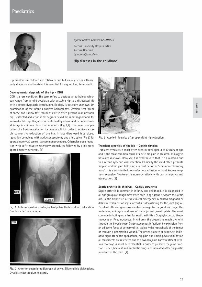
25
Paediatrics
Bjarne Møller-Madsen MD.DMSCi
Aarhus University Hospital NBGAarhus, [email protected]
Hip diseases in the childhood
Hip problems in children are relatively rare but usually serious. Hence, early diagnosis and treatment is essential for a good long term result.
Developmental dysplasia of the hip – DDHDDH is a rare condition. The term refers to acetabular pathology which can range from a mild dysplasia with a stable hip to a dislocated hip with a severe dysplastic acetabulum. Etiology is basically unknown. On examination of the infant a positive Galeazzi test, Ortolani test “clunk of entry” and Barlow test, “clunk of exit” is often present in an unstable hip. Restricted abduction in 90 degrees flexed hip is pathognomonic for an irreducible hip. Diagnosis is confirmed by ultrasound or convention-al X-rays in children older than 4 months (Fig 1,2). Treatment is appli-cation of a flexion-abduction harness or splint in order to achieve a sta-ble concentric reduction of the hip. In late diagnosed hips closed reduction combined with adductor tenotomy and a hip spica (Fig 3) for approximately 20 weeks is a common procedure. Otherwise open reduc-tion with soft tissue release/bony procedures followed by a hip spica approximately 20 weeks. (1)
Fig. 1 Anterior-posterior radiograph of pelvis. unilateral hip dislocation. Dysplastic left acetabulum.
Fig. 2 Anterior-posterior radiograph of pelvis. Bilateral hip dislocations. Dysplastic acetabulum bilateral.
Fig. 3 Applied hip spica after open right hip reduction.
Transient synovitis of the hip – Coxitis simplexTransient synovitis is most often seen in boys aged 3 to 6 years of age and is the most common cause of acute hip pain in children. Etiology is basically unknown. However, it is hypothesized that it is a reaction due to a recent systemic viral infection. Clinically the child often presents limping and hip pain following a recent period of “common cold/runny nose”. It is a self-limited non-infectious effusion without known long-term sequelae. Treatment is non-operatively with oral analgesics and observation. (2)
Septic arthritis in children – Coxitis purulentaSeptic arthritis is common in infancy and childhood. It is diagnosed in all age groups although most often seen in age group newborn to 5 years old. Septic arthritis is a true clinical emergency. A missed diagnosis or delay in treatment of septic arthritis is devastating for the joint (Fig 4). Purulent effusion gives irreversible damage to the joint cartilage, the underlying epiphysis and loss of the adjacent growth plate. The most common infecting organism for septic arthritis is Staphylococcus, Strep-tococcus or Pneumococcus. In children the organisms reach the joint through the blood stream (haematogenous infection); by extension from an adjacent focus of osteomyelitis, typically the metaphysis of the femur or through a penetrating wound. The onset is acute or subacute. Indic-ative signs are septic appearance, hip pain and limping. On examination all movements are restricted due to a swollen joint. Early treatment with-in a few days is absolutely essential in order to preserve the joint func-tion. Hence, bed rest and antibiotic drugs are indicated after diagnostic puncture of the joint. (2)
Paed
iatr
ics

26
Paediatrics
Fig. 4 Septic arthritis caused necrosis in right hip.
Legg-Calve-Perthes disease - LCPLCP is an idiopathic non-vascular necrosis of the proximal femoral epi-physis. The disease appears typically in age group 4–8 years old. Etiolo-gy is basically unknown. Hip, thigh or knee pain combined with restrict-ed range of motion is the most common symptoms. On examination hip movements are restricted due to painful synovitis. In late stages leg length discrepancy may be present. The diagnosis is based on conven-tional X-rays giving the basis for a classification e.g. Herring lateral pil-lar classification (Fig 5). Prognosis is closely associated with age at pres-entation. The best treatment method has not yet been determined. However, treatment at present mainly follows the “containment” princi-ples. 50% of patients will have good results without any treatment. (3)
Fig. 5 Legg-Calve-Perthes disease, Herring type C. Avascular necrosis right hip. Anteroposterior radiograph and frog-leg projection of pelvis.
Slipped capital femoral epiphysis – SCFESCFE is a disorder of the physis of the proximal femur. The femoral neck is slipping off the proximal femoral epiphysis. SCFE occurs typically in age group 10-15 years of old and is more common in boys. The preferred clinical classification is stable /unstable on the basis of the patient’s ability to bear weight. On examination restricted range of hip movement is usual. Obligatory external rotation of hip in flexion is pathognomon-
ic. Diagnosis is confirmed on conventional X-rays, anterior-posterior pro-jection and frog leg positions (Fig 6). Preferred treatment is in-situ pin-ning with a single screw. (4,5)Fig. 6 Anterior-posterior radiograph and frog-leg projection of pelvis. Slipped capital femoral epiphysiolysis left side.
REFERENCESSewell MD, Rosendahl K, Eastwood DM. developmental dysplasia of the 1.
hip. (2009) BMJ. Nov 24; 339:b4454. Kang SN, Sanghera T, Mangwani J, Paterson JM, Ramachandran M.2.
(2009) The management of septic arthritis in children: systematic review of the English language literature. J Bone Joint Surg Br. Sep; 91(9):1127-33.
Wenger DR, Ward WT, Herring JA. Legg-Calvé-Perthes disease. (1991) 3. J Bone Joint Surg Am. 1991 Jun; 73(5):778-88.
Aronsson DD, Loder RT, Breur GJ, Weinstein SL (2006) Slipped Capital 4. Femoral Epiphysis: Current Concepts J Am Acad Orthop Surg; 14:666-679
Loder RT, Aronsson DD, Dobbs MB, et al. (2000) Slipped capital femo-5. ral epiphysis. J Bone Joint Surg [Am]. 2000; 82:1170–1188.

27
Basic Science
Mr. Stephen Cannon
The Royal National Orthopaedic HospitalStanmore, [email protected]
Diagnostic work up and recognition of primary bone tumours
Although the frequency of metastases from primary cancers to bone is unknown, it is very much more common than the incidence of sarcoma. Soft tissue sarcoma has an incidence of around 2,500 cases per annum in the population of the united kingdom (66 Million) and is treated by a number of different surgeons, including orthopaedic surgeons. Bone sarcomata are treated only by orthopaedic surgeons and medical oncol-ogists and the incidence in the united kingdom is around 6 cases per million, per annum. Therefore, in the united kingdom we are likely to see around 360 cases per annum. This rarity of occurrence means that the orthopaedic surgeon must always be wary and suspicious of the probability of the occurrence of a primary bone sarcoma, which usual-ly presents as an uncharacteristic, unrelenting and progressive pain, swelling or pathological fracture.
Rarely in the immature skeleton disorders of growth can occur, partic-ularly around the knee joint. Bone sarcoma is rare beneath the age of five years where the common diagnosis is osteomyelitis, metastatic neu-roblastoma, leukaemia, eosinophilic granuloma or unicameral bone cyst. Primary bone sarcomata, therefore, tend to affect the adolescent popu-lation with an age range between 5 and 20 years and malignant sarco-mas must be differentiated from primary benign disease and fibrous dys-plasia. The commonest sarcoma is osteosarcoma and this has a peak incidence at 13.5 years in girls and 17 years in boys. Ewing’s sarcoma has a similar age distribution to osteosarcoma, but tends to affect slight-ly older patients in addition. The rare condition of chondrosarcoma is very unusual below the age of 20 and tends to afflict the older age group of 50 years and above.
When a bone tumour is considered the first and primary investigation that is required is a simple x-ray and the diagnostic process which fol-lows the recognition of a lesion on the x-ray should be as follows:
What is the lesion doing to the bone?1. What is the bone doing in response to the presence of the lesion 2. has any response been formed?Are there any characteristic features detectable?3.
The presence or absence of response from the host bones usually gives an indication of the degree of rate of growth of the tumour. Well corti-cated areas with a very narrow zone of transition are obviously tumours which have a slower growth rate and allow host-bone to respond, com-pared to those where there is a wide zone of transition or large soft tis-sue mass present. When considering an x-ray a number of features need to be assessed:
The age of the patient.1. The site of the lesion within the skeleton.2. The site of the lesion within the bone – whether it is epiphyseal 3. metaphyseal or diaphyseal. Whether it is intramedullary, cortical or on the surface.4.
The radiological appearances of the lesion itself.5.
The majority of tumours are in fact metaphyseal and intramedullary in their origin. They affect usually the distal femur, the proximal tibia or the proximal humerus, which are areas of rapid growth.
In conclusion, the plain radiograph is the most important investigation for characterisation of the lesion and assessing the general character-istics of the tumour. The MRI is a very important tool for local staging, accurately mapping out the area of involvement of the tumour, but the MRI although useful for characterisation can often be misinterpreted due to reactive bone and soft tissue oedema. Bone tumours generally should be referred to a specialist centre where a multi-disciplinary approach should be taken.
Having established the possible diagnosis of a primary bone tumour by plain x-ray it is important to stage the patient both locally and distally. The local staging essentially takes place by an MRI. Distal staging takes place by CT scanning and Technetium bone scanning or occasionally by PET scanning. Once the lesion has undergone radiological staging then a tissue diagnosis by bone biopsy is required. The usefulness of the var-ious modalities available and their strengths and weaknesses at answer-ing various questions is outlined in Figure 1.
Local staging of the disease essentially wishes to draw out the local extent of the lesion and by use of an MRI of the whole bone define any local skip metastases. The local staging also requires an estimation of the extraosseus extent of the disease, the involvement of soft tissue muscle compartments, the involvement of the joint and the relationship of the tumour to the neurovascular bundle, which is of tantamount importance in considering limb salvage procedures. Magnetic Resonance
Imaging Modalities Primary Bone Tumours
MRI CT Angiogra-phy
Scinitig-raphy
Plain flim
Extraosseous extn. 4.1 3.6 2.9 2.6 1.7
Intraosseous extn. 4.5 4.2 2.9 4.4 3.3
Cortial destruction 3.0 4.0 3.6
Calcification ossification
1.6 3.8 3.0
Periostcal/ enos-teal react
1.6 2.1 3.4
Basi
c Sc
ienc
e
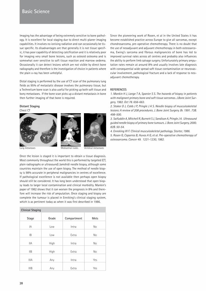
28 28
Imaging has the advantage of being extremely sensitive to bone pathol-ogy. It is excellent for local staging due to direct multi-planer imaging capabilities. It involves no ionising radiation and can occasionally be tis-sue specific. Its disadvantages are that generally it is not tissue specif-ic, it has poor capability of detecting calcification and it is relatively poor for imaging very small bone lesions, such as osteoid osteoma and is somewhat over sensitive to soft tissue reaction and marrow oedema. Occasionally it can detect lesions which are not visible by direct bone radiography and therefore is the investigation of choice in patients where the plain x-ray has been unhelpful.
Distal staging is performed by the use of CT scan of the pulmonary lung fields as 95% of metastatic disease involves the pulmonary tissue, but a Technetium bone scan is also useful for picking up both soft tissue and bony metastases. If the bone scan picks up a distant metastasis in bone then further imaging of that bone is required.
Once the lesion is staged it is important to obtain a tissue diagnosis. Most commonly throughout the world this is performed by targeted (CT, plain radiographs or ultrasound) Jamshidi needle biopsy, although some countries maintain the use of open biopsy. The method of needle biop-sy is 98% accurate in peripheral malignancies in centres of excellence. If pathological excellence is not available then perhaps open biopsy should still be considered. It has long been understood that open biop-sy leads to larger local contamination and clinical morbidity. Mankin’s paper of 1982 shows that it can worsen the prognosis in 8% and there-fore will increase the risk of amputation. Once staging and biopsy are complete the tumour is placed in Enniking’s clinical staging system, which is as pertinent today as when it was first described in 1986.
Since the pioneering work of Rosen, et al in the united States it has become established practice across Europe to give all sarcomas, except chondrosarcoma, pre-operative chemotherapy. There is no doubt that the use of neoadjuvant and adjuvant chemotherapy in both osteosarco-ma, Ewing’s sarcoma and fibrous malignancies of bone has led to improved survival rates across all centres and probably also influences the ability to perform limb salvage surgery. unfortunately primary ampu-tation rates remain at around 8% and usually involves late diagnosis with consequential wide spread soft tissue contamination or neurovas-cular involvement, pathological fracture and a lack of response to neo-adjuvant chemotherapy.
REFERENCESMankin H J, Lange T A, Spanier S S. The hazards of biopsy in patients 1.
with malignant primary bone and soft tissue sarcomas. J.Bone Joint Sur-gery, 1982. Oct 78. 656-663.
Stoker D J, Cobb J P, Pringle J A S. Needle biopsy of musculoskeletal 2. lesions: A review of 208 procedures. J. Bone Joint Surgery. Br. 1991. 73B. 498-500.
Saifuddin A, Mitchell R, Burnett S J, Sandison A, Pringle J A. Ultrasound 3. guided needle biopsy of primary bone tumours. J. Bone Joint Surgery, 2000. 82B. 50-54.
Enneking W F. Clinical musculoskeletal pathology. Storter, 1986.4. Rosen G, Caparros B, Huvos A G, et al. Pre-operative chemotherapy of 5.
osteosarcoma. Cancer 49. 1221-1230, 1982.
Clinical Staging
Stage Grade Compartment Mets
IA Low Intra No
IB Low Extra No
IIA High Intra No
IIB High Extra No
IIIA Any Intra Yes
IIIB Any Extra Yes
Distant StagingChest CT
Lung metastases Mediastinal nodes Vertebral metastases
Basic Science

29 29
Prof. Dr. Miklòs Szendröi
Semmelweis UniversityBudapest, [email protected]
Diagnostic algorithm and treatments options in bone metastasis
Oncology management is becoming an increasingly more serious task in orthopaedic and trauma surgery. A significant number of patients, who have solitary or multiple bone metastases can survive on cytostatic treat-ment for years.The significance of the treatment of bone metastases is indicated by the fact that they are 80-100 times more common than primary malignant bone tumors. Various cancers have very different „bone affinities” as con-cerns their metastases (Table 1.). In 65-85% of bone metastases the primary site of the tumor is in the breast, lung, kidney and prostate. The bones most frequently involved in decreasing sequence are: lumbar, dorsal, cervical spine, ribs, proximal femur and tibia, skull, pelvis, sternum and humerus. Only 1-2% of these secondaries affect the short tubular bones of the hand and foot.
Symptoms: Deep intermittent pain that is independent of the move-ment, often presents weeks or month before the X-ray changes are detected. The case history (primary cancer!) and laboratory tests must be thoroughly evaluated. In 10-30% of cases the first episode is a path-ologic fracture of a lytic metastasis of kidney or lung cancer. Osteoplas-tic metastases of prostate cancer rarely break and have good propensi-ty to heal.
Imaging: In suspected cases, e.g. when there is local bone pain after his-tory of tumor, an X-ray is taken of the area in question and CT, MR (occa-sionally PET-CT) scans are added if necessary. In spine, in the opposite of spondylitis the tumor involves single vertebral bodies, invading the intervertebral space only in later stage. In the long tubular bones, the lesion may be central, though it is more often eccentric, involving the cortex. Periosteal reaction is in most cases absent. Bone scan is also extremely important to decide if the process is single or multiple (Fig. 1.a. and b.).
Prognostic factors: The most sensitive prognostic factor is the origin of the primary tumor (Table 2.). In cases of breast, prostate, thyroid and kidney cancers, the expected survival time is much longer than in cases of lung cancers or bone metastases of melanoma (Fig. 2. and 3.). The life expectancy is poor (Table 3.) when the primary tumor is unknown or inoperable, or when the primary is discovered at the same time as the metastases, if the metastases are inoperable, multiple or multiorganic.
Surgical treatment: The surgical intervention can be palliative or cura-tive. The aims of palliative surgical treatment are: to alleviate the pain, to prevent the imminent fracture, to osteosynthesize and strenghten the bone in case of pathologic fracture using the less invasive technique, to reconstruct the motion and mobility of the patient ensuring a better quality of life. There is a broad range of the possible surgical procedures for reconstruction of the defect, i.g. plating (Fig.4.), intramedullary nail-ing (Fig.5.), curetting the defect and filling up with bone cement or inser-
tion of a normal (Fig.6.) or tumor endoprosthesis. Intramedullary nailing is advantageous for it is stable weight-bearing, and even if the tumor progresses, loosening of the implant is not likely. In 10-20% of the cas-es a curative-type radical tumor excision (Table 4.) is warranted using limb-saving surgery and reconstruction of the defect by modular tumor endoprosthesis or allograft.
REFERENCES1. Baloch KG, Grimer RJ, Carter SR, et al. Radical surgery for the solitary bone metastasis from renal cell carcinoma. J Bone Joint Surg. 82:62-67, 2000.2. DeVita VT, Hellman Jr S, Rosenberg SA. Principles and practice of oncol-ogy 5th ed.Lippincott-Raven Publishers, Philadelphia. Chapter 50. Treat-ment of metastatic cancer. Pp.2570-2585, 1997.3. Jemal A, Siegel R, Ward E et al. Cancer statistics, 2008. CA Cancer J Clin 58:71-96, 2008.4. Jung ST, Ghert MA, Harrelson JM et al.: Treatment of osseous metas-tases in patients with renal cell carcinoma. Clin Orthop Rel Res. 409:223-231, 2003.5. Szendröi M, Sárváry A: Surgery of bone metastases. In: Besznyák I (Ed.) Diagnosis and surgery of organ metastases, 1st ed. Akadémiai Kiadó, Budapest,2001, pp:213-248.
FIGURES
Fig. 1a Diagnostic algorythm at impending fracture (known primary tumor)
Basic Science
Basi
c Sc
ienc
e

30
Fig. 1b Diagnostic algorythm at impending fracture (unknown prima-ry tumor)
Fig. 2 Survival according to the primary site in 209 metastatic patients
Fig. 3 Survival according to the solitary and multiple manifestations of bone metastases in kidney cancer
Fig. 4 Plating with cementation
Fig. 5 Intramedullary nailing
Fig. 6 Conventional cemented revision endoprosthesis
Basic Science

31
TABLES
Basic Science
Basi
c Sc
ienc
e
32
Prof. Dr. Klaus-Peter Günther
University of DresdenDresden, [email protected]
Hip reconstruction: Osteotomy and joint replacement
Several disorders of the growing hip (i.e. developmental dysplasia-DDH, tilt deformity with consecutive impingement, perthes disease) and avas-cular necrosis in the adult age are relevant mechanical risk factors for the development of hip osteoarthritis. In early disease stages with only minor morphological signs of cartilage degeneration osteotomies can help to preserve the joint. Indications are mild pain, absence of advanced radiographic osteoarthritis and an understanding of the procedure by the surgeon as well as the patient.
In adult patients with dysplastic hips pelvic osteotomies are more often performed than femoral osteotomies, as their reorientative potential is higher and they have less disadvantages. Common techniques are the Toennis “Triple osteotomy” and the Ganz “Bernese periacetabular oste-otomy”. Both have a significant potential to correct a pathologic acat-abular coverage, although potential complications are associated with these major surgical procedures (i.e. risk of neurovascular damage, non-union and under-/over-coverage).In avascular necrosis of the femoral head (AVN) femoral osteotomies are a treatment option in limited disease stages (ARCO II-III) with minor defect size (kerboul-angle lower than 200°). In advanced stages or larg-er defect sizes the outcome is not encouraging. The Sugioka “rotation-al osteotomy” is rarely performed and technically very demanding.In patients with a “tilt deformity” due to growth disorders or mild and often unrecognized slipped capital femoral epiphysis (SCFE) symptomat-ic femoroacetabular impingement might result. Surgical treatment options with dislocation of the femoral head through trochanteric oste-otomy and open offset reconstruction have been developed.
In patients with advanced hip osteoarthritis joint preserving osteoto-mies generally are not any more indicated. If conservative treatment options over sufficient time periods (at least 3 to 6 months) fail and patient complain of significant pain and/or functional impairment, total joiunt replacement might be indicated.Different options regarding choice of implant and fixation technique (i.e. “conventional” cemented, cementless, hybrid, hemiarthroplasty, surface replacement, neck preserving stems) as well as bearing materials (uHWM-polyethylene, highly-crosslinked polyethylene, metal-on-metal, ceram-ic-on ceramic) are available. Generally there is a tendency to recom-mend cementless prosthesis with hard bearings in younger and active patients, while cemented implants and conventional bearings are indi-cated in elderly and less active patients. All options, however, have their advantages and also disadvantages in special situations. Therefore gen-eral recommendations regarding the application of certain techniques or materials in any case are not possible. Surgical exposure is possible via anterior, lateral, posterolateral as well as medial approaches and “minimally invasive” procedures have recent-ly been developed. There is not enough evidence until now, however, to recommend these techniques generally.
Although THR is one of the most effective medical procedures, patients and surgeons must be aware of potential complications. Therefore, cer-tain general steps should be done in every hip replacement surgery, which include
controlled/safe patient positioning and check of correct side -adequate soft tissue management (including repeated irrigation -to prevent heterotopic bone formation)intraoperative control of leg legth and offset with trial prosthesis -intra- and or postoperative x-ray control -adequate treatment to prevent periprosthetic infection (single- -shot antibiotics) and thrombosis (pharmaceutical and non-phar-maceutical options)immediate postoperative control of neuro-vascular status of the -operated leg
REFERENCESAustralian Orthopaedic Association National Joint Replacement Reg-1.
istry. Annual Report. Adelaide: AOA; 2009Bernstein P, Thielemann F, Günther K-P, (2007) A Modification of Peria-2.
cetabular Osteotomy Using a Two-Incision Approach. The Open Orthop Journal 1:18-13
Ganz R, Klaue K, Vinh TS, Mast JW (1988) A new periacetabular oste-3. otomy for the treatment of hip dysplasias, technique and preliminary results. Clin Orthop 232:26-36
Ganz R, Gill TJ, Gautier E, Ganz K, Krugel N, Berlemann U (2001) Surgi-4. cal dislocation of the adult hip a technique with full access to the femo-ral head and acetabulum without the risk of avascular necrosis. J Bone Joint Surg Br 83-8:1119-24.
Ganz R, Parvizi J, Beck M, Leunig M, Notzli H, Siebenrock KA (2003) 5. Femoroacetabular impingement: A cause for osteoarthritis of the hip. Clin Orthop Relat Res 417:112-20
Günther KP, Stürmer T, Sauerland S, Zeissig I, Sun Y, Kessler S, Scharf 6. HP, Brenner H, Puhl W (1998) Prevalence of generalised osteoarthritis in patients with advanced hip and knee osteoarthritis: the Ulm Osteoarthri-tis Study. Ann Rheum Dis 57(12):717-23
Pauwels F. Biomechanical principles of varus/valgus intertrochanteric 7. osteotomy in the treatment of osteoarthritis of the hip. In: Schatzker J (Hrsg) The intertrochanteric osteotomy. Berlin Springer 1984
Stöve J, Riederle F, Puhl W, Günther KP. Prädiktoren des Behandlungs-8. verlaufes nach Umstellungsosteotomie bei Hüftkopfnekrose. Z Orthop 2001 139:507-511
Tönnis D (1987) Congenital Dysplasia and Dislocation of the Hip in Chil-9. dren and Adults, New York: Springer p 113-30, 167, 370-80
Tönnis D, Arning A, Bloch M, Heinecke A, Kalchschmidt K, (1994) Tri-10. ple Pelvic Osteotomy. Journal of Pedatric Orthop Part B 3:54-67
Reconstruction

33
Reconstruction
1998 2000 2002
Fig. 1 Rapid OA progression in a patient with insufficient acetabular coverage (CE angle < 18°)
Fig. 2 bilateral staged pelvic reorientation osteotomy due to severe hip dysplasia
Fig. 3 different implants for total hip replacement (from left to right: cemented THR, cementless THR, surface replacement, neck preserving THR)
Fig. 4 preoperative planning in hip replacement is important
Reco
nstr
uctio
n
34
Dr. Martin Pietsch
Orthopedic Hospital Stolzalpe Stolzalpe, [email protected]
Knee: Osteotomy and arthroplasty
Osteotomies around the kneeSeveral treatment options for the osteoarthritis of the knee in the mid-dle-aged patients to preserve the joint are available. Osteotomies around the knee are well known, and different procedures have been reported over years. However, not all results have been described as well. The most important factors for a successful result are a proper patient selection, preoperative planning and a reproducible surgical technique with mod-ern angle stable (locked) implants. The principles are not new: the deformity should be corrected to unload the concerned compartment.
Patient selectionIn most of the patients pain is reduced, and time is gained before the knee replacement is performed. The ideal candidate is under 60 years of age and not obese. The deformity is not over 15° (Table 1). The patient should have a full range of motion and a stable joint. A flexion contrac-ture of 25° is an exclusion according to the ISAkOS recommendations (1). However, what should we do with a patient middle-aged and a flex-ion contracture ? Several authors advise to reduce the posterior tibial slope to get full extension. Others do not accept to change the slope. The dimension of the cartilage lesion of the compartment to be unload-ed is under discussion. Most surgeon likes to have cartilage remained (outerbridge III). Others accept also no cartilage (outerbridge IV). In addi-tion, the dimension of the cartilage lesion of the not affected compart-ment is unclear (maximum outerbridge III or only outerbridge II ?). Most authors accept patellar-femoral arthrosis as a non leading symptom. Smokers have a higher rate of failure regarding bone healing.
Table 1 patient selection for osteotomy [mod. (1)]
PlanningThe minimum of preoperative planning should include the answers of two questions: 1. On which side should the osteotomy be performed and what happens with the joint line; 2. How much correction should be per-formed and what size of the wedge is therefore, needed . The deformi-ty to be corrected should be analyzed on a standing, three-joint radio-graph (Fig. 1), the choice of the side of the osteotomy should consider the joint line. Not every varus deformity should be corrected on the tib-ial side. If the preoperative varus deformity shows the deformity on the femoral side, it is superior to perform the osteotomy (lateral closed wedge osteotomy) on the femoral side to keep the joint line horizontal (Fig. 2). If the joint line cannot be kept horizontal (87± 3°, (2)) with a single oste-otomy on the tibial or femoral side, a double level osteotomy should be taken under consideration. This is usually necessary in hugh corrections, in which the size of the wedge should be separated on the tibial and femoral side.The lateral distal femoral angle (LDFA) and the medial proximal tibial angle (MPTA) could be taken to analyses the deformity (2) (Fig. 1). The varus deformity is overcorrected (maximum valgus of 6°), the valgus is corrected to neutral (Table 2). The overcorrecting should be individual and can be determined by using the Fujisawa point (3) (Fig. 1). The degree of overcorrecting should include the dimension of the cartilage lesion (outerbridge 0 to IV, Table 2) (4). The amount of the wedge to correct the deformity can be calculated by computer, by the trigonometric chart by Hernigou (5) or by the method of Miniaci (6) (Fig. 3). Planning the sagittal alignment takes the posterior tibial slope under consideration, a true lateral view is therefore, necessary. Increasing the tibial slope reduces the posterior translation of the tibia, eliminates hyperextension and takes load from the posterior cruciate ligament (therefore, indicat-
Reconstruction
ideal candidate potential candidate no candidate
isolated pain medial or lateral infect in history complete arthrosis
age of 40 to 60 years age of <40 years and > 60 years
BMI <30 BMI 30 to 40 BMI >40
deformity <15° deformity >15° (double level osteotomy)
full ROM flexion contracture >15° flexion contracture >25°
no patellar-femoral painpatellar-femoral pain non leading symptom
patello-femoral pain leading symptom
cartilage lesion outerbridge I to III cartilage lesion outerbridge IV
stable joint insufficient ACL or PCL medio-lateral instability
non smoker Smoker
35
Reconstruction
ed in posterior instability). Decreasing the tibial slope reduces the ante-rior translation of the tibia, eliminates flexion contracture and stabiliz-es anterior cruciate deficiency (7) . Base for successful modern surgical cartilage repair procedures in the knee joint is a stable joint with a normal limb. A necessary additional osteotomy around the knee should always to be considered.
Table 2 degree of overcorrection of varus into valgus deformity [mod. (4)]
degree of overcorrection
postraumatic, no arthrosis 0 to 2°
ACL lesion 0 to 2°
PCL lesion 2 to 4°
cartilage repair procedure 3 to 5°
osteoarthrosis I and II 2 to 4°
osteoarthrosis III and IV 4 to 6°
Fig.1 a-c LDFA, MPTA and Fujisawa scale
LDFA, MPTA: enables to analyse the deformity: Is the deformity a. (varus, valgus) to be corrected more on the distal femoral side or more on the proximal tibial side ?
LDFA: lateral distal femur angle, standard value: 88 ±3°. MPTA: medial proximal tibia angle, standard value: 87 ±3°
“Joint Line angle”: angle on the lateral femoral side between the b. joint line and the overall biomechanical axis, standard value: 87 ±3°Fujisawa scale: center of the knee 0% to the lateral border as c. 100 %
The overcorrection into valgus can be measured and planned by the crossed mechanical axis of the leg
Correction into valgus Fujisawa
0 to 2 ° 0 to 20 %
2 to 4 ° 20 to 40 %
4 to 6° 40 to 60 %
Fig. 2 a-d Patient 23 years old with medial arthosis Outerbridge III and varus deformity of 6°
LDFA=93°, MPTA 88°, Joint Line Angle: 89° The deformity is a. located on the femoral sideSimulation of medial tibial open wedge osteotomy to 4° of val-b. gus shows that the joint line with 93° is not horizontal (87±3°) Simulation of lateral closed wedge osteotomy to 4° of valgus c. shows that the joint line is correct with 86° after the osteotomyPostoperative after lateral closed wedge osteotomy. Valgus 4°, d. LDFA=88°, MPTA=87°, Joint Line Angle 86°, Wedge= 10 mm
Fig. 3 a,b Planning the size of wedge in a medial open wedge tib-ial osteotomy according to the method of Miniaci (6)
a. long standing radiograph. (1) the new biomechanical axis after the osteotomy is drawn on the radiograph (determined by the Fujisawa point). The osteotomy hinge point (A) is choosen by the surgeon (usual-ly the level of the fibula-tibial joint). a,b The correction angle (X) corre-spond to the “osteotomy angle” (W). The size of the wedge in (mm) can be measured on the cortex.
TechniqueThe medial open wedge osteotomy at the tibia has several advantages compared to the closed wedge osteotomy (Table 3). The surgical tech-nique seems to be quite simply. No muscle has to be separated. No oste-otomy of the fibula is necessary. However, using of locked plates is rec-ommended. No bone substitution in a medial tibial open wedge osteotomy seems to be necessary.
Reco
nstr
uctio
n

36
Table 3 osteotomy on the proximal tibia: lateral closed versus medial open
closed open
surgery fibula less invasivecorrection difficult to be exact less difficultpatella baja not primary primaryslope less morecomplications peroneal palsy nonunionleg length shorter longerTKA difficult less difficult
Total Knee ArthroplastyTotal knee replacement is a very successful procedure. Excellent long-term results have been reported. However, not all patients are satisfied with the prosthesis. There is a gap of 10 to 15 % of the results of the top literature (90 % excellent and good results) and the average sur-geon in the registers (80 % excellent and good results). Early revisions within two years after the implantation have been described. Correct alignment, rotation, balancing and fixation are the base of an excellent result.
AlignmentProper frontal as well as sagittal alignment is important for a good clin-ical outcome and long lifetime of the prosthesis. Computed navigation or a long leg film should be used to get correct alignment. The femoral valgus correction angle should be an individual template on the long radiograph (Fig. 1). The LDFA and the MPTA can be used to verify the frontal cuts (8) (Fig. 2).
Fig. 1 a,b individual valgus cor-rection angle
a. valgus correction angle of 11° in a patient with curved femoral shaft and varus femoral neck b. valgus correction angle of 2° in a patient with valgus femoral neck in a hip prosthesis
Fig. 2 a-d using LDFA, MPTA in alignment verification in TkA
a. long standing radiograph. Analysis of the deformity with LDFA and MPTA. b. LDFA=85°, MPTA=86°c and d. cut verification of the distal femoral cut. C Distal femoral cut of the medial con-dyle shows 10 mm. D Distal femoral cut of the lateral condyle shows 5 mm. The LDFA of 85° is corrected to 90° (1 mm = 1°).
RotationTwo techniques to get correct femoral component rotation have been described: the measured resection technique and the flexion gap bal-anced technique. Both techniques have advantages and disadvantages. The measured resection technique uses bone landmarks (epicondylar axis) to get the correct femoral component rotation and takes away as much as bone is needed (measured resection). The flexion balanced tech-nique uses soft tissue (collateral ligaments) to rotate the femoral com-ponent in the correct rotation. However, there is a clear statement in the literature that the epicondylar axis should be used as the center of rotation in artificial knee joints (9). Theoretically, both methods should come to the same result. To verify epicondylar axis, the medial and lat-eral epicondyles should be identified by preparing the collateral liga-ments. The epicondylar axis is then drawn on the distal femoral surface. The anterior-posterior axis (Whiteside’s line (10)) is also drawn. These two axis allows to check the femoral component rotation (Fig. 3). Tibi-al component rotation is verified by the tibial tubercle (medial to medi-an third) and the attachment of the posterior cruciate ligament (modi-fied (11)) (Fig. 4). A malrotated femoral component leads to an unbalanced flexion gap (lift-off) (12). A malrotated tibial component leads to mala-lignment when a tibial posterior slope is used and patellar maltracking (in fixed bearings).
Fig. 3 a and b femoral component rotationCorrect femoral component rotation: the epicondylar line (ECL) a. is parallel to the dorsal condylar line (DCL) Verifying femoral component rotation by the epicondylar line b. (ECL) and Whiteside’s line (W).The epicondylar line is in 90° to Whiteside’s line.
Fig. 4 a and b tibial component rotationa and b: verifying correct tibial component rotation by the median to the medial tibia tubercle (a) and the attchement of the posterior cruci-ate ligament (a and b).
StabilityThe stability is not only given by the ligaments. The bony gaps should also be matched (Fig. 5). A correct femoral component rotation should be performed. A mismatch between extension to the flexion gap should not be tolerated. A distal re-cut to get more space in extension as well
Reconstruction

37
as using a smaller femoral component (in an anterior referenced pros-thesis) to get more space in flexion should be performed when neces-sary. One of the most important issues for success are the soft tissue release techniques (13) (14). In contract varus deformities the medial soft tissue complex should be adapted to the lateral side. In contract valgus deformities the contract lateral side should be adapted to the medial side (Fig. 6). The valgus deformity can be performed through a medial (“pie crust technique” (15)) as well as through a lateral approach (16).
Fig. 5 a and b stability in TkAMatched extension and flexion gap in total knee arthroplasty. a. The bone gaps are equal in extension and flexion, the ligamen-tous balancing of the medial soft tissue structures are equal to the lateral.Mismatch of the bone gaps. The extension gap > flexion gap. In b. a primary total knee arthroplasty the next smaller femoral com-ponent (anterior referenced system, “down sizing“) will give more space in flexion and match the gaps.
Fig. 6 a and b release in TkAMedial release in varus deformity a.
1 deep medial collateral ligament2 semimembranosous3 superficial medial collateral ligament4 pes anserinus
Lateral release in valgus deformity with lateral approach (16)b. 1 iliotibial band2 lateral collateral ligament3 popliteus muscle4 long head biceps
Controversies and commentsReplacement of the patella or not, posterior stabilized or cruciate retain-ing prosthesis, cementing or not are the old controversies in total knee replacement. Navigation, minimally invasive techniques, mobile or fixed inlays, new materials (trabecular metal, highly cross linked polyethyl-
ene), gender implants, high flexion design are the modern controversies.Patellar replacement: no standard has been shown in the literature (17). In most of the cases with anterior knee pain, it is not a question of replacement of the patellar or not. There are no “hard” facts when to replace and when not. Alignment, rotation, and balancing are more important to get good patellar tracking and to prevent anterior knee pain. Posterior stabilized prostheses are more forgiven compared to cruciate-retaining prostheses (Epicondylar line as the center of rotation in cruci-ate-retaining prosthesis? Change of the joint line is less forgiven in a cruciate-retaining. Posterior cruciate ligament always sufficient?) and cruciate-retaining prosthesis have shown no clinical advantages (18). So why using a prosthesis which is less forgiven but patients have no better clinical outcome? In addition, mobile inlays have not shown a clinical better outcome but also less forgiven (Soft tissue impingement with swelling. Additional dynamic component joint in the joint. Less forgiven using hugh release in severe deformities. Additional interface with small particle wear).Cemented prosthesis is standard for most surgeons. It is cheaper and seems to be save. Cemented primary prosthesis are not worse to revise. Still computed navigation is not standard in modern total knee replace-ment. Of the three basic principles, only frontal alignment can be prop-er be done by navigation.Minimally invasive techniques in total knee arthroplasty have only a short benefit (early rehabilitation) to the patient and are not standard. Modern designs in TkA (gender implants, high flexion implants) cannot achieve better clinical results. A proper surgical technique is still the most important factor for a good clinical outcome and to prevent early revision.
REFERENCESBrinkman JM, Lobenhoffer P, Agneskirchner JD, Staubli AE, Wymenga 1.
AB, van Heerwaarden RJ. Osteotomies around the knee: patient selection, stability of fixation and bone healing in high tibial osteotomies. J Bone Joint Surg Br 2008; 90(12):1548-1557.
Paley D, Tetsworth K. Mechanical axis deviation of the lower limbs. Pre-2. operative planning of multiapical frontal plane angular and bowing deformities of the femur and tibia. Clin Orthop Relat Res 1992; 280:65-71.
Fujisawa Y, Masuhara K, Shiomi S. The effect of high tibial osteotomy 3. on osteoarthritis of the knee. An arthroscopic study of 54 knee joints. Orthop Clin North Am 1979; 10(3):585-608.
Mueller W. Osteotomies around the knee. Instr Course Lect EFORT 4. 2001;34-39.
Hernigou P, Ovadia H, Goutallier D. [Mathematical modelling of open-5. wedge tibial osteotomy and correction tables]. Rev Chir Orthop Repara-trice Appar Mot 1992; 78(4):258-263.
Miniaci A, Ballmer FT, Ballmer PM, Jakob RP. Proximal tibial osteoto-6. my. A new fixation device. Clin Orthop Relat Res 1989;(246):250-259.
Agneskirchner JD, Lobenhoffer P, Wrann CD. Osteotomy and ligament 7. instability:tibial slope corrections and combined procedures around the knee joint. In: Lobenhoffer P, van Heerwaarden RJ, Staubli AE, Jakob RP, eds. Osteotomies around the knee. Stuttgart, New York: Thieme; 2007. 117-130.
Pietsch M, Hofmann S. Value of radiographic examination of the knee 8. joint for the orthopedic surgeon. Radiologe 2006; 46(1):55-64.
Berger RA, Crossett LS, Jacobs JJ, Rubash HE. Malrotation causing 9.
Reconstruction
Reco
nstr
uctio
n
38
Reconstruction
patellofemoral complications after total knee arthroplasty. Clin Orthop Relat Res 1998; 356:144-153.
Whiteside LA, Arima J. The anteroposterior axis for femoral rotation-10. al alignment in valgus total knee arthroplasty. Clin Orthop Relat Res 1995;(321):168-172.
Akagi M, Oh M, Nonaka T, Tsujimoto H, Asano T, Hamanishi C. An anter-11. oposterior axis of the tibia for total knee arthroplasty. Clin Orthop Relat Res 2004; 420:213-219.
Hofmann S, Romero J, Roth-Schiffl E, Albrecht T. Rotational malalign-12. ment of the components may cause chronic pain or early failure in total knee arthroplasty. Orthopade 2003; 32(6):469-476.
Yasgur D.J., Scuderi GR, Insall JN. Medial release for fixed-varus 13. deformity. In: Scuderi GR, Tria A.J.Jr., eds. Surgical techniques in total knee arthroplasty. New York: Springer; 2002. 189-196.
Griffin FM, Scuderi GR, Insall JN. Lateral release for fixed-valgus 14. deformity. In: Scuderi GR, Tria A.J.Jr., eds. Surgical techniques in total knee arthroplasty. New York: Springer; 2002. 197-204.
Ranawat AS, Ranawat CS, Elkus M, Rasquinha VJ, Rossi R, Babhulkar 15. S. Total knee arthroplasty for severe valgus deformity. J Bone Joint Surg Am 2005; 87 Suppl 1(Pt 2):271-284.
Keblish PA. The lateral approach. In: Scuderi GR, Tria A.J.Jr., eds. Sur-16. gical techniques in total knee arthroplasty. New York: Springer; 2002. 137-148.
Calvisi V, Camillieri G, Lupparelli S. Resurfacing versus nonresurfac-17. ing the patella in total knee arthroplasty: a critical appraisal of the avail-able evidence. Arch Orthop Trauma Surg 2009; 129(9):1261-1270.
Jacobs WC, Clement DJ, Wymenga AB. Retention versus sacrifice of 18. the posterior cruciate ligament in total knee replacement for treatment of osteoarthritis and rheumatoid arthritis. Cochrane Database Syst Rev 2005;(4):CD004803.
39
Reconstruction
Dr . Marino Delmi
Clinique des Grangettes Geneva, [email protected]
Reconstructive foot and ankle
Foot and Ankle surgery improved greatly in the last twenty years, improvements dues to better knowledge of physiopathology leading to more sound surgical techniques, improvements in anaesthetic techniques with reduction of pain, adaptation of postoperative cares and better understanding of reasons leading to failure, thus reducing recurrence and complications rates, and improving surgical results. This was accom-panied by a huge development of orthotic, surgical devices and implants.
AnatomyThe surgical anatomy of the Foot and Ankle is all about making access to the major structures without damaging structures in the surgical field. The surface anatomy is therefore vital to planning surgical approaches. The major superficial nerves are the first vital set of structures to avoid in the planning of surgical approach. These include moving from the pos-terior lateral side round to medially and from proximal to distal, the sur-al nerve, the superficial peroneal nerve, the deep peroneal nerve which becomes more superficial distally, the saphenous nerve, the dorsal prop-er nerve of the great toe, the terminal branches of the tibial nerve includ-ing medial and lateral calcaneal nerves and the digital nerves to the toes. Once surgical incisions are planned taking this into account further thought in certain areas needs to be given to the blood supply and ter-minal arteries serving skin. Incisions need to recognise the exact posi-tion of joints in relationship to the skin and general shape of the foot. Particularly coming down the medial column it is surprisingly easy to place incisions distally and access the wrong joint. As the incision deep-ens the major neurovascular bundles become a hazard and as do the dis-tal insertions of tendons.
BiomechanicsPractical biomechanics of the foot needs to recognise how the foot is maintained in a stable state during the act of standing in load bearing and propulsion. Normal gait is divided into stance (= approx 60%) and swing. The foot acts as an energy store and a stabiliser such that in mid-stance the foot is maximally stable and at its flattest position this is dic-tated by the major ligamentous structures especially the plantar fascia, the forefoot is held stably on the floor throughout stance once the foot flat position is obtained. This is effectively guaranteed by the reverse windlass mechanism the energy stored helps to put the foot into a pro-pulsive mode as the body weight passes forwards and the heel rises. The windlass mechanism maintains the forefoot position while also contin-uing to tension the plantar fascia and in doing so the longitudinal arch of the foot raises and the heel goes into varus maximising the drive by an essentially passive mechanism. The role of the long tendons is essen-tially to balance the foot from side to side and cope with varying forc-es. In pathological situations the ability to maintain and balance the stability of the fore foot during stance is lost those intact tendons that resist increased deformity will fail, further deformity will be worsened
by this and an increasing cascade of failure will occur.
HistoryPatients with foot pathology will usually complain of pain, loss of func-tion and increasing deformity to varying degrees. Like in other muscu-loskeletal sites the presence of unremitting pain must be regarded as a red flag to the presence of significant pathology. The site nature and radiation of pain can often help identify the structures involved. The onset and the relationship to specific events can be similarly helpful. Associated symptoms such as tingling numbness and instability should be sort. Pre-existent deformity, disease and family history are all of rel-evance.
ExaminationExamination of the foot needs to be systematised and thorough. A great deal can be learnt about the problem by observing the patient standing walking and by functional tests while standing before the patient is even touched. It is important to assess passive and active movements, motor function and neurology and particularly how these affect the balance of the foot.
ImagingImaging of Foot & Ankle pathologies includes: standard radiographs, uS, MRI, bone scan, CT – arthrography, and Spect-CT. Standard radiographs are cheap, fast to perform, panoramic, ready available, but allow only a limited evaluation of soft tissue and do not assess bone marrow. uS is a very powerful tool for tendons examination, can detect joint effusion, assess synovial hypertrophy and eventually guide a needle puncture. It can detect changes in nerve appearance and a compressive tumor in the tarsal tunnel. Teno-CT is the examination of choice if a longitudinal split or tear of one peroneal or the tibialis posterior tendon is suspected. Spi-ral CT-arthrography is the best imaging technique to evaluate ankle car-tilages. MRI is the best modality to evaluate ankle joints because of its panoramic, multiplanar capabilities and because of its high tissue con-trast. IV Gadolinium helps in the demonstration of synovial hypertrophy. Spect-CT is a relatively new very promising imaging modality.
Conservative treatmentIn many cases of foot pathology, conservative treatment may be the best solution and includes painkillers, steroid local injection, immobilisation, physical therapy, muscle stretching, pads, correction of shoeing, orthot-ic and custom made shoes or boots. Careful examination of the patient will establish the type pathology and lead to an appropriate treatment. In case of unsatisfactory result, surgery can be advocated.
Reco
nstr
uctio
n

40
Ankle
Fig.1 Arthritis of the ankle.Surgery for ankle arthritis (Figure 1) includes: 1. Cheilectomy either arthroscopic or open, 2. Periarticular osteotomies, 3. Arthrodesis either arthroscopic or open (Figure 2), 4. Total Ankle Replacement (TAR) (Fig-ure 3).
Fig. 2 Arthrodesis of the ankle.
Fig. 3 Total ankle replacement (TAR).
Despite of all warnings against it, ankle arthroplasty, in comparison to ankle arthrodesis, is probably the treatment of choice in many advanced ankle arthritis, but surely not for all. Three components TAR, with mobile polyethylene spacer, are the standard in Europe. Indications: good bone stock, adequate vascular status, no immuno-suppression, good align-ment of hindfoot, maintained ankle motion, sufficient medial and later-al ankle stability, contra-lateral ankle arthrodesis, low level of sports activity (bicycle, swimming, walking, golf). Relative contra-indications:
severe osteoporosis, history of osteomyelitis or septic arthritis, segmen-tal bone defect, smoking. Absolute contra-indications: extended AVN, neuroarthropathy (Charcot), important misalignment, massive joint lax-ity (Marfan), highly compromised peri-articular soft tissues, neurologi-cal impairment and high level of sports activity.Etiology of osteochondral lesions or defects (OCD) of the talar dome remains debated and their treatment is still improving. It is staged according to the extent and severity of OCD, with best results being obtained with either arthroscopic debridement and micro-fracture, or open autologous osteochondral transplant (mosaicplasty), or autologous chondrocyte culture and transplantation. Osteochondral allografts or synthetic materials are promising techniques, to be analyzed.Chronic ankle sprains may lead to ankle instability, lateral, medial or rotational, whose treatment is surgical. Ankle sprains may also be the cause of peroneal tendons longitudinal tears, more rare complete rup-tures, and tendons dislocation. All these lesions are often missed and require surgical repair.
Subtalar and ChopartArthrodesis of these joints (Figure 4) remains the treatment of choice in case of advanced posttraumatic or idiopathic subtalar arthrosis, tarsal coalition and severe varus or valgus deformity of the hindfoot. Hindfoot should be placed in slight 5 degrees valgus and position secured with staples, k-wires, or, best, with screws. Post-operative regimen usually includes a casting period of 8 weeks.
Fig. 4 Arthrodesis of the subtalar joint.
FlatfootThe adult acquired flatfoot is a progressive symptomatic collapse of the medial longitudinal arch of the foot. The term “acquired” implies that some structural changes cause the deformity in a foot that was struc-turally normal. The possible etiologies are: biomechanical disorders, neu-romuscular imbalance, tendons impairment (posterior tibial tendon - PTT), a Charcot foot (neuro-arthropathy), post-traumatic sequels, degenerative arthrosis and inflammatory arthritis. Strong correlated fac-tors are age, female patient, obesity and diabetes. The forces exceeding the static and dynamic restraints of the foot create progressive medial structures degenerative dysfunction (PTT - spring and deltoid ligaments) with progressive subluxation at subtalar and midtarsal joints.Chronic stress cause PTT dysfunction and a hypovascular zone makes the tendon prone to degeneration. Stage 1: tenosynovitis\tendinosis - nor-mal tendon length. Stage 2: tendon elongation – flexible hindfoot val-gus. Stage 3: tendon elongation or disruption - fixed hindfoot valgus. Stage 4: rigid hindfoot with ankle involvement. Clinical signs include the “Single heel rise” test with poor or absent evidence of heel varus at heel rise and the “Too many toes” sign.Conservative treatment is preferred as initial protocol. Stabilization and control of affected joints with orthotic can provide the patient a
Reconstruction

41
decreased level of pain and an increased level of function. The orthotic design should be acceptable to the patient’s lifestyle to ensure compli-ancy. In case of symptomatic patients not controlled by conservative treatment or clear progression of the pathology, surgical treatment should be advocated. Depending of the stage and severity of the deform-ity, most frequently bone and soft tissues procedures are associated, with tendon repair \ transfer, tendon (Achilles) lengthening and ligaments (spring, deltoid) repair, calcaneal osteotomies (Figure 5) and arthrodesis.
Fig. 5 Lengthening osteotomy (modified Evans) of the calcaneus.
Achilles tendonAchilles tendon acute ruptures are due to chronic degeneration of the tendon, with failure of the inhibitory mechanism of the musculotendi-nous unit, and risk is increased by corticosteroids (local or systemic) or previous treatment with fluoroquinolone antibiotics and derivatives. Treatments options include: A. Non-operative and B. Operative – Percu-taneous, Mini-invasive or Open. Non operative treatment is an adequate option but treatment is complex. Best option is the surgical mini-inva-sive suture for standard cases with functional post-operative care. This solution combines the advantage of both classical open and conserva-tive modalities, without their complications.Classification of Chronic Achilles tendinopathies includes (Marks): I. Per-itendinitis, II. Pantendinitis (peritendinitis and tendinosis), III. Tendino-sis, IV. Insertional (subcategories frequently coexist). After failure of con-servative measures, surgical treatment can be considered and includes various techniques: Brisement, Percutaneous longitudinal tenotomy, Open debridement, Excision and repair with FHL transfer and V-Y plasty.
Heel Pain – Tarsal tunnelThe heel pain syndrome is a poorly defined entity with numerous etiol-ogies; some of them are very rare while others are extremely frequent. We will concentrate on the two main etiologies: plantar fasciitis and entrapment of the first branch of the lateral plantar nerve. Treatment is first always conservative and carries out good results in 80 to 90% of the patients. It must however be stressed that the healing process can take a long time and both the patient and his doctor must be aware of that. Several therapeutic modalities are necessary. Actually, surgery is necessary in some 5 to 10% of the patients, all etiologies considered. If the diagnostic is carefully established, the surgical release gives favora-ble results in 90% of the entrapment neuropathies and recalcitrant plantar fasciitis.
Sinus tarsi syndromeSinus tarsi syndrome was described by O’Connor in 1958 in the case of old ligament trauma of the ankle; it was characterized by pain on the lateral aspect of the posterior tarsus when walking on uneven ground. The pressure of the lateral aspect of the sinus tarsi provokes an impor-tant pain. The patient describes a subjective instability of the hindfoot. The examination is normal. Standard and stress X-Rays are normal. Many
studies described an important quantity of mechanoreceptors (Paccini corpuscules, Golgi and Ruffini receptors, and nervous fibers) in the sinus tarsi. Electromyocinesiology studies showed dysfunction of the peroneal muscles. The normal pattern of the peronei activity is obtained after injection of local anesthetic in the sinus tarsi. Subtalar arthrography showed disappearance of the micro-recessi normally seen along of the interosseus ligament.The treatment was conservative or surgical. Conservative therapy con-sists in injection of anesthetic and cortisone in the sinus tarsi, along with proprioceptive reeducation. Surgery performs curettage of the sinus tar-si. Success of both conservative and surgical treatment was uncertain. Since use of Ct-scan and MRI, many pathologies have been discovered, such as osteochondral lesions, arthritis, congenital tarsal coalition, etc., and have put suspicion on the diagnosis of sinus tarsi syndrome.In case of painful instability of the hindfoot after a trauma, we advise to perform every possible examination (clinical, X-Rays, Ct-Scan, Bone-Scan, MRI, arthroscopy). Only if all these remain negative, consider the diagnosis of sinus tarsi syndrome.
Charcot (neuro-arthropathic) footJean-Martin Charcot has described first the neuro-arthropathy of the foot in 1868, in relation with the syphilis. Nowadays, the diabetes is the leading cause of this disease, whose etiology is still largely unknown. Mean age of diagnosis is 57, with the diabetes lasting usually for more than 10 years. No difference between male and female patients. Bilat-eralism occurs in 6-40%. Despite the increasing number of Charcot feet, this problem is generally poorly recognized and often poorly managed, leading to a high rate of amputations. The Charcot joint probably has both a vascular and a traumatic etiology. An acute trauma or repetitive microtraumas associated with the impaired sensation caused by the neu-ropathy are the start point of the architectural changes and joint destruc-tion. The presence of excellent circulation is necessary. The Charcot foot may present as a fracture, but more commonly as multiple fracture-dis-locations (Figure 6).
Fig. 6 Charcot arthropathy of the Chopart joints.
Evolution: Eichenholz (1966) has described three stages of development:Stage 1 or fragmentation:1. acute inflammation with bone destruc-tion and dislocation. Clinic: hyperemia, redness, hot swollen jointsStage 2 or coalescence:2. beginning of the reparative process, with bone resorption and callus formation.
Clinic: diminution of edema, warmth and erythema.Stage 3 or consolidation:3. bone healing, usually with residual deformity.
Clinic: “cold” foot, no swelling.Clinical evolution without treatment leads to break down of dislocated joints, with a «rocker bottom» foot deformity, plantar ulcers, infection
Reconstruction
Reco
nstr
uctio
n

42
and eventually amputation. The principles of conservative treatment are:Achieve the third stage of the bony healing with minimal deform-1. ity allowing the use of near normal shoes or easily adaptable orthopaedic shoesMinimize soft tissue problems and ulcerations, avoiding the 2. development of an osteomyelitis leading to an amputationkeep the patient as mobile as possible during the healing process.3. Long period of treatment (normal X 3)4.
The conservative measures include progressive stages:Rest and elevation of the foot to diminish the swelling and rule 1. out osteomyelitisTotal contact cast (change every 5-7 days) or CROW (Charcot 2. Restraining Orthotic Walker)Weight bearing as tolerated and if no progressive foot deforma-3. tionContinue the cast/walker brace as long as the patient has not 4. reached stage 3, clinically and radiologically (usually 4-6 months, but sometimes 12 months). Monthly radiographic controls After consolidation, use of an ankle brace or adapted orthoped-5. ic shoes, and regular medical supervision.
Surgical treatment is indicated in:Acute - stage 11.
failure of conservative treatmenta. progression of deformity or associated osteomyelitisb.
Consolidated - stage 32. recurrent ulcer or joint instabilitya.
and includes various techniques depending of the problem: a. debride-ment only or ostectomy, b. Ilizarov external fixator, c. joint fusion with strong plates or nails (Figure 7). Complications of surgery are post-op-erative infection, not rare and may lead to amputation. Rate increases with co-morbidities, especially smoking and previous active infection. Non-union with rupture of implants are frequent, but it is very often well tolerated and allows walking either with a bracing (AFO) or an adapted shoeing
Fig. 7 Nail arthrodesis of the ankle and subtalar joints.
Hallux valgusHallux valgus (Figure 8) is the most common pathologic condition of the forefoot and much more often seen in women than in men. It is the cause of pain on the medial “bump”, of transfer metarsalgia, and caus-es hammer toes.
Fig. 8 Bilateral hallux valgus.
More that 150 operations have been described for hallux valgus man-agement. They can be divided according to the localization:
Soft tissues1. P1 osteotomies2.
Akina. Distal MT1 osteotomies3.
Chevron (Figure 9)a. Reverdinb.
Mid-shaft MT1 osteotomies4. Scarfa.
Proximal MT1 osteotomies5. Crescentica. Chevronb. Closing or opening wedgec. Ludloffd.
CMT1 arthrodesis (Lapidus) (Figure 10)6. MTP1 arthrodesis7. MTP1 arthroplasties8.
Brandes-keller, Valenti, Mayoa. Prosthesisb.
Or according to the technique:A. OpenB. Minimal invasiveC. Percutaneous
Fig. 9 Distal chevron osteotomy.
Reconstruction

43
Fig. 10 Modified Lapidus (arthrodesis of CMT1) and Akin (P1) osteotomy.
Hallux rigidusArthrosis of the metatarso-phalageal joint of the big toe can be idio-pathic, microtraumatic (dancers), inflammatory (rheumatoid arthritis), due to a chronic misalignment (severe hallux valgus) or due to an oste-ochondritis. Pain and stiffness are the main clinical signs, together with the thickening of MTP1 joint (osteophytes). NSAID drugs, steroid intra-articular injection and orthotic may relieve symptoms. In case of per-sistent pain, surgical options include joint preserving procedures as oste-otomies of P1 (Moeberg) or MT1 (Watermann-Green, Weil), joint debridment with cheilectomy (open or percutaneous), resection arthro-plasties (Valenti, Brandès-keller), MTP1 arthrodesis and prostheses.Arthrodesis of MTP1 joint (Figure 11) is still the gold standard in advanced, stage 3 cases and shows a 90% of satisfaction rate, but positioning is demanding and it requires stability of the internal fixation.
Fig. 11 Arthrodesis of MTP1 joint.
Hammer ToesSurgical correction of hammer toes (Figure 12) often requires an arthro-desis of the proximal interphalangeal (PIP) joint. Most of the time, the joint is maintained with a metallic pin for weeks, with an increased risk of infection, breakage, migration and discomfort for the patient. Fur-thermore, fusion is not always granted after pin removal. use of inter-nal devices (Figure 13) could increase fusion rate, decrease complica-tions and improve patient’s comfort. Percutaneous surgery is an option to be evaluated, especially for metarsalgia.
Fig. 12 Second hammer toe.
Fig. 13 Arthrodesis of PIP joint with memory alloy intramedullary device.
MortonMorton’s syndrome is a very common diagnostic, maybe too often used: its real incidence is not well known and the prolific literature is contro-versial. It concerns an entrapment neuropathy of an inter digital nerve under the inter metatarsal ligament. It can be isolated or, more often, combined with overload pathology of the forefoot. The diagnosis is made essentially clinically. The conservative treatment, with adequate shoes and insoles, is efficient, especially if the symptom lasts for less than one year. The aetiology must be treated. After failure of this approach, the surgical option (Figure 14) must be evaluated carefully: the relatively « easy » procedure of removal of the nerve should not obviate the poten-tial pitfalls, leading sometimes to difficult to manage pain and re oper-ations. Options are injection of phenol and neurolysis by cutting the intermetarsal ligament, percutaneously or endoscopically.
Fig. 14 Excision of Morton’s neuroma through a dorsal third web space incision.
Morton’s nevroma surgery seems very “simple” but is actually very dif-ficult with many complications, bad results and “recurrences”.
Reconstruction
Reco
nstr
uctio
n

44
Reconstruction
What are the reasons for these “bad” results?Differential diagnosis is wide and may lead to wrong treatment: 1. synovitis, bursitis, MTP arthritis, Freiberg’s infarction, stress frac-ture, wart, mechanical hyperpressure, hyperlaxity of a MTP joint, tarsal tunnel syndrome, etc. In this situation, the so-call “recur-rence” of pain is simply the consequence of the false treatment and the persistence of the etiologic problem.
Treatment: treat the cause of the pain!Excision of the nerve is followed by the formation of a stump 2. neuroma. This is a normal process, but if this plantar neuroma is too big or too distal, it will be very painful. Treatment: second excision, more proximal, using the primitive dorsal approach or a plantar incision. In case of persistent problem due to a painful recurring neuroma, a redo with a tubular venous autograft could be the salvage solution.Loss of sensation due to removal of the nerve is also normal, 3. but may be the cause of bothering dysesthesia.
Treatment: no surgery, but conservative measures including pain killer, gabapentine, clonazepam, TENS, “trigger points” desensi-tization, specific insoles, etc.The scar itself, if plantar, is sometimes source of painful intrac-4. table plantar keratosis. Treatment: avoid plantar incision! Specif-ic relief insoles. Surgery: excise the scar – plastic surgery.
REFERENCESOperative Techniques in Foot and Ankle Surgery. Editors: Easley ME and 1.
Wiesel SW. Lippincott Williams & Wilkins, 2011.Reconstructive Foot and Ankle Surgery, 2nd edition. Editor: Myerson 2.
MS. Elsevier Saunders, 2010.Pathologie du Pied et de la Cheville. Editors: Leemrijse T, Valtin B. Else-3.
vier Masson SAS, 2009.Minimally Invasive Foot Surgery. Editors: De Prado M, Ripoll PL, Golanò 4.
P. AYH Publishers, 2009.Patologia del Piede. Techniche chirurgiche, vol I and II. Editor: Barca F. 5.
Timeo Editore, 2008 and 2005.Surgery of the Foot and Ankle, 8th ed. Editors: Coughlin MJ, Mann RA, 6.
Saltzman CL. Mosby Elsevier, 2007.Advanced reconstruction Foot and Ankle. Editor: American Academy 7.
of Orthopaedic Surgeons. AAOS, 2004.McMinn’s Color Atlas of Foot and Ankle Anatomy, 3rd ed. Editors: 8.
Logan BM, Singh D, Hutchings RT. Mosby, 2004.Masters techniques in Orthopaedic Surgery: The Foot and Ankle, 2nd 9.
ed. Editor: Kitaoka HB. Lippincott Williams & Wilkins, 2002. Foot and Ankle Disorders. Editor: Myerson MS. Saunders, 2000.10. Functional reconstruction of the Foot and Ankle. Editor: Hansen TH, 11.
Jr. Lippincott Williams & Wilkins, 2000. An Atlas of Foot and Ankle Surgery. Editors: Wülker N, Stephens M, 12.
Cracchiolo III A. Martin Dunitz, 1998. Surgery of Disorders of the Foot and Ankle, 2nd ed. Editors: Helal B, 13.
Rowley DI, Cracchiolo III A, Myerson MS. Martin Dunitz, 1996. Operative Foot Surgery. Editor: Gould JS. Saunders, 1994.14.
% Good Bad Re-do Numbness Normal shoes
Bennett,1996 85 15 21
Dereymaeker 1996 81 19 30
Jarde, 1995 89 11 41 75
Assmus,1994 81.5 18.5 11.1
Ruuskanen 1994 80 20 9
Keh, 1992 93 7

45
Reconstruction
Dr. Richard Wallensten
Karolinska University HospitalStockholm, [email protected]
Shoulder degenerative disorders
Basic anatomyThe shoulder consists of four joints, the sterno-claviclular, the acromio-clavicular, the gleno-humeral and the thoraco-scapular joint. The latter is not a proper joint but assists in the total range of motion correspond-ing to 30° of flexion-extension. Motion in the sterno- and acromioncla-vicular joints is small and consists of rotation. Most of the shoulder motion occurs in the gleno-humaral joint and consists of both flexion-extension and rotation inwards and outwards. The shoulder is the most mobile joint in the body. The normal motion is flexion 160°, abduction 160°, adduction to reach the opposite shoulder, external rotation 75° and internal rotation to Th 5-6.The gleno-humeral joint has a large head articulating with a small and flat socket that does not cover the head. The surface of the glenoid is just 1/5 of the surface of the humeral head. This means that the stabil-ity of the joint is totally dependent of the function of the surrounding soft tissues: the glenoid labrum, the rotator cuff, the long head of the biceps muscle and to low pressure in the joint cavity. Also the scapula stabilising muscles, trapezius, rhomboids, levator scapule and serratus anterior contribute to shoulder stability and function. The most impor-tant muscle for shoulder function is the deltoid that provides strength and endurance in motion of the arm. The deltoid muscle is innervated by the axillary nerve. The rotator cuff muscles are innervated by the suprascapular nerve ( m. supra- and infraspinatus), the axillary nerve (m. teres minor) and the subscapular nerve (m. subscapularis).
Approaches to the shoulderDeltopectoral approachThe deltopectoral approach is the most commonly used for open shoul-der surgery. It goes from the clavicle to the insertion of the deltoid on the humerus just lateral to the coracoid process following the interval between the deltoid and the pectoralis major. It may be used in its full length but usually one only needs the proximal part or less. After open-ing of the skin and subcutaneous tissue the cephalic vein is held later-ally together with the deltoid muscle and the pectoral muscle medially. The subscapular muscle is transacted close to its insertion and the joint capsule opened. When closing the incision a careful repair of the sub-scapularis is important.
Lateral, transdeltoid approach The lateral, transdeltoid incision is mainly used for surgery on the acromio-clavicular joint, open acromioplasty and rotator cuff repair. The skin incision follows the Langer lines along the acromion. underneath the anterior and middle deltoid bellies are separated and freed subperi-osteally from the acromion. Then the subacromial space is entered and the acromion, a-c-joint and rotator cuff can be addressed. When clos-ing a good repair of the deltoid on the acromion is of great impor-tance.
Posterior approach The posterior approach is not so commonly used but is practical for pos-terior instability, most scapular fractures and can be used for arthro-plasty. The incision frees the deltoid from the scapular spine and then the infraspinatus muscle is separated from the teres minor. The tendon of the infraspinatus is transacted close to the insertion on the humerus and the posterior joint capsule opened.
Clinical diagnosisThe diagnosis of degenerative conditions of the shoulder is based upon a good clinical examination evaluating motion, strength and weakness of the shoulder muscles, instability of the sterno-claviclular, the acromio-clavicular and the gleno-humeral joints and certain clinical tests. The routine examination should include the Neer or Hawkins’s test for impingement, Jobe’s test for supraspinatus and lift off test for subscapu-laris weakness and apprehension test for instability. In addition tender-ness and adduction pain in a-c-joint should be tested.
RadiologyConventional x-rays with AP- and lateral films and special a-c-joint views are the basis for diagnosis of shoulder disorders. ultrasound exam-ination by an experienced examiner is good for rotator cuff pathology. MRI should always be preceded by conventional x-ray and is primarily a study of the soft tissues. For detailed diagnosis of the skeleton CT gives the best information.
ArthroscopyToday arthroscopy is primarily a therapeutic technique and pure diag-nostic arthroscopies are rarely indicated. MRI usually provides sufficient and similar information in a non-invasive way.
The degenerative shoulder Impingement syndromeImpingement syndrome is a pain condition in the shoulder. The name was introduced by Neer who believed the cause to be a conflict between the acromion and coraco-acromial ligament and the subacromial bursa and the rotator cuff on arm elevation. Bigliani-Morrison defined three types of acromial shape where the more curved the acromion is the more common is impingement. This etiology has later been challenged. Impingement syndrome is characterized by pain on raising the arm and when doing overhead work. Night pain and difficulties sleeping on the affected side is typical. It exists on its own or in connection with rota-tor cuff tears. The diagnosis can be established by the patient’s history and a clinical examination. Special clinical tests are the Neer test and the Hawkins’ test which when positive produce the impingement pain. Injection of a local anaesthetic subacromially should take away the pain and make the impingement test negative. Standard x-rays may be nor-
Reco
nstr
uctio
n
46
mal or show curved acromion, sometimes with osteophytes at the tip, and/or arthritic changes in the a-c-joint with inferior osteophytes. MRI may show impression on the rotator cuff by the skeletal changes.Initially impingement syndrome is treated with pain medication, subac-romial injection of steroids and physical therapy. If non-surgical thera-py fails an acromioplasty is indicated. This means increasing the subac-romial space by resection of the tip and bottom surface of the acromion changing the form from curved to flat. Any inferior osteophytes in the a-c-joint are also resected. In Neer’s original description of the opera-tion the coraco-acromial ligament was also removed but later studies have shown that this is not necessary. An acromioplasty may be com-bined with lateral clavicle resection and with rotator cuff repair. Acromi-oplasty can be performed arthroscopically or open and today the former technique is dominating. Rehabilitation after the operation consists of a structured training programme and the outcome may take up to six months to achieve.
Rotator cuff tearsWith normal ageing the rotator cuff becomes thinner and weaker. The tendon tissue degenerates and tendinosis and partial tears occur. They can progress to full thickness ruptures without symptoms but more com-mon is that it is associated with pain and weakness on elevation and external rotation. Rotator cuff tears are more common with increasing age. A trivial trauma or load can change a partial tear to a total rupture. Cuff tears also occur in younger individuals but then usually after a sub-stantial trauma. Dislocation of the shoulder is associated with cuff rup-ture in patients over 40 years of age. Typical clinical symptoms are pain that aggravates with overhead activ-ities and which often disturbs sleep. Weakness on elevation and exter-nal rotation and sometimes loss of those motions are also typical. Jobe´s test (Empty can test) for loss of supraspinatus strength. On the other hand full range of motion is possible even with massive total ruptures.Diagnosis is made by the history and clinical examination. Plain x-rays may show cranial displacement of the humeral head relative to the gle-noid. MRI is diagnostic for the size of the rupture and which tendons are involved. It also shows the condition of the muscle bellies which atrophy with long standing rupture. The muscle is infiltrated by fat and becomes non-functional.Non-surgical therapy consists of physiotherapy aiming at strengthening the shoulder muscles and anti-inflammatory drugs. Subacromial ster-oids injections may give temporary pain relief. When this therapy fails to give sufficient pain decrease surgery is indicated. For patients with no pain but reduced motion and strength surgery is usually not indicat-ed since the outcome regarding these parameters is less predictable.Surgery aims at repairing the torn tendons and reattaches them to the humerus. The repair can be made arthroscopically for minor tears or mini-open (a combination of arthroscopic debridement and mobiliza-tion and open suture through a small transdeltoid incision. Larger tears demand an open repair. If the rupture is massive cuff and the cuff can’t be repaired a tenotomy of an intact long head biceps tendon may reduce pain. Acromioplasty is usually performed to increase the subacromial space and remove impingement on the repaired cuff. For patients in whom the loss of motion is a big problem improvement can be achieved with latissimus dorsi transfer or with a reversed shoul-der prosthesis.Rehabilitation after cuff repair consist of a structured training pro-gramme initially focused on gaining motion and after six to eight weeks strengthening exercises are added. The final outcome may take six to twelve months to achieve.
Primary osteoarthritis This entity is not uncommon in the shoulder. Its most often affects the a-c-joint and less common the gleno-humeral joint. Arthritic changes in the a-c-joint are a common find on x-ray but need not be sympto-matic. Radiological arthritis in the gleno-humeral joint is usually symp-tomatic. Patients complain of pain and limited motion. X-ray films show and decreased joint space and typical inferior osteophyte formation. Treatment modalities are initially conservative (NSAIDS, physiotherapy). When conservative treatment fails a-c-joint arthritis is operated with resection of the most lateral 0.5-1 cm of the clavicle. This procedure gives pain relief but does not affect shoulder function.For gleno-humeral arthritis arthroplasty is indicated. Both hemiarthro-plasty and total joint replacement are used although the latter is pre-ferred when rotator cuff function is intact and glenoid bone stock is enough preserved to permit fixation of a component.
Cuff tear arthropathyA special form of osteoarthritis in the shoulder is seen in combination with massive rotator cuff tears and is called cuff tear arthropathy. It is characterized by advanced arthritic changes, superior migration of the humeral head and absence of cuff function. Standard total shoulder replacement has an increased risk of glenoid loosening so hemiarthro-plasty with or without an extended head is used. Alternatively prosthe-sis with reversed design can give a good function. This prosthesis has the head fixed to the glenoid and the cup in the humerus thus medial-ising the centre of rotation which facilitates arm elevation by the del-toid muscle alone. The lack of rotator cuff function is thus overcome.
Acromio-clavicular osteoarthritisArthritis in the a-c-joint is a common radiological finding but less com-mon as a symptomatic disease. It is characterized by pain on overhead activities, local tenderness and night pain. Injection of a local anaes-thetic relieves the pain and can be used as a clinical diagnostic test. Radiological signs are irregular joint surfaces and osteophytes which can be seen on special views. The surgical treatment is resection of the lat-eral end of the clavicle, so called Mumford procedure. At the same time any concomitant symptomatic rotator cuff disease can be addressed.
Inflammatory arthritis Rheumatoid disease often affects the shoulder. Modern pharmacologi-cal treatment is usually successful and recent progress with anti-TNF drugs have very much slowed down the destructive process. Synovecto-my and arthrodesis was used earlier but today arthroplasty is the pro-cedure of choice when the drug treatment fails. The choice between hemi- and total shoulder replacement is decided by the degree of gle-noid destruction and rotator cuff function. With intact rotator cuff and glenoid total replacement is preferred. When the humeral head still is intact a resurfacing prosthesis on the humerus can be a good alterna-tive to avoid a stem down in the diaphysis in case an elbow prosthesis is in place or indicated later.
Septic arthritis in the shoulderThis is most common in patients with a compromised immune system such as rheumatoid arthritis, post organ transplantation or during chem-otherapy. It can be a potentially life threatening condition caused by a septicaemia and should be treated as such. Diagnosis is clinical with standard laboratory findings such as high white cell count, left shift of white blood cells, high sedimentation rate and elevated C-reactive pro-tein levels. Joint aspiration reveals the causing bacteria. Organisms found
Reconstruction

47
are generally staphylococcus or streptococcus. In mild cases treatment may be antibiotics but in more severe cases it is mandatory to clean out the joint arthroscopically or by open surgery. During the treatment peri-od appropriate i.v. and oral antibiotics are administered.
Adhesive capsulitisAdhesive capsulitis is also called frozen shoulder. It is an idiopathic dis-ease characterized by pain and progressive contracture of the gleno-hu-meral joint. The disease affects the joint capsule but the underlying cause is not known.The disease starts insidiously with slowly increasing shoulder pain espe-cially at night. After 2-6 months increasing stiffness at the gleno-hu-meral joint is noted. Both active and passive motion is reduced and can progress until the joint is completely stiff. This phase takes 3-9 months. The pain then starts to decrease slowly and the mobility of the shoulder comes back over a period of 3-12 months. Adhesive capsulitis thus takes 12-24 months from start to end full recovery is usually achieved. The treatment is symptomatic with analgesics and physiotherapy aiming at preserving the motion. Arthroscopic release of the joint capsule may be indicated when the diagnosis is clear in order to improve motion and shorten the natural history.
REFERENCESRockwood-Matsen-Wirth-Lippitt: The Shoulder. 4th ed., 2 vol., Saun-1.
ders, 2009Iannotti-Williams: Disorders of the Shoulder. 2nd ed., 2 vol. Lippincott 2.
Williams & Wilkins, 2006Frankl, M: Ritator Cuff Deficiency of the Shoulder. Thieme medical Pub-3.
lishers, 2008Dines, D., Laurencin, C., Williams, G.: The Shoulder in Arthritis & Arthro-4.
plasty, Saunders 2009
RECOMMENDED READINGRockwood-Matsen-Wirth-Lippitt: The Shoulder. 4th ed., 2 vol., Saun-1.
ders, 2009Iannotti-Williams: Disorders of the Shoulder. 2nd ed., 2 vol. Lippincott 2.
Williams & Wilkins, 2006Matsen-Lippitt: Shoulder Surgery: Principles and Procedures. 1st ed., 3.
Saunders, 2003.
Rotator cuff testsstrength in external rotation- supraspinatus- infraspinatus- teres minor
Rotator cuff testsstrength in internal rota-tion- subscapularis- lift off test (Gerber’s test)
Impingement testsHawkins
90 ° abduction45 ° flexionelbow in 90 °stress down – internal rotation
Impingement testsNeer
lock scapula elevationraise the arm in internal rotationin the plane of the scapulabefore and after subacromial anaestesia
Impingement and cuff testJobe
30 ° abduction30 ° flexionpronation ”empty can”stress downwards
A-c-joint tests- local pain on palpation- adduction pain- painfree on anaesthesia
Bigliani –Morrison classification
Reconstruction
Reco
nstr
uctio
n

48
Reconstruction
Primary gleno-humeral arthritisseen in 10 - 23 % in autopsy mate-rials
Secondary gleno-humeral arthri-tis- cuff tear arthopathy- post traumatic- post infection- neurological diseases (syringo-myelia, diabetes)- after previous instability opera-tion (post capsulorraphy)
Symptoms of gleno-humeral arthritis- pain at rest- pain on motion- crepitations- reduced ROM
Radiological findingscartilage reduction -osteophytes (bottom of humeral head) -posterior erosion of the glenoid -
Shoulder replacementgood pain relief -quick mobilisation -good function -good long-term resultat -
Neer’s classifikation
Grade I Oedema and haemorrage
typical age < 25
alternative diagnosis shoulder instabilitya-c-arthritis
natural course reversible
treatment non-operative
Neer’s classifikation
Grade II Fibrosis and Tendinitis
typical age 25 - 40
alternative diagnosis frozen shouldercalcifying tendinitis
natural course recurrrent pain with activity
treatment subacromial decom-pression
Neer’s classifikation
Grad III Osteophytes and Cuff tear
typical age > 40
alternative diagnosis cervical radiculitisneoplasm
natural course progressive disability
treatment acromioplasty, cuff repair

49
Reconstruction
Shoulder endoprosthesesHemiprothesi
Bipolar hemiprosthesis Surface replacement
Total prosthesis Reversed total prosthesis
Arthrodesisparetic shoulder, brachial plexus lesions -position 30°- 30°- 30° (flexion– abduction– internal rotation) -stable osteosynthesis -
A-c-joint arthritis- local pain on overhead activities
- night pain - local tenderness - pain in adduction - painfree after local anaesthesia
Acomio-clavicular osteoarthritisx-ray: irregular joint surfaces, osteophytes -special views are needed -
Acromio-clavicular osteoarthritislateral clavicle resection -Mumford procedure -address concomitant symptomatic cuff disease -
Osteoarthritis
Fracture
Rheumatoid Arthritis
Reco
nstr
uctio
n

50
Reconstruction
Frozen shoulder
adhesive capsulitisa specific entityunknown etiologywomen > men40-60 years of ageRisk factors- diabetes, 5 x more common- thyroid disease- cervical disk disease
- increasing pain- increasing restriction of ROM- goes on for 1 – 2 years
Frozen shoulder arthroscopy
Frozen shoulder
- initially non-operative treatment- surgery may be considered after 3-4 months without improve-ment- arthroscopic release possible

51
Reconstruction
Osteoarthritis of the ElbowPrimary osteoarthritis (OA) of the elbow is relatively rare, affecting 2% of the population. The average age of the patient at initial presentation is 50 years old, and the male-to-female ratio is 4:1. The aetiology is unclear, but primary elbow OA is usually associated with a history of heavy use of the arm, such as is the case with manual workers. Although the elbow is a non-weight-bearing joint, studies have reported forces up to 2 times body weight during motions commonly seen in occupa-tional duties such as lifting, moving, and placing 2kg weights. Therefore, individuals who perform strenuous activities, or who require the use of a wheelchair, may be expected to produce large loads across the elbow. Secondary causes of elbow OA include trauma, osteochondritis dissicans, synovial chondromatosis, and valgus extension overload. Patients under the age of forty often have a history of traumatic event.
The symptoms include loss of terminal extension of the dominant elbow, painful catching or clicking, or locking of the elbow. Pain is typically not-ed at the end range of both flexion and extension. Patients report that it is painful to carry heavy objects at the side of the body with the elbow in extension. The arc of motion is restricted by the presence of osteo-phytes, as well as secondary to capsular contracture. Night pain is not typical, and forearm rotation is relatively well-preserved. ulnar neurop-athy is present in up to 50% of patients. The degree of pain and disa-bility varies among patients and is affected by functional demand.
Radiographs show osteophytes involving the coronoid process, coronoid fossa, olecranon, and olecranon fossa. Preservation of the joint space at the ulnohumeral and radiocapitellar joints is common. Loose bodies may be seen, but up to 30% of them are not detected on plain X-Rays.
Conservative treatment includes rest, activity modification, and non-steroidal anti-inflammatory medication (NSAID). Surgery is indicated for those patients who fail to respond to conservative treatment, particu-larly when loss of motion interferes with activities of daily living (loss of extension >30°), when there is painful locking or clicking, or ulnar nerve symptoms are present. Current treatment options include (1) clas-sic open procedure, (2) ulnohumeral arthroplasty, (3) arthroscopic oste-ocapsular arthroplasty, and rarely (4) total elbow replacement.
The classic Outerbridge-kashiwagi (Ok) procedure includes a posterior approach to the elbow with a triceps split, removal of the tip of the ole-cranon, and osteophyte removal through olecranon fossa trephination. Limitations of this procedure are incomplete anterior release and ina-bility for osteophyte removal anteriorly. Any flexion contracture can be more reliably addressed via an ulnohumeral arthroplasty, which is a mod-ification of the Ok procedure including triceps elevation rather than splitting, and a lateral column procedure to perform an anterior capsule release. uInar nerve decompression is advocated if preoperative symp-toms are present, when preoperative flexion is <100°, or when a gain of 30°-40° of flexion is expected. Postoperative complications include ulnar neuropathy, and recurrence of a flexion contracture or osteophytes. Arthroscopic débridement appears to give good results, although cur-rently there is a lack of long term follow-up.
Total elbow arthroplasty is only indicated for patients older than 65 years for whom other interventions have failed, and who are willing to accept low activity levels in regard to the surgical elbow.
The Rheumatoid ElbowRheumatoid arthritis affects 1-2% of the population and involves the elbow joint in 20-50% of the patients. The great majority of patients also have wrist and shoulder involvement. Initially, patients present with a painful stiff elbow. Secondary changes may develop over time, lead-ing to a fixed flexion contracture, pain throughout the range of motion, instability due to soft tissue deterioration, and ulnar and radial nerve neuropathies. Anteroposterior and lateral radiographs of the elbow are needed to stage the disease according to the classification of Larsen or the Mayo Clinic, which are based upon the radiographic degree of joint involvement as well as clinical symptoms. Radiographic signs include periarticular ero-sions, symmetric joint space narrowing, osteopenia, subchondral plate erosions, and finally gross destruction of most or all articular architecture.
Dr. Domizio Suva
University HospitalGeneva, [email protected]
Elbow arthritis
Reco
nstr
uctio
n

52
Nonsurgical management is appropriate for patients with early disease, and includes physical therapy, resting splints, NSAID, and occasional cor-ticosteroid injections. Surgical options include synovectomy (Larsen stag-es 1 to 2), radial head excision, and total elbow arthroplasty (Larsen stages 3 to 4). Open synovectomy is usually performed via a lateral approach. The most common complication is recurrence of pain over time. Arthroscopic synovectomy is less invasive but technically demand-ing and carries the risk of neurovascular injury. Radial head excision is controversial. Rheumatoid arthritis is the primary indication for total elbow arthroplasty. Due to bone loss and soft tissue involvement, semi-constrained implants are the prostheses of choice. Complication rates may be as high as 50% and patient selection is very critical. Age less than 65 years old is only a relative contraindication and total elbow arthroplasty can be performed in low-demand patients with severe dis-ease.
Synovial chondromatosisSynovial chondromatosis is a rare benign pathology of the synovium in which cartilaginous material is formed within synovial tissue. The car-tilaginous nodules may become intraarticular loose bodies or undergo ossification, described as osteochondromatosis. The symptoms are non-specific, and include pain on exertion, swelling, locking symptoms, and flexion/extension deficit. It is a monoarticular process most often occur-ring in middle-aged men, with the knee being the most frequently affect-ed joint. using standard radiographs to diagnose intraarticular chondro-matosis can be difficult when there is no calcification of the cartilage nodules, and magnetic resonance imaging (MRI) or computed tomogra-phy (CT) may be helpful. Treatment consists of open or arthroscopic removal of loose bodies and partial synovectomy. Additional procedures may be necessary according to the local status of the elbow, such as removal of osteophytes, anterior capsulotomy, etc. Recurrence rates between 3-22% have been reported after surgery. Chondromatosis of the elbow frequently leads to secondary osteoarthritis.
Septic olecranon bursitisThis is a common condition that requires prompt recognition and treat-ment in order to avoid potentially life threatening complications. Sep-tic bursitis generally arises after blunt trauma or a superficial wound. Clinically there is local tenderness over the bursa, but the range of motion of the elbow is usually full and pain-free. The diagnosis is based upon clinical evaluation, with standard laboratory findings including elevat-ed white cell count, and high C-reactive protein levels. The organisms that are found are generally staphylococci or streptococci. Treatment consists in incision and drainage with removal of the bursa, and the wound is left open. After several days secondary closure can be per-formed. During the treatment period appropriate antibiotics are admin-istered intravenously.
REFERENCESLieberman JR, AAOS Comprehensive orthopedic review, 2009.1. Gregory DG, Leesa MG. Current concept review. Management of elbow 2.
osteoarthritis. J Bone Joint Surg Am. 2006;88:421-430.Cheung EV, Adams R, Morrey BF. Primary osteoarthritis of the elbow: 3.
current treatment options. J Am Acad Orthop Surg. 2008;16:77-88.Vingerhoeds B, Degreef I, De Smet L. Débridement arthroplasty for oste-4.
oarthritis of the elbow (Outerbridge-Kashiwagi procedure). Acta orthop Belg. 2004;70:306-310.
Kozak TK, Adams RA, Morrey BF. Total elbow arthroplasty in primary 5. osteoarthritis of the elbow. J Arthroplasty. 1998;13:837-842.
Aldridge JM, Lightdale NR, Mallon WJ et al. Total elbow arthroplasty 6. with the Coonrad/Coonrad-Morrey prosthesis. A 10- to 31-year survival analysis. J Bone Joint Surg Br. 2006;88:509-514.
Kauffman JI, Chen AL, Stuchin S, et al. Surgical management of the 7. rheumatoid elbow. J Am Acad Orthop Surg. 2003;11:100-108.
Mansat P. Surgical treatment of the rheumatoid elbow. J Bone Spine. 8. 2001;68:198-210.
Jazrawi LM, Ong B, Jazrawi BS, et al. Synovial chondromatosis of the 9. elbow. A case report and literature review. Am J of Orthopedics. 2001:223-224.
Mueller Th, Barthel Th, Cramer A, et al. Primary synovial chondroma-10. tosis of the elbow. J Shoulder Elbow Surg. 2000;9:319-322.
Kamineni S, O’Driscoll SW, Morrey BF et al. Synovial osteochondro-11. matosis of the elbow. J Bone Joint Surg Br. 2002;84:961-966.
Christensen JH, Poulsen JO. Synovial chondromatosis. Acta Orthop 12. Scand. 1975;46:919-925.
Hoffmeyer P, Chalmers A, Price GE. Septic olecranon bursitis in a gen-13. eral hospital population. CMA Journal. 1980;122:874-876.
Reconstruction

53
Prof. Dr. Pierre Lascombes
Children Hospital CHUNancy, [email protected]
Spine deformities in the childhood
When a child, with a spine deformity, is first seen by an orthopaedic sur-geon, accurate evaluation and documentation of all findings are imper-ative; Family and patient history are taken. Physical examination and imaging evaluation are for most of the cases enough to classify scolio-sis according to their etiologies i.e. idiopathic, congenital, neuro-mus-cular, syndromic or symptomatic. One must keep in mind that idiopath-ic scoliosis in children are painless.We will focus our presentation on idiopathic scoliosis (IS).Low dose x-ray diagnostic imaging such as EOS provides a new modal-ity for three dimensional scoliosis evaluations. Specific criteria are requested for MRI studies i.e. skin abnormalities along the spine, idio-pathic scoliosis in a male, left thoracic, kypho-scoliosis, neurological abnormalities at physical examination including pes cavus and/or uni-lateral amyotrophic calf.
Age-related classification according to age at onset in IS are;For SRS: infantile (0-3Y), Juvenile (3-7Y), adolescent (10-18Y) �and adult (either primary degenerative or “de novo”).For BSS: early onset scoliosis (< 7Y) and late onset. �
Location-related classification in IS are based on apex localisation:Thoracic: apex: T2 to Disc T11/12, �Thoraco-lumbar: apex: T12 to L1, �Lumbar: apex: Disc L1/L2 to L4. �
Several classifications have been proposed. An ideal classification must answer the following items:
Be based on objective criteria for each curve type. �Must be comprehensive and include all curve types. �Must take into consideration the sagittal alignment. �Must help to define treatment/surgery that could be standard- �ized.The reliability inter- and intra-observers must be good. Must be �easy to understand and to apply in the daily practice.Must reflect the current concept of a 3-dimensional analysis of �scoliotic deformities and must consider the skeletal age.
king classification does not answer all these criteria. Lenke’s classifica-tion answers almost all these criteria except that Lenke’s classification is not easy to apply in daily practice covering 54 different types of curve.Aetiology of idiopathic scoliosis is still not fully understood. We will present in our talk the main etiologic hypothesis.Progression-equation which quantifies the risk for progression is a help-ful tool. For example in a child in between 10 and 12 Years of age, if the curve is less than 19° there is a 25% risk for progression. If for the same child the curve is more than 40° the risk for progression is 100%.For scoliosis more than 30° at maturity the curve size still continues to increase during adult life. Thoraco-lumbar curves are the more prone for such an evolution.
The observed deaths are three times the expected deaths for the group of patients with an infantile scoliosis.The frequency of back pain in a scoliotic population is the same as the general population.According to the SRS report from the bracing and non-operative com-mittee published in 2000:
After extensive review, it is our opinion that bracing and surgery are the only scientifically proven methods of treatment for idio-pathic scoliosis. There is no scientific rationale for any other meth-ods of treatment.
Bracing period seems to have no negative long terms effects as meas-ured by Mental subscales of SF-36, patient general well-being index or SRS 22 scale.In early onset idiopathic scoliosis, according to Mehta, serial casting fol-lowed by bracing is the first step of treatment in “malignant” types. If the curve relapsed different surgical techniques can be proposed in a growing child such as Akbarnia dual growth rods (figure 1, 2), Campbell VEPTR (figure 3), Mac Carthy procedure. None of these techniques are complications free. Infection, anchorage breakages of the devices, prox-imal junctional kyphosis, are the more frequent complications. New magnetic devices are experienced to overcome the need for repet-itive surgeries to lengthen the devices.For late onset scoliosis there are a lot of devices available on the mar-ket. Except specific cases anterior approach is less and less used for sur-gical correction (figure 4).Screws rather than hooks are more and more used for curve correction.Hybrid constructs with screws and hooks or screws hooks and ligaments give satisfactory results (figure 5).Modern spine surgery needs to be performed in a secure way the use of intraoperative somatosensory evoked potentials (SSEP) and neurogenic mixed evoked potentials (NMEP). We performed a prospective study on 300 spine monitoring. Values of sensitivity and specificity of the monitoring showed slight differencies between patients under four years old versus older patients. There was no false negative outcome. Various tendencies were highlighted. There were more true positive alerts for secondary aetiologies than for idio-pathic ones, for revision spine surgeries than for index ones, for boys than for girls.Idiopathic scoliotic deformities range from a 10° right thoracic curva-ture in a fourteen old girl for who no treatment is needed to a 70° cur-vature in a 2 years old patient. For the latest if serial casting and brac-ing are not working the treatment is still challenging and a wide area of research is still open.
Acknowledgment to Prof. Gerard Bollini, Marseille, France for his partic-ipation to this lecture.
Spine (incl. Trauma)
Spin
e (in
cl. T
raum
a)
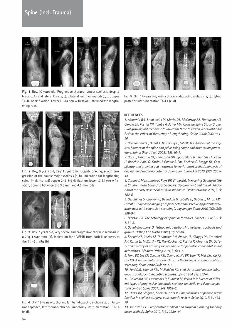
54
REFERENCESAkbarnia BA, Breakwell LM, Marks DS, McCarthy RE, Thompson AG, 1.
Canale SK, Kostial PN, Tambe A, Asher MA; Growing Spine Study Group. Dual growing rod technique followed for three to eleven years until final fusion: the effect of frequency of lengthening. Spine 2008; (33): 984-90.
Berthonnaud E., Dimnt J., Roussouly P., Labelle H.J. Analysis of the sag-2. ittal balance of the spine and pelvis using shape and orientation param-eters. Spinal Disord Tech 2005; (18): 40-7.
Bess S, Akbarnia BA, Thompson GH, Sponseller PD, Shah SA, El Sebaie 3. H, Boachie-Adjei O, Karlin LI, Canale S, Poe-Kochert C, Skaggs DL. Com-plications of growing-rod treatment for early-onset scoliosis: analysis of one hundred and forty patients. J Bone Joint Surg Am 2010; (92): 2533-43.
Corona J, Matsumoto H, Roye DP, Vitale MG. Measuring Quality of Life 4. in Children With Early Onset Scoliosis: Development and Initial Valida-tion of the Early Onset Scoliosis Questionnaire. J Pediatr Orthop 2011; (31): 180-5.
Deschênes S, Charron G, Beaudoin G, Labelle H, Dubois J, Miron MC, 5. Parent S. Diagnostic imaging of spinal deformities: reducing patients radi-ation dose with a new slot-scanning X-ray imager. Spine 2010 (20); (35): 989-94.
Dickson RA. The aetiology of spinal deformities. Lancet 1988; (331): 6. 1151-5.
Duval-Beaupère G. Pathogenic relationship between scoliosis and 7. growth. Orthop Clin North 1988; (19): 58-64.
Elsebai HB, Yazici M, Thompson GH, Emans JB, Skaggs DL, Crawford 8. AH, Karlin LI, McCarthy RE, Poe-Kochert C, Kostial P, Akbarnia BA. Safe-ty and efficacy of growing rod technique for pediatric congenital spinal deformities. J Pediatr Orthop 2011; (31): 1-5.
Fong DY, Lee CF, Cheung KM, Cheng JC, Ng BK, Lam TP, Mak KH, Yip PS, 9. Luk KD. A meta-analysis of the clinical effectiveness of school scoliosis screening. Spine 2010; (35): 1061-71.
Ford DM, Bagnall KM, McFadden KD, et al. Paraspinal muscle imbal-10. ance in adolescent idiopathic scoliosis. Spine 1984; (9): 373-6.
Gauchard GC, Lascombes P, Kuhnast M, Perrin P. Influence of differ-11. ent types of progressive idiopathic scoliosis on static and dynamic pos-tural control. Spine 2001; (26): 1052-8.
Hicks JM, Singla A, Shen FH, Arlet V. Complications of pedicle screw 12. fixation in scoliosis surgery: a systematic review. Spine 2010; (35): 465-70.
Johnston CE. Preoperative medical and surgical planning for early 13. onset scoliosis. Spine 2010; (35): 2239-44.
Fig. 1 Boy, 10 years old. Progressive thoraco-lumbar scoliosis, despite bracing. AP and lateral Xray (a, b). Bilateral lengthening rods (c, d) : upper T4-T6 hook fixation. Lower L3-L4 screw fixation. Intermediate length-ening rods.
Fig. 2 Boy, 6 years old, 22q11 syndrome. Despite bracing, severe pro-gression of the double major scoliosis (a, b). Indication for lengthening spinal implants (c, d) : upper 2nd-3rd rib fixation, lower L3-L4 screw fix-ation, domino between the 3.5 mm and 4.5 mm rods.
Fig. 3 Boy, 7 years old, very severe and progressive thoracic scoliosis in a 22q11 syndrome (a). Indication for a VEPTR from both iliac crests to the 4th-5th ribs (b).
Fig. 4 Gril, 19 years old, thoraco lumbar idiopathic scoliosis (a, b). Ante-rior approach, left thoraco-phreno-lumbotomy, instrumentation T11-L4 (c, d).
Fig. 5 Girl, 14 years old, with a thoracic idiopathic scoliosis (a, b). Hybrid posterior instrumentation T4-L1 (c, d).
Spine (incl. Trauma)

55
Spine (incl. Trauma)
Kelly DM, McCarthy RE, McCullough FL, Kelly HR. Long-term outcomes 14. of anterior spinal fusion with instrumentation for thoracolumbar and lum-bar curves in adolescent idiopathic scoliosis. Spine 2010 15; (35): 194-8.
King HA, Moe JH, Bradford DS, Winter RB. The selection of fusion lev-15. els in thoracic idiopathic scoliosis. J Bone Joint Surg Am 1983; (65): 1302-13.
Kouwenhoven JWM, Castelein RM. The pathogenesis of adolescent 16. idiopathic scoliosis. Spine 2008; (33): 2898-908.
Lenke LG, Betz RR, Clements D, Merola A, Haher T, Lowe T, Newton P, 17. Bridwell KH, Blanke K.Curve prevalence of a new classification of opera-tive adolescent idiopathic scoliosis: does classification correlate with treatment? Spine 2002; (27): 604-11.
Lenke LG, Edwards CC 2nd, Bridwell KH. The Lenke classification of 18. adolescent idiopathic scoliosis: how it organizes curve patterns as a tem-plate to perform selective fusions of the spine. Spine 2003; (28): S199-207.
Li M, Ni J, Fang X, Liu H, Zhu X, He S, Gu S, Wang X. Comparison of 19. selective anterior versus posterior screw instrumentation in Lenke5C ado-lescent idiopathic scoliosis. Spine 2009; (34): 1162-6.
Lonner BS, Auerbach JD, Estreicher MB, Kean KE. Thoracic pedicle 20. screw instrumentation: the learning curve and evolution in technique in the treatment of adolescent idiopathic scoliosis. Spine 2009; (34): 2158-64.
Lonner BS, Auerbach JD, Sponseller P, Rajadhyaksha AD, Newton PO. 21. Variations in pelvic and other sagittal spinal parameters as a function of race in adolescent idiopathic scoliosis. Spine 2010; (35): E374-7.
Lowe TG, Edgar M, Margulies JY, et al. Etiology of idiopathic scoliosis: 22. current trends in research. J Bone Joint Surg 2000; (82): 1157-68.
Machida M, Dubousset J, Imamura Y, Iwaya T, Yamada T, Kimura J, 23. Toriyama S. Pathogenesis of idiopathic scoliosis : SEPs in chicken with experimentally induced scoliosis and in patients with idiopathic scolio-sis. J Pediatr Orthop 1994; (14): 329-335
Mahar AT, Bagheri R, Oka R, Kostial P, Akbarnia BA. Biomechanical 24. comparison of different anchors (foundations) for the pediatric dual grow-ing rod technique. Spine J 2008 ; (8): 933-9.
McCarthy RE, Sucato D, Turner JL, Zhang H, Henson MA, McCarthy K. 25. Shilla growing rods in a caprine animal model: a pilot study. Clin Orthop Relat Res. 2010; (468): 705-10.
McCarthy RE. Scoliosis Research Society (SRS) meeting introduces 26. new ideas, questions old beliefs, and challenges us to think in new and exciting ways. Spine 2003; (28): 2181.
Milbrandt TA, Singhal M, Minter C, McClung A, Talwalkar VR, Iwinski 27. HJ, Walker J, Beimesch C, Montgomery C, Sucato DJ. A comparison of three methods of pain control for posterior spinal fusions in adolescent idiopathic scoliosis. Spine 2009; (34): 1499-503.
Negrini S, Minozzi S, Bettany-Saltikov J, Zaina F, Chockalingam N, 28. Grivas TB, Kotwicki T, Maruyama T, Romano M, Vasiliadis ES. Braces for idiopathic scoliosis in adolescents. Spine 2010; (35): 1285-93.
Negrini S, Minozzi S, Bettany-Saltikov J, Zaina F, Chockalingam N, 29. Grivas TB, Kotwicki T, Maruyama T, Romano M, Vasiliadis ES. Braces for idiopathic scoliosis in adolescents. Cochrane Database Syst Rev. 2010 Janv 20; (1). CD006850.
Ogilvie J. Adolescent idiopathic scoliosis and genetic testing. Curr Opin 30. Pediatr 2010; (22): 67-70.
Reinker K, Simmons JW, Patil V, Stinson Z. Can VEPTR(®) Control Pro-31. gression of Early-onset Kyphoscoliosis?: A Cohort Study of VEPTR(®) Patients With Severe Kyphoscoliosis. Clin Orthop Relat Res. 2010 Nov 30.
Roach JW, Mehlman CT, Sanders JO. Does the outcome of adolescent 32.
idiopathic scoliosis surgery justify the rising cost of the procedures? J Pedi-atr Orthop 2011; (31): S77-80.
Robin, G.C., et al., Scoliosis in the elderly: a follow-up study. Spine 33. 1982; (7): 355-9.
Samdani AF, Ranade A, Saldanha V, Yondorf MZ. Learning curve for 34. placement of thoracic pedicle screws in the deformed spine. Neurosur-gery 2010; (66): 290-5.
Samdani AF, Ranade A, Sciubba DM, Cahill PJ, Antonacci MD, Clem-35. ents DH, Betz RR. Accuracy of free-hand placement of thoracic pedicle screws in adolescent idiopathic scoliosis: how much of a difference does surgeon experience make? Eur Spine J 2010; (19): 91-5.
Schiller JR, Thakur NA, Eberson CP. Brace management in adolescent 36. idiopathic scoliosis. Clin Orthop Relat Res 2010; (468): 670-8.
Schneider, T., Schieber, C., Eeckhoudt, L., Gollier, C. Economics of Radi-37. ation Protection: Equity Considerations. Theory and Decision, 1997; (43): 241-251.
Simoneau M, Richer N, Mercier P, Allard P, Teasdale N. Sensory dep-38. rivation and balance control in idiopathic scoliosis adolescent. Exp Brain Res 2006; (170): 576-82.
Sucato DJ. Management of severe spinal deformity: scoliosis and 39. kyphosis. Spine 2010; (35): 2186-92.
Wang WJ, Yeung HY, Chu WC, Tang NL, Lee KM, Qiu Y, Burwell RG, 40. Cheng JC. Top theories for the etiopathogenesis of adolescent idiopathic scoliosis. J Pediatr Orthop 2011; (31): S14-27.
Westrick ER, Ward WT. Adolescent idiopathic scoliosis: 5-year to 41. 20-year evidence-based surgical results. J Pediatr Orthop. 2011; (31): S61-8.
White KK, Song KM, Frost N, Daines BK. VEPTR™ Growing Rods for Ear-42. ly-onset Neuromuscular Scoliosis: Feasible and Effective. Clin Orthop Relat Res 2011.
Yang JS, McElroy MJ, Akbarnia BA, Salari P, Oliveira D, Thompson GH, 43. Emans JB, Yazici M, Skaggs DL, Shah SA, Kostial PN, Sponseller PD. Grow-ing rods for spinal deformity: characterizing consensus and variation in current use. J Pediatr Orthop. 2010; (30): 264-70.
Zhu F, Chu WC, Sun G, Zhu ZZ, Wang WJ, Cheng JC, Qiu Y. Rib length 44. asymmetry in thoracic adolescent idiopathic scoliosis: is it primary or sec-ondary? Eur Spine J 2011; (20): 254-9.
Spin
e (in
cl. T
raum
a)

56
Spine (incl. Trauma)
Prof. Dr. Benny Dahl
Rigshospitalet National University Hospital of DenmarkCopenhagen, [email protected]
Management of spine fractures
IntroductionWithin the last two decades the treatment of spine fractures has changed resulting in more patients being operated for spine fractures. This is espe-cially the case for thoracolumbar fractures; possibly due to the devel-opment in spinal implant systems and anesthesiological techniques resulting in an acceptable morbidity; even in complex surgical proce-dures. There is, however, still a modest number of randomized, clinical trials comparing conservative and surgical treatment of spine fractures, and some authors have advocated that conservative treatment of unstable fracture with orthosis, should be considered in some cases to avoid the complications related to surgery. Other groups have argued that the mor-bidity related to a posterior thoracolumbar instrumentation is so low that the benefits outweigh the risks, and results in early mobilization, better long-term spinal balance and possibly a better health economic outcome. The reason for this discussion can to a large extend be attributed to the ongoing discussion about fracture classification. Furthermore, the choice of surgical technique and optimal timing, especially in polytrauma, is not clarified. Finally, the literature consists of studies that to a varying degree include patients with neurological compromise. This review will focus in these aspects of mangagement of spine fractures.
Anatomy and epidemiologyThe majority of patients sustaining a structural spine injury have a blunt trauma, either isolated or in combination with other injuries and it is estimated that between 1 and 3% of patients treated in emergency cent-ers have sustained a structural spine injury [1]Approximately 20% of patients received at a major trauma facility will have a spinal fracture and half of these patients will have a severe spine injury [2] [3]. using one of the most established trauma scoring systems, a “severe spine injury” is defined as an injury corresponding to an Abbre-viated Injury Scale (AIS) of > 2 [3]. If this injury is located at the tho-racic level the risk of an associated significant thoracic injury is more than 90%. In patients received on a Level I trauma center the majority of fractures are located at the thoracolumbar region from T11 to L2, and 54% of all patients have their fracture in association with another injury [4]. The risk of an associated injury is highest in patients with a cervical frac-ture. Approximately 20% of the patients have an additional spine fracture, and the likelihood of multisegmental fractures correlated with the number of associated injuries. E.g. a patient with a multilevel spinal frac-ture had more than 95% risk of an associated injury. These findings cor-respond to a previous study and underline the importance of always hav-ing the suspicion of one or more spinal injuries in a patient with polytrauma [5].
Primary treatmentA primary evaluation of a patient suspected of having a spine fracture is mandatory and should follow the guidelines issued by the Advanced Trauma Life Support (ATLS) protocol for the initial management of trau-ma patients [6]. This includes neurological evaluation, logroll, inspec-tion and palpation of the spine and rectal examination. The patient’s neurologic status is evaluated according to the principles described by the American Spinal Injury Association [7]. This results in a classification into ASIA grade A (complete paraplegia), B, C, D, or E (normal motor and sensory function) [7].
In the polytrauma patient with a spine fracture, the concept of “spine damage control” has been introduced [8]. The rationale being that ini-tial fixation of the spine within 8 hours after the injury, postponing pos-sible final surgery until after day three, reduces the risk of pulmonary complications and stay in the Intensive Care unit (ICu), compared to pri-mary final treatment. An initial posterior fixation can be performed with-in 30 to 60 min, which results in less contribution to the inflammatory response compared to a 2 to 3 hour procedure; in some cases even com-bined anterior-posterior surgery.
ClassificationThe comprehensive description of spinal stability provided by White and Panjabi is not optimal in a clinical setting [9]. The first classification sys-tem to be used for this purpose was the description of the three column spine [10]. This system however, did not provide any suggestion regard-ing treatment strategy, and the ideal classification system should reflect increasing severity of the fracture, and provide the surgeon with a tool to be used in the selection of surgical procedure.
Upper cervical spineIn the upper cervical spine, from occiput to C2, some characteristic frac-ture patterns occur. These are: fracture of the Atlas (Jefferson fracture), odontoid fracture of C2 and Hangman’s fracture (traumatic spondy-lolisthesis of C2). The majority of these fractures can be treated conserv-atively, although surgical treatment of odontoid fractures can be con-sidered [11] [12]
Subaxial spineThe biomechanical characteristics of the subaxial spine require certain considerations in the classification of injuries in this region. These include vertebral body translation, facet joint fractures and dislocations, disc rupture with spinal compromise and posterior ligamentous injury [13]. Consequently, supplemental radiographic examinations are often required, such as CT with 3D reconstruction and MRI.
AO classification of thoracolumbar fracturesIn the AO classification system the fracture patterns compression, dis-
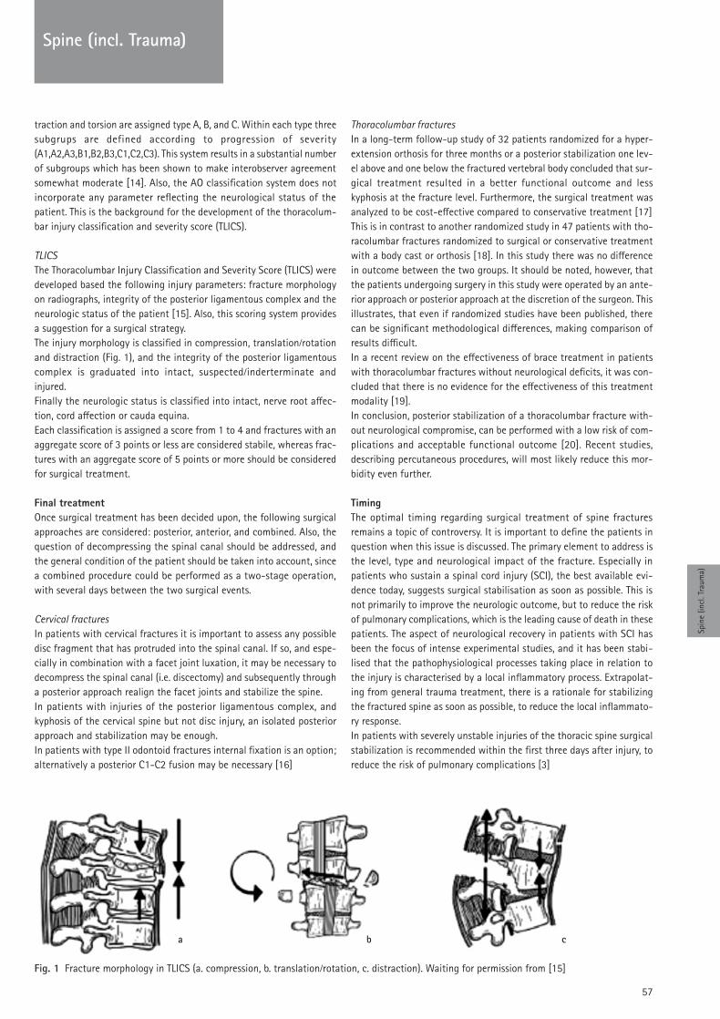
57
traction and torsion are assigned type A, B, and C. Within each type three subgrups are defined according to progression of severity (A1,A2,A3,B1,B2,B3,C1,C2,C3). This system results in a substantial number of subgroups which has been shown to make interobserver agreement somewhat moderate [14]. Also, the AO classification system does not incorporate any parameter reflecting the neurological status of the patient. This is the background for the development of the thoracolum-bar injury classification and severity score (TLICS).
TLICSThe Thoracolumbar Injury Classification and Severity Score (TLICS) were developed based the following injury parameters: fracture morphology on radiographs, integrity of the posterior ligamentous complex and the neurologic status of the patient [15]. Also, this scoring system provides a suggestion for a surgical strategy. The injury morphology is classified in compression, translation/rotation and distraction (Fig. 1), and the integrity of the posterior ligamentous complex is graduated into intact, suspected/inderterminate and injured. Finally the neurologic status is classified into intact, nerve root affec-tion, cord affection or cauda equina. Each classification is assigned a score from 1 to 4 and fractures with an aggregate score of 3 points or less are considered stabile, whereas frac-tures with an aggregate score of 5 points or more should be considered for surgical treatment.
Final treatment Once surgical treatment has been decided upon, the following surgical approaches are considered: posterior, anterior, and combined. Also, the question of decompressing the spinal canal should be addressed, and the general condition of the patient should be taken into account, since a combined procedure could be performed as a two-stage operation, with several days between the two surgical events.
Cervical fracturesIn patients with cervical fractures it is important to assess any possible disc fragment that has protruded into the spinal canal. If so, and espe-cially in combination with a facet joint luxation, it may be necessary to decompress the spinal canal (i.e. discectomy) and subsequently through a posterior approach realign the facet joints and stabilize the spine. In patients with injuries of the posterior ligamentous complex, and kyphosis of the cervical spine but not disc injury, an isolated posterior approach and stabilization may be enough. In patients with type II odontoid fractures internal fixation is an option; alternatively a posterior C1-C2 fusion may be necessary [16]
Thoracolumbar fractures In a long-term follow-up study of 32 patients randomized for a hyper-extension orthosis for three months or a posterior stabilization one lev-el above and one below the fractured vertebral body concluded that sur-gical treatment resulted in a better functional outcome and less kyphosis at the fracture level. Furthermore, the surgical treatment was analyzed to be cost-effective compared to conservative treatment [17]This is in contrast to another randomized study in 47 patients with tho-racolumbar fractures randomized to surgical or conservative treatment with a body cast or orthosis [18]. In this study there was no difference in outcome between the two groups. It should be noted, however, that the patients undergoing surgery in this study were operated by an ante-rior approach or posterior approach at the discretion of the surgeon. This illustrates, that even if randomized studies have been published, there can be significant methodological differences, making comparison of results difficult. In a recent review on the effectiveness of brace treatment in patients with thoracolumbar fractures without neurological deficits, it was con-cluded that there is no evidence for the effectiveness of this treatment modality [19]. In conclusion, posterior stabilization of a thoracolumbar fracture with-out neurological compromise, can be performed with a low risk of com-plications and acceptable functional outcome [20]. Recent studies, describing percutaneous procedures, will most likely reduce this mor-bidity even further.
TimingThe optimal timing regarding surgical treatment of spine fractures remains a topic of controversy. It is important to define the patients in question when this issue is discussed. The primary element to address is the level, type and neurological impact of the fracture. Especially in patients who sustain a spinal cord injury (SCI), the best available evi-dence today, suggests surgical stabilisation as soon as possible. This is not primarily to improve the neurologic outcome, but to reduce the risk of pulmonary complications, which is the leading cause of death in these patients. The aspect of neurological recovery in patients with SCI has been the focus of intense experimental studies, and it has been stabi-lised that the pathophysiological processes taking place in relation to the injury is characterised by a local inflammatory process. Extrapolat-ing from general trauma treatment, there is a rationale for stabilizing the fractured spine as soon as possible, to reduce the local inflammato-ry response.In patients with severely unstable injuries of the thoracic spine surgical stabilization is recommended within the first three days after injury, to reduce the risk of pulmonary complications [3]
Fig. 1 Fracture morphology in TLICS (a. compression, b. translation/rotation, c. distraction). Waiting for permission from [15]
a b c
Spine (incl. Trauma)
Spin
e (in
cl. T
raum
a)
58
Spine (incl. Trauma)
ConclusionAll patients suspected of having a structural lesion of the spine under-go neurologic evaluation upon the arrival to the emergency facility. Most fractures can be assessed on plain radiographs combined with CT, and diagnosing one fracture should raise the suspicion of additional frac-tures. The elements in the classification of a spine injury are: injury mor-phology, posterior ligamentous injury and neurologic status. If surgical treatment is considered “spine damage control” is an option, since pos-terior stabilization can be obtained with low morbidity in less than one hour of surgery, allowing additional procedures to planned after the inflammatory state of the patient has settled.
REFERENCESHoffman, J.R., et al., Validity of a set of clinical criteria to rule out inju-1.
ry to the cervical spine in patients with blunt trauma. National Emergen-cy X-Radiography Utilization Study Group. N Engl J Med, 2000. 343(2): p. 94-9.
Albert, T.J. and D.H. Kim, Timing of surgical stabilization after cervical 2. and thoracic trauma. Invited submission from the Joint Section Meeting on Disorders of the Spine and Peripheral Nerves, March 2004. J Neuro-surg Spine, 2005. 3(3): p. 182-90.
Schinkel, C., et al., Timing of thoracic spine stabilization in trauma 3. patients: impact on clinical course and outcome. J Trauma, 2006. 61(1): p. 156-60; discussion 160.
Leucht, P., et al., Epidemiology of traumatic spine fractures. Injury, 2009. 4. 40(2): p. 166-72.
Harris, M.B. and R.K. Sethi, The initial assessment and management of 5. the multiple-trauma patient with an associated spine injury. Spine (Phi-la Pa 1976), 2006. 31(11 Suppl): p. S9-15; discussion S36.
Anderson, P.A., et al., Clearing the cervical spine in the blunt trauma 6. patient. J Am Acad Orthop Surg, 2010. 18(3): p. 149-59.
Mirza, S.K., et al., Classifications of thoracic and lumbar fractures: 7. rationale and supporting data. J Am Acad Orthop Surg, 2002. 10(5): p. 364-77.
Stahel, P.F., et al., Advocating “spine damage control” as a safe and 8. effective treatment modality for unstable thoracolumbar fractures in pol-ytrauma patients: a hypothesis. J Trauma Manag Outcomes, 2009. 3: p. 6.
Panjabi, M.M. and A.A. White, 3rd, Basic biomechanics of the spine. 9. Neurosurgery, 1980. 7(1): p. 76-93.
Denis, F., The three column spine and its significance in the classifi-10. cation of acute thoracolumbar spinal injuries. Spine, 1983. 8(8): p. 817-831.
Li, X.F., et al., A systematic review of the management of hangman’s 11. fractures. Eur Spine J, 2006. 15(3): p. 257-69.
Hsu, W.K. and P.A. Anderson, Odontoid fractures: update on manage-12. ment. J Am Acad Orthop Surg, 2010. 18(7): p. 383-94.
Bono, C.M., et al., Measurement techniques for lower cervical spine 13. injuries: consensus statement of the Spine Trauma Study Group. Spine (Phila Pa 1976), 2006. 31(5): p. 603-9.
Wood, K.B., et al., Assessment of two thoracolumbar fracture classi-14. fication systems as used by multiple surgeons. J Bone Joint Surg Am, 2005. 87(7): p. 1423-9.
Vaccaro, A.R., et al., A new classification of thoracolumbar injuries: 15. the importance of injury morphology, the integrity of the posterior liga-mentous complex, and neurologic status. Spine (Phila Pa 1976), 2005. 30(20): p. 2325-33.
Przybylski, G.J., J.S. Harrop, and A.R. Vaccaro, Closed management of 16. displaced Type II odontoid fractures: more frequent respiratory compro-
mise with posteriorly displaced fractures. Neurosurg Focus, 2000. 8(6): p. e5. Siebenga, J., et al., Treatment of traumatic thoracolumbar spine frac-17.
tures: a multicenter prospective randomized study of operative versus nonsurgical treatment. Spine (Phila Pa 1976), 2006. 31(25): p. 2881-90.
Wood, K., et al., Operative compared with nonoperative treatment of 18. a thoracolumbar burst fracture without neurological deficit. A prospec-tive, randomized study. J Bone Joint Surg Am, 2003. 85-A(5): p. 773-81.
Giele, B.M., et al., No evidence for the effectiveness of bracing in 19. patients with thoracolumbar fractures. Acta Orthop, 2009. 80(2): p. 226-32.
Gelb, D., et al., Successful treatment of thoracolumbar fractures with 20. short-segment pedicle instrumentation. J Spinal Disord Tech, 2010. 23(5): p. 293-301.
59
Spine (incl. Trauma)
Prof. Dr. Enric Caceres Palou
Hospital Vall d’HebronBarcelona, [email protected]
Degenerative spine disease
Degenerative disc diseaseThe prevalence of the degenerative disc disease is very high, 95% of peo-ple are noted to some degree of degenerative changes, and also one third of asymptomatic individuals will have lumbar MRI that demonstrates degeneration.However, numerous studies have found that degeneration changes to the intervertebral disc do not correlate with patient symptoms and in general we need to classified symptomatic patients in a real diagnosis.What black disc means?Clinically nothing, except in asymptomatic young patients without any other diagnosis.From the iconographic point of view is equivalent to the image in MRI with the disk degeneration IV degree and imply biochemical and struc-tural changes.The outer annulus in a normal disk is mostly type I collagen; inner nucle-us is type II collagen. With age, the ratio of keratin sulphate to chon-droitin sulphate increases and water content decreases, leading to a cas-cade of secondary degenerative events, staring with disc height loss, increased segment motion, compensatory osteophytes, buckling of lig-amentous, and facet arthritis.In young people will be symptomatic and structural biochemistry chang-es modify mechanical properties. These changes are described by a radi-ologist in 1988 showing changes in the signal of bone narrowing the intervertebral disc, with different clinical significance. It’s useful like a non-invasive tool
Modic I show decreased signal in T1 and increased signal in T2 �and correlated with Bone oedema. There are a strong correlation within Modic I and positive discographyModic II shows increased signal in T1 and increased signal in T2 �correlated with fat degeneration. Modic III with decrease of signal in T1 and T2 correlates with �sclerosis
HIZ High intensity Zone it’s another fundamental sign in MRI, brilliant signal in MRA correlate tears of the annulus, and have a direct correlation with pos-itive discography and histologically contends granuloma vascularized tissue, and not always has relationship with clinical symptoms. High-light with Gadolinium
Finally in cases of failure of conservative treatment discography could be helpful to discriminated the pain discal generator, and the main sig-nificance isn’t the image is it the pain concordance with habitual pain-ful sensation. No predicts the success of the surgical treatmentIn summary the clinical significance of black disk depends number one of clinical picture of discogenic lumbar pain, no radicular pain and social normal behaviour. Image of disc degeneration in MRI, some cases with HIZ and the use of discriminative pain by discography. We can reject other aetiology by facet joint injection
Basis of treatment it’s the information to the patient and healthy back behaviour.More o less surgical treatment it’s no evident, special some less invasive treatments like IDET intervertebral disc electrotermocoagulation and Total Disc Replacement, dynamic stabilization or circumferential fusion aren’t no good evidence in outcomes.The fundamentals of this kind of treatment is to cancel the pain gener-ator in disc disease.
Lumbar disk herniation (LDH)Lumbar disk herniation (LDH) has been defined as a focal displacement of nucleus, annulus or end plate material beyond the osseous confines of vertebral body, resulting in displacement of nerve root and or thecae sac.The location its a few more frequent in L4-L5 level, in poster lateral posi-tion, central location will cause lumbar pain without sciatica, and foram-inal location more frequent in elderly people.The main clinical sign it’s radiculopathy with leg pain, more than low back pain, dermatomal irradiation and increase the pain in sitting posi-tion and forward bending improving with bed rest.Physical examination includes dermatome pain irradiation description by the patient, reflex evaluation and straight-leg raise test that increas-es nerve root tension generating pain. Patellar reflex corresponding with L4 level, and L5 level haven’t, own reflex.This is the diagram of physical examination of S1 disturb with Achilles tendon reflex correlation.In order to obtain good diagnosis outcomes after treatment the corre-lation sheep between physical examination, symptomatology and imag-ing test are fundamental.Plain radiography and CT scan are not often helpful.Myelography despairs in normal diagnosis today and MRI it’s the elec-tive test for diagnosis.CT scan + discography it’s useful in recurrence cases diagnosisNatural history of LD Herniation has a good prognostic with conserva-tive treatment.Consistent in short rest, pharmacs to relax muscles, analgesics and non-steroids. In cases without improvement with pharmacologic treatment will be necessary epidural injection of corticosteroid.Without neurologic changes we wait for around two months conserva-tive treatment before surgery. In case of necessity of surgical treatment a plain and conventional discectomy it’s the elective procedure for this condition.
Spinal stenosisAnother diagnoses its spinal stenosis or changes between relationship with contends of the canal and the size with neurological compression
Spin
e (in
cl. T
raum
a)

60
The origin will be by degeneration or by congenital narrowing of the size of the canal. Degeneration of segmental vertebral structures like, facet joints intervertebral disc and ligamentum flavum decrease de size of the canal.The compromise could be central, or periferical by osteophytes or facet degeneration. In the first case predefines neurologic claudication and in the second one radicular signs.It can be by degeneration present starting from fifth decade of live or congenital like achondroplasy.Plane radiographies, CT scans and MRI are good tools to perform diag-nosis and also electromyography findings can it help to diagnoses. Ini-tially conservative treatment could be favourable. Claudication, severe neurological changes, or failure to improve with CT could be indications for operative treatment.Natural history without surgical treatment will be favourable in around 50% of cases.Surgical treatment requires preoperative medical evaluation, with sav-ing blood techniques. The position in the table in surgical procedure is very important in order to decrease de blooding.Surgical procedure consists in more or less wide laminectomy with lat-eral decompression in function of radicular previous compression. In female cases or strong decompression and poster lateral fusion could be necessary.If deformity joint to stenosis its present like degenerative unisegmental spondylolistesis, we need to add circumferential fusion with pedicle instrumentation.There are few works with randomized treatment of canal stenosis. The figures of good results are around 72 %, and in case of associated deformity, add some kind of fusion technique improves the results. But… one of the problems of surgical treatment of spinal stenosis will be the de-rotation of results. In a series of 88 patients with a follow-up between 3 and 7 years the figures they pass to eleven per cent in poor results to 43% seventeen per cent was re-operate.
There are a strong relationship between worse results and age and comorbidities. The presence of more than three comorbidities increases largely hospital stage, cost and dependence In case of association of deformity the results improve dramatically with fusion with figures of 96% of good results in front off 42% without fusionIn summary we have defined dark disk symptomatic in young patients, and to obtain success in treatment we need strong agreement between patient diagnosis and treatment, without no consensus in surgical treat-ment. Lumbar DH its defined by positive radicular tension signs, with specific radicular pain and good results in conservative and surgical treatment. And finally spinal stenosis has a good natural history with high incidence of surgical complications.
REFERENCESDegenerative disk disease
Malvimaara A, Hakkinen U, Aro T, Heinrichs ML, Koskenniemi L, Kuos-1. ma E, Lappi S, Paloheimo R, Servo C, Vaaranen V, Hernberg S (1995)
Lindstrom I, Ohlund C, Eek C, Wallin L, Peterson LE, Fordyce WE (1992) 2. The effect of graded activity on patients with subacute low back pain: a randomized prospective clin- ical study with an operant-conditioning behavioural approach. Physical Therapy 72: 279 – 293
Frost H, Klaber Moffett JA, Bergman JA, Spengler D (1995) Randomised 3. controlled trial for evaluation of fitness programme for patients with chronic low back pain. Br Med J 310:152 – 154
Van Tulder M, Koes B, Malmivaara A (2006) Outcome of non-invasive 4. treatment modali- ties on back pain: an evidence-based review. Eur Spine J 15:S64–S81
Abenhaim L, Rossignol M, Valat JP, Nordin M, Avouac B, Blotman F, 5. Charlot J, Dreiser RL, Legrand E, Rozenberg S, Vautravers P (2000) The role of activity in the therapeutic management of back pain. Report of the International Paris Task Force on Back Pain. Spine 25:1S–33S
Cherkin DC, Deyo RA, Battie M, et al. (1998) A comparison of physical 6. therapy, chiro- practic manipulation, and provision of an educational booklet for the treatment of patients with low back pain. N Engl J Med 339:1021–9
Mannion AF, Taimela S, Muntener M, Dvorak J (2001) Active therapy 7. for chronic low back pain: part 1. Effects on back muscle activation, fat-igability, and strength. Spine 26:897 – 908
Kaser L, Mannion AF, Rhyner A, Weber E, Dvorak J, Muntener M (2001) 8. Active therapy for chronic low back pain: part 2. Effects on paraspinal muscle cross-sectional area, fiber type size, and distribution. Spine 26:909–19
Mannion AF, Junge A, Taimela S, Muntener M, Lorenzo K, Dvorak J 9. (2001) Active ther- apy for chronic low back pain: part 3. Factors influ-encing self-rated disability and its change following therapy. Spine 26:920–9
Lumbar disk herniationWilliams RW (1978) Microlumbar discectomy: a conservative surgical 1.
approach to the virgin herniated lumbar disc. Spine 3:175–82 Atlas SJ, Keller RB, Wu YA, Deyo RA, Singer DE (2005) Long-term out-2.
comes of surgical and non-surgical management of sciatica secondary to a lumbar disc herniation: 10 year results from the Maine Lumbar Spine Study. Spine 30:927–935
Balague F, Nordin M, Sheikhzadeh A, Echegoyen AC, Brisby H Hooge-3. woud HM, Fred- man P (1999)
Weber H (1983) Lumbar disc herniation. A controlled, prospective study 4. with ten years of observation. Spine 8:131–140
Weinstein JN, Lurie JD, Tosteson TD, et al. (2006) Surgical vs nonopera-5. tive treatment for lumbar disk herniation. The Spine Patient Outcomes Research Trial (SPORT), a ran- domized trial. JAMA 296:2441–2450
Weinstein JN, Lurie JD, Tosteson TD, et al. (2006) Surgical vs nonopera-6. tive treatment for lumbar disk herniation. The Spine Patient Outcomes Research Trial (SPORT) observa- tional cohort. JAMA 296:2451–2459
Spinal stenosisAmundsen T, Weber H, Nordal HJ, Magnaes B, Abdelnoor M, Lilleas F 1.
(2000) Lumbar spinal stenosis: conservative or surgical management? A prospective 10-year study. Spine 25(11):1424–35
Grob D, Humke T, Dvorak J (1995) Degenerative lumbar spinal stenosis. 2. Decompression with and without arthrodesis. J Bone Joint Surg Am 77:1036–41
Herkowitz HN, Kurz LT (1991) Degenerative lumbar spondylolisthesis 3. with spinal ste- nosis. A prospective study comparing decompression with decompression and inter- transverse process arthrodesis. J Bone Joint Surg Am 73:802–8
Fischgrund JS, Mackay M, Herkowitz HN, Brower R, Montgomery DM, 4. Kurz LT (1997) Degenerative lumbar spondylolisthesis with spinal steno-sis: a prospective, randomized study comparing decompressive laminec-tomy and arthrodesis with and without spinal instrumentation. Spine 22(24):2807–12
Spine (incl. Trauma)

61
Sports / Knee soft-tissue
1. IntroductionThe etymology of the term meniscus comes from the Greek, “meniskos” which means a crescent-moon shape.The morphology of the meniscus strongly resembles this shape.The kinematics of the menisci in humans is quite asymmetric, with the lateral meniscus being much more mobile than that of the medial menis-cus.Because the medial compartment of the knee is substantially more con-strained than that of the lateral compartment, chronic ligamentous inju-ries – such as that of the anterior cruciate ligament which results in pathological increases in AP translation – often is associated with increased frequency of damage of the medial meniscus compared to that of the lateral meniscus.All of the various components of the knee are important to normal func-tioning of this living, self-maintaining transmission system. (Figure 1) A knee that has sustained a tear of the meniscus can be thought of as a transmission that has a damaged bearing.
Fig. 1 All of the various components of the knee are important to nor-mal functioning of this living self-maintaining transmission system (Sott Dye, courtesy Werner Müller)
2. Meniscus repair2.1 BiologySince kohn and Siebert’s study in 1989, the biomechanical basis of meniscus repair – and meniscus repair itself – has significantly evolved. Evaluation of the first-generation repair techniques with sutures showed that the biomechanical conditions of meniscal repair were dependent on the anatomy of the meniscus, the quality of this tissue and the type of suture and suture material.Studies published in the 90s and in the current decade evaluated the second and third-generation repair devices. While the second-generation devices represented a significant step for-ward with respect to the invasiveness of surgery, their biomechanical
properties were generally inferior to those of the “gold standard” sutures. However, as biomechanical testing became more complex with the intro-duction of cyclic loading, the evaluation of meniscus repair could be extended to include criteria such as the resistance of the repair and gap-ping of the tear site under more physiologic loading conditions. The third-generation flexible suture anchors meet both the criteria of minimal invasiveness and biomechanical properties, which are comparable to those obtained with suture techniques.These anchors as well as improved all-inside suture techniques will prob-ably represent the first choice of meniscal repair techniques in the com-ing years. From a scientific point of view, further studies should be performed to achieve a better understanding of the forces acting on meniscus repair under certain pathologic conditions and of the biomechanical proper-ties of regenerated or “healed” meniscus tissue after repair.
2.2 TechniquesSaving the meniscus, especially in young patients, to decrease the risk of secondary osteoarthritis is challenging. Meniscal repair techniques are well established and allow surgeons to address tears of different complexity and location. There exists no universal technique, but rath-er several techniques which are adapted to different indications. Even if all-inside fourth-generation devices are now the gold standard in the majority of cases, inside-out, outside-in, and even open techniques are still indicated in selected cases. The ultimate goal is to achieve a strong repair. In the future, the next step will be biological meniscus repair by intro-ducing factors such as stem cells, growth factors, or cytokines at the site of the repair to enhance healing. These can be regarded as biological mediators, which regulate key processes in tissue repair (cell prolifera-tion, directed cell migration, cell differentiation, and extracellular matrix synthesis).
2.3 ResultsMenisci are no vestigial structures, but form an integral part of the ’self-maintaining transmission system’ which the knee joint is.Minimal tissue resection, which very often can be described as ’ade-quate’, e.g. leaving the meniscal rim, should be the rule. Care should be taken to resect what has been torn and remove meniscal tissue only to avoid any further impingement that may remain sensitive to rotational painful stress and may thus produce clinical symptoms. Arthroscopic techniques allow for repeat surgery, which may be required in case of persistent mechanical derangement. However, the fulcrum to proceed to repeat arthroscopy surgery needs to remain clinical. All too often, repeat surgery does not alter the clinical findings if it is based on – needless - imaging alone. Therefore, potential meniscal repair is warranted in all cases where
Prof. René Verdonk
Gent University HospitalGent, [email protected]
ACL, PCL, collaterals, meniscus
Spor
ts /
knee
sof
t-tis
sue

62
Sports / Knee soft-tissue
meniscal resection has been considered. Full options remain when, in addition to partial resection, suture of the meniscal remnant to the meniscal wall appears to be required.Biomechanical investigation and testing of meniscal repair devices has received ample consideration. While tensile forces, which are of lesser importance in clinical practice, have been extensively investigated, shear-ing forces acting on the meniscus are of paramount clinical importance but cannot be reliably reproduced in in-vitro studies.Experience has taught us that a red-on-red tear heals spontaneously within four to six weeks, provided that the necessary immobilization is applied. (Figure 2) The purpose of meniscal stabilization is to safely bridge this period in order for the scar tissue to heal and stabilize the lesion. Because in vivo testing is not possible as yet, clinicians investigate implant material by essentially focusing on material properties, safety guidelines and ease of insertion, with convincing evidence based on physiological meniscal healing.
Fig. 2 The healing rate in meniscal repair is highly satisfactory when considering CT scan arthrography (courtesy Société Française d’Arthroscopie)
The implants developed in recent years allow for arthroscopic meniscal suturing all around the meniscal rim. Good stabilization is obtained in the majority of cases. Average results are defined as up to 80% of clin-ical healing at long-term follow-up. Failures are mainly due to improp-er indications or knee joint instability. Less well-documented reasons could be poor meniscal tissue, low cellularity and thus poor healing response. These findings are obviously difficult to document, but are rec-ognized when at surgery yellowish degeneration of the meniscal core is found, which is often related to age and overload and compromises the healing response.One of these ’degenerative’ findings is the meniscal cyst. Prone to increased shearing forces at its fixation around the popliteus muscle tendon, the lateral meniscus may sustain a horizontal tear associated with cyst formation. Depending on its intra-articular ’opening’, the symp-tomatic cyst needs to be resected and the torn meniscus repaired. Repair is mandatory at all costs in order to avoid underlying cartilage degen-eration.
2.4 RehabilitationAlthough evolving continuously, concepts of postoperative rehabilita-tion after meniscus repair still remain controversial. Two rehabilitation protocols, applied in clinical practice, are described in the current liter-ature: the conventional and the accelerated rehabilitation protocol. The optimum rehabilitation is yet to be identified, and the lack of scientific data in the literature does not allow us to endorse a specific rehabilita-tion program.
However, it is the authors’ opinion that all intrinsic factors should be taken into account when designing a rehabilitation program. Individu-alizing the rehabilitation according to the size and type of meniscal tear, vascular supply, localization, concomitant procedure, and presence of other intra-articular disorders (ACL, cartilage lesions,...) seems to be an interesting concept. If less than two intrinsic risk factors are present, healing will occur fairly rapidly and the risk of failure is low, so that an accelerated rehabilitation protocol is recommended. However, the pres-ence of more than two risk factors (e.g., a large tear in a red-white zone) increases the risk of meniscus failure and slow healing, and in this case a more conservative approach is probably the best guarantee for suc-cess.However, well-designed longitudinal studies are mandatory to deter-mine the actual efficacy of this rehabilitative approach with regard to patient function and satisfaction.
3. Meniscus replacementCarl Wirth and Gabriela von Lewinski investigated the basic science in meniscal transplantation. The interest taken by their German group in meniscal transplants was fueled by clinical needs.The concept of the meniscus also being a stabilizing structure in the knee joint is not new, but they were the first to consider the meniscus as a primary stabilizer after knee ligament injury and repair. Simply removing the meniscus had proven deleterious to the long-term results after ligament repair.In animal experiments, the authors were able to show healing after meniscal allograft implantation.Also in human clinical studies, satisfactory healing occurred at the meniscosynovial junction, but whether this was also true for the menis-cal horns remained a controversial issue.Horn fixation is indeed mandatory for true hoop stress protection.In addition, animal experiments showed increased cartilage degradation when the normal anatomy had not been restored. Nowadays, no clear consensus is available on whether bone fixation of meniscal allografts is mandatory for normal homeostasis. Choosing allograft tissue such as meniscal tissue, although of limited availability, is a logical option.Deep-freezing appears to be the most accepted method of preservation and standards of procurement have been well established.If procured in a sterile fashion, the allografts can be used when the tis-sue bank has found the donor to be free of transmissible diseases. When harvesting has been done in an unsterile fashion, the issue of sterility requires appropriate attention and management. Avoiding irradiation as such is essential in order not to be detrimental to meniscal structure and thus good postoperative function.However, national laws and regulations can interfere with good clinical practice on grounds of legal constraints based on earlier infringements and exposures. Meniscal surgery, as it started in the 1990s, required an open approach because at that time arthroscopic meniscal fixation devices were limit-ed and not really appropriate. In the early beginning, meniscal trans-plantation was very often associated with other repair surgery (mostly ligamentous).Open surgery is also required for bone plug fixation and to obtain ele-mentary stability.It is only because meniscal surgery and repair indications have increased that arthroscopic transplantation has been initiated.Without bone plug fixation the technique becomes an arthroscopic soft-tissue procedure, also with use of improved fixation and stabilization
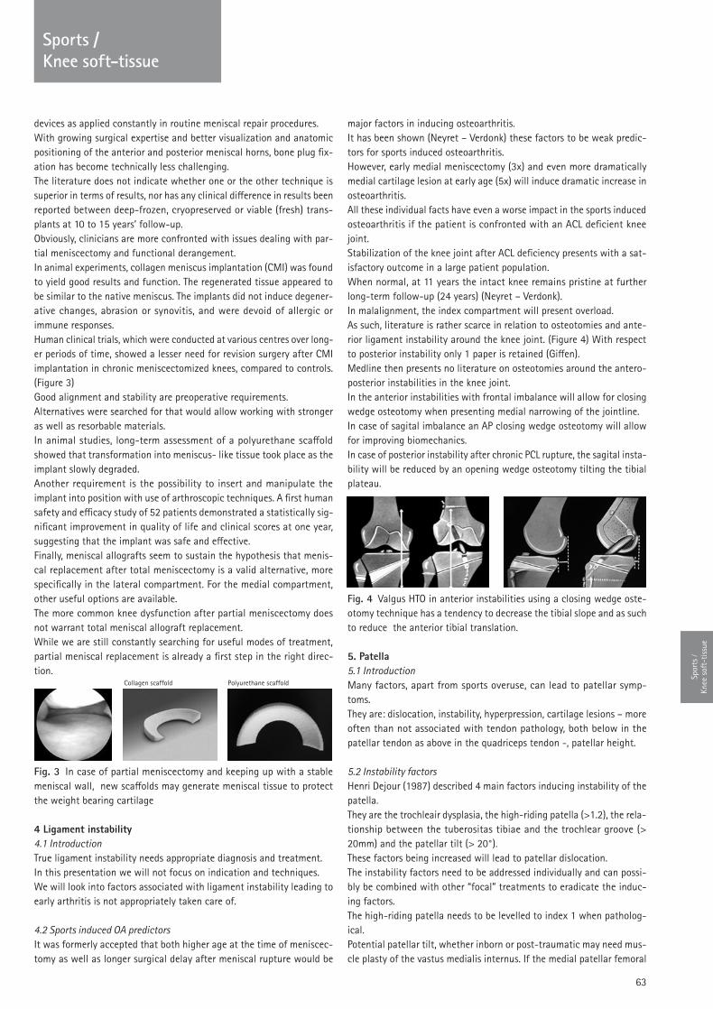
63
devices as applied constantly in routine meniscal repair procedures.With growing surgical expertise and better visualization and anatomic positioning of the anterior and posterior meniscal horns, bone plug fix-ation has become technically less challenging. The literature does not indicate whether one or the other technique is superior in terms of results, nor has any clinical difference in results been reported between deep-frozen, cryopreserved or viable (fresh) trans-plants at 10 to 15 years’ follow-up.Obviously, clinicians are more confronted with issues dealing with par-tial meniscectomy and functional derangement. In animal experiments, collagen meniscus implantation (CMI) was found to yield good results and function. The regenerated tissue appeared to be similar to the native meniscus. The implants did not induce degener-ative changes, abrasion or synovitis, and were devoid of allergic or immune responses. Human clinical trials, which were conducted at various centres over long-er periods of time, showed a lesser need for revision surgery after CMI implantation in chronic meniscectomized knees, compared to controls. (Figure 3)Good alignment and stability are preoperative requirements.Alternatives were searched for that would allow working with stronger as well as resorbable materials.In animal studies, long-term assessment of a polyurethane scaffold showed that transformation into meniscus- like tissue took place as the implant slowly degraded.Another requirement is the possibility to insert and manipulate the implant into position with use of arthroscopic techniques. A first human safety and efficacy study of 52 patients demonstrated a statistically sig-nificant improvement in quality of life and clinical scores at one year, suggesting that the implant was safe and effective.Finally, meniscal allografts seem to sustain the hypothesis that menis-cal replacement after total meniscectomy is a valid alternative, more specifically in the lateral compartment. For the medial compartment, other useful options are available.The more common knee dysfunction after partial meniscectomy does not warrant total meniscal allograft replacement.While we are still constantly searching for useful modes of treatment, partial meniscal replacement is already a first step in the right direc-tion.
Fig. 3 In case of partial meniscectomy and keeping up with a stable meniscal wall, new scaffolds may generate meniscal tissue to protect the weight bearing cartilage
4 Ligament instability4.1 Introduction True ligament instability needs appropriate diagnosis and treatment.In this presentation we will not focus on indication and techniques.We will look into factors associated with ligament instability leading to early arthritis is not appropriately taken care of.
4.2 Sports induced OA predictorsIt was formerly accepted that both higher age at the time of meniscec-tomy as well as longer surgical delay after meniscal rupture would be
major factors in inducing osteoarthritis.It has been shown (Neyret – Verdonk) these factors to be weak predic-tors for sports induced osteoarthritis.However, early medial meniscectomy (3x) and even more dramatically medial cartilage lesion at early age (5x) will induce dramatic increase in osteoarthritis.All these individual facts have even a worse impact in the sports induced osteoarthritis if the patient is confronted with an ACL deficient knee joint.Stabilization of the knee joint after ACL deficiency presents with a sat-isfactory outcome in a large patient population.When normal, at 11 years the intact knee remains pristine at further long-term follow-up (24 years) (Neyret – Verdonk).In malalignment, the index compartment will present overload. As such, literature is rather scarce in relation to osteotomies and ante-rior ligament instability around the knee joint. (Figure 4) With respect to posterior instability only 1 paper is retained (Giffen). Medline then presents no literature on osteotomies around the antero-posterior instabilities in the knee joint.In the anterior instabilities with frontal imbalance will allow for closing wedge osteotomy when presenting medial narrowing of the jointline. In case of sagital imbalance an AP closing wedge osteotomy will allow for improving biomechanics. In case of posterior instability after chronic PCL rupture, the sagital insta-bility will be reduced by an opening wedge osteotomy tilting the tibial plateau.
Fig. 4 Valgus HTO in anterior instabilities using a closing wedge oste-otomy technique has a tendency to decrease the tibial slope and as such to reduce the anterior tibial translation.
5. Patella5.1 IntroductionMany factors, apart from sports overuse, can lead to patellar symp-toms.They are: dislocation, instability, hyperpression, cartilage lesions – more often than not associated with tendon pathology, both below in the patellar tendon as above in the quadriceps tendon -, patellar height.
5.2 Instability factorsHenri Dejour (1987) described 4 main factors inducing instability of the patella.They are the trochleair dysplasia, the high-riding patella (>1.2), the rela-tionship between the tuberositas tibiae and the trochlear groove (> 20mm) and the patellar tilt (> 20°).These factors being increased will lead to patellar dislocation.The instability factors need to be addressed individually and can possi-bly be combined with other “focal” treatments to eradicate the induc-ing factors.The high-riding patella needs to be levelled to index 1 when patholog-ical.Potential patellar tilt, whether inborn or post-traumatic may need mus-cle plasty of the vastus medialis internus. If the medial patellar femoral
Collagen scaffold Polyurethane scaffold
Sports / Knee soft-tissue
Spor
ts /
knee
sof
t-tis
sue

64
Sports / Knee soft-tissue
ligament is ruptured because of an earlier dislocation, it needs to be reconstituted anatomically.In case of frank trochlear dysplasia (grade B and D), a trochlear plasty may be necessitated. In case of abnormal TT- TG it needs to be reduced to former and normal measurements.Secondary factors such as malalignment do not need to be addressed in casu.
6 Cartilage6.1 IntroductionCartilage repair is physiologically non existent. Surgical approaches have been described using local regeneration using the micro-fracture tech-nique. Cell transplantation, both autologous as well as allotransplantation of both individual cells, cultured cells or as allografts has been used.It appears that both in symptoms and in treatment, sizing is essential for inducing valuable results.In addition, correct alignment is a prerequisite to support good clinical results.
6.2 Cartilage lesionsArøen et al. (2004) and Curl (1997) have investigated the number of car-tilage lesions in routine arthroscopy. Cartilage lesions in excess of 2cm² are present in more than 66% of all cases.However, only 20% present with a localized defect.11% are rated grade 3 or 4 according to ICRS.Only a very limited number of 6% present the same grade 3 – 4 ICRS in a defect that is in access of 2cm². Figure 5It is generally accepted that only lesions in the weight bearing zone in excess of 2cm² can be symptomatic in need and are in potential need of treatment.
Fig. 5 Limited and focal fresh cartilage lesions can be considered for surgery when limited in extent.
It is also common knowledge that degenerative lesions need an appro-priate treatment that does not relate to cartilage treatment in itself.
6.3 TreatmentsNowadays small lesions tend to present with good results when treat-ed with microfracture approach as described by Steadman.The edges of the lesions need to be debrided until sharp.The micro-fracture technique necessitates perforation until the subchon-dral lamina every 2 to 3 mm in order to expect bleeding to occur. This suggests the potential of mesenchymal stem cells to approach the lesions surface in order to generate repair cartilage tissue.
Gobbi (2005) has investigated a level 4 comparative study looking into long-term results in young athletes (38y.) retaining pain relief at 6 years, however with lower and sports activities in 80% of cases.Gudas (2005) looked into younger sportsmen and investigated in a ran-domized prospective level 1 study microfracture versus mosaicplasty in a 3 year minimal follow-up study.Lesions were grade 3 to 4. Size, 2,8 cm² and comparing microfracture versus mosaicplasty.There was a distinct advantage in the use of the mosaicplasty treatment versus microfracture.
7 ConclusionIn painful cartilage lesions in the young athlete, the ideal candidate presents with age below 40. The lesions should be fresh (below 3 months). It is essential to have a lower end BMI (<30kg/m²) in normal axial align-ment without any associated lesions. Small sizing of maximum 3cm² is prerequisite.
REFERENCESR. Verdonk (2010) Meniscal repair: Biomechanics. In: The Meniscus, edi-1.
tors R. Verdonk and PH. Beaufils. In press.Dye SF (1996) The knee as a Biologic Transmission with an Envelope of 2.
Function. Clin Orthop Rel Res 325: 10-18.Müller W (1982) Le Genou. Springer Verlag, Berlin3. Dye SF (1987) An evolutionary perspective of the Knee. J. Bone Joint 4.
Surg 7:976-983Seil R, Pape D. (2010) Meniscal repair: biomechanics. In: The Meniscus, 5.
editors R. Verdonk and PH. Beaufils. In pressCharrois O (2008) Enquête de pratique SFA/ESSKA/SOFCOT. In: Sympo-6.
sium on Le ménisque latéral. Congrèsde la Société Francaise d’Arthroscopie, Paris
Seil R, Rupp S, Krauss PW, Benz A, Kohn D (1998) Comparison of initial 7. fixation strength between biodegradable and metallic interference screwsand a press-fit fixation technique in a porcine model. Am J Sports Med 26(6):815–819
Seil R, Rupp S, Dienst M, Müller B, Bonkhoff H, Kohn D (2000) Chondral 8. lesions after arthroscopic meniscus repair using meniscus arrows. Arthros-copy 16(7): E17 37. Seil R, Rupp S, Kohn D (2000) Cyclic testing of menis-cal sutures. Arthroscopy 16:505–510
Seil R, Rupp S, Jurecka C, Rein R, Kohn D (2001) Der Einfluß verschieden-9. er Nahtstärken auf das Verhalten von Meniskusnähten unter zyklischer Zugbelastung. Unfallchirurg 104(5):392–398
Seil R, Rupp S, Mai C, Pape D, Kohn D (2001) The footprint of menis-10. cus fixation devices on the femoral surface of the medial meniscus: a bio-mechanical cadaver study. ISAKOS congress, Montreux 40. Seil R, Rupp S, Jurecka C, Georg T, Kohn D (2003) Réparation méniscale par fixations biodégradables: etude biomécanique comparative. Rev Chir Orthop 89:35–43
Jouve F, Ovadia H. (2010) Meniscal repair: Technique. In: The Menis-11. cus, editors R. Verdonk and PH. Beaufils. In press
Albrecht-Olsen P, Kristensen G, Tormala P (1993) Meniscus bucket-12. handle fixation with an absorbable Biofix tack: development of a new technique. Knee Surg Sports Traumatol Arthrosc 1:104–106
DeHaven KE (1990) Decision-making features in the treatment of 13. meniscal lesions. Clin Orthop 252:49–54
DeHaven KE, Black K, Griffiths HJ (1989) Open meniscus repair. Tech-14. nique and two to nine year results. Am J Sports Med 17:788–795
McDermott ID, Richards SW, Hallam P, Tavares S, Lavelle JR, Amis AA 15.
65
(2003) A biomechanical study of four different meniscal repair systems, comparing pull-out strengths and gapping under cyclic loading. Knee Surg Sports Traumatol Arthrosc 11:23–29
R. Verdonk (2010) Synthesis. In: The Meniscus, editors R. Verdonk and 16. PH. Beaufils. In press.
E. Witvrouw (2010) Rehabilitation. In: The Meniscus, editors R. Ver-17. donk and PH. Beaufils. In press.
Jokl P, Stull PA, Lynch JK et al. (1989) Independent home versus super-18. vised rehabilitation following arthroscopic knee surgery: a prospective randomized trial. Arthroscopy, vol 5, pp 298-305.
McLaughlin J, DeMaio M, Noyes FR et al (1994) Rehabilitation after 19. meniscus repair. Orthopedics, vol 17, pp 465-471.
Shelbourne KD, Patel DV, Adsit WS et al. (1996) Rehabilitation after 20. meniscal repair. Clin Sports Med, vol 15, pp 595-612.
Giffin JR, Vogrin TM, Zantop T, Woo SL, Harner CD, Effects of increas-21. ing tibial slope on the biomechanics of the knee. Am J Sports Med. 2004 Mar;32(2):376-82
Arøen A, Løken S, Heir S, Alvik E, Ekeland A, Granlund OG, Engebret-22. sen L. Articular cartilage lesions in 993 consecutive knee arthroscopies. Am J Sports Med. 2004 Jan- Feb;32(1):211-5.
Curl WW, Smith BP, Marr A, Rosencrance E, Holden M, Smith TL., The 23. effect of contusion and cryotherapy on skeletal muscle microcirculation. J Sports Med Phys Fitness. 1997 Dec;37(4):279-86
Gobbi A., Nunag P., Malinowski K., Treatment of full thickness chon-24. dral lesions of the knee with microfracture in a group of athletes. Volume 13, Number 3 / April, 2005
Gudas R, Kalesinskas R., Kimtys V, Stankevicius E, Toliusis V, Berno-25. tavicius G, Smailys A. A Prospective Randomized Clinical Study of Mosa-ic Osteochondral Autologous Transplantation Versus Microfracture for the Treatment of Osteochondral Defects in the Knee Joint in Young Ath-letes. Arthroscopy: The Journal of Arthroscopic and Related Surgery Sep-tember 2005 (Vol. 21, Issue 9, Pages 1066-1075)
Sports / Knee soft-tissue
Spor
ts /
knee
sof
t-tis
sue

66
Trauma
Prof. Dr. Ulrich Stöckle
Klinikum rechts der Isar der TU MünchenMunich, [email protected]
Fractures: Pelvic ring und acetabular fractures
1. Pelvic ring injuriesJust 2% of all injuries are pelvic ring injuries with about 70% being sta-ble anterior pelvic ring fractures in the elderly caused by a simple fall. unstable pelvic ring injuries are caused by a high energy trauma and are part of a polytrauma in one third of the cases. These unstable pelvic ring injuries with disruption of the posterior pelvic ring can be life-threat-ening because of massive retroperitoneal bleeding. Therefore primary stabilisation of the pelvic ring needs to be included into the polytrauma algorithm. Definitive stabilisation with internal fixation is to be per-formed as secondary procedure after stabilisation of the general condi-tion of the patient.
1.1. AnatomyThe osseous pelvic ring consists of two ossa innominata and the sacral body. The stability of the pelvic ring is achieved by the ligamentous struc-tures in the symphyseal area and the SI joints. Herby the posterior sac-ro-iliac ligaments are the strongest ligaments in the whole human body. Forces of more than 1000N are needed to produce a disruption of the posterior pelvic ring. Therefore unstable pelvic ring injuries often are associated with high energy trauma and polytrauma. Iliac artery and vein as well as the lumbal nerve plexus are running in front of the sacrum. Major bleeding can be caused by disruptions of the posterior pelvic ring, caused mainly by bleeding out of the sacral venous plexus and fracture sites. In only 20% arterial bleeding is the reason for hemodynamic instability.
1.2. DiagnosticsBesides the clinical exam and stability test, the plain X-ray as pelvic view is the base to assess a pelvic ring injury. Additional 45° tilted projections like Inlet and Outlet view can show horizontal and vertical displacements respectively. The computer-tomography is strongly recommended for further evaluation of a pelvic ring injury and for preoperative planning. In up to 70 percent a posterior pelvic ring lesion can only be detected in the CT. Besides a minimum of 2mm cuts additional 2D reconstructions in sagittal and coronal direction are helpful. MRI is rarely needed in the acute setting. An angiography can be applied in cases with suspect for additional vascular injury and existing setup for embolisation.A precise neurologic exam is mandatory in pelvic ring injuries because of the close relation to the lumbar nerve plexus.
1.3. ClassificationThe most common classification used is the AO Classification, based on the Tile classification. The main criterion in this classification is the remaining stability of the pelvic ring. Stable A-type injuries are differ-entiated from partially unstable B-type injuries and completely unsta-ble C-type injuries:
A-type injuries, stable lesions:A1: iliac wing fractures, apophyseal fracturesA2: anterior pelvic ring fracturesA3: transverse sacral fractures
B-type injuries, partially unstable lesions, horizontal instability:B1: open book injuriesB2: lateral compression type injuriesB3: bilateral B-type injuries
C-type injuries, lesions with complete disruption of the posterior pelvic ring, horizontal and vertical instability:
C1: unilateral vertical instabilityC2: one side vertical instability, other side B-type injury with hor-izontal instabilityC3: bilateral vertically unstable lesion
In C-type injuries the disruption of the posterior pelvic ring can be tran-siliacal, through the SI joint or transsacral.
1.4. TreatmentThe assessment of stability of the pelvic ring and appropriate classifica-tion is a mandatory prerequisite for the adequate therapy.A-type injuries can be treated conservatively most of the times. This means mobilisation with symptom-adapted weight bearing. In case of persisting pain a CT is recommended to exclude a lesion of the posteri-or pelvic ring. Surgical therapy is only recommended for apophyseal frac-tures in adolescents, iliac wing fractures with major displacement and transverse sacral fractures with neurology.B-type injuries need a differentiated therapy concept. Open book inju-ries (B1 injuries) are usually treated with open reduction and plate fix-ation (4 hole LCDCP). Lateral compression injuries (B2) with stable impac-tion can be treated conservatively with partial weight bearing on the injured side. Lateral compression injuries with major displacement and/ or overlap in the anterior pelvic ring need fixation of the anterior pelvic ring, preferably with an external fixator.As the posterior pelvic ring is not disrupted completely, B-type injuries are treated appropriately with stabilisation of the anterior pelvic ring.C-type injuries are characterized by complete disruption of the poste-rior pelvic ring through the ilium, the SI joint or sacrum. Depending on the concomitant bleeding and general condition of the patient, a pri-mary external stabilisation of the pelvis is necessary to stabilize the patient. Besides a simple sheet around the pelvis as “in field maneuver”, the external fixator and pelvic C-clamp are used for this primary stabi-lisation. The C-clamp is an emergency device to stabilize the posterior pelvic ring, thus reducing the intrapelvine volume and the bleeding. The best indications are pure SI disruptions or injuries with major fragments. Contraindication is a transiliac fracture, because of the risk of penetrat-ing pins. The alternative method for external stabilisation of the pelvic ring is the external fixator. Preferably the Schanz’ screw are placed supra-acetab-
67
Trauma
ular because of better biomechanical stability compared to pins into the iliac crest.Definitive stabilisation is usually performed as a secondary procedure after 5-7 days depending on the general condition of the often poly-traumatized patient. If closed reduction is possible, sacral fractures and SI disruptions can be stabilized with SI screw fixation in a minimal inva-sive technique. If open reduction is necessary, direct plating is preferred for sacral fractures using a posterior approach and anterior plating with 2 plates for SI disruptions. Transiliac dislocation fractures are treated with open reduction and plate fixation from anterior.For stable fixation and good results, C-type injuries need fixation of the posterior and anterior pelvic ring. Stabilisation of the anterior pelvic ring is achieved at least with an external fixator.
1.5. Results Stable A-type injuries have good results with conservative treatment. In case of persisting pain in A-type injuries a CT is strongly recommended to exclude additional lesions in the posterior pelvic ring, thus resulting in a B-type injury. For B-type injuries, functional results usually are good. Overall outcome is depending on the extent of urogenital and neurolog-ical impairments, especially in B1 and B3 injuries. For C-type injuries the stable reconstruction of the pelvic ring with less than 1cm displacement is a mandatory prerequisite for good results. This can be achieved in more than 70% of the cases. Nevertheless the overall result is depending main-ly on the neurological impairments and in case of a polytrauma on the result of the other injuries.
2. Acetabular fractures2.1. AnatomyThe acetabulum consists of an anterior and a posterior column. In radi-ographs the iliopectineal line corresponds to the anterior column, the ilioischial line to the posterior column. Additionally the acetabular roof, the anterior wall and the posterior wall can be evaluated.
2.2. DiagnosticsThe pelvic X-ray is the base for diagnostics. All lines need to be evalu-ated in comparison to the noninjured side. Interruptions of the ilio-pectineal line show a fracture of the anterior column, interruptions of the ilioischial line a fracture of the posterior column. Additional evalu-ation is possible with the 45° degree tilted iliac view and obturator view. In the iliac view the anterior wall and posterior column are to be seen the best, in the obturator view the anterior column and posterior wall. The additional computertomography is recommended for all acetabular fractures. Intraarticular fragments, marginal impaction of the joint and the full extent of the fracture can be visualized within the axial cuts and the 2D reconstructions. The 3D reconstruction can be helpful to plan the appropriate approach for reconstruction.
2.3. ClassificationThe most common classification is the Letournel classification. Within this classification 5 simple fractures and 5 combined fractures are dif-ferentiated. Simple fracture does not mean easy to treat, but a single fracture line. Anterior wall, posterior wall, anterior column, posterior col-umn and transverse fracture are these basic fractures. Posterior wall fractures are often associated with a posterior dislocation of the femo-ral head. Combined fractures are posterior column posterior wall, trans-verse posterior wall, anterior column posterior hemitransverse, T-type and both column fractures. Both column fractures are characterized by
complete separation of the fractured joint from that part of the iliac bone still being attached to the SI joint.
2.4. TreatmentNondisplaced fractures, fractures below the weight bearing area and fractures with displacement less then 2mm can be treated conservative-ly. unstable fractures and fractures with displacement more than 2mm within the weightbearing area are recommended to be treated opera-tively. The choice of the appropriate approach is essential for anatomic reduction and stable fixation. Fractures with more anterior pathology (anterior wall, anterior column, some transverse, anterior column pos-terior hemitransverse and most both column fractures) are treated with an anterior, ilioinguinal approach. The posterior kocher-Langenbeck approach is used for fractures with more posterior pathology (posterior wall, posterior column, some transverse, transverse posterior wall, pos-terior column posterior wall, T-type). More than 90% of all acetabular fractures can be treated by these two classic approaches. Just rarely extended approaches are needed for fractures in which manipulation and fixation from anterior and posterior is necessary. For specific frac-ture patterns minimal invasive approaches can be applied, especially with intraoperative 3D C-arm imaging and / or navigation.After reconstruction and fixation of acetabular fractures, partial weight bearing with 15kg is necessary for 12 weeks.
2.5. ResultsFor good functional long term results near anatomic reduction is essen-tial. With appropriate diagnostics, exact classification, the right approach and extensive expertise good reduction of acetabular fractures with less than 1mm step or gap is possible in up to 80% of the fractures. In case of near anatomic reduction about 75% will have good long term results.
REFERENCESLetournel E., The treatment of acetabular fractures through the ilioin-1.
guinal approach. Clin Orthop Relat Res. 1993 Jul;(292):62-76. PMID: 8519138 [PubMed - indexed for MEDLINE]
Helfet DL, Schmeling GJ., Management of complex acetabular frac-2. tures through single nonextensile exposures. Clin Orthop Relat Res. 1994 Aug;(305):58-68. PMID: 8050248 [PubMed - indexed for MEDLINE]
Matta JM., Fractures of the acetabulum: accuracy of reduction and 3. clinical results in patients managed operatively within three weeks after the injury. J Bone Joint Surg Am. 1996 Nov;78(11):1632-45. PMID: 8934477 [PubMed - indexed for MEDLINE]
Kreitner KF, Mildenberger P, Rommens PM, Thelen M. Rofo.; 2000 4. Jan;172(1):5-11. [Rational diagnostic imaging of pelvic and acetabulum injuries]. PMID: 10719457 [PubMed - indexed for MEDLINE]
Gettys FK, Russell GV, Karunakar MA., Open treatment of pelvic and 5. acetabular fractures. Orthop Clin North Am. 2011 Jan;42(1):69-83, vi. PMID: 21095436 [PubMed - indexed for MEDLINE]
Bates P, Gary J, Singh G, Reinert C, Starr A. Percutaneous treatment of 6. pelvic and acetabular fractures in obese patients. Orthop Clin North Am. 2011 Jan;42(1):55-67, vi. PMID: 21095435 [PubMed - indexed for MEDLINE]
Stöckle U, Schaser K, König B. Image guidance in pelvic and acetabu-7. lar surgery--expectations, success and limitations. Injury. 2007 Apr;38(4):450-62. Epub 2007 Apr 2. PMID: 17403522 [PubMed - indexed for MEDLINE]
Giannoudis PV, Pohlemann T, Bircher M. Pelvic and acetabular surgery 8.
Trau
ma

68
Trauma
within Europe: the need for the co-ordination of treatment concepts. Inju-ry. 2007 Apr;38(4):410-5. Epub 2007 Mar 29. PMID: 17397845 [PubMed - indexed for MEDLINE]
H Tscherne, T Pohlemann, Unfallchirurgie 1998. Buch. XVI, 498 S.: 800 9. s/w-Abbildungen, 30 s/w-Tabellen. Hardcover Springer Berlin ISBN 978-3-540-62481-3
Fig.1a,b AO Classification for pelvic ring injuries and treatment options
Fig. 2a-c Classification of acetabular fractures, results after operative treatment.

69
Trauma
Dr. Olivier Borens, MER
university HospitalLausanne, [email protected]
Fractures: Hip, femur, knee and tibia
1. Intracapsular hip fracturesIntroduction: The absolute number of intracapsular hip fractures has doubled since the mid 1960s leading to an immense increase of the cost. These fractures occur more often in women than in men and more fre-quently and in younger individuals than intertrochanteric fractures (Park-er). There is a direct correlation between low BMD, low BMI, decreased activity, poor balance, impaired vision and fractures in the elderly. Fem-oral neck fractures in patients < 50 years make up only 3%-5% and are usually due to high-energy trauma. In this age group 20% of femoral neck fractures are associated with femoral shaft fractures and are missed in up to 30% !
Anatomy: The vascular supply to the proximal femur, including the fem-oral head, originates from the medial and lateral femoral circumflex arteries, branches of which form an extracapsular arterial ring at the base of the femoral neck. The LFCA being the main artery for blood sup-ply to the head.
Diagnosis: Clinical signs and symptoms can vary according to the type of fracture. In valgus impacted, incomplete or nondisplaced fractures weight bearing can still be possible while patients with displaced frac-tures have shortening of the affected limb with external rotation, mild abduction and are unable to weight bear. Standard antero-posterior and cross-table lateral RX are required for the diagnosis of a neck fracture. In doubt T1-weighted MRI study will confirm or deny the presence of a fracture in 100%.
Classification: The Garden classification based on the amount of dis-placement on the antero-posterior radiograph is the most commonly accepted system. There are nondisplaced (GI and II) and the displaced (GIII and IV) fractures. The AO system classification ranges from 31.B1.1. to 31.B.3.3.
undisplaced fracture (Garden 1 & 2), Displaced fracture (Garden 3 & 4)
Treatment: A femoral neck fracture in a young patient should be con-sidered an orthopaedic emergency, elderly patients should be operated as soon as possible once they are medically cleared and ideally within 48h. Young patients with a displaced femoral neck fracture should ben-efit from anatomic reduction of the fracture. If this is not possible by closed means surgical dislocation of the hip by trochanter flip or a
Watson-Jones approach should be discussed. Patients with non- or minimally displaced fracture have a low risk for osteonecrosis and nonunion if the fracture is fixed. A displaced fracture places the patient at a high risk of about 25% (range, 10%-45%) of osteonecrosis and nonunion. In elderly patients displaced femoral neck fractures should thus be considered for prosthetic replacement. If frac-ture fixation is chosen, best results will be achieved with screws. In non-comminuted fractures placing more than 3 screws (or 2 pins) offers no mechanical advantage. In fractures with posterior comminution a forth screw can provide better stability (kauffmann). If arthroplasty is chosen bipolar hemiarthroplasty as compared to mono-polar hemi arthroplasty demonstrates a trend to better scores for pain, social function and mobility.Total hip replacement, initially used mainly for fracture patients with severe preexisting hip disease, is considered now more and more for patients with a life expectancy of more than 5 to 10 years.
Complications: The incidence of osteonecrosis ranges from 10% to 45% adequate and timely reduction being the most important preventing fac-tors. The reported incidence of non-union ranges from 10% to 30% and MRI is indicated to assess the viability of the femoral head. For young patients osteotomy can be indicated whereas prosthetic replacement will be most adequate in elderly patients.
2. Intertrochanteric femur fracturesDiagnosis: Intertrochanteric fractures are rare in young patients but pos-sible in high-energy trauma. In this context they usually come with more soft tissue injury, more fracture displacement and bony fragmentation than in elderly patients. If standard X-ray permits not to make the diag-nosis MRI is nowadays the examination of choice for confirming or deny-ing the fracture. A multitude of classifications exists with the kyle and AO classification being the most widely used.
Treatment: Surgical stabilization is the treatment of choice. Early sur-gery (within 24-48h) is associated with a reduction in 1-year mortality in all but the most medically unstable patients. Sliding hip screws (SHS) and intramedullary nails are the implants most widely used but the 95° blade plate for reverse oblique fractures and modern angular stable (LCP) plates can be a valuable option. If a SHS or an intramedullary nail are used the correct placement of the screw in the femoral head are of great-est importance. Optimal position is determined by the tip-apex distance (Baumgaertner).
Trau
ma

70
Trauma
3. Fractures of the femoral diaphysis Currently antegrade reamed intramedullary nailing is the treatment of choice for femoral shaft fractures with union rates of at least 95%, infec-tion rates of < 1% and low clinically significant malunion rates.
Diagnosis: Patients with femoral shaft fractures have limb shortening, swelling and pain. Except in elderly patients the fracture is usually the result of high-energy trauma and associated injuries are common and should actively been looked for. At initial evaluation the pelvis, ipsilat-eral knee and foot, neurovascular status should be checked. Initial radi-ological workup should include hip and knee in order to rule out ipsilat-eral femoral neck fracture. In doubt CT scan of the pelvis.
Classification: Femoral shaft fractures are classified in proximal third, middle third or distal third fractures, most often the AO classification is used (32.A.1 – 32.C.3).
Treatment: Nonsurgical treatment with only traction can be used but comes with shortening, rotational malunion and knee stiffness and is thus not the treatment of choice. EX FIX is used in severe open fractures as well as for the initial stabilization in hemodynamically unstable poly- trauma patients and in patients with vascular injuries. Plating with MIPO technique has lately become more and more popular but in most hands results in a higher incidence of infection, nonunion and implant failure than does IM-nailing (Bostman). Reamed antegrade nailing is consid-ered to be the gold standard for the treatment of femoral shaft frac-tures. Retrograde femoral nails are mainly being used in obese patients, floating knee situations, ipsilateral shaft and neck fractures, pelvic ring fractures and in pregnancy. The results between antegrade and retro-grade nailing are almost similar.
4. Intraarticular fractures of the distal femurIn younger patients usually due to high-energy trauma with associated local injuries (cartilage, ligaments, skin, muscle) and systemic compli-
cations. In elderly patients osteoporotic bone increases the difficulty of successful treatment. In more complex intraarticular fractures preoper-ative planning with the help of a CT scan usually helps a great deal.
Classification: usually the AO classification is being used.
Treatment: For successful treatment 4 conditions must be fullfilled: 1) anatomic reduction of fragments particularly joint fragments; 2) pres-ervation of blood supply; 3) stable internal fixation; 4) early, active mobi-lization. Nowadays retrograde IM- nails and angular stable plates intro-duced in a MIPO technique are typically used. The 95°fixed-angle plate remains an option.
5. Fractures of the tibial plateauThese fractures occur when the femoral condyle impacts the tibia with a varus or valgus force. This can occur alone or combined with an axial compression force. Lateral plateau fractures are most common followed by bicondylar and finally medial fractures. Schatzker described in his simple and widely used classification system 6 different fracture types: Pure split fractures (Type I) occur mainly in young patients whereas split-depression fractures (Type II) are more common in the elderly. Type III describe pure depression fractures of the lateral plateau; Type IV stand for medial plateau, Type V fractures are bicondylar and finally type VI come with a metapyseal-diaphyseal separation.
Treatment: Articular congruity, limb alignment and stability are para-mount for a good functional result. If minor displacement is present (lat-
A B C

71
Trauma
eral fracture with <5° of valgus, articular incongruity <3mm) non-sur-gical treatment with delayed weight bearing for 8 weeks followed by partial weight bearing for another 4 weeks can be chosen. Medial frac-tures with any displacement need to be fixed. Surgical treatment can be pure percutaneous screw fixation, with or without arthroscopic visual-ization or open/closed reduction with internal fixation. External fixation temporary or definitive using non-spanning hybrid fixators can be an option. The fracture fixation technique needs to be adapted to the type of fracture and the soft tissue situation. Patient with complex tibial pla-teau fractures should be informed that regardless of the fracture fixa-tion mode chosen the potential for a poor outcome with early degener-ative arthritis is high. These patients need a long-term follow up as posttraumatic arthritis can take up to 5-7 years to develop.
6. Fractures of the tibial diaphysisFractures of the tibial shaft are the most frequent long bone fracture and present with a wide amount of different problems from the mini-mally displaced closed fracture to the severe open fracture with bone loss and vascular lesion.
Classification: The most common classification scheme is the AO classi-fication dividing bony injuries into simple (Type A), wedge (Type B) and complex (Type C) fractures. As there is a high amount of soft tissue inju-ries the Oestern/Tscherne classification for closed fractures and the Gust-illio/Anderson classification for open fractures are being used.
Treatment: All tibia shaft fracture needs treatment. This can go from simple splinting to amputation. Acceptable displacement for conserva-tive treatment with a cast or brace are: angular malalignment of ≤ 5° in all planes, rotational malalignment within 10°, and < 10-12mm of shortening (Brumback). Plate osteosynthesis, after having fallen out of favor because of frequent soft tissue healing problems, presents with a revival since the introduction of MIPO techniques with LCP plates. Plates are usually used if there is metaphyseal extension of the fracture. Exter-nal fixation is used for bony and soft tissue stabilization and can be used for definitive treatment but comes with a higher rate of malalignmemt than does IM nailing. Reamed locked nailing is actually the gold stand-ard for the treatment of tibial midshaft fractures.
7. Fractures of the tibial plafondThese fractures, also referred to as pilon fractures, are defined by the disruption of the weight bearing articular surface of the tibia most com-monly due to an axial loading mechanism. The ipsilateral fibula is frac-tured in 75% of the time (Rüedi).
Evaluation/classification: In pilon fractures look for associated lesion. Radiographic evaluation consists of standard X-ray exam but is often completed by CT-scan to provide the surgeon with a better understand-ing of the fracture for preoperative planning. AO classification is next to the Rüedi/Allgöwer classification the most widely used classification system.
Treatment: Nonsurgical treatment with cast and splint is generally cho-sen for non-displaced fractures or non-ambulators. If surgical treatment is opted for timing and soft tissue management are important. The saf-est approach is to delay the operation until the soft-tissue envelope can tolerate surgical aggression. In the time between the accident and the
definitive stabilization complex fractures are generally immobilized with joint spanning external fixation. This leaves the time for CT-evaluation and planning of the surgery (Span-Scan-Plan). Definitive fixation can be done with standard ORIF, MIPO, Hybrid Ex Fix or Ilizarov. The technique has to be adapted to the personality of the fracture and the soft-tissue condition.
Complications: Wound breakdown is a major problem after surgical treat-ment of pilon fractures (10%-15%). Infection occurs in 4%-35%. Often plastic surgeons need to be consulted in order to resolve soft –tissue problems. Malunion and nonunion can lead to the need for arthrodesis.
REFERENCESCaviglia HA, Osorio PQ, Comando D. Classification and diagnosis of int-1.
racapsular fractures of the proximal femur. Clin Orthop Relat Res 2002; (399): 17-27.
Cho MR, Lee SW, Shin DK, et al. A predictive method for subsequent 2. avascular necrosis of the femoral head (AVNFH) by observation of bleed-ing from the cannulated screw used for fixation of intracapsular femoral neck fractures. 2007.
Baumgaertner MR, Curtin SL, Lindskog DM, et al. The value of the tip-3. apex distance in predicting failure of fixation of pertrochanteric fractures of the hip. J Bone Joint Surg (Am 1995); 77(7): 1058–1064.
Collinge C, Liporace F, Koval K, Gilbert GT. Cephalomedullary screws as 4. the standard proximal locking screws for nailing femoral shaft fractures. J Orthop Trauma 2010; 24 (12): 717-22.
Weinlein J, Schmidt A. Acute compartment syndrome after tibial pla-5. teau fracture – beware. J Knee Surg 2010; 23 (1) 9-16.
Thomas Ch, Athanisov A, Wullschleger M, Schuetz M. Current concepts 6.
Trau
ma

72
in tibial plateau fractures. Acta Chir Orthop Traumatol Cech 2009; 76 (5): 363-73.
Musahl V, Tarkin I, Kobbe P, Tzioupis C, Siska PA, Pape HC. New trends 7. and techniques in open reduction and internal fixation of fractures of the tibial plateau. J Bone joint Surg (Br) 2009; 91 (4): 426-33.
Della Rocca GJ, Crist BD. External fixation versus conversion to 8. intramedullaray nailing for definitive management of closed fractures of the femoral and tibial shaft. J Am Acad Orthop Surg 2006; 14(10) 124-7.
Borens O, Kloen P, Richmond J, Roederer G, Levine DS, Helfet DL. Min-9. imally invasive treatment of pilon fractures with a low profile plate: Pre-liminary results in 17 cases. Arch Orthop Trauma Surg 2009; 129(5) 649-59.
Trauma

73
Trauma
Dr. Mathieu Assal
University HospitalGeneva, [email protected]
Fractures: Pilon, ankle, talus and calcaneus
1. Fractures: AnkleMalleolar injuries generally result from rotational forces.Injuries comprise both bony and soft tissue structures.Bony anatomy: Medial malleolus, fibula/lateral malleolus, posterior malleolus.Soft tissue anatomy: Collateral ligaments (deltoid; talofibular and cal-caneofibular ligaments; syndesmotic complex consisting of anterior and posterior inferior tibiofibular ligaments, interosseous ligament, interos-seous membrane.Radiographs: Anteroposterior, lateral and mortise (internal rotation 15°) views. Must have full leg films. key factors to look at: Medial clear space, tibiofibular overlap, posterior articular surface of tibia (posterior malle-olus), fibular length.Classification: AO: 44-A (Weber A), Infrasyndesmotic: The fibular frac-ture is below the syndesmotic ligament; 44-B (Weber B), Transsyn-desmotic fibular fracture: The fibular fracture begins at the level of the joint line and extends proximally. The integrity of the syndesmotic liga-ments cannot be determined from radiographs; 44-C (Weber C), Supra-syndesmotic fibular fracture. The fibular fracture is above the syndesmot-ic ligaments. A very high Type C fracture is also called a Maisonneuve fracture. The fibula fracture is at the neck of the fibula and the medial lesion is either a rupture of the deltoid ligament or a fracture of the medial malleolus.Goal of treatment is to restore normal articular anatomy of the ankle joint. Fibula must be restored to normal length and rotation. Integrity of the tibiofibular bond (syndesmotic ligaments and interosseous mem-brane) must be assured. Medial malleolus demands an anatomic reduc-tion. Posterior malleolus requires anatomic reduction if fragment size is >20% of articular surface. Patient is placed in the supine position with a flank bump. Tourniquet is used. Meticulous care for the soft tissue envelope is mandatory (little or no retraction).Weber A: The fibular fracture is transverse. Fixation is with tension band wire, lag screw, or plate, or no fixation if nondisplaced. An associated medial malleolar fracture with be an axial shear with possible small impaction of articular surface at medial corner of joint. This requires open reduction internal fixation (ORIF) with lag screws and usually an antiglide 1/3rd tubular plate. Any impacted articular surface must be reduced prior to rigidly fixing the medial malleolus.Weber B: The fibular fracture is oblique from proximal posterior to dis-tal anterior. It requires anatomic reduction restoring length and rota-tion. Fixation is in one of three ways: (1) Two or more lags crews with-out plate; (2) A single anterior to posterior lag screw and 1/3 tubular plate laterally as neutralization; or (3) Posterior antglide 1/3rd tubular plate with posterior to anterior lag screw. It is then mandatory to check for syndesmosis stability by means of a hook test (see below)Weber C: The fibular fracture requires an anatomic reduction restoring length and alignment. Fixation depends upon the fracture pattern. Will
almost always require a plate (1/3rd tubular or 3.5mm standard plate), and lag screw if possible. By definition, the syndesmosis is unstable, but can be confirmed by a hook testHook test is performed after reduction and fixation of the fibula. A small hook is used to pull the fibula away from the tibia while controlling movement with the image intensifier. If the tibiofibular connection is disrupted the talus will follow the fibula laterally. Stabilization of the syndesmosis with position screws: Generally use a single 3.5mm cortical position screw through three cortices (lateral fib-ula, medial fibula, lateral tibia). In cases where the fibula has not be internally fixed, such as with a Maisonneuve fracture, one should use a 3.5mm cortex screw through four cortices (the screw passes through the medial tibial cortex), and consideration given for two 3.5mm screws.After stabilization of the lateral complex in a Weber B or C fracture any associated medial malleolar fracture should be treated by ORIF. An ana-tomic reduction is required and fixation is usually by means of two 4.0mm partially threaded cancellous screws. With smaller fragments a tension band wire can be used. If the medial injury is a deltoid ligament rupture it is treated nonoperatively. If the posterior malleolar fracture involves >20% of the articular sur-face of the distal tibia it should be fixed with one or two screws (depend-ing upon the size of the fragment) from either anterior to posterior or posterior to anterior. The screws should be inserted so as to provide inter-fragmentary compression (lag screws). In some cases, particularly in more osteoporotic bone, a posterior antiglide plate could be used.Beware of an “isolated” lateral malleolar fracture and normal medial clear space. There may be an associated deltoid ligament rupture (“bimalleolar equivalent”) and the patient should have a stress test (ever-sion/external rotation) to see if there is a lateral talar shift. If there is no talar shift the fracture can be treated nonoperatively.Postoperative management consists of a posterior splint for six weeks (to prevent ankle equinus and also to allow for good soft tissue healing), during which ankle range of motion exercises are permitted. Very limit-ed weight-bearing (10kg) is permitted. At six weeks a follow-up radio-graph is obtained, and if all is satisfactory the splint is discarded and progressive weight-bearing is begun.
Illustrative case (pictures 1, 2, 3 and 4) 54 yo male who sustained an ankle injury. Preoperative radiographs (pictures 1 and 2) show a Weber B bimalleolar fracture. ORIF was performed and the postoperative radi-ographs show anatomic reduction and stable fixation (pictures 3 and 4).
Trau
ma
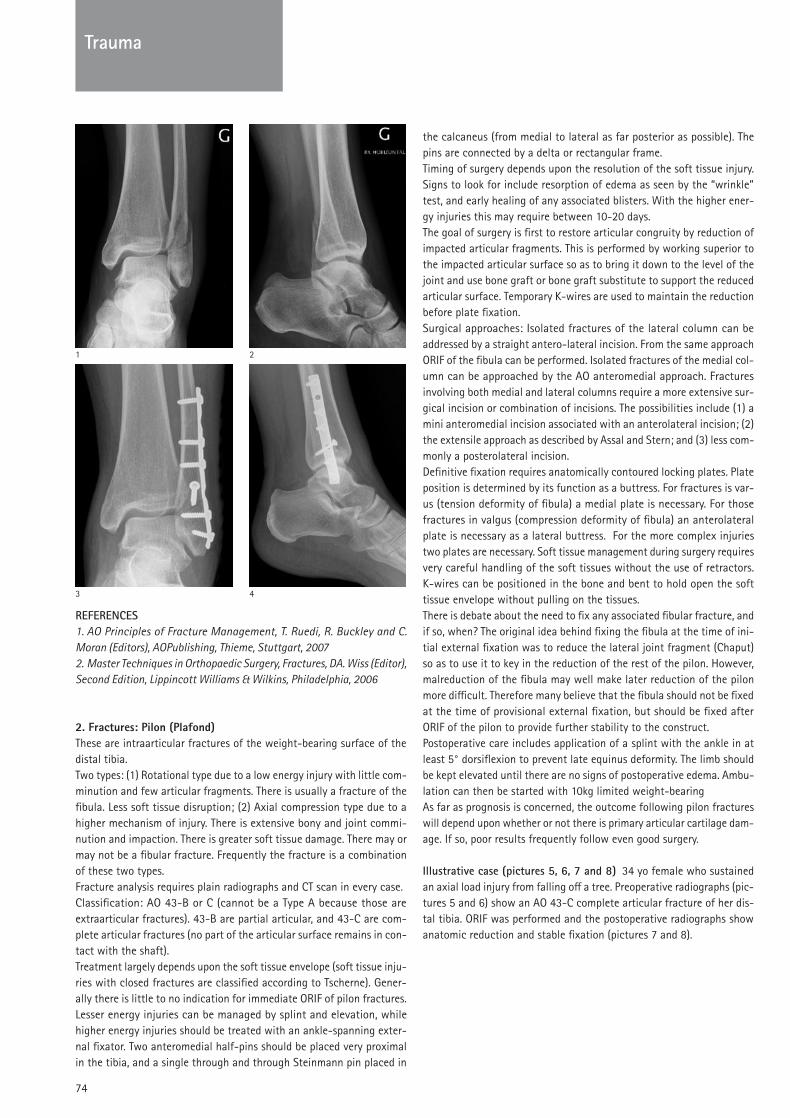
74
Trauma
REFERENCESAO Principles of Fracture Management, T. Ruedi, R. Buckley and C. 1.
Moran (Editors), AOPublishing, Thieme, Stuttgart, 2007Master Techniques in Orthopaedic Surgery, Fractures, DA. Wiss (Editor), 2.
Second Edition, Lippincott Williams & Wilkins, Philadelphia, 2006
2. Fractures: Pilon (Plafond) These are intraarticular fractures of the weight-bearing surface of the distal tibia. Two types: (1) Rotational type due to a low energy injury with little com-minution and few articular fragments. There is usually a fracture of the fibula. Less soft tissue disruption; (2) Axial compression type due to a higher mechanism of injury. There is extensive bony and joint commi-nution and impaction. There is greater soft tissue damage. There may or may not be a fibular fracture. Frequently the fracture is a combination of these two types. Fracture analysis requires plain radiographs and CT scan in every case. Classification: AO 43-B or C (cannot be a Type A because those are extraarticular fractures). 43-B are partial articular, and 43-C are com-plete articular fractures (no part of the articular surface remains in con-tact with the shaft).Treatment largely depends upon the soft tissue envelope (soft tissue inju-ries with closed fractures are classified according to Tscherne). Gener-ally there is little to no indication for immediate ORIF of pilon fractures. Lesser energy injuries can be managed by splint and elevation, while higher energy injuries should be treated with an ankle-spanning exter-nal fixator. Two anteromedial half-pins should be placed very proximal in the tibia, and a single through and through Steinmann pin placed in
the calcaneus (from medial to lateral as far posterior as possible). The pins are connected by a delta or rectangular frame.Timing of surgery depends upon the resolution of the soft tissue injury. Signs to look for include resorption of edema as seen by the “wrinkle” test, and early healing of any associated blisters. With the higher ener-gy injuries this may require between 10-20 days. The goal of surgery is first to restore articular congruity by reduction of impacted articular fragments. This is performed by working superior to the impacted articular surface so as to bring it down to the level of the joint and use bone graft or bone graft substitute to support the reduced articular surface. Temporary k-wires are used to maintain the reduction before plate fixation.Surgical approaches: Isolated fractures of the lateral column can be addressed by a straight antero-lateral incision. From the same approach ORIF of the fibula can be performed. Isolated fractures of the medial col-umn can be approached by the AO anteromedial approach. Fractures involving both medial and lateral columns require a more extensive sur-gical incision or combination of incisions. The possibilities include (1) a mini anteromedial incision associated with an anterolateral incision; (2) the extensile approach as described by Assal and Stern; and (3) less com-monly a posterolateral incision.Definitive fixation requires anatomically contoured locking plates. Plate position is determined by its function as a buttress. For fractures is var-us (tension deformity of fibula) a medial plate is necessary. For those fractures in valgus (compression deformity of fibula) an anterolateral plate is necessary as a lateral buttress. For the more complex injuries two plates are necessary. Soft tissue management during surgery requires very careful handling of the soft tissues without the use of retractors. k-wires can be positioned in the bone and bent to hold open the soft tissue envelope without pulling on the tissues. There is debate about the need to fix any associated fibular fracture, and if so, when? The original idea behind fixing the fibula at the time of ini-tial external fixation was to reduce the lateral joint fragment (Chaput) so as to use it to key in the reduction of the rest of the pilon. However, malreduction of the fibula may well make later reduction of the pilon more difficult. Therefore many believe that the fibula should not be fixed at the time of provisional external fixation, but should be fixed after ORIF of the pilon to provide further stability to the construct. Postoperative care includes application of a splint with the ankle in at least 5° dorsiflexion to prevent late equinus deformity. The limb should be kept elevated until there are no signs of postoperative edema. Ambu-lation can then be started with 10kg limited weight-bearingAs far as prognosis is concerned, the outcome following pilon fractures will depend upon whether or not there is primary articular cartilage dam-age. If so, poor results frequently follow even good surgery.
Illustrative case (pictures 5, 6, 7 and 8) 34 yo female who sustained an axial load injury from falling off a tree. Preoperative radiographs (pic-tures 5 and 6) show an AO 43-C complete articular fracture of her dis-tal tibia. ORIF was performed and the postoperative radiographs show anatomic reduction and stable fixation (pictures 7 and 8).
1
3
2
4

75
Trauma
REFERENCESAO Principles of Fracture Management, T. Ruedi, R. Buckley and C. 1.
Moran (Editors), AOPublishing, Thieme, Stuttgart, 2007Master Techniques in Orthopaedic Surgery, Fractures, DA. Wiss (Edi-2.
tor), Second Edition, Lippincott Williams & Wilkins, Philadelphia, 2006
3. Fractures: TalusInjuries are typically high energy and one should search for other mus-culoskeletal injuries.The talus articulates with seven joints: both malleoli, the pilon, the three subtalar joints, and the navicular. The talus is mostly articular (70% covered by cartilage). Any fracture is an articular fracture.The talus is composed of three parts: (1) Body, which includes the dome; (2) Neck; and (3) Head.Fracture analysis includes anteroposterior, lateral and mortise views of the ankle, as well as anteroposterior, lateral and Canale (specific view of the talar neck) views of the foot. A CT scan is essential to clearly understand fracture morphology.A particular problem with fractures of the talus relates to its blood sup-ply, which while is both extra- and intraosseous it is prone to disruption by the injury. The blood supply is coming from the three main arteries
of the leg: the tibialis anterior, tibialis posterior, and the fibular arteries. One of the complications of fractures of the talus is avascular necrosis of the talar body, as well as osteoarthritis.Talar body fractures: Generally caused by axial loading secondary to a fall from a height or a motor vehicle accident. It is frequently associat-ed with either tibiotalar or subtalar dislocation. Closed reduction of any dislocation should be performed immediately after plain radiographs are available, and before CT imaging. If there is no stability after closed reduction, or there is soft tissue compromise, a provisional external fix-ator should be applied as provisional fixation. Definitive reduction and fixation can wait until soft tissues are stable.For definitive surgery anatomic reduction with stable internal fixation is the goal. The surgical approach will be dictated by the nature of the fracture. There are two basic approaches: (1) Medial approach through the tibialis anterior and tibialis posterior, which can be extended prox-imally to incorporate a medial malleolar osteotomy for better visualiza-tion of the talar dome; (2) Anterolateral approach just lateral to the extensor digitorum longus and extends from anterior to the lateral malle-olus towards the fourth toe; and (3) Posteromedial approach with the patient supine in the interval between the posterior tibial tendon and flexor digitorum longus tendon. It addresses those fractures involving the very back of the talus (Posteromedial talar body fracture).The fractures are first preliminarily stabilized with k-wires. Definitive fixation has changed from large fragment screws and plates to small and mini implants. Any screws that must pass through articular carti-lage have to be countersunk to avoid prominence of the screw head. Bone grafting is occasionally required to support reduced articular seg-ments that have been disimpacted. If a medial malleolar osteotomy is performed local cancellous bone can be used as graft.Postoperative management includes a posterior splint. The patient is advised to ambulate with no more than 5kg of weight-bearing and remove the splint daily for range of motion exercises.Talar neck fractures: They account for 50% of all talar fractures. While neck fractures are not intraarticular, they disrupt the normal relation-ship between the articular facets of the subtalar joint. They are usually cause by forceful dorsiflexion, such as with higher energy motor vehi-cle accidents when the foot is pushing the brake pedal (historically with early airplane crashes and thus given the name, “aviator’s astragalus”). Anatomic reconstruction is critical to restore proper joint mechanics.Typically patients present with swelling and gross deformity. All obvious dislocations should be reduced as an emergency after obtaining plain radiographs, and before obtaining CT scans. The standard radiographs are mentioned above. The most widely used classification of talar neck fractures is that of Hawkins. Three groups were originally described. Group 1 fractures are nondisplaced (absolutely nondisplaced; not common). Group 2 are talar neck fractures where the talus is subluxated or dislocated from the sub-talar joint. Typically there is a varus and rotational deformity. Group 3 are talar neck fractures where the body of the talus is dislocated from both the subtalar and ankle joints. Immediate closed reduction should be attempted as an emergency, and if not possible emergent open reduc-tion is required. The talar body may have dislocated Posteromedial com-pressing the neurovascular bundle. Later, a “Type” 4 was added by Canale and kelly, thus confusing “groups” with “types.” Group 4 fractures have all the characteristics of Group 3, with the addition of a dislocation of the head of the talus at the talonavicular joint.Definitive surgical treatment is almost always performed with two inci-sions, medial and lateral (dual approach). The medial incision is made between the tibialis anterior and posterior tendons, thus providing expo-
Trau
ma
5
7
6
8

76
sure to the medial neck. Fractures are typically more comminuted medi-ally as the neck displaces into varus. This incision can be carried proxi-mally in order to perform a medial malleolar osteotomy. A lateral incision begins just anterior to the fibula, lateral to the peroneus tertius and common extensor tendons and extends distally to the fourth toe. The lateral incision allows for evaluation of length and alignment as there is less comminution laterally. One must be careful not to injure the super-ficial peroneal nerve.Stripping of soft tissue should be avoided to preserve the remaining talar blood supplyFixation can be from distal to proximal or vice-versa. With distal to prox-imal fixation screws will pass through the articular surface of the talar head and thus must be countersunk below the cartilage. Lag screws can be used for simple fractures, but with comminution one should not use lag screws but instead fully threaded position screws to avoid shorten-ing of the neck. Alternatively, screws can be placed from the posterior aspect of the talus across the neck fracture into the head. These screws can be placed just inferior to the posterior articular cartilage. In frac-tures with greater comminution minifragment plates are used to pre-serve length and alignment and no compression is used.Postoperative management is similar to that described above for talar body fractures.
Illustrative case (pictures 9, 10, 11 and 12) 22 yo male involved in a car accident. Preoperative radiographs (pictures 9) show a talar neck fracture type Hawkins III, both subtalar and tibio-talar joints being dis-located. ORIF was performed through combined medial and lateral inci-sions (dual approaches). The postoperative radiographs show anatomic reduction and stable fixation (pictures 11, 12 and 13).
REFERENCESAO Principles of Fracture Management, T. Ruedi, R. Buckley and C. 1.
Moran (Editors), AOPublishing, Thieme, Stuttgart, 2007Master Techniques in Orthopaedic Surgery, Fractures, DA. Wiss (Editor), 2.
Second Edition, Lippincott Williams & Wilkins, Philadelphia, 2006
4. Fractures: CalcaneusThe calcaneus is the most frequently fractured tarsal bone (60% of all tarsal fractures). Most intraarticular calcaneus fractures are secondary to an axial load applied directly to the heel, and usually from a fall from a height or motor vehicle accident. Extraarticular fractures are caused more frequently by a twisting or avulsion.Intraarticular fractures result in two primary fracture lines. The first starts at the crucial angle of Gissane and results from the lateral process of the talus acting as wedge dividing the calcaneus into an anterior and a posterior half. The anteromedial fragment remains in its relationship with the talus, and the posterolateral fragment displaces laterally into varus. Secondary fracture lines form in the sagittal plane along the length of the calcaneus in an anterior-posterior direction. They may extend into the calcaneocuboid joint, split the anterior facet, or exit medially or lat-erally through the body. Higher energy fractures result in greater com-minution.One must be aware of additional musculoskeletal injuries as axial load-ing can frequently result in additional injuries to the lower extremities or lumbar spine (careful examination!).Initial treatment for al calcaneus fractures should be application of a bulky dressing and splint and the limb elevated. Most patients require hospital admission. One must carefully check the neurovascular status and also be alert for possible development of a compartment syndrome (reported to occur in up to 10% of patients with a calcaneus fracture). One must periodically assess the soft tissue envelope as these high ener-gy injuries result in substantial amounts of edema and hematoma, and frequently result in the development of fracture blisters. If the soft tis-sues are threatened from within by a displaced fracture fragment urgent reduction is required to avoid full-thickness skin necrosis. Fortunately, open fractures are not common. If present, the traumatic wound tends to be medial caused by a sharp spike of the medial wall created by the primary fracture line as the tuberosity dislocates laterally.Radiographic examination consists of plain radiographs which include a lateral of the foot and ankle, a mortise view of the ankle, and dorso-plantar and oblique X-rays of the foot. In addition, a Harris or axial view is obtained. Contralateral foot and ankle views may be obtained for com-parison. A CT scan is mandatory to understand the fracture morphology.There are primarily three classification systems for calcaneus fractures. The first is from Essex-Lopresti where intraarticular fractures are clas-sified into two broad types, tongue-type and joint depression. It helps to point out those fractures that might be amenable to minimally inva-sive techniques. A second classification is that if Sanders, and is based upon the number and location of posterior facet fragments as seen on the CT scan. Third is the AO classification where the calcaneus is coded as 81.2, and fractures are divided into types where A are extraarticular fractures, B are intraarticular fractures, and C represent fracture-dislo-cations. Further subdivision into groups and subgroups follows from these broad types. Definitive treatment of calcaneus fractures is either nonoperative or operative. The factors to consider in the treatment decision relate to the patient, the fracture, and surgeon experience. Patient factors include the ability to understand the injury and cooperate with treatment, and sit-
Trauma
9
11
10
12
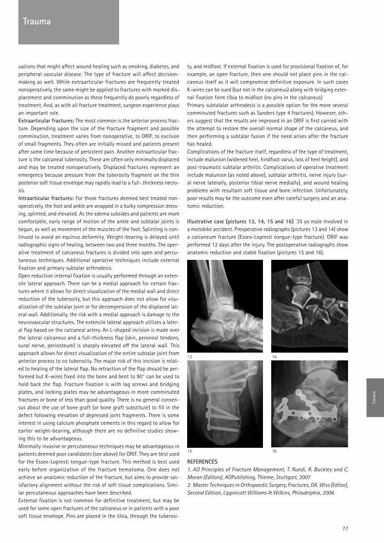
77
Trauma
uations that might affect wound healing such as smoking, diabetes, and peripheral vascular disease. The type of fracture will affect decision-making as well. While extraarticular fractures are frequently treated nonoperatively, the same might be applied to fractures with marked dis-placement and comminution as these frequently do poorly regardless of treatment. And, as with all fracture treatment, surgeon experience plays an important role.Extraarticular fractures: The most common is the anterior process frac-ture. Depending upon the size of the fracture fragment and possible comminution, treatment varies from nonoperative, to ORIF, to excision of small fragments. They often are initially missed and patients present after some time because of persistent pain. Another extraarticular frac-ture is the calcaneal tuberosity. These are often only minimally displaced and may be treated nonoperatively. Displaced fractures represent an emergency because pressure from the tuberosity fragment on the thin posterior soft tissue envelope may rapidly lead to a full-.thickness necro-sis.Intraarticular fractures: For those fractures deemed best treated non-operatively, the foot and ankle are wrapped in a bulky compression dress-ing, splinted, and elevated. As the edema subsides and patients are more comfortable, early range of motion of the ankle and subtalar joints is begun, as well as movement of the muscles of the foot. Splinting is con-tinued to avoid an equinus deformity. Weight-bearing is delayed until radiographic signs of healing, between two and three months. The oper-ative treatment of calcaneus fractures is divided into open and percu-taneous techniques. Additional operative techniques include external fixation and primary subtalar arthrodesis.Open reduction internal fixation is usually performed through an exten-sile lateral approach. There can be a medial approach for certain frac-tures where it allows for direct visualization of the medial wall and direct reduction of the tuberosity, but this approach does not allow for visu-alization of the subtalar joint or for decompression of the displaced lat-eral wail. Additionally, the risk with a medial approach is damage to the neurovascular structures. The extensile lateral approach utilizes a later-al flap based on the calcaneal artery. An L-shaped incision is made over the lateral calcaneus and a full-thickness flap (skin, peroneal tendons, sural nerve, periosteum) is sharply elevated off the lateral wall. This approach allows for direct visualization of the entire subtalar joint from anterior process to co tuberosity. The major risk of this incision is relat-ed to healing of the lateral flap. No retraction of the flap should be per-formed but k-wires fixed into the bone and bent to 90° can be used to hold back the flap. Fracture fixation is with lag screws and bridging plates, and locking plates may be advantageous in more comminuted fractures or bone of less than good quality. There is no general consen-sus about the use of bone graft (or bone graft substitute) to fill in the defect following elevation of depressed joint fragments. There is some interest in using calcium phosphate cements in this regard to allow for earlier weight-bearing, although there are no definitive studies show-ing this to be advantageous.Minimally invasive or percutaneous techniques may be advantageous in patients deemed poor candidates (see above) for ORIF. They are best used for the Essex-Lopresti tongue-type fracture. This method is best used early before organization of the fracture hematoma. One does not achieve an anatomic reduction of the fracture, but aims to provide sat-isfactory alignment without the risk of soft tissue complications. Simi-lar percutaneous approaches have been described.External fixation is not common for definitive treatment, but may be used for some open fractures of the calcaneus or in patients with a poor soft tissue envelope. Pins are placed in the tibia, through the tuberosi-
ty, and midfoot. If external fixation is used for provisional fixation of, for example, an open fracture, then one should not place pins in the cal-caneus itself as it will compromise definitive exposure. In such cases k-wires can be sued (but not in the calcaneus) along with bridging exter-nal fixation form tibia to midfoot (no pins in the calcaneus)Primary subtalalar arthrodesis is a possible option for the more several comminuted fractures such as Sanders type 4 fractures). However, oth-ers suggest that the results are improved in an ORIF is first carried with the attempt to restore the overall normal shape of the calcaneus, and then performing a subtalar fusion if the need arises after the fracture has healed.Complications of the fracture itself, regardless of the type of treatment, include malunion (widened heel, hindfoot varus, loss of heel height), and post-traumatic subtalar arthritis. Complications of operative treatment include malunion (as noted above), subtalar arthritis, nerve injury (sur-al nerve laterally, posterior tibial nerve medially), and wound healing problems with resultant soft tissue and bone infection. unfortunately, poor results may be the outcome even after careful surgery and an ana-tomic reduction.
Illustrative case (pictures 13, 14, 15 and 16) 35 yo male involved in a motobike accident. Preoperative radiographs (pictures 13 and 14) show a calcaneum fracture (Essex-Lopresti tongue-type fracture). ORIF was performed 12 days after the injury. The postoperative radiographs show anatomic reduction and stable fixation (pictures 15 and 16).
REFERENCESAO Principles of Fracture Management, T. Ruedi, R. Buckley and C. 1.
Moran (Editors), AOPublishing, Thieme, Stuttgart, 2007Master Techniques in Orthopaedic Surgery, Fractures, DA. Wiss (Editor), 2.
Second Edition, Lippincott Williams & Wilkins, Philadelphia, 2006
Trau
ma
13
15
14
16

78
Distal Radius FracturesThe recent developments of many osteosynthesis and fixation devices are the reason or the consequences of the rapid changes in the treat-ment of distal radial fractures (DRF). From a relative conservative poli-cy of treatment we are facing now a very aggressive treatment with open reduction and internal fixation. Confusion is very often done between the different fractures types, the character of the injury and not least the patient groups, their age and activities level or expected activity levels. We have to be careful and not treat all patients in the same way using the latest implant presented on the market. We cannot deny the economical factor and accept that this abundance of implants is driven by the manufacturers who envisage a huge new market. How-ever we are still missing evidence based research which argues defini-tively for the new approach.
1. EpidemiologyDistal radial fractures are common fractures accounting for about one-sixth of the fractures treated at emergency rooms or one-tenth of the total number of fractures in adults over 35 years. The incidence of DRF is approximately 19–43 per 10000 inhabitants annually with females outnumbering males in overall distribution 4:1. In Sweden, the incidence in the city of Malmö had almost doubled from the 1950s to the 1980s. This change over time could not be explained by an increase in diag-nosed DRF as the incidence of shaft fractures of the forearm remained the same. The overall ageing of the population and an increased inci-dence of osteoporosis may offer an explanation. This trend can be reversed with community interventions which promote health-educa-tion programs that address dietary intake, physical activity, smoking hab-its and environmental risk factors for osteoporosis and falls. Over the last decades there has been an increase in incidence especially in the age group greater than 60 years. The higher incidence among older wom-en could be explained by the increasing incidence of osteoporosis. A screening of patients with wrist fractures between the ages of 50–75 years revealed that only 19% had normal BMD in the hip and vertebrae. The occurrence of a DRF can be used as a predictor for a later hip frac-ture. In a Swedish study an overall relative risk to sustain a hip fracture after a previous DRF was 1.54 for women and 2.27 for men and in an American study the relative risk for a hip fracture was 1.4 for women and 2.7 for men. For DRF in younger patients the proportions of men and women are equal. These fractures are often the result of a high energy trauma and should therefore be treated differently than the osteoporot-ic fractures. The fractures in younger patients are more often intra-ar-ticular and associated with a high incidence of ligamentous injuries with the scapho-lunate ligament being the most commonly injured. As the fracture is so common, it imposes large costs to society. In Sweden, the costs in the year following the fracture were 2147€, including both direct and indirect costs resulting in an annual cost to the country of about 50 million Euro for the adult (7,26 million persons) population (November
2007). However, costs for fractures after the first year, such as costs for surgery of malunions, are not taken to account. With an increasing pro-portion of elderly people, not only in the western communities but also in the developing countries, the DRF remains an important and increas-ing economical problem that has to be assessed. However, not only the costs of the fracture are of importance, but also the outcome and disa-bility from the patients’ perspective and therefore reliable objective measurements are of importance.
2. Results and how to measure itThe final result of a fracture can be difficult to define and measure. The type of the injury, the expectation of the patient and/or the medical team may have an impact on the real appreciation of the quality of the result. Various modalities have to be considered, such as the subjective, objec-tive and economical outcome; a broad view which incorporates pain, range of motion and cosmetic appearance was suggested by Colles as “One consolation only remains, that the limb will at some remote period again enjoy perfect freedom in all of its motions and be completely exempt from pain: the deformity, however, will remain undiminished through life”. This description of the outcome following a DRF is still valid today as found and described by kopylov et al. in a 30 year follow up of 76 patients with most patients experiencing a good long-term outcome. In a short-er perspective it is somewhat different. In our practice and for research purposes we use the following tool in assessment of the results.
2.1 Objective parameters: The range of motion is measured in the three axes of rotation around the wrist joint. Extension and flexion as well as radial and ulnar deviation take place in the radio-carpal joint and were measured and expressed as one parameter as these could be regarded as one motion around the radio-ulnar and dorso-volar axis. Forearm rotation takes place in the distal and proximal radio-ulnar joints around the longitudinal axis. Grip strength, the next objective clinical parame-ter of interest, is measured with the Jamar dynamometer, expressed in kg and related to the strength of the contra-lateral hand. Grip strength in an older population has been shown to correlate well to the health related quality of life measured by the SF- 36.
2.2 Radiographs: Radiographs were first used for examination of DRF at the end of the nineteenth century. Since then, radiographic examination has improved technically and forms a basis of classification and out-come. However, it has in some studies been shown to correlate poorly with final clinical outcome and the inter-observer reliability and intra-observer reproducibility of different radiographic classifications is low. In a recent study, the radiographic appearance in the initial radiograph, radial shortening > 2mm, dorsal angulation >15 degrees, and radial angulation >10 degrees were each significantly associated with a poor-er DASH score.
Trauma
Prof. Dr. Philippe Kopylov
Malmö-Lund University HospitalLund, [email protected]
Fractures of distal radius and scaphoid and wrist instability

79
2.3 Subjective parameters: In recent years there has been interest in the development of patient related outcome scores – generic, region specif-ic and organ or joint specific. The DASH is one of the most commonly used region specific scoring systems for the upper extremity. DASH is an abbreviation for Disabilities of the Arm Shoulder and Hand, initially pub-lished, and later corrected, as the Disabilities of the Arm, Shoulder and Head. DASH is a self-administered questionnaire developed by the AAOS and the Institute for Work & Health in Canada (http://www.dash.iwh.on.ca/). DASH has been translated and validated in many languages for general use in upper extremity disorders but not specifically for DRF. A change in mean DASH score of 10 points after an intervention such as surgery is considered as minimally important change. As the original DASH with 30 questions and items, is sometime perceived as difficult to work with and time consuming, a shorter form, QuickDASH has been developed. It consists of eleven questions from the original DASH and correlate excellently with the standard DASH.
3. Treatment Alternatives3.1 Non Invasive techniques3.1.1 Conservative Closed reduction and splinting is still today the most commonly used method of treatment in the DRF. The type of splinting is of importance as is the position to immobilize. In supination there is less likelihood of radiolocation. In the Cochrane data base report on closed reduction methods, only three randomized or quasi-randomized studies were found including 404 patients. Many methods of closed reduction have been developed during the years but there is no evidence based on randomized studies to support the choice of a closed reduc-tion method. Handoll and Madok found more studies (33), when also systematically evaluating non-randomized reports of methods of closed reduction. Even in this study, there is no robust evidence to support any treatment in favour to another and the authors simply recommend the use of a method with which the practitioner is familiar. In many cases conservative treatment, however, is not enough and especially for pri-marily or secondarily unstable fractures, surgical options are needed.
3.1.2 External Fixation External fixation of DRF has been in use for more than three decades. In Sweden, it is considered to be the standard meth-od for operative treatment of the fracture- and for this reason it can be chosen as the method of reference to which newer methods can be com-pared. External fixation uses ligamentotaxis to both reduce as well as to keep the fracture in position during healing. The recommended time for immobilization varies, ranging from 4 weeks to 6 weeks. In general, long immobilization time increases the risk for reflex sympathetic dystrophy (RSD). The traction of the wrist ligaments may cause stiffness and there-fore dynamic fixation with an articulated device or non-bridging fixa-tion has been proposed with better results reported than for tradition-al bridging technique A recent randomized study was unable to find any difference between the bridging and the non-bridging external fixator in regard to clinical results in elderly patients.
3.1.3 Pinning Other closed reduction techniques includes fixation of the fracture by pinning. Various techniques have been described such as intrafocal pinning, intrafocal intramedullary pinning or pinning in com-bination with external fixation. In the Cochrane report on percutaneu-os pinning of DRF it is stated that the high rate of complications casts some doubt on their general.
3.2 Open Surgery 3.2.1 Plates For volarly dislocated fractures especially of the Barton or
Smith type, a volar plate is preferably used. For other types of DRF, oth-er techniques have been considered. Standard AO-plates and screws can be used with good results, however, to get a good stability, usually two or more columns of the radial cortex has to be fixated to achieve good results.
3.2.2 Fragment specific A fragment specific system addresses the radial and ulnar columns separately as well as single fracture fragments both dorsally and at the volar rim by a combination of plates, pins and screws. It is primarily based on pinning of the fracture but since additional sta-bility is needed to prevent the pins from bending or the fragments from sliding on the pins, a stabilizing plate to secure the pins has been add-ed. In addition, wire forms to support the subchondral bone or small fragments can be used. The system is low profile and offers good stabil-ity. The surgical approach is determined by the type of fracture and the type of fixation needed to address the fragments.
3.2.3 Volar locking plates The newest concept, the volar locking plates with angle stable screws or pegs is becoming widely used as it offers stability and a safe approach to the fracture. The fracture is approached from the volar side using the Henry approach just radially to the flexor carpi radials, ulnarly to the radial artery. This offers an easy access to the volar part of the radius. The volar locking plate has, in biomechani-cal testing, been shown to be sufficiently stable for fixation of the dor-sally comminuted fracture and has been shown to offer equivalent sta-bility when compared to the fragment specific fixation.
4. FutureFor many it seems that the volar locking plates as given the final solu-tion to the treatment of DRF. From the existing literature it is clear that volar locking plates can be used successfully in both intra and extraar-ticular DRF. However this treatment is not without complications. The evolution of treating DRF according to the reports in conferences and the publishing results is to treat all fractures undepending types, injury mechanism or age by the same method: open reposition and internal fixation with in the most cases volar locking plates. using this approach we sure overtreat an undefined number of patients with an increased morbidity and potential complication rates and without control of the potential benefits in term of increase quality of the results for each patient. Therefore, studies of sufficient quality are lacking particularly in important broad diagnoses such as the DRF. The randomized studies most often are limited in size and large differences are necessary to show statistically significant differences. We no longer look upon the DRF as a homogenous entity but instead as a rather heterogenic group. In our department in Lund (Sweden) we have an ongoing registration with a prospective follow up of the DRF with the DASH. This will allow us to pick out smaller groups, analyze the result and perhaps change the treat-ment for that specific group. Ideally, the registry works as a hypothesis generating tool for selection of randomized studies as the next step.
5. A treatment ProtocolA standardized treatment program, based on the radiographic appear-ance but taking in account the age and the demands of the patients when selecting the proper treatment was developed by “The consensus group for distal radius fracture in southern Sweden” in2004. This group consisting of dedicated surgeons from the orthopaedic and hand sur-gery departments in south of Sweden and with special interests in the treatment of DRF analysed the literature at that time and defined accord-ing to it the following protocol (Fig. 1).
Trauma
Trau
ma

80
The treatment protocol is meant to be used as a guideline for treatment but a strict compliance to it is not expected. In a prospective follow up of a large number of patients collected from the previously mentioned DASH – registry have shown that a treatment protocol is of value and might help us to select the optimal treatment for each patient.
6. ConclusionThe use of a standardized treatment protocol may make it possible to select the patients with DRF for appropriate treatment. The chosen treat-ment will guarantee in each case the expected results with an almost, but not fully, normalized function at one year. All fracture types inde-pendently their severity will reach the same good results. There is no evi-dence based reason, with the actual knowledge in 2009 to apply a stand-ardized treatment with volar locking plate to all patients and/or type of DRF. Further studies on this subject are needed and might change the actual standard of care in the future. We always have to be aware of the morbidity of the applied treatment.Ackowledgments. I want to thank Dr A. Abramo from the Hand unit Dept of Orthopedics in Lund for his help in the redaction of this manuscript.
Scaphoid fracturesThe scaphoid a mobile link between the proximal and distal row of the carpus is the most commonly fractured carpal bone. The scaphoid frac-ture is relatively frequent in the younger adults and decrease with age. The fracture of the scaphoid is difficult to diagnosis and difficult to treat. The very often missed diagnosis because of the difficulty to see the frac-ture on plain Xrays associated to the long healing time in relation with the poor vascularisation of this bone leads to a great number of scaphoid non unions with carpus collapse and secondary osteoarthritis of the wrist (SNAC). In all wrist trauma the scaphoid fracture can be evocated and has to be treated as a scaphoid fracture until it can be denied.
1. Diagnosis1.1 Clinical. The scaphoid fracture is caused by fall on an extended hand and needs a relative high energy trauma. Stress fractures are uncom-mon. Classically the pain localisation after such fracture makes the clin-ical diagnostic easy. A swelling and pain at palpation on the radial side of the wrist distal to the styloid and between the long and short thumb extensor tendons are typical. But sometimes the complaints are vague, the pain located round or dorsal to the wrist. The traction on the thumb can help in diagnosis but the most important remains that scaphoid frac-tures has to be evocated in all wrist trauma.
1.2 Radiographic. Many scaphoid fractures are undisplaced and difficult to see on plain Xrays. Only the bone resorption which appears in the fracture line after some days or the displacement with a gap between the fractured scaphoid parts make the diagnosis possible. The initial X rays of the scaphoid needs to be careful, to the AP view and lateral pro-jections so called scaphoid projections has to be added. In ulnar devia-tion of the wrist the scaphoid will be extended and examinated in all its length.
1.3 MRI. This exam is very useful in the diagnosis of scaphoid fracture and gives also information on eventual fracture of other carpal bone or ligaments injury. The marrow oedema describes as bone bruise can be sources of over diagnostic and for this reason the images of the MRI has to be correlated to a careful and critical clinical exam before diagnosis of one or the other injury is accepted. The high price of this investiga-
tion and the difficulties in many hospitals of doing MRI in emergency are important drawbacks.1.4 CT bone scan. This exam recognizes the fracture very early, is sensi-tive and specific but unfortunately gives no information about eventu-al associated ligament injuries.
1.5 Scintigram. The low specificity of this exam make the us of the scin-tigrafy more rare nowadays
2. ClassificationAccording to J C Botelheiro the diagnosed scaphoid fracture has to be classified essentially to define the correct treatmenrt. He proposed to consider these following important points
The fracture location: proximal, middle or distal third. Tuberosity -undisplaced or displaced -The presence of comminution -The age of the fracture -Association of the scaphoid fracture as a part of a more severe -midcarpal injury/luxation
3. TreatmentThe classical conservative treatment, with cast, remains the rule for the majority of scaphoid fractures. New is that the displacement, the com-minution or the localisation of the fracture with their consequences as malposition or non union risk make that we have to consider in an increasing number of cases other treatment alternatives. The cast to be used has been discussed in many publications without giving a consen-sus. It seems that the elbow do not need to be included in the immobi-lization. In our facility we use a “scaphoid cast” that includes the base of the thumb placed in abduction, immobilize the MCP I leaves the IP free. The wrist is immobilized with the second and third metacarpal in the axis of the radius.
3.1 Tuberosity fractures. This scaphoid fracture localisation has a god prognosis. The healing is relatively easy with almost no problem except a long disability and pain at the base of the thumb. A scaphoid cast can be applied for a period of 4-5 weeks in order to reduce the pain during healing.
3.2 Undisplaced Middle third fracture. This fracture normally healed with out problems if the treatment is applied early. A scaphoid cast for a pre-liminary period of at least 6-8 weeks is recommended. After this time X-rays (without cast) will give information about the healing process. If the healing on X-rays can be confirmed by the absence of pain at pal-pation, the patient is allowed to start mobilization but a new X-Rays has to confirm the healing at 6 months in order to recognize an eventual painless scaphoid non union. When bone healing on X-Rays at 6-8 weeks is associated with remaining pain at palpation the immobilization time with cast has to be prolongated. Immobilization for more than 3 months is usually worthless.
3.3 Displaced Middle third fracture. The deformity, consequence of the fracture displacement with the hump back deformity and the shorten-ing of the scaphoid, the comminution making the fractures unstable are strong argues for the necessity of reposition and internal fixation. The higher risk of non union of this fracture type is also in favour of the sur-gical treatment. In selected cases the conservative treatment is still acceptable.
Trauma
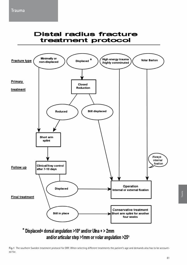
81
Trauma
Fig.1 The southern Sweden treatment protocol for DRF. When selecting different treatments the patient’s age and demands also has to be account-ed for.
Trau
ma

82
3.4 Proximal third fracture. The high risk of non union and/or necrosis of the proximal pole following this fracture are type is in strong favour of surgical treatment. Not only stabilization is here necessary but bone transplantation, vascularised or not, seems to be of importance.
4. Technical considerationsAlmost all surgeons have nowadays recognized the advantages of can-nulated compression screws in the treatment of scaphoid fractures or scaphoid non unions. With help of these devices or instruments the treat-ment has been facilitated and can very often be done without opening. Arthroscopy has here an important place and can help visualizing the reduction of the fracture. However it’s still questionable if bone trans-plantation can be done without opening the fracture. The surgeon who has to treat scaphoid fracture or their complications needs to have a good knowledge of carpus anatomy, fixation techniques and wrist arthroscopy. He also needs knowledge about vascularised bone trans-plantation techniques described for carpus indications.
5. Scaphoid non unionunfortunately the treatment described above does not guarantee heal-ing of all scaphoid fractures. On the top of that, scaphoid non union can develops without symptoms with a patient who never was in contact with the physician or has forgotten or neglected the initial trauma. The non union can be the source of the instability of the carpus followed by a development of a SNAC wrist (Scaphoid Non union Advanced Collaps) and osteoarthritis. The Non union does not necessary leads to a SNAC and is not always symptomatic. The treatment of a scaphoid non union is only indicated in the absence of osteoarthritis. The non union has to be treated by reposition of the scaphoid, cleaning of the non union site, bone transplantation (vascularised or not) and fixation.
6. Treatment algorithmCombination of a positive clinical examination associated with a posi-tive X-rays may possible to start the treatment according to fracture type, localisation and displacement. With a negative X-rays the suspi-cion of a scaphoid fracture remains and the treatment is started with a scaphoid cast for a period of 2 weeks. After this delay a new clinical examination and a conventional radiographic examination of the scaphoid are performed. If both are negative the scaphoid fracture can be denied. With a negative X-Rays and the remaining presence of symp-toms the fracture of the scaphoid has to be proved or denied by other examination as MRI or CT bone scan. Only if one or the other of these 2 exams are negative can, in theses cases, the scaphoid fracture be denied (Fig 1).
7. ConclusionThe proper diagnosis of a scaphoid fracture has to be done early in order to immobilize and stabilize the fracture as soon as possible. This can influence positively the outcome of these fractures with a high poten-tial of non union or bone necrosis. A scaphoid fracture has to be sus-pected in all wrist traumas and treated until the fracture can be denied. The use of a standardized treatment algorithm may make it possible to select the patients for appropriate treatment. The chosen treatment will be chosen in accordance to the fracture type, localization and displace-ment. The arthroscopic procedures are very important in the treatment of this injury but cannot solve all problems. Bone graft including vascu-larised bone grafts are often needed for the treatment of the difficult cases.
REFERENCES AND ADDITIONAL READINGSAbramo A, Kopylov P, Tagil M. Evaluation of a treatment protocol in 1.
distal radius fractures: a prospective study in 581 patients using DASH as outcome. Acta Orthop 2008;79-3:376-85.
Alnot JY . Les fractures et pseudarthroses polaires proximales du 2. scaphoide carpien.Rev. Chir Orthop 1988;74: 740-43
AmadioPC, Berqvist TH,Smith DK et al.Scaphoid malunion, J Hand Surg 3. (am)1989; 14:679-87
Andersen DJ, Blair WF, Steyers CM, Jr., Adams BD, el-Khouri GY, Brand-4. ser EA. Classification of distal radius fractures: an analysis of interobserv-er reliability and intraobserver reproducibility. J Hand Surg [Am] 1996;21-4:574-82.
Arora R, Lutz M, Hennerbichler A, Krappinger D, Espen D, Gabl M. Com-5. plications following internal fixation of unstable distal radius fracture with a palmar locking-plate. J Orthop Trauma 2007;21-5:316-22.
Atroshi I, Brogren E, Larsson GU, Kloow J, Hofer M, Berggren AM. Wrist-6. bridging versus non-bridging external fixation for displaced distal radius fractures: a randomized assessor-blind clinical trial of 38 patients fol-lowed for 1 year. Acta Orthop 2006;77-3:445-53.
Beaton DE, Wright JG, Katz JN. Development of the QuickDASH: com-7. parison of three item-reduction approaches. J Bone Joint Surg Am 2005;87-5:1038-46.
Botelheiro JC. Fractures of the scaphoid: Diagnosis and management. 8. EFFORT European Instructional lectures Madrid Springer Ed, Bentley G 2010;79-84.
Dias JJ, Thompson J, Barton NJ et al. Suspected scaphoid fractures. The 9. value of radiographs. J Bone Joint surg Br 1990;72:98-101
Downing ND, Karantana A. A revolution in the management of frac-10. tures of the distal radius? J Bone Joint Surg Br 2008;90-10:1271-5.
1Gelberman RH, MenonJ. The vascularity of the scaphoid bone J Hand 11. Surg Am 1980; 5:508-513
Gummesson C, Atroshi I, Ekdahl C. The disabilities of the arm, shoul-12. der and hand (DASH) outcome questionnaire: longitudinal construct valid-ity and measuring self-rated health change after surgery. BMC Muscu-loskelet Disord 2003;4-1:11.
Handoll HH, Madhok R. Closed reduction methods for treating distal 13. radial fractures in adults. Cochrane Database Syst Rev 2003-1:CD003763.
Handoll HH, Vaghela MV, Madhok R. Percutaneous pinning for treat-14. ing distal radial fractures in adults. Cochrane Database Syst Rev 2007-3:CD006080.
Herbert TJ, Fisher WE. Management of the fractured scaphoid bone 15. using a new bone screw. J Bone Joint surg 1984; 66B: 114-23.
Hudak PL, Amadio PC, Bombardier C. Development of an upper extrem-16. ity outcome measure: the DASH (disabilities of the arm, shoulder and hand) [corrected]. The Upper Extremity Collaborative Group (UECG). Am J Ind Med 1996;29-6:602-8.
Kopylov P, Johnell O, Redlund-Johnell I, Bengner U. Fractures of the 17. distal end of the radius in young adults: a 30-year follow-up. J Hand Surg [Br] 1993;18-1:45-9.
Mathoulin C, Haerle M, Vandeputte G.Vascularized bone graft in car-18. pal bone reconstruction, Ann Chir Plast Esthet. 2005 Feb;50(1):43-8.
McQueen MM. Redisplaced unstable fractures of the distal radius. A 19. randomised, prospective study of bridging versus non-bridging external fixation. J Bone Joint Surg Br 1998;80-4:665-9.
Rikli DA, Regazzoni P. The double plating technique for distal radius 20. fractures. Tech Hand Up Extrem Surg 2000;4-2:107-14.
Trauma

83
Trauma
Fig. 2 Treatment algorithm of diagnosis of a scaphoid fracture
Trau
ma

84
For shoulder pathology the major reference is: The Shoulder, Fourth Edi-tion. Editors CA Rockwood, FA Matsen, MA Wirth, SB Lippitt. 2009, Phil-adelphia, Saunders Elsevier. In the AO manual that covers all important fixation techniques the reader will fact pertinent facts related to the topic at hand: AO Principles of Fracture Management. Second expanded edition, TP Rüedi, RE Buckley, CG Moran. 2007, Georg Thieme Verlag. For the latest in fracture fixation techniques the reader is also invited to vis-it the AO surgery reference site: AO surgery reference: http://www.aofoun-dation.org. Another most useful publication containing pertinent facts related to orthopaedics and musculoskeletal trauma is the AAOS Com-prehensive Orthopaedic Review. Jay R. Lieberman, MD, Editor 2009 Rose-mont, IL, American Academy of Orthopaedic Surgeons. For surgical approaches the most useful reference is without doubt: Surgical Expo-sures in Orthopaedics: The Anatomic Approach. Hoppenfeld S, deBoer P, Buckley R. Lippincott Williams & Wilkins; 2009, Fourth Edition.
Clavicle fracturesClavicular fractures are some of the most common fractures account-ing for 5% to 10% of all fractures and 35% to 45% of shoulder girdle injuries. The clavicle struts the shoulder girdle.Clavicular fractures are the result of falls, rarely direct trauma and rare-ly secondary to metastatic disease.Clinical examination shows deformity of the shoulder girdle; a careful neurovascular examination must be performed due to the vicinity of fragile structures deep to this unprotected and subcutaneous bone.Diagnosis necessitates an AP X-ray of the clavicle and often an AP view of the whole shoulder girdle will be of help to comparatively determine the amount of displacement. In some rare cases a CT will define the frac-ture.
Fractures of the clavicle are divided into proximal third, mid-third and distal third.
Proximal third fractures �usually conservative treatment will be sufficient, if displaced will benefit from fracture fixation, preferably with a plate. Beware of free pins that tend to migrate.Mid-third fractures �If little displacement is present conservative treatment with a sling will be sufficient. In cases of displacement >100% or > 2 cm of shortening, fixation is indicated. Flail chest, scapulotho-racic dissociation, fractures menacing the integrity of the skin or open fractures are also indications for operative fixation. Activ-ities such as professional cycling cannot tolerate unequal clavic-ular lengths and in these cases reconstruction is indicated. Fixation may be accomplished with a 3.5 mm reconstruction or dynamic compression with or without locked screws. Nails of dif-ferent types have been advocated and reported to be successful by many authors.
Robertson C, Celestre P, Mahar A, Schwartz A. Reconstruction plates for stabilization of mid-shaft clavicle fractures: differences between non-locked and locked plates in two different positions. J Shoulder Elbow Surg. 2009;18(2):204-9.Lee YS, Lin CC, Huang CR, Chen CN, Liao WY. Operative treatment of mid-clavicular fractures in 62 elderly patients: Knowles pin versus plate. Ortho-pedics. 2007;30(11):959-64.Canadian Orthopaedic Trauma Society Trial. Midshaft Clavicular Frac-tures. A Multicenter, Randomized Clinical Nonoperative Treatment Com-pared with Plate Fixation of Displaced. J Bone Joint Surg Am.2007;89:1-10.
Distal third fractures �In case of displacement > 100%, skin menace or open fracture fixation is indicated. Depending on the size of the distal frag-ment the surgical intervention can vary from simple excision, to figure of 8 wiring with pins, to heavy sutures to specific plates or hook plates. If the coracoclavicular ligaments are compromised (Neer type II fractures) coracoclavicular fixation (sutures or screws) may be indicated.
Khan LA, Bradnock TJ, Scott C, Robinson CM. Fractures of the clavicle. J Bone Joint Surg Am. 2009;91(2):447-60.Neer CS II. Fractures of the distal third of the clavicle. Clin Orthop. 1968;58:43–50.Fann CY, Chiu FY, Chuang TY, Chen CM, Chen TH. Transacromial Knowles
Prof. Dr. Pierre Hoffmeyer
University HospitalGeneva, [email protected]
Upper Limb Trauma: Shoulder girdle, proximal humerus, humeral shaft
Neer Type 1
Neer Type 2
Neer Type 3
Trauma

85
pin in the treatment of Neer type 2 distal clavicle.fracturesA prospective evaluation of 32 cases. J Trauma. 2004;56(5):1102-5; discussion 1105-6.Goldberg JA, Bruce WJ, Sonnabend DH, Walsh WR. J Shoulder Elbow Surg. Type 2 fractures of the distal clavicle: a new surgical technique. 1997;6(4):380-2.The Shoulder, Fourth Edition. Editors CA Rockwood, FA Matsen, MA Wirth, SB Lippitt. 2009, Philadelphia, Saunders Elsevier.Muramatsu K, Shigetomi M, Matsunaga T, Murata Y, Taguchi T. Use of the AO hook-plate for treatment of unstable fractures of the distal clavicle. Arch Orthop Trauma Surg. 2007;127(3):191-4.
ComplicationsInfections, nonunions or neurovascular compromise dominate the scene. The Shoulder, Fourth Edition. Editors CA Rockwood, FA Matsen, MA Wirth, SB Lippitt. 2009, Philadelphia, Saunders Elsevier.Endrizzi DP, White RR, Babikian GM, Old AB. Nonunion of the clavicle treated with plate fixation: a review of forty-seven consecutive cases. J Shoulder Elbow Surg. 2008;17(6):951-3.
Sternoclavicular dislocations Anatomy �With relatively no osseous constraints, stability is provided by the anterior capsular ligament, the posterior capsular ligament, and a joint meniscus. The costoclavicular and interclavicular lig-aments provide adjunct stability. Antero-superior dislocation �unstable and needs surgical intervention for stability. usually reassurance and conservative treatment will suffice however.Postero-inferior dislocation �This is potentially a life threatening situation. Symptoms are related to the posterior structures under compression (dyspnea, dysphagia, vascular compromise or thrombosis). CT is helpful to make the diagnosis. Reduction under anaesthesia with a bolster under the dorsal spine and simultaneously pulling the arm in extension while grabbing the clavicle end with a towel clip will usually reduce the clavicle that will stay stable.Beware of fractures passing trough the proximal growth plate,
which is the last to ossify at age 25.
Jaggard MK, Gupte CM, Gulati V, Reilly P. A comprehensive review of trau-ma and disruption to the sternoclavicular joint with the proposal of a new classification system. J Trauma. 2009;66(2):576-84.
Acromioclavicular dislocationsusually a consequence of a fall on the tip of the shoulder in a young to middle-aged male athlete, the acromion is pushed downwards and the coraco- and acriomio-clavicular ligaments are damaged to varying degrees along with a displacement of the clavicle with respect to the shoulder girdle.The patient presents with a deformity due to the antero-inferior posi-tion of the shoulder girdle. Check for instability in the frontal and trans-verse planes. Inspect the skin to rule out abrasions.AP X-rays of the shoulder, Zanca views (10°-15° cephalic tilt) and axil-lary views are necessary and sufficient. An AP X-ray view of the shoul-der girdle is a useful adjunct. Stress views are not necessary.AC dislocations are classified according to Rockwood:Type I: Strain without tear, Type II tearing of AC ligaments, Type III: Tear-ing of AC and CC ligaments (Trapezoid and conoid), Type IV: posterior displacement of the clavicle in relation to the acromion. Type V: More than 100% displacement with tearing of AC and CC ligaments and over-lying trapezius muscle.Types I and II need conservative treatment.Surgery is usually recommended in types IV and V. Type III is controver-sial in frail patients in may be recommended. The techniques may involve coraco-clavicular screws, CC and AC heavy sutures or tapes or transar-ticular pinning. In long standing cases the Weaver-Dunn procedure is recommended, with removal of 1 cm of the distal clavicle and using the coraco-acro-mial ligament as a substitute inserted into the hollowed out distal clav-icle. Hook plates are used by some authors but will require reoperation for their removal.
The Shoulder, Fourth Edition. Editors CA Rockwood, FA Matsen, MA Wirth, SB Lippitt. 2009, Philadelphia, Saunders Elsevier.AAOS Comprehensive Orthopaedic Review. Jay R. Lieberman, MD, Editor 2009 Rosemont, IL, American Academy of Orthopaedic Surgeons
Trauma
Trau
ma
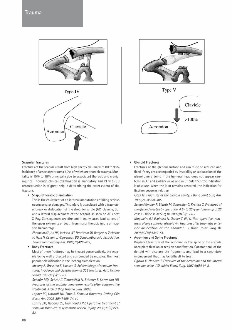
86
Scapular fracturesFractures of the scapula result from high energy trauma with 80 to 95% incidence of associated trauma 50% of which are thoracic trauma. Mor-tality is 10% to 15% principally due to associated thoracic and cranial injuries. Thorough clinical examination is mandatory and CT with 3D reconstruction is of great help in determining the exact extent of the fracture.
Scapulothoracic dissociation �This is the equivalent of an internal amputation entailing serious neurovascular damages. This injury is associated with a traumat-ic break or dislocation of the shoulder girdle (AC, clavicle, SC) and a lateral displacement of the scapula as seen on AP chest X-Ray. Consequences are dire and in many cases lead to loss of the upper extremity or death from major thoracic injury or mas-sive haemorrage.Ebraheim NA, An HS, Jackson WT, Pearlstein SR, Burgess A, Tscherne H, Hass N, Kellam J, Wipperman BU. Scapulothoracic dissociation. J Bone Joint Surgery Am, 1988;70,428-432,Body Fractures �Most of these fractures may be treated conservatively, the scap-ula being well protected and surrounded by muscles. The most popular classification is the Ideberg classification.Ideberg R, Grevsten S, Larsson S. Epidemiology of scapular frac-tures. Incidence and classification of 338 fractures. Acta Orthop Scand. 1995;66(5):395-7.Schofer MD, Sehrt AC, Timmesfeld N, Störmer S, Kortmann HR. Fractures of the scapula: long-term results after conservative treatment. Arch Orthop Trauma Surg. 2009.Lapner PC, Uhthoff HK, Papp S. Scapula fractures. Orthop Clin North Am. 2008 ;39(4):459-74, vi.Lantry JM, Roberts CS, Giannoudis PV. Operative treatment of scapular fractures: a systematic review. Injury. 2008;39(3):271-83.
Glenoid Fractures �Fractures of the glenoid surface and rim must be reduced and fixed if they are accompanied by instability or subluxation of the glenohumeral joint. If the humeral head does not appear cen-tered in AP and axillary views and in CT cuts then the indication is absolute. When the joint remains centered, the indication for fixation becomes relative.Goss TP. Fractures of the glenoid cavity. J Bone Joint Surg Am. 1992;74-A:299-305.Schandelmaier P, Blauth M, Schneider C, Krettek C. Fractures of the glenoid treated by operation. A 5- to 23-year follow-up of 22 cases. J Bone Joint Surg Br. 2002;84(2):173-7Maquieira GJ, Espinosa N, Gerber C, Eid K. Non-operative treat-ment of large anterior glenoid rim fractures after traumatic ante-rior dislocation of the shoulder. J Bone Joint Surg Br. 2007;89(10):1347-51.Acromion and Spine Fractures �Displaced fractures of the acromion or the spine of the scapula need plate fixation or tension band fixation. Constant pull of the deltoid will displace the fragments and lead to a secondary impingement that may be difficult to treat.Ogawa K, Naniwa T. Fractures of the acromion and the lateral scapular spine. J Shoulder Elbow Surg. 1997;6(6):544-8.
Trauma

87
Classification of fractures of the scapular neck: TP Goss. J Am Acad Orthop Surg 1995;3:22-33
Classification of the scapular body : Ideberg R, Grevsten S, Larsson S. Epidemiology of scapular fractures. Incidence and classification of 338 fractures. Acta Orthop Scand. 1995;66(5):395-7.
Glenohumeral dislocationFor shoulder pathology the major reference is: The Shoulder, Fourth Edi-tion. Editors CA Rockwood, FA Matsen, MA Wirth, SB Lippitt. 2009, Phil-adelphia, Saunders Elsevier. In the AO manual that covers all important fixation techniques the reader will fact pertinent facts related to the topic at hand: AO Principles of Fracture Management. Second expanded edition, TP Rüedi, RE Buckley, CG Moran. 2007, Georg Thieme Verlag. For the latest in fracture fixation techniques the reader is also invited to vis-it the AO surgery reference site: AO surgery reference: http://www.aofoun-dation.org. Another most useful publication containing pertinent facts related to orthopaedics and musculoskeletal trauma is the AAOS Com-prehensive Orthopaedic Review. Jay R. Lieberman, MD, Editor 2009 Rose-mont, IL, American Academy of Orthopaedic Surgeons. For surgical approaches the most useful reference is without doubt: Surgical Expo-sures in Orthopaedics: The Anatomic Approach. Hoppenfeld S, deBoer P, Buckley R. Lippincott Williams & Wilkins; 2009, Fourth Edition.
IntroductionDislocation applies to a complete loss of contact between two joint sur-faces. Subluxation implies partial loss of contact. Laxity is the result of a clinical examination showing more than “normal” passive motion or translation. Instability is a subjective sensation described by the patient that includes subluxation up to dislocation.Glenohumeral instability is a spectrum that includes hyperlaxity and traumatic dislocation whether anterior or posterior or multidirectional. Classification of the different types of instability include:
Traumatic anterior dislocation: Accidental fall �Traumatic posterior dislocation: Accidental fall �Atraumatic instability due to capsular stretching because repeat- �ed “micro-trauma”Multidirectional instability due to capsular laxity �
PathoanatomyTraumatic anterior instability �Generally accompanied by a tear of the capsulo-labral complex that sometimes includes osseous fragments off from the glenoid rim: The Bankart lesion.Anterior capsular stretching.In many cases a bony trough in the posterior-superior region of the head will be caused by impaction against the glenoid rim sometimes leading up to a fracture of the greater tuberosity: The Hill-Sachs lesion.Traumatic posterior instability �The inferior glenohumeral ligament is the main restraint in abduc-tion/external rotation and found to be torn or detached in all cas-es of traumatic dislocation.Generally accompanied by detachment and stretching of the pos-terior capsulo-ligamentous complex, rarely with osseous lesions involving the glenoid rim: The reverse Bankart lesion.Posterior capsular stretching. Impaction of the anterior region of the head just medial to the lesser tuberosity leading up to a head-split fracture: The reverse Hill-Sachs lesion.
Anterior dislocation injuries
Hill-Sachs impression; Bankart bony and labral; capsular stretching.
Dislocation and instability typesAnterior dislocationusually related to sports activities (soccer, skiing etc.) or falls. Recurrence rates are high in patients below 20 yrs (up to 90%), between
Trauma
Trau
ma
88
20 and 40 yrs 60% recurrence rates, above 40 yrs 10%. These numbers vary depending on authors but trends remain.Clinical examination is dominated by apprehension in abduction and external rotation. Signs of generalized laxity are often present: Antero-posterior drawer, inferior sulcus sign, joint hyperlaxity (fingers, thumb, elbow).In acute cases axillary nerve injury occurs in 5% of patients.Imaging involves AP and axillary views. Arthro-CT scans delineate pre-cisely bony morphology of fractures; Hill-Sachs lesions, glenoid brim fractures or rounding are well visualized. MRI may be helpful but bony lesions are poorly demonstrated.
Treatment for acute dislocations -AFTER diagnostic X-Rays: Reduction techniques include, after neurovascular testing, Stimson (Patient prone, arm hanging with 1 to 3 kg weights attached to the wrist), Saha (slow elevation in the plane of the scapula), Kocher (Adduction in internal rot fol-lowed by abduction in ext rotation), Traction after intra-articu-lar injection of lidocaïne or equivalent, Davos (Patient to cross his fingers around his flexed knee and with elbows extended is instructed to slowly bend backwards), Hippocrates technique (anesthetized, traction on the arm and with foot in the axilla which should be replaced by a towel) should only be performed when the non traumatic techniques have failed. Postreduction treatment includes, after neurovascular testing, immobilisation in internal rotation or in an external rotation splint. (The rationale for the external rotation immobilisation is to force the Bankart lesion to stay fixed to the anterior glenoid rim pressured by the subscapularis). Immobilisation should be 2 to 4 weeks followed by strengthening exercises.Treatment for recurrent dislocations -Surgical indications for stabilisation include one episode of dis-location too many, or severe apprehension. Techniques include capsulorraphy, Bankart lesion refixation, bony augmentation if severe rounding or fracture of the rim.Open or arthroscopic techniques are both suitable. Closed arthro-scopic techniques are advocated in traumatic Bankart lesions, open techniques are recommended in cases of capsular stretch-ing or large Hill-Sachs lesions. Recurrence rates range between 5% and 30% depending on technique used, strength of recon-struction and patient compliance.Patients are immobilized from 3 to 6 weeks in internal rotation; rehabilitation emphasizes muscular strengthening in the first weeks followed by range of motion exercises. Patients are advised to avoid contact sports for a year following stabilisation.
Posterior dislocationFall on outstretched hand, seizures or electrical shocks are the main causes. AP and axillary X-rays for diagnosis. Relatively rare; less than 5% of all instabilities. Beware of the diagnosis: The cardinal sign is active and passive limitation of external rotation. On the AP X-ray, the joint space is not visible and the axillary is always diagnostic. In doubt a CT will solve the issue.
Treatment for acute dislocations -If a small i.e. less than 10% reverse Hill-Sachs is present, gentle traction will generally reduce the shoulder which should then be immobilized in an external rotation splint for three to 6 weeks with a rehabilitation programme to follow.If a large Hill-Sachs lesion is present, reduction under anaesthe-sia may be necessary followed by the McLaughlin procedure
where through an anterior deltopectoral incision the head is gen-tly levered out and the subscapularis or the osteotomized lesser tuberosity is sutured or screwed into the bony defect. External rotation immobilisation 4 to 6 weeks followed by a rehabilita-tion programme. Treatment for recurrent dislocations -If no major Hill-Sachs lesion is present a posterior approach with a cruciate capsulorraphy and fixation of the reverse Bankart lesion is performed. A bone graft from the spine of the scapula or the iliac crest may be necessary if a bony defect is present. If a major Hill-Sachs lesion is present a McLaughlin procedure will be necessary and if insufficient an adjunct posterior proce-dure may be necessary.
Multidirectional dislocationThis applies to young patients with laxity and instability in more than in one direction, i.e. anterior and posterior or posterior and inferior or all three. Cardinal signs are hyperlaxity, sulcus sign and anterior and pos-terior drawer signs all causing discomfort or apprehension.Standard X-rays, arthro-CT or MRI will delineate the existing lesions. Surgery is indicated only after one year of serious muscle strengthening physiotherapy and exercises. The most commonly accepted operation is Neer’s capsular shift which may be performed through an anterior deltopectoral approach but in certain cases may need an adjunct posterior approach. The axillary nerve must be protected during this demanding and complex intervention. 6 weeks of immobilisation in neutral (handshake) position is necessary follwed by a muscle strengthening programme.
Chronic dislocation usually seen in debilitated patients. The best option may be no treat-ment. In cases of chroni pain and discomfort shoulder fusion may be another option. Some authors advocate the reverse prosthesis but the danger of dislocation is great.
Recurrent dislocation in the elderly patientOften these dislocations are associated with minor trauma. A massive rotator cuff tear is the usual cause. If repairable the supra and infrasp-inatus lesions should be repaired. If not repairable the reverse prosthe-sis may be an option and if not fusion may have to be performed.
The Shoulder, Fourth Edition. Editors CA Rockwood, FA Matsen, MA Wirth, SB Lippitt. 2009, Philadelphia, Saunders Elsevier.AAOS Comprehensive Orthopaedic Review. Jay R. Lieberman, MD, Editor 2009 Rosemont, IL, American Academy of Orthopaedic Surgeons.Shoulder Reconstruction. CS Neer. W.B. Saunders Company (January 1990)
Proximal humerus fracturesFor shoulder pathology the major reference is: The Shoulder, Fourth Edi-tion. Editors CA Rockwood, FA Matsen, MA Wirth, SB Lippitt. 2009, Phil-adelphia, Saunders Elsevier. In the AO manual that covers all important fixation techniques the reader will fact pertinent facts related to the topic at hand: AO Principles of Fracture Management. Second expanded edition, TP Rüedi, RE Buckley, CG Moran. 2007, Georg Thieme Verlag. For the latest in fracture fixation techniques the reader is also invited to vis-it the AO surgery reference site: AO surgery reference: http://www.aofoundation.org. Another most useful publication containing pertinent
Trauma
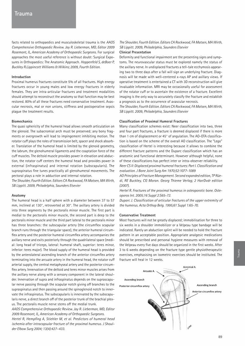
89
facts related to orthopaedics and musculoskeletal trauma is the AAOS Comprehensive Orthopaedic Review. Jay R. Lieberman, MD, Editor 2009 Rosemont, IL, American Academy of Orthopaedic Surgeons. For surgical approaches the most useful reference is without doubt: Surgical Expo-sures in Orthopaedics: The Anatomic Approach. Hoppenfeld S, deBoer P, Buckley R.Lippincott Williams & Wilkins; 2009, Fourth Edition.
IntroductionProximal humerus fractures constitute 5% of all fractures. High energy fractures occur in young males and low energy fractures in elderly females. They are intra-articular fractures and treatment modalities should attempt to reconstruct the anatomy so that function may be best restored. 80% of all these fractures need conservative treatment. Avas-cular necrosis, mal or non unions, stiffness and postoperative sepsis plague the treatment results.
BiomechanicsThe quasi sphericity of the humeral head allows smooth articulation on the glenoid. The subacromial arch must be preserved; any bony frag-ments or overgrowth will lead to impingement inhibiting motion. The rotator cuff plays the roles of transmission belt, spacer and shock absorb-er. Translation of the humeral head is limited by the glenoid geometry, the labrum, the glenohumeral ligaments and the coaptation force of the cuff muscles. The deltoid muscle provides power in elevation and abduc-tion, the rotator cuff centers the humeral head and provides power in external (infraspinatus) and internal rotation (subscapularis). The supraspinatus fine tunes practically all glenohumeral movements. The pectoral plays a role in adduction and internal rotation. The Shoulder, Fourth Edition. Editors CA Rockwood, FA Matsen, MA Wirth, SB Lippitt. 2009, Philadelphia, Saunders Elsevier
AnatomyThe humeral head is a half sphere with a diameter between 37 to 57 mm, inclined at 130°, retroverted at 30°. The axillary artery is divided into three segments by the pectoralis minor muscle. The first part is medial to the pectoralis minor muscle, the second part is deep to the pectoralis minor muscle and the third part lateral to the pectoralis minor has three branches: the subscapular artery (the circumflex scapular branch runs through the triangular space), the anterior humeral circum-flex artery and the posterior humeral circumflex artery accompanies the axillary nerve and exits posteriorly through the quadrilateral space (medi-al: long head of triceps, lateral: humeral shaft, superior: teres minor, inferior: teres major). The blood supply of the humeral head is provided by the anterolateral ascending branch of the anterior circumflex artery terminating into the arcuate artery in the humeral head, the rotator cuff arterial supply, the central metaphyseal artery and the posterior circum-flex artery. Innervation of the deltoid and teres minor muscles arises from the axillary nerve along with a sensory component in the lateral shoul-der. Innervation of supra and infraspinatus depends on the suprascapu-lar nerve passing through the scapular notch giving off branches to the supraspinatus and then passing around the spinoglenoid notch to inner-vate the infraspinatus. The subscapularis is innervated by the subscapu-laris nerve, a direct branch off of the posterior trunk of the brachial plex-us. The pectoralis muscle nerve stems off the medial trunk. AAOS Comprehensive Orthopaedic Review. Jay R. Lieberman, MD, Editor 2009 Rosemont, IL, American Academy of Orthopaedic Surgeons.Hertel R, Hempfing A, Stiehler M, et al. Predictors of humeral head ischemia after intracapsular fracture of the proximal humerus. J Shoul-der Elbow Surg 2004; 13(4):427–433.
The Shoulder, Fourth Edition. Editors CA Rockwood, FA Matsen, MA Wirth, SB Lippitt. 2009, Philadelphia, Saunders ElsevierClinical PresentationDeformity and functional impairment are the presenting signs and symp-toms. The neurovascular status must be explored namely the status of the axillary nerve. In undisplaced fractures a tell-tale ecchymosis appear-ing two to three days after a fall will sign an underlying fracture. Diag-nosis will be made with well-centered x-rays AP and axillary views. If operative treatment is entertained a CT with 3D reconstruction will give invaluable information. MRI may be occasionally useful for assessment of the rotator cuff or to ascertain the existence of a fracture. Excellent imaging is the only way to accurately classify the fracture and establish a prognosis as to the occurrence of avascular necrosis. The Shoulder, Fourth Edition. Editors CA Rockwood, FA Matsen, MA Wirth, SB Lippitt. 2009, Philadelphia, Saunders Elsevier
Classification of Proximal Humeral FracturesMany classification schemes exist: Neer classification into two, three and four part fractures, a fracture is deemed displaced if there is more than 1 cm of displacement or 45° of angulation. The AO-OTA classifica-tion is based on the scheme of the overall AO classification. The “Lego” classification of Hertel is interesting because it allows to combine the different fracture patterns and the Duparc classification which has an anatomic and functional determinant. However although helpful, none of these classifications has perfect inter or intra-observer reliability. Neer CS II: Displaced proximal humeral fractures: Part I. Classification and evaluation. J Bone Joint Surg Am 1970;52:1077-1089 AO Principles of Fracture Management. Second expanded edition, TP Rüe-di, RE Buckley, CG Moran. Georg Thieme Verlag; 2 Har/Dvdr edition (2007).Hertel R. Fractures of the proximal humerus in osteoporotic bone. Oste-oporos Int. 2005;16 Suppl 2:S65-72. Duparc J. Classification of articular fractures of the upper extremity of the humerus. Acta Orthop Belg. 1995;61 Suppl 1:65-70.
Conservative TreatmentMost fractures will not be greatly displaced; immobilisation for three to six weeks in a shoulder immobilizer or a Velpeau type bandage will be indicated. Rarely an abduction splint will be needed to hold the fracture pattern in an acceptable position. Appropriate analgesic medications should be prescribed and personal hygiene measures with removal of the Velpeau every five days should be organized in the first weeks. After 3 to 6 weeks depending on the fracture type gentle physiotherapeutic exercises, emphasising on isometric exercises should be instituted. The fracture will heal in 12 weeks.
Trau
ma
Trauma

90
Vascular anatomy of the Humeral Head
Surgical ApproachesDelto-pectoral approach �The cephalic vein should if possible be preserved. The axillary nerve must be palpated in front of the subscapularis. If the long biceps tendon is not anatomically replaced a tenodesis is in order.Trans-deltoid approach �The deltoid should not be split further than 5 cm distal to the acromion to protect the axillary nerve.Posterior approach �A deltoid split will lead to the unfraspinatus which may have to be detached to access the capsule for arthrotomy. Rarely used approach in the trauma setting.
The Shoulder, Fourth Edition. Editors CA Rockwood, FA Matsen, MA Wirth, SB Lippitt. 2009, Philadelphia, Saunders ElsevierAAOS Comprehensive Orthopaedic Review. Jay R. Lieberman, MD, Editor 2009 Rosemont, IL, American Academy of Orthopaedic Surgeons.
Operative techniquesIsolated greater tuberosity fractures �Displacement of more than 0,5 to 1cm warrants operative treat-ment. usually a trans-deltoid approach with suture fixation, sometimes augmented by isolated screws or perhaps a plate in case of a large fragment.
Transdeltoid approach and osteosuture of displaced fracture of the great-er tuberosity
Displaced lesser tuberosity fractures �Anatomic reduction and fixation with screws is warranted to pre-serve subscapularis function.Two part displaced surgical neck fracture �Plating or IM nailing can be used successfully in this indication.
Two part fracture fixed with a locking plate
Three part fractures �In strong bone percutaneous pinning may be used although accu-rate reduction is best achieved with an open technique. Some authors favour locked nailing for these fractures. In weak bone a deltopectoral approach with plate fixation with or without fixed angle screws or an osteosuture technique will be indicated. The biceps if well aligned in the bicipital groove is a precious indica-tor as to reduction accuracy. It is wise to check the reduction before closure with an X-ray or an image intensifier.Four part fractures �Prosthetic replacement respecting height, version and tuberosi-ty fixation will be used in the elderly patient. In high demand young patients it is probably best to attempt plate osteosynthe-sis with angle stable screws. This is an acceptable solution only if an adequate anatomical reconstruction has been achieved. If not, a hemiarthroplasty with careful reconstruction of the tuber-osities is an acceptable option.
Three part fracture fixation with a locking screw plate
Three part fracture fixation with a third tubular 3.5 locking screw plate
Trauma
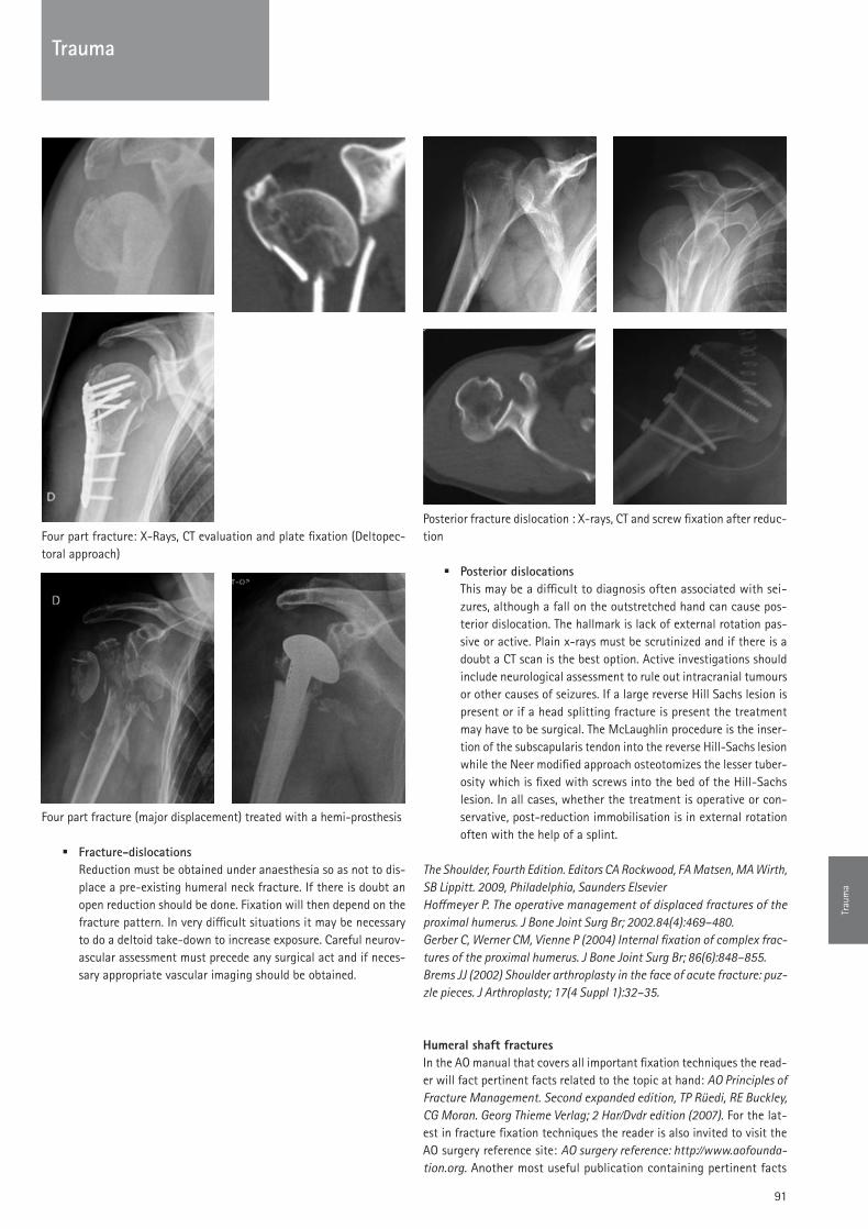
91
Four part fracture: X-Rays, CT evaluation and plate fixation (Deltopec-toral approach)
Four part fracture (major displacement) treated with a hemi-prosthesis
Fracture-dislocations �Reduction must be obtained under anaesthesia so as not to dis-place a pre-existing humeral neck fracture. If there is doubt an open reduction should be done. Fixation will then depend on the fracture pattern. In very difficult situations it may be necessary to do a deltoid take-down to increase exposure. Careful neurov-ascular assessment must precede any surgical act and if neces-sary appropriate vascular imaging should be obtained.
Posterior fracture dislocation : X-rays, CT and screw fixation after reduc-tion
Posterior dislocations �This may be a difficult to diagnosis often associated with sei-zures, although a fall on the outstretched hand can cause pos-terior dislocation. The hallmark is lack of external rotation pas-sive or active. Plain x-rays must be scrutinized and if there is a doubt a CT scan is the best option. Active investigations should include neurological assessment to rule out intracranial tumours or other causes of seizures. If a large reverse Hill Sachs lesion is present or if a head splitting fracture is present the treatment may have to be surgical. The McLaughlin procedure is the inser-tion of the subscapularis tendon into the reverse Hill-Sachs lesion while the Neer modified approach osteotomizes the lesser tuber-osity which is fixed with screws into the bed of the Hill-Sachs lesion. In all cases, whether the treatment is operative or con-servative, post-reduction immobilisation is in external rotation often with the help of a splint.
The Shoulder, Fourth Edition. Editors CA Rockwood, FA Matsen, MA Wirth, SB Lippitt. 2009, Philadelphia, Saunders ElsevierHoffmeyer P. The operative management of displaced fractures of the proximal humerus. J Bone Joint Surg Br; 2002.84(4):469–480.Gerber C, Werner CM, Vienne P (2004) Internal fixation of complex frac-tures of the proximal humerus. J Bone Joint Surg Br; 86(6):848–855.Brems JJ (2002) Shoulder arthroplasty in the face of acute fracture: puz-zle pieces. J Arthroplasty; 17(4 Suppl 1):32–35.
Humeral shaft fracturesIn the AO manual that covers all important fixation techniques the read-er will fact pertinent facts related to the topic at hand: AO Principles of Fracture Management. Second expanded edition, TP Rüedi, RE Buckley, CG Moran. Georg Thieme Verlag; 2 Har/Dvdr edition (2007). For the lat-est in fracture fixation techniques the reader is also invited to visit the AO surgery reference site: AO surgery reference: http://www.aofounda-tion.org. Another most useful publication containing pertinent facts
Trau
ma
Trauma

92
related to orthopaedics and musculoskeletal in general trauma is the AAOS Comprehensive Orthopaedic Review. Jay R. Lieberman, MD, Editor 2009 Rosemont, IL, American Academy of Orthopaedic Surgeons. For sur-gical approaches the most useful reference is without doubt: Surgical Exposures in Orthopaedics: The Anatomic Approach. Hoppenfeld S, deBo-er P, Buckley R. Lippincott Williams & Wilkins; Fourth Edition edition 2009.
IntroductionFractures of the humeral shaft represent 2-3% of fractures and are dis-tributed in a bimodal mode. High energy fractures are typical of young-er patients while low energy fractures are more commonly seen in the elderly osteoporotic individual. These fractures are the result of blunt trauma resulting from a fall and more rarely from gunshot wounds or war injuries. Sports are associated with these fractures such as hang-glider inuries or result from arm wrestling. Pathologic fractures are also commonly seen arising from bony fragilisation resulting from metastas-es (8% of humeral fractures). All neoplastic diseases solid or haemato-logical may cause metastatic disease. The neoplasms most frequently involved are those arising from: Breast, kidney, thyroid, lung, prostate or multiple myeloma. Chronic osteomyelitis either primary or associated with haemoglobinopathies may also cause associated fractures.Ekholm R, Adami J, Tidermark J, Hansson K, Törnkvist H, Ponzer S Frac-tures of the shaft of the humerus. An epidemiological study of 401 frac-tures. J Bone Joint Surg Br. 2006;88(11):1469-73.Sarahrudi K, Wolf H, Funovics P, Pajenda G, Hausmann JT, Vécsei V. Sur-gical treatment of pathological fractures of the shaft of the humerus. J Trauma. 2009;66(3):789-94.Frassica FJ, Frassica DA. Metastatic bone disease of the humerus. J Am Acad Orthop Surg. 2003;11(4):282-8.
BiomechanicsThe main forces acting on the humerus are torsional.
AnatomyThe main anatomical feature is the medial to lateral posteriorly running spiral groove housing the radial nerve beginning at 20 cm medially from the distal articular surface and ending 14 cm proximal to the distal joint surface. The radial nerve is reported to be injured on average in 11.8% in fractures of the humeral shaft. Shao YC, Harwood P, Grotz MR, Limb D, Giannoudis PV. Radial nerve pal-sy associated with fractures of the shaft of the humerus: a systematic review. J Bone Joint Surg Br. 2005;87:1647–1652.AAOS Comprehensive Orthopaedic Review. Jay R. Lieberman, MD, Editor 2009 Rosemont, IL, American Academy of Orthopaedic Surgeons.
Classification of humeral shaft fracturesThe AO classification is popular. It classifies the fracture patterns of the humeral shaft as type A (simple, transverse or spiral) type B (wedge with a butterfly fragment) and type C (segmental or comminuted fragments). Open fractures are classified according to Gustilo and Anderson: Type I inside-out (< 1 cm), Type II outside-in (> 1cm), Type III A (open, osseous coverage possible), type III B ( open, necessitating a local or free flap), Type III C (Open fracture with vascular injury). AO Principles of Fracture Management. Second expanded edition, Tho-mas P Rüedi, Richard E Buckley, Christopher G Moran. Georg Thieme Ver-lag; Har/Dvdr edition (2007).Gustilo RB, Mendoza RM, Williams DN. Problems in the management of type III (severe) open fractures. A new classification of type III fractures.
J Trauma 1984, 24:742-746.Gustilo RB, Anderson J. Prevention of infection in the treatment of one thousand and twenty-five open fractures of long bones: Retrospective and prospective analyses. J Bone Joint Surg Am 1976;58:453-458.
Clinical presentationA deformed extremity is present. Look for neurovascular injuries (radial nerve) carefully before any manipulation of the injured extremity. Plain X-rays including the shoulder and elbow are generally sufficient in acute traumatic cases. MRI, CT, Bone scintigraphy are useful in special situa-tions such as chronic infection, metastatic or primary tumors.
Conservative treatmentusually if conservative treatment is chosen the patient is first immobi-lised in a Velpeau type bandage and after two to three weeks when swell-ing has diminished a functional brace is applied. There exist no guide-lines but many agree that planar angulations of 20° sagittally and 15° frontally, malrotations up to 15°, and shortening up to 3 cm are accept-able. According to Sarmiento the most common complication of con-servative functional bracing is varus angulation:16%>10°-20°.Sarmiento A, Latta LL. Humeral diaphyseal fractures: functional bracing. Unfallchirurg. 2007;110(10):824-32. Sarmiento A, Zagorski JB, Zych GA: Functional bracing for the treatment of fractures of the humeral diaphysis. J Bone Joint Surg Am 2000;82:478-486.
Surgical ApproachesAntero-lateral approach �The radial nerve may be identified in the intermuscular groove between the brachialis and the brachioradialis. It is followed up into its entry into the groove. The brachialis is then split to reveal the entire length of the shaft if necessary.
Mekhail AO, Checroun AJ, Ebraheim NA, Jackson WT, Yeasting RA.Exten-sile approach to the anterolateral surface of the humerus and the radial nerve. J Shoulder Elbow Surg. 1999;8(2):112-8.
Posterior approach �The radial nerve is identified running obliquely from medial to lateral under the heads of the triceps. The ulnar nerve runs along the medial border of the medial head of the triceps. This is not a suitable approach for proximal fractures because of the deltoid insertion.
Zlotolow DA, Catalano LW 3rd, Barron OA, Glickel SZ. Surgical exposures of the humerus. J Am Acad Orthop Surg. 2006;14(13):754-65.
Operative treatment indicationsThe list is not exhaustive and includes the following: Open fractures, bilateral fractures, vascular injury, immediate radial nerve palsy, float-ing elbow, failure of closed treatment, pathologic fractures (bone metas-tases), brachial plexus injury, and obesity.
IM Nailing �Nailing is an advantageous minimally invasive technique that is suitable for unstable fractures. Control of rotation is achieved with locking bolts. Shoulder pain is common after anterograde nailing. Non-unions are more common with nailing than with plating. Anterograde -Care must be taken with an adequate point of entry; most nails
Trauma

93
enter through the cartilaginous surface of the head thus mini-mizing injury to the rotator cuff.Retrograde -Entry point must be well above the olecranon fossa to avoid fra-gilizing the distal humerus. This is not suited for distal frac-tures.
Plating �using a lateral or a posterior approach, plates suitable to the anatomy (broad plates in a large bone, narrow plates in a small bone) should be used. Attempts at minimally invasive approach-es with incisions proximally and distally (radial nerve) allowing closed plate insertions are being developed. Locked screws may be useful in osteoporotic bone. union rates of more than 94% are achieved with plating.
External fixation �Indicated in polytrauma (Staged in damage control orthopaed-ics), open fractures or situations were formal osteosynthesis with nailing or plating is not possible. Open approaches are recom-mended to avoid injuring nerves. If the elbow must be spanned it is preferable to insert the pins in the ulna.
Bhandari M, Devereaux PJ, McKee MD: Compression plating versus intramedullary nailing of humeral shaft fractures—A meta-analysis. Acta Orthop 2006;77:279-284. Popescu D, Fernandez-Valencia JA, Rios M, Cuñé J, Domingo A, Prat S. Internal fixation of proximal humerus fractures using the T2-proximal humeral nail. Arch Orthop Trauma Surg. 2008 Park JY, Pandher DS, Chun JY,et al. Antegrade humeral nailing through the rotator cuff interval: a new entry portal. J Orthop Trauma. 2008;22(6):419-25.
Trau
ma
Trauma

94
Prof. Dr. Pierre Hoffmeyer
University HospitalGeneva, Switzerland
Upper Limb Trauma: Elbow and forearm
IntroductionThe most authoritative and comprehensive textbook about the elbow is certainly The Elbow and Its Disorders, 4th Edition, Elsevier 2008 by Ber-nard F. Morrey, MD. In it, the student will find a compilation of the most recent knowledge of all aspects of elbow pathology.For the latest in fracture fixation techniques the reader is invited to vis-it the AO surgery reference site: AO surgery reference: http://www.aofoun-dation.org. Another most useful publication containing pertinent facts related to orthopaedics and musculoskeletal in general trauma is the AAOS Comprehensive Orthopaedic Review. Jay R. Lieberman, MD, Editor 2009 Rosemont, IL, American Academy of Orthopaedic Surgeons.
Facts about the elbowThe distal humerus is an arch subtended by two columns of equal impor-tance. The trochlea is a pulley like structure covered by cartilage in a 300° arc. The articular portion of the distal humerus in the lateral plane is inclined 30° anterior with respect to the axis of the humerus, the fron-tal plane is tilted 6° into valgus, and in the transverse plane is rotated medially about 5°. The capitellum is a half sphere covered anteriorly with cartilage. The radial head is asymmetric and has two articular interac-tions: The proximal ulno-radial joint and the radio-humeral joint. It has approximately a 240° of articular cartilage coverage which leaves 120° of non cartilage covered area amenable to hardware fixation. The head and neck have an angle of 15° in valgus. The proximal ulna has a coro-noid process that has an area equivalent to the radial head. There is no cartilage in the middle of the sigmoid notch. The joint is angled 30° pos-teriorly in the lateral plane; 1° to 6° in the frontal plane. The carrying angle is the angle between the humerus and the ulna with the elbow extended fully and it varies between 11°-14° in men and 13°-16° in women. The capsule attaches anteriorly above the coronoid and radial fossae and just distal to the coronoid. Posteriorly it attaches above the olecranon fossa, follows the columns and distally attaches along the articular margins of the sigmoid notch. The normal elbow has a range from 0° or slightly hyperextended to 150° of flexion, pronation is 75° and supination is 85°. A 3° to 4° varus-valgus laxity has been measured during F/E. The rotation of the forearm is around an oblique axis pass-ing through the proximal and distal radio-ulnar joints. The primary stat-ic stabilizers of the elbow are the ulno-humeral articulation and the col-lateral ligaments. The secondary static stabilizers are the capsule, the radiohumeral articulation and the common flexor and extensor tendon origins. The dynamic stabilizers are all the muscles that cross the elbow (Anconeus, triceps, brachialis). Finally all forces that cross the elbow joint are directed posterior and this has implications in surgical proce-dures around the elbow, in the design of elbow prosthesis, and in reha-bilitation programmes.The Elbow and Its Disorders, Editor BF Morrey, 4th Edition, Elsevier 2008.
Approaches to the elbowLateral approach
Kocher (radial head fracture, lat collat reconstruction)Interval between the anconeus and extensor carpi ulnaris
Column (Stiff elbow)Extensor carpi radialis longus and distal fibers of the brachial radialis elevated from the lateral column and epicondyle. Brachialis muscle sep-arated from the anterior capsule; safe if the joint penetrated at the radi-ocapitellar articulation. Triceps may be elevated posterior giving access to the olecranon fossa.
Anterior approach Henry (PIN, proximal radius, tumors)
After an appropriately curving incision to avoid the flexor crease, bra-chioradialis and brachialis are gently separated to find the radial nerve. Follow the nerve to the arcade of Frohse where the motor branch plung-es into the supinator to course dorsally in the forearm then elevate supi-nator from its radial insertion laterally thus protecting motor branch in the supinator mass.
Medial approachOver the top Hotchkiss approach(Coronoid fracture type 1: transolecranon suture)50:50 split in the flexor-pronator mass anterior to the ulnar nerve.Natural split : Taylor and Scham (Coronoid fracture type 2-3 with plate fixation).Elevation of the entire flexor-pronator mass, from the dorsal aspect to the volar aspect.
Boyd Posterolateral Exposure (Radial head, proximal radius) The ulnar insertion of the anconeus and the origin of the supinator mus-cles are elevated subperiosteally. More distally, the subperiosteal reflec-tion includes the abductor pollicis longus, the extensor carpi ulnaris, and the extensor pollicis longus muscles. The origin of the supinator at the crista supinatorus of the ulna is released, and the entire muscle flap is retracted radially, exposing the radiohumeral joint. The posterior inter-osseous nerve is protected in the substance of the supinator, which must be gently retracted
Posterior approach (Fractures distal humerus, arthroplasty, stiff elbow)Posterior approach with extensile exposure of the distal humerus: �Bilaterotricipital approach (Alonso-Llames) with lateral and medi- -al retraction of the triceps.Triceps Splitting (Campbell) -Olecranon osteotomy: Extra-articular, chevron or straight. -Triceps sparing elevation of triceps according to Gschwend -(osseous) or Morrey-Bryan (subperiosteal).
Trauma
95
Triceps reflecting anconeus pedicle approach (TRAP) O’Driscoll. -The Elbow and Its Disorders, 4th Ed, Elsevier 2008 Ed. Morrey BF.Fracture of the Anteromedial Facet of the Coronoid Process. Surgical Tech-nique. Ring D, Doornberg JN. J Bone Joint Surg Am. 2007;89:267-283.A posteromedial approach to the proximal end of the ulna for the inter-nal fixation of olecranon fractures. Taylor TK, Scham SM. J Trauma. 1969;9:594-602.AO surgery reference: http://www.aofoundation.org.Surgical Exposures in Orthopaedics: The Anatomic Approach. Hoppenfeld S, deBoer P,Buckley R.Lippincott Williams & Wilkins; Fourth Edition edi-tion 2009.
Fractures and dislocations Fractures of the distal humerusThese fractures are relatively rare and constitute about 2% of all frac-tures, but represent a 1/3 of all elbow fractures. They most commonly occur in patients in the 6th decade and above and are frequently asso-ciated with osteoporosis. These fractures are frequently comminuted and operative fixation is therefore technically difficult. Fractures of the dis-tal humerus are articular fractures characteristically unstable and prone to displacement. Only in exceptional circumstances is non-operative treatment warranted. For the best results operative intervention pro-viding accurate reduction and stable fixation is therefore indicated in these complex fractures. Before intervening careful physical examina-tion is necessary and specifically ascertaining the neurovascular status of the involved extremity. Compartment syndromes, a menacing com-plication with a devastating outcome must be diagnosed early and aggressively treated with fasciotomy. The goal of the treatment is to obtain a stable construct restoring the anatomy and allowing for early motion so as to restore function and strength to the elbow joint. The anatomy of the distal humerus is complex and for practical purpos-es the two column concept is the best suited. It may be described as two columns, the lateral and the medial, providing the stable structure upon which the articular epiphysis, trochlea and capitellum, is anchored.
Many classifications exist; the one best suited being the AO classifica-tion which includes type A or extra-articular fracture patterns, type B or partial intra-articular fractures and type C, the most complex, with intra-articular separations and comminution involving the whole joint.To aid in classification it may be useful to obtain x-rays of the contral-ateral elbow, to perform CT scanning and perhaps most helpful to obtain traction X-rays. Certain fractures such as capitellum fractures are diffi-cult to diagnose and therefore all imaging modalities must be obtained in case of doubt. All these modalities will aid in the diagnosis of the frac-ture pattern and influence the approach and fixation modalities. AO surgery reference: http://www.aofoundation.org.
Once the decision to operate is taken, it must be decided on how the patient should be positioned during the intervention. This will depend on the fracture pattern and on the patient’s condition. The decision should be made in accordance with the anaesthesiologists. For fractures involving the lateral column only a supine approach will be chosen while for fractures involving the medial column or both columns a decubitus lateral position or a ventral position may be necessary to perform a pos-terior approach, it must be noted that in these positions it will be nigh impossible to access to the front of the elbow, however having to do so
is very rare. Also, the surgeon must be very careful of the positioning of the contralateral limb, head and neck, to avoid injury due to compres-sion. The use of a tourniquet is debatable, if the fracture is uncomplicated and the operation is anticipated to be short, a tourniquet will provide a bloodless field and will be useful. In case of a complex fracture, para-doxically, I tend not to use a tourniquet which might have to be inflat-ed for too long a time and I prefer instead to perform careful haemos-tasis during the approach and operate in the driest field possible.
The lateral approach will be directly on the lateral column dissecting off sharply the insertions of the brachioradialis and the extensor carpi radialis longus and brevis from the lateral supracondylar ridge in front and the triceps in the back. The common extensor origin is then sharp-ly lifted off of the epicondyle anteriorly and if necessary the anconeus posteriorly. Proximal extension must be done with caution because of the radial nerve. The joint capsule is incised and elevated to view the capitellum and the radial head.
The medial approach is useful for fractures of the epitrochlea and the ulnar nerve must be carefully identified before inserting screws.
The posterior approach will be useful for fractures involving the medi-al or both columns and with intra-articular fractures of the trochlea. The incision will be midline, swerving laterally around the olecranon and in line with ulnar shaft. In all cases the ulnar nerve must be visualized and protected. The nerve is easy to find, lying almost subcutaneously at the medial edge of the triceps three finger breadths above the olecranon, once identified it is followed over the epitrochlea into the common flex-or mass avoiding injury to the motor branch of the flexor carpi ulnaris that it penetrates between its ulnar (posterior) and humeral (anterior) heads. At the end of the operation it must be decided whether to trans-pose the nerve anteriorly into a subcutaneous pocket or not. Personal-ly, I avoid this if possible because it renders redo surgery extremely dif-ficult if the whereabouts of the transposed nerve are not exactly described. The next difficulty is exposing the fracture. If there is wide-spread comminution of the trochlea it is wise to proceed to an osteot-omy of the olecranon which may be chevron shaped or transverse. The near cortex is cut with an oscillating saw for precision but the articular cortex should be broken off with an osteotome allowing for perfect repo-sition. At the end of the intervention the olecranon must be repositioned and fixed using a tension band with k-wires or a single 6.5 mm spong-iosa screw. In the case the hole may be drilled before osteotomy ensur-ing a good reposition. In cases where the fracture of the trochlea is sag-ittal with no comminution a bilateral tricipital reflecting approach as described by Alonso-LLames may be used or alternatively a triceps reflecting anconeus pedicle approach (TRAP) as described by O’Driscoll may be used. It is best to avoid triceps cutting (V-Y) approaches for they tend to weaken the extensor mechanism without really being efficacious for exposure.
Isolated fractures of the capitellum are approached laterally and must be repositioned and fixed with two posterior to anterior small fragment 3.5 mm lag screws or with Herbert type screws.
Isolated fractures of the epitrochlea are approached medially and fixed in place with a lag screw after careful reposition. The ulnar nerve must be protected.
Trau
ma
Trauma
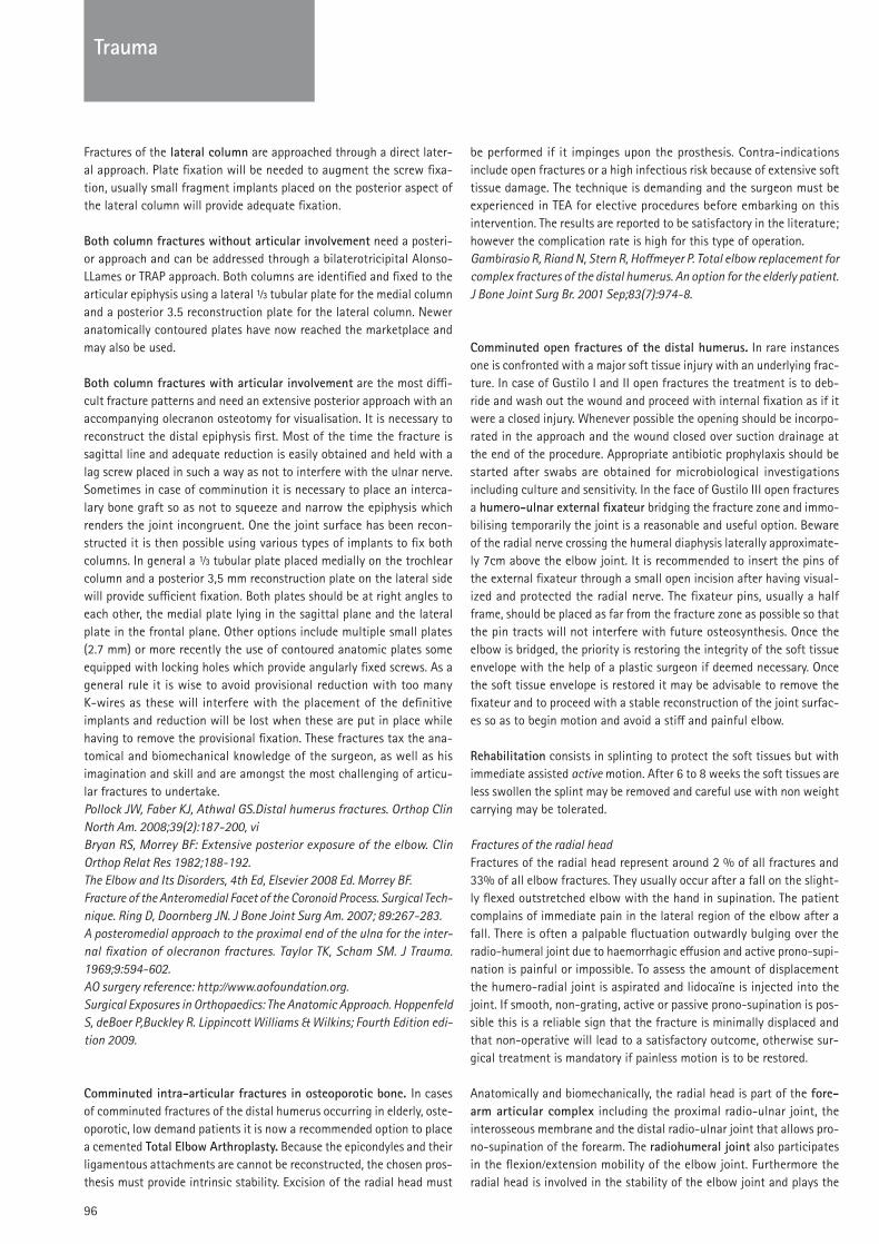
96
Fractures of the lateral column are approached through a direct later-al approach. Plate fixation will be needed to augment the screw fixa-tion, usually small fragment implants placed on the posterior aspect of the lateral column will provide adequate fixation.
Both column fractures without articular involvement need a posteri-or approach and can be addressed through a bilaterotricipital Alonso-LLames or TRAP approach. Both columns are identified and fixed to the articular epiphysis using a lateral 1/3 tubular plate for the medial column and a posterior 3.5 reconstruction plate for the lateral column. Newer anatomically contoured plates have now reached the marketplace and may also be used.
Both column fractures with articular involvement are the most diffi-cult fracture patterns and need an extensive posterior approach with an accompanying olecranon osteotomy for visualisation. It is necessary to reconstruct the distal epiphysis first. Most of the time the fracture is sagittal line and adequate reduction is easily obtained and held with a lag screw placed in such a way as not to interfere with the ulnar nerve. Sometimes in case of comminution it is necessary to place an interca-lary bone graft so as not to squeeze and narrow the epiphysis which renders the joint incongruent. One the joint surface has been recon-structed it is then possible using various types of implants to fix both columns. In general a 1/3 tubular plate placed medially on the trochlear column and a posterior 3,5 mm reconstruction plate on the lateral side will provide sufficient fixation. Both plates should be at right angles to each other, the medial plate lying in the sagittal plane and the lateral plate in the frontal plane. Other options include multiple small plates (2.7 mm) or more recently the use of contoured anatomic plates some equipped with locking holes which provide angularly fixed screws. As a general rule it is wise to avoid provisional reduction with too many k-wires as these will interfere with the placement of the definitive implants and reduction will be lost when these are put in place while having to remove the provisional fixation. These fractures tax the ana-tomical and biomechanical knowledge of the surgeon, as well as his imagination and skill and are amongst the most challenging of articu-lar fractures to undertake.Pollock JW, Faber KJ, Athwal GS.Distal humerus fractures. Orthop Clin North Am. 2008;39(2):187-200, viBryan RS, Morrey BF: Extensive posterior exposure of the elbow. Clin Orthop Relat Res 1982;188-192. The Elbow and Its Disorders, 4th Ed, Elsevier 2008 Ed. Morrey BF.Fracture of the Anteromedial Facet of the Coronoid Process. Surgical Tech-nique. Ring D, Doornberg JN. J Bone Joint Surg Am. 2007; 89:267-283.A posteromedial approach to the proximal end of the ulna for the inter-nal fixation of olecranon fractures. Taylor TK, Scham SM. J Trauma. 1969;9:594-602.AO surgery reference: http://www.aofoundation.org.Surgical Exposures in Orthopaedics: The Anatomic Approach. Hoppenfeld S, deBoer P,Buckley R. Lippincott Williams & Wilkins; Fourth Edition edi-tion 2009.
Comminuted intra-articular fractures in osteoporotic bone. In cases of comminuted fractures of the distal humerus occurring in elderly, oste-oporotic, low demand patients it is now a recommended option to place a cemented Total Elbow Arthroplasty. Because the epicondyles and their ligamentous attachments are cannot be reconstructed, the chosen pros-thesis must provide intrinsic stability. Excision of the radial head must
be performed if it impinges upon the prosthesis. Contra-indications include open fractures or a high infectious risk because of extensive soft tissue damage. The technique is demanding and the surgeon must be experienced in TEA for elective procedures before embarking on this intervention. The results are reported to be satisfactory in the literature; however the complication rate is high for this type of operation.Gambirasio R, Riand N, Stern R, Hoffmeyer P. Total elbow replacement for complex fractures of the distal humerus. An option for the elderly patient. J Bone Joint Surg Br. 2001 Sep;83(7):974-8.
Comminuted open fractures of the distal humerus. In rare instances one is confronted with a major soft tissue injury with an underlying frac-ture. In case of Gustilo I and II open fractures the treatment is to deb-ride and wash out the wound and proceed with internal fixation as if it were a closed injury. Whenever possible the opening should be incorpo-rated in the approach and the wound closed over suction drainage at the end of the procedure. Appropriate antibiotic prophylaxis should be started after swabs are obtained for microbiological investigations including culture and sensitivity. In the face of Gustilo III open fractures a humero-ulnar external fixateur bridging the fracture zone and immo-bilising temporarily the joint is a reasonable and useful option. Beware of the radial nerve crossing the humeral diaphysis laterally approximate-ly 7cm above the elbow joint. It is recommended to insert the pins of the external fixateur through a small open incision after having visual-ized and protected the radial nerve. The fixateur pins, usually a half frame, should be placed as far from the fracture zone as possible so that the pin tracts will not interfere with future osteosynthesis. Once the elbow is bridged, the priority is restoring the integrity of the soft tissue envelope with the help of a plastic surgeon if deemed necessary. Once the soft tissue envelope is restored it may be advisable to remove the fixateur and to proceed with a stable reconstruction of the joint surfac-es so as to begin motion and avoid a stiff and painful elbow.
Rehabilitation consists in splinting to protect the soft tissues but with immediate assisted active motion. After 6 to 8 weeks the soft tissues are less swollen the splint may be removed and careful use with non weight carrying may be tolerated.
Fractures of the radial headFractures of the radial head represent around 2 % of all fractures and 33% of all elbow fractures. They usually occur after a fall on the slight-ly flexed outstretched elbow with the hand in supination. The patient complains of immediate pain in the lateral region of the elbow after a fall. There is often a palpable fluctuation outwardly bulging over the radio-humeral joint due to haemorrhagic effusion and active prono-supi-nation is painful or impossible. To assess the amount of displacement the humero-radial joint is aspirated and lidocaïne is injected into the joint. If smooth, non-grating, active or passive prono-supination is pos-sible this is a reliable sign that the fracture is minimally displaced and that non-operative will lead to a satisfactory outcome, otherwise sur-gical treatment is mandatory if painless motion is to be restored.
Anatomically and biomechanically, the radial head is part of the fore-arm articular complex including the proximal radio-ulnar joint, the interosseous membrane and the distal radio-ulnar joint that allows pro-no-supination of the forearm. The radiohumeral joint also participates in the flexion/extension mobility of the elbow joint. Furthermore the radial head is involved in the stability of the elbow joint and plays the
Trauma

97
role of a secondary stabilizer. If the ulnar collateral ligaments and the distal radioulnar joint are intact, the radial head plays no role in the sta-bility of the elbow and may therefore safely be removed if necessary. However, in the absence of the radial head and disruption of the distal radioulnar ligaments (Essex-Lopresti lesion), the radius will migrate prox-imally and more so when there is an associated tear of the interosseous membrane. In these circumstances a relative over-lengthening of the ulna will occur at the wrist entailing painful dysfunction. Also, in the absence of the radial head, valgus instability will occur at the elbow if the ulnar collateral ligament is torn or elongated.
Fracture classification of radial head fractures:Various classification schemes have been proposed:Mason Classification:
Type I: Non-displacedType II: Displaced marginal fracturesType III: Comminuted fracturesType IV: Associated with elbow dislocation
Hotchkiss modification Type I: No surgery Type II: Displaced but fixableType III: Displaced and unfixable
As a general rule displaced fractures need surgical intervention, mini-mal displacement may benefit from Open Reduction and Internal Fixa-tion (ORIF) and highly displaced or comminuted fractures may necessi-tate excision in case of a stable Distal Radio-ulnar Joint and intact interosseous membrane and prosthetic replacement if these conditions are not met.
ApproachThe approach is basically lateral starting obliquely from the supracondy-lar ridge over the radio-humeral joint and trough the kocher interval between the anconeus and the extensor carpi ulnaris. This protects the motor branch of the radial nerve (Avoid placing a Hohmann type retrac-tor over the anterior neck of the radius) and the approach is sufficient-ly anterior to spare the ulnar collateral ligament which will not be inad-vertently severed. The capsule is revealed and an arthrotomy is performed exposing the radial head. The annular ligament is spared.
TechniqueReconstructible fractures: After assessment they are fixed using small fragment 2.0 or 2.7 AO or Herbert type screws. Sometimes the use of a mini blade plate type of implant may be necessary. Radial head excision: If excision is necessary be sure that all fragments are excised by reconstructing the head on the instrument table. The head should be removed at the level of the annular ligament. The elbow and wrist should be closely assessed for stability and the lateral collateral reconstructed if necessary.
Prosthetic replacement: It is necessary to provide stability by inserting a radial head prosthesis in cases of fracture dislocations with either fron-tal plane instability such as with extensive tearing of the lateral collat-eral ligaments or with longitudinal instability with tearing of the inter-osseous membrane or DRuJ. Today, the accepted prosthesis is metallic with or without a moving or floating (bipolar) head and a stem that may be cemented or non-cemented. In some cases the prosthesis may be left permanently in place while in other instances such as in very young patients it may be useful to remove the prosthetic head used as a tem-porary spacer once healing of the ligamentous complex has occurred. Silastic implants once in vogue are now generally abandoned because of the risk of a destructive synovial inflammatory response due to frag-mentation of the prosthesis leading to the accumulation of irritative particulate matter. Furthermore biomechanical studies have shown that these prostheses are not stiff enough to allow anatomic healing of torn ligaments.
RehabilitationAs general rule rehabilitation must be begun early and motion should be started within days of the intervention in case of operative treatment or diagnosis in case of conservative treatment. After a few days of rest start by gentle active flexion exercises going from 90° to 110° then progress with active extension exercises ranging from 120° to 30° as tolerated. After two to three weeks gentle active prono-supination exer-cises are begun. An articulated brace is useful for protection in cases of instability
The ESSEX LOPRESTI injuryThis injury was described in 1951 by Essex-Lopresti and associates as a severely comminuted fracture of the radial head with tearing of the interosseous membrane and disrupting the DRuJ. The diagnosis is clin-ical and radiological. X-rays of the whole forearm are necessary. The radial head must imperatively be fixed or replaced with a prosthesis and the DRuJ has to be stabilized with a cross pin left in situ for 4 to 6 weeks. (See below).Frankle MA, Koval KJ, Sanders RW, Zuckerman JD: Radial head fractures with dislocations treated by immediate stabilization and early motion J Shoulder Elbow Surg 1999;8:355-356. Cooney WP. Radial head fractures and the role of radial head prosthetic replacement: current update. Am J Orthop. 2008;37(8 Suppl 1):21-5.
Fractures of the olecranonFractures of the olecranon usually occur after falls directly on the elbow point. They are frequently seen in the osteoporotic patient.There are various classifications; the most popular are the Mayo classi-fication:Type I: undisplacedType II: Displaced but stable elbow (Noncomminuted: A /Comminuted: B)Type III: Displaced and unstable elbow (Noncomminuted: A / Comminut-ed: B)
The AO classification (Complex: includes the proximal forearm segment: radius and ulna):A: Extra-articular fractures B: Intra-articular fracturesC: Fractures of both olecranon and radius
A treatment plan must be elaborated. The great majority of these frac-tures are displaced and the question arises as to what is the best suit-
Trau
ma
Trauma

98
ed technique.
ApproachThe surgical approach is straightforward. The ulna is subcutaneous; the patient may be in a supine or lateral decubitus position with the arm resting on a support. A tourniquet may be used. The incision follows the shaft of the ulna and some recommend arcing the incision radially to avoid the tip of the olecranon and also to avoid a scar over the ulnar nerve.
TechniqueClearly transverse fractures are best treated by a technique associating k-wires and tension band cerclage such as described by the AO Group. The technique must be meticulously followed and especially the place-ment of the k-wires must be parallel, 5 to 6 cm long, and the tip should be into the opposite cortex distally to the coronoid and the ends must be bent at 180° and deeply buried into the triceps and olecranon. Com-minuted fractures will require a plating technique (3.5 LCP or DCP plates) augmented by longitudinal screws (so-called homerun screws). If the fracture is oblique and is near the coronoid a compression screw will be most useful. The main drawbacks of these techniques lie in the high reoperation rate that all authors mention. k-wires tend to back out and must be carefully followed and plates usually present some degree of discomfort and are best removed after adequate consolidation. A more recently described complication is the interference of k-wires or screws placed in the proximal ulna with the proximal radius. Some implants are too long and either impinge or are screwed into the radial head or bicip-ital tuberosity. This is not an easy diagnosis post-operatively and free-dom of prono-supination must be carefully ascertained at the end of the surgical reconstruction.
RehabilitationThe patient is placed into a backslab at 80° of flexion and gentle active flexion and extension exercises are started as tolerated. The olecranon is protected for 6 to 8 weeks before any weight bearing exercises are started.
Fractures of the coronoidThis is usually associated with dislocations of the elbow. Regan and Mor-rey have classified these injuries into:Type I: Fracture of the tipType II: Less than 50% of the height of the coronoidType III: More than 50%A and B types signify no or associated dislocation.Some have added a:Type IV: Fracture of the sublime tubercle.
Type I fractures are generally stable and do not need fixation if the elbow is stable. Types III and IV need surgery to insure stability of the elbow
because the medial collateral ligament attaches to the medial coronoid and instability will occur if the bony fragments are not fixed.More recently O’Driscoll has modified this classification: into In this classification all types 2 and 3 need fixation and especially if asso-ciated with a dislocation or a radial head fracture. Plain x-rays and pref-erably a CT scan should be used for making the diagnosis and classify-ing the lesions. Small lesions can be fixed by transolecranon sutures. The fragment is approached from a medial incision in an “Over the top” as approach described by Hotchkiss Large fragments are approached by a posteromedial route.In very unstable elbows a hinged external fixateur device will provide stability while allowing early motion.Regan W, Morrey B. Fractures of the coronoid process of the ulna. J Bone Joint Surg Am 1989;71:1348–1354.)O’Driscoll SW, Jupiter JB, Cohen MS, Ring D, McKee MD. Difficult elbow fractures: pearls and pitfalls. Instr Course Lect 2003;52:113–134.Hotchkiss RN. Fractures and dislocations of the elbow. In: Rockwood CA, Green DP, eds. Rockwood and Green’s fractures in adults. Vol 1. 4th ed. Philadelphia, PA: Lippincott Williams & Wilkins; 1996:929–1024.
Dislocation of the elbow The mechanism is usually a fall on the outstretched hand with the elbow in a varus position. The primary lesion is a tear of the lateral collateral ligament from the lateral humeral insertion and as the mechanism of dislocation the capsule is then torn anteriorly , the coronoid may be damaged by the ram effect of the trochlea and finally the medially col-lateral may be torn also, leading to a very unstable position. In 5 to 10% of cases a fracture of the radial head may be associated as well as more rarely a fracture of the capitellum. Neurovascular injuries occur infre-quently but must be looked for. The median nerve may be stretched by the front riding humerus, and this is the most frequent neurological inju-ry, however the radial nerve and the ulnar nerve may also be damaged. The brachial artery may suffer an intimal tear or a rupture while it is stretched out over the protruding distal humerus and very rarely the skin may split leading to an open injury. A compartment syndrome is always a possibility and the patient must be monitored.Dislocations of the elbow are classified as anterior (rare), posterior (most common) and divergent (very rare the radial head will be separated from the ulna and the annular ligament is torn).The elbow, after proper radiographic and clinical assessment should be reduced, general anaesthesia may be necessary, and tested for stability: varus, valgus and postero-lateral. Postero-lateral rotatory instability occurs when the ulno-radial bloc dislocates off of the humerus lateral-ly in supination and upon reduction in pronation a clunk is heard and felt. The elbow is then flexed to past 90° and held in a splint in prona-tion. An X-ray is then taken to determine that the reduction is adequate. After 5 to 7 days the elbow is moved, first in flexion then extended as
Type 1: Tip fractures Type 2 : Anteromedial fractures Type 3 : Base of coronoid fractures
Trauma

99
tolerated in an active-assisted mode. A hinged splint may be worn and after 3 to 6 weeks all immobilisation if motion has returned and the patient feels stable all splints are removed.Indications for surgery range from incarceration of a bony fragment in the joint space, to vascular impairment or gross instability usually asso-ciated with a coronoid fracture (see above). Late contracture or heter-otopic bone may also lead to surgery at a later stage if mobility is severe-ly limited (>30° of flexion deformity). Late instability may also necessitate surgery and the use of a hinged fixator allowing stable dis-traction of the joint surfaces and concomitant mobilisation.O’Driscoll SW, Morrey BF, Korinek S, An KN. Elbow subluxation and dislo-cation: A spectrum of instability. Clin. Orthop. Relat. Res. 280:186, 1992
Medial instability of the elbowThrowing athletes may develop medial instability due to medial ulnar collateral ligament (MuCL) stretching out or tearing. The patient may experience a pop or a tearing sensation during a throw. Physical exam-ination includes looking for ulnar neuritis and Tinel’s sign. The elbow is stressed in valgus at 25° of flexion and the MuCL is palpated for taught-ness. Further diagnostic imaging using plain stress-test x-rays, dynam-ic ultrasound or Arthro-MRI will fine tune the diagnosis. MuCL recon-struction using a figure of eight tendon graft as described originally by F Jobe and refined and modified more recently may then be performed.Safran M, Ahmad CS, El Attrache NS. Ulnar collateral ligament of the elbow. Arthroscopy 21:1381, 2005.
Postero-lateral rotatory instability of the elbowAfter injury or dislocation of the elbow the patient may develop a con-dition where recurrently he has the impression of the elbow popping or giving way or even dislocating. The symptoms are on the lateral side where the patient often has pain and discomfort. Clinical testing will reproduce the sensation of pain and instability when the elbow is stressed in valgus and supination. An audible pop can occur during this manoeu-vre. It signifies that the radius and the ulna although firmly attached by the annular ligament, slip out laterally as a unit from the capitellum because of a tear of the ulnar lateral collateral ligament that laterally unites the humerus to the supinator crista of the lateral ulna. Repair may be accomplished by a tendon graft uniting the humerus to the supi-nator crista of the ulna and passing under the radial head.Nestor BJ, O’Driscoll SW, Morrey BF. Ligamentous reconstruction for pos-terolateral rotatory instability of the elbow: J Bone Joint Surg Am. 1992;74(8):1235-41.
The stiff elbowThe normal elbow has a range from 0° or slightly hyperextended to 150° of flexion, pronation is 75° and supination is 85°. The stiff elbow becomes a clinical problem when the functional arc accepted in flexion/extension diminishes beyond 130°-30°- 0. Very severe stiffness occurs when the total arc is less than 30°, severe stiffness is when the arc is between 31° and 61°, moderate between 61° and 90° and minimal when the arc is greater than 90°. A 100° range of pron/supination (50° pronation and 50° of supination) is necessary for normal function although as a rule lack of pronation is in general less tolerated than lack of supination. If no bony abnormalities are present the lateral column procedure, where the anterior and posterior contracted capsule is excised from a lateral approach after detaching the distal fibers of the brachioradialis and the extensor carpi radialis longus is recommended. Medial release detach-ing the flexor-pronator mass is performed in case of arthritic osteo-phytes, caring for the ulnar nerve. It may be combined with the lateral
column procedure. For the rehabilitation it is important to immobilize during night-time the elbow in the position of greatest motion loss. If extension is to be gained the elbow should be immobilized in extension during night-time and flexion during the day.For more complex conditions with bony deformity, ectopic bone, major osteophytes overgrowth or posttraumatic conditions a posterior approach with of sculpturing of deformed bony surfaces, excision of new bone formation and sectioning of restraining tissues will have to be performed. In some of these cases a hinged uni or bilateral humero-ulnar external fixator allowing controlled motion will need to be used. The ulnar nerve will need special care and transposition may be indicated in some cas-es. The radial nerve may be at risk when external fixation is used.Some authors in cases of minimal or moderate stiffness have used arthro-scopic release techniques Ball CM, Meunier M, Galatz LM, Calfee R, Yamaguchi K. Arthroscopic treatment of post-traumatic elbow contracture. J Shoulder Elbow Surg 2002;11:624-629.Mansat P, Morrey BF. The column procedure: A limited lateral approach for extrinsic contracture of the elbow. J Bone Joint Surg Am 1998;80:1603-1615.Morrey BF. The posttraumatic stiff elbow. Clin Orthop Relat Res 2005;26-35.
Tendon ruptures and athletic injuries Distal Biceps Tendon RupturesThe distal biceps is the most commonly ruptured tendon around the elbow. This usually occurs with heavy lifting. The patient reports hear-ing a pop or a crack in the anterior region of his elbow. In the hours that follow the injury an ecchymosis may discolour the antecubital fold. The biceps muscle belly does not retract immediately because it is held down by the lacertosus fibrosus. The patient will have near normal flexion extension strength but will complain of weakness in supination. In an active population the treatment is usually surgical and a two incision reattachment technique as described by Morrey yields satisfactory results. When using this technique care must be taken not to come into contact with the proximal ulna when bringing the distal biceps through the ulno-radial space so as to avoid an osseous synostosis. Gentle flex-ion-extension exercises follow the surgery and at 6 weeks a full return to activity is permitted.Rupture of the brachialis and of the triceps tendons have been report-ed. These are rare injuries and the best surgical treatment consists in suturing the ruptured tendons. Papandrea RF: Two-incision distal biceps tendon repair, in Yamaguchi K, King GJW, McKee O’Driscoll SW : Advanced Reconstruction Elbow. Rose-mont, IL, American Academy of Orthopaedic Surgeons, 2006, pp 121-128. Lateral Epicondylitis (Tennis Elbow)The most comprehensive description of the pathoanatomy of epicondyli-tis is Nirschl’s. The essentially this is an overuse lesion causing tearing of the extensor carpi radialis brevis tendon at its distal humerus inser-tion. Diagnosis is made by eliciting pain on palpation of the lateral epi-condyle, wrist extension against resistance as long finger extension against resistance will also produce pain at the elbow in case of epi-condylitis. All other conditions leading to elbow pain such as carpal tun-nel, radial nerve entrapment under the arcade of Frohse or radiohumer-al arthritis should be eliminated. Adjunct imaging such as plain x-rays will not be specific and MRI may be used to image a tear or an edema-
Trau
ma
Trauma

100
tous area in the region of insertion. Treatment consists of modifying activity, steroidal infiltration, adapted physiotherapy and in case of a long duration of symptoms surgical excision of the ECRB tendon, situ-ated under the Extensor Carpi Radialis Longus tendon. Most authors rec-ommend open procedures although success has been reported using arthroscopic techniques. A characteristic angiofibroblastic hyperplasia-tendinosis has been described by Nirschl which characteristically dem-onstrates little inflammatory cells. Postoperative treatment consists of a protective splint followed by gentle motion as tolerated with full func-tion possible 6 to 8 weeks postoperatively. Kraushaar BS, Nirschl RP. Tendinosis of the elbow (tennis elbow). Clinical features and findings of histological, immunohistochemical, and electron microscopy studies. J Bone Joint Surg Am 1999;81:259-278. Nirschl RP, Pettrone F. Tennis elbow: The surgical treatment of lateral epi-condylitis. J. Bone Joint Surg Am. 1979; 61:832.
Medial Epicondylitis Rarely, in the competitive athlete pain will develop following overuse of the flexor-pronator complex. Again the treatment should first be con-servative. If symptoms persist surgical excision of the diseased part of the medial conjoint tendon of the flexor-pronator complex may be con-sidered. In some cases a transposition of the ulnar nerve completes the procedure. Vangsness CT, Jobe FW. Surgical treatment of medial epicondylitis. Results in 35 elbows. J Bone Joint Surg Br. 1991;73(3):409-11.
Osteochondritis DissecansRare condition affecting mostly skeletally immature patients involved in receptive throwing sports. Symptoms include pain, flexum deformity or catching and locking. This should be differentiated from Panner disease which is an osteochondrosis not requiring treatment. A classification has been evolved that spans from the simple cartilage fissures (I) to the detachment of a large fragment (IV) of cartilage. Treatment is at first conservative with activity modification and if not successful can go, depending on severity, from simple drilling of the lesion to complex mosaicplasty. Baumgarten TE, Andrews JR, Satterwhite YE: The arthroscopic classifica-tion and treatment of osteochondritis dissecans of the capitellum. Am J Sports Med 1998;26:520-523.
Septic olecranon bursitisThis is a potentially life threatening condition caused by a septicaemia originating from an infected bursa under tension. Generally seen in debil-itated patients but can arise without a clear cause or after minor trau-ma in an otherwise healthy individual. Diagnosis is clinical with stand-ard laboratory findings such as high white cell count, left shift of white blood cells, high sedimentation rate and elevated C-reactive protein lev-els. Organisms found are generally staphylococcus or streptococcus. In mild cases treatment may be antibiotics and splinting. In more severe cases it is mandatory to incise the bursa and leave open, to immobilise in a splint and after a few days to perform a closure secondarily. During the treatment period appropriate IV and oral antibiotics are adminis-tered.Pien FD, Ching D, Kim E. Septic bursitis: Experience in a community prac-tice. Orthopaedics 1991; 14:981Infectious olecranon and patellar bursitis: short-course adjuvant antibi-otic therapy is not a risk factor for recurrence in adult hospitalized patients. Perez C, Huttner A, Assal M, Bernard L, Lew D, Hoffmeyer P, Uçkay I. J Antimicrob Chemother. 2010 Mar 1. [Epub ahead of print]
ForearmIn the AO manual that covers all important fixation techniques the read-er will fact pertinent facts related to the topic at hand: AO Principles of Fracture Management. Second expanded edition, TP Rüedi, RE Buckley, CG Moran. Georg Thieme Verlag; 2 Har/Dvdr edition (2007). For the lat-est in fracture fixation techniques the reader is also invited to visit the AO surgery reference site: AO surgery reference: http://www.aofounda-tion.org. Another most useful publication containing pertinent facts related to orthopaedics and musculoskeletal in general trauma is the AAOS Comprehensive Orthopaedic Review. Jay R. Lieberman, MD, Editor 2009 Rosemont, IL, American Academy of Orthopaedic Surgeons. For sur-gical approaches the most useful reference is without doubt: Surgical Exposures in Orthopaedics: The Anatomic Approach. Hoppenfeld S, deBo-er P, Buckley R. Lippincott Williams & Wilkins; Fourth Edition 2009.
Introduction forearmThe forearm must be considered as a whole functioning joint allowing pronation of 75° and supination of 85°. The interosseous membrane plays a major stabilising role.AAOS Comprehensive Orthopaedic Review. Jay R. Lieberman, MD, Editor 2009 Rosemont, IL, American Academy of Orthopaedic Surgeons.
Classification of forearm fracturesThe AO classification is popular. It classifies the fracture patterns of radi-al and ulnar shafts as type A (simple, transverse or spiral) type B (wedge with a butterfly fragment) and type C (segmental or comminuted frag-ments). Open fractures are classified according to Gustilo and Anderson: Type I inside-out (< 1 cm), Type II outside-in (> 1cm), Type III A (open but osseous coverage possible), type III B ( open necessitating a local or free flap), Type III C any open fracture with vascular injury. Specific to the forearm are the Monteggia fracture pattern (Fracture of the ulna with dislocation of the radial head). Bado classification:
I Anterior radial head dislocation and proximal ulnar shaft frac-ture (apex anterior)II Posterior or postero-lateral radial head dislocation and proxi-mal ulnar shaft fracture (apex posterior)III Lateral radial head dislocation and proximal ulnar shaft frac-ture (apex posterior)IV Anterior radial head dislocation and proximal ulnar and radi-al shaft fracture (apex posterior)
Konrad GG, Kundel K, Kreuz PC, Oberst M, Sudkamp NP. Monteggia frac-tures in adults: long-term results and prognostic factors. J Bone Joint Surg Br. 2007;89(3):354-60.
Trauma
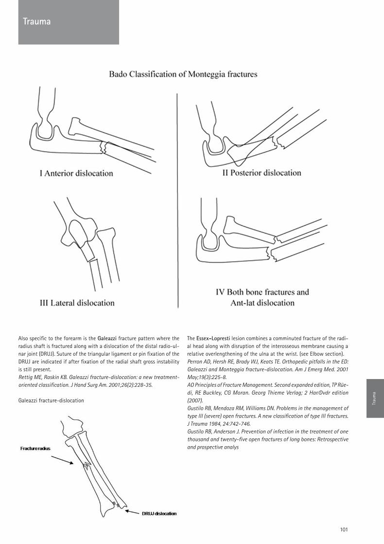
101
Also specific to the forearm is the Galeazzi fracture pattern where the radius shaft is fractured along with a dislocation of the distal radio-ul-nar joint (DRuJ). Suture of the triangular ligament or pin fixation of the DRuJ are indicated if after fixation of the radial shaft gross instability is still present.Rettig ME, Raskin KB. Galeazzi fracture-dislocation: a new treatment-oriented classification. J Hand Surg Am. 2001;26(2):228-35.
The Essex-Lopresti lesion combines a comminuted fracture of the radi-al head along with disruption of the interosseous membrane causing a relative overlengthening of the ulna at the wrist. (see Elbow section).Perron AD, Hersh RE, Brady WJ, Keats TE. Orthopedic pitfalls in the ED: Galeazzi and Monteggia fracture-dislocation. Am J Emerg Med. 2001 May;19(3):225-8.AO Principles of Fracture Management. Second expanded edition, TP Rüe-di, RE Buckley, CG Moran. Georg Thieme Verlag; 2 Har/Dvdr edition (2007).Gustilo RB, Mendoza RM, Williams DN. Problems in the management of type III (severe) open fractures. A new classification of type III fractures. J Trauma 1984, 24:742-746.Gustilo RB, Anderson J. Prevention of infection in the treatment of one thousand and twenty-five open fractures of long bones: Retrospective and prospective analys
Galeazzi fracture-dislocation Trau
ma
Trauma

102
Clinical presentation of forearm fracturesA deformed extremity is present. Look for neurovascular injuries (radial nerve) carefully before any manipulation of the injured extremity. Plain X-rays including the shoulder and elbow are generally sufficient in acute traumatic cases. MRI, CT, Bone scintigraphy are useful in special situa-tions such as chronic infection, metastatic or primary tumors.
Conservative treatmentThere is practically no place for conservative treatment in adult both bone forearm fractures. Isolated fractures of the ulnar shaft may be treated by functional bracing but the rate of non-union remains high and many authors recommend immediate plate fixationMackay D, Wood L, Rangan A. The treatment of isolated ulnar fractures in adults: a systematic review. Injury. 2000;31(8):565-70.
Surgical ApproachesAnterior (Henry) approach �Anatomic approach but with some soft tissue stripping. Allows exposure of the whole radius.Dorsal Thompson approach �Danger to the Posterior Interosseous Nerve (PIN). http://www.wheelessonline.com/ortho/dorsal_approach_thomp-sonDirect approach �The direct approach is best suited for the ulna.
Mekhail AO, Ebraheim NA, Jackson WT, Yeasting RA. Vulnerability of the posterior interosseous nerve during proximal radius exposures. Clin Orthop Relat Res. 1995 Jun;(315):199-208.Erratum in: Clin Orthop 1997;(334):386.
Operative treatment indications: The forearm constitutes a joint and in the adult the treatment is anatomical reduction and fixation
IM Nailing �Difficult to guarantee stable fixation and anatomic fixation with these devices.Anterograde -Nails for the ulna are in use and being developedRetrograde -Nails for the radius may be usedPlating �3,5 mm plates should be used and never semi or third tubular
type plates. 6 cortices on each side of the fracture should be used. Some authors
Both bones fracture of the forearm fixed anatomically with rigid com-pression plating
External fixationIn case of open fractures an external fixation may be applied. For the ulna the pins may be applied closed but for the proximal radius an open approach allowing to identify the pertinent neurovascular structures should be performed.Hertel R, Pisan M, Lambert S, Ballmer FT. Plate osteosynthesis of diaphy-seal fractures of the radius and ulna. Injury. 1996;27(8):545-8.Chapman MW, Gordon JE, Zissimos AG. Compression-plate fixation of acute fractures of the diaphyses of the radius and ulna. J Bone Joint Surg Am. 1989;71(2):159-69.Lindvall EM, Sagi HC. Selective screw placement in forearm compression plating: results of 75 consecutive fractures stabilized with 4 cortices of screw fixation on either sideof the fracture. J Orthop Trauma. 2006;20(3):157-62; discussion 162-3.
Essex-Lopresti fracture-dislocation
Trauma

103
Notes

104
Notes