new and rare metridia from antarctic and subantarctic ... · new and rare metridia from antarctic...
TRANSCRIPT

О Zoological Institute, St.Petersburg, 2001
New and rare Metridia from Antarctic and Subantarctic waters (Copepoda, Calanoida: Metridinidae)
E.L. Markhaseva
Markhaseva, E.L. 2001. New and rare Metridia from Antarctic and Subantarctic waters (Copepoda, Calanoida: Metridinidae) Zoosystemalica Rossica, 9(1), 2000: 43-75.
Metridia ferrarii sp. n. and M. pseudoasymmetrica sp n. are described from females and males, both species collected in the South Atlantic and South-Eastcrn Pacific (Southern Ocean) Redescriptions are given for M. asymmetrica Brodsky, 1950, M. ornata Brodsky, 1950, M. princeps Giesbrecht, 1889 and M. macrura Sars, 1905. Females of M. ferrarii are clearly distinguished from the related metridinids in the presence of collar on both sides of cephalosome; males differ from those of M. princeps in the strongly asymmetrical P5 left coxopod, absence of pointed attenuations on left and right second segments of antennule and some other characters. M. ferrarii differs from M. ornata mostly in the longer caudal rami, longer hook-like spines at P2 Enp and the shape of the genital somite. Females of M. pseudoasymmetrica differ from those of the related species M. asymmetrica mainly in the shape and location of spermathecae (oval and overlapping in lateral view in M. psedoasym-metrica vs. round and not overlapping in M. asymmetrica) and in presence of distinct indentation of the genital somite (in lateral view); males of M. pseudoasymmetrica are very similar to those of M. asymmetrica, but differ in setation of left P5 and absence of short spinules at the second urosomal somite on the right and presence of short hairs at the fourth urosomal somite on the left.
E.L. Markhaseva, Zoological Institute, Russian Academy of Sciences, Universiletskayanab. 1, St.Petersburg 199034, Russia
The genus Metridia includes 21 species (Bradford-Grieve, 1999), plus M. andraeana and M. trispinosa so poorly described by Brady (1918) that their status is not clear. Six species in the genus Metridia are characterized as large-sized, between 5.8 mm (M. ignota Esterly, 1906) and 10.5 mm (M. macrura Sars, 1905). Total lengths of other species typically are about 2-3 mm; 4.5 mm of M. longa (Lubbock, 1854) and M. okhotensis Brodsky, 1950 are the largest mean in this group. Large-sized species of Metridia are difficult to identify, due to incomplete original descriptions and a limited number of figures.
Eight Metridia species, of them the large-sized M. princeps Giesbrecht, 1889 and M. macrura Sars, 1905, have been reported from the Antarctic and Subantarctic waters by Ra-zouls (1995) and Seret (1979) respectively.
Metridinids from the samples collected in the 4th, 5th, 8-12th and 23rd cruises of Eltanin south of 55°S in the South Atlantic and South-Eastern Pacific (Southern Ocean) and deposited in the National Museum of Natural History, Smithsonian Institution (Washington,
D.C.) were examined (Table 1). Six known species of Metridia were found in the collection: M. curticauda Giesbrecht, 1889, M. ger-lachei Giesbrecht, 1902, M. lucens Boeck, 1864, M. ornata Brodsky, 1950 (recorded from Antarctic and Subantarctic for the first time), M. princeps Giesbrecht, 1889, and M. venusta Giesbrecht, 1889. Two new species, M. ferrarii sp. n. and M. pseudoasymmetrica sp. п., are described here. The latter is related to M. asymmetrica Brodsky, 1950, which is redescribed here.
A redescription of M. ornata Brodsky, 1950 and of M. asymmetrica Brodsky, 1950 are given from the type specimens at the Zoological Institute, Russian Academy of Sciences (St.Petersburg); brief redescriptions of M. princeps Giesbrecht, 1889 and M, macrura Sars, 1905 are given,to make possible comparisons with other large-sized Metridia species (M. alata Roe, 1975; M. bicornuta Davis 1949 and M. ignota Esterly, 1906).
The following abbreviations are used in the descriptions: Al, antennule; A2, antenna; Enp, endopod; Exp, exopod; Gn, gnathobase; Md, mandible; Mdp, mandibular palp; Mxl, maxil-

44 E.L. Markhaseva: Antarctic Metridia • ZOOSYST. ROSSICA Vol. 9
Cruise Station Date Coordinates Depth (m)
Species
4 99 12.07.1962 51°30'S 77°35'W 1208-1219 M. princeps (2 9, 1 d") 123 28 07.1962 57°09'S 63°43'W 2439 M. ferrarii (6 9, 2 d")
M pseudoasymmetrica (5 9) 149 12.08.1962 58°31'S 65°17'W 2105 M. ornata (1 9)
M. ferrarii (3 9) M. pseudoasymmetrica (4 9, 1 a")
154 16.08.1962 56°43'S 64°28'W 2105 M. ferrarii (3 9) M. pseudoasymmetrica (2 9)
5 262 19.10.1962 62°26'S 67°45'W 2428 M. ornata (3 9, 3 d) 296 28.10.1962 63°57'S 71°19'W 2489 M. ferrarii (1 9)
8 578 19.04.1963 57°17'S 27°22'W 1464-1867 M. ferrarii (1 9) 580 21.04.1963 57°23'S 2 3 ° i r W 3074 M. ornata (3 9, 3 a")
M. ferrarii (1 9) 636 20.05.1963 59°37'S 24°28'W 5722-5856 M. pseudoasymmetrica (1 9, 1 d")
9 687 26 08.1963 55°24'S 37°57'W 2214 M. ornata (1 9, 3 d \ 1 CV) M. ferrarii (1 9) M. pseudoasymmetrica (2 9, 1 <f)
10 868 25.11.1963 57°06'S 78°56'W 997-1230 M. ferrarii (2 9) M. princeps (3 9, 1 CIV)
874 27.11.1963 56°06'S 79°04'W 1491 M. ferrarii (2 9, 1 cf) 11 891 04.01.1964 59°50'S 114°53'W 1347-1702 M. ferrarii (1 9)
895 06.01.1964 60°48'S 1 14 051'W 2315 M. ferrarii (14 9, 6 d") M. princeps (1 9, 1 CV)
901 07.01.1964 62°11'S I15°02'W 3477-3678 M. ornata (5 9 , 3 d") M. ferrarii (2 9, 1 c?-) M pseudoasymmetrica (7 9)
918 15.01.1964 66°44'S 115°13'W 1885 M. ferrarii (10 9. 5 a - ) Л/. pseudoasymmetrica (8 9, 2 d")
941 23.01 1964 70°01'S 98°43'W 2562 M ornata ( 1 9 ) M. ferrarii (\ 9, 1 d")
944 24.01.1964 69°06'S 95°02'W 3029 M ornata (4 9 , 2 c/) M. ferrarii Q 9, 1 cO M pseudoasymmetrica (2 9)
12 1014 19.03.1964 65°08'S 47°45'W 1025-1153 M. ferrarii (1 9) M. pseudoasymmetrica (\ 9)
23 1610 07.04 1966 63°28'S 94°I3'W 1250 M ferrarii (4 9, 1 cf) M. pseudoasymmetrica (\ 9)
1615 09.04.1966 62°13'S 95°39'W 800-1025 M princeps (2 9) 1666 26.04.1966 62°30'S 108°35'W 1783-2117 M. ferrarii (3 9, 1 d*)
lule; Мл7 Z./7, praecoxal arthrite (= first internal lobe); M J 7 L / 2 , coxal endite (= second internal lobe); Л/х/ basal endites (= third and fourth internal lobes); МсУ Z-е/, coxal epipodite (= first external lobe); Л/х/ £e2, basal exite (= second external lobe); Mx2, maxilla; Mx2 Lil-2, praecoxal endites (= first and second lobes); Mx2 ЫЗ-4, coxal endites (= third and fourth lobes); Mx2 Ы5-6, basal endites (= fifth and sixth lobes); Mxp, maxilliped; PI-P4, swimming legs 1-4; Pr, prosome; Ur, urosome.
The names of the institutions in which the material is deposited are abbreviated as fol
lows: USNM - National Museum of Natural History, Smithsonian Institution, Washington, D.C.; ZISP - Zoological Institute, Russian Academy of Sciences, St.Petersburg.
All scale lines equal 0.1 mm. Small italic numbers on the figures of antennule designate the successive number of articulated segments.
Metridia ferrarii sp.n.
(Figs 1-59)
Holotype. 9, 9.5 mm, 69°06'S, 95°02'W, from IK.MWT tows taken in Eltanin cruise 11, Sta. 944, 3029 m vertical haul, 24.1.1964, USNM No. 296429.
Table 1. ELTANIN stations where large-sized species of Metridia and M. pseudoasymmetrica sp n. were collected
ZOOSYST. ROSSICA Vol. 9 E.L. Markhaseva: Antarctic Metridia 45
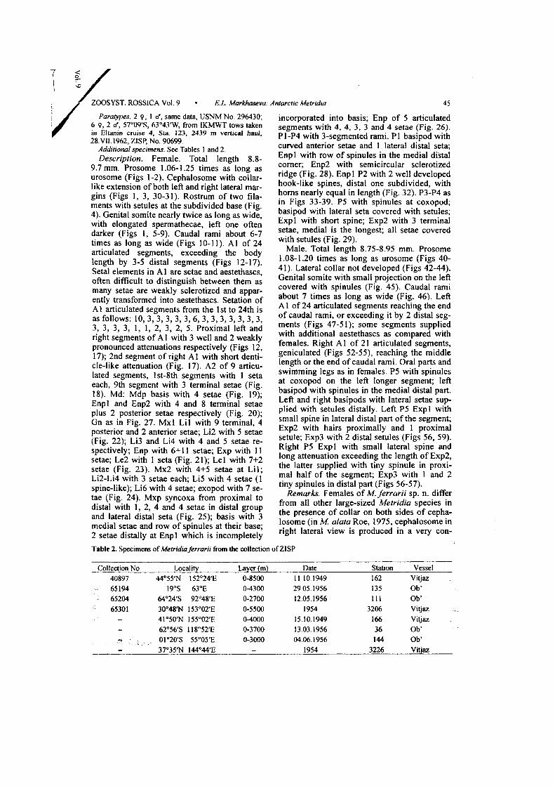
Paratypes. 2 9, 1 d*, same data, USNM No. 296430; 6 9, 2 rf, 57°09'S, 63°43'W, from IKMWT tows taken in Eltanin cruise 4, Sta. 123, 2439 m vertical haul, 28.VII. 1962, ZISP, No. 90699.
Additional specimens. See Tables 1 and 2. Description. Female. Total length 8.8-
9.7 mm. Prosome 1.06-1.25 times as long as urosome (Figs 1-2). Cephalosome with collarlike extension of both left and right lateral margins (Figs 1, 3, 30-31). Rostrum of two filaments with setules at the subdivided base (Fig. 4). Genital somite nearly twice as long as wide, with elongated spermathecae, left one often darker (Figs 1, 5-9). Caudal rami about 6-7 times as long as wide (Figs 10-11). A l of 24 articulated segments, exceeding the body length by 3-5 distal segments (Figs 12-17). Setal elements in A l are setae and aestethascs, often difficult to distinguish between them as many setae are weakly sclerotized and apparently transformed into aestethascs. Setation of Al articulated segments from the 1st to 24th is as follows: 10, 3 , 3 , 3, 3, 3, 6, 3, 3, 3, 3, 3, 3 , 3 , 3, 3, 3, 3, 1, 1, 2 , 3 , 2, 5. Proximal left and right segments of A l with 3 well and 2 weakly pronounced attenuations respectively (Figs 12, 17); 2nd segment of right A l with short denticle-like attenuation (Fig. 17). A2 of 9 articulated segments, lst-8th segments with 1 seta each, 9th segment with 3 terminal setae (Fig. 18). Md: Mdp basis with 4 setae (Fig. 19); Enpl and Enp2 with 4 and 8 terminal setae plus 2 posterior setae respectively (Fig. 20); Gn as in Fig. 27. Mxl Lil with 9 terminal, 4 posterior and 2 anterior setae; Li2 with 5 setae (Fig. 22); Li3 and Li4 with 4 and 5 setae respectively; Enp with 6+11 setae; Exp with 11 setae; Le2 with 1 seta (Fig. 21); Lei with 7+2 setae (Fig. 23). Mx2 with 4+5 setae at Li l ; Li2-Li4 with 3 setae each; Li5 with 4 setae (1 spine-like); Li6 with 4 setae; exopod with 7 setae (Fig. 24). Mxp syncoxa from proximal to distal with 1, 2, 4 and 4 setae in distal group and lateral distal seta (Fig. 25); basis with 3 medial setae and row of spinules at their base; 2 setae distally at Enpl which is incompletely
incorporated into basis; Enp of 5 articulated segments with 4, 4, 3 , 3 and 4 setae (Fig. 26). P1-P4 with 3-segmented rami. PI basipod with curved anterior setae and 1 lateral distal seta; Enpl with row of spinules in the medial distal corner; Enp2 with semicircular sclerotized ridge (Fig. 28). Enpl P2 with 2 well developed hook-like spines, distal one subdivided, with horns nearly equal in length (Fig. 32). P3-P4 as in Figs 33-39. P5 with spinules at coxopod; basipod with lateral seta covered with setules; Expl with short spine; Exp2 with 3 terminal setae, medial is the longest; all setae covered with setules (Fig. 29).
Male. Total length 8.75-8.95 mm. Prosome 1.08-1.20 times as long as urosome (Figs 40-41). Lateral collar not developed (Figs 42-44). Genital somite with small projection on the left covered with spinules (Fig. 45). Caudal rami about 7 times as long as wide (Fig. 46). Left A1 of 24 articulated segments reaching the end of caudal rami, or exceeding it by 2 distal segments (Figs 47-51); some segments supplied with additional aestethascs as compared with females. Right A l of 21 articulated segments, geniculated (Figs 52-55), reaching the middle length or the end of caudal rami. Oral parts and swimming legs as in females. P5 with spinules at coxopod on the left longer segment; left basipod with spinules in the medial distal part. Left and right basipods with lateral setae supplied with setules distally. Left P5 Expl with small spine in lateral distal part o f the segment; Exp2 with hairs proximally and 1 proximal setule; ЕхрЗ with 2 distal setules (Figs 56, 59). Right P5 Expl with small lateral spine and long attenuation exceeding the length of Exp2, the latter supplied with tiny spinule in proximal half o f the segment; ЕхрЗ with 1 and 2 tiny spinules in distal part (Figs 56-57).
Remarks. Females of M. ferrarii sp. n. differ from all other large-sized Metridia species in the presence o f collar on both sides o f cephalosome (in M. alata Roe, 1975, cephalosome in right lateral view is produced in a very con-
Table 2. Specimens of Metridia ferrarii from the collection of ZISP
Collection No. Locality Layer (m) Date Station Vessel 40897 44°55'N 152°24'E 0-8500 11.10.1949 162 Vitjaz 65194 19°S 63°E 0-4300 29.05.1956 135 Ob-65204 64°24'S 92°48'E 0-2700 12.05.1956 111 Ob' 65301 30°48Т^ 153°02'E 0-5500 1954 3206 Vitjaz
- 41°50ТМ I55°02'E 0-4000 15.10.1949 166 Vitjaz
- 62°56'S 118°52'E 0-3700 13.03.1956 36 Ob'
- 01°20'S 55°05'E 0-3000 04.06.1956 144 Ob' - 37°35Тч1 144°44'E - 1954 3226 Vitjaz

46 E.L. Markhaseva: Antarctic Metridia • ZOOSYST. ROSSICA Vol. 9
spicuous wing; however, it differs significantly in shape from that in M. ferrarii).
Females of M. ferrarii differ from females of M. ornata Brodsky, 1950 in the much longer spermathecae (Figs 5-9; cf. Figs 196-197, 199-203) and larger hook-like spines at P2 Enpl (Fig. 32; cf. Figs 213-214). They differ from females of M. princeps in the less swollen genital somite (Figs 6-9; cf. Fig. 153); presence of a short denticle-like attenuation on the second segment of right Al (Figs 12, 17; cf. Fig. 157), and absence of short spinules on the lateral distal part of PI Enp2 (Fig 32; cf. Fig. 162). Males of the new species differ from those of M princeps in the strongly asymmetrical P5 left coxopod (Fig. 56), much longer than right (nearly symmetrical in M. princeps, Fig. 176); absence o f sharp attenuation on left and right second segments of A l (Figs 47, 52) (at least one of A l is with sharp attenuation in M. princeps, Figs 177, 183); absence of suture in the left P5 ЕхрЗ (Fig. 57) (the suture subdividing the segment into more or less sclerotized parts is present in M. princeps, Fig. 175); presence of spinules only at left P5 coxopod (Fig. 56) (in M. princeps, spinules present on both left and right coxopod, Fig. 176). Males of M.
ferrarii differ from those of M. ornata mostly in the longer caudal rami (length/width ratio about 7.0 in M. ferrarii vs. 4.8-6.6 in M. ornata), longer hook-like spines at P2 Enp compared to those in M. ornata (Fig. 226), and shape of genital somite (Fig. 45; cf. Fig. 224).
Type locality. Antarctic, 69°06'S, 95°02'W. Etymology. The name honours Frank D. Fer
rari's contributions to the systematics of ca-lanoid copepods.
Metridia pseudoasymmetrica sp. n. (Figs 60-110)
Holotype. 9, 3.8 mm, 69°06'S, 95°02'W, from IKMWT tows taken in Eltanin cruise 11, Sta. 944, 3029 m vertical haul, 24.1.1964, USNM No. 296447.
Paratypes. 1 9, 3.8 mm, same data, USNM No. 296448; 4 9 (3.31, 3.39, 3 42, 3.6 mm), 1 <t (2.87 mm), 58°31'S, 65°17'W, from IKMWT tows taken in Eltanin cruise 4, Sta. 149, 2105 m vertical haul, 12.VIII. 1962, USNM No. 296449; 8 9 (3.55, 3.6, 3.7, 3.8 mm), 2 <f (3.45 and 3.5 mm), 66°44'S, 115°13'W, from IKMWT tows taken in Eltanin cruise 11, Sta 918, 1885 m vertical haul, 15.1.1964, ZISP No 90700
Additional material. See fable I. Description. Female. Total length 3.31-3.80
mm. Prosome 1.6-2.3 times as long as urosome, similar in shape to that of M. asymmetrica (Fig. 111). Genital somite nearly twice as long as wide, with asymmetrical oval spermathecae, left one situated more anterior than
right one (Figs. 60-66). Caudal rami about 1.7 times as long as wide (Fig. 60). A l of 24 articulated segments reaching Ur3 or the end of caudal rami; 1st proximal segment of A l without well pronounced attenuations; 2nd, 4th, 5th and 6th segments with short sharp attenuation each (Figs 79-80). Setation of Al segments from proximal to distal is as follows: 10, 4, 4, 4, 4, 4, 6, 4, 3, 4, 3, 3, 3, 3, 3, 3, 3, 3, 1, 1, 2, 3, 6. A2 of 9 articulated segments; lst-8th segments with 1 seta each; terminal segment with 3 terminal setae (Figs 71-72 ). Md: basis with 4 setae; Enpl and Enp2 with 4 and 8 terminal plus 2 posterior seta respectively; Gn as in Fig. 73. Mxl Lil with 9 terminal, 4 posterior and 2 anterior setae; Li2 with 5 setae; Li3 and Li4 with 3 and 5 setae respectively; Enp with 6+10 setae; Exp with 11-12 setae (Figs 74-75); Le2 with 1 small short seta; Lei with 7+2 setae (Fig. 74). Mx2 with 4+5 setae at Li l ; Li2-Li4 with 3 setae each; Li5 with 4 setae (1 spinelike); Li6 with 3 setae (Fig. 77); Exp with 7 setae (Fig. 76). Mxp syncoxa from proximal to distal with 1, 2, 4 and 4 setae medially and lateral distal seta; basis with 3 medial setae and row of spinules at their base; Enpl with 2 setae; Enp2-6 with 4, 4, 3, 3 and 4 setae (Fig. 78). P1-P4 with 3-segmented rami (Figs 67, 86-89, 91). PI basipod with curved anterior setae and distal lateral seta; Enpl with row of hairs in the outer corner (Fig 67). Enpl P2 with I well developed, subdivided, hook-like spine and proximal triangular attenuation o f the segment (Figs 83-86). P3-P4 as in Figs 87-91. P5 without spinules at coxopod; basipod with thin lateral seta; Expl usually without, rarely with seta (Fig. 69); Exp2 with 3 terminal setae, medial is the longest, usually they have setules, but in some specimens are nude (Figs 68, 70).
Male. Total length 2.87-3.5 mm. Prosome 1.4-1.5 times as long as urosome (Figs 92-93). Urosome with small spinulated swellings on the left side of Ur2-Ur4 (Fig. 94). Caudal rami about twice as long as wide (Fig. 94). Left A1 20-segmented, geniculated, reaching the end of caudal rami (Figs 95-99). Shape of P5 segments strongly varies depending on view (Figs 100-109). Left P5 Expl with long attenuation in the medial part of the segment; Exp2 with small spine and ЕхрЗ with tiny setules terminally (Figs 100-105). P5 with hairs at medial swelling of right basipod (Figs 100, 107, 110). Right P5 Expl with small lateral spine in distal part; Exp2 with 2 lateral spine-like attenuations; ЕхрЗ with 1 and 2 tiny spinules in distal part (Figs 100, 106-109).
Remarks. M. pseudoasymmetrica sp. n. is similar to M. asymmetrica Brodsky, 1950, but

ZOOSYST. ROSSICA Vol. 9 • E.L. Markhaseva: Antarctic Metridia 47
females of the new species differ in the shape and location of spermathecae (oval and overlapping in lateral view in M. pseudoasymmetrica, Figs 63-66 vs. round and not overlapping in lateral view in M. asymmetrica, Figs 114, 116), distinct indentation in the posterior third of the genital somite in lateral view (Fig. 61) (indentation weakly pronounced in M. asymmetrica, Figs 114, 116), and the subdivided hook-like spine at P2 Enpl with markedly unequal horns (Figs 83-86) (the horns are slightly unequal in length in M. asymmetrica, Figs 125-126). Males of these 2 species are very similar, but M. pseudoasymmetrica differs in the presence of small setae supplied with setules in left lateral distal part of P5 basipod and small distal lateral seta at P5 left Exp 1, and absence of short spinules at Ur2 on the right and presence of short hairs at Ur4 on the left.
Type locality. Antarctic, 69°06'S, 95°02'W. Etymology. The name of the species alludes
to its similarity to M. asymmetrica.
Metridia asymmetrica Brodsky, 1950 (Figs 111-147)
Material examined. Syntypes, 9 and a", Pacific Ocean, Kamchatka, 90 miles SE off cape Shipunsky, collected by ice-breaker "Seventy Polyus" in 1946, vertical haul 4000-1000 m. ZISP No. 40672.
Description. Female. Total length 3.9-4.2 mm. Prosome nearly twice as long as urosome (Fig. 111). Rostrum of two well developed filaments (Fig. 112). Genital somite 1.7-1.8 times as long as wide, asymmetrical, with left side more swollen (Figs 111, 113), with round spermathecae situated asymmetrically: left one directly above right one (Figs 113-116). Caudal rami about twice as long as wide (Fig. 113). A l of 24 articulated segments reaching anal somite or nearly the middle length of caudal rami; proximal segments of A l with well pronounced attenuations (Figs 121, 123). Oral parts and Mxp as described for M. pseudoasymmetrica. PI very similar to that of M pseudoasymmetrica (Figs 117-118). Enpl P2 with 1 well pronounced, subdivided, hook-like spine and proximal hook-like attenuation of the segment (Figs 125-126). P3-P4 as in M. pseudoasymmetrica (Fig. 127). P5 without spinules on coxopod; basipod with thin lateral seta in distal corner; Expl without seta; Exp2 with 3 terminal setae without setules, medial is the longest, small subterminal seta is present at right P5 (Fig. 120).
Male. Total length 3.0-3.5 mm. Prosome 1.8-2.0 times as long as urosome. Ur2 with small anterior swelling covered with spinules on the
left and on the right; Ur3 with short spinules in left lateral anterior part (Fig. 130). Caudal rami about 1.9 times as long as wide (Fig. 130). Left A l 20-segmented, geniculated, reaching Ur4 (Figs 132-135); right Al reaching caudal rami. Shape of P5 segments strongly varies depending of view (Figs 136-147). P5 with spinules at medial swelling of right basipod (Figs 136-137). Left P5 Expl with long attenuation in the medial part of the segment; ЕхрЗ with tiny setules terminally (Figs 142-143, 146-147). Right P5 Expl with lateral spine (Fig. 138); Exp2 with 2 lateral spine-like attenuations (Figs 137, 139), ЕхрЗ with 1 and 2 tiny spinules in distal part (Figs 139-141).
Metridia princeps Giesbrecht, 1889 (Figs 148-183)
Material examined: see Tables 1 and 3. Description. Female. Total length 7.4-
7.8 mm (8.1-8.5 after Bradford-Grieve, 1999). Prosome 1.33-1.47 times as long as urosome. Cephalosome without collar on both left and right sides (Figs 148-149, 150). Rostrum of 2 filaments at subdivided base (Fig. 152). Genital somite 1.8-2.0 times as long as wide, with elongate spermathecae, right one usually darker (Figs 153-156). Caudal rami about 6 times as long as wide. A1 of 24 articulated segments, exceeding the body length by 4-5 distal segments (Figs 157-161). In both left and right A l , 1st articulated segment with 3 well pronounced attenuations, 2nd segment with a long, pointed, denticle-like attenuation, 4th segment with a smaller denticle-like attenuation, (Fig. 157). Oral parts and Mxp as in M. ferrarii. P1 basipod with curved anterior setae; Enpl with row of spinules at anterior corner; both Enpl and Enp2 with well sclerotized lateral parts of segments, those of Enp2 with short spinules (Fig. 162). Enpl P2 with 2 well developed hook-like spines, distal one subdivided, with long horns (Figs 163-164). P3-P4 Exp as in Figs 165-166. P5 with spinules at coxopod, basipod with lateral seta covered with setules, Expl with short spine, Exp2 with 3 terminal setae, medial is the longest, all setae with setules (Fig. 167).
Male. Total length 7.4 mm (7.0-8.0 mm after Bradford-Grieve, 1999). Cephalosome collar absent (Figs 168-169). Genital somite with small projection on the right lacking spinules; spinules present at the posterior prosomal somite on the left (Fig. 170). Urosomal somites lacking spinules (Figs 170-171). Caudal rami about 6 times as long as wide (Fig. 173). Right A l geniculated (Figs 177-182); denticle-like

48 E.L. Markhaseva: Antarctic Metridia • ZOOSYST. ROSSICA Vol. 9
Table 3. Specimens of Metridia princeps from the collection of Z1SP
Collection No. Locality Layer (m) Date Station Vessel 40898 41°50'N 55°02'E 0-4000 15 10.1949 166 Vitjaz 65190 06°50'N 87°37'E 0-500 09.06 1957 - Ob' 65191 06°44'S 59°21'E 0-2700 02 06.1957 141 Ob' 65192 05°44'S 87°06'F. - 08.05.1957 321 Ob' 65193 19°09'S 63°07'E 0-2700 29.05.1956 135 Ob' 65195 00°51'N 54°27'E 0-1500 04.06.1956 145 Ob-65197 63°19'S I35*U*E 0-2600 22 03.1956 48 Ob' 65199 45°26'S 125°52'E 0-2200 04 05.1956 97 Ob' 65200 37°12'S 67°28'E 0-2000 24.05 1956 127 Ob' 65201 34°18'S 66°49'E 0-1100 25.05.1956 128 Ob' 65202 19°09'S 63°07'E 0-3300 29 05.1956 135 Ob-65203 03°09'N 53°45'E 0-3350 05 06.1956 146 Ob' 65206 31°20'S 66°04'E 0-2200 25.05.1956 129 Ob' 65207 01°20'S 55°05'E 0-3000 04 06.1956 144 Ob' 65208 05°05'S 57°34'E - 02.06.1956 142 Ob' 65210 10°54'S 94°57'E 0-1000 01 05.1957 312 Ob-65212 32°27'S 75°44'E 0-1900 20 05 1958 442 Ob' 65213 31°38'S 80°42'E 0-2300 18 05.1958 439 Ob' 65214 31°59'S 78°27'E 0-2300 19.05.1958 440 Ob' 90695 37°35'N 144°44'E - 1454 3226 Vitjaz
attenuations at proximal segments are developed to different extent in different specimens and in the specimens from different localities (Figs 177, 182-183). P5 in the specimens examined with long attenuation at left Expl . P5 with spinules at coxopod on the left and on the right; right basipod with hairs in the medial part distally. Both left and right basipods with lateral seta distally; setae covered with setules (Figs 174, 176). Left P5 ЕхрЗ with weakly pronounced suture (Fig. 175) subdividing segment into better and weaker sclerotized parts, however, contrary to Sars (1924-1925), segment is not subdivided into two separate parts.
Remarks. Differences between M. princeps and M. ferrarii are listed in the remarks to the description of M. ferrarii (see above). Females of M. princeps differ from those of M. ornata in the larger spines at P2 Enpl (Figs 163-164; cf. Figs 212-214), more swollen genital somite, which is about 1.8 times as long as wide (Fig. 153) (about 2.3 times as long as wide in M. ornata, Figs 196, 201-203). In both males and females of M. princeps, the second segment of at least one of A l is with a pointed, long attenuation (Figs 157, 183), while in M. ornata this attenuation is shorter (Figs 204, 233).
Geographical distribution. The species is widely distributed in the Pacjfic, Atlantic and Indian Oceans and in the Southern Ocean (Ra-zouls, 1995). It was not found north of 65°N (Jespersen, 1940) and is not reported south of
71°S (Farran, 1929). The type locality of this species is the tropical Pacific at about 3°S, 99°W (after Giesbrecht, 1892). Probably the distributional range for this species is not so wide, as it could have been mixed with other large-sized Metridia, the taxonomic status of which are not clear.
Metridia ornata Brodsky, 1950 (Figs 184-243)
Material examined. See Tables 1 and 4. Description. Female. Total length 7.5-
8.1 mm. Prosome 1.2-1.4 times as long as urosome. Cephalosome without collar on either side (Figs 184-187, 192-194). Rostrum of two haired filaments at subdivided base (Fig. 198). Genital somite nearly 2.1 times as long as wide (dorsal view), with elongate spermathecae, both either dark or light (Figs 196-203). Caudal rami about 5-6 times as long as wide (Figs 189-191). A1 o f 24 articulated segments, by 2-3 distal segments exceeding the body length (Figs 204-209). Both left and right proximal segments of A l with 3 pronounced attenuations (Fig. 204). Oral parts and Mxp as in M. ferrarii. P1-P4 with 3-segmented rami (Figs 210-212, 215). PI basipod with curved anterior setae; Enpl with row of spinules in anterior lateral corner; Enp2 with semicircular sclerotized ridge (Fig. 210). Enpl P2 with 2 hook-like spines, distal one subdivided, horns

ZOOSYST. ROSSICA Vol. 9 • E.L. Markhaseva: Antarctic Metridia
Table 4. Specimens of Metridia ornata from the collection of ZISP
49
Collection No Locality Layer (m) Date Station Vessel 40763 (syntypes) Pacific Ocean, Kamchatka,
90 mil SE of Shipunsky 1000-4000 25.08.1946 1 Severnyi
Polyus 40765 57°25'N 175°43'E 1000-3700 23.08 1950 524 Vitjaz 65298 37°35'N 144°44'E - 1954 3226 Vitjaz 65299 46°07'N 155°16'E - 24.05.1953 2119 Vitjaz 65304 62°56'S 118°52'E 0-3700 13.03.1956 36 Ob' 90690 41°50'N 155°02'E 0-4000 15.10.1949 166 Vitjaz 90691 44°55'N 152°24'E 0-8500 11.10.1949 162 Vitjaz 90692 46°22'N 145°54'E 0-3000 30.04.1932 9 Gagara
are very short (Figs 212-214). P5 with spinules at coxopod; basipod with setuled or nude lateral seta; Expl with short spine latero-distally; Exp2 with 3 terminal setae, medial is the longest, setae with or without hairs (Figs 216-220).
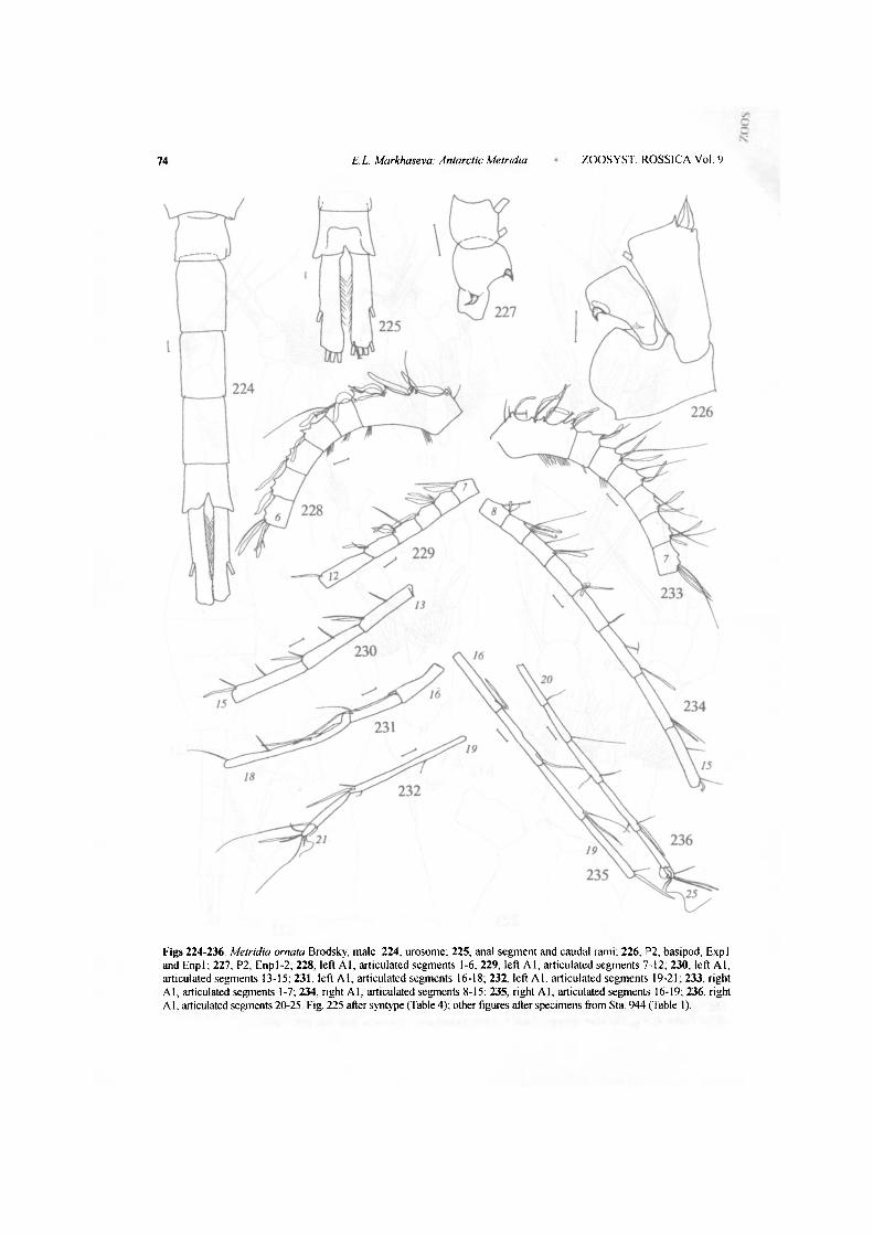
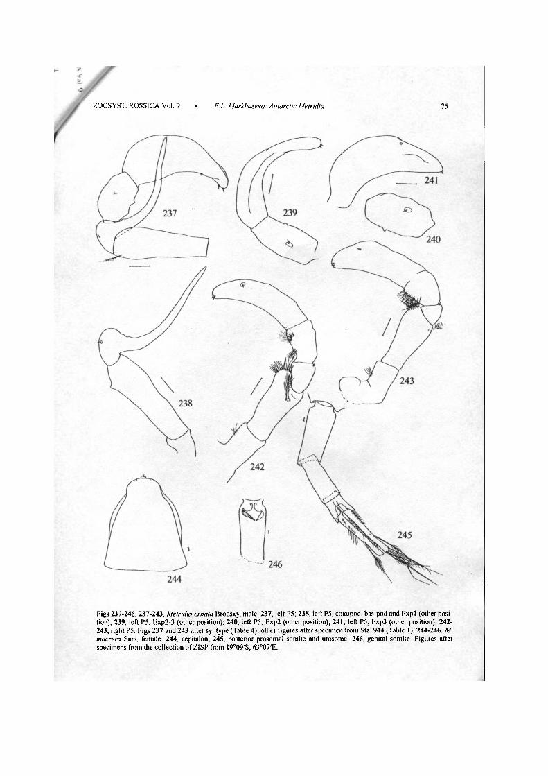
Male. Total length 7.0-7.4 mm. Prosome 1.10-1.20 times as long as urosome. Collar absent (Figs 221-222). Genital somite without hairs at small projection on the right (Fig. 224). Caudal rami about 4.8-6.6 times as long as wide (Figs 224, 225) . Left A l geniculated, reaching the end of anal segment (Figs 228-232). Right Al reaching the middle length of caudal rami. P5 with spinules at the right, longer coxopod; right basipod with spinules in the medial distal part. Both left and right basipods with setuled lateral setae distally. Right P5 Expl with small spine in lateral distal part of the segment; Exp2 with hairs; ЕхрЗ with 3 tiny setules distally (Figs 242-243). Left P5 Expl with small lateral spine and long medial attenuation exceeding the length of the Exp2, the latter supplied with tiny spinule in the middle; ЕхрЗ with 1+2 tiny spinules in distal part (Figs 237-241).
Remarks. Differences between M. ornata and M. ferrarii are listed in the remarks for M. ferrarii, these with M. princeps are listed in the remarks to M. princeps. In the original description of M. ornata (Brodsky, 1950), the type locality is given as 35°N, 125°W, however the original label of the type specimen is as following: Pacific Ocean, Kamchatka, 90 miles SE of cape Shipunsky. This species apparently can be considered as bipolar: its northernmost record is at 57°N in the Bering Sea (Table 4) and southernmost at 70°S (see Table 1).
Metridia macrura Sars, 1905 (Figs 244-246)
Material examined 1 9 , I9°09'S, 63°07'E, total vertical haul 4300 m, 29.V. 1956, Sta. 135, R/V Ob', ZISP.
The female specimen available for examination was damaged and thus not measured. The species differs from all large-sized Metridia species in the longest caudal rami: they are nearly 9 times as long as wide in the examined specimen and are known as about 12 times as long as wide (Sars, 1924, fig. 5).
Acknowledgements
The author thanks the US Antarctic Programme at the National Museum of Natural History and NSF at the Smithsonian Institution (Washington, D C . ) for financial support of the study. The research of the collections deposited in the Zoological Insitute, Russian Academy of Sciences, has been fulfilled within the framework and owing the financial support of the Subprogramme "Studies and Investigations of the Antarctica" FGP "World Ocean" project No. 16 "Conducting of the Mul-tidisciplinary Study of the Antarctic Biota". The work was carried out using scientific collections of the Zoological Institute, Russian Academy of Sciences, which obtain financial support from the Science and Technology Ministry of the Russian Federation (Reg. No 99-03-16).
References
Bradford-Grieve, J.M. 1999. The Marine Fauna of New Zealand: Pelagic Calanoid Copepoda: Bathy-ponliidae, Arietellidae, Augaptilidae, Heterorhabdi-dae, Lucicutiidae, Metridinidae, Phyllopodidae, Centropagidae, Pseudodiaptomidae, Temoridae, Candaciidae, Pontellidae, Sulcanidae, Acartiidae, Tortanidae. NIWA Biodiversity Memoir 111: 1-268.
Brady, G.S. 1918. Copepoda Scientific Reports of the Australasian Expedition. 1911-1914 (C), 5(3): 1-48.
Brodsky, K.A. 1950. Calanoida of the Far Eastern seas and Polar basin of the USSR Opredeliteli po faune SSSR, 35: 1-442. (In Russian).
Giesbrecht, W. 1892. Systematik und Faunistik der pelagischen Copepoden des Golfes von Neapel Fauna und Flora des Golfes von Neapel und der an-grenzenden Meeresabschnitte, 19: 1-831, 54 pis.
Farran, G.P. 1929. Copepoda. Nat. Hist. Rep. Terra NovaExped. (Zool)., 8(3): 203-306.

50 E.L. Markhaseva: Antarctic Metridia ZOOSYST. ROSSICA Vol. 9
Jespersen, P. 1940. Non-parasitic Copepoda In: The Zoology of Iceland, 3(33): 1-116.
Razouls, C. 1995 Diversity et repartition geog-raphique chez les copepodes pclagiques. Ann Inst Oceanogr, 71(2): 81-404.
Sars, G.O. 1924-1925 Copipodes particulierement bathypelagiques provenant des campagnes scienti-f'iques du Prince Albert ler de Monaco. Resultats des Campagnes Scientifiques accomplies par le
Prince Albert 1, Monaco, 69: Atlas. 1924, 127 pis; text, 1925, 408 pp
Seret, C. 1979. Taxonomie, biologie et biogeographie des copepodes pelagiques recoltes au cours de la campagne MD 03 du "Marion-Dufresne" (ile.s Crozet, Kerguelen et Heard) These Doctorat, Uni-versite de Paris. 187 pp (cited after http://www obs-banyuls.fr/razouls/webcd/ SUZANNE1 htm).
Received 21 June 2000

ZOOSYST. ROSSICA Vol. 9 E. L. Markhaseva: Antarctic Metridia 51
Figs 1-11. Metridia ferrarii sp. п., female. 1, 2, general view (dorsal and left lateral); 3, cephalosome (ventral view); 4, rostrum, 5, genital somite (ventral view); 6-9, the same (right lateral view); 10, 11, caudal rami (dorsal and right lateral view). Fig. 6 after female from Sta. 1014; Figs 8-9 after females from Sta. 154 (Table 1); other figures from holotype.

Figs 12-18. Metridia ferrarii sp. п., female, holotype. 12, left A l , articulated segments 1-8; 13, left A l , articulated segments 9-12; 14, left A l , articulated segments 13-15; 15, left Al , articulated segments 16-20; 16, left A l , articulated segments 21-24; 17, right Al articulated segments 1-6; 18, A2.
52 E.L Markhaseva: Antarctic Metridia • ZOOSYST. ROSSICA Vol. 9

ZOOSYST. ROSSICA Vol. 9 E.L. Markhaseva: Antarctic Metridia 53
Figs 19-26. Metridia ferrarii sp. п., female, holotype. 19, Mdp, basis; 20, Md, Enp and Exp; 21, Mxl, Li3, Li4, Enp, Exp and Le2; 22, Mxl, Li 1 and Li2; 23, Mxl , Lei; 24, Mx2; 25, Mxp, syncoxa; 26, Mxp, basis and Enp.

Figs 27-31. Metridia ferrarii sp. п., female, holotype. 27, Md Gn; 28, PI; 29, P5; 30, 31, cephalosome (lateral and dorsal
EL. Markhaseva: Antarctic Melndia • ZOOSYST. ROSSICA Vol. 9 54

Figs 32-39. Metridia ferrarii sp. n. female. 32, P2, coxopod, basipod, Enp and ExpI-2; 33, P2, Exp2-3; 34, P3, coxopod, basipod, Enp and Expl-2; 35-36, РЗ, ЕхрЗ; 37, P4, coxopod and basipod; 38, P4, Enp; 39, P4, Exp Fig. 36 after paratype No 296430; other figures after holotype.
ZOOSYST. ROSSICA Vol 9 • EL. Markhaseva: Antarctic Metridia 55

56 E.L. Markhaseva: Antarctic Metridia ZOOSYST. ROSSICA Vol. 9
- d 42
Figs 40-46 Metridia ferrarii sp. п., male. 40, 41, general view (dorsal and left lateral); 42, cephalosome (dorsal view), 43, cephalosome (right lateral view); 44, cephalosome (ventral view); 45, posterior prosomal and genital somite (dorsal view) 46 caudal rami (dorsal view). Fig. 42 after male from Sta. 895 (Table 1); odier figures after paratype No. 296430.

ZOOSYST. ROSSICA Vol. 9 E. L. Markhaseva: Antarctic Metridia 57
Figs 47-59. Metridia ferrarii sp. п., male 47, left A l , articulated segments 1 -6; 48, left A l , articulated segments 7-11; 49, left A l , articulated segments 12-15; 50, left A l , articulated segments 16-19; 51, left A l , articulated segments 20-24; 52, right A l , articulated segments 1-12; 53, right A l , articulated segments 13-15; 54, right A l , articulated segments 16-18; 55, right Al , articulated segments 19-21; 56, P5; 57, right P5, ЕхрЗ; 58, right P5, coxopod, basipod and Expl-2; 59, left P5, Exp2-3. Fig. 56 after male from Sta. 895 (Table 1); other figures after paratype No. 296430.

58 E.L. Markhaseva: Antarctic Metridia ZOOSYST. ROSSICA Vol. 9
Figs 60-70. Metridia pseudoasymmetrica sp. п., female. 60, Ur (dorsal view); 61, Ur (left lateral view); 62-64, genital somite (ventral view); 65-66, genital somite (left lateral view); 67, PI; 68-70, P5. Figs 60-62, 67-68 after holotype; Fig. 70 after female from Sta. 123; other figures after female from Sta. 154 (Table 1).

ZOOSYST. ROSSICA Vol. 9 • E.L Markhaseva: Antarctic Metridia 59
Figs 71-78. Metridiapseudoasymmetrica sp. п., female, holotype. 71, A2; 72, A2, Exp (other position); 73, Md; 74, Mxl; 75, Mxl, Exp (other limb of holotype); 76, Mx2, Lil-Li6; 77, Mx2, Li5-6 and exopod; 78, Mxp

Figs 79-80. Metridia pseudoasymmetrica sp. п., female from Sta. 918 (Table 1). 79, right AI; 80, left A1.

Figs 81-94. Metridia pseudoasymmetrica sp. п., female (81-91) and male (92-94). 81, P2, coxopod and basipod; 82, P2, margin of ЕхрЗ laterally; 83-85, P2, Enpl (different positions); 86, P2, Exp2-3 and Enp; 87, P3, coxopod, basipod and Enp; 88, P3, Exp; 89, P4, basipod and Enp; 90, P4, coxopod; 91, P4, Exp; 92, general lateral view; 93, general dorsal view; 94, Ur (dorsal view). Figs 82-85,87-91 after holotype; Figs 81, 86 after female from Sta 123 (Table 1); Figs 92-93 after male from Sta 918 (Table 1); Fig. 94 after male from Sta. 687 (Table 1)
ZOOSYST. ROSSICA Vol. 9 • E.L. Markhaseva: Antarctic Metridia 61

Figs 95-110. Metridia pseudoasymmetrica sp. п., male. 95, left A l , articulated segments 1-5; %, left A l , articulated segments 6-11; 97, left A l , articulated segments 12-16; 98, left A l , articulated segments 16-18; 99, left A l , articulated segments 19-20; 100, P5, 101-102. left P5 (different positions); 103-105, left P5, Exp2-3 (different positions); 106-107, right P5 (different positions); 108, right P5, Exp2-3, 109, right P5, ЕхрЗ (other view than 108); 110, right P5, basipod, lateral swelling. Figs 102-104, 106 and 109-110 after male from Sta. 687 (Table 1); other figures after male paratype No 90700
6 2 E.L. Markhaseva: Antarctic Metridia • ZOOSYST ROSSICA Vol. 9

ZOOSYST. ROSSICA Vol. 9 • E.L. Markhaseva: Antarctic Metridia 63
Figs 111-120. Metridia asymmetrica Brodsky, female (syntype). I l l , general dorsal view; 112. cephalosome (right lateral view); 113, posterior somite of prosome and Ur (dorsal view); 114, the same (left lateral view); 115, genital somite (ventral view); 116, genital somite (right lateral view); 117, PI, coxopod and basipod; 118, PI, Exp; 119, PI, Enp; 120, P5.

64 E.L. Markhaseva: Antarctic Metridia • ZOOSYST ROSSICA Vol. 9
Figs 121-131. Metridia asymmetrica Brodsky, syntypes, female (121-127) and male (128-131) 121, right A1, articulated segments 1-6; 122, right A l , articulated segments 7-9; 123, left A l , articulated segments 1-4; 124, left A1, articulated segments 5-9; 125, P2; 126, P2, Enpl; 127, P3, Exp; 128, cephalosome (dorsal view); 129, cephalosome (left lateral view); 130, posterior somite of prosome and urosome (dorsal view); 131, posterior somite of prosome and tirosome (left lateral view).
> <
ZOOSYST. ROSSICA Vol. 9 • E.L. Markhaseva: Antarctic Metridia 65
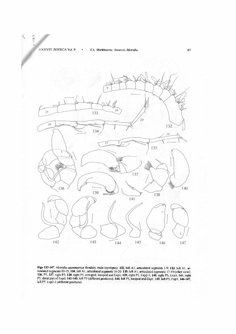
Figs 132-147. Metridia asymmetrica Brodsky, male (syntypes). 132, left A l , articulated segments 1-9; 133, left A l , articulated segments 10-15; 134, left A l , articulated segments 16-20; 135, left A l , articulated segments 17-19 (other view); 136, P5; 137, right P5; 138, right P5, coxopod, basipod and Expl; 139, right P5, Exp2-3; 140, right P5, ЕхрЗ; 141, right P5, distal part of ЕхрЗ; 142-143, left P5 (different positions); 144, left P5, basipod and Expl; 145, left P5, Expl; 146-147, left P5, Exp2-3 (different positions).

66 E.L. Markhaseva: Antarctic Metridia • ZOOSYST. ROSSICA Vol. 9
Figs 148-156. Metridia princeps Giesbrecht, female. 148, general right lateral view; 149. general dorsal view; 150, cephalosome (right lateral view); 151, anterior part of cephalosome; 152, rostrum; 153, urosome (right lateral view); 154-156. genital somite (ventral view). Figs 148, 150-151, 153-154 after specimen from Sta. 99; Figs 149, 152, 155 after specimen from Sta. 1615 (Table 1); Fig. 156 after specimen No 40898 (Table 3).

ZOOSYST ROSSICA Vol. 9 E.L. Markhaseva: Antarctic Metridia 67
Figs 157-167. Metridia princeps Giesbrecht, female. 157, left A l , articulated segments 1-6; 158, left A l , articulated segments 7-11; 159, left A l , articulated segments 12-15; 160, left A l , articulated segments 16-18; 161, left A l , articulated segments 19-24; 162, PI, Enp; 163, P2, Enp; 164, P2, Enpl; 165, P3, Exp; 166, P4, Exp; 167, P5. Figs 157-164, 167 after specimen from Sta. 99; Figs 165-166 after specimen from Sta. 1615 (Table 1).

Figs 168-176. Metridia princeps Giesbrecht, male 168, general dorsal view; 169, cephalosome (right lateral view); 170, posterior prosomal somite and urosome; 171, the same, right lateral view; 172, posterior prosomal somite and genital somite (dorsal view); 173, anal somite and caudal rami (dorsal view); 174, left P5; 175, left P5, ЕхрЗ; 176, P5, coxopod and right leg. Fig. 170 after specimen No. 40898 (see Table 3); other figures after specimen from Sta. 99 (Table 1).
N
68 E.L. Markhaseva: Antarctic Metridia • ZOOSYST ROSSICA Vol. 9

ZOOSYST. ROSSICA Vol 9 E.L. Markhaseva: Antarctic Metridia 69
Figs 177-187. 177-183. Metridia princeps Giesbrecht, male. 177, right A l , articulated segments 1-12; 178, right A l , articulated segments 13-16; 179, right A l , articulated segments 15-17; 180, right A1, articulated segments 17-19; 181, right Al , articulated segments 20-22; 182, left A l , articulated segments 1-4; 183, right A l , articulated segments 1-4. 184-187. M. ornata Brodsky, female. 184,186, general dorsal view; 185,187, general right lateral views. Figs 182-183 after specimen from Sta. 99 (Table 1); Figs 177-181 after specimen 40898 (Table 3); Figs 184-185 after specimen No 90690 (Table 4); Figs 186-187 after specimen from Sta. 944 (Table 1).
70 E.L. Markhaseva: Antarctic Metridia ZOOSYST. ROSSICA Vol 9
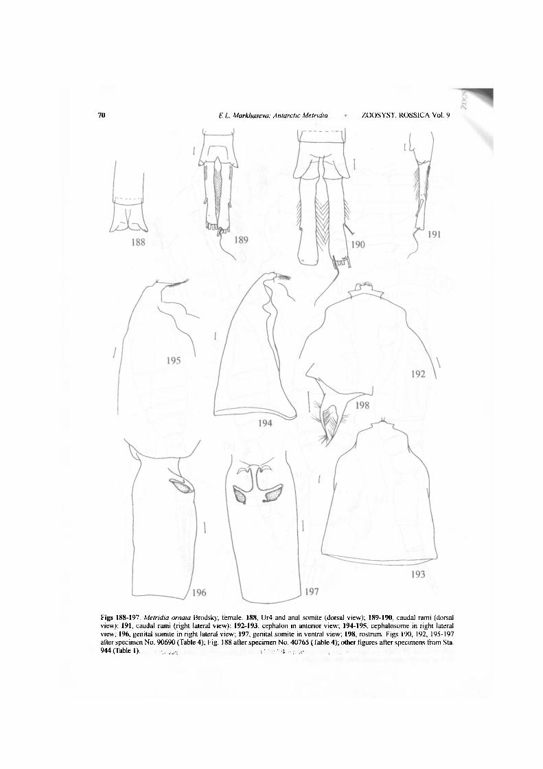
Figs 188-197 Metridia ornata Brodsky, female. 188, Ur4 and anal somite (dorsal view); 189-190, caudal rami (dorsal view); 191, caudal rami (right lateral view); 192-193, cephalon in anterior view; 194-195, cephalosome in right lateral view, 196, genital somite in right lateral view; 197, genital somite in ventral view, 198, rostrum. Figs 190, 192, 195-197 after specimen No. 90690 (Table 4); Fig. 188 after specimen No. 40765 (Table 4); other figures after specimens from Sta. 944 (Table 1).

ZOOS YST ROSSICA Vol. 9 • E.L Markhaseva: Antarctic Metridia 71
rigs 199-209. Metridia ornata Brodsky, female 199-200, genital somite in ventral view; 201-203, genital somite in right lateral view; 204, right A l , articulated segments 1-6; 205, right AI , articulated segments 7-10; 206, right A l , articulated segments 11-15; 207, right A l , articulated segments 16-18; 208, right A l , articulated segments 19-22; 209, right A l , articulated segments 23-24. Figs 204-209 after specimen No. 90690 (Table 4), Figs 202-203 after specimens No. 40765 (Table 4); other figures after specimens from Sta. 944 (Table 1).
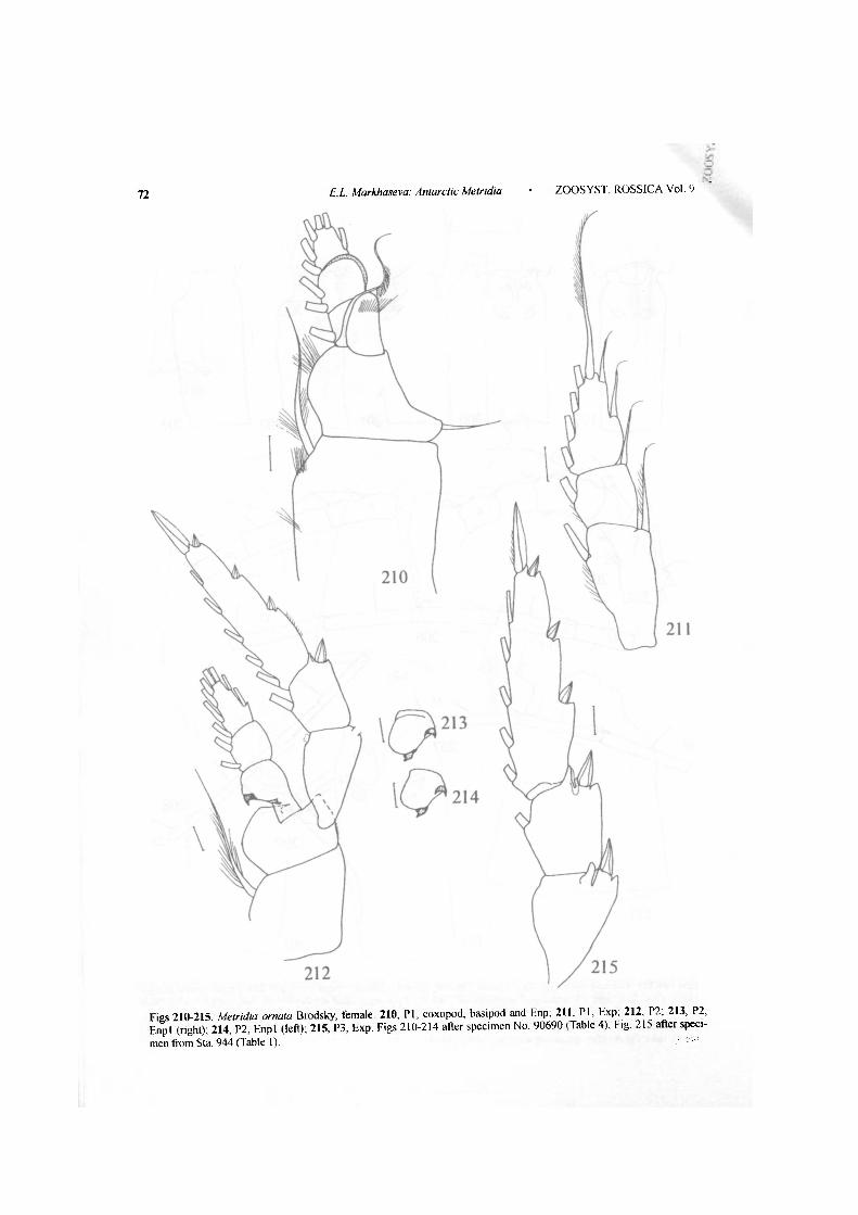
Figs 210-215 Afefrtfta ornata Brodsky, female 210, PI, coxopod, basipod and Enp; 211, PI, Exp; 212, P2; 213, P2, Enpl (right); 214, P2, Enpl (left); 215, P3, Exp. Figs 210-214 after specimen No. 90690 (Table 4), Fig. 215 after specimen from Sta. 944 (Table 1).
72 E.L. Markhaseva: Antarctic Metridia • ZOOSYST ROSSICA Vol. 9
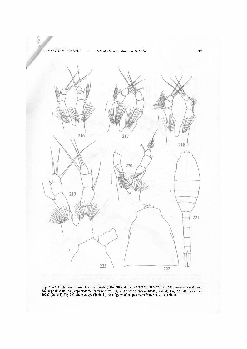
Figs 216-223. Metridia ornata Brodsky, female (216-220) and male (221-223). 216-220, P5; 221, general dorsal view; 222, cephalosome; 223, cephalosome, anterior view. Fig. 219 after specimen 90690 (Table 4), Fig. 220 after specimen 40765 (Table 4), Fig. 223 after syntype (Table 4), other figures after specimens from Sta 944 (Table 1).
ZOOSYST. ROSSICA Vol. 9 • E.L Markhaseva: Antarctic Metridia 73
71 E. L. Markhaseva: Antarctic Metridia ZOOSYST. ROSSICA Vol. 9
Figs 224-236. Metridia ornata Brodsky, male 224, urosome: 225, anal segment and caudal rami, 226, P2, basipod, Expl and Enpl: 227, P2, Enpl-2, 228, left A l , articulated segments 1-6; 229, left A l , articulated segments 7-12; 230, left A l , articulated segments 13-15; 231, left A l , articulated segments 16-18; 232, left A1, articulated segments 19-21; 233, right Al , articulated segments 1-7: 234, right Al , articulated segments 8-15; 235, right Al , articulated segments 16-19; 236. right Al, articulated segments 20-25 Fig. 225 after syntype (Table 4); other figures after specimens from Sta. 944 (Table 1).
ZOOSYST. ROSSICA Vol. 9 • E.L. Markhaseva: Antarctic Metridia 75
Figs 237-246. 237-243. Metridia ornata Brodsky, male. 237, left P5; 238, left P5, coxopod, basipod and Fxpl (other position); 239, left P5, Kxp2-3 (other position); 240, left P5, F.xp2 (oilier position); 241, left P5, F.xp.3 (other position); 242-243, right P5. Figs 237 and 243 after syntype (Table 4); other figures after specimen from Sta. 944 (Table 1). 244-246. M. macrura Sars, female. 244, ccphalon; 245, posterior prosomal somite and urosome; 246, genital somite. Figures after specimens from the collection of ZISP from I9°09'S, 63°07'E.