new 1,3-diarylureas linked by c-c suzuki coupling to the
TRANSCRIPT

1
New 1,3-diarylureas linked by C-C Suzuki coupling to the methyl
3-aminothieno[3,2-b]pyridine-2-carboxylate moiety: synthesis and
fluorescence studies in solution and in lipid membranes
Maria-João R. P. Queiroz,a,*
Daniela Peixoto,a Ana Rita O. Rodrigues,
b Pedro M. F. Mendes,
b
Cátia N. C. Costa,b Paulo J. G. Coutinho,
b Elisabete M. S. Castanheira
b,*
aDepartamento/Centro de Química, Universidade do Minho, Campus de Gualtar, 4710-057 Braga, Portugal
bCentro de Física (CFUM), Universidade do Minho, Campus de Gualtar, 4710-057 Braga, Portugal
_________________________________________________________________________
REVISED VERSION
Abstract
New six fluorescent 1,3-diarylureas linked by C-C Suzuki coupling to the 6-position of the methyl
3-aminothieno[3,2-b]pyridine-2-carboxylate moiety were prepared by reaction of the amino groups on
the ortho or meta positions relative to the C-C bond of the Suzuki coupling products, with different
para-substituted arylisocyanates (H, OMe, CN), in high to excellent yields. The fluorescence
properties of the 1,3-diarylureas in solution and in lipid membranes of egg-yolk phosphatidylcholine
(Egg-PC), dipalmitoyl phosphatidylcholine (DPPC), dipalmitoyl phosphatidylglycerol (DPPG) or
dioctadecyldimethylammonium bromide (DODAB), with or without cholesterol (Ch), were studied.
The six 1,3-diarylureas have reasonable fluorescence quantum yields in several solvents (0.02 F
0.69) and present a moderately solvent sensitive emission, but are not fluorescent in alcohols and
water. The compounds bearing the arylurea moiety in the meta position relative to the C-C bond,
especially with the OMe and CN substituents, present the better solvatochromic properties.
Incorporation of the six compounds in lipid membranes indicates that all the compounds are deeply
located in the hydrophobic region of the lipid bilayers, feeling the transition between the rigid gel
phase and fluid phases.
_________________________________________________________________________
Keywords: 1,3-Diarylureas; Thieno[3,2-b]pyridines; Fluorescence; Lipid membranes; Fluorescence probes
1. Introduction
Unsymmetrical 1,3-diarylureas have attracted much attention due to their diverse
applications in agriculture, medicine, petrochemicals, supramolecular chemistry (anion
receptors), biology and as important intermediates and bifunctional organocatalysts in organic
synthesis [1-3].

2
Thienopyridines including their 1,3-diarylurea derivatives have shown different biological
activities, namely as antitumoral agents [4] and receptor tyrosine kinase inhibitors [5].
In this work, six new 1,3-diarylureas were prepared by reaction of aminated compounds,
resulting from Suzuki coupling of methyl 3-amino-6-bromothieno[3,2-b]pyridine-2-
carboxylate and ortho or meta pinacolborane ester anilines [6], with different para-substituted
arylisocyanates (H, OMe, CN).
Due to the potential biological activity of the new compounds, their interaction with lipid
membranes is of particular interest. The photophysical properties of these thieno[3,2-
b]pyridine 1,3-diarylurea derivatives in solution and in lipid bilayers were studied. Lipid
membranes were composed of neutral/zwitterionic phospholipids (DPPC – dipalmitoyl
phosphatidylcholine; Egg-PC – egg yolk phosphatidylcholine), anionic phospholipids (DPPG
– dipalmitoyl phosphatidylglycerol) or synthetic cationic lipids (DODAB –
dioctadecyldimethylammonium bromide). The incorporation of cholesterol, an important
component of most natural membranes, may increase the stability of the lipid aggregates by
modulating the fluidity of the lipid bilayer, preventing crystallization of the phospholipid acyl
chains and providing steric hindrance to their movement [7].
Fluorescence anisotropy measurements can give relevant information about the compounds
behavior and location in the lipid membranes, namely if they are located deeply in the lipid
bilayer, feeling the differences between the rigid gel phase and the fluid liquid-crystalline
phase of the lipids.
2. Experimental
2.1. Synthesis
2.1.1.General Remarks
Melting points (ºC) were determined in a SMP3 Stuart apparatus and are uncorrected. 1
H and
13C NMR spectra were recorded on a Bruker Avance III at 400 and 100.6 MHz or on a Varian
Unity Plus at 300 and 75.4 MHz, respectively. Heteronuclear correlations, 1H-
13C, HMQC or
HSQC were performed to attribute some signals.
HRMS data were recorded using a method of direct injection by ESI-TOF by the mass
spectrometry service of the University of Vigo, Spain.
3
The reactions were monitored by thin layer chromatography (TLC) in aluminium plates
covered with a layer of silica gel 60 (Macherey-Nagel) of 0.2 mm, with UV254 fluorescence
indicator.
2.1.2. General procedure for the synthesis of 1,3-diarylureas (Scheme 1): Compounds 1a-b
[6] and different arylisocyanates (1 equiv.) in 6 mL CH2Cl2: THF (1:1) were left stirring at
room temperature for 16 h. If a precipitate didn’t come out after this time, hexane (3-5 mL)
was added to the mixture to precipate the product. This was filtered under vacuum to give the
corresponding 1,3-diarylureas.
2.1.2.1. Methyl 3-amino-6-[3-(3-phenylureido)phenyl]thieno[3,2-b]pyridine-2-carboxylate
(2a): From compound 1a (80.0 mg, 0.270 mmol) and phenylisocyanate (32.0 mg) compound
2a was isolated as a yellow solid (100 mg, 90%), m.p. 226-226.5 ºC. 1H NMR (400 MHz,
DMSO-d6): δ 3.83 (3H, s, OMe), 6.93 (2H, br s, NH2), 6.95-6.99 (1H, m, ArH), 7.26-7.30
(2H, m, ArH), 7.40-7.52 (5H, m, ArH), 7.91 (1H, broad s, 2’-H), 8.62 (1H, d, J = 2Hz, 7-H),
8.75 (1H, br s, NH), 8.82 (1H, br s, NH), 8.93 (1H, d, J = 2 Hz, 5-H) ppm. 13
C NMR (400
MHz, DMSO-d6): δ 51.5 (OMe), 97.4 (C), 117.0 (2’-CH), 118.3 (2CH) 118.4 (CH), 121.0
(CH), 121.9 (CH), 128.8 (2×CH), 129.3 (7-CH), 129.7 (CH), 133.9 (C), 135.1 (C), 137.3 (C),
139.6 (C), 140.5 (C), 145.4 (C), 145.5 (5-CH), 147.9 (C), 152.6 (C=O), 164.6 (C=O) ppm.
HRMS (ESI-TOF) Calcd. for C22H19N4O4S [M+H]+ 419.1172; found 419.1188.
2.1.2.2. Methyl 3-amino-6-{3-[3-(4-methoxyphenyl)ureido]phenyl}thieno[3,2-b]pyridine-2-
carboxylate (2b): From compound 1a (150 mg, 0.540 mmol) and 4-methoxyphenylisocyanate
(80.0 mg) compound 2b was isolated as a yellow solid (217 mg, 90%), m.p. 243-244 ºC. 1H
NMR (400 MHz, DMSO-d6): δ 3.71 (s, 3H, OMe), 3.83 (s, 3H, OMe), 6.87 (2H, d, J = 9.2
Hz, 3’’ and 5’’-H), 6.93 (2H, br s, NH2), 7.36 (2H, d, J = 9.2 Hz, 2’’ and 6’’-H), 7.38-7.50
(3H, m, ArH), 7.89 (1H, br s, 2’-H), 8.55 (1H, br s, NH), 8.61 (1H, d, J = 2.0 Hz, 7-H), 8.73
(1H, broad s, NH), 8.92 (1H, d, J = 2.0 Hz, 5-H) ppm. 13
C NMR (100.6 MHz, DMSO-d6): δ
51.6 (OMe), 55.2 (OMe), 97.4 (C), 114.0 (3’’and 5’’-CH), 116.9 (2’-CH), 118.3 (CH), 120.2
(2’’and 6’’-CH), 120.8 (CH), 129.3 (7-CH), 129.7 (CH), 132.6 (C), 133.9 (C), 135.2 (C),
137.3 (C), 140.7 (C), 145.4 (C), 145.5 (5-CH), 147.9 (C), 152.8 (C=O), 154.6 (C), 164.6
(C=O) ppm. HRMS (ESI-TOF) Calcd. for C23H21N4O4S [M+H]+
449.1284; found 449.1284.

4
2.1.2.3. Methyl 3-amino-6-{3-[3-(4-cyanophenyl)ureido]phenyl}thieno[3,2-b]pyridine-2-
carboxylate (2c): From compound 1a (100 mg, 0.330 mmol) and 4-cyanophenylisocyanate
(50.0 mg) compound 2c was isolated as a yellow solid (120 mg, 80%), m.p. 264-265 ºC. 1H
NMR (300 MHz, DMSO-d6): δ 3.83 (3H, s, OMe), 6.94 (2H, br s, NH2), 7.45-7.47 (2H, m,
ArH), 7.49-7.54 (1H, m, ArH), 7.65 (2H, d, J = 9.0 Hz, 2’’ and 6’’-H), 7.73 (2H, d, J = 9.0
Hz, 3’’ and 5’’-H), 7.90 (1H, br s, 2’-H), 8.62 (1H, d, J = 2 Hz, 7-H), 8.92 (1H, d, J = 2.0 Hz,
5-H), 9.01 (1H, br s, NH), 9.30 (1H, br s, NH) ppm. 13
C NMR (75.4 MHz, DMSO-d6): δ
51.6 (OMe), 97.4 (C), 103.4 (C), 117.4 (2’-CH) 118.1 (2’’ and 6’’-CH), 118.7 (CH) 119.3
(C), 121.5 (CH), 129.4 (7-CH), 129.8 (CH), 133.3 (3’’ and 5’’-CH), 133.9 (C), 135.0 (C),
137.4 (C), 140.0 (C), 144.1 (C), 145.5 (C), 145.6 (5-CH), 147.9 (C), 152.2 (C=O), 164.6
(C=O) ppm. HRMS (ESI-TOF) Calcd. for C23H18N5O3S [M+H]+
444.1125; found 444.1120.
2.1.2.4. Methyl 3-amino-6-[2-(3-phenylureido)phenyl]thieno[3,2-b]pyridine-2-carboxylate
(3a): From compound 1b (125 mg, 0.420 mmol) and phenylisocyanate (50.0 mg) compound
3a was isolated as a yellow solid (120 mg, 75%), m.p. 226-227 ºC. 1H NMR (400 MHz,
DMSO-d6): δ 3.83 (3H, s, OMe), 6.91-6.95 (m, 1H, ArH), 6.97 (2H, br s, NH2), 7.17-7.25
(3H, m, ArH), 7.35-7.44 (4H, m, ArH), 7.86 (1H, br s, NH), 8.00 (1H, dd, J = 8.0 and 1.2
Hz, ArH), 8.43 (1H, d, J = 2 Hz, 7-H), 8.65 (1H, d, J = 2 Hz, 5-H), 8.79 (1H, br s, NH) ppm.
13C NMR (100.6 MHz, DMSO-d6): δ 51.6 (OMe), 97.3 (C), 118.0 (2×CH), 121.8 (CH),
122.6 (CH) 123.5 (CH), 128.8 (2×CH) 128.9 (CH), 129.1 (C), 130.7 (CH), 132.1 (7-CH),
133.7 (C), 133.8 (C), 136.5 (C), 139.6 (C), 145.3 (C), 147.3 (5-CH), 148.0 (C), 152.6 (C=O),
164.6 (C=O) ppm. HRMS (ESI-TOF) Calcd. for C22H19N4O3S [M+H] +
419.1172; found
419.1180.
2.1.2.5. Methyl 3-amino-6-{2-[3-(4-methoxyphenyl)ureido]phenyl}thieno[3,2-b]pyridine-2-
carboxylate (3b): From compound 1b (100 mg, 0.330 mmol) with 4-
methoxyphenylisocyanate (50.0 mg) compound 3b was obtained (100 mg, 70%), m.p. 243-
244 ºC. 1H NMR (400 MHz, DMSO-d6): δ 3.68 (3H, s, OMe), 3.83 (3H, s, OMe), 6.81 (2H,
d, J = 9.2 Hz, 3” and 5”- H), 6.97 (2H, br s, NH2), 7.15-7.19 (1H, m, ArH), 7.25 (2H, d, J =
9.2 Hz, 2” and 6”-H), 7.32-7.35 (1H, m, ArH), 7.38-7.42 (1H, m, ArH), 7.77 (1H, br s, NH),
7.99 (1H, br d, J = 8.4 Hz, ArH), 8.42 (1H, d, J = 2.0 Hz, 7-H), 8.63 (1H, br s, NH), 8.64 (1H,
d, J = 2.0 Hz, 5-H). 13
C (100.6 MHz, DMSO-d6): δ 51.6 (OMe), 55.1 (OMe), 97.2 (C), 99.7
(C), 114.0 (3’’ and 5’’-CH), 119.8 (2’’and 6’’-CH), 122.3 (CH), 123.2 (CH) 128.9 (CH),
130.7 (CH), 132.1 (7-CH), 132.6 (C), 133.8 (C), 136.7 (C), 145.3 (C), 147.3 (5-CH), 148.0

5
(C), 152.7 (C=O), 154.4 (C), 164.6 (C=O) ppm. HRMS (ESI-TOF) Calcd. for C23H21N4O4S
[M+H]+
449.1284; found 449.1280.
2.1.2.6. Methyl 3-amino-6-{2-[3-(4-cyanophenyl)ureido]phenyl}thieno[3,2-b]pyridine-2-
carboxylate (3c): From compound 1b (140 mg, 0.470 mmol) and 4-cyanophenylisocyanate
(69.0 mg) compound 3c was isolated as a yellow solid (120 mg, 75%), m.p. 210-211 ºC. 1H
NMR (400 MHz, DMSO-d6): δ 3.83 (3H, s, OMe), 6.96 (2H, br s, NH2), 7.22-7.26 (1H, m,
ArH), 7.36-7.39 (1H, m, ArH), 7.52-7.55 (1H, m, ArH), 7.53 (2H, d, J = 8.8 Hz, 2’’ and 6’’-
H), 7.67 (2H, d, J = 8.8 Hz, 3’’ and 5’’-H), 7.93-7.95 (1H, m, ArH), 8.07 (1H, br s, NH), 8.44
(1H, d, J = 2 Hz, 7-H), 8.65 (1H, d, J = 2.0 Hz, 5-H), 9.28 (1H, br s, NH) ppm. 13
C NMR
(100.6 MHz, DMSO-d6): δ 51.6 (OMe), 97.3 (C), 103.3 (C), 117.9 (2’’ and 6’’-CH), 119.3
(C) 123.2 (CH), 124.2 (CH) 129.0 (CH), 129.8 (C), 130.8 (CH), 132.1 (7-CH), 133.3 (3’’ and
5’’-CH), 133.6 (C), 133.7 (C), 135.8 (C), 144.1 (C), 145.3 (C), 147.2 (5-CH), 147.9 (C),
152.3 (C=O), 164.6 (C=O) ppm. HRMS (ESI-TOF) Calcd. for C23H18N5O3S [M+H]+
444.1125; found 444.1129.
2.2. Lipid membranes preparation
All the solutions were prepared using spectroscopic grade solvents and ultrapure water (Milli-
Q grade). 1,2-Dipalmitoyl-sn-glycero-3-phosphocholine (DPPC), 1,2-diacyl-sn-glycero-3-
phosphocholine from egg yolk (egg-PC), 1,2-dipalmitoyl-sn-glycero-3-[phospho-rac-(1-
glycerol)] (sodium salt) (DPPG) and cholesterol were obtained from Sigma-Aldrich and
dioctadecyldimethylammonium bromide (DODAB) from Tokyo Kasei (lipid structures are
shown below).

6
For lipid vesicles preparation, the ethanolic injection method was used [8-10]. For egg-PC
membranes preparation, defined volume of stock solution of lipid (86.2 mM) and each
compound (0.3 mM) in ethanol were injected together, under vigorous stirring, to an aqueous
buffer solution (10 mM Tris, pH=7.4), at room temperature. A similar procedure was adopted
for DPPC, DODAB and DPPG vesicles, but the injection of the required amounts of stock
solutions of lipid (50 mM for DPPC, 20 mM for DODAB and 26.8 mM for DPPG) and
compounds in ethanol was done at 60 ºC, well above the melting transition temperature of
each lipid (ca. 41 ºC for DPPC [11], ca. 45 ºC for DODAB [12], and 39.6 ºC for DPPG [13]).
In all cases, the final lipid concentration was 1 mM, with a compound/lipid molar ratio of
1:333.
2.3. Spectroscopic measurements
Absorption spectra were recorded in a Shimadzu UV-3101PC UV-Vis-NIR
spectrophotometer. Fluorescence measurements were performed using a Fluorolog 3
spectrofluorimeter, equipped with double monochromators in both excitation and emission,
Glan-Thompson polarizers and a temperature controlled cuvette holder. Fluorescence spectra
were corrected for the instrumental response of the system.
For fluorescence quantum yield determination, the solutions were previously bubbled for
20 minutes with ultrapure nitrogen. The fluorescence quantum yields (s) were determined
using the standard method (equation 1) [14,15]. Quinine sulfate in H2SO4 0.05 M was used as
reference, r = 0.546 at 25 ºC [16].
r2rrs
2ssrs nFAnFA (1)
where A is the absorbance at the excitation wavelength, F the integrated emission area and n
the refraction index of the solvents used. Subscripts refer to the reference (r) or sample (s)
compound. The absorbance value at excitation wavelength was always less than 0.1, in order
to avoid inner filter effects.
Solvatochromic shifts can be described by the Lippert-Mataga equation (2), which relates
the energy difference between absorption and emission maxima to the orientation
polarizability, [17,18]
constfhcR
3
2
flabs2
4
1
0
(2)

7
where abs is the wavenumber of maximum absorption, fl is the wavenumber of maximum
emission, = e – g is the difference in the dipole moment of solute molecule between
excited (e) and ground (g) states, R is the cavity radius (considering the fluorophore a point
dipole at the center of a spherical cavity immersed in the homogeneous solvent), and f is
the orientation polarizability given by (eq. 3):
12
1
12
1
2
2
n
nf , (3)
where is the static dielectric constant and n the refractive index of the solvent.
An alternative expression, proposed by Bakhshiev, takes into account the angle, , between
the ground and excited state dipole moments of the fluorophore [19,20]:
constnfhcR
,cos22
4
1 2eeg
2g3flabs
0
(4)
where 2eeg
2g cos2 is equivalent to
2
ge
and
2
1
2
1,
2
2
n
nnf (5)
The steady-state fluorescence anisotropy, r, is calculated by
VHVV
VHVV
2 IGI
IGIr
(6)
where IVV and IVH are the intensities of the emission spectra obtained with vertical and
horizontal polarization, respectively (for vertically polarized excitation light), and
HHHV IIG is the instrument correction factor, where IHV and IHH are the emission
intensities obtained with vertical and horizontal polarization (for horizontally polarized
excitation light).
3. Results and discussion
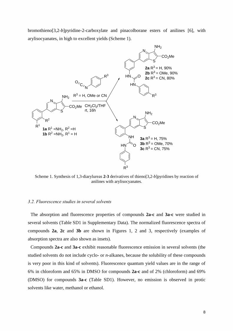
3.1. Synthesis
New 1,3-diarylureas 2a-c and 3a-c were prepared by reaction of the aromatic amino groups
of the Suzuki coupling products 1a-b, earlier obtained by us from methyl 3-amino-6-
8
bromothieno[3,2-b]pyridine-2-carboxylate and pinacolborane esters of anilines [6], with
arylisocyanates, in high to excellent yields (Scheme 1).
N
S
NH2
CO2Me
NC
O
CH2Cl2/THFrt, 16h
R2
1a R1 =NH2, R2 =H
1b R2 =NH2, R1 = H
R3 = H, OMe or CN
N
S
NH2
CO2Me
NH
HN O
R3
3a R3 = H, 75%
3b R3 = OMe, 70%
3c R3 = CN, 75%
R1
N
S
NH2
CO2Me
HN
HN
O
R3
R3
2a R3 = H, 90%
2b R3 = OMe, 90%
2c R3 = CN, 80%
Scheme 1. Synthesis of 1,3-diarylureas 2-3 derivatives of thieno[3,2-b]pyridines by reaction of
anilines with arylisocyanates.
3.2. Fluorescence studies in several solvents
The absorption and fluorescence properties of compounds 2a-c and 3a-c were studied in
several solvents (Table SD1 in Supplementary Data). The normalized fluorescence spectra of
compounds 2a, 2c and 3b are shown in Figures 1, 2 and 3, respectively (examples of
absorption spectra are also shown as insets).
Compounds 2a-c and 3a-c exhibit reasonable fluorescence emission in several solvents (the
studied solvents do not include cyclo- or n-alkanes, because the solubility of these compounds
is very poor in this kind of solvents). Fluorescence quantum yield values are in the range of
6% in chloroform and 65% in DMSO for compounds 2a-c and of 2% (chloroform) and 69%
(DMSO) for compounds 3a-c (Table SD1). However, no emission is observed in protic
solvents like water, methanol or ethanol.

9
Figure 1. Normalized fluorescence spectra of 310-6
M solutions of compound 2a in several solvents
(exc=370 nm). Inset: Absorption spectrum of 10-5
M solutions of 2a in dioxane and acetonitrile, as
examples.
Figure 2. Normalized fluorescence spectra of 310-6
M solutions of compound 2c in several solvents
(exc=370 nm). Inset: Absorption spectrum of 10-5
M solutions of 2c in dioxane and acetonitrile, as
examples.

10
Figure 3. Normalized fluorescence spectra of 310
-6 M solutions of compound 3b in several solvents
(exc=370 nm). Inset: Absorption spectrum of 10-5
M solutions of 3b in dioxane and acetonitrile, as
examples.
This behavior, already observed for di(hetero)arylether derivatives of thieno[3,2-b]pyridines
recently synthesized [21], can be due to specific solute-solvent interactions by hydrogen
bonds with protic solvents, namely by protonation of the nitrogen atom of the pyridine ring.
The same explanation can justify the low fluorescence quantum yields obtained in
chloroform, as the formation of hydrogen bonds between chloroform and proton acceptor
molecules has been already described [22].
The six compounds studied here are more fluorescent in polar solvents like
dimethylformamide and dimethylsulfoxide, which is also a common behavior with the
di(hetero)arylether derivatives of thieno[3,2-b]pyridines studied earlier [21]. Also, the
substituent does not seem to have a significant influence in the fluorescence quantum yield
values (Table SD1).
For all compounds, significant red shifts are observed for emission in polar solvents (34-36
nm between chloroform and dimethylsulfoxide for compounds 2a-c and 20-25 nm for 3a-c).
In the absorption spectra, the red shifts are negligible (Table SD1), indicating that solvent
relaxation after photoexcitation plays an important role, especially for compounds 2a-c. This
indicates that the 1,3-arylurea substituent in the meta position relative to C-C bond contributes
to increase the intramolecular charge transfer (ICT) character of the excited state.

11
The solvatochromic plots for compounds 2a–c and 3a–c, shown in Figures 4 and 5, are
reasonably linear, the slope being larger for compound 2c. This predicts a higher ICT
character of the excited state for this compound, maybe related with the position of the
arylurea moiety together with the presence of a nitrile substituent.
Figure 4. Solvatochromic plots (equation 4) for compounds 2a-c. Solvents: 1 - Dioxane; 2 -
chloroform; 3 - ethyl acetate; 4 - dichloromethane; 5 - dimethylsulfoxide; 6 - N,N-dimethylformamide;
7 - acetonitrile (values of and n were obtained from ref. [23]).
Figure 5. Solvatochromic plots (equation 4) for compounds 3a-c. Solvents: 1 - Dioxane; 2 -
chloroform; 3 - ethyl acetate; 4 - dichloromethane; 5 - dimethylsulfoxide; 6 - N,N-dimethylformamide;
7 - acetonitrile (values of and n were obtained from ref. [23]).

12
The slopes of the solvatochromic plots are much lower for compounds 3a-c, being similar
for these three molecules. This indicates that the charge transfer character of the excited state
is much lower for the arylureas in the ortho position relative to the thieno[3,2-b]pyridine-2-
carboxylate moiety.
From ab initio molecular quantum chemistry calculations, obtained with Gaussian 09
software [24] and use of a 6-311+G(dp) basis set at the DFT (CAM-B3LYP/AUTO) level of
theory [24,25] in gas phase, the cavity radius (R) and the ground state dipole moment (g)
were determined for the six compounds (Table 2). The use of CAM-B3LYP functional was
needed, as initial trials with the simpler B3LYP gave unrealistic underestimates of the
HOMO-LUMO band gap (<500 nm). This is a known problem with B3LYP functional in the
description of excited states with charge transfer character [25]. The optimized geometries of
the ground state and the first excited state were obtained with a smaller basis set (3-21G+*)
and are similar in the groups of compounds 2a-c and 3a-c (Figures 6 and 7). In the case of
excited state calculations, a time dependent density functional method was used (TD-SCF
DFT). In Table 1, the dihedral angle defined by the two molecular planes and the angles for
the N-C-N and C-S-C chemical bonds (in the thienopyridine moiety) are indicated, evidencing
notable differences in geometries between the two sets of compounds. Comparing the ground
and excited state geometries, a small decrease (ca. 10º in 2a-c and 5º in 3a-c) of the dihedral
angle is observed in the excited state (Table 1), together with a 2º increase in the C-S-C bond
angle for all compounds.
Table 1. Dihedral angle , N-C-N bond angle and C-S-C bond angle in the ground and first singlet
excited state geometries for compounds 2a-c and 3a-c.
Compound State Dihedral angle N-C-N angle C-S-C angle
2a Ground -46.6º 112.6º 90.1º
Excited -36.8º 112.8º 92.6º
2b Ground -46.9º 112.5º 90.1º
Excited -36.6º 112.8º 92.6º
2c Ground -47.0º 112.5º 90.1º
Excited -36.1º 112.7º 92.6º
3a Ground 94.0º 112.3º 90.1º
Excited 89.6º 112.5º 92.7º
3b Ground 94.7º 112.2º 90.1º
Excited 89.0º 112.5º 92.8º
3c Ground 94.0º 112.2º 90.1º
Excited 89.8º 112.4º 92.7º
The directions of the calculated dipole moments in the ground and excited states are also
indicated in Figures 6 and 7, evidencing an increase in magnitude and a change of direction in

13
the excited state dipole moment vector relative to the ground state one. This clearly indicates
that the angle between the two dipole moment vectors cannot be neglected and must be
considered in the solvatochromic plots (Bakhshiev’s equation (4)). Nevertheless, the change
in the dipole moment direction is very small for compounds 2c and 3a. In the excited state,
the dipole moment vectors for all compounds point to the side of the thienopyridine moiety.
Figure 6. Optimized geometries of compounds 2a-c obtained by Gaussian 09 software (grey: C atoms;
white: H atoms; red: O atoms; blue: N atoms; yellow: S atoms). Above: ground state; below: lowest
excited singlet state. The arrows indicate the direction of the dipole moment.
Figure 7. Optimized geometries of compounds 3a-c obtained by Gaussian 09 software (grey: C atoms;
white: H atoms; red: O atoms; blue: N atoms; yellow: S atoms). Above: ground state; below: lowest
excited singlet state. The arrows indicate the direction of the dipole moment.

14
The absolute value of the difference in the excited and ground state dipole moment vectors,
estimated from the solvatochromic plots (Figures 4 and 5) and from molecular quantum
mechanical calculations, is presented in Table 2, for each compound. The obtained values are
very similar and, therefore, both methods point to the presence of a charge transfer
mechanism in the excited state, more pronounced for compounds 2a-c. Compounds bearing
an electron-donating group (-OCH3 in 2b) or an electron-withdrawing group (-CN in 2c) have
a higher ICT character of the excited state than compound 2a.
Table 2. Cavity radius (R), ground (g) and excited state (e) dipole moments obtained from
theoretical calculations, and absolute value of the dipole moment difference ( ge
), from quantum
mechanical calculations and from the solvatochromic plots.
Compound
Cavity
radius, R
(Å)
Ground state
dipole moment,
g (D)
Excited state dipole
moment, e (D), from
theoretical
calculations
ge
(D)
from theoretical
calculations
ge
(D) from
solvatochromic
plots
2a 5.6 3.1 6.1 5.1 5.2
2b 6.2 4.7 7.3 5.2 5.8
2c 5.9 4.9 10.1 5.3 6.2
3a 5.7 2.2 6.7 4.6 4.1
3b 6.0 3.3 7.8 4.5 4.4
3c 5.9 7.4 10.2 4.7 4.2
Figure 8 displays the representation of electronic density difference between the lowest
excited state and the ground state, for the lowest excited state optimized geometry (relaxed S1
state). In general, it can be observed that electron density variations reside mostly on the
thienopyridine-2-carboxylate moiety. This justifies that the compounds do not exhibit a
noticeable influence of the arylurea substituent (-OCH3 or -CN) in their photophysical
properties, namely in the fluorescence quantum yields. The more prominent features in Figure
8 are an electron density transfer from the amino group linked to the thiophene ring and its
sulfur atom to the nitrogen atom in the pyridine moiety and to the carboxylate group. In the π-
electron system, alternating increases and decreases of electronic density are observed. This
confirms the ICT character of the first excited state of these compounds.

15
Figure 8. Representation of the electronic density difference for compounds 2a-c and 3a-c (optimized
geometry for the lowest excited singlet state) at an iso level of 0.0004; green regions: loss of electronic
density; red regions: enrichment of electronic density.
The photophysical behavior of the six compounds shows that they can be considered as
solvatochromic probes, especially compounds 2a-c. The sensitivity of the fluorescence
emission to the fluorophore environment can be very useful when probing the
location/behavior of these compounds in lipid membranes.
3.3. Fluorescence studies in lipid membranes
Fluorescence experiments of the six compounds incorporated in lipid membranes of several
compositions were carried out. These lipid aggregates were composed either by neat
phospholipids, or by phosphatidylcholines with cholesterol (Ch), for a better simulation of the
biological membranes. In fact, the vesicles composed of 70% Egg-PC and 30% cholesterol
(Egg-PC:Ch 7:3) are often used as models of the biological membranes [26,27].
Lipid membranes of neat DPPC (zwitterionic), DPPG (anionic), DODAB (cationic), Egg-PC
(zwitterionic, composed of a phosphatidylcholine mixture), Egg-PC:Ch 7:3 and DPPC:Ch
7:3, with incorporated compounds, were prepared and the fluorescence emission was
monitored in both gel (below the main transition temperature, Tm) and liquid-crystalline
(above Tm) phases of the phospholipids. At room temperature, the phospholipids DPPC,
DODAB and DPPG are in ordered gel phase, where the hydrocarbon chains are fully
extended and closely packed. The melting transition temperature of Egg-PC is very low [28]
and this lipid is in the fluid liquid-crystalline phase at room temperature.

16
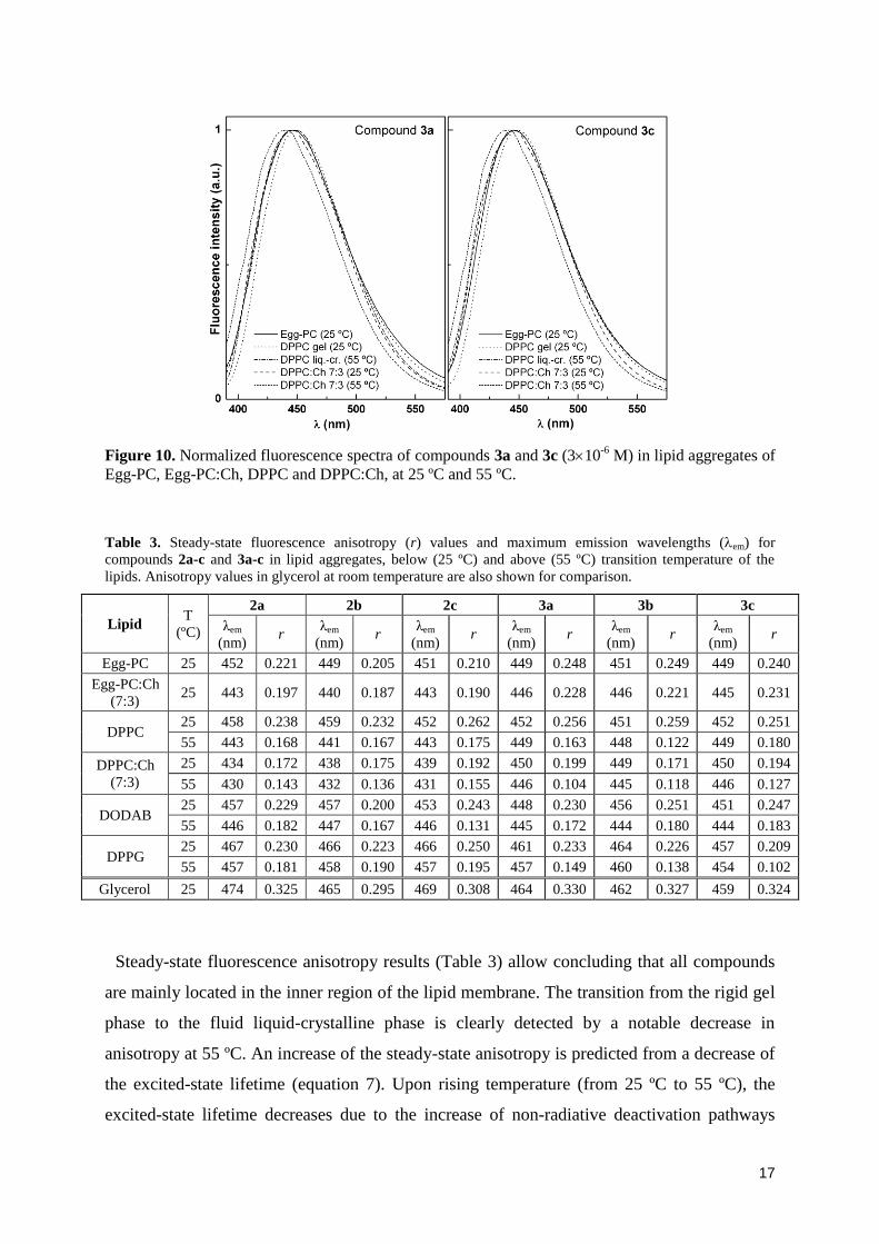
Fluorescence spectra of compounds incorporated in these lipid aggregates are presented in
Figures 9 and 10. All the six compounds exhibit reasonable fluorescence emission when
incorporated in lipid membranes, indicating that they are mainly located in the region of the
lipid bilayer, as they are not fluorescent in alcohols or water. The maximum emission
wavelengths in lipid membranes generally point to a hydrophobic medium for all compounds
in these lipid aggregates, feeling an environment with polarity near dioxane or less polar than
dioxane (Table 3). A slightly more hydrated environment is anticipated for the six compounds
in DPPG vesicles at 25 ºC, considering the values of the maximum emission wavelengths.
Fluorescence anisotropy (r) measurements can give relevant information about the location
of the compounds in liposomes, as r increases with the rotational correlation time of the
fluorescent molecule (and, thus, with the viscosity of the fluorophore environment) [29].
Steady-state anisotropy relates to both the excited-state lifetime and the rotational correlation
time of the fluorophore [29],
c0
111
rr (7)
where r0 is the fundamental anisotropy, is the excited-state lifetime and c is the rotational
correlation time.
The fluorescence steady-state anisotropies of compounds 2a-c and 3a-c in lipid membranes
are shown in Table 3. Anisotropy values in glycerol at room temperature were also
determined for comparison.
Figure 9. Normalized fluorescence spectra of compounds 2a and 2c (310-6
M) in lipid aggregates of
Egg-PC, Egg-PC:Ch, DPPC and DPPC:Ch, at 25 ºC and 55 ºC.
17
Figure 10. Normalized fluorescence spectra of compounds 3a and 3c (310-6
M) in lipid aggregates of
Egg-PC, Egg-PC:Ch, DPPC and DPPC:Ch, at 25 ºC and 55 ºC.
Table 3. Steady-state fluorescence anisotropy (r) values and maximum emission wavelengths (em) for
compounds 2a-c and 3a-c in lipid aggregates, below (25 ºC) and above (55 ºC) transition temperature of the
lipids. Anisotropy values in glycerol at room temperature are also shown for comparison.
Lipid T
(ºC)
2a 2b 2c 3a 3b 3c
λem
(nm) r
λem
(nm) r
λem
(nm) r
λem
(nm) r
λem
(nm) r
λem
(nm) r
Egg-PC 25 452 0.221 449 0.205 451 0.210 449 0.248 451 0.249 449 0.240
Egg-PC:Ch
(7:3) 25 443 0.197 440 0.187 443 0.190 446 0.228 446 0.221 445 0.231
DPPC 25 458 0.238 459 0.232 452 0.262 452 0.256 451 0.259 452 0.251
55 443 0.168 441 0.167 443 0.175 449 0.163 448 0.122 449 0.180
DPPC:Ch
(7:3)
25 434 0.172 438 0.175 439 0.192 450 0.199 449 0.171 450 0.194
55 430 0.143 432 0.136 431 0.155 446 0.104 445 0.118 446 0.127
DODAB 25 457 0.229 457 0.200 453 0.243 448 0.230 456 0.251 451 0.247
55 446 0.182 447 0.167 446 0.131 445 0.172 444 0.180 444 0.183
DPPG 25 467 0.230 466 0.223 466 0.250 461 0.233 464 0.226 457 0.209
55 457 0.181 458 0.190 457 0.195 457 0.149 460 0.138 454 0.102
Glycerol 25 474 0.325 465 0.295 469 0.308 464 0.330 462 0.327 459 0.324
Steady-state fluorescence anisotropy results (Table 3) allow concluding that all compounds
are mainly located in the inner region of the lipid membrane. The transition from the rigid gel
phase to the fluid liquid-crystalline phase is clearly detected by a notable decrease in
anisotropy at 55 ºC. An increase of the steady-state anisotropy is predicted from a decrease of
the excited-state lifetime (equation 7). Upon rising temperature (from 25 ºC to 55 ºC), the
excited-state lifetime decreases due to the increase of non-radiative deactivation pathways

18
(mainly the rate constant for internal conversion S1→S0). Instead of an expected rise in
anisotropy (equation 7), a decrease is observed, which can only be attributed to a diminution
of the rotational correlation time of the fluorophore, that arises from the decrease of
membrane microviscosity upon changing from the gel to the liquid-crystalline phase.
Fluorescence anisotropy values for the compounds incorporated in lipid membranes exhibit
also a significant decrease when lipid aggregate fluidity increases by addition of cholesterol.
Simultaneously, a blue shift in emission spectra (Figures 9 and 10 and Table 3) is observed
when fluidity increases, either by phase transition to the liquid-crystalline phase or by
cholesterol addition. These changes are much more pronounced for compounds 2a-c and may
indicate a relocation of the fluorophores in a less hydrated environment. The decrease in local
microviscosity can facilitate a deeper penetration of these molecules in lipid bilayers. The
results obtained here point to a promising utility of the new diarylureas to monitor changes in
fluidity of lipid membranes, especially compounds 2a-c.
Microviscosity in hydrophobic domains of microheterogeneous systems has been measured
using the widely known fluorescence probe 1,3-bis-(1-pyrenyl)propane (BPP) [30-35]. This
probe can form intramolecular excimers and is highly sensitive to constraints imposed by its
environment, reported by variations in the ratio between excimer and monomer emission
intensities. The new diarylurea derivatives 2a-c and 3a-c showed in this work an interesting
potential as fluidity probes for lipid membranes (especially 2a-c) through their intrinsic
photophysical properties, rather than by the relative efficiency of a dynamic conformational
change leading to an intramolecular pyrene excimer formation, as observed in BPP. However,
further studies are needed to assess the utility of these new probes for microviscosity
determinations of other types of microheterogeneous systems already studied using BPP, like
micelles, liquid crystals, lipoproteins and synthetic polymers [30, 33-35].
4. Conclusions
New six fluorescent 1,3-diarylureas in the thieno[3,2-b]pyridine series were prepared by a
reaction of ortho and meta aminated Suzuki coupling products obtained by the formation of a
C-C bond in the 6-position of the thieno[3,2-b]pyridine moiety, with different para-
substituted arylisocyanates (H, OMe or CN).
The six compounds exhibit reasonable fluorescence quantum yields in several solvents and
present a moderately solvent sensitive emission, but are not fluorescent in alcohols and water.

19
Compounds 2b and 2c with the arylurea moiety in the meta position and bearing a OMe or
CN group, are the ones with better solvatochromic properties.
Incorporation of compounds 2a-c and 3a-c in lipid membranes indicate that all compounds
are deeply located in the hydrophobic region of the lipid bilayers, feeling the transition
between the rigid gel phase and the liquid-crystalline phase. The results obtained point to a
promising utility of these compounds to monitor changes in fluidity of lipid membranes,
especially compounds 2a-c. Moreover, due to the potential biological activity of these new
compounds, their interaction with lipid membranes is of particular interest.
Supplementary Data
Table SD1. Maximum absorption (abs) and emission wavelengths (em), molar absorption
coefficients () and fluorescence quantum yields (F) for compounds 2a-c and 3a-c in several
solvents.
Acknowledgements
To the Foundation for the Science and Technology (FCT, Portugal) for inancial support to
the NMR portuguese network (PTNMR, Bruker Avance III 400-Univ. Minho). To the FCT
and FEDER (European Fund for Regional Development)-COMPETE-QREN-EU for financial
support to the Research Centres, CQ/UM [PEst-C/QUI/UI0686/2011 (FCOMP-01-0124-
FEDER-022716)] and CFUM [PEst-C/FIS/UI0607/2011 (F-COMP-01-0124-FEDER-
022711)], and to the research projects PTDC/QUI/81238/2006 (FCOMP-01-0124-FEDER-
007467) (photophysical studies) and PTDC/QUI-QUI/111060/2009 (F-COMP-01-0124-
FEDER-015603) (organic synthesis).
References
[1] V. Amendola, L. Fabbrizzi, L. Mosca, Anion recognition by hydrogen bonding: urea-based
receptors, Chem. Soc. Rev. 39 (2010) 3889-3915.
[2] K. Lang, J. Park, S. Hong, Urea/transition-metal cooperative catalyst for anti-selective
asymmetric nitroaldol reactions, Angew. Chem. Int. Ed. 51(7) (2012) 1620–1624.
[3] J. G. Kim, Y. Takami, T. Mizugami, K. Beppu, T. Fukuda, I. Kataoka, CPPU application on
size and quality of hardy kiwifruit, Sci. Hortic. 110 (2006) 219-222.
[4] I. Hayakama, R. Shioya, T. Agatsuma, H. Furokawa, Y. Sugano, Thienopyridine and
benzofuran derivatives as potent anti-tumor agents possessing different structure-activity
relationship, Bioorg. Med. Chem. 14 (2004) 3411-3414.

20
[5] H. R. Heyman, R. R. Frey, P. F. Bousquet, G. A. Cunha, M. D. Moskey, A. A. Ahmed, N. B.
Soni, P. A. Marcotte, L. J. Pease, K. B.Glaser, M. Yates, J. J. Bouska, D. H. Albert, C. L. Black
Schaefer, P. J. Dandliker, K. D.Stewart, P. Rafferty, S. K. Davidsen, M. R. Michaelides, M. L.
Curtin, Thienopyridine urea inhibitors of KDR kinase, Bioorg. Med. Chem. Lett. 17 (2007)
1246-1249.
[6] M.-J. R. P. Queiroz, R. C. Calhelha, L. A. Vale-Silva, E. Pinto, R. T. Lima, M. H. Vasconcelos,
Efficient synthesis of 6-(hetero)arylthieno[3,2-b]pyridines by Suzuki-Miyaura coupling.
Evaluation of growth inhibition in human tumor cell lines, SARs and effects on the cell cycle,
Eur. J. Med. Chem. 45 (2010) 5628-5634.
[7] Y. Malam, M. Loizidou, A. M. Seifalian, Liposomes and nanoparticles: nanosized vehicles for
drug delivery in cancer, Trends in Pharmacol. Sci. 30 (2009) 592-599.
[8] S. Batzri, E. D. Korn, Single bilayer liposomes prepared without sonication, BBA:
Biomembranes 298 (1973) 1015-1019.
[9] J. M. H. Kremer, M. W. J. v. d. Esker, C. Pathmamanoharan, P. H. Wiersema, Vesicles of
variable diameter prepared by a modified injection method, Biochemistry 16 (1977) 3932-3935.
[10] J. R. Nordlund, C. F. Schmidt, S. N. Dicken, T. E. Thompson, Transbilayer distribution of
phosphatidylethanolamine in large and small unilamellar vesicles, Biochemistry 20 (1981)
3237-3241.
[11] B. R. Lentz, Membrane “fluidity” as detected by diphenylhexatriene probes, Chem. Phys.
Lipids 50 (1989) 171-190.
[12] E. Feitosa, P. C. A. Barreleiro, G. Olofsson, Phase transition in dioctadecyldimethylammonium
bromide and chloride vesicles prepared by different methods, Chem. Phys. Lipids 105 (2000)
201-213.
[13] J. S. Vincent, S. D. Revak, C. D. Cochrane, I. W. Levin, Interactions of model human
pulmonary surfactants with a mixed phospholipid bilayer assembly: Raman spectroscopic
studies, Biochemistry 32 (1993) 8228-8238.
[14] J. N. Demas, G. A. Crosby, Measurement of photoluminescence quantum yields – Review, J.
Phys. Chem. 75 (1971) 991-1024.
[15] S. Fery-Forgues, D. Lavabre, Are fluorescence quantum yields so tricky to measure? A
demonstration using familiar stationery products, J. Chem. Educ. 76 (1999) 1260-1264.
[16] S. R. Meech, D. Phillips, Photophysics of some common fluorescence standards, J. Photochem.
23 (1983) 193-217.
[17] J. R. Lakowicz, Principles of Fluorescence Spectroscopy, Kluwer Academic/Plenum Press, New
York, 1999.
[18] N. Mataga, T. Kubota, Molecular Interactions and Electronic Spectra, Marcel Dekker, New
York, 1970.
[19] N. G. Bakhshiev, Universal molecular interactions and their effects on the position of the
electronic spectra of molecules in two component solutions. I. Theory (liquid solutions), Opt.
Spectrosc. 10 (1961) 379-384.
[20] N. G. Bakhshiev, Universal molecular interactions and their effects on the position of the
electronic spectra of molecules in two component solutions, Opt. Spectrosc. 12 (1962) 309-313;
Opt. Spectrosc. 13 (1962) 24-29.
[21] M.-J. R. P. Queiroz, S. Dias, D. Peixoto, A. R. O. Rodrigues, A. D. S. Oliveira, P. J. G.
Coutinho, L. A. Vale-Silva, E. Pinto, E. M. S. Castanheira, New potential antitumoral
di(hetero)arylether derivatives in the thieno[3,2-b]pyridine series: Synthesis and fluorescence
studies in solution and in nanoliposomes, J. Photochem. Photobiol. A: Chemistry 238 (2012)
71-80.

21
[22] (a) K. C. James, P. R. Noyce, Hydrogen bonding between testosterone propionate and solvent in
chloroform-cyclohexane solutions, Spectrochim. Acta A 27 (1971) 691-696. (b) G. R. Wiley, S.
I. Miller, Thermodynamic parameters for hydrogen-bonding of chloroform with Lewis bases in
cyclohexane - Proton magnetic-resonance study, J. Am. Chem. Soc. 94 (1972) 3287-3293.
[23] D. R. Lide (Ed.), Handbook of Chemistry and Physics, 83th Edition, CRC Press, Boca Raton,
2002.
[24] Gaussian 09, Revision A.02, M. J. Frisch, G. W. Trucks, H. B. Schlegel, G. E. Scuseria, M. A.
Robb, J. R. Cheeseman, G. Scalmani, V. Barone, B. Mennucci, G. A. Petersson, H. Nakatsuji,
M. Caricato, X. Li, H. P. Hratchian, A. F. Izmaylov, J. Bloino, G. Zheng, J. L. Sonnenberg, M.
Hada, M. Ehara, K. Toyota, R. Fukuda, J. Hasegawa, M. Ishida, T. Nakajima, Y. Honda, O.
Kitao, H. Nakai, T. Vreven, J. A. Montgomery, Jr., J. E. Peralta, F. Ogliaro, M. Bearpark, J. J.
Heyd, E. Brothers, K. N. Kudin, V. N. Staroverov, R. Kobayashi, J. Normand, K. Raghavachari,
A. Rendell, J. C. Burant, S. S. Iyengar, J. Tomasi, M. Cossi, N. Rega, J. M. Millam, M. Klene,
J. E. Knox, J. B. Cross, V. Bakken, C. Adamo, J. Jaramillo, R. Gomperts, R. E. Stratmann, O.
Yazyev, A. J. Austin, R. Cammi, C. Pomelli, J. W. Ochterski, R. L. Martin, K. Morokuma, V.
G. Zakrzewski, G. A. Voth, P. Salvador, J. J. Dannenberg, S. Dapprich, A. D. Daniels, Ö.
Farkas, J. B. Foresman, J. V. Ortiz, J. Cioslowski, and D. J. Fox, Gaussian, Inc., Wallingford
CT, 2009.
[25] (a) T. Yanaia, D. P. Tewb, N. C. Handy, A new hybrid exchange–correlation functional using
the Coulomb-attenuating method (CAM-B3LYP), Chem. Phys. Lett. 393 (2004) 51-57. (b) M.
J. G. Peach, T. Helgaker, P. Sałek, T. W. Keal, O. B. Lutnæs, D. J. Tozer, N. C. Handy,
Assessment of a Coulomb-attenuated exchange–correlation energy functional, Phys. Chem.
Chem. Phys. 8 (2006) 558-562.
[26] C. Toniolo, M. Crisma, F. Formaggio, C. Peggion, V. Monaco, C. Goulard, S. Rebuffat, B.
Bodo, Effect of N-acyl chain length on the membrane-modifying properties of synthetic
analogs of the lipopeptaibol trichogin GA IV, J. Am. Chem. Soc. 118 (1996) 4952-4958.
[27] M. Crisma, A. Barazza, F. Formaggio, B. Kaptein, B. Q. Broxterman, J. Kamphuis, C. Toniolo,
Peptaibolin: synthesis, 3D-structure, and membrane modifying properties of the natural
antibiotic and selected analogues, Tetrahedron 57 (2001) 2813-2825.
[28] D. Papahadjopoulos, N. Miller, Phospholipid model membranes. I. Structural characteristics of
hydrated liquid crystals, Biochim. Biophys. Acta 135 (1967) 624-638.
[29] B. Valeur, Molecular Fluorescence - Principles and Applications, Weinheim, Wiley-VCH,
2002.
[30] R. L. Melnick, H. C. Haspel, M. Goldenberg, L. M. Greenbaum, S. Weinstein, Use of
fluorescent probes that form intramolecular excimers to monitor structural changes in model and
biological membranes, Biophys. J. 34 (1981) 499-515.
[31] L. M. Almeida, W. L. Vaz, K. A. Zachariasse, V. M. Madeira, Fluidity of sarcoplasmic
reticulum membranes investigated with dipyrenylpropane, an intramolecular excimer probe,
Biochemistry 21 (1982) 5972-5977.
[32] S. Kang, I. G. Kang, I. Yun, Determination of microviscosity and location of 1,3-di(1-
pyrenyl)propane in brain membranes, Arch. Pharm. Res. 20 (1997) 1-6.
[33] E. Szajdzinska-Pietek, M. Wolszczak, A. Plonka, S. Schlick, Fluorescence studies of self-
assembling in aqueous solutions of poly(ethylene-co-methacrylic acid) (EMAA) ionomers, J.
Am. Chem. Soc. 120 (1998) 4215-4221.
[34] Y. Díaz-Fernández, S. Rodríguez-Calvo, A. Pérez-Gramatges, P. Pallavicini, S. Patroni, C.
Mangano, Effect of surfactant structure on the residual fluorescence of micelle-based
fluorescent probes, J. Colloid Interface Sci. 313 (2007) 638-644.

22
[35] Y. A. Díaz-Fernández, E. Mottini, L. Pasotti, E. F. Craparo, G. Giammona, G. Cavallaro, P.
Pallavicini, Multicomponent polymeric micelles based on polyaspartamide as tunable
fluorescent pH-window biosensors, Biosens. Bioelectron. 26 (2010) 29-35.