neuropsychology of chronic painovercomingpain.com/wp-content/uploads/2015/06/neuropsychology... ·...
TRANSCRIPT
The Neuropsychotherapist issue 14 May 201514
&Neuropsychology
of Chronic Pain
Eye Movement Desensitization & Reprocessing
Mark Grant
www.neuropsychotherapist.com 15The Neuropsychotherapist
Kur
han/
Bigs
tock
.com
Understanding Pain in the BrainThe brain is a hierarchical system for receiving
and processing experiential information. Sensory information (e.g., nociception) enters via the brain stem and travels up through the areas of the brain responsible for emotional processing (the mid-brain), before reaching the prefrontal cortex (the executive brain) where attention and a cognitive re-sponse occur. Horizontally, the brain is divided into left and right. Although controversy still rages about the significance of this division, it has been estab-lished that the left and right hemispheres contribute uniquely to how we think, feel, and respond to expe-rience (McGilchrist, 2009). For example, neuroimag-ing studies have shown that the right anterior cingu-late cortex (ACC) is activated in pain sufferers with unilateral pain, regardless of which side of the body the pain is located. A recent neuroimaging study of brain processing of acute pain found activation was either localized exclusively or strongly lateralized in the right hemisphere for many areas of the cortex (e.g., superior, middle, and inferior frontal gyrus, an-terior cingulate, inferior parietal lobe). It is thought that there is a right lateralized attentional system, which acts to alert the organism to intermittent pain (Symonds, Gordon, Bixby, & Mande, 2006).
The main areas of the brain that are involved in pain (the pain circuit) include the thalamus, the so-matosensory cortex, the ACC, the amygdala, the hippocampus, and the prefrontal cortex (PFC). The thalamus receives sensory information and relays it to the somatosensory cortex. It acts as both a pro-cessing and a relay center for nociceptive informa-tion, especially via ascending pain pathways. The so-matosensory cortex and the thalamus are involved in the sensory-discriminative component of pain. The thalamus and the ACC also mediate the affec-tive-motivational components of pain. In the ACC, this activation is directly related to the unpleas-
antness of pain and is accompanied by activation of subcortical areas such as the amygdala and the cerebellum. In conjunction with the amygdala, the hippocampus mediates anxiety and the fear of pain, and pain memories via aversive associative learning. The frontal and prefrontal cortex mediates attention and cognitive aspects of pain, including the anticipa-tion of pain (Lorenz, Minoshima, & Kasey, 2003). See Figure 1 for a summary of ascending brain pathways involved in pain.
Reflecting the neuroplastic nature of the brain, repeated activation of the pain circuit leads to pain memories. A pain memory is best described as a learned pattern of brain activity that recreates pain-ful experience in the absence of ongoing external nociceptive input. This is the central sensitization model of pain. A pain memory may originate from a physical insult, but it is maintained by various ab-normalities in the central nervous system—includ-ing increased cortisol (Vachon-Presseau et al., 2013), random firing in damaged nociceptors (Flor, 2002), reduced hippocampal volume and a loss of grey matter (Henry, Chiodo, & Yang, 2011), prefrontal cortical hyperactivity (Apkarian, Thomas, Krauss, & Szeverenyi, 2001), and thickening of the somatosen-sory cortex in migraine sufferers (Kim et al., 2014). Where these abnormalities lead to pain as a result of learning processes it is known as “kindling” (Ray & Zbik, 2002).
Different pain theories draw on different aspects of these changes. A. D. (“Bud”) Craig (2003) posits a circuit between the VMpo—the ventromedial poste-rior nucleus—(a thalamic relay) and its parieto-insu-lar target, the interoceptive cortex, which is active in response to all health-related sensations emanating from the body. In his view, pain leads to a remapping of the interoceptive cortex in the right anterior insu-la of humans, that is, of “how you feel”. Craig notes that the interoceptive cortex exists only in primates and is enormously enlarged in humans. He regards
As Milton Erickson used to say, when a patient comes to you tremendously handi-capped, how handicapped is he really? What brain cells does he have unused? The brain is a misunderstood and under-utilized resource in almost every therapeutic situation. Every neuropsychotherapist realizes this and is committed to learning
the most effective ways of harnessing its resources. This article will briefly summarize the current state of our understanding of pain from a neuropsychological perspective and its implications for treatment. Based on brain changes detected in recipients of eye movement desensitization and reprocessing therapy (EMDR), it is proposed that this method meets many of the criteria of neuropsychotherapy. This will be illustrated through a case study of EMDR treatment of a chronic pain/PTSD sufferer.

The Neuropsychotherapist issue 14 May 201516
pain as part of a specific homeostatic response con-sisting of physical sensations, represented in the pa-rieto-insular cortex, and an emotional component, represented in the anterior cingulate (Craig, 2003).
In an earlier article in this journal about emo-tional style, pain, and the brain, James Alexander proposed that chronic pain is maintained by forced overstimulation of the NAcc (a cluster of neurons at the head of the anterior cingulate cortex that gener-ate motivation and reward) by the prefrontal cortex in people who are not naturally optimistic. Drawing on Sarno’s TMS model (Sarno, 1998), Alexander sug-gests this leads to a kind of false optimism that shuts out the emotional reality of chronic pain without re-ally addressing it (Alexander, 2013).
Neurological changes associated with pain also occur in relation to numerous other causal factors in-cluding physical pathology, genetics, psychological trauma, stress, and even culture. For example, com-plex regional pain syndrome (CRPS) is caused by a disturbance of the sympathetic nervous system—the network of nerves located along the spinal cord that controls bodily functions such as the opening and closing of blood vessels and sweat glands. And fibromyalgia, which involves increased sensitivity to pain and fatigue, is often associated with PTSD (Roy-Byrne, Smith, Goldberg, Afari, & Buchwald, 2004).
Therapeutic ImplicationsNeurological factors are increasingly being used
as a basis for psychological pain treatments (Ray & Zbik, 2002; Lorenz et al., 2003; Alexander, 2013; Grant, 2015). Neuroscience raises the possibility of developing new treatments (or refining existing ones) that cohere more with brain structure and functioning and ought therefore to be more effec-tive. For example, Fuch (2004, pp. 480–481) has sug-gested that the “brain is an organ of transformation which may be addressed by input to different hierar-chical levels …‘top-down’… and …‘bottom-up’. In or-der to produce lasting effects, psychotherapy needs to restructure neural networks, particularly in the subcortical limbic system.”
Van der Kolk (n.d.) offers some detail on this in terms of the role of different brain structures in the neocortex and the lower brain regions:
Top-down processing is based on cogni-tion and is operated by the neocortex, which allows for high-level executive functioning by observing, monitoring and planning. It can only effectively function if the input from low-er brain levels is inhibited … Bottom-up pro-cessing, by itself, does not resolve trauma, but if the patient is directed to track and articulate sensorimotor experience while consciously in-hibiting emotions, content and interpretative
Prefontalcortex
(attention, cognition)‘there is something
wrong with me’
Insula(feelings, sensations)
‘I feel bad’
PAINBrain stem
(motor and sensory input)
Somatosensory cortex
(pain sensations; hot, cold, throbbing etc)
Anterior Cingulate Cortex
(thoughts, feelings) ‘ouch’
Parietal lobe(sensory information,
proprioception) damaged body schema
Amygdala(fear)
Thalamus(relay station)
Hippocampus(memory)
memories of pain
Figure 1. Ascending pathway of brain structures involved in pain.
www.neuropsychotherapist.com 17The Neuropsychotherapist
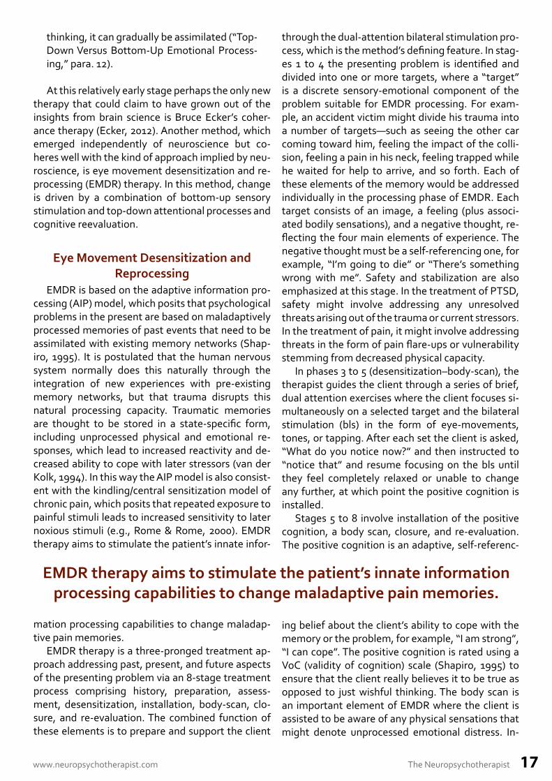
thinking, it can gradually be assimilated (“Top-Down Versus Bottom-Up Emotional Process-ing,” para. 12).
At this relatively early stage perhaps the only new therapy that could claim to have grown out of the insights from brain science is Bruce Ecker’s coher-ance therapy (Ecker, 2012). Another method, which emerged independently of neuroscience but co-heres well with the kind of approach implied by neu-roscience, is eye movement desensitization and re-processing (EMDR) therapy. In this method, change is driven by a combination of bottom-up sensory stimulation and top-down attentional processes and cognitive reevaluation.
Eye Movement Desensitization and Reprocessing
EMDR is based on the adaptive information pro-cessing (AIP) model, which posits that psychological problems in the present are based on maladaptively processed memories of past events that need to be assimilated with existing memory networks (Shap-iro, 1995). It is postulated that the human nervous system normally does this naturally through the integration of new experiences with pre-existing memory networks, but that trauma disrupts this natural processing capacity. Traumatic memories are thought to be stored in a state-specific form, including unprocessed physical and emotional re-sponses, which lead to increased reactivity and de-creased ability to cope with later stressors (van der Kolk, 1994). In this way the AIP model is also consist-ent with the kindling/central sensitization model of chronic pain, which posits that repeated exposure to painful stimuli leads to increased sensitivity to later noxious stimuli (e.g., Rome & Rome, 2000). EMDR therapy aims to stimulate the patient’s innate infor-
mation processing capabilities to change maladap-tive pain memories.
EMDR therapy is a three-pronged treatment ap-proach addressing past, present, and future aspects of the presenting problem via an 8-stage treatment process comprising history, preparation, assess-ment, desensitization, installation, body-scan, clo-sure, and re-evaluation. The combined function of these elements is to prepare and support the client
through the dual-attention bilateral stimulation pro-cess, which is the method’s defining feature. In stag-es 1 to 4 the presenting problem is identified and divided into one or more targets, where a “target” is a discrete sensory-emotional component of the problem suitable for EMDR processing. For exam-ple, an accident victim might divide his trauma into a number of targets—such as seeing the other car coming toward him, feeling the impact of the colli-sion, feeling a pain in his neck, feeling trapped while he waited for help to arrive, and so forth. Each of these elements of the memory would be addressed individually in the processing phase of EMDR. Each target consists of an image, a feeling (plus associ-ated bodily sensations), and a negative thought, re-flecting the four main elements of experience. The negative thought must be a self-referencing one, for example, “I’m going to die” or “There’s something wrong with me”. Safety and stabilization are also emphasized at this stage. In the treatment of PTSD, safety might involve addressing any unresolved threats arising out of the trauma or current stressors. In the treatment of pain, it might involve addressing threats in the form of pain flare-ups or vulnerability stemming from decreased physical capacity.
In phases 3 to 5 (desensitization–body-scan), the therapist guides the client through a series of brief, dual attention exercises where the client focuses si-multaneously on a selected target and the bilateral stimulation (bls) in the form of eye-movements, tones, or tapping. After each set the client is asked, “What do you notice now?” and then instructed to “notice that” and resume focusing on the bls until they feel completely relaxed or unable to change any further, at which point the positive cognition is installed.
Stages 5 to 8 involve installation of the positive cognition, a body scan, closure, and re-evaluation. The positive cognition is an adaptive, self-referenc-
ing belief about the client’s ability to cope with the memory or the problem, for example, “I am strong”, “I can cope”. The positive cognition is rated using a VoC (validity of cognition) scale (Shapiro, 1995) to ensure that the client really believes it to be true as opposed to just wishful thinking. The body scan is an important element of EMDR where the client is assisted to be aware of any physical sensations that might denote unprocessed emotional distress. In-
EMDR therapy aims to stimulate the patient’s innate information processing capabilities to change maladaptive pain memories.

The Neuropsychotherapist issue 14 May 201518
structing the client to “mentally scan your body for any signs of distress” also helps ensure that any dis-sociated material, or feelings the client is unaware of, are identified and addressed. In the closure stage the session is concluded with the therapist ensuring the client is emotionally stable and then instruct-ing them to keep a record of any upsetting memo-ries or events between sessions. These records may indicate that there are aspects of the problem that still need to be processed and become the basis for future targets for processing. The last session in the process (stage 8) always begins with a reevaluation of the targets processed in the previous session, followed by a review of potential targets the client needs to address next.
EMDR therapy is a well-established PTSD treat-ment that has gained clinical and institutional ac-ceptance based on extensive research. With over 20 published studies, including two controlled studies and many observational studies, chronic pain is the next most researched application of the method. EMDR therapy has been found to alleviate sensory and emotional distress associated with chronic pain, and gains are often well-maintained (Schneider, Hofman, Rost, & Shapiro, 2008; van Rood & de Roos, 2009; Grant, 2014). The method appears to be most effective with pain associated with PTSD.
EMDR and the BrainEMDR therapy’s unusual mechanism of action—
dual focus of attention paired with bilateral stimu-lation—together with increased interest in the role of the brain in psychological problems has spurred consideration of the method’s coherency with brain functioning. There are a number of theories that will probably all be found to represent pieces of the same puzzle of a unified model of human informa-tion processing. The parietal lobe activation model, for example, posits that EMDR harnesses attention and episodic memory to stimulate reintegration of damaged body schema in the parietal cortex. An-other theory posits that the bls element of EMDR activates the lateral cerebellum and subsequently the ventrolateral and central lateral thalamic nuclei, which facilitates the repair and integration of so-matosensory, memorial, cognitive, emotional, and synchronized hemispheric functioning. The low fre-quency stimulation model posits that EMDR takes advantage of the fact that memory traces in the hip-pocampus and amygdala become labile during acti-vation. When coupled with the low frequency stimu-lation provided by bls, this leads to de-potentiation of limbic synapses. Research indicates that the eye movements play a significant role in the changes as-sociated with EMDR (Lee & Cuijpers, 2012).
SPECT scans of EMDR recipients (mostly PTSD sufferers) have shown changes in brain activity in
EMDR therapy’s unusual mechanism of action—dual focus of attention paired with bilateral stimulation—together with increased interest in the role of
the brain in psychological problems has spurred consideration of the method’s
coherency with brain functioning.
Dph
iMan
/Big
stoc
k.co
m
www.neuropsychotherapist.com 19The Neuropsychotherapist
the left frontal ACC, left frontal cortex, and occipi-tal and temporal lobes, in the opposite direction to those associated with PTSD. Treatment effects of EMDR include reduced emotionality (van den Hout et al., 2011), relaxation, and decreased autonomic arousal (Elofsson, von Schèele, Theorell, & Sönder-gaard, 2008; Söndergaard & Elofsson, 2008). These findings are indicative of: (a) emotional regulation due to increased activity of the prefrontal lobe, (b) inhibition of limbic over-stimulation by increased regulation of the association cortex, (c) reduction in the intrusion and over-consolidation of traumatic episodic memory due to the reduction of temporal lobe activity, (d) the reduction of occipitally medi-ated flashbacks, and (e) the induction of a functional balance between the limbic and prefrontal areas. Consistent with these hypothesized brain effects, recipients of EMDR often describe decreased nega-tive cognitive activity, feeling relaxed, and feeling detached from the problem, or distancing effect (Grant, 2014).
These changes suggest EMDR stimulates the repair of failures in cognitive, memorial, affective, somatosensory, and interhemispheric integration. In considering how these effects might ameliorate pain, Ray & Zbik (2002) have suggested that EMDR separates and permanently de-augments the affec-tive component of traumatic memories and pain. They suggest that this gives EMDR an added dimen-sion in contrast with more traditional approaches (e.g., CBT) that may improve a person’s perception of pain and quality of life but don’t generally offer affective change. Notwithstanding this, CBT has been found to stimulate increased grey matter in
the dorsolateral prefrontal cortex, which was associ-ated with decreased catastrophizing (Seminowicz et al., 2013). Certainly the nature and location of brain changes associated with EMDR, as well as the meth-od’s modus operandi and results, suggest EMDR is consistent with the type of integrated bottom-up approach recommended by neuroscientists.
To date there are over 20 published reports re-garding EMDR treatment of pain, with most indi-cating that the method is effective, particularly in
treating pain associated with PTSD. Based on this research (including by this author), a treatment manual, Pain Control with EMDR (Grant, 2015), has been developed. Undoubtedly, the treatment of pain is an area of urgent need. The following case illustrates some of the practices and procedures in-volved in EMDR, and how their coherence with brain science enhances treatment outcomes.
EMDR Treatment of Chronic Pain Sufferer with PTSD
Angela (35) sought help to cope with chronic pain in her right arm following a car accident five years previously. She also had a complex PTSD as a result of a combination of her 15 years’ service as a police officer and the various traumas she experienced or witnessed during that time. Angela was raised by loving adoptive parents. She was married and had two children of her own. In terms of the accident the medical diagnosis was whiplash. Despite exten-sive medical and psychological treatment, including surgery (a fusion at C-4, 5, and 6), three weeks as an inpatient at a leading trauma hospital, and pre-scription medication in the form of analgesics and anti-depressants, Angela continued to suffer from constant pain and depression. Angela’s self-esteem and body-image had suffered considerably: she de-scribed her right arm, which was encased in a protec-tive brace, as basically a lump of dead, aching flesh. She felt useless and a burden on her family, with whom she was often irritable and tense. In addition to its effects on her functioning as a wife and mother, Angela had not been able to return to her career as a
policewoman, which was a devastating blow to her self-esteem. In addition to her depressed mood she was experiencing recurring nightmares and intrusive thoughts about the accident and its aftermath.
Angela was referred to me to see if EMDR could help. In addition to obtaining her history and not-ing her depression and PTSD, I assessed Angela as having a basically stable personality and strong pre-morbid ego functioning. Angela was a phlegmatic, practical person who generally preferred to keep
The nature and location of brain changes associated with EMDR, as well as the method’s modus operandi and results, suggest EMDR is consistent with the type of integrated bottom-up approach rec-
ommended by neuroscientists.
The Neuropsychotherapist issue 14 May 201520
her emotions to herself. She appeared stoic and un-complaining, although she was clearly in the midst of a devastating ordeal. These are important quali-ties when qualifying a patient for EMDR: they indi-cate a capacity for sustained focused attention, a capacity to observe symptoms in a detached way, and a readiness to engage in free association rela-tively safely. Angela had no resistance to coming to therapy although she was not sure how it could help with her very real pain. I briefly described EMDR and the accelerated information processing model, the concept of neuroplasticity, and how it was possible to change pain memories through targeted brain stimulation. Angela indicated that these ideas gave her a different understanding of her problems.
Taking into account her past trauma and past and present pain, it was decided that Angela’s pre-
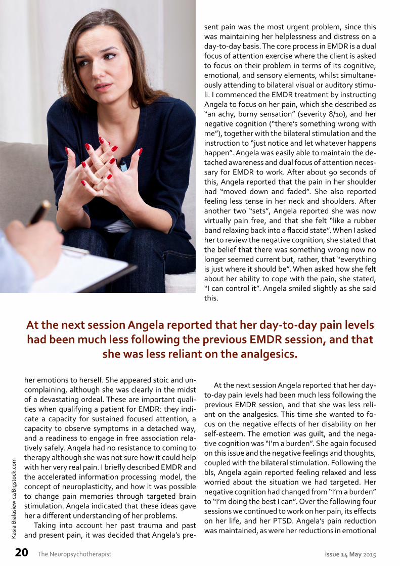
sent pain was the most urgent problem, since this was maintaining her helplessness and distress on a day-to-day basis. The core process in EMDR is a dual focus of attention exercise where the client is asked to focus on their problem in terms of its cognitive, emotional, and sensory elements, whilst simultane-ously attending to bilateral visual or auditory stimu-li. I commenced the EMDR treatment by instructing Angela to focus on her pain, which she described as “an achy, burny sensation” (severity 8/10), and her negative cognition (“there’s something wrong with me”), together with the bilateral stimulation and the instruction to “just notice and let whatever happens happen”. Angela was easily able to maintain the de-tached awareness and dual focus of attention neces-sary for EMDR to work. After about 90 seconds of this, Angela reported that the pain in her shoulder had “moved down and faded”. She also reported feeling less tense in her neck and shoulders. After another two “sets”, Angela reported she was now virtually pain free, and that she felt “like a rubber band relaxing back into a flaccid state”. When I asked her to review the negative cognition, she stated that the belief that there was something wrong now no longer seemed current but, rather, that “everything is just where it should be”. When asked how she felt about her ability to cope with the pain, she stated, “I can control it”. Angela smiled slightly as she said this.
At the next session Angela reported that her day-to-day pain levels had been much less following the previous EMDR session, and that she was less reli-ant on the analgesics. This time she wanted to fo-cus on the negative effects of her disability on her self-esteem. The emotion was guilt, and the nega-tive cognition was “I’m a burden”. She again focused on this issue and the negative feelings and thoughts, coupled with the bilateral stimulation. Following the bls, Angela again reported feeling relaxed and less worried about the situation we had targeted. Her negative cognition had changed from “I’m a burden” to “I’m doing the best I can”. Over the following four sessions we continued to work on her pain, its effects on her life, and her PTSD. Angela’s pain reduction was maintained, as were her reductions in emotional
At the next session Angela reported that her day-to-day pain levels had been much less following the previous EMDR session, and that
she was less reliant on the analgesics.
Kas
ia B
iala
siew
icz/
Bigs
tock
.com
www.neuropsychotherapist.com 21The Neuropsychotherapist
distress and disability. After six sessions Angela was experiencing minimal pain, she was able to do a lot more around the house and with her children, she was using almost no medication, and her depression and PTSD symptoms had decreased to the point where she no longer met the diagnostic criteria for either condition. In a moving session towards the end of her treatment, Angela described how joyful she felt to be able to tell her children “Mommy can give you proper cuddles now”. Physically, Angela’s body-image had changed. She exclaimed, “It’s like I have two arms again!” Angela also felt more like her-self—not her old self, but like someone who could cope with life as it was.
This case illustrates how EMDR can facilitate changes in the sensory-emotional component of chronic pain, particularly when associated with trau-ma. It also demonstrates some of the practical ways in which a brain-based psychotherapy can enhance treatment. For example, introducing EMDR in terms of neuroplasticity prepared this client for the possi-bility that change might be possible and that some-thing different might be going to happen. It would not have made any difference if Angela was resist-ant or skeptical, other than possibly having to help
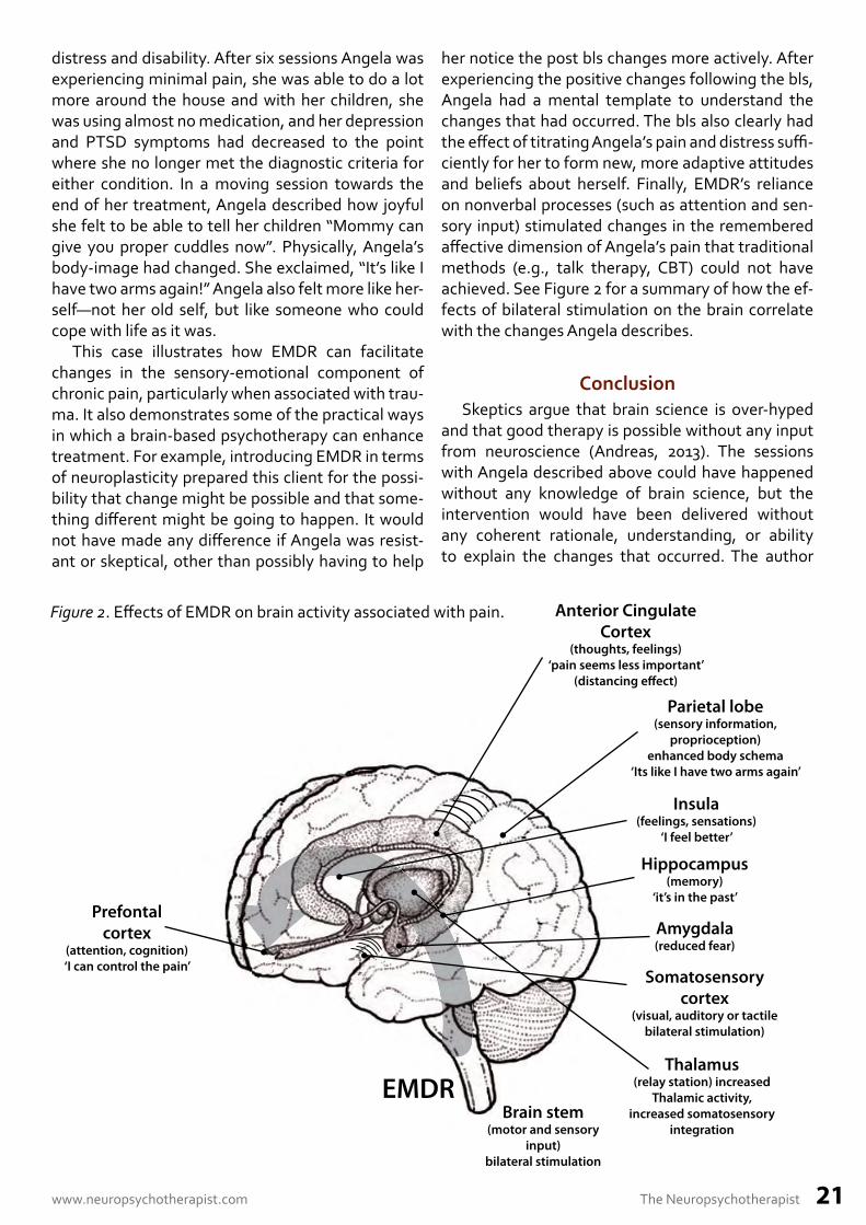
her notice the post bls changes more actively. After experiencing the positive changes following the bls, Angela had a mental template to understand the changes that had occurred. The bls also clearly had the effect of titrating Angela’s pain and distress suffi-ciently for her to form new, more adaptive attitudes and beliefs about herself. Finally, EMDR’s reliance on nonverbal processes (such as attention and sen-sory input) stimulated changes in the remembered affective dimension of Angela’s pain that traditional methods (e.g., talk therapy, CBT) could not have achieved. See Figure 2 for a summary of how the ef-fects of bilateral stimulation on the brain correlate with the changes Angela describes.
ConclusionSkeptics argue that brain science is over-hyped
and that good therapy is possible without any input from neuroscience (Andreas, 2013). The sessions with Angela described above could have happened without any knowledge of brain science, but the intervention would have been delivered without any coherent rationale, understanding, or ability to explain the changes that occurred. The author
Prefontalcortex
(attention, cognition)‘I can control the pain’
Insula(feelings, sensations)
‘I feel better’
EMDRBrain stem
(motor and sensory input)
bilateral stimulation
Somatosensory cortex
(visual, auditory or tactile bilateral stimulation)
Anterior Cingulate Cortex
(thoughts, feelings)‘pain seems less important’
(distancing e� ect)
Parietal lobe(sensory information,
proprioception) enhanced body schema
‘Its like I have two arms again’
Amygdala(reduced fear)
Thalamus(relay station) increased
Thalamic activity, increased somatosensory
integration
Hippocampus(memory)
‘it’s in the past’
Figure 2. Effects of EMDR on brain activity associated with pain.
The Neuropsychotherapist issue 14 May 201522
has found neuroscience indispensable in terms of assessing clients, understanding the processes of changes, and matching treatments to individual cli-ent’s personality characteristics. This learning has been stimulated by EMDR, which made no sense in terms of existing paradigms when I first learned it in the early 1990s. A combination of curiosity and dissatisfaction with existing treatments drove me to look deeper and led to a cascade of insights that have changed my understanding of what is possible in psychotherapy.
As I learned about the limbic system and the over-whelming power of emotions to influence thoughts, it made sense that trying to change how clients felt by changing their thoughts was a very slow and inef-ficient process. In fact I learned over many years that it was affect, rather than the client’s ability to appear coherent, that was the most telling clinical indicator. From sitting with clients I gradually learned to rec-ognize when there was a disconnect between a cli-ent’s story and their felt experience, and how to ac-cess dissociated emotion using the EMDR protocol or sometimes just by working with a bodily sensa-tion. Not directly asking clients to connect their feel-ings with their experience often made it easier for them to work with otherwise unspoken, unbearable memories. I integrate EMDR with elements of other therapies, including CBT, Ericksonian hypnosis, ha-komi and gestalt, but EMDR is my favorite tool, and it continues to stimulate curiosity about the brain.
ReferencesAndreas, S. (2013, July). Therapy isn’t brain sci-
ence. Psychotherapy Networker. Retrieved from http://www.psychotherapynetworker.org/maga-zine/recentissues/2013-julaug/item/2158-thera-py-isnt-brain-science
Alexander, J. (2013, March). Emotional style, chronic pain, and their neurological underpinnings. The Neuropsychotherapist, 2–11. Retrieved from http://www.neuropsychotherapist.com/emo-tional-style doi:10.12744/tnpt.21.03.2013.01
Apkarian, A. V., Thomas, P. S., Krauss, B. R., & Sze-verenyi, N. M. (2001). Prefrontal cortical hyper-activity in patients with sympathetically medi-ated chronic pain. Neuroscience Letters, 311(3), 193–197.
Aubert-Khalfa, S., Roques, J., & Blin, O. (2008). Evidence of a decrease in heart rate and skin conductance responses in PTSD sufferers after a single EMDR session. Journal of EMDR Practice and Research, 2(1), 51–56.
Australian Centre for Posttraumatic Mental Health. (2013). Australian guidelines for the treatment of adults with acute stress disorder and posttraumat-ic stress disorder. Melbourne, Victoria, Australia: ACPMH.
Bergmann, U. (2010). EMDR’s neurobiological mechanisms of action: A survey of 20 years of searching. Journal of EMDR Practice and Re-search, 4(1), 22–44.
Butler, A. C., Chapman, J. E., Forman, E. M., & Beck,
A. T. (2006). The empirical status of cognitive-behavioral therapy: A review of meta-analyses. Clinical Psychology Review, 26(1), 17–31.
Craig, A. D. (2003). Interoception: The sense of the physiological condition of the body. Current Opinion in Neurobiology, 13(4), 500–505.
Ecker, B., Ticic, R., & Hulley, L. (2012). Unlocking the emotional brain: Eliminating symptoms at their roots using memory reconsolidation. New York, NY: Routledge.
Elofsson, U., von Schèele, B., Theorell, T., & Sönder-gaard, H. P. (2008). Physiological correlates of eye movement desensitization and reprocessing. Journal of Anxiety Disorders, 22(4), 622–34.
Flor, H. (2002). Painful memories. Can we train chronic pain patients to ‘forget’ their pain? EBMO Reports, 3(4), 288–291.
Fuchs, T. (2004). Neurobiology and psychotherapy: An emerging dialogue. Current Opinion in Psy-chiatry, 17, 479–485. Retrieved from
Grant, M. (2014, August). Eye movement desensi-tization reprocessing treatment of chronic pain. OA Musculoskeletal Medicine, 2, 17. Retrieved from http://www.oapublishinglondon.com/arti-cle/1430
Grant, M. (2015). Pain control with EMDR (5th ed.). Kew, Victoria, Australia: Author.
Henry, D. E., Chiodo, A. E., & Yang. W. (2011). Cen-tral nervous system reorganization in a variety of chronic pain states: A review. PM&R: Journal of Injury, Function and Rehabilitation, 3(12), 1116–1125.
Kim, J. H., Kim, J. B., Sui, S. I., Seo, W. K., Oh, K., & Koh, S. B. (2014). Thickening of the somatosen-sory cortex in migraine without aura. Cephalal-gia, 34(14), 1125–1133.
Lee, C. W., & Cuijpers, P. (2012). A meta-analysis of the contribution of eye movements in processing emotional memories. Journal of Behavior Therapy and Experimental Psychiatry, 44(2), 231–239.
Lorenz, J., Minoshima, S., & Kasey, K. (2003). Keep-ing pain out of mind: The role of the dorsolateral
www.neuropsychotherapist.com 23The Neuropsychotherapist
prefrontal cortex in pain modulation, Brain, 126, 1079–1091.
doi: http://dx.doi.org/10.1093/brain/awg102 McGilchrist, I. (2009). The master and his emissary:
The divided brain and the making of the Western world. New Haven, CT: Yale University Press.
Ray, A. L., & Zbik, A. (2002). Cognitive behavioral therapies and beyond. In Tollison, C. D., Satterth-waite, J. R., & Tollison, J. W. (Eds), Practical pain management (pp. 189–207). Philadelphia, PA: Lippincott Williams & Wilkins.
Rome, H. P., Jr., & Rome, J. D. (2000). Limbically Augmented Pain Syndrome (LAPS): Kindling, corticolimbic sensitization, and convergence of affective and sensory symptoms in chronic pain disorders. Pain Medicine, 1(1), 7–13.
Roy-Byrne, P., Smith, W. R., Goldberg, J., Afari, N., & Buchwald, D. (2004). Post-traumatic stress disorder among patients with chronic pain and chronic fatigue. Psychological Medicine, 34(2), 363–368.
Sarno, J. (1998). The mindbody prescription: Healing the body, healing the pain. New York, NY: Warner Books.
Seminowicz, D. A., Shpaner, M., Keiser, M. L., Krauthamer, G. M., Mantegma, J., Dumas, J. A., . . . Naylor, M. R. (2013). Cognitive-behavioral therapy increases prefrontal cortex gray mat-ter in patients with chronic pain. Journal of Pain, 14(12), 1573–84.
Shapiro, F. (1995). Eye movement desensitization and reprocessing: Basic principles, procedures and protocols. New York, NY: Guilford Press.
Schneider, J., Hofman, A., Rost, C., & Shapiro, F. (2008). EMDR in the treatment of chronic phan-tom limb pain. Pain Medicine, 9(1), 76–82.
Söndergaard, H. P., & Elofsson, U. (2008). Psycho-physiological studies of eye movement desen-sitization and reprocessing. Journal of EMDR Practice and Research, 2(4), 282–288.
Symonds, L. L, Gordon, N. S., Bixby J. C., & Mande, M. M. (2006). Right-lateralized pain processing in the human cortex: An FMRI study. Journal of Neurophysiology, 95, 3823–3830.
Vachon-Presseau, E., Roy, M., Martel, M. O., Caron, E., Marin M. F., Chen J., . . . Rainville, P. (3013). The stress model of chronic pain: Evidence from basal cortisol and hippocampal structure and function in humans. Brain, 136, 815–827. doi:10.1093/brain/aws371
Van den Hout, M. A., Engelhard, I. M., Rijkeboer, M. M., Koekebakker, J., Hornsveld, H., Leer, A., . . . Akse, N. (2011). EMDR: Eye movements superior to beeps in taxing working memory and reducing vividness of recollections. Behavior Research and Therapy, 49(2), 92–98.
Van der Kolk, B. A. (1994). The body keeps the score: Memory and the evolving psychobiology of posttraumatic stress disorder. Harvard Review of Psychiatry, 1(5), 253–265.
Van der Kolk, B. A. (n.d.). EMDR and the lessons from neuroscience research. Retrieved from http://www.emdr.org.il/dls/1.html
Van Rood, Y. R., & de Roos, C. (2009). EMDR in the treatment of medically unexplained symptoms: A systematic review. Journal of EMDR Practice and Research, 3(4), 248–263.
Williams, A. C., Eccleston, C., & Morley, S. (2012). Psychological therapies for the management of chronic pain (excluding headache) in adults. The Cochrane Database of Systematic Reviews, 11, CD007407. doi:10.1002/14651858.CD007407.pub3.
Mark Grant, MA, is a clinical psychologist with over 20 years’ experience in the treatment of stress, trauma, and pain. Mark’s main interest and experience is in the role of negative emotion (resulting from stress) as a cause and an effect of health prob-lems. On the basis of recent discoveries from brain scans, Mark regards emotion as the key to many psychological problems, but also a powerful resource. In association with this, he is interested in treatments which harness people’s emotional resources. Find out about Mark Grant at http://overcomingpain.com
The Neuropsychotherapist issue 14 May 201524
Change Your Brain, Change Your PainMark Grant, MA
Based on the latest discoveries about brain structure and functioning, this book and CD set explains how physical and emotional pain are linked in the brain and how to overcome pain by reversing the brain processes that maintain it.
The book begins by explaining how the brain works and how brain functioning is disrupted by stress. Stress is one of the leading causes of pain. Readers are then shown how to create the conditions where healing can occur including safety and support, reconnecting with feelings and improved sleep and physical well-being.
With the CD readers are then taught how to used focused attention, auditory stimulation and memory to stimulate unconscious non-verbal areas of the brain where pain is stored. Dual Attention Stimulus (DAS) as the process is known, is a treatment element of EMDR, a cutting-edge treatment for PTSD. Because of the
close connection these areas have with the body, they enable faster and more effective al-teration of pain memories.
One of the key ideas of this book is that any kind of pain other than acute physical pain involves memory, and memories are changeable.
The CD of accompanying brain stimulation exercises has to be purchased as a separate item (both are offered at a discounted price). These materials will help you access a different part of your brain!
Available from Amazon.com or visit overcomingpain.com.
Also available from Amazon.com by Mark Grant
Pain Control with EMDR
This 240 page manual describes how to use EMDR in the treatment of chronic pain. Includes protocol, de-sensitization guidelines, assessment tools and over 30 pages of treatment aids and resources based on the in-formation processing model.

www.neuropsychotherapist.com 25The Neuropsychotherapist
www.tnptinstitute.comThe Neuropsychotherapy Institute.
The neuropsychotherapy institute learning platform has been created for psychothera-pists, psychologists, and other mental health professionals, to educate them in the new par-adigm of neuropsychotherapy for more effective clinical practice.
Neuropsychotherapy is a multidisciplinary perspective on mental well-being that looks to neuroscience and other related fields of human biology and psychology to enhance the clinical practice of talking therapies. The Neuropsychotherapy Institute will provide you with a sound foundational understanding of the neurobiology of mental life and how that knowl-edge can inform psychotherapy and increase the effectiveness of your practice. The Insti-tute offers courses on a Continuing Education or Professional Development credit basis* as each unit keeps you up-to-date with the latest science and practice of psychotherapy.