neuropsychiatry block - ksumscksumsc.com/download_center/2nd/1) neuropsychiatry block/female...pool...
TRANSCRIPT

NeuroPsychiatry Block Spinal Cord Functions & Spinal
Reflexes Prof. Faten Zakareia
Physiology Department College of Medicine King Saud University
2017 Email: [email protected] Ext:52736

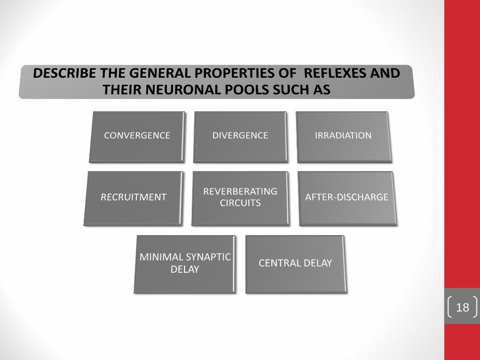
Objectives: Upon completion of this lecture, students should be able to:
- Appreciate the two-way traffic along the spinal cord . -- Describe the organization of the spinal cord for motor functions(anterior horn cells& interneurons & neuronal pools) - Describe the physiological role of the spinal cord in spinal reflexes & reflex arc components
--Classify reflexes into superficial and deep; monosynaptic & polysynaptic -Describe withdrawal reflex & crossed extensor reflex --Recognize the general properties of properties of spinal cord reflexes -Reference book/Chapter 55 (Guyton & Hall)
- Review of Human physiology by Ganong (last edition)

The spinal cord has 31 pairs of spinal nerves Each spinal nerve has has ventral & dorsal roots : • The dorsal(posterior) root contains afferent (sensory) nerves coming from receptors . • The cell body of these neurons is located in dorsal ( posterior ) root ganglion ( DRG) • The ventral(anterior) root carries efferent (motor) fibers • The cell-body of these motor fibres is located in the ventral (anterior ) horn of the spinal cord
THE SPINAL CORD

Functions of the Spinal Cord 1- The two-way traffic along the spinal cord A-Sensory signals from receptors enter the cord through the sensory (posterior) roots, then every sensory signal travels to two separate destinations:
1-One branch of the sensory nerve terminates in the gray matter of the cord and elicits local segmental cord reflexes
2-Another branch transmits signals to higher levels in the cord , or to the brain stem, or even to the cerebral cortex through:-
A- spinal ascending sensory tracts as:_ - Dorsal Column Tracts ( Gracile &Cuneate ) - Lateral & Anterior Spinothalamic Tract. - Spinocerebellar Tracts
B- Spinal decending motor tracts:-Motor signals & brain motor commands pass through descending motor tracts & then to spinal efferent motor nerves to skeletal muscles to execute motor functions

2- Initiate spinal reflexes As withdrawal & stretch reflex

The organization of the spinal cord for motor functions (anterior horn cells& interneurons& neuronal pools)
Located in each segment of the anterior horns of the cord gray matter , several thousand neurons that are 50
to 100 percent larger than others neurons .They give rise to the nerve fibers that leave
the cord in the ventral roots and directly innervate the skeletal muscle fibers.
• 1-Alpha motor neurons:_
• -They give rise to large type A alpha motor nerve fibers,
• 2-Gamma motor neurons:-
-smaller gamma motor neurons
-They transmit impulses through much smaller type A gamma motor nerve fibers

Q-What is the motor unit?

Spinal reflexes
What is a reflex?
-Functional unit of CNS,
rapid, automatic ,
involuntary response to a
stimulus that involve
spinal neurons only
-example/pinprick causes
withdrawal. R

Components of the reflex arc
REFLEX ARC IS THE PATHWAY FOLLOWED BY NERVE IMPULSES THAT PRODUCE A REFLEX IS A REFLEX ARC (REFLEX CIRCUIT)
9

SENSORY RECEPTOR |1|
10

SENSORY NEURON |2|
11

INTEGRATING CENTRE|3|
12

- Interneurons & interneuron pool
- Interneurons are present in the gray matter in the dorsal
horns, the anterior horns, and the intermediate areas between
them.
- -30 times as numerous as the anterior motor neurons, small and
highly excitable, often exhibiting spontaneous activity
- Different types of neuronal circuits are found in the interneuron pool (parallel and reverberating circuits).
- Have diverging, converging, and repetitive-discharge
- They are (excitatory or inhibitory).

• Renshaw Cells :- • - As the anterior motor neuron axon leaves the
body of the neuron, sends collateral branches to
adjacent Renshaw cells.
• - These are inhibitory cells that transmit
inhibitory signals to the surrounding motor
neurons BY Lateral inhibition/ So stimulation
of one motor neuron tends to inhibit adjacent
motor neurons .
• -This lateral inhibition helps to focus or sharpen
the signals from each excited motor neuron
• allow transmission of the primary signal in the
desired direction& suppressing the tendency for
signals to spread laterally)

-Efferent neuron
15

- MOTOR NEURON|4|( Efferent neurons)
- Anterior Horn Cells (Motor neurons)
of spinal cord supplying skeletal muscle:
1. alpha motor neurons :- large cells, with large mylinated fibres (axons) form 70% of ventral root - supply extrafusal muscle fibres (2/3 Of skeletal muscle fibers)
2. Gamma motor neurons :- smaller cells- with thiner axons form 30 % of ventral root - supply
intrafusal muscle fibres (muscle spindles=1/3 Of skeletal muscle fibers)

EFFECTOR|5|
17
18

- Sensory afferent enter spinal cord via dorsal(posterior) root, as they enter the neuronal pool undergo:
1- Divergence help to spread a single stimulus to a
wide area of the spinal cord( amplification of signal)
- it is important for weak signals to excite far
greater numbers of nerve fibers leaving the pool.
2-Convergence :- signals from multiple inputs unit
to excite a single neuron
-multiple action potentials converging on the neuron
provide enough spatial summation to bring the neuron
to the threshold required for discharge.
- (multiple stimuli summate & collect together at the
same time)

• CONVERGENCE |
20
DIVERGENCE

3-Reciprocal inhibition circuits -Stimulation of flexors muscle accompanied by inhibition of
extensors through inhibitory interneurons , the neuronal
circuit that causes this reciprocal relation is called
reciprocal innervation_
-Reflex contraction of an agonist muscle is accompanied by
inhibition of the antagonist. -the input fibre directly excites the excitatory output pathway, but it stimulates an intermediate inhibitory neuron (neuron 2), which secretes a different type of transmitter substance to inhibit the second output pathway from the pool. (3)
-Value/ preventing over activity in many parts of the spinal cord.
21

Neuronal pool circuits 1- Parallel 2-Reverbrating
1-Parallel circuits //afferent and
efferent are parallel to each other
(input parallel to output)

• 4-Reverberatory (Oscillatory) Circuit
• 1-The simplest reverberatory circuits involves only a single
neuron
• -the output neuron sends a collateral nerve fiber back to its own
dendrites or soma to restimulate the input neuron itself & so the
circuit may discharge repetitively for a long time and causes
signal prolongation (Allow prolonged discharge of the same
motor neurons by a single stimulus
• - Amore complex circuits in which both facilitatory and
inhibitory fibers involved on the reverberating circuit.
• A facilitatory signal enhances the intensity and frequency of
reverberation, whereas an inhibitory signal depresses or stops the
reverberation.
• - Most reverberating pathways are constituted of many parallel
fibers


5-After-discharge:-
- A prolonged maintained output discharge of AHCs called after -discharge, lasting a few milliseconds or many minutes after the incoming signal is over.
- After- discharge occurs due to the following:-
- 1-Synaptic After-discharge.
When excitatory synapses discharge on the surfaces of dendrites or soma of a neuron, a postsynaptic electrical potential ( PSP)develops in the neuron and lasts for milliseconds. As long as this potential lasts, it can continue to excite the neuron to transmit (a series of continuous repetitive discharges) .
- 2- Reverbrating circuits
- Presence of reverberating circuit restimulate AHCs

6-SYNAPTIC DELAY ( central delay) -Is the time of reflex to pass through neurons of the spinal cord
-
-The minimal period of time required for transmission of a neuronal signal from
a presynaptic neuron to a postsynaptic neuron, is SYNAPTIC DELAY.
-Equals 0.5 ms /synapse ( it is long in polysynaptic Reflex).
- It is > 2 ms in the withdrawal R (polysynaptic Reflex)
-Number of synapses in a reflex = central delay / 0.5ms
-for knee jerk it equals 0.6 msc = one synapse
26

Types of spinal reflexes -According to number of neurons:-
Monosynaptic
Sensory axon (afferent)synapse directly with anterior horn cell- (No interneuron )
Ex.Stretch reflex
Polysynaptic
Sensory axon (afferent)synapse with one or
more interneuron
Ex.Withdarwal,abdominal reflexes, visceral

Types of reflexes -According to site of the receptor:-
(A)Deep Reflexes:- by stimulation of receptors deep in muscle and tendons
(1) Stretch Reflexes (Tendon jerks) ,they are monosynaptic : such as knee-jerk ( patellar reflex ) and ankle jerk . The receptor for all these is the muscle spindle ( is located deep within the muscle
itself (2) Inverse Stretch Reflex ( Golgi Tendon organ reflex ) , polysynaptic : The receptor is called Golgi Tendon Organ present deep in the muscle tendon
(B) Superficial Reflexes
Are polysynaptic reflexes . The receptor are superficial in the skin . Examples are
Withdrawal, abdominal reflexes and plantar reflex
©Visceral:-by stimulation of receptors in wall of viscera
As Micturition, defecation

SUPERFICIAL AND DEEP REFLEXES
29

Withdrawal reflex(flexor reflex)
(Nociceptive Reflex)
-A superficial polysynaptic reflex Stimulation of pain receptors of hand(a pin- prick, heat, or a wound)>>>>>> impulses to SC in A delta or C fibres >>>>>>> interneurons pool >> motor neurons >> stimulate hand flexor muscles >>move the hand away from the injurious stimulus.
characterised by :_ 1- diverging circuits to spread the reflex to the necessary muscles for withdrawal (2) reciprocal inhibition circuits Circuits to inhibit the antagonist muscles -Stimulation of flexors muscle accompanied by inhibition of extensors through inhibitory interneurons


3- RECRUITMENT :
- Gradual activation of more number of motor neurons (AHCS) on stim of afferent nerve in a reflex arc by maintained, repetitive stimulus
Cause/ 1-different conduction velocities of afferents some are slowly
&others are rapidly conducting fibres 2-different number of interneurons with short & long pathways to
the motor neurons (AHCs ) (impulses do not reach AHCs at same time but reach them gradually, so maintained stimulation allow more neurones to be stimulated)
Motor unit recruitment : If a repetitive &stronger stimulus is maintained, there
will be gradual increase in the force of the muscle contraction until the maximum force is reached , due to gradual recruitment/activation of more and more motor neurons

4- After-discharge CIRCUITS:- Circuits to cause afterdischarge lasting many fractions of a second after the stimulus is over.
-The duration of after-discharge depends on the
intensity of the sensory stimulus that elicited the reflex
Cause/ -Presence of reverberating circuit restimulate AHCs -Value /prolong the protective response of reflex

5- IRRADIATION :- - spread of impulses up & down to different segments and motor neurons in the S.C
A strong stim in sensory afferent irradiate to many segments of S.C due to divergence
****The extent of the response in a reflex depends on the intensity of the stimulus.
- The more intense the stimulus >>>> greater spread of activity in the spinal cord >>>involving more & more motor neurons>>>more response
- Weak stim-----irradiates to small number of neurons ,
so it causes weak flexion of limb
- Strong stim---- irradiates to large number of neurons ,
so it causes withdrawal of affected limb &
extension of opposite limb.(as in crossed extensor reflex)

• 6- “local sign” Pattern of Withdrawal.
The pattern of withdrawal that results when the flexor reflex
is elicited depends on which sensory nerve is stimulated.
• - Thus, a pain stimulus on the inward side of the arm
elicits not only contraction of the flexor muscles of the arm
but also contraction of abductor muscles to pull the arm
outward.
• This is called the principle of “local sign”

THE WITHDRAWAL REFLEX |
REFLEX FLEXOR REFLEX OR WITHDRAWAL REFLEX
CLINICAL TEST | STIMULUS
SHARP PAINFUL STIMULUS (STEPPING ON NAIL)
RESPONSE LIMB IS RAPIDLY WITHDRAWN
SENSORY RECEPTOR
CUTANEOUS SKIN AND PAIN RECEPTORS
SYNAPSES INVOLVED
POLYSYNAPTIC (VIA INTERNEURON)
EFFECTS ON MUSCLE
CONTRACTS FLEXOR MUSCLE
OTHER EFFECTS RELAXES (-) EXTENSOR MUSCLE OF SAME LIMB
REVERSE EFFECT ON OPPOSITE LIMB (CROSS EXTENSOR REFLEX)
FUNCTION PROTECTIVE – WITHDRAWAL FROM PAINFUL STIMULUS
CROSS EXTENSOR AIDS IN MAINTAINING POSTURE WHEN OPPOSING LEG IS LIFTED
http
s://m
usom
.ma
rsha
ll.ed
u/a
na
tom
y/g
rossho
m/a
llpp
t/pdf/S
pin
alre
flexe
s.
pd
f
36

Crossed Extensor Reflex:- Crossed extensor reflex supporting the body weight against gravity While pushing
the body away from the injurious agent by withdrawal R ,the Flexion and withdrawal of the
stimulated limb >> extension of the opposite limb
- Occurs with strong stimulus only. why?
Signals from sensory neurons cross to the opposite side of the cord to excite extensor muscles
motor neurons.
-It does not begin until 200 to 500 milliseconds after onset of the initial pain stimulus, because
many interneurons are involved in the circuit between the incoming sensory neuron and the
motor neurons of the opposite side of the cord
-After the painful stimulus is removed, the crossed extensor reflex has an even longer period of
afterdischarge, results from reverberating circuits among the interneuronal cells.
The prolonged afterdischarge is of benefit in holding the body away from the painful object
- Mostly in the lower limb to support balance.
- It has reciprocal innervations occurs also in crossed extensor reflex. How?
-flexors in the opposite limb are inhibited while extensors are excited supporting the body weight
against gravity during withdrawal .R

Crossed extensor reflex