neuralprecursorcell-expresseddevelopmentallydown ... · mass spectrometry—enzymatic digests of...
TRANSCRIPT

Neural Precursor Cell-expressed Developmentally Down-regulated Protein 4-2 (Nedd4-2) Regulation by 14-3-3 ProteinBinding at Canonical Serum and Glucocorticoid Kinase 1(SGK1) Phosphorylation Sites*□S
Received for publication, August 12, 2011 Published, JBC Papers in Press, September 7, 2011, DOI 10.1074/jbc.M111.293233
Sindhu Chandran‡, Hui Li§, Wuxing Dong‡, Karolina Krasinska¶, Chris Adams¶, Ludmila Alexandrova¶, Allis Chien¶,Kenneth R. Hallows§, and Vivek Bhalla‡
From the ‡Division of Nephrology, Department of Medicine, Stanford University School of Medicine, Stanford, California 94305, the§Renal-Electrolyte Division, Department of Medicine, University of Pittsburgh School of Medicine, Pittsburgh, Pennsylvania 15261,and the ¶Mass Spectrometry Facility, Stanford University, Stanford, California 94305
Background: Coordinate regulation by kinases and 14-3-3 proteins regulates sodium transport through phosphorylationand inhibition of E3 ligases.Results: Phosphorylation at similar but distinct target motifs can either inhibit or stabilize E3 ligases.Conclusion: E3 ligases integrate multiple kinase inputs to regulate sodium transport and protein stability.Significance:These findings broaden our knowledge of how E3 ligases and sodium transport are regulated by phosphorylation.
Regulation of epithelial Na� channel (ENaC)-mediated trans-port in the distal nephron is a critical determinant of blood pres-sure in humans. Aldosterone via serum and glucocorticoidkinase 1 (SGK1) stimulates ENaC by phosphorylation of the E3ubiquitin ligaseNedd4-2,which induces interactionwith 14-3-3proteins. However, themechanisms of SGK1- and 14-3-3-medi-ated regulation of Nedd4-2 are unclear. There are three canon-ical SGK1 target sites onNedd4-2 that overlapphosphorylation-dependent 14-3-3 interaction motifs. Two of these are termed“minor,” and one is termed “major,” based on weak or strongbinding to 14-3-3 proteins, respectively. By mass spectrometry,we found that aldosterone significantly stimulates phosphoryl-ation of a minor, relative to the major, 14-3-3 binding site onNedd4-2. Phosphorylation-deficient minor site Nedd4-2mutants bound less 14-3-3 than did wild-type (WT) Nedd4-2,and minor site Nedd4-2 mutations were sufficient to inhibitSGK1 stimulation of ENaC cell surface expression. Asmeasuredby pulse-chase and cycloheximide chase assays, a major bindingsite Nedd4-2 mutant had a shorter cellular half-life than WTNedd4-2, but this property was not dependent on binding to14-3-3. Additionally, a dimerization-deficient 14-3-3� mutantfailed to bind Nedd4-2. We conclude that whereas phosphoryl-ation at theNedd4-2major site is important for interactionwith14-3-3 dimers, minor site phosphorylation by SGK1may be therelevant molecular switch that stabilizes Nedd4-2 interaction
with 14-3-3 and thus promotes ENaC cell surface expression.We also propose that major site phosphorylation promotes cel-lular Nedd4-2 protein stability, which potentially represents anovel form of regulation for turnover of E3 ubiquitin ligases.
Nedd4-2,2 a member of the homology to the E6-associatedprotein C terminus (HECT) family of E3 ubiquitin ligases (1, 2),is a physiologically important regulator of the epithelial Na�
channel (ENaC) (1, 3). ENaCcomprises�,�, and� subunits andreabsorbs Na� across several epithelia, including the distalnephron (4). Nedd4-2 is a potent inhibitor of ENaC activity invivo. Liddle syndrome, a severe form of inherited hypertension,results from mutations in ENaC that abolish interaction withNedd4-2 and thus cause renal Na� retention (5, 6). Moreover,Nedd4-2 knock-out mice have salt-sensitive hypertension (7).In cultured cells Nedd4-2 ligase activity decreases ENaC cellsurface expression and Na� current (8). Potential effects ofNedd4-2-mediated ENaC ubiquitination include increasedendocytosis from the plasma membrane and enhanced protea-somal and/or lysosomal degradation (9, 10). Furthermore,Nedd4-2 may decrease ENaC open probability by limitingENaC cell surface residence time and thus proteolytic cleavageof its � and � subunits (11).
Phosphorylation is an important mechanism for the regula-tion of Nedd4-2 and other E3 ligases. For example, AMP-acti-vated protein kinase and others negatively regulate ENaCthrough stimulatory phosphorylation of Nedd4-2, whichenhances ENaC interaction orNedd4-2 ubiquitin ligase activity(12, 13). Conversely, serum and glucocorticoid kinase 1 (SGK1)and protein kinase A (PKA) can positively regulate ENaC
* This work was supported, in whole or in part, by National Institutes of HealthGrants T32 DK007357-26 (to S. C.), R01 DK075048 (to K. R. H), and K08DK071648 (to V. B.). This work was also supported by National KidneyFoundation Young Investigator Grant YIB787 (to V. B.) and by NationalCenter for Research Resources Award S10RR027425 (to the Stanford Uni-versity Mass Spectrometry Facility).
□S The on-line version of this article (available at http://www.jbc.org) containssupplemental Figs. 1– 4, Table 1, Experimental Procedures, and additionalreferences.
1 To whom correspondence should be addressed: Division of Nephrology,780 Welch Rd., Suite 106, Stanford University, Palo Alto, CA 94304. Fax:650-721-1443; E-mail: [email protected].
2 The abbreviations used are: Nedd4-2, neural precursor cell-expressed,developmentally down-regulated protein 4-2; DD, dimerization-deficient;ENaC, epithelial Na� channel; EYFP, enhanced YFP; mpkCCDc14, mousepolarized kidney cortical collecting duct; MS/MS, tandem MS; SGK1, serumand glucocorticoid kinase 1; xNedd4-2, Xenopus Nedd4-2.
THE JOURNAL OF BIOLOGICAL CHEMISTRY VOL. 286, NO. 43, pp. 37830 –37840, October 28, 2011© 2011 by The American Society for Biochemistry and Molecular Biology, Inc. Printed in the U.S.A.
37830 JOURNAL OF BIOLOGICAL CHEMISTRY VOLUME 286 • NUMBER 43 • OCTOBER 28, 2011

through inhibitory phosphorylation ofNedd4-2 at three canon-ical sites. Alanine mutations at these three sites diminish SGK1and PKA-stimulated ENaC cell surface expression and activity(1, 14). SGK1 and/or PKA-mediated Nedd4-2 phosphoryla-tion-dependent interaction with 14-3-3 scaffolding proteinsprevents subsequent interaction with and ubiquitination ofENaC (15–17). Aldosterone stimulates SGK1 expression,whereas vasopressin activates PKA. Therefore, Nedd4-2 is aconvergence point for hormonal regulation of epithelial Na�
transport in the distal nephron.The mechanisms of aldosterone-mediated regulation of
Nedd4-2 by 14-3-3 are poorly characterized. In general, 14-3-3proteins bind as dimers to phosphorylated ligands (e.g.Nedd4-2) at one high affinity (major) site, which facilitates theinteraction with a lower affinity (minor) site (18). 14-3-3 pro-teins propagate kinase-dependent signaling events by regulat-ing their target proteins via several mechanisms, includingactivity, subcellular localization, and/or stability. Alanine sub-stitution at either minor (Ser-338, Thr-363) or major (Ser-444)sites on Nedd4-2 (Xenopus numbering) disrupt SGK1 stimula-tion of ENaC (1, 14, 15). Also, abrogation of binding to phos-phorylated Ser-444 via a S444Amutant or Ser(P)-444 antibodydecreases binding of Nedd4-2 to 14-3-3 (15–17). However, inheterologous expression systems we have reported that SGK1-dependent enhancement of Nedd4-2 phosphorylation at Ser-338 (�4.5-fold) and Thr-363 (�40-fold) is substantially greaterthan at Ser-444 (1.5-fold) (13). Moreover, we and others haveobserved a high level of basal phosphorylation at Ser-444 incells (13, 19). These findings suggest that this residue may notbe the principal regulatory site of aldosterone-dependent stim-ulation of ENaC.We have identified a significant role for Nedd4-2 minor site
phosphorylation as a trigger for enhanced interaction with14-3-3 and thus aldosterone-mediated ENaC stimulation. Ourdata also suggest that basal major site phosphorylation, whichalso participates in interaction with dimeric 14-3-3, is impor-tant for regulating the cellular stability of Nedd4-2.
EXPERIMENTAL PROCEDURES
Antibodies—A rabbit polyclonal antibody recognizing theWW2 domain of Nedd4 and Nedd4-2 (Millipore) was usedfor immunoprecipitation of endogenous Nedd4-2, and a rab-bit polyclonal Nedd4-2 antibody (Abcam) was used forimmunoblotting. Rabbit serum against Ser(P)-328 of mouseNedd4-2 (equivalent to Ser-444 of Xenopus Nedd4-2(xNedd4-2) was a gift from Jon Loffing (University of Zurich)(19). Rabbit polyclonal antibodies were used for detection ofendogenous SGK1 (Sigma), E-cadherin (Sigma), and 14-3-3proteins (Santa Cruz Biotechnology). Commercial antibod-ies against epitope tags were also used: HA (Roche AppliedScience), FLAG (Sigma), and GFP (Clontech; to detectEYFP), and 1D4 (Millipore).DNA Constructs—All FLAG-tagged xNedd4-2 cDNAs and
constitutively activemouse SGK1 (S422D) were subcloned intoa modified pcDNA3 vector (pMO) using PCR-directedmutagenesis. An HA-tagged dimerization-deficient mutantwas created using site-directed mutagenesis (QuikChange;Stratagene) of the following mouse 14-3-3� amino acids: E5K,
LAE12,13,14QQR, Y85Q,M88N, and E90Q, adapted fromTvi-zion et al. (20). EYFP-tagged R18WT (wild-type) or R18M(mutant) were gifts fromHaian Fu (Emory University) (21). Allmutants and constructs were confirmed by DNA sequencing.Cell Culture—Human embryonic kidney (HEK-293T) cells
and mouse polarized kidney cortical collecting duct (mpkC-CDc14) cells were maintained and cultured as described previ-ously (15).Co-immunoprecipitation of Endogenous Nedd4-2—mpkC-
CDc14 cells were plated on collagen-coated 1.0-cm2 Transwells(Corning Costar) at superconfluent density. When monolayersreached a transepithelial resistance �1000 ohm-cm2 (22), cellswere incubated in serum-free, supplement-free medium for24 h prior to treatment. Following treatment for 3 h with etha-nol (vehicle) or 1 �M aldosterone (Sigma), cells were lysed in150 �l of lysis buffer (10 mM Tris-HCl (pH 7.4), 50 mM EDTA,0.4% deoxycholate acid, and 1% Nonidet P-40) containing pro-tease inhibitors (1mMphenylmethylsulfonyl fluoride (PMSF), 1mM, benzamidine, and 1� Complete Protease Inhibitor Mix-ture (Roche Applied Science)) and phosphatase inhibitors (25mM sodium fluoride, 2 �M microcystin-LR, and 1� Phospha-tase-Inhibitor Mixture Sets I and II (Calbiochem)). Immuno-precipitation was performed overnight with anti-Nedd4 anti-body and protein A-agarose (Santa Cruz Biotechnology) withgentle rocking at 4 °C. Controls for immunoprecipitation wereperformed using a concentration of normal IgG (Santa CruzBiotechnology) equivalent to that of the primary precipitatingantibody. Samples were separated by SDS-PAGE and stainedwith Coomassie Blue or transferred to nitrocellulose forimmunoblotting.Relative Quantitation of Nedd4-2 Phosphorylation Sites by
Mass Spectrometry—The band containing Nedd4-2 wasexcised from a Coomassie-stained gel (see Co-immunoprecipi-tation section, above), followed by in-gel digestion with trypsinand addition of acrylamide as the alkylating agent as describedpreviously (23), with the addition of Protease Max (Promega)for increased peptide recovery and shorter digestion times.Extracted peptides were dried in a SpeedVac to completion.Prior to analysis, a fraction of the peptide pool was reconsti-tuted using 97.8% water, 2% acetonitrile, 0.2% formic acid andinjected onto a self-packed fused silica 150-�m inner diameterC18 column. The LC was an Eksigent nano2D LC, run at 600nl/min. The LCgradientwas either 60 or 80min, linear from2%mobile phase B to 35% mobile phase B followed by a ramp to65%mobile phase B. The eluatewas nanoelectrosprayed using aMichrom Advance source (Auburn) into an LTQ OrbitrapVelos mass spectrometer (MS) (Thermo Fisher), which was seton data-dependent acquisition mode, sequencing using HCDand/or CAD the top eight most intense ions of charge state 2�or greater. The. RAW files were converted offline to mzXMLformat using a msconvert script and searched using a Sorcererwork station with a Sequest search engine. The static modifica-tion was Cys propionamide, and variable modifications wereMet oxidation and Ser, Thr, or Tyr phosphorylation. Thesearched results were uploaded into a Scaffold viewer for easiervisualization. For relative quantitation of changes in phospho-rylation stoichiometry, the ratio of the intensity of peaks inextracted ion chromatograms of phosphorylated and nonphos-
Characterization of SGK1 Phosphorylation Sites on Nedd4-2
OCTOBER 28, 2011 • VOLUME 286 • NUMBER 43 JOURNAL OF BIOLOGICAL CHEMISTRY 37831

phorylated versions of the same peptides acquired by MS werecompared. The identified nonphosphorylated and phosphoryl-ated peptides encompassing residues equivalent to Ser-338,Thr-363, and Ser-444 were then synthesized in vitro (Anaspec)and used for determination of absolute quantitation of endog-enous Nedd4-2.Absolute Quantitation of Nedd4-2 Phosphorylation Sites by
Mass Spectrometry—Enzymatic digests of Nedd4-2 were sepa-rated by LC-MS/MS using an Agilent 1100HPLC system. Sam-ples (10 �l) were injected in duplicate onto a reverse phasecolumn (KinetexC18, 50� 2.1mm, 2.6-�mparticle size, 100-Åpore size; Phenomenex). Components were eluted at 25 °C at aflow rate of 250 nl/min. The gradient HPLC method consistedof the following: 5%B, 0–0.3min, linear ramp to 35%B at 6minand to 60% B at 7 min, followed by ramp back to 10% B at 7.5min and reequilibration at 10%B for 2.5min, for a total run timeof 10 min. Mobile phases were: A � 0.1% formic acid in water(v/v); and B � 0.1% formic acid in acetonitrile (v/v). The LCeluate was introduced into theMS ion source without splitting.A Quattro Premier triple quadrupole mass spectrometer(Waters-Micromass) equipped with a standard electrosprayion source was used for detection. TheMS was operated with acapillary voltage of 3.5 kV, source temperature of 120 °C, des-olvation temperature of 350 °C, and collision gas (argon) pres-sure of 2.8� 10�3 mbar. Positive ion selected reactionmonitormode was used for monitoring the transitions of ions for eachanalyte (supplemental Table 1).
The lower limit of quantitation, defined as 10:1 signal-to-noise ratio for the analytes varied (supplemental Table 1).Standard calibration curves were obtained over the concentra-tion range 0.0025–2 pmol/�l. A 13C-5/15N-valine-labeled pep-tide corresponding to nonphosphorylated Ser-338 was spikedinto each sample and calibration curve solutions at 1 �M andserved as an internal standard. Instrumentation control anddata analyses were accomplished using MassLynx 4.1 andQuanLynx 4.1 software (Waters-Micromass).Co-immunoprecipitation of Nedd4-2 and 14-3-3 in HEK-
293T Cells—Cells were transiently transfected as indicatedwith FLAG-taggedNedd4-2, SGK1 S422D,HA-tagged 14-3-3�,EYFP-tagged R18 peptide, and as described previously (13).Immunoprecipitation was performed with M2 anti-FLAG(Sigma) or anti-HA (Roche Applied Science) antibody conju-gated to resin, and samples were separated by SDS-PAGE andimmunoblotted with anti-FLAG or anti-HA-horseradish per-oxidase 3F10. Endogenous 14-3-3 isoforms were detected withanti-pan-14-3-3 (�-19) antibody. Scanning densitometry wasmeasured using ImageJ software, and values for immunopre-cipitated 14-3-3 were normalized to Nedd4-2 within the samelane.ENaC Surface Biotinylation Assay—HEK-293T cells were
co-transfected with � (HA-tagged), �, and � subunits of mouseENaC, FLAG-Nedd4-2, and SGK1. Two days after transfection,cells were washed with modified (M)-PBS (Invitrogen D-PBSwith 1 mM MgCl2 and CaCl2 (pH 7.8)), and then cell surfaceproteins were labeled with 0.5 mg/ml sulfo-NHS-biotin(Pierce) in M-PBS for 30 min on ice and then quenched with100 mM glycine in M-PBS for 10 min at 4 °C. After washingthree times with M-PBS, cells were lysed in Nonidet P-40 lysis
buffer (0.4% sodium deoxycholate, 1% Nonidet P-40, 63 mmEDTA, 50mmTris-HCl (pH 8), and protease inhibitors). Bioti-nylated (cell surface) and interacting proteins were isolated byincubating cell lysate with immobilized NeutrAvidin beads(Pierce) overnight with gentle rocking at 4 °C. Following sepa-ration by SDS-PAGE, biotinylated HA-tagged � ENaC wasdetected by immunoblotting and quantified by scanning densi-tometry. Anti-pan-cadherin antibody was used as a loadingcontrol for cell surface proteins.Pulse-Chase Assays—HEK-293T cells were co-transfected
with 1 �g of WT or mutant FLAG-tagged Nedd4-2 (3 �g forS444A mutant) constructs with 1 �g of EYFP-tagged WT ormutant R18 peptide. Twenty-four hours after transfection,cells were starved in cysteine- and methionine-free modifiedEagle’s medium (MEM) for 30 min and then radiolabeledwith 0.05 mCi/ml Tran35S-label (MP Biomedicals) for 15min. Cells were chased in serum-free MEM at 37 °C for 0 to8 h. At the appropriate chase time, cells were rinsed twicewith PBS and lysed in radioimmune precipitation assaybuffer (10 mM Tris-HCl (pH 7.4), 0.15 M NaCl, 1% TritonX-100, 1% deoxycholate, 1% Nonidet P-40, 0.1% SDS) sup-plemented with phosphatase and protease inhibitors. FLAG-Nedd4-2 was immunoprecipitated using M2 anti-FLAGantibody coupled to protein A/G beads (Pierce), as describedpreviously (13). Immunoprecipitated FLAG-Nedd4-2 wasresolved by SDS-PAGE, and dried gels were analyzed using aPhosphorImager (Bio-Rad). The detected bands were quan-titated using Quantity One software (Bio-Rad). For experi-ments with S444A Nedd4-2, values for each time point werefitted to an exponential decay curve using IGOR-Pro 4.0software (WaveMetrics), and half-life values were calculatedfrom the decay constant obtained from each fitted curve.The relative protein amount (N(t)) at each time point wasmodeled as N(t) � N0�exp(�t/�), where N0 is the proteinamount at time 0, and inverse tau (1/�) is the time constant.Half-life (t1⁄2) values were calculated as t1⁄2� ��ln2.Cycloheximide Chase Assays—HEK-293T cells were
co-transfected with 0.4 �g of WT or mutant FLAG-taggedNedd4-2 (0.8 �g for S444A mutant) constructs with 0.4 �g ofWT or mutant R18. Twenty-four hours after transfection, cellswere treated with 100 �g/ml cycloheximide (Sigma) in Dulbec-co’s modified Eagle’s medium (DMEM) and then chased at37 °C for 0 to 8 h. At the appropriate chase time, cells wererinsed twice with cold PBS and lysed in radioimmune precipi-tation assay buffer supplemented with phosphatase and prote-ase inhibitors. Proteins were resolved by SDS-PAGE andimmunoblotted with anti-FLAG-HRP. The detected bandswere then quantitated using Quantity One software (Bio-Rad).For S444A Nedd4-2, relative values for each time point werefitted to an exponential decay curve as described above forpulse-chase assays.Statistical Analysis—Data were obtained from experiments
performed three to five times, and values are presented asmean � S.E. p values were calculated by analysis of variance orStudent’s t test as appropriate. A p value of �0.05 was deemedto be statistically significant.
Characterization of SGK1 Phosphorylation Sites on Nedd4-2
37832 JOURNAL OF BIOLOGICAL CHEMISTRY VOLUME 286 • NUMBER 43 • OCTOBER 28, 2011

RESULTS
Aldosterone Induces Significant Nedd4-2 Phosphorylation ata Minor Relative to the Major Site—We first tested whetherconstitutively active SGK1 (S422D) phosphorylates heterolo-gously expressed Nedd4-2 in HEK-293T cells. Althoughincreased 14-3-3 binding to immunoprecipitated Nedd4-2occurredwith co-expression of SGK1, there was no appreciableincrease in phosphorylation of the Ser-444 residue in Nedd4-2under these conditions, as assayed by a phospho-specific anti-body generated against the major 14-3-3 binding site onNedd4-2 (Fig. 1, A and B). We then examined an aldosterone-stimulated time course ofmajor site phosphorylation of endog-enous Nedd4-2 in mpkCCDc14 cells. Despite a robust increasein SGK1 expression starting at 3 h, we were unable to detect asignificant increase in phosphorylation at Ser-444 at any timepoint from 0 to 24 h (Fig. 1C). Tomeasure the ratio of phospho-rylated residues in endogenous Nedd4-2 obtained frommpkC-CDc14 cell lysates, we analyzed Nedd4-2 protein usingLC-MS/MS and compared the peak intensity of phosphoryl-ated and nonphosphorylated peptides (Fig. 1D and supplemen-tal Fig. 1). After treatment of mpkCCDc14 cells with vehicle for3 h, the major site was phosphorylated (76%). By relative quan-titation, there was only a modest 13% increase in major sitephosphorylation with aldosterone treatment.We performed an additional absolute quantitation analysis
using LC-MS/MS and peptide standards to measure more pre-cisely the amount of major site phosphorylation (supplementalFig. 1). Using this approach (Fig. 1E) there was no significantincrease in major site phosphorylation in the presence versusabsence of aldosterone treatment (23.4 � 1.1 versus 24.2 � 6.6nM, respectively). Nonphosphorylated peptides encompassingthis residuewere not consistently found above the lower limit ofquantitation. However, Thr-363, one of the two minor 14-3-3binding sites in Nedd4-2, wasmore phosphorylated with aldos-terone treatment versus vehicle in both relative (38% increase)and absolute (26.1 � 2.4 versus 16.6 � 0.8 nM, respectively)quantitation assays. As further confirmation, nonphosphory-lated peptides at Thr-363 decreased in aldosterone versus vehi-cle-treated samples (19.6 � 0.7 versus 24.7 �/2.1 nM, respec-tively). Ser-338, the other minor site, was not morephosphorylated in aldosterone- versus vehicle-treated samples.Neither nonphosphorylated nor phosphorylated peptides sur-rounding Ser-338 were detectable by our absolute quantitationmethod due to poor ionization efficiency (supplemental Table1). Taken together, themajor site ofNedd4-2 is phosphorylatedat base line and does not significantly increase with SGK1 co-transfection or aldosterone treatment. In contrast, phosphoryl-ation at the Thr-363 minor site is significantly and relativelyfurther enhanced in aldosterone-treated mpkCCDc14 cells. Wefocused on the role of the minor sites to understand better themechanisms of aldosterone-stimulated inhibition of Nedd4-2.Phosphorylation-deficientMutants of Nedd4-2 atMinor Sites
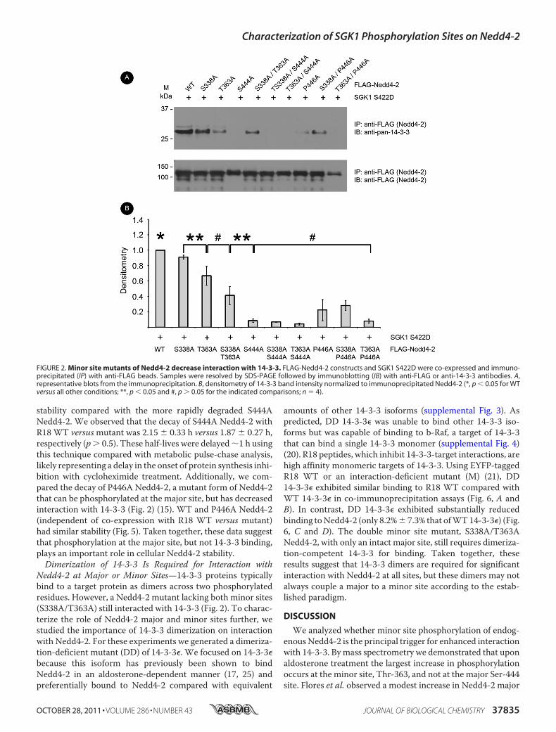
Decrease Interaction with 14-3-3 Proteins—In co-immunopre-cipitation experiments from HEK-293T cells transfected withSGK1 and either WT or mutant Nedd4-2 (S338A, T363A, orS338A/T363A), we found that interaction with endogenous14-3-3 proteins was decreased (by 9 � 3%, 33 � 12%, and 59 �
12%, respectively) with mutant Nedd4-2 expression relative tothat with WT Nedd4-2 expression (Fig. 2). T363A Nedd4-2decreased interaction with 14-3-3 more than S338A. A doubleminor site Nedd4-2 mutant (S338A/T363A) did not signifi-cantly decrease 14-3-3 interaction further compared withT363A Nedd4-2 alone. The major site Nedd4-2 mutants(S444A and P446A, which diminishes 14-3-3 binding withoutdisrupting phosphorylation at Ser-444) (15) also showedmark-edly diminished binding compared withWTNedd4-2 (by 92�2% and 78 � 14%, respectively) (Fig. 2). The remaining doublemutants, each of which included aminor site mutation, did notexhibit significantly different binding to 14-3-3 than did theircorresponding single major site mutants. Taken together, themajor site is critical, but the minor sites are also important forinteraction with 14-3-3. We next tested the roles of theseNedd4-2 mutants to alter cell surface ENaC expression.SGK1DoesNot Augment Cell Surface ENaC in the Presence of
Minor Site Mutants of Nedd-2—In surface biotinylation assaysof HEK-293T cells co-transfected with �-, �-, and �-ENaC,SGK1, andWTormutant Nedd4-2, we first confirmed that cellsurface expression of �-ENaC was decreased when co-ex-pressed withWTNedd4-2, but restored with added SGK1 (Fig.3A, first four lanes), as described previously (24). In the pres-ence of SGK1, compared with WT Nedd4-2 co-expression,Nedd4-2 harboring Ala mutations at Ser-338 or Thr-363decreased cell surface �-ENaC (by 59 � 8% and 76 � 5%,respectively) (Fig. 3B). Consistent with the greater inhibitoryeffect of the T363A mutation on the binding of 14-3-3 toNedd4-2 (Fig. 2), the Nedd4-2 T363A mutant more stronglyattenuated cell surface �-ENaC expression compared with theS338Amutant. The ability of SGK1 to enhance�-ENaC surfaceexpression in the presence of Nedd4-2was also attenuatedwithinclusion of major site Nedd4-2 mutants (e.g. by 71% � 11%with the P446Amutant), but not significantlymore thanT363ANedd4-2 alone (Fig. 3B). In summary, mutations at minor siteswere sufficient to attenuate SGK1-induced cell surface expres-sion of �-ENaC significantly.Phosphorylation at the Major Site Is Important for Nedd4-2
Protein Stability—We next examined the role for phosphoryl-ation at the major site. We consistently observed that, despitetransfection with three times as much plasmid DNA, steady-state expression of S444ANedd4-2 inHEK-293T cells was sub-stantially decreased compared with that of WT and minor sitemutants HEK-293T (Fig. 4A). Thus, we studied whether majorsite phosphorylation is important forNedd4-2 cellular stability.Using [35S]Met/Cys labeling of newly synthesized proteins andpulse-chase analysis in HEK-293T cells, we observed thatS444A Nedd4-2 had a substantially shorter cellular half-lifethan WT Nedd4-2 (Fig. 4B). To examine further whetherNedd4-2 major site interaction with 14-3-3 proteins is impor-tant for Nedd4-2 cellular stability, we sought to disrupt 14-3-3binding without eliminating phosphorylation at the major site.Thus, we co-expressed a generalized inhibitor of 14-3-3-targetinteractions, R18 WT peptide versus an inert control R18 M(mutant) (21). Although R18 WT but not R18 M inhibited theNedd4-2–14-3-3 interaction by 75.6% � 0.10% under theseconditions (supplemental Fig. 2), there was no significantchange in the decay forWT or S444A using these peptides. The
Characterization of SGK1 Phosphorylation Sites on Nedd4-2
OCTOBER 28, 2011 • VOLUME 286 • NUMBER 43 JOURNAL OF BIOLOGICAL CHEMISTRY 37833

decay of S444A with R18 WT versusmutant was 0.98 � 0.18 hversus 1.17 � 0.06 h, respectively (p � 0.4). WT Nedd4-2 didnot substantially decay over this time interval, and thus itsexpression could not be fitted to an exponential decay curve.We further examined the stability of WT versus S444A
Nedd4-2 using cycloheximide treatment to inhibit new proteinsynthesis and then following the decay of protein expressionover time. Using the cycloheximide chase approach, weobserved similar findings to metabolic pulse-chase assays (Fig.5). WT Nedd4-2 with R18WT versusmutant showed a similar
FIGURE 1. Evaluation of major and minor site phosphorylation of Nedd4-2. A, FLAG-Nedd4-2 and SGK1 S422D constructs co-expressed and immunopre-cipitated with anti-FLAG beads. Samples were resolved by SDS-PAGE followed by immunoblotting (IB) with the indicated antibodies. Representative blots fromthe immunoprecipitation (IP) and whole cell lysate (WCL) are shown (n � 3). B, densitometry of Ser(P)-444 band intensity normalized to immunoprecipitatedNedd4-2 (#, p � 0.05 for the indicated comparisons; n � 4). C, immunoblot analysis of SGK1, total Nedd4-2, and Ser-444 phosphorylation (arrow) in aldosterone-treated mpkCCDc14 cell lysates. mpkCCDc14 cells were grown to high resistance on permeable supports and then treated with 1 �M aldosterone for theindicated time intervals. A representative blot is shown (n � 3). D, relative quantitation analysis of Nedd4-2 peptides by mass spectrometry using LC-MS/MS.Ratio of phosphorylated peptide to total nonphosphorylated plus phosphorylated peptide (percentage) for each of the three described SGK1 phosphorylationsites (Ser-338, Thr-363, and Ser-444) under different conditions. mpkCCDc14 cells were treated with vehicle (100% ethanol) (light bars) or 1 �M aldosterone (darkbars) for 3 h prior to harvest of Nedd4-2. Value above each peptide indicates intensity ratio of aldosterone to vehicle. E, absolute quantitation of Nedd4-2peptides by mass spectrometry using LC-MS/MS and internal peptide standards. Samples treated with vehicle (light bars) or aldosterone (dark bars) aregrouped by the indicated Nedd4-2 peptide. Neither peptide with Ser-338 or nonphosphorylated Ser-444 were consistently detected above the lower limit ofquantitation using this method and thus are not shown (*, p � 0.05; #, p � 0.46; n � 3). Designation of major and minor sites is based on predicted affinity of14-3-3 binding at each individual site. Phosphorylated residues are numbered according to Xenopus Nedd4-2, but are conserved from Xenopus to mouse tohuman.
Characterization of SGK1 Phosphorylation Sites on Nedd4-2
37834 JOURNAL OF BIOLOGICAL CHEMISTRY VOLUME 286 • NUMBER 43 • OCTOBER 28, 2011
stability compared with the more rapidly degraded S444ANedd4-2. We observed that the decay of S444A Nedd4-2 withR18WT versusmutant was 2.15 � 0.33 h versus 1.87 � 0.27 h,respectively (p� 0.5). These half-lives were delayed1 h usingthis technique compared with metabolic pulse-chase analysis,likely representing a delay in the onset of protein synthesis inhi-bition with cycloheximide treatment. Additionally, we com-pared the decay of P446A Nedd4-2, a mutant form of Nedd4-2that can be phosphorylated at the major site, but has decreasedinteraction with 14-3-3 (Fig. 2) (15). WT and P446A Nedd4-2(independent of co-expression with R18 WT versus mutant)had similar stability (Fig. 5). Taken together, these data suggestthat phosphorylation at the major site, but not 14-3-3 binding,plays an important role in cellular Nedd4-2 stability.Dimerization of 14-3-3 Is Required for Interaction with
Nedd4-2 at Major or Minor Sites—14-3-3 proteins typicallybind to a target protein as dimers across two phosphorylatedresidues. However, a Nedd4-2 mutant lacking both minor sites(S338A/T363A) still interacted with 14-3-3 (Fig. 2). To charac-terize the role of Nedd4-2 major and minor sites further, westudied the importance of 14-3-3 dimerization on interactionwith Nedd4-2. For these experiments we generated a dimeriza-tion-deficient mutant (DD) of 14-3-3�. We focused on 14-3-3�because this isoform has previously been shown to bindNedd4-2 in an aldosterone-dependent manner (17, 25) andpreferentially bound to Nedd4-2 compared with equivalent
amounts of other 14-3-3 isoforms (supplemental Fig. 3). Aspredicted, DD 14-3-3� was unable to bind other 14-3-3 iso-forms but was capable of binding to b-Raf, a target of 14-3-3that can bind a single 14-3-3 monomer (supplemental Fig. 4)(20). R18 peptides, which inhibit 14-3-3-target interactions, arehigh affinity monomeric targets of 14-3-3. Using EYFP-taggedR18 WT or an interaction-deficient mutant (M) (21), DD14-3-3� exhibited similar binding to R18 WT compared withWT 14-3-3� in co-immunoprecipitation assays (Fig. 6, A andB). In contrast, DD 14-3-3� exhibited substantially reducedbinding toNedd4-2 (only 8.2%� 7.3% that ofWT14-3-3�) (Fig.6, C and D). The double minor site mutant, S338A/T363ANedd4-2, with only an intact major site, still requires dimeriza-tion-competent 14-3-3 for binding. Taken together, theseresults suggest that 14-3-3 dimers are required for significantinteraction with Nedd4-2 at all sites, but these dimers may notalways couple a major to a minor site according to the estab-lished paradigm.
DISCUSSION
We analyzed whether minor site phosphorylation of endog-enousNedd4-2 is the principal trigger for enhanced interactionwith 14-3-3. Bymass spectrometry we demonstrated that uponaldosterone treatment the largest increase in phosphorylationoccurs at the minor site, Thr-363, and not at the major Ser-444site. Flores et al. observed a modest increase in Nedd4-2 major
FIGURE 2. Minor site mutants of Nedd4-2 decrease interaction with 14-3-3. FLAG-Nedd4-2 constructs and SGK1 S422D were co-expressed and immuno-precipitated (IP) with anti-FLAG beads. Samples were resolved by SDS-PAGE followed by immunoblotting (IB) with anti-FLAG or anti-14-3-3 antibodies. A,representative blots from the immunoprecipitation. B, densitometry of 14-3-3 band intensity normalized to immunoprecipitated Nedd4-2 (*, p � 0.05 for WTversus all other conditions; **, p � 0.05 and #, p � 0.05 for the indicated comparisons; n � 4).
Characterization of SGK1 Phosphorylation Sites on Nedd4-2
OCTOBER 28, 2011 • VOLUME 286 • NUMBER 43 JOURNAL OF BIOLOGICAL CHEMISTRY 37835

site phosphorylation with short term aldosterone treatment(19). We employed a similar model system, mpkCCDc14 cells,and perhaps subtle changes in the cells or cell culture techniqueor choice of phosphatase inhibitors may have contributed tothe magnitude of differences at the major site between thisstudy and the earlier study. Additionally, we compared relativechanges in phosphorylation at both major and minor sites. Wefound that, although aldosterone may modestly increase phos-phorylation at the major site, the increase in minor site phos-phorylation is substantially greater. We also found consistentresults in two cell culture systems. Moreover, we have utilizedboth densitometric analysis and mass spectrometry to confirmdifferences at these phosphorylation sites. Two studies have
examined the relative importance of each of the SGK1/PKAphosphorylation motifs within Nedd4-2 using site-directedmutagenesis. These studies showed that mutation of the majorsite, Ser-444 (or its equivalent), played a dominant role overmutation of one of theminor sites in disrupting SGK1- or PKA-inducible Na� transport (1, 14). Furthermore, an antibodydirected against the phosphorylated major site also decreased14-3-3 binding and aldosterone-stimulated ENaC current (17).Our quantitative phosphorylation analysis prompts a refine-ment of the model by suggesting that although the major site iscritical for 14-3-3 binding and ENaC regulation, it tends to beconstitutively phosphorylated (Fig. 1). Thus, phosphorylationof a lower affinity minor site on Nedd4-2 (e.g. Thr-363) may
FIGURE 3. Minor site mutants of Nedd4-2 decrease cell surface expression of �-ENaC. FLAG-Nedd4-2 constructs, SGK1 S422D, and ���-ENaC (HA-�-ENaC)were transfected into HEK-293T cells and surface proteins labeled by biotinylation for subsequent affinity purification with neutravidin-linked beads. Sampleswere resolved by SDS-PAGE followed by immunoblotting (IB) with the indicated antibodies. E-cadherin was probed as a surface protein loading control. A,representative blots from the pulldown and whole cell lysates (WCL). B, densitometry of cell surface �-ENaC (cleaved � uncleaved band intensity) normalizedto �-ENaC in the whole cell lysate (*, p � 0.05 and #, p � 0.05 for the indicated comparisons; n � 4).
Characterization of SGK1 Phosphorylation Sites on Nedd4-2
37836 JOURNAL OF BIOLOGICAL CHEMISTRY VOLUME 286 • NUMBER 43 • OCTOBER 28, 2011

serve as the physiologically relevant SGK1-dependent molecu-lar switch. This paradigm differs from that involving otherSGK1 targets such as the transcription factor Forkhead,FKHRL1, wherein SGK1 robustly phosphorylates both majorandminor 14-3-3 binding sites to induce nuclear localization ofFKHRL1 (26). Our findings imply that SGK1 integrates analdosterone input signal with signals via other kinases (whichphosphorylate the major site) to inhibit Nedd4-2 and to stimu-late ENaC-mediated Na� transport. Candidate kinases for themajor site include Akt, PKA, and I�B kinase-� (27, 28).We next investigated whether mutation of minor sites is suf-
ficient to inhibit SGK1-mediated 14-3-3 interaction and
Nedd4-2 inhibition. Alanine substitutions at minor sites, Ser-338 or Thr-363, decreased the interaction between Nedd4-2and 14-3-3 (Fig. 2). Moreover, SGK1 could not rescue cell sur-face expression of �-ENaC when co-expressed with theseminor site Nedd4-2 mutants (Fig. 3). Finally, expression of theT363A mutant decreased both 14-3-3 interaction withNedd4-2 and SGK1 stimulation of ENaC surface expression toa greater extent than did expression of the S338A Nedd4-2mutant. These results are consistent with our mass spectrom-etry data andwith previous electrophysiologic data which dem-onstrated that the minor sites are important for regulatingENaC-mediated Na� transport and that Thr-363 may be more
FIGURE 4. Phosphorylation at the major site in Nedd4-2, but not 14-3-3 binding, is important for cellular stability as assessed by metabolic pulse-chase analysis. FLAG-Nedd4-2 and either wild-type (WT) or mutant (M) EYFP-R18 constructs were transfected into HEK-293T cells and radiolabeled with[35S]Met/Cys for 15 min before chase with nonradioactive buffer. Nedd4-2 was immunoprecipitated with anti-FLAG beads at indicated times after metaboliclabeling. Samples were resolved by SDS-PAGE and then assayed by exposure to a phospho-screen. A, representative blot of steady-state expression of WT andmutant Nedd4-2 24 h after transfection (n � 5). B, detection of [35S]Met/Cys-labeled Nedd4-2 by PhosphorImager after pulse-chase at indicated times. Arepresentative image is shown. C, labeled protein abundance relative to time 0 fitted to an exponential decay curve using IGOR-Pro 4.0 software as describedunder “Experimental Procedures.” Decay curves for WT (circles) and major site mutant S444A Nedd4-2 (squares) with R18 WT (filled, dashed lines) or mutant(open, solid lines) are superimposed (n � 3).
Characterization of SGK1 Phosphorylation Sites on Nedd4-2
OCTOBER 28, 2011 • VOLUME 286 • NUMBER 43 JOURNAL OF BIOLOGICAL CHEMISTRY 37837

important for the effects of SGK1 than Ser-338 (14). Interest-ingly, Ser-338 may preferentially determine regulation ofNedd4-2 and ENaC by vasopressin/PKA (14). HumanNedd4-2is variably spliced at several exons, and one isoform (KIAA0439,GenBank accession no. AB007899) lacks only exon 12 and theresidue equivalent to Thr-363 (29). Thus, it would be interest-ing to determine whether a higher proportion of this Nedd4-2variant might protect against hypertension in different popula-tions. Modulating regulation of this critical variant may alsoprovide a selective drug target to disrupt aldosterone stimula-tion of ENaC activity.Why is themajor site phosphorylated under base-line condi-
tions? Although the major site is necessary for interaction with14-3-3 and inhibition of Nedd4-2, phosphorylation at this sitealone is not sufficient to stimulate ENaC maximally becauseSGK1 cannot effectively augment ENaC activity when co-ex-pressedwith aminor siteNedd4-2mutant. Furthermore,majorsite mutations result in markedly lower steady-state expressionof Nedd4-2 protein in transfected cells. We tested the hypoth-esis that constitutive phosphorylation at the major site is nec-essary for Nedd4-2 stability. Using pulse-chase assays we dem-onstrated that the S444A phosphorylation-deficient mutanthas amuch shorter half-life than doesWT (Fig. 4). R18 peptide,which decreases Nedd4-2–14-3-3 interaction without elimi-
nating phosphorylation at the major site, did not decrease thehalf-life of Nedd4-2, which is consistent with a 14-3-3-inde-pendent effect on stability. Also consistent with this view, aP446ANedd4-2mutant, which selectively reduces 14-3-3 bind-ing but not Ser-444 phosphorylation, also did not diminish thehalf-life of Nedd4-2.We cannot exclude that relative instabilityof the major site mutant could be due to nonspecific (or evenspecific) changes in protein folding. The prevention of majorsite phosphorylation with this mutant could induce changes inconformation, either locally or globally, that enhance its target-ing for degradation in cells. Major site phosphorylation maythus be an important prerequisite to confer Nedd4-2 stability.The importance of phosphorylation at this residue is supportedby our finding that the major site is highly phosphorylated incells under base-line conditions (Fig. 1). Furthermore, themajor site mutant Nedd4-2 robustly inhibits ENaC-mediatedNa� current, arguing that this mutation does not disrupt eitherthe interaction with ENaC subunits or the ligase activity ofNedd4-2 (1, 14, 15). Thus, one or more kinases appear to pre-serve a high basal state of major site phosphorylation, whichmaintains Nedd4-2 expression independent of 14-3-3 interac-tion at themajor site. Future experiments to delineate the phys-iologically relevant kinases that phosphorylate the major siteshould enhance our understanding of the regulation of
FIGURE 5. Phosphorylation at the major site in Nedd4-2 but not 14-3-3 binding is important for cellular stability by cycloheximide chase analysis.FLAG-Nedd4-2 and EYFP-R18 constructs were transfected into HEK-293T cells and treated with 100 �g/ml cycloheximide before chase with radioimmuneprecipitation assay buffer. A, detection of Nedd4-2 protein expression levels by immunoblotting after cycloheximide treatment for the various indicated times.A representative blot is shown. B, protein abundance relative to time 0 at the indicated times. Decay curves for WT (circles), P446A (triangles), and major sitemutant S444A Nedd4-2 (squares) with R18 WT (filled, dashed lines) or mutant (open, solid lines) are superimposed (n � 3).
Characterization of SGK1 Phosphorylation Sites on Nedd4-2
37838 JOURNAL OF BIOLOGICAL CHEMISTRY VOLUME 286 • NUMBER 43 • OCTOBER 28, 2011

Nedd4-2 stability. To the best of our knowledge, this suggests anovel mechanism by which the cellular stability and expressionlevels of E3 ubiquitin ligases may be regulated through phos-phorylation.We speculate that this systemprovides an efficientmethod to fine tune the control of Na� transport by permittingboth ENaC inhibition in the absence of hormonal stimulationand ENaC stimulation in the presence of hormonal stimulationvia the displacement of Nedd4-2 from ENaC by phosphoryla-tion of a minor site, enhanced 14-3-3 binding, and Nedd4-2sequestration from ENaC.We have demonstrated that a double minor site mutant
Nedd4-2, with only the major SGK1/PKA phosphorylationmotif available, has 40–50% of the binding to 14-3-3 thatoccurs withWTNedd4-2. These data suggest that 14-3-3 couldbind in three possible states at the major site: as a monomer, asa dimer in a trans-configuration with the major site of anotherNedd4-2 molecule or another 14-3-3-binding protein (e.g.AS160) (30), or finally as a dimer in a cis configuration by uti-lizing an unrecognized fourth 14-3-3 binding site on Nedd4-2.However, we have also shown that, in contrast to the mono-meric 14-3-3 targets R18 WT or b-Raf, Nedd4-2 bound onlyweakly to DD 14-3-3. We speculate that 14-3-3 dimers bindNedd4-2 at amajor andminor site upon aldosterone treatment,
but 14-3-3 proteins can bind at the major site and possibly afourth binding site on Nedd4-2. This latter dimeric configura-tion would be in equilibrium with the classic dimer across onemajor, Ser-444, and one minor site, Ser-338 or Thr-363. Wecannot rule out the possibility that 14-3-3 proteins could bindas a dimer across both minor sites upon aldosterone stimula-tion. However, neither phosphorylated motif has a Pro at the�2 position, and thus, they are not perfect consensusmotifs forinitial binding to 14-3-3 (31).In conclusion, the complexity of phosphorylation and 14-3-
3-mediated regulation of Nedd4-2 demonstrates additionalcontrols on ENaC-mediatedNa� transport and ubiquitin ligaseactivity, in general. The results of this study refine the role of14-3-3 proteins in the regulation of SGK1 stimulation of ENaCand expand the role of phosphorylation in the regulation ofNedd4-2.
Acknowledgments—We thank Alan Pao and David Pearce forfeedback.
REFERENCES1. Debonneville, C., Flores, S. Y., Kamynina, E., Plant, P. J., Tauxe, C.,
Thomas, M. A., Münster, C., Chraïbi, A., Pratt, J. H., Horisberger, J. D.,
FIGURE 6. Dimerization is required for Nedd4-2–14-3-3 interaction, and dimers can bind to the major site independent of minor sites. EYFP-tagged R18peptide (monomeric target of 14-3-3; either WT or mutant (M)), FLAG-Nedd4-2, HA-14-3-3�, and/or SGK1 S422D constructs were co-expressed and immuno-precipitated (IP) with anti-HA or anti-FLAG beads. Samples were resolved by SDS-PAGE followed by immunoblotting (IB) with the indicated antibodies. A,representative blots from the immunoprecipitated and whole cell lysates (WCL) are shown for immunoprecipitation of wild-type (WT) or DD HA-tagged 14-3-3�with R18 peptides or WT Nedd4-2. B, densitometry of R18 band intensity normalized to immunoprecipitated 14-3-3 (#, p � 0.05; n � 4). C, representative blotsfrom the immunoprecipitated and whole cell lysates for immunoprecipitation of WT or double minor site mutant (S/T-A) Nedd4-2 with WT or DD 14-3-3�. D,densitometry of 14-3-3 band intensity normalized to immunoprecipitated WT Nedd4-2 (*, p � 0.01; n � 4).
Characterization of SGK1 Phosphorylation Sites on Nedd4-2
OCTOBER 28, 2011 • VOLUME 286 • NUMBER 43 JOURNAL OF BIOLOGICAL CHEMISTRY 37839

Pearce, D., Loffing, J., and Staub, O. (2001) EMBO J. 20, 7052–70592. Staub, O., Dho, S., Henry, P., Correa, J., Ishikawa, T., McGlade, J., and
Rotin, D. (1996) EMBO J. 15, 2371–23803. Snyder, P. M., Steines, J. C., and Olson, D. R. (2004) J. Biol. Chem. 279,
5042–50464. Bhalla, V., and Hallows, K. R. (2008) J. Am. Soc. Nephrol. 19, 1845–18545. Michlig, S., Harris, M., Loffing, J., Rossier, B. C., and Firsov, D. (2005)
J. Biol. Chem. 280, 38264–382706. Schild, L., Lu, Y., Gautschi, I., Schneeberger, E., Lifton, R. P., and Rossier,
B. C. (1996) EMBO J. 15, 2381–23877. Shi, P. P., Cao, X. R., Sweezer, E.M., Kinney, T. S.,Williams, N. R., Husted,
R. F., Nair, R., Weiss, R. M., Williamson, R. A., Sigmund, C. D., Snyder,P. M., Staub, O., Stokes, J. B., and Yang, B. (2008) Am. J. Physiol. Renal.Physiol. 295, F462–470
8. Abriel, H., and Horisberger, J. D. (1999) J. Physiol. 516, 31–439. Kabra, R., Knight, K. K., Zhou, R., and Snyder, P. M. (2008) J. Biol. Chem.
283, 6033–603910. Malik, B., Yue, Q., Yue, G., Chen, X. J., Price, S. R.,Mitch,W. E., and Eaton,
D. C. (2005) Am. J. Physiol. Renal. Physiol. 289, F107–11611. Knight, K. K., Olson, D. R., Zhou, R., and Snyder, P. M. (2006) Proc. Natl.
Acad. Sci. U.S.A. 103, 2805–280812. Bhalla, V., Oyster, N. M., Fitch, A. C., Wijngaarden, M. A., Neumann, D.,
Schlattner, U., Pearce, D., and Hallows, K. R. (2006) J. Biol. Chem. 281,26159–26169
13. Hallows, K. R., Bhalla, V., Oyster, N. M., Wijngaarden, M. A., Lee, J. K., Li,H., Chandran, S., Xia, X., Huang, Z., Chalkley, R. J., Burlingame, A. L., andPearce, D. (2010) J. Biol. Chem. 285, 21671–21678
14. Snyder, P. M., Olson, D. R., Kabra, R., Zhou, R., and Steines, J. C. (2004)J. Biol. Chem. 279, 45753–45758
15. Bhalla, V., Daidié, D., Li, H., Pao, A. C., LaGrange, L. P., Wang, J., Vande-walle, A., Stockand, J. D., Staub, O., and Pearce, D. (2005)Mol. Endocrinol.19, 3073–3084
16. Ichimura, T., Yamamura, H., Sasamoto, K., Tominaga, Y., Taoka, M.,Kakiuchi, K., Shinkawa, T., Takahashi, N., Shimada, S., and Isobe, T.(2005) J. Biol. Chem. 280, 13187–13194
17. Liang, X., Peters, K. W., Butterworth, M. B., and Frizzell, R. A. (2006)J. Biol. Chem. 281, 16323–16332
18. Yaffe, M. B. (2002) FEBS Lett. 513, 53–5719. Flores, S. Y., Loffing-Cueni, D., Kamynina, E., Daidié, D., Gerbex, C., Cha-
banel, S., Dudler, J., Loffing, J., and Staub,O. (2005) J. Am. Soc. Nephrol. 16,2279–2287
20. Tzivion, G., Luo, Z., and Avruch, J. (1998) Nature 394, 88–9221. Masters, S. C., and Fu, H. (2001) J. Biol. Chem. 276, 45193–4520022. Carattino, M. D., Edinger, R. S., Grieser, H. J., Wise, R., Neumann, D.,
Schlattner, U., Johnson, J. P., Kleyman, T. R., and Hallows, K. R. (2005)J. Biol. Chem. 280, 17608–17616
23. Shevchenko, A., Tomas, H., Havlis, J., Olsen, J. V., and Mann, M. (2006)Nat. Protoc. 1, 2856–2860
24. Soundararajan, R., Melters, D., Shih, I. C., Wang, J., and Pearce, D. (2009)Proc. Natl. Acad. Sci. U.S.A. 106, 7804–7809
25. Liang, X., Butterworth, M. B., Peters, K. W., Walker, W. H., and Frizzell,R. A. (2008) J. Biol. Chem. 283, 27418–27425
26. Brunet, A., Park, J., Tran, H., Hu, L. S., Hemmings, B. A., and Greenberg,M. E. (2001)Mol. Cell. Biol. 21, 952–965
27. Lee, I. H., Dinudom, A., Sanchez-Perez, A., Kumar, S., and Cook, D. I.(2007) J. Biol. Chem. 282, 29866–29873
28. Edinger, R. S., Lebowitz, J., Li, H., Alzamora, R., Wang, H., Johnson, J. P.,and Hallows, K. R. (2009) J. Biol. Chem. 284, 150–157
29. Itani, O. A., Stokes, J. B., and Thomas, C. P. (2005) Am. J. Physiol. Renal.Physiol. 289, F334–346
30. Liang, X., Butterworth, M. B., Peters, K.W., and Frizzell, R. A. (2010)Mol.Biol. Cell 21, 2024–2033
31. Muslin, A. J., Tanner, J. W., Allen, P. M., and Shaw, A. S. (1996) Cell 84,889–897
Characterization of SGK1 Phosphorylation Sites on Nedd4-2
37840 JOURNAL OF BIOLOGICAL CHEMISTRY VOLUME 286 • NUMBER 43 • OCTOBER 28, 2011