neural mechanisms mediating optimism bias
TRANSCRIPT

LETTERS
Neural mechanisms mediating optimism biasTali Sharot1,2, Alison M. Riccardi1, Candace M. Raio1 & Elizabeth A. Phelps1
Humans expect positive events in the future even when there is noevidence to support such expectations. For example, people expectto live longer and be healthier than average1, they underestimatetheir likelihood of getting a divorce1, and overestimate their pro-spects for success on the job market2. We examined how the braingenerates this pervasive optimism bias. Here we report that thistendency was related specifically to enhanced activation in theamygdala and in the rostral anterior cingulate cortex when ima-gining positive future events relative to negative ones, suggesting akey role for areas involved in monitoring emotional salience inmediating the optimism bias. These are the same regions that showirregularities in depression3, which has been related to pessi-mism4. Across individuals, activity in the rostral anterior cingulatecortex was correlated with trait optimism. The current study high-lights how the brain may generate the tendency to engage in theprojection of positive future events, suggesting that the effectiveintegration and regulation of emotional and autobiographicalinformation supports the projection of positive future events inhealthy individuals, and is related to optimism.
People tend to make overly confident, positive predictions aboutthe future, which are often inaccurate1,2. The tendency to expect goodthings in the future is known as optimism. Extreme optimism can beharmful as it can promote an underestimation of risk and poorplanning5. In contrast, a pessimistic view is correlated with severityof depression symptoms3. A moderate optimistic illusion, however,can motivate adaptive behaviour in the present towards a future goal,and has been related to mental6 and physical7 health. How does thehealthy brain generate the tendency to create images of positivefuture events?
It has been suggested that imagining future events demands a sys-tem that calls on the past to retrieve pieces of information that are thenreconstructed to form representations of possible future scenarios8,9.In accordance with this proposal, it has been shown that imagining thefuture depends on the same neural networks that are active whenrecalling the past8,10,11. One possibility is that in optimists this networkis more active when imagining future positive events than negativeones. It is also possible that other brain areas modulate this network ina way that biases it to engage in positive future projections.
To examine the neurobiological basis of optimism we collectedfunctional magnetic resonance imaging (fMRI) data while partici-pants thought of autobiographical events related to a description of alife episode (for example, ‘winning an award’ or ‘the end of a roman-tic relationship’). The word ‘past’ or ‘future’ indicated if they shouldthink of an event that occurred in the past or one that might occur inthe future. Trials were classified into positive, negative and neutralaccording to participants’ ratings. The mean number of trials rated asneutral was negligible, and thus all contrasts of interest were con-ducted between positive and negative trials only.
After scanning, participants rated their memories and projectionson six factors12 related to their subjective experience. The mean scoresfor these ratings and statistical analysis are presented in Table 1 andSupplementary Table 1. Finally, participants completed the LOT-R(Life Orientation Test-Revised) scale that measures trait optimism13
(see Methods for details).As represented in Table 1 and Supplementary Table 1, future posi-
tive events were rated as more positive than past positive events, andwere imagined to be closer in temporal proximity then future nega-tive events and all past events (Fig. 1a). Negative future events wereexperienced with a weaker subjective sense of pre-experiencing, andwere more likely to be imagined from an outsider viewing in, thanpositive future events and all past events (Fig. 1b). The more optim-istic participants were, as indicated by the LOT-R scores, the morelikely they were to expect positive events to happen closer in thefuture than negative events, and to experience them with a greatersense of pre-experiencing (Fig. 1c, d).
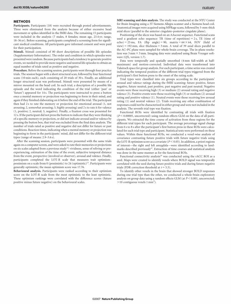
Next we examined the fMRI data to identify the underlying neuralmechanisms. Functional regions of interest (ROIs) were identified bycontrasting all trials with fixation (P , 0.00005, uncorrected). Thefour regions that survived this threshold (Fig. 2a, b) included therostral anterior cingulate cortex (rACC, Fig. 2b), extending intothe ventral medial prefrontal cortex at a more lenient threshold(see Fig. 3a, c), the posterior cingulate cortex (PCC, Fig. 2b), andthe dorsal medial prefrontal cortex (Fig. 2b), all of which have beenreported to have a key role in retrieval of autobiographical memoryand in imagining future events8. The fourth region was the rightamygdala, which has been shown to be important for emotion’s
1Department of Psychology, New York University, New York 10003, USA. 2Wellcome Department of Imaging Neuroscience, Institute of Neurology, University College London, LondonWC1N 3BG, UK.
Table 1 | Means and statistics for subjective ratings according to trial type
Scale Future Past
Positive Negative t-test (P) Correlation withoptimism
Positive Negative t-test (P) Correlation withoptimism
Valence 2.44 4.34 ,0.001 NS 2.77 4.07 ,0.01 NSArousal 3.56 3.23 5 0.05 NS 3.50 3.61 NS NSTime of event 2.37 3.08 ,0.005 ,0.005 3.01 3.00 NS NSSubjective temporal distance 3.00 3.47 NS NS 3.33 3.40 NS NSPerspective 2.96 3.37 ,0.025 NS 2.92 2.91 NS NSReliving or pre-experiencing 3.59 3.07 ,0.025 ,0.025 3.80 3.85 NS NSVividness 3.66 3.28 NS NS 3.90 4.19 NS NS
Valence (1, positive; 6, negative), arousal (1, low; 6, high), time of event (years from present, 1–5), subjective temporal distance (1, near; 6, far), perspective (1, involved; 6, observer), reliving or pre-experiencing (1, low; 6, high) and vividness (1, low; 6, high). NS, not significant.
Vol 450 | 1 November 2007 | doi:10.1038/nature06280
102Nature ©2007 Publishing Group

influence on autobiographical memory14 (Fig. 2a; the left amygdalawas also observed at a more lenient threshold, Fig. 3b, d). Amongthese ROIs, the blood-oxygenation-level-dependent (BOLD) signalin the amygdala and rACC was reduced when imagining negativefuture events relative to positive future events and to all past events(Fig. 2c, d). The differences in BOLD signal between positive andnegative future events remained significant when controlling for dif-ferences in the sense of pre-experiencing (amygdala, P , 0.01; rACC,P , 0.05). For structural ROI analysis (Supplementary Fig. 1) andexploratory whole-brain analysis, see Supplementary Information.
To examine if these differences were related to optimism acrossindividuals, we identified voxels within the four functional ROIs inwhich changes in BOLD signal during future positive trials relative tofuture negative trials were correlated with participants’ optimismscore on the LOT-R scale (see Supplementary Information for addi-tional analysis and discussion). We found a positive correlation inthe rACC (Fig. 2e). No significant correlation with optimism wasobserved in the other ROIs. Finally, a functional connectivity analysisrevealed a strong correlation between activity in the rACC and acti-vity in the amygdala bilaterally while imagining future positiveevents; this correlation was weaker and less extensive when imaginingfuture negative events (Fig. 2f).
Our behavioural results suggest that, whereas the past isconstrained, the future is open to interpretation allowing subjectsto distance themselves from possible negative events and movecloser toward positive ones. Across individuals, this tendency wasassociated with trait optimism. The brain imaging findings providea possible mechanism mediating the behavioural observations.
Future Past
Sub
ject
ive
inte
nsity
Tim
e fr
om p
rese
nt (y
ears
)
753 9 11 13 15
753 9 11 13 15
Optimism
1
2
3
4
5
1
2
3
4
5
Future Past
PositiveNegative
PositiveNegative
–2.5–2.0–1.5–1.0–0.5
00.51.0
Optimism
*
a b
c d
*
Tim
e in
futu
re fr
om p
rese
nt,
pos
itive
– n
egat
ive
(yea
rs)
Pre
-exp
erie
ncin
g,p
ositi
ve –
neg
ativ
e
–1.0–0.5
00.51.01.52.02.53.0
Figure 1 | Optimism related to expected time of event and sense of pre-experiencing future events. a, Positive events are perceived to be closer intime to the present than negative future events and all past events.b, Negative future events are experienced with a weaker sense of pre-experiencing than positive future events and all past events. c, The differencein expected time of event for future positive and negative events correlatedwith the optimism level defined by ranking participants according to theirLOT-R scores (r 5 20.74, P , 0.005). d, The difference in pre-experiencingfor positive and negative events correlated with trait optimism (r 5 0.58,P , 0.025). N 5 15; error bars, 6s.e.m.; *P , 0.05.
–0.2
–0.1
03 5 7 9 11 13 15
0.1
0.2
0.3
0.4
0.5
0.6
0.7
Optimism
x = –11
a
y = –9 R L
b
rACC
PCC
DMPFC
Amygdala0
0.05
0.1
0.15
0.2
0.25
0.3
0.35
0.4
0
0.05
0.1
0.15
0.2
0.25
0.3
0.35
0.4
0.45
0.5
PastFuture
PastFuture
Sig
nal c
hang
e (%
) S
igna
l cha
nge
(%)
PositiveNegative
PositiveNegative
c
d
*
*
f
LRR
Func
tiona
l con
nect
ivity
with
rA
CC
Future negative
Future positive
Sig
nal c
hang
e in
the
rA
CC
(%)
futu
re p
ositi
ve –
futu
re n
egat
ive
e
Amygdala, y = –9
Figure 2 | Activity in the amygdalaand rACC and its relation tooptimism. ROIs defined bycontrasting all trials with fixation(shown at P , 0.0005, uncorrected)reveal activity of a, the amygdala(Talarich coordinates of peak voxel:20, 29, 214) and b, the rACC(Broadman Area (BA) 32: 211, 42,21), the PCC (BAs 30 and 31: 24,247, 19) and the dorsal medialprefrontal cortex (DMPFC; BA 6:28, 12, 52). BOLD signals in bothc, the amygdala and d, the rACCreduced while imagining negativefuture events. e, BOLD signal in therACC during future positive eventsversus future negative eventscorrelated with trait optimism(29, 39, 22; r 5 0.5, P , 0.05).f, Functional connectivity map withrACC as the seed reveals extensivecorrelation with the amygdalasignal during future positive trials,and restricted correlation duringfuture negative trials. The redsquare in a indicates the regionshown in f. N 5 15; error bars,6s.e.m.; *P , 0.05.
NATURE | Vol 450 | 1 November 2007 LETTERS
103Nature ©2007 Publishing Group
Reduced BOLD signal was observed in the amygdala and rACC dur-ing imagination of negative future events relative to positive futureevents and all past events, suggesting that the optimism bias may berelated to a reduction in negative future thought.
The amygdala has a documented role in the modulation by emo-tion of cognitive processes including memory and decision making(for a review, see ref. 15). Our data extend the role of the amygdala toinclude simulation of future emotional events. Previous studiesexamining the neural mechanisms underlying imagination of non-emotional future events did not observe amygdala involvement8,10.Thus, as in the case of memory, we suggest that the amygdala may beselectively engaged in imagining future emotional events, rather thanserving a general function in imagining the future.
The rACC has strong reciprocal connection with the amygdala andother regions that convey emotional and motivational information16.It has been implicated in tasks involving self-reflection, such as mak-ing positive self-referential judgments17, reflecting on hopes anddreams18, indicating preferences19 and judging the trustworthinessof others20. This has caused some researchers to suggest that wheninformation is judged to be self-relevant, activity in the rACC willconvey the valence17. However, others have suggested a moregeneral role for the rACC in assessing the salience of emotional andmotivational information and regulating emotional responsesaccordingly21. Consistent with this notion it has been shown thatthe rACC’s response to positive and negative stimuli reflects situ-ational specificity. For example, both the rACC and amygdala weremore sensitive to positive stimuli in subjects that focused on obtain-ing goals (promotional context) and to negative stimuli in subjectsthat focused on avoiding failure (prevention context)22. Further-more, changes in rACC activity have been related to extinction offear conditioning23, and to reduced anxiety during shock expect-ancy24; this possibly indicates diminished salience of negative stimulithat may consequently lower amygdala activity23.
Consistent with these previous studies, we suggest that in the cur-rent study the rACC is tracking the subjective salience of the stimuliby assessing emotional, motivational and autobiographical infor-mation, and possibly by regulating such signals. This suggestion is
supported by the strong functional connectivity observed betweenthe rACC and amygdala during imagination of positive future events.In addition, individual differences in trait optimism were related tothe relative level of rACC engagement when imagining positivefuture scenarios versus negative ones. We speculate that rACC acti-vity during imagination of future events reflects a self-regulatoryfocus that underlies a bias in attention and vigilance towards positivefuture events and away from negative ones, modulating engagementof other regions that provide emotional and autobiographicalinformation that is processed and recombined to construct futurescenarios.
These findings may provide insight to the mechanisms underlyingdepression. Depressive symptoms are associated with pessimism4
and with difficulties in creating detailed images of future events25.It has been suggested that malfunction of a neural pathway incorpo-rating the rACC and the amygdala may cause depression by leading todecreased regulatory affects of the rACC over the amygdala and otherregions involved in emotional processing3. Future studies are neededto determine whether these abnormalities are related directly to thebreakdown of optimism in depression, specifically because it relatesto the way in which depressed patients imagine future events.
One should note that when comparing past and future trials we arealso comparing imagining and remembering. Thus, our data cannotindicate whether the positivity bias is a function of time (that is, it willemerge only when thinking about the future) or whether it reflects atendency to engage in positive thought when not constrained byreality. We speculate that the difference is not between past andfuture, although in practice the optimism bias manifests itself inthoughts of the future because the absence of factual constraintsleaves plenty of room to alter expectations.
Expecting positive events, and generating compelling mentalimages of such events, may serve an adaptive function by motivatingbehaviour in the present towards a future goal. We do not suggestthat simulating negative events is not important for survival. How-ever, pondering on such events may interfere with daily activities bypromoting negative effects such as anxiety and depression. The cur-rent study highlights how the brain may generate the tendency toengage in the projection of positive future events, suggesting that therACC modulates activity in brain areas that are involved in emotionalprocessing and autobiographical retrieval to create positive images ofthe future.
METHODS SUMMARY
Functional MRI images were collected from participants while they thought of
autobiographical events related to a description of a life episode that appeared on
screen for 14 s. Either the word ‘past’ or ‘future’ indicated if they should think of
an event that occurred in the past or that might occur in the future. The parti-
cipants were instructed to press a button once the memory or projection was
beginning to form in their mind, and again when they finished elaborating on it.
They then had 2 s to rate the memory or projection for emotional arousal and 2 s
to rate it for valence. There were 80 trials that were classified into positive,
negative or neutral according to the participants’ ratings.
After scanning, participants rated their memories or projections on six fac-
tors12 related to their subjective experience, and completed the LOT-R scale that
measures trait optimism13. Participants were ranked according to their optimism
score on the LOT-R scale. These optimism rankings were correlated with the
difference in scores (future positive minus future negative) on the behavioural
scales.
For each participant a time series was created indicating the temporal position
of the different trial types triggered from the participant’s first button press.
Functional ROIs were identified by contrasting all trials with fixation
(P , 0.00005, uncorrected) using random effects GLM (General Linear
Model) on the data of all participants. The time courses of activation were
extracted from these regions for each trial type and participant for statistical
analysis. Next, we identified voxels within the ROIs in which changes in BOLD
signal during future positive trials relative to future negative trials were corre-
lated with the LOT-R scores. Additionally, functional connectivity maps26 were
created for the different trial types (False Discovery Rate (FDR) correction
threshold at t 5 5.5).
Future eventsPast events
Amygdala, R Amygdala, LAmygdala, R Amygdala, L
VMPFC
PCC
VMPFC
PCC
DMPFC
c
d
a
b
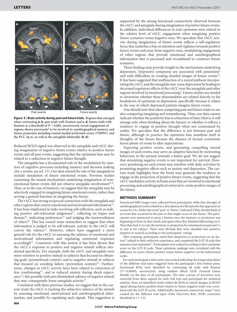
Figure 3 | Brain activity during past and future trials. Regions that emergedwhen contrasting a, b, past trials with fixation and c, d, future trials withfixation at a threshold of P , 0.005, uncorrected, reveal engagement ofregions shown previously8 to be involved in autobiographical memory andfuture projection including ventral medial prefrontal cortex (VMPFC) andthe PCC (a, c), as well as the amygdala bilaterally (b, d).
LETTERS NATURE | Vol 450 | 1 November 2007
104Nature ©2007 Publishing Group

Full Methods and any associated references are available in the online version ofthe paper at www.nature.com/nature.
Received 7 July; accepted 19 September 2007.Published online 24 October 2007.
1. Weinstein, N. D. Unrealistic optimism about future life events. J. Pers. Soc. Psychol.39, 806–820 (1980).
2. Hoch, S. Conterfactual reasoning and accuracy in predicting personal events.J. Exp. Psychol. 11, 719–731 (1984).
3. Drevets, W. C. et al. Subgenual prefrontal cortex abnormalities in mood disorders.Nature 386, 824–827 (1997).
4. Strunk, D. R., Lopez, H. & DeRubeis, R. J. Depressive symptoms are associatedwith unrealistic negative predictions of future life events. Behav. Res. Ther. 44,861–882 (2006).
5. Lovallo, D. & Kahneman, D. Delusions of success: how optimism underminesexecutives’ decisions. Harv. Bus. Rev. 56, 56–63 (2003).
6. Taylor, S. E. & Brown, J. D. Illusion and well-being: a social psychologicalperspective on mental health. Psychol. Bull. 103, 193–210 (1988).
7. Scheier, M. F. & Carver, C. S. Dispositional optimism and physical well-being: theinfluence of generalized outcome expectancies on health. J. Pers. 55, 169–210(1987).
8. Addis, D. R., Wong, A. T. & Schacter, D. L. Remembering the past and imaginingthe future: common and distinct neural substrates during event construction andelaboration. Neuropsychologia 45, 1363–1377 (2007).
9. Tulving, E. in The Missing Link in Cognition (eds Terrace, H. S. & Metcalfe, J.) 3–56(Oxford Univ. Press, New York, 2005).
10. Okuda, J. et al. Thinking of the past and past: the roles of the frontal pole and themedial temporal lobe. Neuroimage 19, 1369–1380 (2003).
11. Hassabis, D., Kumaran, D., Vann, S. D. & Marguire, E. A. Patients withhippocampal amnesia cannot imagine new experiences. Proc. Natl Acad. Sci. USA104, 1726–1731 (2007).
12. D’Argembeau, A. & Van der Linden, M. Influence of affective meaning on memoryfor contextual information. Emotion 4, 173–188 (2004).
13. Scheier, M. F., Carver, C. S. & Bridges, M. W. Distinguishing optimismfrom neuroticism (and trait anxiety, self-mastery, and self-esteem): areevaluation of the Life Orientation Test. J. Pers. Soc. Psychol. 67, 1063–1078(1994).
14. Sharot, T., Martorella, E. A., Delgado, M. R. & Phelps, E. A. How personalexperience modulates the neural circuitry of memories of September 11. Proc. NatlAcad. Sci. USA 104, 389–394 (2007).
15. Phelps, E. A. Emotion and cognition: insights from studies of the human amygdala.Annu. Rev. Psychol. 57, 27–53 (2006).
16. Vogt, B. A. & Pandya, D. N. Cingulate cortex of the rhesus monkey: II. corticalafferents. J. Comp. Neurol. 262, 271–289 (1987).
17. Moran, J. M., Macrae, C. N., Heatherton, T. F., Wyland, C. L. & Kelley, W. M.Neuroanatomical evidence for distinct cognitive and affective components of self.J. Cogn. Neurosci. 18, 1586–1594 (2006).
18. Johnson, S. C., Baxter, L. C., Wilder, L. S., Pipe, J. G., Heiserman, J. E. & Prigatano,G. P. Neural correlates of self-reflection. Brain 125, 1808–1814 (2002).
19. Paulus, M. P. & Frank, L. R. Ventromedial prefrontal cortex activation is critical forpreference judgments. Neuroreport 14, 1311–1315 (2003).
20. Winston, J. S., Strange, B. A., O’Doherty, J. & Dolan, R. J. Automatic and intentionalbrain responses during evaluation of trustworthiness of faces. Nature Neurosci. 5,277–283 (2002).
21. Bush, G., Luu, P. & Posner, M. I. Cognitive and emotional influences in anteriorcingulated cortex. Trends Cogn. Sci. 14, 215–222 (2000).
22. Cunningham, W. A., Raye, C. L. & Johnson, M. K. Neural correlates of evaluationassociated with promotion and prevention regulatory focus. Cogn. Affect. Behav.Neurosci. 5, 202–211 (2005).
23. Phelps, E. A., Delgado, M. R., Nearing, K. I. & LeDoux, J. E. Extinction learning inhumans: role of the amygdala and vmPFC. Neuron 43, 897–905 (2004).
24. Simpson, J. R., Snyder, A. Z. Jr, Gusnard, D. A. & Raichle, M. E. Emotion-inducedchanges in human medial prefrontal cortex: I. during cognitive task performance.Proc. Natl Acad. Sci. USA 98, 688–693 (2001).
25. Williams, J. M., Ellis, N. C., Tyers, C., Healy, H., Rose, G. & MacLeod, A. K. Thespecificity of autobiographical memory and imageability of the future. Mem.Cognit. 24, 116–125 (1996).
26. Roebroeck, A., Formisano, E. & Goebel, R. Mapping directed influence over thebrain using Granger causality and fMRI. Neuroimage 25, 230–242 (2005).
27. Pruessner, J. C. et al. Volumetry of hippocampus and amygdala with high-resolution MRI and three-dimensional analysis software: minimizing thediscrepancies between laboratories. Cereb. Cortex 10, 433–442 (2000).
Supplementary Information is linked to the online version of the paper atwww.nature.com/nature.
Acknowledgements This study was supported by the NIMH (E.A.P.), the SeaverFoundation (grant to NYU’s Center for Brain Imaging), and a Margaret and HermanSokol Postdoctoral Fellowship (T.S.). We thank S. H. Mulhern, D. C. Johnson andJ. K. Szary for help in data analysis, and D. Schiller, J. H. McDermott and Y. Trope fordiscussion.
Author Contributions T.S. designed the study. T.S. and E.A.P. interpreted the dataand wrote the paper. T.S. and A.M.R. developed stimuli, gathered behavioural pilotdata, and conducted behavioural data analysis. T.S. gathered fMRI data. T.S.conducted neuroimaging analyses with the help of A.M.R. and C.M.R., and withadvice of E.A.P. Whole-brain exploratory analysis was conducted by C.M.R.
Author Information Reprints and permissions information is available atwww.nature.com/reprints. Correspondence and requests for materials should beaddressed to E.A.P. ([email protected]).
NATURE | Vol 450 | 1 November 2007 LETTERS
105Nature ©2007 Publishing Group
METHODSParticipants. Participants (18) were recruited through posted advertisements.
Three were eliminated from the analysis because of either excessive head
movement or spikes identified in the fMRI data. The remaining 15 participants
were included in the analysis (7 males, 8 females; mean age, 23.4 yr; range,
18–36 yr). Before scanning, participants completed a screening form for signifi-
cant medical conditions. All participants gave informed consent and were paid
for their participation.
Stimuli. Stimuli consisted of 80 short descriptions of possible life episodes
(Supplementary Information). The order and condition in which episodes were
presented were random. Because participants had a tendency to generate positive
events, we needed to provide more negative and neutral life episodes to obtain an
equal number of trials rated as positive and negative.
Procedure. Before the scanning session, participants went through four practice
trials. The session began with a short structural scan, followed by four functional
scans (10 min each), each consisting of 20 trials of 30 s. Finally, an additional
longer structural scan was performed. Stimuli were presented by means of a
mirror mounted on the head coil. In each trial, a description of a possible life
episode and the word indicating the condition of the trial (either ‘past’ or
‘future’) appeared for 14 s. The participants were instructed to press a button
once a mental memory or projection was beginning to form in their mind, and
again if they finished elaborating on it before the end of the trial. The participant
then had 2 s to rate the memory or projection for emotional arousal (1, not
arousing; 2, somewhat arousing; 3, highly arousing) and 2 s to rate it for valence
(1, positive; 2, neutral; 3, negative). Finally, a fixation cross was presented for
12 s. If the participant did not press the button to indicate that they were thinking
of a specific memory or projection, or did not indicate arousal and/or valence by
pressing the button box, that trial was excluded from the final data analysis. The
number of trials rated as positive and negative did not differ for future or past
conditions. Reaction times, indicating when a mental memory or projection was
beginning to form in the participants’ mind, did not differ for the different trial
types (range of means: 2.9–3.6 s).
After the scanning session, participants were presented with the same trials
again on a computer screen, and were asked to rate their memories or projections
on six scales adapted from a previous study12: vividness, sense of reliving or pre-
experiencing, estimation of the time of the event, subjective temporal distance
from the event, perspective (involved or observer), arousal and valence. Finally,
participants completed the LOT-R scale that measures trait optimism–
pessimism on a scale from 0 (pessimistic) to 24 (optimistic)13. Participants were
generally optimistic; the mean optimism score was 17.76.
Behavioural analysis. Participants were ranked according to their optimism
score on the LOT-R scale from the most optimistic to the least optimistic.
These optimism rankings were correlated with the difference scores (future
positive minus future negative) on the behavioural scales.
MRI scanning and data analysis. The study was conducted at the NYU Centerfor Brain Imaging using a 3T Siemens Allegra scanner and a Siemens head coil.
Anatomical images were acquired using MPRage scans, followed by 3-mm-thick
axial slices (parallel to the anterior cingulate–posterior cingulate plane).
Positioning of the slices was based on an AAscout sequence. Functional scans
used a gradient echo sequence: TR (time of repetition) 5 2 s, TE (time of
echo) 5 25 ms, FA (flip angle) 5 90, matrix 5 64 3 64, FOV (field of
view) 5 192 mm, slice thickness 5 3 mm. A total of 39 axial slices parallel to
the AC–PC plane were sampled for whole-brain coverage. The in-plane resolu-
tion was 3 mm 3 3 mm. Imaging data were analysed using Brain Voyager soft-
ware (QX, version 1.7).
Data were temporally and spatially smoothed (4 mm full-width at half-
maximum) and motion-corrected. Individual data were transformed into
Talairach space for group analysis. For each participant, a time series was created
indicating the temporal position of the different trial types triggered from the
participant’s first button press to the onset of the rating scale.
Trial types were classified into six groups according to the participants’
arousal and valence ratings during the fMRI scanning: future positive, future
negative, future neutral, past positive, past negative and past neutral. Negativeevents were those receiving high (3) or medium (2) arousal rating and negative
valence (3). Positive events were those receiving high (3) or medium (2) arousal
rating and positive valence (1). Neutral events were those receiving low arousal
rating (1) and neutral valence (2). Trials receiving any other combination of
responses could not be characterized in either group and were not included in the
analysis. The seventh trial type was fixation.
Functional ROIs were identified by contrasting all trials with fixation
(P , 0.00005, uncorrected) using random effects GLM on the data of all parti-
cipants. We extracted the time course of activation from these regions for the
different trial types for each participant. The average percentage signal change
from 6 s to 8 s after the participant’s first button press in these ROIs were calcu-
lated for each trial type and participant. Statistical tests were performed on these
values. Within these functional ROIs, we conducted a voxel-wise analysis of
covariance contrasting future positive trials with future negative trials using
the LOT-R optimism score as a covariate (P , 0.05). In addition, a priori regions
of interest—the right and left amygdala—were identified according to land-
marks described previously27. Extraction of time courses and statistical analysis
was done in the same manner as for the functional ROIs.Functional connectivity analysis26 was conducted using the rACC ROI as a
seed. Maps were created to identify voxels where BOLD signal was temporally
correlated with the seed during future positive trials and during future negative
trials (FDR correction threshold at t 5 5.5).
To identify other voxels in the brain that showed stronger BOLD responses
during one trial type than the other, we conducted a whole-brain exploratory
analysis on group data using a random effects GLM (at P , 0.001, uncorrected;
.10 contiguous voxels 1 mm3).
doi:10.1038/nature06280
Nature ©2007 Publishing Group