neural basis of speech 2/29/00. neuron neuron = nervous system cell –neuron cell body (contains...
TRANSCRIPT
Neural Basis of Speech
2/29/00
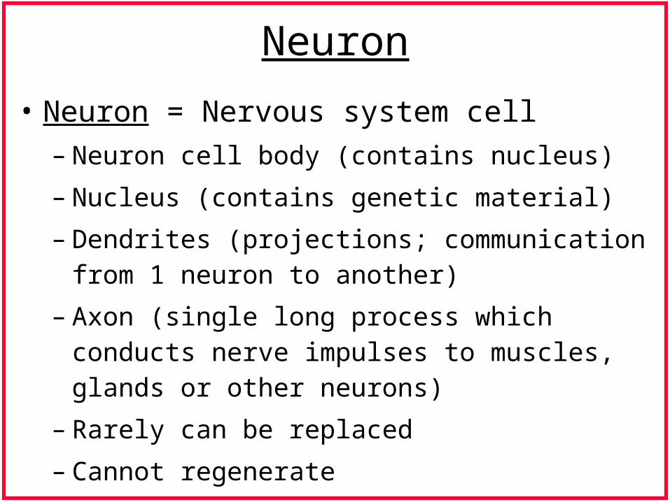
Neuron
• Neuron = Nervous system cell
– Neuron cell body (contains nucleus)
– Nucleus (contains genetic material)
– Dendrites (projections; communication from 1 neuron to another)
– Axon (single long process which conducts nerve impulses to muscles, glands or other neurons)
– Rarely can be replaced
– Cannot regenerate
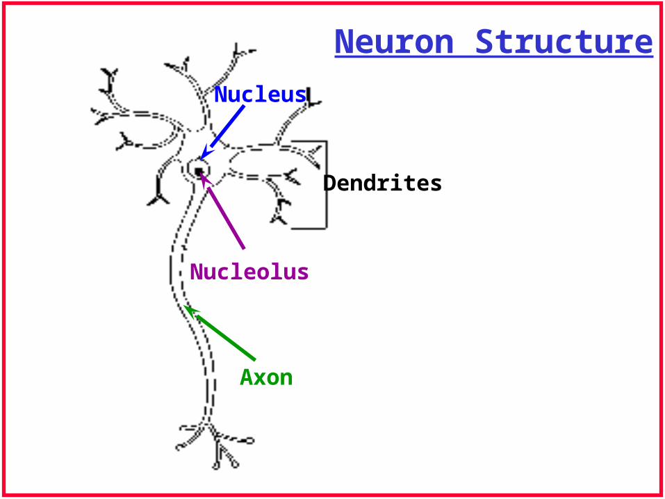
Neuron Structure
Nucleus
Nucleolus
Axon
Dendrites
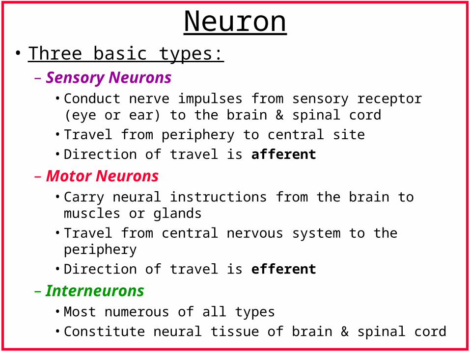
Neuron• Three basic types:
– Sensory Neurons• Conduct nerve impulses from sensory receptor (eye or ear) to
the brain & spinal cord
• Travel from periphery to central site
• Direction of travel is afferent
– Motor Neurons• Carry neural instructions from the brain to muscles or glands
• Travel from central nervous system to the periphery
• Direction of travel is efferent
– Interneurons• Most numerous of all types
• Constitute neural tissue of brain & spinal cord
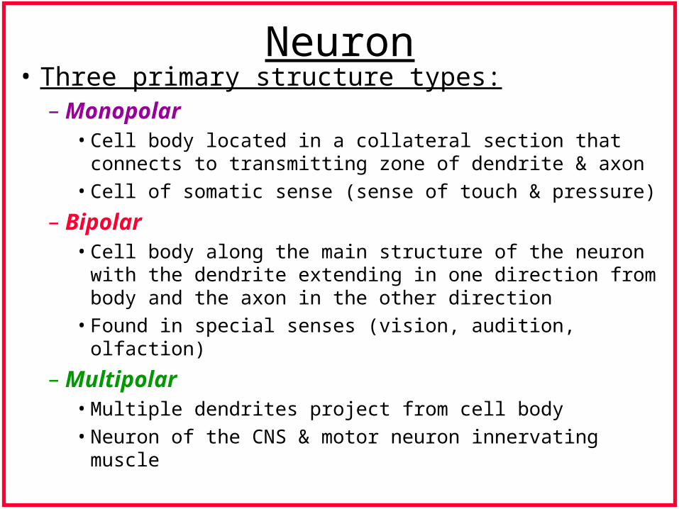
Neuron• Three primary structure types:
– Monopolar• Cell body located in a collateral section that connects to
transmitting zone of dendrite & axon
• Cell of somatic sense (sense of touch & pressure)
– Bipolar• Cell body along the main structure of the neuron with the
dendrite extending in one direction from body and the axon in the other direction
• Found in special senses (vision, audition, olfaction)
– Multipolar• Multiple dendrites project from cell body
• Neuron of the CNS & motor neuron innervating muscle
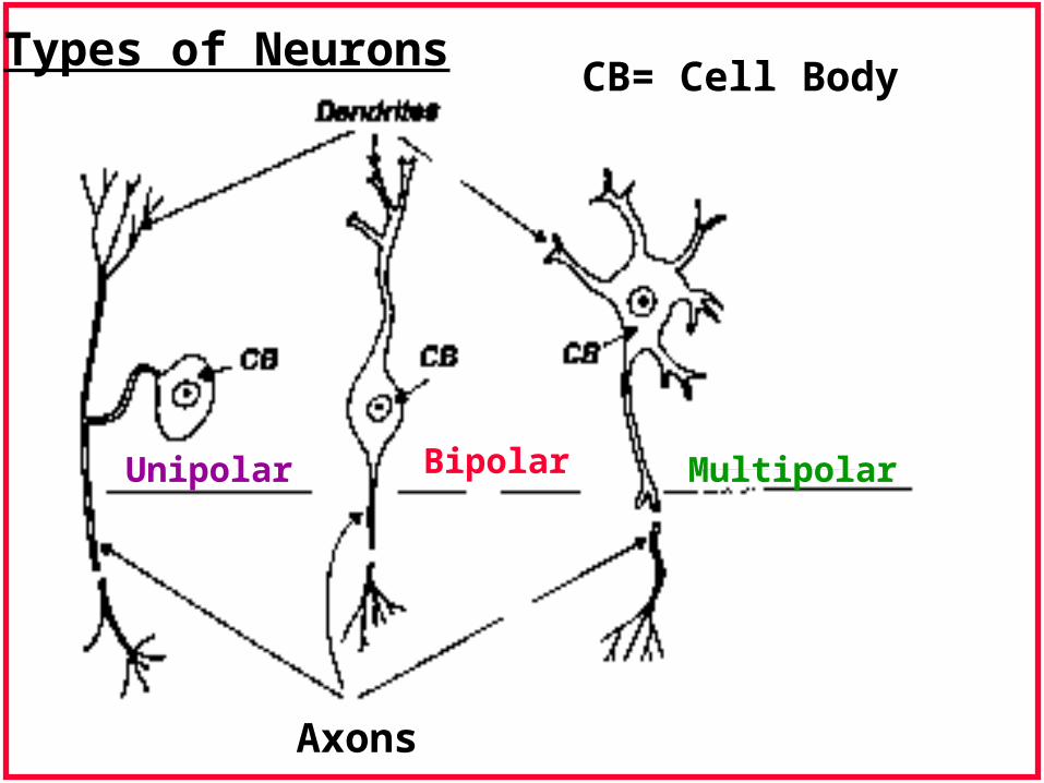
CB= Cell Body
Unipolar Bipolar Multipolar
Axons
Types of Neurons
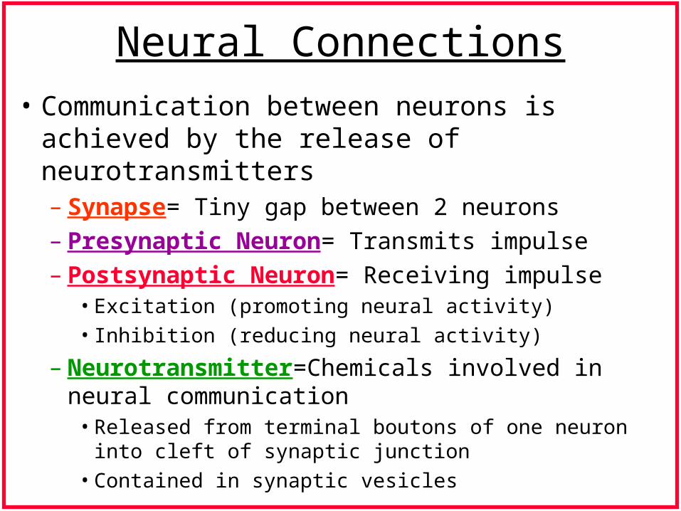
Neural Connections
• Communication between neurons is achieved by the release of neurotransmitters– Synapse= Tiny gap between 2 neurons– Presynaptic Neuron= Transmits impulse– Postsynaptic Neuron= Receiving impulse
• Excitation (promoting neural activity)
• Inhibition (reducing neural activity)
– Neurotransmitter=Chemicals involved in neural communication
• Released from terminal boutons of one neuron into cleft of synaptic junction
• Contained in synaptic vesicles
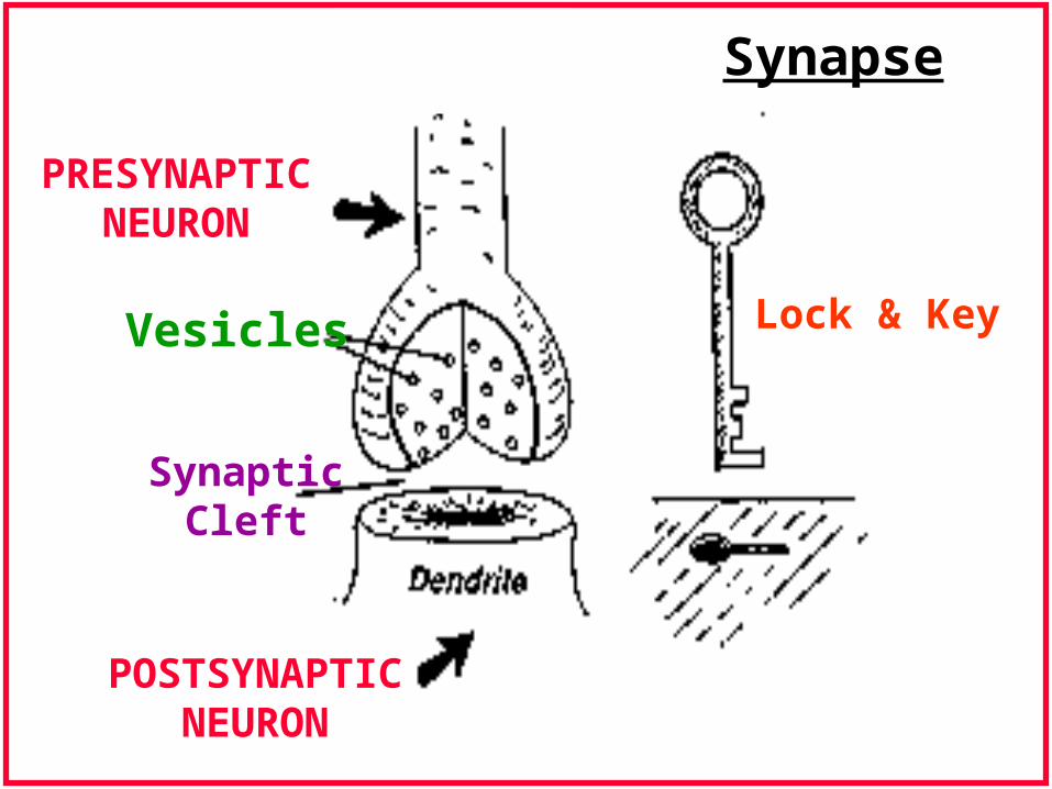
PRESYNAPTICNEURON
POSTSYNAPTICNEURON
Vesicles
SynapticCleft
Lock & Key
Synapse
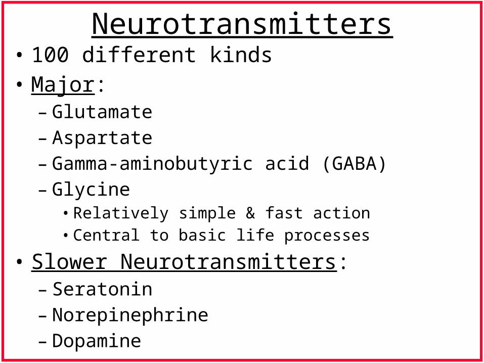
Neurotransmitters• 100 different kinds• Major:
– Glutamate– Aspartate– Gamma-aminobutyric acid (GABA)– Glycine
• Relatively simple & fast action• Central to basic life processes
• Slower Neurotransmitters:– Seratonin– Norepinephrine– Dopamine
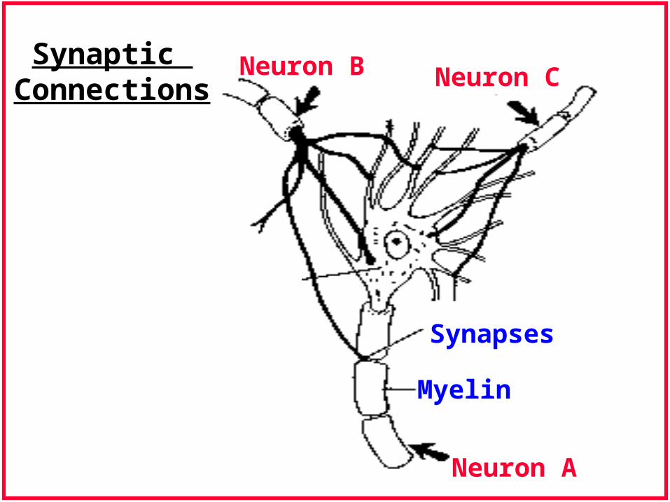
Synaptic Connections
Neuron B Neuron C
Neuron A
Synapses
Myelin
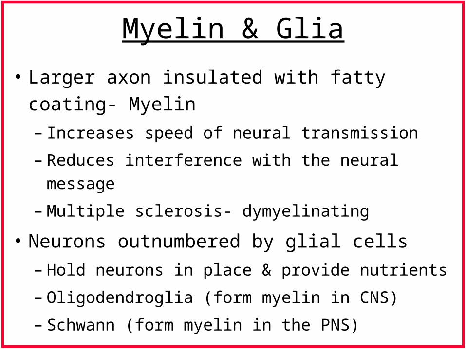
Myelin & Glia
• Larger axon insulated with fatty coating- Myelin
– Increases speed of neural transmission
– Reduces interference with the neural message
– Multiple sclerosis- dymyelinating
• Neurons outnumbered by glial cells
– Hold neurons in place & provide nutrients
– Oligodendroglia (form myelin in CNS)
– Schwann (form myelin in the PNS)
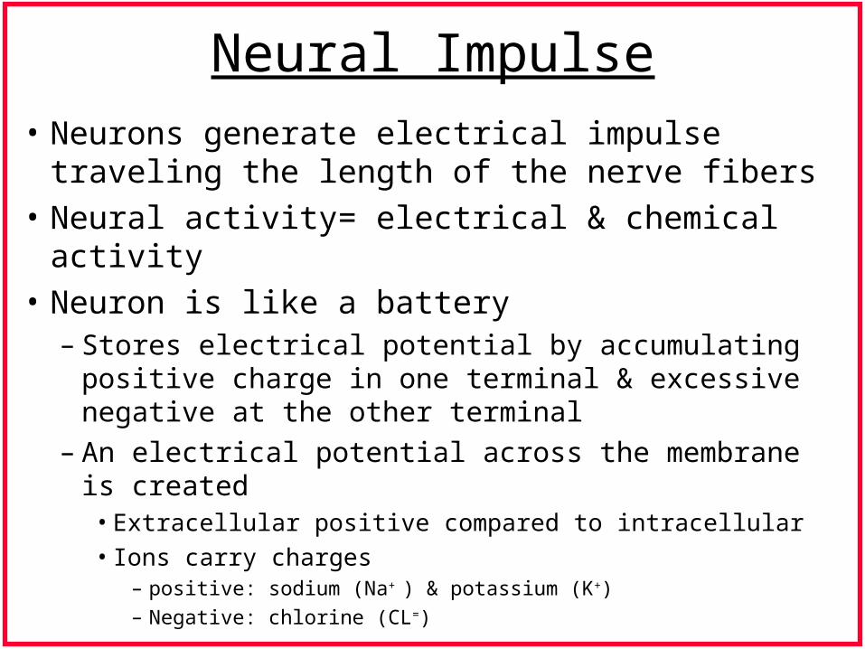
Neural Impulse
• Neurons generate electrical impulse traveling the length of the nerve fibers
• Neural activity= electrical & chemical activity
• Neuron is like a battery– Stores electrical potential by accumulating positive
charge in one terminal & excessive negative at the other terminal
– An electrical potential across the membrane is created• Extracellular positive compared to intracellular
• Ions carry charges – positive: sodium (Na+ ) & potassium (K+)
– Negative: chlorine (CL=)
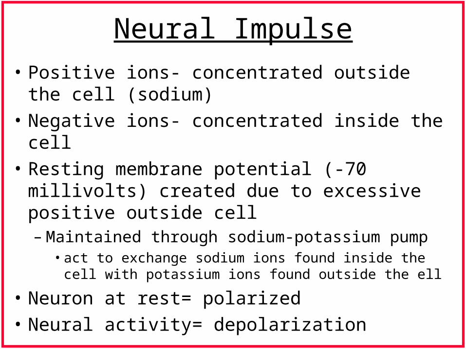
Neural Impulse
• Positive ions- concentrated outside the cell (sodium)
• Negative ions- concentrated inside the cell
• Resting membrane potential (-70 millivolts) created due to excessive positive outside cell– Maintained through sodium-potassium pump
• act to exchange sodium ions found inside the cell with potassium ions found outside the ell
• Neuron at rest= polarized
• Neural activity= depolarization
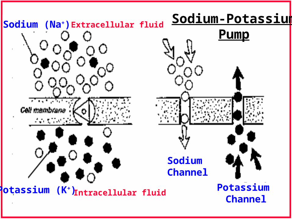
Sodium (Na+)
Potassium (K+)
Extracellular fluid
Intracellular fluid
Sodium Channel
Potassium Channel
Sodium-PotassiumPump
Neural Impulse
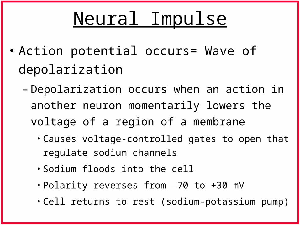
• Action potential occurs= Wave of depolarization
– Depolarization occurs when an action in another
neuron momentarily lowers the voltage of a region of a
membrane
• Causes voltage-controlled gates to open that regulate sodium
channels
• Sodium floods into the cell
• Polarity reverses from -70 to +30 mV
• Cell returns to rest (sodium-potassium pump)
Neural Impulse
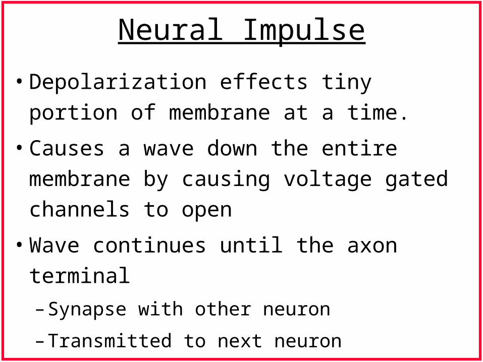
• Depolarization effects tiny portion of
membrane at a time.
• Causes a wave down the entire membrane by
causing voltage gated channels to open
• Wave continues until the axon terminal
– Synapse with other neuron
– Transmitted to next neuron
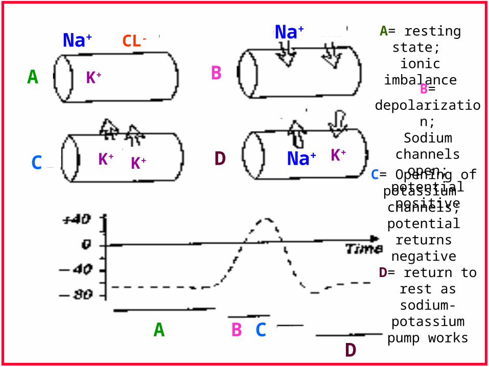
Na+ Na+CL-
K+
K+ K+
A
A
B
B
C
C
D
D
K+Na+
A= resting state; ionic imbalance
B= depolarization;Sodium
channels open; potential positive
C= Opening of potassium
channels; potential returns negative
D= return to rest assodium-potassium
pump works
Neuroanatomy of the Vocal Mechanism

Neuroanatomy of the vocal Mechanism
• Volitional control of muscles of the larynx resides in the
brain.
• Connecting points in brain that have a role in control of
phonation: cortex, subcortical areas, midbrain & medulla.
• Next slides will briefly review phonation neuroanatomy
& neurophysiology.
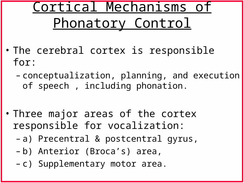
Cortical Mechanisms of Phonatory Control
• The cerebral cortex is responsible for:– conceptualization, planning, and execution of speech ,
including phonation.
• Three major areas of the cortex responsible for vocalization: – a) Precentral & postcentral gyrus, – b) Anterior (Broca’s) area, – c) Supplementary motor area.
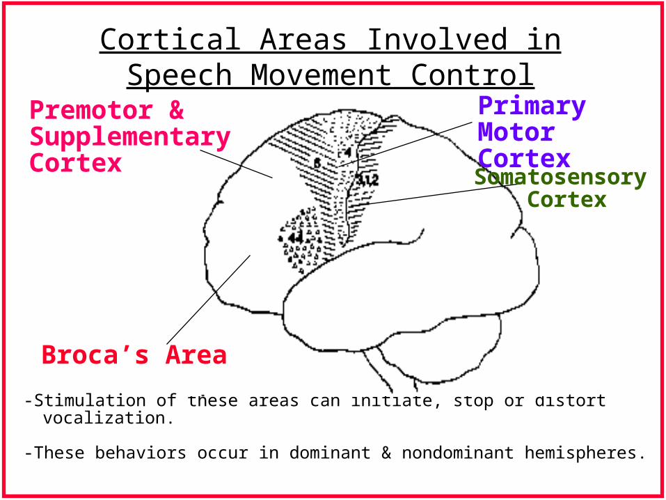
Cortical Areas Involved in Speech Movement Control
-Stimulation of these areas can initiate, stop or distort vocalization.
-These behaviors occur in dominant & nondominant hemispheres.
Premotor & SupplementaryCortex
Broca’s Area
Primary Motor Cortex
Somatosensory Cortex
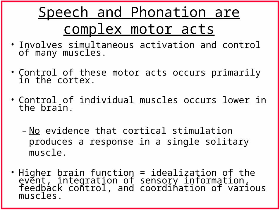
Speech and Phonation are complex motor acts
• Involves simultaneous activation and control of many muscles.
• Control of these motor acts occurs primarily in the cortex.
• Control of individual muscles occurs lower in the brain.
– No evidence that cortical stimulation produces a response in a single solitary muscle.
• Higher brain function = idealization of the event, integration of sensory information, feedback control, and coordination of various muscles.
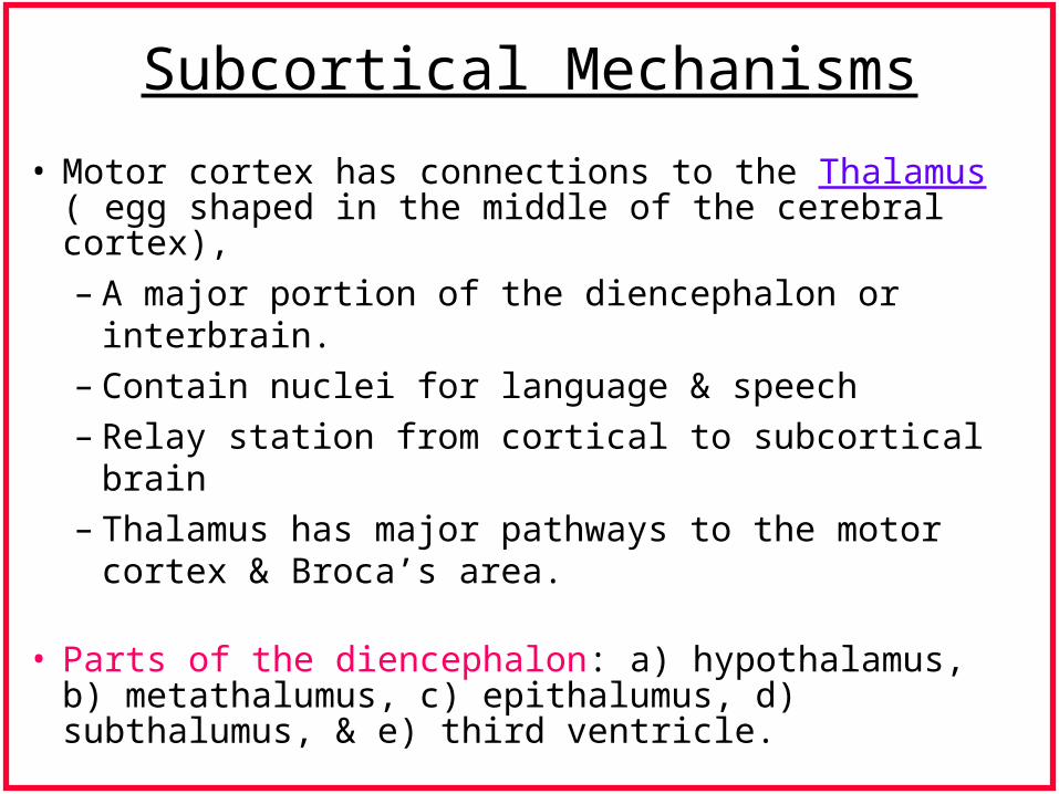
Subcortical Mechanisms
• Motor cortex has connections to the Thalamus ( egg shaped in the middle of the cerebral cortex),
– A major portion of the diencephalon or interbrain.– Contain nuclei for language & speech– Relay station from cortical to subcortical brain– Thalamus has major pathways to the motor cortex &
Broca’s area.
• Parts of the diencephalon: a) hypothalamus, b) metathalumus, c) epithalumus, d) subthalumus, & e) third ventricle.
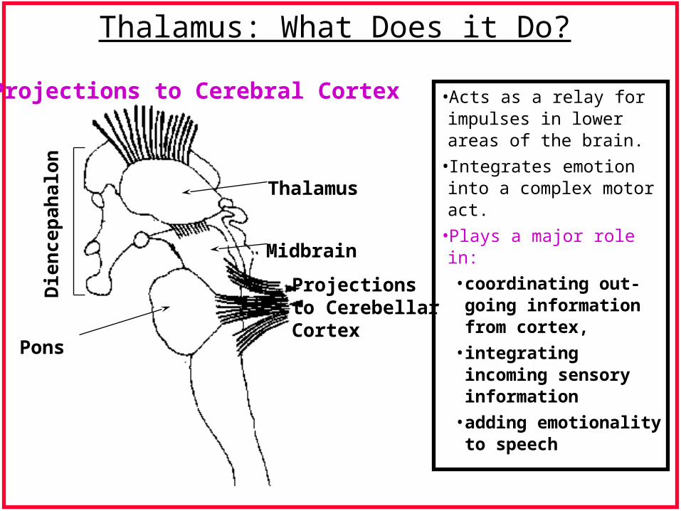
Thalamus: What Does it Do?
•Acts as a relay for impulses in lower areas of the brain.
•Integrates emotion into a complex motor act.
•Plays a major role in:
• coordinating out-going information from cortex,
• integrating incoming sensory information
• adding emotionality to speech
Thalamus
Projections to Cerebral Cortex
Midbrain
Projectionsto CerebellarCortex
Die
nce
pah
alon
Pons

Nuclei in thalamus that project to parts of the cerebral cortex
• Motor area receives its projections from the ventrolateral nucleus.
• 1971- ventrolateral nucleus shown to be responsible for initiation of speech movements & control of loudness, pitch, rate & articulation.
• Broca’s area- receives connections from dorsomedian nuclei.
VentralLateral Ventral posterior
Lateral
DorsalMedian
to & from Prenucleus
to & fromSup. Parietal Lobule
to & fromParietal Lobe
LateralDorsal
MassaIntermedia
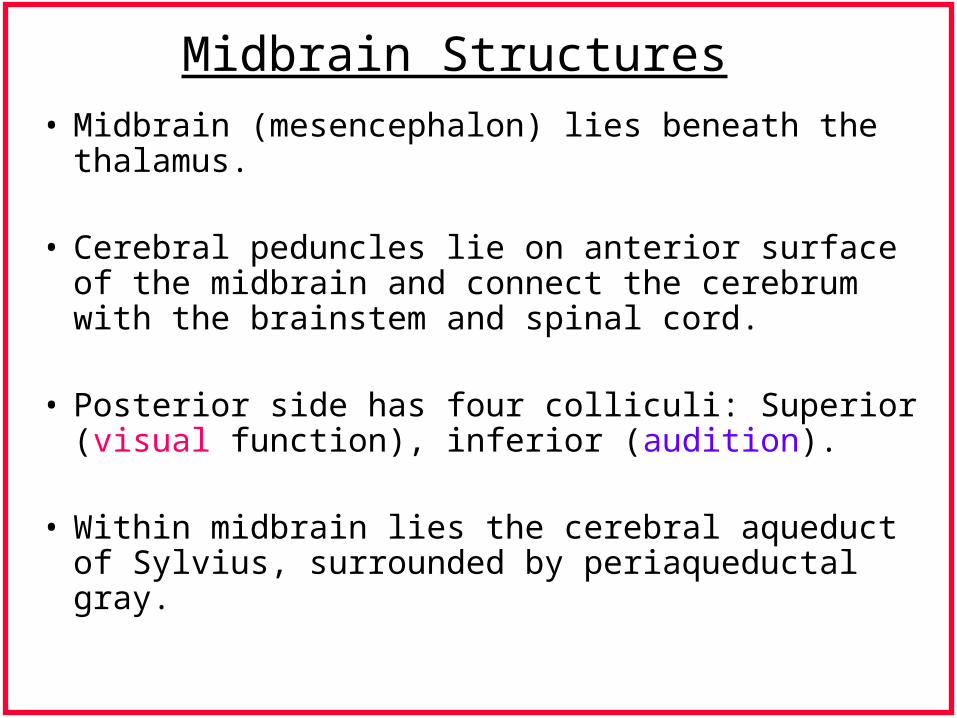
Midbrain Structures
• Midbrain (mesencephalon) lies beneath the thalamus.
• Cerebral peduncles lie on anterior surface of the midbrain and connect the cerebrum with the brainstem and spinal cord.
• Posterior side has four colliculi: Superior (visual function), inferior (audition).
• Within midbrain lies the cerebral aqueduct of Sylvius, surrounded by periaqueductal gray.
Periaqueductal Gray: What does it do?
• Stimulation of dorsal and ventrolateral areas of periaqueductal gray = activity in some laryngeal muscles.
• 1985- Larson reported some cells in ventrolateral area stimulate muscle activity, whereas some suppress activity.
• Periaqueductal gray is an intermediate area between recognition of a stimulus and the production of a motor act.
Brainstem
• Bilateral structures in brainstem implicated in the neural control of phonation:
• Nucleus ambiguus
• Nucleus tractus solitarii
• Nucleus parabrachialis
• How do we know these structures are involved in phonation?
Yoshida, Mitsumasu, Hirano Study
• Traced connections among brainstem structures.
• Injected tracer chemical into one nucleus ambiguus.
• Found evidence of tracer throughout the contralateral nuclei, nuclei tractus solitarri bilaterally, in nucleus parabrachialis and bilaterally in the lateral and ventrolateral parts of the periaqueductal gray area, with a predominance ipsilaterally.
• Conclusion: Many interconnections bilaterally among the nucleus ambiguous, nucleus tractus solitarri, and motor roots of vagus.
Cerebellum
• Structure lying posterior to the midbrain area.
• Implicated in the control of movement.• Three main portions: a) vermis, b) pars
intermedia, c) hemispheres• Consists of many traverse folia- increases
surface area.• Fissura prima- fissure separating anterior &
posterior lobes.
References:• Colton, R.H. & Casper, J.K.,(1990), Understanding Voice
Problems: A physiological perspective for diagnosis and treatment,, Williams & Wilkins.
• Bhatnager, S.C. & Andy, O.J., (1995), Neuroscience for the study of communicative disorders, Williams & Wilkins.
• Kuehn, D.P., Lemme, M.L. & Baumgartner, J.M., (1989), Neural basis of speech, hearing, and language, College- Hill Press.
• Lieberman, M., (1991), Neuroanatomy made easy and understandable, Aspen Publishers.
• Netsell, R., (1985), Speech and language evaluation in neurology-adult disorders, Grune & Stratton.
• Poritsky, R., (1992), Neuroanatomy: a functional atlas of parts & pathways, Mosby-Year Book.