neelay kothari, md march 12th, 2015 - wordpress.com · 12 mm in diameter focus of ring ......
TRANSCRIPT

Neelay Kothari, MD March 12th, 2015


Estimated 1.18 million people living with HIV (2008)
20% believed to be unaware of their infection
Males 75%, Females 25%
Rates of infection 8x higher amongst blacks than whites


0
2000
4000
6000
8000
10000
12000
14000
16000
18000
1981
1983
1985
1987
1989
1991
1993
1995
1997
1999
2001
2003
2005
2007
2009
2011
2013
Year`
Nu
mb
er
Prevalence

34 year old Somali female with negative PMH
Two month history of progressive dyspnea, dry cough, headache, loss of appetite
Acutely worse over the past 3 days
Exam (pertinent positives)
Febrile to 102.9 degrees
Dyspneic and tachypneic
Scattered crackles on lung exam

There is a very extensive diffuse hazy ground-glass opacity in virtually all the lungs, more severely involving the dependent portion of the lower lung fields, atelectases, as well as mild air bronchogram

Rapid HIV Test positive CD4+ count = 14 HIV Viral Load = 649,000

Which of the following is most likely?
(A) Pulmonary tuberculosis
(B) Pulmonary MAI infection
(C) Pneumocystis infection
(D) Toxoplasmosis pneumonia
(E) Cryptococcal pneumonia

How should diagnosis of pneumocystis pneumonia be made?
(A) Blood fungal cultures
(B) Sputum stain for pneumocystis
(C) Sputum fungal cultures
(D) Serology
Sputum stain positive for pneumocystis organisms

CD4 < 200 in 90%; Mean CD4 = 79
Before HAART: 70-80% got PCP without prophylaxis
Incidence markedly down with HAART
Risk of relapse 65 % w/o secondary prophylaxis

Symptoms usually of insidious onset Fever
Dyspnea
Non-productive cough
Often have oral thrush co-infection
Findings
Hypoxemia
Diffuse bilateral interstitial infiltrates (can have normal CXR)
LDH elevation (non-specific)
Pneumothorax

Histopathologic examination
PCR Testing
Beta-d-glucan assay
Not able to culture organism
Organisms persist for days to weeks
OK to start empiric treatment if high index of suspicion

Histopathology (microscopy with staining): Sensitivity in HIV patients
Induced sputum 50-90%
Bronch with BAL 90-99%
Transbronch biopsy 95-100%
Open lung biopsy 95-100%
Lower yield in non-HIV patients
Lower organism burden

PCR testing
Increased diagnostic yield, especially in non-HIV patients
Beta-d-glucan in serum
Up to 92% sensitivity
Not specific for pneumocystis

TMP-Sulfa is drug of choice
15-20 mg/kg q6-8h x 21 days
Intolerance common (rash, pancytopenia, hepatitis)
Survival rate 91% if complete treatment, 31% if change to pentamidine
Other options (mild-to-moderate disease)
Dapsone + TMP (similar efficacy, more pills)
Primaquine + clindamycin
Pentamidine
Atovaquone
Other options (severe disease)
IV Pentamidine (more toxic – hypoglycemia, metabolic, hepatitis, pancreatitis, neutropenia)
Primaquine + clindamycin
Trimetrexate (not available anymore)

Allow 7-10 d for response; PATIENCE! May deteriorate early Keep dry! Follow glucose on pentamidine Steroids: If PO2 < 70 or A-a gradient > 35 mmHg
Reduced early deterioration in oxygenation, decreased early and intermediate mortality
Prednisone 40 mg bid x 5d,20 mg bid x 5 d, 20 mg qd to day 21

Indications for primary prophylaxis CD4 < 200 (consider if 200-250 and dropping fast) Oropharyngeal candidiasis
Secondary prophylaxis History of pneumocystis (until immune
reconstitution)
Atovaquone 1500 mg po daily
Dapsone + pyrimethamine + leucovorin
Atovaquone + pyrimethamine + leucovorin

TMP-SMX 1 DS or SS daily Dapsone 100 mg daily Pentamidine Inhalation-300 mg q mo by neb
Non-compliant pts
More peripheral PCP and pneumothoraces
Atovaquone 1500 mg po daily Dapsone + pyrimethamine + leucovorin Atovaquone + pyrimethamine + leucovorin

2 years later….
Patient presents with difficulty swallowing, mouth ulcers, weight loss, fever, and pain in chest
Has been non-adherent to HIV treatment and prophylactic medications, missed several clinic visits
CD4 count 20, HIV viral load > 100,000
Exam: several ulcers in mouth, + thrush
Possible causes of patient’s symptoms?
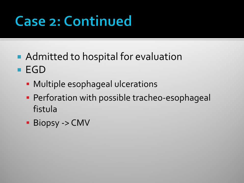
Admitted to hospital for evaluation EGD
Multiple esophageal ulcerations
Perforation with possible tracheo-esophageal fistula
Biopsy -> CMV

Typically causes disease with CD4 < 50 Chorioretinitis
30% incidence in AIDS patients (before HAART)
Initially unilateral – will spread to other eye
Floaters, visual loss, decreased acuity
Diagnosis: clinical ophthalmology evaluation
Treatment: systemic and local (val)ganciclovir
Esophagitis / colitis Fever, weight loss, diarrhea, odynophagia
Can have hemorrhage / perforation rarely
Neurologic disease: dementia, ventriculoencephalitis, ascending polyradiculomyelopathy
Pulmonary disease: significance often unclear

66 year old Native American male Presenting with partial seizure with left face
twitching, left arm rhythmic movements, and slurred speech
Past Medical History: CAD, hyperlipidemia, hypertension
Social History: divorced, works as mechanic and farmer, lives alone

12 mm in diameter focus of ring
enhancement at the corticomedullary
junction of the lateral aspect of the
precentral gyrus of the posterior right
frontal lobe in the expected location of the
primary motor cortex. There is vasogenic
edema in the adjacent white matter of this
gyrus

Differential Diagnosis?
Further workup?

12 mm in diameter focus of ring
enhancement at the corticomedullary
junction of the lateral aspect of the
precentral gyrus of the posterior right
frontal lobe in the expected location of the
primary motor cortex. There is vasogenic
edema in the adjacent white matter of this
gyrus
1.1 cm peripherally enhancing lesion within
the posterolateral right frontal lobe that has
developed worsening non-enhancing
increased T2 signal in the subcortical white
matter compatible with edema
3 Days Later

Laboratory Evaluation
HIV antibody POSITIVE
CD4 count = 32
HIV viral load = 315,000
Toxoplasma IgG POSITIVE

EPIDEMIOLOGY
Variable seroprevalence 15% in United States
50-75% in parts of Europe
Transmission Undercooked meat with tissue
cysts
Oocysts in cat feces (sporulation requires 24 hours)
Not person-to-person
CLINICAL
Disease risk CD 4 count usually < 50
33% will get disease without prophylaxis
Symptoms Focal encephalitis
Headache, confusion, focal motor weakness
Fever
Seizures
ExtraCNS disease rare

Clinical presentation Serology 95 % sensitive CT or MRI-Multiple, bilateral,hypodense,
ring-enhancing mass lesions with predilection for B.G. and corticomedullary junction
Commonest cause of focal brain lesion in AIDS
Reduced in HAART Era
Clinical and radiographic response to Rx
Usually better in 2 wks

Differential Diagnosis
CNS Lymphoma
Toxoplasmosis
Tuberculosis
Cryptococcus
Other: Bacterial / PML / Chagas disease
Often treat empirically for toxoplasmosis
Biopsy if fail to respond

Pyrimethamine(200mg x 1 then 50-100 mg qd) + Sulfadiazine(1-2 g q 6h) Leucovorin to prevent BM suppression
80-90 % respond in 2-6 wks
Relapse 90 % w/o maintenance
f/u CT or MRI in 2-4 wks
Treatment at least 6 weeks, often longer if incomplete response
Chronic maintenance / secondary prevention: Pyrimethamine 25-50 mg daily + sulfadiazine 2-4 grams daily + leucovorin
Other regimens Pyrimethamine + Clindamycin Pyrimethamine + Azithromycin or Clarithromycin Atovaquone +/- Pyrimethamine

Indication: CD4 < 100 and Toxo IgG + TMP-SMX 1 DS tab daily is first line Other options
TMP-SMX 1 DS PO tiw
TMP-SMX 1 SS PO daily
Dapsone 50 mg PO daily + pyrimethamine 50 mg PO weekly + leucovorin 25 mg PO weekly
Dapsone 200 mg + pyrimethamine 75 mg + leucovorin 25 mg) PO weekly
Atovaquone 1,500 mg +/- pyrimethamine 25 mg + leucovorin 10 mg) PO daily

43 year old female originally from Kenya 2-4 week history of
Left sided weakness
10 lb weight loss
Mental slowing
Fatigue and sleepiness
Past medical history: anemia Exam: slow to respond to questions, mild left
sided weakness, white coating on tongue


Laboratory Evaluation
HIV Antibody POSITIVE
CD4 count = 16
HIV Viral Load = 3,220,000
RPR Negative
CSF
▪ 1 WBC, 1 RBC, protein 26, glucose 54
▪ Cryptococcal antigen negative
▪ JC virus PCR POSITIVE

Patient was started on HIV treatment
Unfortunately her mental status continued to deteriorate
Transitioned to comfort care and died

JC Virus – 85% of population seropositive CD4+ count usually < 100 Clinical Presentation
Demyelinating lesions
Focal neurologic deficits
Progressive over weeks to months
Seizures in 20%
Imaging: Multiple nonenhancing white matter lesions
No mass effect
CSF analysis: PCR positive in 70-90% if not on HAART

Treatment
No specific treatment
Reverse immunosuppression ▪ Start HAART immediately
Prognosis
High morbidity and mortality
Half get better with HAART, though often have residual deficits
Lower CD4 count at presentation associated with poor outcome

46 year old male previously healthy Chief complaint: headache
Present for approximately one month
Sudden onset, with gradual worsening since
Bilateral, retro-orbital and into top of head Initially seen in Minute Clinic
Presumed sinusitis, prescribed amoxicillin
Not better, prescribed azithromycin
Not better, then prescribed Vicodin 6 days prior to admission

Associated symptoms Nausea and vomiting x 2 weeks
15 pound weight loss
Brief 30-60 second episodes of dizziness
Intermittent photophobia
Ringing sensation in right ear
Feels somewhat depressed Per family
Slow mentation and speech
Increased sleepiness

Works as nail technician, unable to work recently
No tobacco, occasional cigar, occasional alcohol (2 drinks per week), denies other drug use
Travel: recent travel to England No pets, animal or tick exposures, sick
contacts Enjoys gardening

VS Temp 98.1, P 75, R 16, BP 131/90 Weight 130 lb General: flat and depressed affect, slow
speech HEENT: whitish patches on tongue Skin: 3-4 mm skin colored papules present on
forehead, shoulder, sternum Remainder of exam unremarkable

HEAD MRI Focally increased signal intensity on
FLAIR and T2-weighted images involving the caudate nuclei, putamina, globus pallidus nuclei and the thalami bilaterally.
No associated hemorrhage, midline shift or hydrocephalus.
Patchy restricted diffusion in the same distribution, but to a lesser extent than the areas of T2 prolongation.

WBC 4.3, Hgb 12.7, plt 202 Creatinine 0.97 Liver function tests normal Rapid HIV test positive Lumbar puncture
Opening pressure 31cm
0 RBC, 74 WBC (90% L)
Protein 76, glucose 45
Gram stain: no PMNs, 2+ yeast
RPR negative
Cryptococcal antigen positive (1:2048)

Can infect any organ Most common sites of
infection
Lungs: nodules, masslike infiltrate, adenopathy, lung cavitation, lobar infiltrates
CNS: subacute meningitis or meningoencephalitis
Skin: papules with soft or ulcerated center

CD4 count usually < 50 Usually subacute presentation
Meningitis or meningoencephalitis
Fever, malaise, headache
CSF findings
Opening pressure > 20 cm in 75%
Mildly increased protein, glucose low to normal
Lymphocytic pleocytosis or normal WBC
Positive serum cryptococcal antigen (93-99%)

Altered mental status Positive cryptococcal BC CSF Ag titer > 1:1024 + CSF India Ink CSF Cell Count <20

Initial Treatment Amphotericin + flucytosine x 2 weeks minimum Monitor flucytosine levels Monitor renal function / can use liposomal ampho Serial therapeutic lumbar puncture to reduce ICP
Follow up therapy Lumbar puncture at 2 weeks to check culture Once clinically better and if culture negative, can switch to
fluconazole 400 mg po daily x 8 weeks
Chronic maintenance therapy Fluconazole 200 mg po daily Until immune reconstitution

Randomized trial in 64 patients
Ampho monotherapy
Ampho + Flucytosine
Ampho + Fluconazole
Ampho + Flucytosine + Fluconazole
Ampho + Flucytosine had more rapid CSF sterilization (0.15-0.23 log CFU per day)
No association with clinical outcomes
Brouwer et al, Lancet, 2004.

Observational Study
208 patients
Outcome: death or mycologic failure at 2 weeks
Ampho + flucytosine: 26% failure
Other treatments: 56% failure
Dromer et al, PLOS One, 2008

Treatment:
Amphotericin B 0.7 mg/kg IV daily
Flucytosine 25 mg/kg po q6hours
Repeat lumbar puncture
Opening pressure = 8 cm
Additional laboratory testing
CD4 count 68, viral load 1,090,000
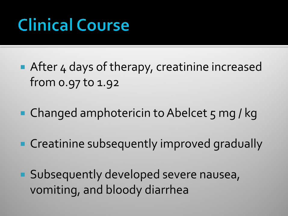
After 4 days of therapy, creatinine increased from 0.97 to 1.92
Changed amphotericin to Abelcet 5 mg / kg
Creatinine subsequently improved gradually
Subsequently developed severe nausea, vomiting, and bloody diarrhea

Underwent EGD and colonoscopy with biopsy
Results: 1. Atypical colitis, suspicious for drug induced (possibly flucytosine). This is a
morphologically unusual colitis, which is histologically very similar to the colitis one sees in patients with mycophenolate mofetil colitis.
2. No viral inclusions noted
3. No evidence of idiopathic inflammatory bowel disease or opportunistic infection

Headache improved, mentation improved Repeat lumbar puncture at ~2 weeks
Cryptococcal antigen 1:256
Culture with 1+ cryptococcus
▪ Fluconazole MIC = 16
▪ Flucytosine MIC > 32 (RESISTANT)
Antifungal therapy was changed to fluconazole

Gradual improvement in CNS and gastrointestinal symptoms
Eventually started HAART approximately 2 months after initial diagnosis
Now doing well
CD4 count improved from 38 to 263
No CNS sequelae

Ubiquitous in environment Typically CD4 count < 50 (20-40% incidence) Clinical presentation
Often non-specific
Fever, night sweats, fatigue, weight loss, diarrhea
Pulmonary presentation less common
Anemia, elevated alkaline phosphatase
Hepatomegaly, splenomegaly
Diagnosis: culture from sterile site

Treatment: multi-drug therapy
Clarithromycin (or azithromycin)
Ethambutol
Rifabutin
Prophylaxis – rule out active infection first
Azithromycin 1200 mg weekly is preferred
Rifabutin is an option
▪ More drug interactions
▪ Rule out active TB

Most common HIV-related illness globally Cause of death for 13% of those with AIDS CD4 most often 300-500 Reactivation most common 10 % risk per yr of reactivation v 10% lifetime risk if
immunocompetent Presentation typical of reactivation only if intact
immune system In AIDS extrapulmonary in 2/3 Only 30-50 % with AIDS have + PPD(>5mm)
Retest once CD4 count >200

Most common neoplasm in AIDS Usually gay males HHV-8-DNA + > 90% HIV + KS Localized or widespread visceral disease
GI,Lung, LN Violaceous lesions Bx-mimics BA
Rx = IFN, Vinblastine, VCR,VP-16,Adriamycin Responses Poor
HAART-good responses, sometimes complete remission
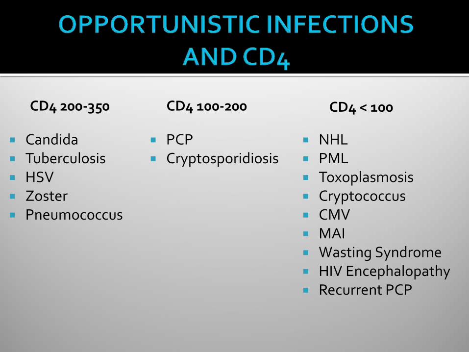
CD4 100-200
PCP Cryptosporidiosis
CD4 < 100
NHL PML Toxoplasmosis Cryptococcus CMV MAI Wasting Syndrome HIV Encephalopathy Recurrent PCP
Candida Tuberculosis HSV Zoster Pneumococcus
CD4 200-350
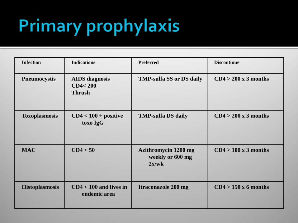
Infection Indications Preferred Discontinue
Pneumocystis AIDS diagnosis
CD4< 200
Thrush
TMP-sulfa SS or DS daily CD4 > 200 x 3 months
Toxoplasmosis CD4 < 100 + positive
toxo IgG
TMP-sulfa DS daily CD4 > 200 x 3 months
MAC CD4 < 50 Azithromycin 1200 mg
weekly or 600 mg
2x/wk
CD4 > 100 x 3 months
Histoplasmosis CD4 < 100 and lives in
endemic area
Itraconazole 200 mg CD4 > 150 x 6 months

Parodoxical clinical worsening after starting HAART despite improved immune function
Due to inflammatory response against infectious antigen CD 4 count usually < 50 Usually within 6 weeks, though can be several months Common pathogens: MAC, TB, CMV, cryptococcus,
pneumocystis, JC virus Can have atypical presentation Treatment: continue HAART if possible, treat infection,
anti-inflammatory therapy

Opportunistic infections are often the initial presentation of persons with undiagnosed HIV infection
Important to recognize syndromes, test for HIV infection when indicated
Treatment of HIV infection reduces risk for opportunistic infections