necropsy report for sperm whale stranded on …csi.whoi.edu/sites/default/files/mh 02 673 pm...
TRANSCRIPT

1
Necropsy Report for Sperm Whale Stranded on Nantucket June 7th 2002 General Observations Field Number: MH02-673-Pm, CCSN 02-163Pm Time and Date discovered: 0600 June 07 2002 Location of Stranding: Great Point, Nantucket 41 22 N, 070 01 W Dates of Necropsy: June 10, 11 and 17 2002 Necropsy Location: Transfer Station, City of New Bedford, MA Species: Sperm whale (Physeter macrocephalus) Sex: Male Lengths:
Total Length 14.65m Skull Length 5.2m Snout to eye 3.9m. Snout to Temporomandibular joint 4.5m Fluke width 4.13m
Weight: 90,000lbs (by DN Kelly Inc. Travelift) Age: Pending analysis of tooth. Length/ weight curve for male sperm whales from Best (1970) would suggest animal to be a mature male of about 30 years old (Best, PB. 1970. “The sperm whale off the west coast of South Africa”. Div. Sea. Fisheries Inv. Rep. 79 page 9). Condition Code: 2-3 (Early stages of autolysis). Human Interaction: No evidence Date and Time of Death: Not known, but assume close to time of discovery given lack of skin blisters and scavenging. Necropsy Team Leader and author of this report: Michael Moore, Woods Hole Oceanographic Institution, Woods Hole MA 02543 [email protected] 508 289 3228 telephone. On-Site Coordinator: Katie Touhey, Cape Cod Stranding Network Off-Site Coordinator: Connie Merigo, New England Aquarium Recorder and Photographer: Caroline Harper (WHOI)
History 0600 June 7th 2002, a sperm whale was reported dead on the beach a mile south of Great Point lighthouse on the NE corner of Nantucket, MA. The animal was first sighted, according to the Nantucket Marine Mammal Stranding Team, with its tail pointing out to sea. The animal appeared to be freshly dead at the time of stranding. Girth appeared to be small for the length of the animal. It was then pounded with heavy surf on the 7th. Beach assessment by staff from the New England Aquarium, Mass Wildlife and WHOI, on June 8th lead to the conclusion that necropsy at that site was impractical, given the presence of endangered bird species precluding the use of heavy machinery. The New Bedford Whaling Museum (NBWM) was known to be interested in acquiring a sperm whale skeleton. A 20’ x 4” cargo strap was wound around the tail stock, the flukes cut off short, and a 600’ floating tow hawser shackled to the strap. The animal was towed to New Bedford Harbor on June 9th 2002 by the Tug Jaguar. The animal was lifted by a Travelift at DN Kelleys in Fairhaven, weighed, and deposited on a flatbed trailer for trucking to the New Bedford city transfer station. It arrived at the necropsy site 0030 June 10th (see above). A necropsy was conducted on June 10, 11 and 17 2002. On the first day, the blubber coat and lower jaw were removed and the lower intestines examined. On the second day the post cranial skeleton was removed, and the lungs, heart, stomach and bones examined. On the third day the remaining
2
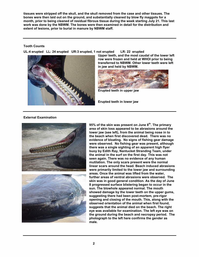
tissues were stripped off the skull, and the skull removed from the case and other tissues. The bones were then laid out on the ground, and substantially cleaned by blow fly maggots for a month, prior to being cleaned of residual fibrous tissue during the week starting July 21. This last work was done by the NBWM. The bones were then examined in detail for the distribution and extent of lesions, prior to burial in manure by NBWM staff. Tooth Counts UL:4 erupted LL: 24 erupted UR:3 erupted, 1 not erupted LR: 22 erupted
Upper teeth, and the most caudal of the lower left row were frozen and held at WHOI prior to being transferred to NBWM. Other lower teeth were left in jaw and held by NBWM.
Erupted teeth in upper jaw Erupted teeth in lower jaw
External Examination
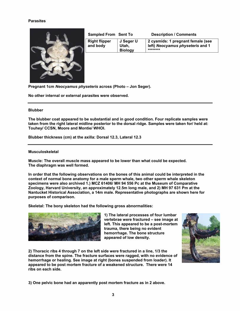
95% of the skin was present on June 8th. The primary area of skin loss appeared to be abrasions around the lower jaw (see left), from the animal being nose in to the beach when first discovered dead. There was no evidence of bloating. No signs of fishing gear damage were observed. No fishing gear was present, although there was a single sighting of an apparent high flyer buoy by Edith Ray, Nantucket Stranding Team, under the animal in the surf on the first day. This was not seen again. There was no evidence of any human mutilation. The only scars present were the normal linear scars around the head. Beach induced abrasions were primarily limited to the lower jaw and surrounding areas. Once the animal was lifted from the water, further areas of ventral abrasions were observed. The skin was in good general condition. As the day of June 8 progressed surface blistering began to occur in the sun. The blowhole appeared normal. The mouth showed damage by the lower teeth on the upper gums, suggesting there had been post-mortem, pre-rigor opening and closing of the mouth. This, along with the observed orientation of the animal when first found, suggests that the animal died on the beach. The right eye was available for examination. The left eye was on the ground during the beach and necropsy period. The photograph to the left here confirms the gender as male.
3
Parasites
Sampled From Sent To Description / Comments
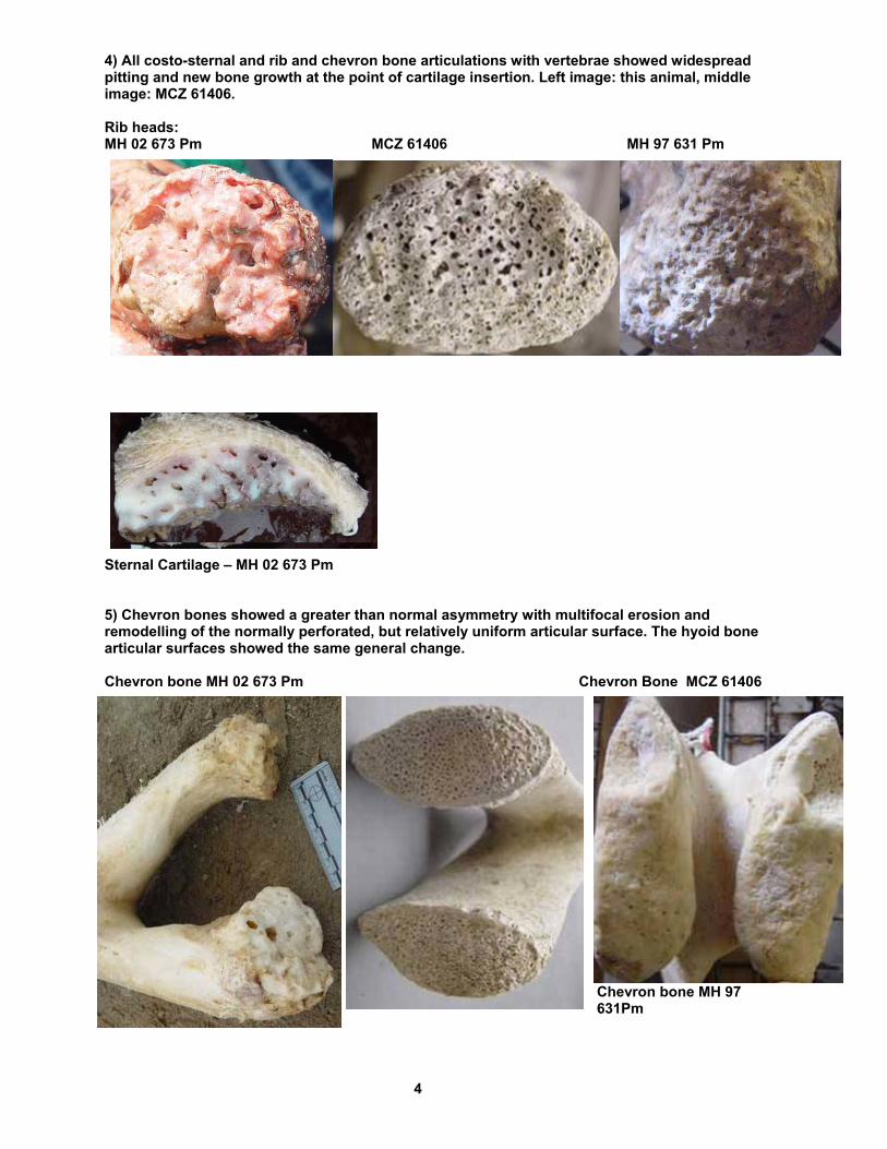
Pregnant 1cm Neocyamus physeteris across (Photo – Jon Seger). No other internal or external parasites were observed. Blubber The blubber coat appeared to be substantial and in good condition. Four replicate samples were taken from the right lateral midline posterior to the dorsal ridge. Samples were taken for/ held at: Touhey/ CCSN, Moore and Montie/ WHOI. Blubber thickness (cm) at the axilla: Dorsal 12.3, Lateral 12.3 Musculoskeletal Muscle: The overall muscle mass appeared to be lower than what could be expected. The diaphragm was well formed. In order that the following observations on the bones of this animal could be interpreted in the context of normal bone anatomy for a male sperm whale, two other sperm whale skeleton specimens were also archived 1.) MCZ 61406/ MH 94 556 Pc at the Museum of Comparative Zoology, Harvard University, an approximately 12.5m long male, and 2) MH 97 631 Pm at the Nantucket Historical Association, a 14m male. Representative photographs are shown here for purposes of comparison. Skeletal: The bony skeleton had the following gross abnormalities:
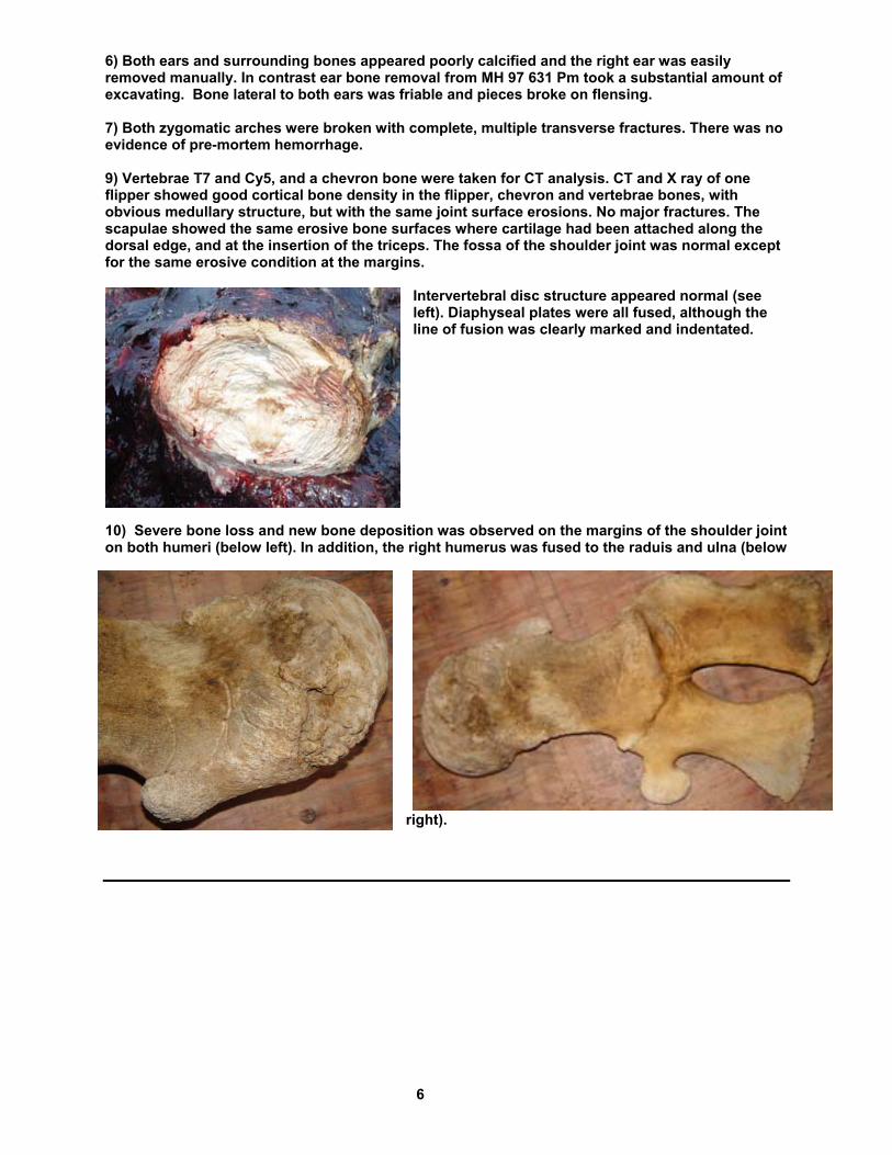
1) The lateral processes of four lumbar vertebrae were fractured – see image at left. This appeared to be a post-mortem trauma, there being no evident hemorrhage. The bone structure appeared of low density.
2) Thoracic ribs 4 through 7 on the left side were fractured in a line, 1/3 the distance from the spine. The fracture surfaces were ragged, with no evidence of hemorrhage or healing. See image at right (bones suspended from loader). It appeared to be post mortem fracture of a weakened structure. There were 14 ribs on each side. 3) One pelvic bone had an apparently post mortem fracture as in 2 above.
Right flipper and body
J Seger U Utah, Biology
2 cyamids: 1 pregnant female (see left) Neocyamus physeteris and 1 ********
4
4) All costo-sternal and rib and chevron bone articulations with vertebrae showed widespread pitting and new bone growth at the point of cartilage insertion. Left image: this animal, middle image: MCZ 61406. Rib heads: MH 02 673 Pm MCZ 61406 MH 97 631 Pm
Sternal Cartilage – MH 02 673 Pm 5) Chevron bones showed a greater than normal asymmetry with multifocal erosion and remodelling of the normally perforated, but relatively uniform articular surface. The hyoid bone articular surfaces showed the same general change. Chevron bone MH 02 673 Pm Chevron Bone MCZ 61406
Chevron bone MH 97 631Pm

5
CT scan of above chevron bone from this animal
6) Sternum and skull bone surface appeared rough, and pitted with new bony growth. This was also seen to a lesser degree in MH 97 631 Pm Sternum – this animal Sternum MCZ 61406 Sternum MH 97 631 Pm
In the skull this change was primarily in the caudal half, with the dorsal surface showing extensive remodeling, especially on the left side caudal to the internal nares. The bone surface had the texture of a lava flow. The bony density around each temporal bone caudal to the mandibular joint was friable, spongy and soft (see left below). The vomer was very spongy. The right parietal bone was thinned, to the extent that in two places there was an actual fistula in the bone plate (see right below).
6
6) Both ears and surrounding bones appeared poorly calcified and the right ear was easily removed manually. In contrast ear bone removal from MH 97 631 Pm took a substantial amount of excavating. Bone lateral to both ears was friable and pieces broke on flensing. 7) Both zygomatic arches were broken with complete, multiple transverse fractures. There was no evidence of pre-mortem hemorrhage. 9) Vertebrae T7 and Cy5, and a chevron bone were taken for CT analysis. CT and X ray of one flipper showed good cortical bone density in the flipper, chevron and vertebrae bones, with obvious medullary structure, but with the same joint surface erosions. No major fractures. The scapulae showed the same erosive bone surfaces where cartilage had been attached along the dorsal edge, and at the insertion of the triceps. The fossa of the shoulder joint was normal except for the same erosive condition at the margins.
Intervertebral disc structure appeared normal (see left). Diaphyseal plates were all fused, although the line of fusion was clearly marked and indentated.
10) Severe bone loss and new bone deposition was observed on the margins of the shoulder joint on both humeri (below left). In addition, the right humerus was fused to the raduis and ulna (below
right).
7
Respiratory
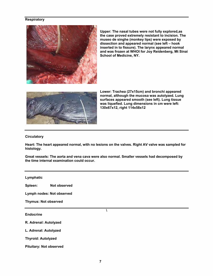
Upper: The nasal tubes were not fully explored,as the case proved extremely resistant to incision. The museo de singhe (monkey lips) were exposed by dissection and appeared normal (see left – hook inserted in to fissure). The larynx appeared normal and was frozen at WHOI for Joy Reidenberg, Mt Sinai School of Medicine, NY.
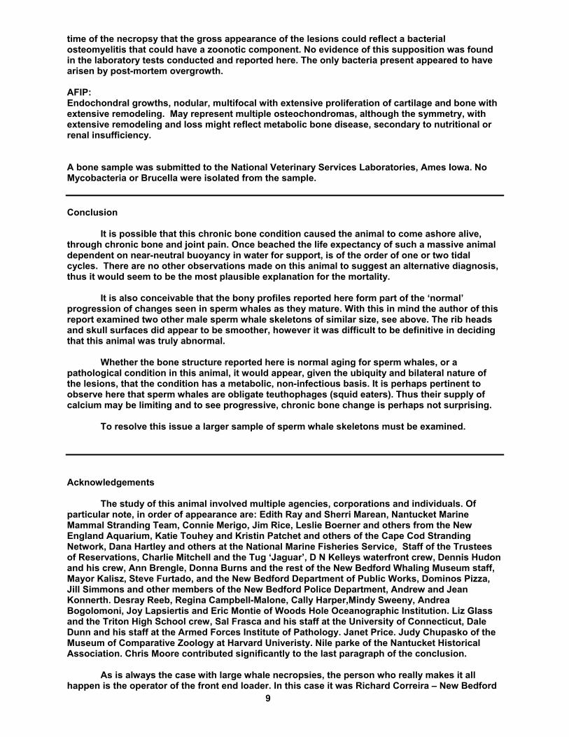
Lower: Trachea (27x15cm) and bronchi appeared normal, although the mucosa was autolyzed. Lung surfaces appeared smooth (see left). Lung tissue was liquefied. Lung dimensions in cm were left: 130x67x12, right 114x58x12
Circulatory Heart: The heart appeared normal, with no lesions on the valves. Right AV valve was sampled for histology. Great vessels: The aorta and vena cava were also normal. Smaller vessels had decomposed by the time internal examination could occur. Lymphatic Spleen: Not observed Lymph nodes: Not observed Thymus: Not observed
\
Endocrine R. Adrenal: Autolyzed L. Adrenal: Autolyzed Thyroid: Autolyzed Pituitary: Not observed

8
Urinary R. Kidney: Autolyzed L. Kidney: Autolyzed Bladder: complete and normal but empty. Digestive Esophagus: Normal, with a good mucosa. Stomach: Normal. A small amount of flaky debris was present in the fundic portion. Stomach contents: A small square of fishing net was present in the pylorus. No other contents. Intestines: Sampled for histology Liver: Autolyzed Pancreas / Pancreatic ducts: Autolyzed Reproductive One testis sampled for histology Nervous/ Sensory Eyes: Appeared normal grossly. Right eye sampled for histology Spinal Cord: Autolyzed Brain: Autolyzed Carcass disposition 6 chevron bones, 3 hyoids, 28 ribs, 1 sternum, 2 scapulae, 2 flippers, 2 pelvic bones, the backbone and cranium and two mandibles with teeth were retained by the New Bedford Whaling Museum. Soft tissue waste was disposed via leakproof dumpsters at the Crapo Hill landfill. Laboratory Studies Samples for soft tissue and bone histology were submitted to Connecticut Veterinary Diagnostic Laboratory (CVDL) and the Armed Forces Institute of Pathology (AFIP). Reports are given below. In summary the following diagnoses were made: CVDL: Articular surfaces: Chondrodegeneration, erosion, ulceration, multifocal, chronic, marked with osteolysis and proliferation. Indicating articular cartilage degeneration, with osteolysis and proliferation. Etiology unclear, but premortem, with no indication of any infectious disease. Ziehl-Neelsen staining failed to show any definitive acid-fast bacteria. There was a concern raised at the
9
time of the necropsy that the gross appearance of the lesions could reflect a bacterial osteomyelitis that could have a zoonotic component. No evidence of this supposition was found in the laboratory tests conducted and reported here. The only bacteria present appeared to have arisen by post-mortem overgrowth. AFIP: Endochondral growths, nodular, multifocal with extensive proliferation of cartilage and bone with extensive remodeling. May represent multiple osteochondromas, although the symmetry, with extensive remodeling and loss might reflect metabolic bone disease, secondary to nutritional or renal insufficiency. A bone sample was submitted to the National Veterinary Services Laboratories, Ames Iowa. No Mycobacteria or Brucella were isolated from the sample. Conclusion
It is possible that this chronic bone condition caused the animal to come ashore alive, through chronic bone and joint pain. Once beached the life expectancy of such a massive animal dependent on near-neutral buoyancy in water for support, is of the order of one or two tidal cycles. There are no other observations made on this animal to suggest an alternative diagnosis, thus it would seem to be the most plausible explanation for the mortality.
It is also conceivable that the bony profiles reported here form part of the ‘normal’ progression of changes seen in sperm whales as they mature. With this in mind the author of this report examined two other male sperm whale skeletons of similar size, see above. The rib heads and skull surfaces did appear to be smoother, however it was difficult to be definitive in deciding that this animal was truly abnormal.
Whether the bone structure reported here is normal aging for sperm whales, or a pathological condition in this animal, it would appear, given the ubiquity and bilateral nature of the lesions, that the condition has a metabolic, non-infectious basis. It is perhaps pertinent to observe here that sperm whales are obligate teuthophages (squid eaters). Thus their supply of calcium may be limiting and to see progressive, chronic bone change is perhaps not surprising.
To resolve this issue a larger sample of sperm whale skeletons must be examined.
Acknowledgements The study of this animal involved multiple agencies, corporations and individuals. Of particular note, in order of appearance are: Edith Ray and Sherri Marean, Nantucket Marine Mammal Stranding Team, Connie Merigo, Jim Rice, Leslie Boerner and others from the New England Aquarium, Katie Touhey and Kristin Patchet and others of the Cape Cod Stranding Network, Dana Hartley and others at the National Marine Fisheries Service, Staff of the Trustees of Reservations, Charlie Mitchell and the Tug ‘Jaguar’, D N Kelleys waterfront crew, Dennis Hudon and his crew, Ann Brengle, Donna Burns and the rest of the New Bedford Whaling Museum staff, Mayor Kalisz, Steve Furtado, and the New Bedford Department of Public Works, Dominos Pizza, Jill Simmons and other members of the New Bedford Police Department, Andrew and Jean Konnerth. Desray Reeb, Regina Campbell-Malone, Cally Harper,Mindy Sweeny, Andrea Bogolomoni, Joy Lapsiertis and Eric Montie of Woods Hole Oceanographic Institution. Liz Glass and the Triton High School crew, Sal Frasca and his staff at the University of Connecticut, Dale Dunn and his staff at the Armed Forces Institute of Pathology. Janet Price. Judy Chupasko of the Museum of Comparative Zoology at Harvard Univeristy. Nile parke of the Nantucket Historical Association. Chris Moore contributed significantly to the last paragraph of the conclusion. As is always the case with large whale necropsies, the person who really makes it all happen is the operator of the front end loader. In this case it was Richard Correira – New Bedford
10
DPW. There are few of his profession who can lay claim, as he can, to having successfully wrestled a 10 ton sperm whale case into a dumpster.
DEPARTMENT OF THE ARMYARMED FORCES INSTITUTE OF PATHOLOGY
WASHINGTON, DC 20306~
REflLYTOATT1!NnDN OF
October 16, 2002
2833931 00ANIMAL, CETACEAN SPERM WHALEMH02-673-PM TDKMLF/DGDlkmr
Michael MooreMS33WHOIWoods Hole, MA 02543
AFI? REPORT:
MH02-673-?M Heart; atrioventricular valve; aorta; esophagus; stomach, pyloricand fundic; small intestine; testicle; skeletal muscle; connective tissue at ribfracture; costochondral junction: No significant lesions, Sperm Whale (Physetermacrocephalus), cetacean.
COMMENT: This young adult male sperm whale was found dead"in Nantucket,Massachusetts. Significant gross findings include periosteal proliferation withwidespread pitting of the sternum, multiple skull bones, and the articular surfacesof the costo-sternal, rib and chevron bones. The cause of death is not evident fromhistological evaluation of the above tissues; however, severe postmortem autolysishampered evaluation and may mask lesions. There is no evidence of infectiousdisease in any of the specimens examined histologically thus far. No acid-fastbacteria are evident in the above sections. We received the additional tissues yousent, namely multiple sections of bone with the described lesions. The bones andthe eye sent with the original samples are undergoing histologic processing. Anaddendum report will be sent once these tissues are examined .
.•..-
ADDENDUM: 16 October 2002
Bone, sternum, rib, temporal bone (per contributor) and unspecified sites:Endochondral growths, nodular, multifocal, with extensive remodeling.
Department of Veterinary Pathology.Building 54,Room G-1l7,14lh St. and Alaska Ave NW,Washingtou. D.C, 20306-6000Phone: 202-782-2600 DSN: 662-2600 fax: 202-782-9150 Email: [email protected] Website: bttp:lfwww.afip.orglvetpath
11
-22833931
COMMENT: This case was studied in consultation with the Department ofOrthopedic Pathology. The nodular growths present on multiple bones are unusualin our experience. Histologically, these nodules consist of a proliferation ofcartilage and bone with extensive remodeling. These growths may representmultiple osteochondromas, but we can't be certain of this. The symmetrical natureof the lesions, extensive bone remodeling and loss, and the lack of inflammationalso suggest the possibility of metabolic bone disease, as might occur secondary tonutritional or renal insufficiency. We wish we could provide a clearer understandingof the pathogenesis of this condition. Postmortem autolysis prevented histologicalprocessing of the eye. Thank you for submitting this fascinating case; it will bemaintained in the Registry of Veterinary Pathology for possible further study in thefuture.
New versions of our Consultation Request form (DO 2834), Necropsy Report (DO1626) and Necropsy Protocol (TB Med 283) are available at www.afip.org/vetpath.
Michelle L. Fleetwood, DVM, Diplomate, ACVPChief, Consultation BranchDepartment of Veterinary Pathology
~"61JG. Dunn, DVM, Diplomate, ACVPLTC, VC, USAChief, Consultation and Training Division
CPT: 88323. 88036, 88312\.71
Department of Veterinary Pathology,Building 54,Room G-117,14th St. and Alaska Ave NW.Wuhill£lOD, D.C. 20306-6000Phone: 202-182-2600 DSN: 662-2600 fix: 202-782-9150 Email: [email protected] Website: hUp:llwww.afip.orglvelpath
12
Connecticut Veterinary Diagnostic Laboratory
Accession: 5211-02Report dace: 8-2-02/SJGOwner: Woods Hole Oceanographic (508) 289-3395Coordinator: Or. S. Frasca, Jr.
UnIversitY of Connecticut61 N. Eagleville Rd. U·89Storrs, CT 06269-3089860-486-3738FAX: 860-486-3936
SUBMISSIONSUMMARY
Taken: 6-10-02Received: 6-18-02
WOODS HOLE OCEANOGRAPHIC (Acct: 11581)ATTN: MOORE, DR. MICHAELMS#33, REDFIELD 2-44266 WOODS HOLE RD.WOODS HOLE, MA 02543
(50B) 457-2134 (Please fax results)** PRELIMINARY REPORT it it
SpeciesMmammal
Animals. Tests, 0
Completedo
ANIMAL ID: ,: MH-02-673-PM/SPERM WHALE, Mmammal, MSample Taken; 6-10-02 Completed; 6-24-02/10:488
CLINICAL DATA 6-24-02/10:48a
Hist/Clin Sum:
Sample: Fixed Tissue
Please refer to the History/Clinical Summary for CVOL Accession No. 5162-02 for a completehistory. This accession is for additional specimens from the same animal, which were receivedat a later date.The following frozen and fixed specimens were submitted for histOlogy and/or culture: (1) headsof 3rd ond 4th right. and 5th left ribs; (21 sternum - ventral granulation; (3) temporal bone.
DIAGNOSES 7-23-02/10:06a
Morphological:Articular surfaces, multiple. Chondrodegeneration, erosion and ulceration, multifocal,chronic, marked, with osteolysis and proliferation.
Final:
Anlcular cartilage degeneration, with osteolysis and proliferation.
Comments;
The etiology of these lYtic areas of cartilage and bone is not apparent, but the reactiveproliferation indicates a pre-mortem event. There is no indication of an infectious diseaseprocess within bone or cartilage, and the observed bacteria are Iikelv the result Of postmortemovergrowth. Tissue samples have been forwarded to the National Veterinary Services laboratory
13
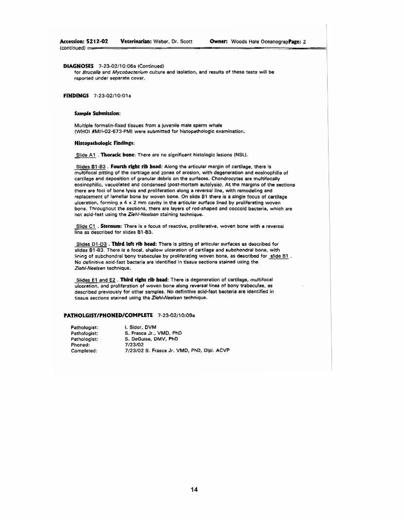
Owner: Woods Hole OceanograpPage: 2Veterinarian: Weber. Dr. ScottAccession: 5212-02(continued) =============;::;:;;;lii="""====-__""""""","======"""""'========-===!
DIAGNOSES 7-23-02/10:06a (Continued)for Brucel/8 and Mycobacterium culture and isolation, and results of these tests will bereported under separate cover.
fINDINGS 7-23-02/10:01 a
Samp(e Submission:
Multiple formalin-fixed tissues from a juvenile male sperm whale(WHOI #MH-02-673-PM) were submitted for histopathologic examination.
Histopathologic Findings;
Slide A1 . Thoracic bone: There are no significant histologic lesions (NSl).
Slides B1·83 . Fourth risht rib head: Along the articular margin of cartilage, there ismultifocal pitting of the cartilage and zones of erosion, with degeneration and eosinophilia ofcartilage and deposition of granular debris on the surfaces. Chondrocytes are multifocallyeosinophilic, vacuolated and condensed Cpost-mortem autolysis). At the margins- of the sectionsthere are foci of bone lysis and proliferation along a reversal line, with remodeling andreplacement of lamellar bone by woven bone. On slide B1 there is a single focus of cartilageulceration, forming a 4 x 2 mm cavity in the articular surface lined by proliferating wovenbone. Throughout the sections, there are layers of rod-shaped and coccoid bacteria, which arenot acid-fast using the Ziehl-Neelsen staining technique.
Slide C1 . Sternum: There is a focus of reactive, proliferative. woven bone with a reversalline as described for slides 81-B3.
Slides 01-03 • third 1m rib head: There is pitting of articular surfaces as described forslides B1·83. There ;s a focal. shallow ulceration of cartilage and svbchondral bone, withlining of subchondral bony trabeculae by proliferating woven bone, as described for slide 81No definitive aCid-fast bacteria are identified in tissue sections stained using theZiehl-Neelsen technique.
Slides El and E2 . third l'ilht rib head: There is degeneration of cartilage, multifocalulceration, and proliferation of woven bone along reversal lines of bony trabeculae, 8S
described previously for other samples. No definitive acid-fast bacteria are identified intissue sections $teined using the Ziehl-Nee/sen technique.
PATHOLGISTIPHONED/COMPLETE 7-23-02/10:09a
Pathologist:Pathologist:Pathologist:Phoned:Completed:
I. Sidor, DVMS. Frasca Jr., VMO, PhDS. DeGuise, OMV, PhO7/23/027/23/02 S. Frasca Jr. VMD, PhD, Dip!. ACVP
14

Connecticut Veterinary Diagnostic Laboratory
Accession: 5152-02Repon date: 8-2-02/SJGOwner: Woods Hole Oceanoaraphic (508) 289-J195Coordinator: Dr. S. Frasca. Jr.
University of Conneetieut61 N. Eagleville Rd. U-89Storrs, CT 06269-3089860-486-3738FAX: 860-486-3936
SUBMISSIONSUMMARY
WOODS HOLE OCEANOGRAPHICATIN: MOORE, DR. MICHAELMS#33. REDFIELD 2-44266 WOODS HOLE RD.WOODS HOLE, MA 02543
(Acet: 11 581 )
SpeciesMmammal
Animals1
Taken: 6-10-02Received: 6-14-02
Tests Completedo 0
(50S} 457-2134 (Please tax results)!fit PREUMINARY REPORT ••
PATHOLOGY _.-:===-=======~===-------===-
ANIMAL ID: 1: MH-02-673-PM/SPERM WHALE, Mmammal, MSample Taken: 6·10-02 Completed: 7-23-02/9:00a
DIAGNOSES 7-23-02/9:00a
Morphological:
Sample: Fixed Tissue
Huldple dssues examined. Postmortem autolysis and bacterial overgrowth.
Cost05temal junction, cartilage. Chondrolysis, multifocal. moderate, with intralesionalbacteria, interpreted 8S postmortem autolytic finding.
Final:
Postmortem autolysis and bacterial overarowdt, with dtondrolysis in Amp!es of costostemaljoint.
Comments:
The degree of postmortem autolysis precludes a detailed description of many of the tissuesexamined, which is to be expected given the history and condition of the specimen. It isdebatable whether the lytic changes in cartilage actually represent significant antemortempathology, given the fact that postmortem autolysis and bacterial overgrowth obfuscatehistologic interpretation of these tissue sections. Tissue samples have been forwarded to theNational Veterinary Services Laboratory for microbiological culture and isolation of Brucellaand Mycobacterium species, and results of these diagnostic tests wilt be reported underseparate cover.
15

Owner: Woods Hole OceanograpPage: 2Veterinarian: Weber, Dr. ScottAccession: 5152-02(continued) ==================-===--=""""'=-=.....""""........====-====.........
FINDINGS 7·23·02/8:50" (Continued)
FINDINGS 7-23-02/8:50a
Sample Submission:
Multiple formalin-fixed tissues irom a juvenile male sperm whale (WHOI #MH-02-573-PM) weresubmitted for histopathologic examination.
Hiscopatholosk Findinp:
Slides A1-A3 . Atrioventricular valve: There is diffuse post-mortem 8utolysis (PMA), withnumerous colonies of short, wide bacilli scattered throughout interstitium and adherent toexternal surfaces. A large fibrin clot with numerous bacterial colonies is located within thelumen of an artery. Bacteria stain blue, Le. are not acid-fast, using the Ziehl-Nee/senacid-fast technique.
Slides 81 &. 82 . Connective tissue at n"l» fradare site: PMA. Around degenerate myofibers isgranular eosinophilic cellulClr debris and numerous short bacilli with bulbs or rounded ends(consistent with spores of Clostridium spp.). Bacteria stain either pale pink or blue, i.e.are not acid-fast, using the Ziehl-Nee/sen acid-fast technique. '
Slide C2A . Sclera, right eye: There is moderate post-mortem overgrowth of bacterial colonieswithin interlacing bundles of dense fibr.ous connective tissue (collagen). Bacteria stain blue,i.e. are not acid-fsst, using the Ziehl·Neelsen acid-fast technique.
Slides 01 to 05 . Costosternal joint: Within cartilage there are multifocal. large (2 to 5 mmdiameter), irregular areas of postmortem lysis, lined by thin layers of granular debris andnumerous bacteria. Using a Brown and BrtJnn Tissue Gram Stain, bacteria are a mixture ofgram-negative coccobacilli and stout Gram·positive rods (interpreted as postmortem bacterialovergrowth). Bacteria are not acid·fast using the Ziehl-Nee/sen 8cid·faststaining technique.Few bacterial colonies penetrate into deeper tissue around lytic regions.
Slides E1 through E8 . Testis, intestine, dense and loose connettiv. twue (mulciplesections), skeletal ft1UScle,hearc and aorta: Postmortem autolysis and bacterial overgrowth; nosignificant histologic lesions (some sections uninterpretablel. No acid-fast bacteria areidentified in tissue sections stained using the Ziehl·Neelsen acid-fast technique.
PATHOlGIST/PHOHED/COMPLETE 7·23-02/9:01 a
Pathologist:Pathologist:Pathologist:Phoned:Completed:
l. Sidor. DVMS. Frasca Jr., VMD, PhDS. OeGuise, DMV, PhD7123/027/23/02 S. Frasca Jr. VMD, PhD. Dipl. ACVP
16

10/09/02 WED 16:43 F~~ 860 486 3936iU/U~/~uu~ U~;JU r~A iOiOOOJfJiO
CVDL UCONNU~VA m!~~V~A~l~~!AL
!4J001IgJ002
Nation~1 Veterinary Se~vices Laboratories1600 Vayton Road Ames, Iowa SOOlD
Phone (515) bb3-72b~ Fa~ (515) ~b3-7~97
Tubercu10sis Labo~atory Test Report PageAHn: m l(!hae I mOOre.
], 0 f 1 50s~157- oJloL!FEDE~AL RELAY SERVIC£ eVoice/TT¥/ASCIIISpanish)
1-800-881-8339Accession: 182b39
Establishm~nt No.: CTCONN
CONNECTICUT V~TERINA~Y DIAG. LABU-30a~, ~1 N. ~AGLE~!LLE RD.
STORRS, C1 Ob2b9-3089Fax: 660461.393b
Preservation:R@tain Tag:
Date Received: ~/16/c002
~ererral Number:
Owner: WOODS HOLE OCEANOGRAPHIC INSTICity: ~OO)S HOLEState: MA
Species: Marine Ma~mal
NVSL II> Casett Salllp hi ID Animal III
511US5 0249~], 5152-02 MH-02-~73-pn
Bact@rial Isolation'Diagnosis! No Isolation Made
TissuesBone fragment
No Mycobacteria was isolated fro. the specimen sUbmiteed·
548458 020254 5~5a-02 MH-02-b?3-pn81 e~cc@rial Isolation
No Brucella was isol.ted from the bone sample·
l>ISTRIeUTION:Submitti!1"10/09/200.2CALS ~eportO
Approved By: Janet Payeur 7/02/20D2National Veterinary Services Laboratories
17