nanotechnology in medicin
TRANSCRIPT
8/2/2019 nanotechnology in medicin
http://slidepdf.com/reader/full/nanotechnology-in-medicin 1/21
INTRODUCTION
Nanotechnology:-
Nanotechnology is defined as the study and use of structures between 1
nanometer and 100 nanometers in size. To give you an idea of how small that is,
it would take eight hundred 100 nanometer particles side by side to match the
width of a human hair .
Nanotechnology involves manipulating properties and structures at the
nanoscale, often involving dimensions that are just tiny fractions of the width of
a human hair. Nanotechnology is already being used in products in its passive
form, such as cosmetics and sunscreens, and it is expected that in the coming
decades, new phases of products, such as better batteries and improved
electronics equipment, will be developed and have far-reaching implications.
One area of nanotechnology application that holds the promise of providing
great benefits for society in the future is in the realm of medicine.
Nanotechnology is already being used as the basis for new, more effective drug
delivery systems and is in early stage development as scaffolding in nerve
regeneration research. Moreover, the National Cancer Institute has created the
Alliance for Nanotechnology in Cancer in the hope that investments in this
branch of nanomedicine could lead to breakthroughs in terms of detecting,
diagnosing, and treating various forms of cancer.
Nanotechnology medical developments over the coming years will have a wide
variety of uses and could potentially save a great number of lives.
Nanotechnology is already moving from being used in passive structures to
active structures, through more targeted drug therapies or “smart drugs.” These
new drug therapies have already been shown to cause fewer side effects and be
1
8/2/2019 nanotechnology in medicin
http://slidepdf.com/reader/full/nanotechnology-in-medicin 2/21
more effective than traditional therapies. In the future, nanotechnology will also
aid in the formation of molecular systems that may be strikingly similar to
living systems. These molecular structures could be the basis for the
regeneration or replacement of body parts that are currently lost to infection,
accident, or disease. These predictions for the future have great significance not
only in encouraging nanotechnology research and development but also in
determining a means of oversight. The number of products approaching the
FDA approval and review process is likely to grow as time moves forward and
as new nanotechnology medical applications are developed.
2
8/2/2019 nanotechnology in medicin
http://slidepdf.com/reader/full/nanotechnology-in-medicin 3/21
WHAT IS MEDICAL NANOTECHNOLOGY?
Medical nanotechnology is a branch of nanotechnology which applies principles
in this field to health care issues. Nanotechnology is a broad spectrum of
scientific endeavours which involves manufacturing and machining which take
place on a molecular scale. There are a number of potential applications for
medical nanotechnology, and in its early phases, many people were quite
excited about the huge changes which could occur in the medical world with the
assistance of medical technology.
Because nanotechnology operates on such a small scale, it offers the
opportunity to create precisely targeted surgical instruments, drug delivery
systems, and implants. Nanobots, for example, could be used to perform a non-
invasive medical imaging study inside the body, or to perform surgical
procedures. Nanomaterials can also be implanted into the body; for example,
someone with a badly damaged bone or joint could be treated with nanoparticles
which would promote new growth, regrowing the damaged tissue.
Medical nanotechnology also makes cell repair on a molecular level possible,
and provides a number of opportunities for medication administration. Drugs
developed through nanotechnology could directly penetrate cells, for example,
or nanoparticles could be designed to target cancer cells, delivering medication
or providing a focal point for radiation. Medical nanotechnology can also be
used to make biosensors which can be implanted into patients for monitoring,
along with medical devices which are designed to be permanently implanted
such as pacemakers.
This field also has a number of implications for prosthetics. Nanomaterials
could be used to give people greater control over prosthetic limbs, and
potentially to do things like restoring function to the eyes. Several militaries
have invested in medical nanotechnology for the purpose of developing new
3
8/2/2019 nanotechnology in medicin
http://slidepdf.com/reader/full/nanotechnology-in-medicin 4/21
treatments for injured soldiers. The field also creates a potential for the
development of devices which could enhance human function, much to the
delight of science fiction authors around the world.
HISTORY OF NANOTECHNOLOGY
4
8/2/2019 nanotechnology in medicin
http://slidepdf.com/reader/full/nanotechnology-in-medicin 5/21
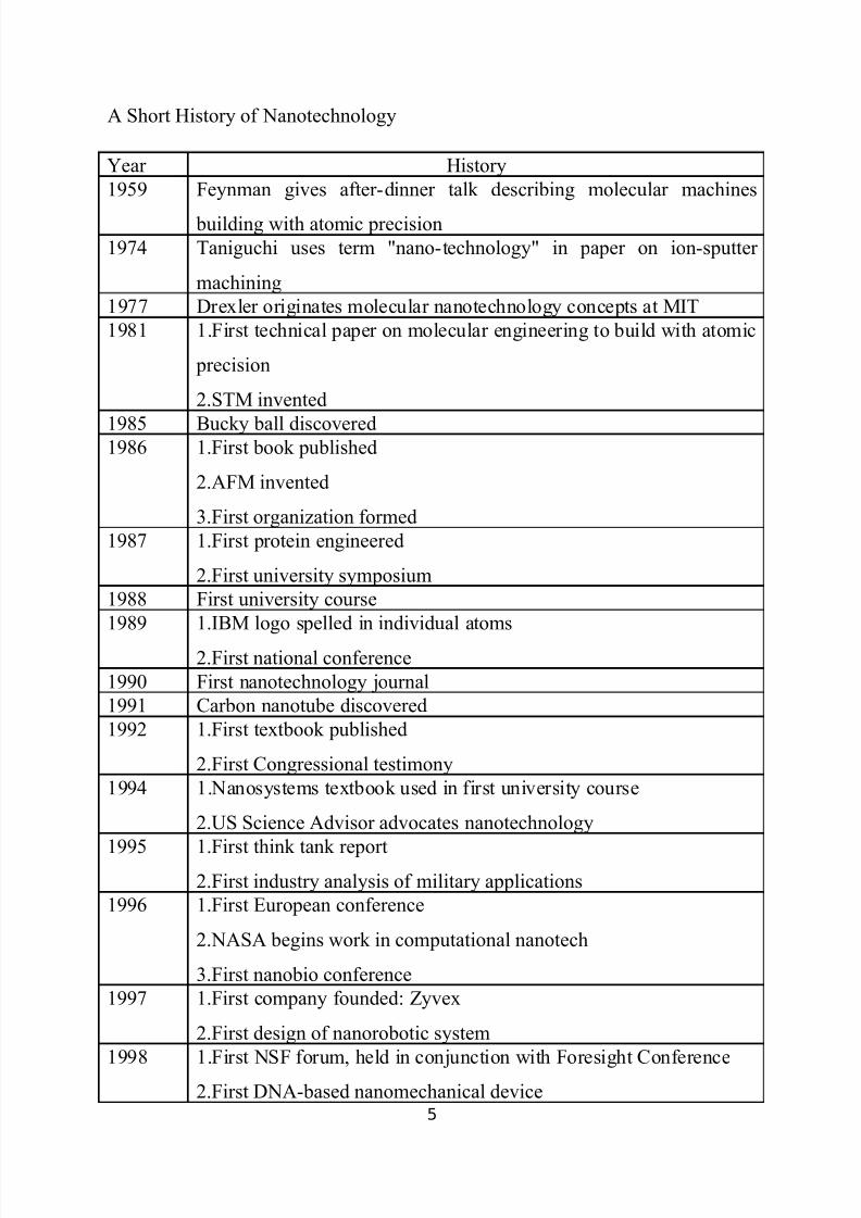
A Short History of Nanotechnology
Year History
1959 Feynman gives after-dinner talk describing molecular machines
building with atomic precision
1974 Taniguchi uses term "nano-technology" in paper on ion-sputter
machining
1977 Drexler originates molecular nanotechnology concepts at MIT
1981 1.First technical paper on molecular engineering to build with atomic
precision
2.STM invented
1985 Bucky ball discovered1986 1.First book published
2.AFM invented
3.First organization formed
1987 1.First protein engineered
2.First university symposium
1988 First university course
1989 1.IBM logo spelled in individual atoms
2.First national conference
1990 First nanotechnology journal
1991 Carbon nanotube discovered
1992 1.First textbook published
2.First Congressional testimony
1994 1.Nanosystems textbook used in first university course
2.US Science Advisor advocates nanotechnology
1995 1.First think tank report2.First industry analysis of military applications
1996 1.First European conference
2.NASA begins work in computational nanotech
3.First nanobio conference
1997 1.First company founded: Zyvex
2.First design of nanorobotic system
1998 1.First NSF forum, held in conjunction with Foresight Conference2.First DNA-based nanomechanical device
5
8/2/2019 nanotechnology in medicin
http://slidepdf.com/reader/full/nanotechnology-in-medicin 6/21
1999 1.First Nanomedicine book published
2.First safety guidelines
3.Congressional hearings on proposed National Nanotechnology
Initiative
2001 1.First report on nanotech industry
2.U.S. announces first center for military applications
2002 1.First nanotech industry conference
2.Regional nanotech efforts multiply
2003 1.Congressional hearings on societal implications
2.Call for balancing NNI research portfolio
2004 1.First policy conference on advanced nanotech2.First center for nanomechanical systems
2005 At Nanoethics meeting, Roco announces nanomachine/nanosystem
project count has reached 300
2006 National Academies nanotechnology report calls for experimentation
toward molecular manufacturing
2007 Feynman Prize in Nanotechnology awarded for construction of
molecular machine systems that function in the realm of Brownianmotion, and molecular machines based upon two-state mechanically
interlocked compounds
2008 1.Technology Roadmap for Productive Nanosystems released
2.Protein catalysts designed for non-natural chemical reactions
2009 1.An improved walking DNA nanorobot
2.Structural DNA nanotechnology arrays devices to capture
molecular building blocks
3.Design 'from scratch' of a small protein that performed the function
performed by natural globin proteins
4.Organizing functional components on addressable DNA scaffolds
2010 1.DNA-based 'robotic' assembly begins
2011 1.First programmable nanowire circuits for nanoprocessors
2.DNA molecular robots learn to walk in any direction along a
branched track
6
8/2/2019 nanotechnology in medicin
http://slidepdf.com/reader/full/nanotechnology-in-medicin 7/21
3.Mechanical manipulation of silicon dimers on a silicon surface
NANOBIOSENSORS
INTRODUCTION:
A biosensor is generally defined as a measurement system that consists of a
probe with a sensitive biological recognition element, or bioreceptor, a
physicochemical detector component, and a transducer in between.
Nanosensors are any biological, chemical, or surgery sensory points used to
convey information about nanoparticle to the macroscopic world. Their use
mainly include various medicinal purposes and as gateways to building other
nanoproducts, such as computer chips that work at the nanoscale and
nanorobots.
Components:-
7
8/2/2019 nanotechnology in medicin
http://slidepdf.com/reader/full/nanotechnology-in-medicin 8/21
1) BIORECEPTORS
2) TRANSDUCERS
3) DETECTORS
1. Bioreceptors:- It is a sensitive biological element. The interaction of an
analyte, e.g. a particular chemical component, virus or micro-organism, with the
bioreceptor is designed to generate an effect picked up by a transducer, which
converts the information into a measurable effect by the detector, for instance
an electric signal. Bioreceptors are used because of their specificity. They
enable measurement with minimum interference from other components in
complex mixtures. The bioreceptor is a biological molecule (e.g., an
antibody/antigen, DNA, protein, or enzyme), or a living biological system (e.g.,
cells, tissues, or whole organisms) that utilises a biochemical mechanism of
recognition. The sampling component of a biosensor contains a bio-sensitive
layer that can either contain bioreceptors or be made of bioreceptors covalently
attached to the transducer.
2. Transducer -Transduction can be accomplished by optical, electrochemical,
and mass detection methods.
3. Detection techniques:-
1. ELECTROCHEMICAL METHODS
Electrochemical biosensors are generally fairly simple devices. There are three
types utilising electrical current, potential or resistive changes:
1. Amperometric biosensors:- which determine the electric current associated
with the electrons involved in redox processes;
2. Potentiometric biosensors:- which use ion-selective electrodes to determine
changes in the concentration of chosen ions (e.g.hydrogen ions); and
8
8/2/2019 nanotechnology in medicin
http://slidepdf.com/reader/full/nanotechnology-in-medicin 9/21
3.Conductimetric biosensors:- which determine conductance changes associated
with changes in the ionic environment.
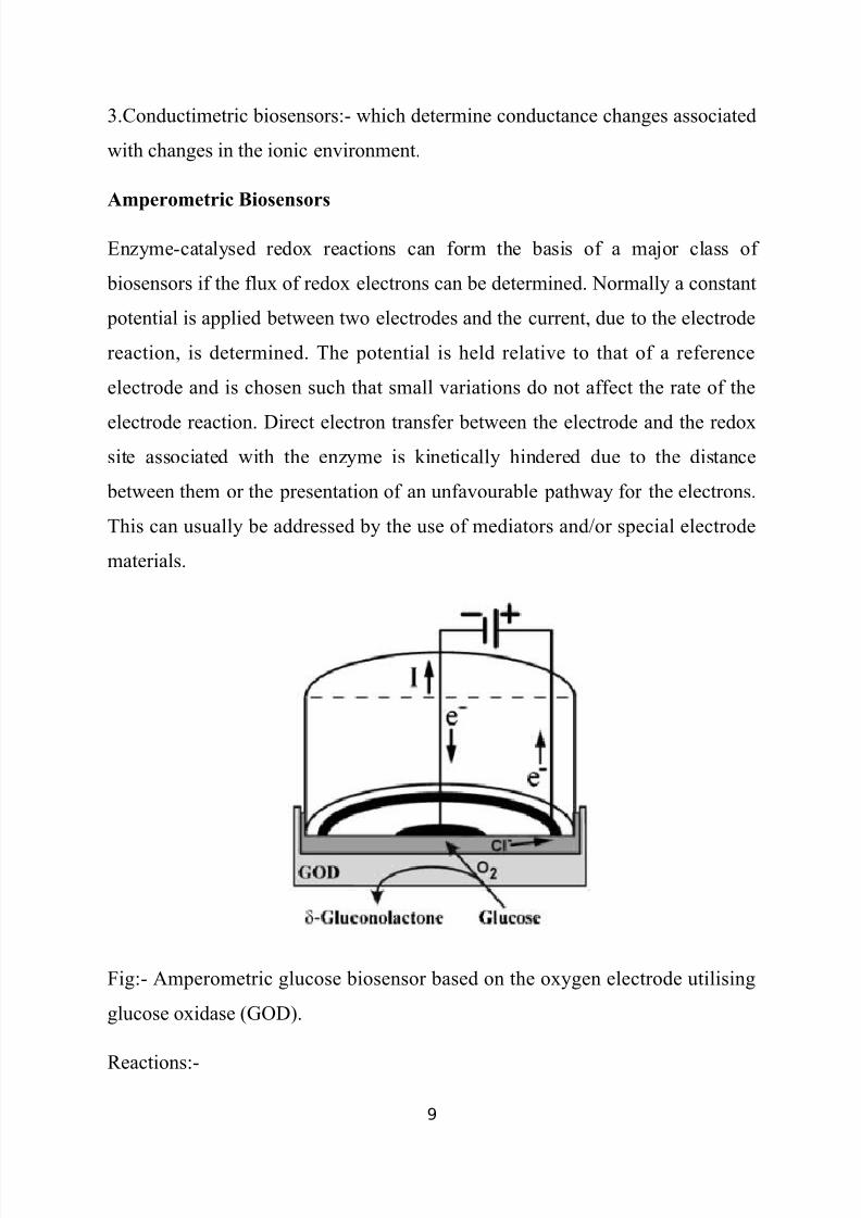
Amperometric Biosensors
Enzyme-catalysed redox reactions can form the basis of a major class of
biosensors if the flux of redox electrons can be determined. Normally a constant
potential is applied between two electrodes and the current, due to the electrode
reaction, is determined. The potential is held relative to that of a reference
electrode and is chosen such that small variations do not affect the rate of the
electrode reaction. Direct electron transfer between the electrode and the redoxsite associated with the enzyme is kinetically hindered due to the distance
between them or the presentation of an unfavourable pathway for the electrons.
This can usually be addressed by the use of mediators and/or special electrode
materials.
Fig:- Amperometric glucose biosensor based on the oxygen electrode utilising
glucose oxidase (GOD).
Reactions:-
9
8/2/2019 nanotechnology in medicin
http://slidepdf.com/reader/full/nanotechnology-in-medicin 10/21
The first and simplest biosensor was based on this principle. It was for
the determination of glucose and made use of the Clark oxygen electrode.
Figure shows a section through such a simple amperometric biosensor. A
potential of 0.6V is applied between the central platinum cathode and the
surrounding silver/silver chloride reference electrode (the anode). Dissolved
molecular oxygen at the platinum cathode is reduced and the circuit is
completed by means of the saturated KCl solution. Only oxygen can be reduced
at the cathode due to its covering by a thin Teflon or polypropylene membrane
through which the oxygen can diffuse but which acts as a barrier to other
electroactive species.
The biocatalyst is retained next to the electrode by means of
a membrane, which is permeable only to low molecular weight molecules
including the reactants and products. Glucose may be determined by the
reduction in the dissolved oxygen concentration when the redox reaction,
catalysed by glucose oxidase occurs:
It is fortunate that this useful enzyme is also one of the most stable
oxidoreductases found.
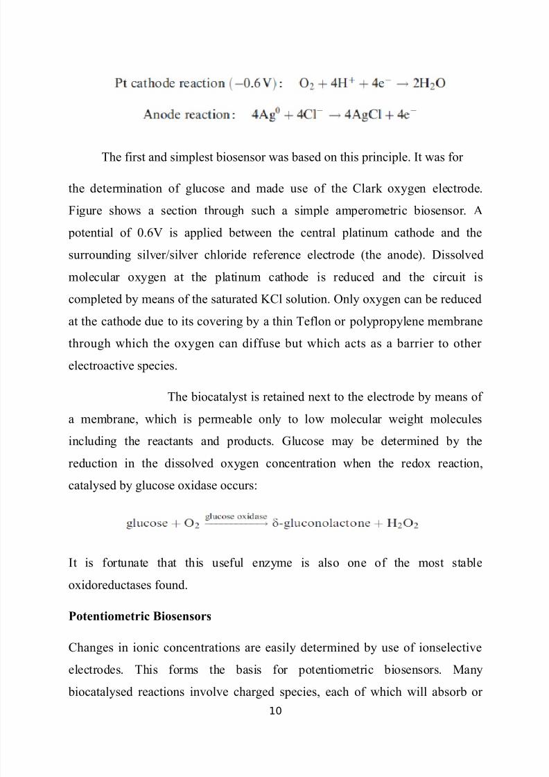
Potentiometric Biosensors
Changes in ionic concentrations are easily determined by use of ionselective
electrodes. This forms the basis for potentiometric biosensors. Many
biocatalysed reactions involve charged species, each of which will absorb or
10
8/2/2019 nanotechnology in medicin
http://slidepdf.com/reader/full/nanotechnology-in-medicin 11/21
release hydrogen ions according to their pKa and the pH of the environment.
This allows a relatively simple electronic transduction using the commonest ion
selective electrode, the pH electrode. Potentiometric biosensors can be
miniaturised by the use of field effect transistors (FETs).
Fig :- A FET-based potentiometric biosensor.
Ion-selective field effect transistors (ISFETs) are low-cost devices that are in
mass production. Figure shows a diagrammatic cross-section through an npn
hydrogen ion-responsive ISFET with an approximately 0.025mm2 biocatalytic
membrane covering the ion-selective membrane. The build-up of positive
charge on this surface (the gate) repels the positive holes in the p-type silicon,
causing a depletion layer and allowing the current to flow. The reference
electrode is usually an identical ISFET without any biocatalytic membrane. A
major practical problem with the manufacture of such enzyme-linked FETs
(ENFETs) is protection of the silicon from contamination by the solution, hence
the covering of waterproof encapsulant. Because of their small size, they only
require minute amounts of biological material and can be produced in a form
whereby they can determine several analytes simultaneously. A further
11
8/2/2019 nanotechnology in medicin
http://slidepdf.com/reader/full/nanotechnology-in-medicin 12/21
advantage is that they have a more rapid response rate compared with the larger,
sluggish ion-selective electrode devices. The enzyme may be immobilised to the
silicon nitride gate using polyvinylbutyral deposited by solvent evaporation and
cross-linked with glutaraldehyde. Such devices still present fabrication
problems such as reproducibility, drift, sensitivity to light and the need for on-
chip temperature compensation.
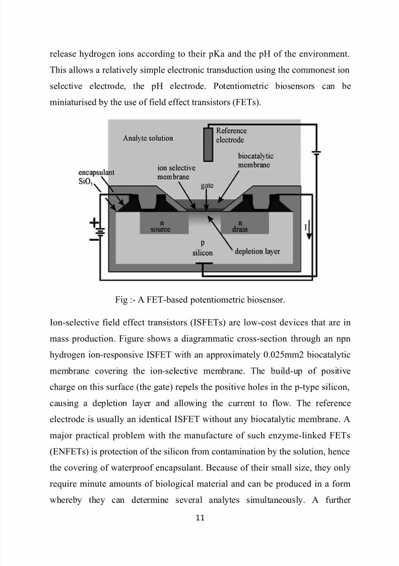
Conductimetric Biosensors
Many biological processes involve changes in the concentrations of ionic
species. Such changes can be utilised by biosensors that detect changes inelectrical conductivity. A typical example of such a biosensor is the urea sensor,
utilising immobilised urease and used as a monitor during renal surgery and
dialysis. The reaction gives rise to a large change in ionic concentration at pH
7.0, making this type of biosensor particularly attractive for monitoring urea
concentrations.
12
8/2/2019 nanotechnology in medicin
http://slidepdf.com/reader/full/nanotechnology-in-medicin 13/21
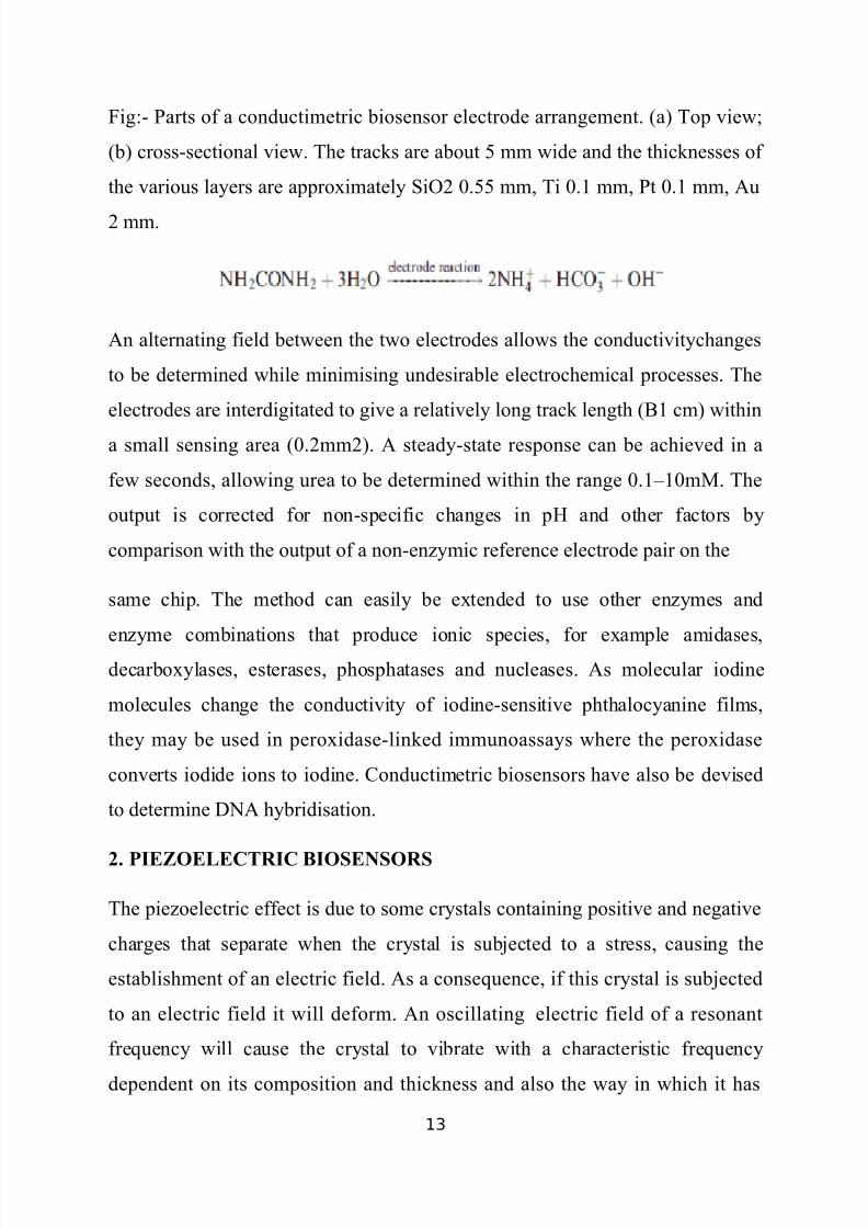
Fig:- Parts of a conductimetric biosensor electrode arrangement. (a) Top view;
(b) cross-sectional view. The tracks are about 5 mm wide and the thicknesses of
the various layers are approximately SiO2 0.55 mm, Ti 0.1 mm, Pt 0.1 mm, Au
2 mm.
An alternating field between the two electrodes allows the conductivitychanges
to be determined while minimising undesirable electrochemical processes. The
electrodes are interdigitated to give a relatively long track length (B1 cm) within
a small sensing area (0.2mm2). A steady-state response can be achieved in a
few seconds, allowing urea to be determined within the range 0.1–10mM. The
output is corrected for non-specific changes in pH and other factors by
comparison with the output of a non-enzymic reference electrode pair on the
same chip. The method can easily be extended to use other enzymes and
enzyme combinations that produce ionic species, for example amidases,
decarboxylases, esterases, phosphatases and nucleases. As molecular iodine
molecules change the conductivity of iodine-sensitive phthalocyanine films,
they may be used in peroxidase-linked immunoassays where the peroxidase
converts iodide ions to iodine. Conductimetric biosensors have also be devised
to determine DNA hybridisation.
2. PIEZOELECTRIC BIOSENSORS
The piezoelectric effect is due to some crystals containing positive and negative
charges that separate when the crystal is subjected to a stress, causing the
establishment of an electric field. As a consequence, if this crystal is subjected
to an electric field it will deform. An oscillating electric field of a resonant
frequency will cause the crystal to vibrate with a characteristic frequency
dependent on its composition and thickness and also the way in which it has
13
8/2/2019 nanotechnology in medicin
http://slidepdf.com/reader/full/nanotechnology-in-medicin 14/21
been cut. As this resonant frequency varies when molecules adsorb on the
crystal surface, a piezoelectric crystal may form the basis of a biosensor. Even
small changes in resonant frequencies are easy to determine with precision and
accuracy using straightforward electronics. Differences in mass, even as small
as 1 ng cm–2, can be measured when adsorbed on the sensing surface. Changes
in frequency are generally determined relative to a similarly treated reference
crystal but without the active biological material. As an example, a biosensor
for cocaine in the gas phase may be made by attaching cocaine antibodies to the
surface of a piezoelectric crystal. This biosensor changes frequency by about 50
Hz for a 1 ppb atmospheric cocaine sample and can be reused after flushing for
a few seconds with clean air. The relative humidity of the air is important,
because if it is too low the response is less sensitive and if it is too high the
piezoelectric effect may disappear altogether. Cocaine in solution can be
determined after drying such biosensors.
Enzymes with gaseous substrates or inhibitors can also be attached to suchcrystals, as has been proved by the production of biosensors for formaldehyde
incorporating formaldehyde dehydrogenase and for organophosphorus
insecticides incorporating acetyl cholinesterase. One of the drawbacks
preventing the more widespread use of piezoelectric biosensors is the difficulty
in using them to determine analytes in solution. The frequency of a piezoelectric
crystal depends on the liquid’s viscosity, density and specific conductivity.
Under unfavourable conditions, the crystal may cease to oscillate completely.
There is also a marked effect of temperature due to its effect on viscosity. The
binding of material to the crystal surface may be masked by other
intermolecular effects at the surface and bulk viscosity changes consequent
upon even small concentration differences. There is also the strong possibility
of interference due to non-specific binding.
14
8/2/2019 nanotechnology in medicin
http://slidepdf.com/reader/full/nanotechnology-in-medicin 15/21
Antibody–antigen binding can be determined by measuring the frequency
changes in air after drying the crystal. Such procedures, although sensitive, are
difficult to reproduce repetitively, as the antibody layer may be partially lost
when the antigen is removed. However, oneshot biosensors have been
developed, using this principle, for the detection of several food contaminants
such as enterobacteria.
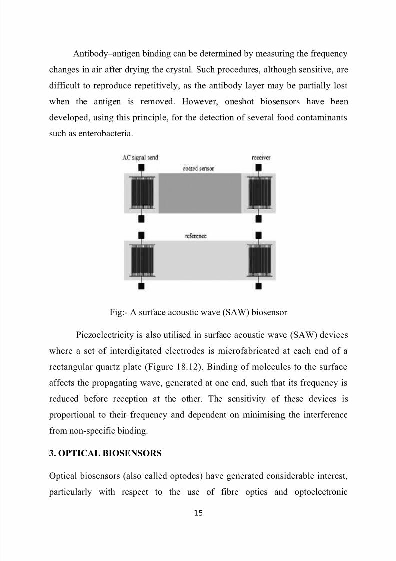
Fig:- A surface acoustic wave (SAW) biosensor
Piezoelectricity is also utilised in surface acoustic wave (SAW) devices
where a set of interdigitated electrodes is microfabricated at each end of a
rectangular quartz plate (Figure 18.12). Binding of molecules to the surface
affects the propagating wave, generated at one end, such that its frequency is
reduced before reception at the other. The sensitivity of these devices is
proportional to their frequency and dependent on minimising the interference
from non-specific binding.
3. OPTICAL BIOSENSORS
Optical biosensors (also called optodes) have generated considerable interest,
particularly with respect to the use of fibre optics and optoelectronic
15

8/2/2019 nanotechnology in medicin
http://slidepdf.com/reader/full/nanotechnology-in-medicin 16/21
transducers. These allow the safe non-electrical remote sensing of materials in
hazardous or sensitive (i.e. in vivo) environments. An advantage of optical
biosensors is that no reference sensor is needed; a comparative signal is
generally easily generated by splitting the light source used by the sampling
sensor. A simple example of an optical biosensor is the fibre optic lactate sensor
which senses changes in molecular oxygen concentrations by determining its
quenching of a fluorescent dye:
Fig:- A fibre optic lactate biosensor
quenching and consequentially causing an increase in the fluorescence output.
Simple colorimetric changes can be monitored in some biosensor
configurations. A lecithin biosensor has been developed containing
phospholipase D, choline oxidase and bromothymol blue. The change in pH,
16
8/2/2019 nanotechnology in medicin
http://slidepdf.com/reader/full/nanotechnology-in-medicin 17/21
due to the formation of the acid betaine from the released choline, causes a
change in the bromothymol blue absorbance at 622 nm. Gasphase reactions can
also be monitored. For example, alcohol vapour can be detected by the colour
change of a dry dispersion of alcohol oxidase and peroxidase plus the redox dye
2,6-dichloroindophenol.
One of the most widely established biosensor technologies is the lowtechnology
single-use colorimetric assay based on a paper pad impregnated with reagents.
This industry revolves mainly round blood and urine analysis with test strips
costing only a few cents. A particularly important use for these colorimetric test
strips is the detection of glucose. In this case, the strips contain glucose oxidase
and horseradish peroxidase together with a chromogen (e.g. o-toluidine) which
changes colour when oxidised by the peroxidase-catalysed reaction with the
hydrogen peroxide produced by the aerobic oxidation of glucose:
The colour produced can be evaluated by visual comparison with a test
chart or by the use of a portable reflectance meter. Many test strips incorporate
anti-interference layers to produce more reproducible and accurate results. It is
possible to link up luminescent reactions to biosensors, as light output is a
relatively easy phenomenon to transduce to an electronic output. As an
example, the reaction involving immobilised (or free) luciferase can be used to
detect the ATP released by the lysis of microorganisms:
This allows the rapid detection of urinary infections by detecting the microbial
content of urine samples.
17
8/2/2019 nanotechnology in medicin
http://slidepdf.com/reader/full/nanotechnology-in-medicin 18/21
NANOPARTICLES
In nanotechnology, a particle is defined as a small object that behaves as
a whole unit in terms of its transport and properties. Particles are further
classified according to size : in terms of diameter, coarse particles cover a range
between 10,000 and 2,500 nanometers. Fine particles are sized between 2,500
and 100 nanometers. Ultrafine particles, or nanoparticles are sized between 100
and 1 nanometers.
Nanoclusters have at least one dimension between 1 and 10 nanometers and a
narrow size distribution. Nanopowders are agglomerates of ultrafine particles,
nanoparticles, or nanoclusters. Nanometer-sized single crystals, or single-
domain ultrafine particles, are often referred to as nanocrystals.
Nanoparticle research is currently an area of intense scientific interest due to a
wide variety of potential applications in biomedical, optical and electronic
fields.
Properties of Nanoparticles:-
A nanometer is one-billionth of a meter (10-9 m); a sheet of paper is about
100,000 nanometers thick. These nanoparticles give us the ability to see cells
and molecules that we otherwise cannot detect through conventional imaging.
The ability to pick up what happens in the cell, to monitor therapeutic
intervention and to see when a cancer cell is mortally wounded or is actually
activated is critical for the successful diagnosis and treatment of this disease.
For drug delivery in cancer we have “Nano scale devices”. Nanoscale devices
are 102 to 104 times smaller than human cells but are similar in size to large
biomolecules such as enzymes and receptors. Nanoscale devices smaller than 50
nm can easily enter most cells, and those smaller than 20 nm can move out of
blood vessels as they circulate through the body. Nanodevices are suitable to
18
8/2/2019 nanotechnology in medicin
http://slidepdf.com/reader/full/nanotechnology-in-medicin 19/21
serve as customized, targeted drug delivery vehicles to carry large doses of
chemotherapeutic agents or therapeutic genes into malignant cells while sparing
healthy cells.
Quantum Dots (QDs)
Quantum dots are unique in their far reaching possibilities in many avenues of
medicine. A QD is a fluorescent nanoparticle that has potential to be used as a
sensitive probe for screening cancer markers in fluids, as a specific label for
classifying tissue biopsies and as a high resolution contrast agent for medical
imaging, which is capable of detecting even the smallest of tumors. These particles have the unique ability to be sensitively detected on a wide range of
length scales, from macroscale visualization, down to atomic resolution using
electron microscopy. Using Quantum dots (QDs), the drug delivery particles are
injected in to blood stream until they find the cancer cells, to which the
antibodies adhere. Infrared light shining on the suspected cancer site penetrates
the tissues and cause the quantum dots to radiate photons. The photons pinpointthe cancer cell’s location and also cause the release of the Taxol (an
anticancerous drug), which can then attack and kill the cancer cells. Quantum
dots are tiny crystals that glow when stimulated by ultraviolet light. The
wavelength, or color, of the light depends on the size of the crystal. Latex beads
filled with these crystals can be designed to bind to specific DNA sequences. By
combining different sized quantum dots within a single bead, scientists can
create probes that release distinct colors and intensities of light. When the
crystals are stimulated by UV light, each bead emits light that serves as a sort of
spectral bar code, identifying a particular region of DNA. The diversity of
quantum dots will allow scientists to create many unique labels, which can
identify numerous regions of DNA simultaneously. This will be important in the
detection of cancer, which results from the accumulation of many different
19
8/2/2019 nanotechnology in medicin
http://slidepdf.com/reader/full/nanotechnology-in-medicin 20/21
changes within a cell. Another advantage of quantum dots is that they can be
used in the body, eliminating the need for biopsy.
Nanotechnology may also be useful for developing ways to eradicate
cancer cells without harming healthy, neighboring cells. Scientists hope to use
nanotechnology to create therapeutic agents that target specific cells and deliver
their toxin in a controlled, time-released manner.
CONCLUSION
20

8/2/2019 nanotechnology in medicin
http://slidepdf.com/reader/full/nanotechnology-in-medicin 21/21
Nanotechnology is definitely a medical boon for diagnosis, treatment and
prevention of cancer disease. It will radically change the way we diagnose, treat
and prevent cancer to help meet the goal of eliminating suffering and death
from cancer. Although most of the technologies described are promising and fit
well with the current methods of treatment, there are still safety concerns
associated with the introduction of nanoparticles in the human body.
21