nanotechnology: from in vivo imaging system to … from in vivo imaging ... tions ranging from...
TRANSCRIPT
NANO REVIEW Open Access
Nanotechnology: from In Vivo ImagingSystem to Controlled Drug DeliveryMaria Mir, Saba Ishtiaq, Samreen Rabia, Maryam Khatoon, Ahmad Zeb, Gul Majid Khan, Asim ur Rehman*
and Fakhar ud Din*
Abstract
Science and technology have always been the vitals of human’s struggle, utilized exclusively for the development ofnovel tools and products, ranging from micro- to nanosize. Nanotechnology has gained significant attention due to itsextensive applications in biomedicine, particularly related to bio imaging and drug delivery. Various nanodevices andnanomaterials have been developed for the diagnosis and treatment of different diseases. Herein, we have describedtwo primary aspects of the nanomedicine, i.e., in vivo imaging and drug delivery, highlighting the recentadvancements and future explorations. Tremendous advancements in the nanotechnology tools for the imaging,particularly of the cancer cells, have recently been observed. Nanoparticles offer a suitable medium to carryoutmolecular level modifications including the site-specific imaging and targeting. Invention of radionuclides, quantumdots, magnetic nanoparticles, and carbon nanotubes and use of gold nanoparticles in biosensors have revolutionizedthe field of imaging, resulting in easy understanding of the pathophysiology of disease, improved ability to diagnoseand enhanced therapeutic delivery. This high specificity and selectivity of the nanomedicine is important, and thus, therecent advancements in this field need to be understood for a better today and a more prosperous future.
Keywords: Nanotechnology, Nanocomposites, In vivo imaging, Drug delivery and pharmaceutical nanosystems
ReviewIntroductionAs a matter of fact, nanotechnology is making progressthrough all imperative fields of engineering and science,and scientists are revolutionizing all the industries andhuman lives by designing things capable of working onthe smallest scale length, atom by atom [1]. Nanotech-nology involves the study of eminently small structures.Nanotechnology can be defined comprehensively as thestudy, creation, design, synthesis, and implementation offunctional materials, systems, and devices through con-trolling matter within the size range of 1–100 nm at thenanometer scale. Moreover, the manipulation of innova-tive phenomena and improved properties of matter atthis nanometer scale, also referred as molecular nano-technology, is a magical point on scale length wheresmallest man-made appliances encounter the moleculesand atoms of the universe [2–4].The early inception of the concept of nanotechnology
and nanomedicine sprang from the discerning idea of
Feynman that tiny nanorobots and related devices could bedeveloped, fabricated, and introduced into the human bodyto repair cells at molecular level. Although later in the1980s and 1990s, this innovative concept was advocated inthe famous writings of Drexler [5, 6], and in 1990s and2000s in the popular writings of Freitas [7, 8]. Feynmanoffered the first known proposal for a nanomedical proced-ure to cure heart disease. In general, miniaturization ofmedical tools will provide more accurate, controllable,reliable, versatile, cost-effective, and quick approaches forimproved quality of human life [9]. In 2000, for the veryfirst time, National Nanotechnology Initiative waslaunched; then from onwards, modeling of electronics andmolecular structures of new materials, establishment ofnanoscale photonic and electronic devices [10, 11], devel-opment of 3D networking, nanorobotics [12], and advent ofmulti-frequency force microscopy [13] have paved the wayfor emergence of molecular nanotechnology.Nanoparticles are considered as the essential building
blocks of nanotechnology. Presence of strong chemicalbonds, extensive delocalization of valence electrons varyingwith size, and structural modifications in nanoparticles lead* Correspondence: [email protected]; [email protected]
Department of Pharmacy, Quaid-I-Azam University, Islamabad, Pakistan
© The Author(s). 2017 Open Access This article is distributed under the terms of the Creative Commons Attribution 4.0International License (http://creativecommons.org/licenses/by/4.0/), which permits unrestricted use, distribution, andreproduction in any medium, provided you give appropriate credit to the original author(s) and the source, provide a link tothe Creative Commons license, and indicate if changes were made.
Mir et al. Nanoscale Research Letters (2017) 12:500 DOI 10.1186/s11671-017-2249-8

to different physical and chemical properties includingmelting points, optical properties, magnetic properties, spe-cific heats, and surface reactivity. These ultrafine nanoparti-cles exhibit completely new and improved properties ascompared to their bulk counterpart due to variation in spe-cific characteristics such as size, distribution, and of the par-ticles which give rise to larger surface area to volume ratio[14–16]. As the field of nanostructured materials has beenevolved, many different labels and terminologies are beingused including 3D nanoparticle, nanocrystals, nanofilms,nanotubes, nanowires, and quantum dots with promisingpotential of infinite number of properties [17]. Because ofthe variety of potential applications (including industrialand military), governments have invested billions of dollarsin nanotechnology research. The USA has invested 3.7billion dollars through its National Nanotechnology Initia-tive, and European Union has also subsidized 1.2 billion,and 750 million dollars were invested by Japan [18].Today, nanotechnology is one of the most innovative,
vanguard areas of scientific study, and it continues toprogress at staggering rates [19]. Through advancementin nanotechnology, many state-of-the-art technologiesbecame available for the drug delivery. Researchers haveextensively investigated the potential of nanodevices fortarget specific and controlled delivery of various micro-and macromolecules including drugs, proteins, mono-clonal antibodies, and DNA (deoxyribonucleic acid) inmultifarious biomedical applications like cancer [20, 21],vaccination [22], dental [23], inflammatory [24], andother health disorders. It is therefore a need of the dayto demonstrate efficient use of nanotechnology applica-tions ranging from in-vivo imaging system to controlleddrug delivery, to mark the current progress and getdirections for impending research in medical fields.
Pharmaceutical NanosystemsPharmaceutical nanotechnology can be classified into twomain categories of nanotools, i.e., nanomaterials andnanodevices. Nanomaterials can be further categorized onthe basis of three basic parameters including structure,dimension, and phase composition. Nanostructures arefurther classified into polymeric and non-polymeric struc-tures including nanoparticles, micelles, dendrimers, drugconjugates, metallic nanoparticles, and quantum dots [25].On the basis of their dimensions, nanomaterials are classi-fied in four groups, i.e., zero, one, two, and three nanodi-mension materials. According to phase composition, thesenanomaterials can be categorized in three groups.Nanodevices are subdivided in three groups, includingmicroelectromechanical systems/nanoelectromechanicalsystem (MEMS/NEMS), microarrays, and respirocytes.These structures and devices can be fabricated with a highdegree of functional property for use in medicine to inter-act with cells at a molecular level, thus allowing an extent
of integration between biological systems and latest tech-nology that was not achievable previously [26]. Detailedclassification of pharmaceutical nanotools is describedwith their examples in Table 1.
Manufacturing ApproachesNanosizing technologies have achieved great importancefor the formulation of poorly water soluble drugs. By re-ducing the particle size to nanoscale range, the dissolutionrate and bioavailability increase because of the increase insurface area, according to the Noyes-Whitney equation[27]. Approaches used for the manufacturing materials arecategorized into bottom up techniques, top down tech-niques, and the combination of bottom up and top downtechniques. Bottom up techniques involve built up ofmolecules. Some of the techniques that follow bottom upapproach for manufacturing of nanoscale materials in-clude liquid phase techniques based on inverse micelles,chemical vapor deposition (CVD), sol-gel processing, andmolecular self-assembly. The components produced bybottom up are significantly stronger than the macroscalecomponents because of the covalent forces that hold themtogether. In top down techniques, materials are micron-ized by cutting, carving, and molding for manufacturingof nanomaterials. Examples include milling, physical vapordeposition, hydrodermal technique electroplating, andnanolithography [28]. Different manufacturing approacheswith their respective types are described in Table 2.
Biomedical Applications of Advanced NanotechnologyImagingTremendous advancements were reported during the lastdecade, using the nanotechnology tools for the imagingand therapy in research particularly targeting the cancercells. Nanoparticles, with size 10–100 nm, offer a verysuitable medium to carry out molecular level modifica-tions such as the site-specific imaging and targeting incancer cells [29]. The following section summarizes somerecent advancement in the imaging techniques.
Radionuclide ImagingBecause of the inability of small molecules to be viewed withthe noninvasive technique, the site-targeted contrast agentsare employed to identify a selected biomarker that is impos-sible to be separated from the normal surrounding tissues[30]. The radionuclide imaging has been developed with theconcept that the expressed protein is probed with a radio-pharmaceutical or isotope-labeled agent or cell and istracked further in vivo [31]. The positron emission tomog-raphy (PET) imaging is used in the cancer patientssuccessfully to image the multidrug resistance through P-glycoprotein transport using 99 m tetrofosmin and sestamibias the radiolabeled substrates for the P-glycoprotein [32, 33].The mechanism of imaging is determined by the type of
Mir et al. Nanoscale Research Letters (2017) 12:500 Page 2 of 16
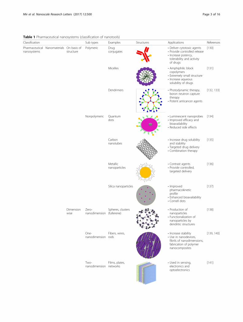
Table 1 Pharmaceutical nanosystems (classification of nanotools)
Classification Sub types Examples Structures Applications References
Pharmaceuticalnanosystems
Nanomaterials On basis ofstructure
Polymeric Drugconjugates
• Deliver cytotoxic agents• Provide controlled release• Increase potency,tolerability and activityof drugs
[130]
Micelles • Amphiphilic blockcopolymers
• Extremely small structure• Increase aqueoussolubility of drugs
[131]
Dendrimers • Photodynamic therapy,boron neutron capturetherapy
• Potent anticancer agents
[132, 133]
Nonpolymeric Quantumdots
• Luminescent nanoprobes• Improved efficacy andbioavailability
• Reduced side effects
[134]
Carbonnanotubes
• Increase drug solubilityand stability
• Targeted drug delivery• Combination therapy
[135]
Metallicnanoparticles
• Contrast agents• Provide controlled,targeted delivery
[136]
Silica nanoparticles • Improvedpharmacokineticprofile
• Enhanced bioavailability• Cornell dots
[137]
Dimensionwise
Zero-nanodimension
Spheres, clusters(fullerene)
• Production ofnanoparticles
• Functionalization ofnanoparticles bydendritic structures
[138]
One-nanodimension
Fibers, wires,rods
• Increase stability• Use in nanodevices,fibrils of nanodimensions,fabrication of polymernanocomposites
[139, 140]
Two-nanodimension
Films, plates,networks
• Used in sensing,electronics andoptoelectronics
[141]
Mir et al. Nanoscale Research Letters (2017) 12:500 Page 3 of 16

modality used for the imaging such as nanocarriers includ-ing liposomes [34], dendrimers [35], Bucky balls [36], andnumerous polymers and copolymers [37]. They can be filledwith the large number of imaging particles such as opticallyactive compounds and radionuclides for the detection withimaging equipment. The BODIPY (boron dipyrromethane)-labeled jasplakinolide analogs have been used to visualizethe long lived actin filaments inside the living cells [38, 39].The enormous growth of nanotechnology is leading the
research in the molecular imaging with many contrastagents. To obtain an appropriate imaging, the contrastagent selected should have longer half-life, low background
signal, specific epitope binding, and enhanced contrast tonoise enhancement. Large number of carrier availability isable to define more advancements in imaging with particu-lar focus on the molecular and cellular mechanisms of thedisease; this will create more opportunities for the rationaldevelopment of imaging and drug delivery systems [30].
Quantum DotsSemiconductor quantum dots are now used as a new class offluorescent labels. These semiconductor nanocrystals are apromising tool for visualization of the biological cells owingto their easy surface chemistry, allowing biocompatibility and
Table 1 Pharmaceutical nanosystems (classification of nanotools) (Continued)
Three-nanodimension
Tri and tetrapods, nanocombs
• Used in separation,catalytic, biomedicaland heat transfer
[142]
Phasecompositionwise
Single phasesolids
Amorphousparticles andlayers
• Increase drug solubility• Increase the shelf lifeof drugs
[143]
Multi-phasesolids
Matrix composites • Long term, repeated,on demand delivery ofdrugs for pain,chemotherapy, and insulin
[144]
Multi-phasesystem
Colloids, ferrofluids
• Diagnosis and drugtargeting
• Deliver vaccines, toxoids,anticancer, gene andanti HIV drugs
[145]
Nanodevices NEMS/MEMS • Microscopic devices with length morethan 100 nm but less than 1 mm, possesscombined electrical and mechanicalcomponents
• Used for optical activities, electronic orbiological applications and micro machines
[146]
Microarrays • Mapping of biological pathways, analysisof bio molecular interactions, assaydevelopment for compound screening,delivery of protein and peptides
[147]
Respirocytes • Artificial nanospherical robotic erythrocyteswith internal pressure 1000 atm of combinedoxygen and carbon dioxide
• Preserve living tissues, treat anemia, asphyxia,and other respiratory problems
[148]
Mir et al. Nanoscale Research Letters (2017) 12:500 Page 4 of 16
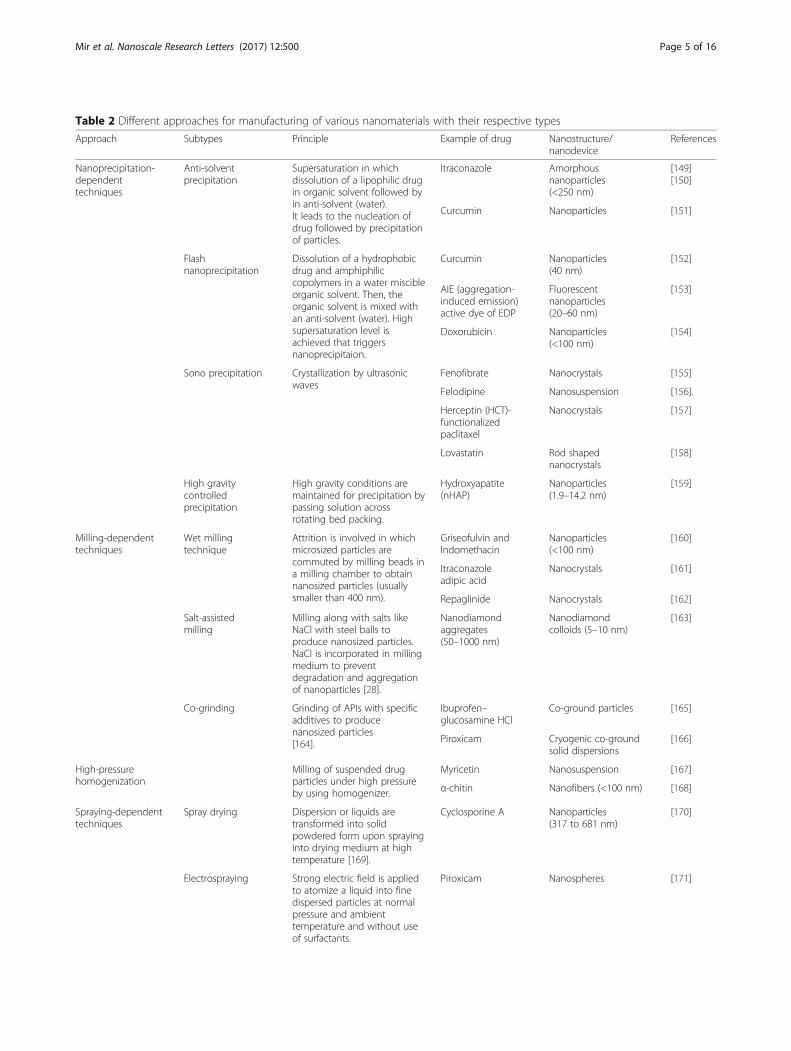
Table 2 Different approaches for manufacturing of various nanomaterials with their respective types
Approach Subtypes Principle Example of drug Nanostructure/nanodevice
References
Nanoprecipitation-dependenttechniques
Anti-solventprecipitation
Supersaturation in whichdissolution of a lipophilic drugin organic solvent followed byin anti-solvent (water).It leads to the nucleation ofdrug followed by precipitationof particles.
Itraconazole Amorphousnanoparticles(<250 nm)
[149][150]
Curcumin Nanoparticles [151]
Flashnanoprecipitation
Dissolution of a hydrophobicdrug and amphiphiliccopolymers in a water miscibleorganic solvent. Then, theorganic solvent is mixed withan anti-solvent (water). Highsupersaturation level isachieved that triggersnanoprecipitaion.
Curcumin Nanoparticles(40 nm)
[152]
AIE (aggregation-induced emission)active dye of EDP
Fluorescentnanoparticles(20–60 nm)
[153]
Doxorubicin Nanoparticles(<100 nm)
[154]
Sono precipitation Crystallization by ultrasonicwaves
Fenofibrate Nanocrystals [155]
Felodipine Nanosuspension [156].
Herceptin (HCT)-functionalizedpaclitaxel
Nanocrystals [157]
Lovastatin Rod shapednanocrystals
[158]
High gravitycontrolledprecipitation
High gravity conditions aremaintained for precipitation bypassing solution acrossrotating bed packing.
Hydroxyapatite(nHAP)
Nanoparticles(1.9–14.2 nm)
[159]
Milling-dependenttechniques
Wet millingtechnique
Attrition is involved in whichmicrosized particles arecommuted by milling beads ina milling chamber to obtainnanosized particles (usuallysmaller than 400 nm).
Griseofulvin andIndomethacin
Nanoparticles(<100 nm)
[160]
Itraconazoleadipic acid
Nanocrystals [161]
Repaglinide Nanocrystals [162]
Salt-assistedmilling
Milling along with salts likeNaCl with steel balls toproduce nanosized particles.NaCl is incorporated in millingmedium to preventdegradation and aggregationof nanoparticles [28].
Nanodiamondaggregates(50–1000 nm)
Nanodiamondcolloids (5–10 nm)
[163]
Co-grinding Grinding of APIs with specificadditives to producenanosized particles[164].
Ibuprofen–glucosamine HCl
Co-ground particles [165]
Piroxicam Cryogenic co-groundsolid dispersions
[166]
High-pressurehomogenization
Milling of suspended drugparticles under high pressureby using homogenizer.
Myricetin Nanosuspension [167]
α-chitin Nanofibers (<100 nm) [168]
Spraying-dependenttechniques
Spray drying Dispersion or liquids aretransformed into solidpowdered form upon sprayinginto drying medium at hightemperature [169].
Cyclosporine A Nanoparticles(317 to 681 nm)
[170]
Electrospraying Strong electric field is appliedto atomize a liquid into finedispersed particles at normalpressure and ambienttemperature and without useof surfactants.
Piroxicam Nanospheres [171]
Mir et al. Nanoscale Research Letters (2017) 12:500 Page 5 of 16
hereto conjugation with elongation of fluorescence time [29,40]. The visualization properties of quantum dots (fluores-cence wavelength) are strongly size dependent. The opticalproperties of quantum dots depend upon their structure asthey are composed of an outer shell and a metallic core. Forinstance, grapheme quantum dots (GQD), a type of greenfluorescence carbon nanomaterials, are made by cuttinggrapheme oxide solvothermally and are found to be dominat-ing the visualization properties [41].Quantum dot core is usually made up of cadmium selen-
ide, cadmium sulfide, or cadmium telluride. The outer shellis fabricated on the core with high band gap energy in orderto provide electrical insulation with preservation of fluores-cence properties of quantum dots. The fine-tuned core andshells with different sizes and compositions with visualizationproperties of specific wavelength provide a large number ofbiomarkers [40]. Quantum dots are conjugated with differentligands in order to obtain specific binding to biological re-ceptors. The tumor-targeting ligands are linked with amphi-philic polymer quantum dots and used to carry out theimaging studies of prostate cancer in mice [42]. Similarly,quantum dots offer significant advantages over the conven-tional dyes such as narrow bandwidth emission, higherphoto stability, and extended absorption spectrum for thesingle excitation source. Moreover, the challenge of hydro-phobicity in quantum dots has been overcome by making
them water soluble. An example of the aqueous quantumdots with long retention time in biological fluids is the devel-opment of highly fluorescent metal sulfide (MS) quantumdots fabricated with thiol-containing charged groups [43].Furthermore, the unique fluorescence properties of quantumdots made them suitable imaging tools for the cancer cells[42]. Quantum dots linked with A10 RNA aptamer conju-gated with doxorubicin (QD-Apt-Dox) is the example of tar-geted cancer cell imaging [44]. However, increased toxicity ofquantum dots has been observed due to the incorporation ofheavy metals, resulting in their limited use for the in vivo im-aging. Nevertheless, recent approaches focus on the reduc-tion in toxicity and the enhancement of biocompatibility ofquantum dots to the body cells. It is also worth to mentionthat quantum dots with the diameter less than 5.5 nm arerapidly and efficiently excreted from the urine resulting in re-duced toxicity. This phenomenon was exhibited by the syn-thesis of cadmium free, CulnS2/ZnS (copper indium sulfide/zinc sulfide) as the core and shell of the quantum dots,which resulted in enhanced stability in the living cells forlymph node imaging with a clear reduction in acute localtoxicity [45, 46].
BiosensorsOne of the greatest achievements in nanomaterials since lastfew years is the development of biosensors. Biosensors are
Table 2 Different approaches for manufacturing of various nanomaterials with their respective types (Continued)
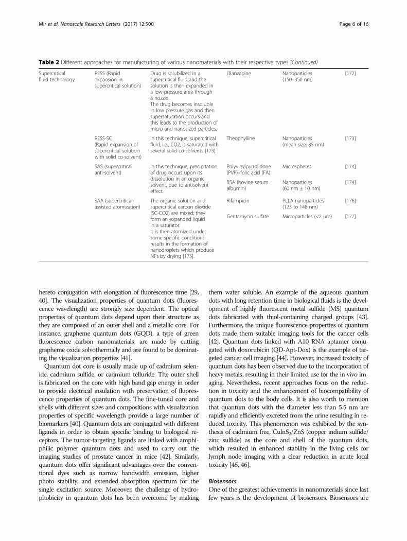
Supercriticalfluid technology
RESS (Rapidexpansion insupercritical solution)
Drug is solubilized in asupercritical fluid and thesolution is then expanded ina low-pressure area througha nozzle.The drug becomes insolublein low pressure gas and thensupersaturation occurs andthis leads to the production ofmicro and nanosized particles.
Olanzapine Nanoparticles(150–350 nm)
[172]
RESS-SC(Rapid expansion ofsupercritical solutionwith solid co-solvent)
In this technique, supercriticalfluid, i.e., CO2, is saturated withseveral solid co solvents [173].
Theophylline Nanoparticles(mean size: 85 nm)
[173]
SAS (supercriticalanti-solvent)
In this technique, precipitationof drug occurs upon itsdissolution in an organicsolvent, due to antisolventeffect.
Polyvinylpyrrolidone(PVP)–folic acid (FA)
Microspheres [174]
BSA (bovine serumalbumin)
Nanoparticles(60 nm ± 10 nm)
[174]
SAA (supercritical-assisted atomization)
The organic solution andsupercritical carbon dioxide(SC-CO2) are mixed; theyform an expanded liquidin a saturator.It is then atomized undersome specific conditionsresults in the formation ofnanodroplets which produceNPs by drying [175].
Rifampicin PLLA nanoparticles(123 to 148 nm)
[176]
Gentamycin sulfate Microparticles (<2 μm) [177]
Mir et al. Nanoscale Research Letters (2017) 12:500 Page 6 of 16

the devices that contain the biological sensing element thatis either connected or integrated in the transducer. Biosen-sor exhibits their action by recognition of specific moleculesin the body on the basis of their structure including anti-body antigen, enzyme substrate, and receptor hormone. Thetwo major properties of biosensor including their specificityand selectivity are dependent upon this recognition system.These basic properties of the biosensors are most import-antly used for the concentration that is proportional to thesignals [47–49].In order to produce the biosensor with high efficiency,
the substrate selected for the sensing material dispersion isprerequisite. Different types of nanomaterial includingquantum dots [50], magnetic nanoparticles [51], carbonnanotubes (CNTs) [52], and gold nanoparticles (GNPs) [53]are applied to the biosensors. The distinctive chemical,physical, magnetic, optical, and mechanical properties ofnanomaterial lead to their increased specificity and sensitiv-ity for detection. Biosensors containing GNPs have offereda compatible environment for the biomolecules that has in-creased the immobilized biomolecules concentration onthe surface of electrode. It has resulted in enhancedsensitivity of the biosensors [54, 55]. The most widely usedelectrode surfaces within the biosensors are the glassy car-bon electrode (GCE), which are modified from GNPs.Moreover, they have shown best sensitivity as well as elec-trochemical stability. In this regards, methylene blue (MB)and GNPs are easily assembled and modified through layerby layer (LBL) technique in the form of films on GCE, inorder to detect the concentration of human chorionic go-nadotrophin (HCG) [56]. Owing to the large surface areacontained by the nanoparticles in order to load anti-HCG,these immunosensors have their potential to be used fordetecting the concentrations of HCG in the human bloodor urine samples. Similarly, CNTs have found greatapplications in biomedical engineering, bio-analysis, bio-sensing, and nanoelectronics [57–59]. Moreover, multi-walled carbon nanotubes (MWNT) in the form of bio-nanocomposite layers of polymers have the potential to beused for the DNA detection [60]. Furthermore, magneticnanoparticles have also found wide applications because oftheir magnetic properties, including magnetic resonanceimaging (MRI) contrast agent [61], hyperthermia [62], im-munoassay [63], tissue repair [64], cell separation [65],GMR-sensor [66], and drug or gene delivery [67].Likewise, a new type of magnetic chitosan microspheres
(MCMS) has also been produced by simply using chitosanand carbon-coated magnetic nanoparticles [68]. In thisstudy, hemoglobin was also immobilized successfully onthe MCMS modified GCE surface by using glutaraldehydeas the crosslinking agent. Another important applicationof biosensors is in the optical technology, which includesthe detection of various kinds of DNA oligonucleotides byusing SsDNA–CNT probes as the biosensors [69].
Similarly, liposome-based biosensors have also gainedconsiderable attention as they have been used in the mon-itoring of the organophosphorus pesticides, includingparaoxon and dichlorvos on the minimum levels [70].
Magnetic NanoparticlesMagnetic nanoparticles (MNPs) provide exclusive mag-netic properties as they have the ability to work at the mo-lecular or cellular level of the biological interactions,which make them the best compounds as contrast agentsin MRI and as carriers in drug delivery. The recentadvancements in nanotechnology have gained attention asit helped in the modification of the properties and featuresof MNPs for the biomedical applications. In this respect,the liver tumor and metastasis imaging via RES-mediateduptake of superparamagnetic iron oxides (SPIOs) has beenshown to be capable of the differentiation of the lesionsthat are as small as only 2–3 mm [70, 71]. Moreover, theseultra-small supermagnetic iron oxides (USPIOs) are alsovery effective in the imaging of the metastasis of thelymph nodes with only 5 to 10 mm of diameter [72].Furthermore, importance of this noninvasive approachhas also been shown in the detection of the lymphaticdissemination as it is considered an important part in thestaging as well as in identifying the treatment approachesfor the breast colon and prostate cancers [73].
Drug DeliveryNanotechnology is an attractive tool for disciplines ran-ging from materials science to biomedicine because oftheir different physical, optical, and electronic characteris-tics. The most effective research areas of nanotechnologyare nanomedicine that applies nanotechnology principlesfor the treatment, prevention, and diagnosis of diseases.Moreover, many products of nanomedicine have beenmarketed due to the surge in nanomedicine researchduring the past few decades, around the globe. Currently,nanomedicine is influenced by drug delivery systems,accounting for more than 75% of the total sales [74]. Inthis regards, nanoparticle-based drug delivery platformshave gain the trust of scientists for being the mostappropriate vehicles in addressing the pharmacokineticdrawbacks associated with conventional drug formulations[75]. Hence, various nanoforms have been attempted asdrug delivery systems such as liposomes, solid lipid nano-particles, dendrimers, and solid metal-containing NPs, toenhance the therapeutic efficacy of drugs [76, 77]. Someof the major fields of interest are discussed below.
OphthalmologyDrug delivery through the ophthalmic route is highly at-tractive yet challenging for the pharmaceutical scientists.The eye is a tiny intricate organ with multi-compartments.Its biochemistry, physiology, and anatomy have made it
Mir et al. Nanoscale Research Letters (2017) 12:500 Page 7 of 16

most impermeable to the xenobiotic. Common conditionsthat demand ocular administration contain the eyeinfections such as, conjunctivitis along with the corneal dis-orders like glaucoma. The most common drug classes usedin the ocular delivery include mydriatics or cycloplegics mi-otics, anti-infective, anti-inflammatory, diagnostics, and sur-gical adjuvants. For the small ocular irregularity, genetherapy is required too, and a large amount of work is be-ing conducted within this area. Nanocarrier supported ap-proaches have got attention of the scientists for theirsuitability and specificity. It has been reported that par-ticulate delivery system such as microspheres and nano-particles and vesicular carriers like liposomes, niosomes,pharmacosomes, and discomes improved the pharmacoki-netic and pharmacodynamics properties of various typesof drug molecules [76]. Many novel controlled drug deliv-ery systems have been emerged including hydrogels,muco-adhesive polymers, microemulsions, dendrimers,iontophoretic drug delivery, siRNA-based approaches,stem cells technology, non-viral gene therapy, and lasertherapy with the sclera plugs [78]. Different systems fordrug delivery are costumed for the delivery of drugthrough the ocular route. The chief goal of all the drugdelivery systems is to improve the residence period,enhance the corneal permeability, and liberate the drug atposterior chamber of eye, leading to increased bioavailabil-ity and improved patient compliance [79].Abrego et al. prepared PLGA (poly lactic co-glycolic
acid) nanoparticles of pranoprofen for ophthalmic deliv-ery in the form of hydrogel. This hydrogel formulationhave suitable rheological and physicochemical propertiesfor the ocular delivery of pranoprofen with improved bio-pharmaceutical outline of the drug. Moreover, it intensi-fied the local anti-inflammatory and analgesic results ofthe drug, resulting in improved patient’s compliance [80].In another study, cefuroxim loaded nanoparticles of chi-tosan were developed using a double crosslinking indouble emulsion technique. The inference point outchitosan-gelatin particles as potently practical candidatesfor DD at intraocular level [81]. Moreover, diclofenacloaded N-trimethyl chitosan nanoparticles (DC-TMCNs)were developed for ophthalmic use to improve ocular bio-availability of the drug [82]. Furthermore, nanosizedsupramolecular assemblies of chitosan-based dexametha-sone phosphate have been developed for improved pre-corneal drug residence time due to its muco-adhesivecharacteristics. These nanoparticles interact strongly withboth ocular surface and drug and protect the drug frommetabolic degradation leading to extended pre-cornealresidence [83]. Glaucoma, an ophthalmic disease, wastreated with brimonidine-based loaded sustained releasesolid lipid nanoparticles using glyceryl monostearate assolid lipid [84, 85]. Similarly, daptomycin-loadedchitosan-coated alginate (CS-ALG) nanoparticles were
developed with a suitable size for ocular applications andhigh encapsulation efficiency (up to 92%). This study re-vealed that daptomycin nanocarrier system could be usedin future to deliver this antibiotic directly into the eye, inorder to act as a prospective therapy against bacterial en-dophthalmitis and as an efficient alternative to chitosannanoparticles [86].One of the major causes of short- and long-term failure
of grafts in the corneal transplantation is the immunologicgraft rejection. For this purpose, PLGA-based biodegrad-able nanoparticle system of dexamethasone sodiumphosphate (DSP) was prepared, resulting in the sustainedrelease of the corticosteroids in order to prevent therejection of corneal graft [87]. Moreover, MePEG-PCL(polyethylene glycol-poly caprolactone) nanoparticles ofcurcumin were reported, and they showed increased effi-ciency, enhanced retention of curcumin in the cornea, andsignificant improvement in prevention of the corneal neo-vascularization over free curcumin [88]. Likewise, silvernanoparticle-infused tissue adhesive (2-octyl cyanoacryl-ate) were developed with enhanced mechanical strengthand antibacterial efficacy. These doped adhesive (silvernanoparticles) supported the use of tissue adhesives as aviable supplement or alternative to sutures [89].
PulmonologyLung diseases probably asthma, chronic obstructive pul-monary disease (COPD), and lung cancer have a high oc-currence and are often life threatening. For instance, it isdescribed that COPD is the fourth major cause of death,and lung carcinoma is the most prevailing cause of cancerdeaths worldwide. Nanoparticles are scrutinized as achoice to improve therapy of these severe diseases [90].Various drug-laden nanoparticles have been utilized fortheir local and systemic effects in the treatment of lungdiseases. Delivery of curative agents to the place of actionfor lung diseases may permit for effective treatment ofchronic lung infections, lung cancers, tuberculosis, andother respiratory pathologies [91]. The nanocarriers usedfor this purpose include liposomes, lipid- or polymer-based micelles, dendrimers, and polymeric NPs [92]. Poly-meric NPs are of prenominal interest, as the polymers canbe co-polymerized, surface modified, or bio-conjugatedfor ameliorate targeting capacity and distribution of theencapsulated agents. The generally used nanocarriers inpulmonary drug delivery contain natural polymers such asgelatin, chitosan, and alginate and synthetic polymers likepoloxamer, PLGA, and PEG [93].It was observed that PLGA NPs exhibit the most
convenient set of characteristics as carriers for pulmonaryprotein/DNA delivery while gelatin NPs are an agreeablereciprocal choice [94]. Similarly, anisotropic or Janus parti-cles of doxorubicin and curcumin were formulated to cargothe anticancer drugs for the treatment of lung cancer
Mir et al. Nanoscale Research Letters (2017) 12:500 Page 8 of 16

through inhalation. The particles were formulated by usingthe biocompatible and biodegradable materials binarymixtures. These particles did not exhibit geno- and cyto-toxic consequence. The cancer cells internalize these Janusparticles and massed them in the nucleus and cytoplasmleading to prolonged retention. Moreover, polyamidoamine(PAMAM) dendrimers were evaluated as nanocarriers forpulmonary delivery of the model weakly soluble anti-asthma pharmaceutical beclometasone dipropionate (BDP)using G3, G4 and G4 [12] dendrimers. This study showedthat BDP-dendrimers have potential for pulmonary inhal-ation using air-jet and vibrating-mesh nebulizers. Further-more, it was observed that the aerosol characteristics wereinfluenced by nebulizer design rather than dendrimers gen-eration [95]. Additionally, engineered nanoparticles (ENP),composed of inorganic metals, metal oxides, metalloids, or-ganic biodegradable, and inorganic biocompatible polymerswere used efficiently as carriers for the vaccine and drugdelivery and for the management of a variety of lung dis-eases. Properties and efficacious effects of ENPs on lungsare represented in Fig. 1. Inorganic ENP (silver, gold, andcarbon ENP), metal oxides ENP (iron oxide, zinc oxides,and titanium dioxide), and organic ENP (Lipid-based,polysaccharide-based, polymer matrix-based) were devel-oped and evaluated for pulmonary immune hemostasis. Aswell as being relatively secure carriers, modern studies indi-cated ENP cable of supervening beneficial outcomes withanti-inflammatory properties (e.g., silver and polystyrene)
and imprinting of the lung which present the maintenanceof immune homeostasis (e.g., polystyrene). Further knowingof the mechanisms may help in better understanding theuseful effects of ENP on pulmonary immune homeostasisand/or management of inflammatory lung disease [96].It is important to state that functionalized cationic lipo-
polyamine (Star: Star-mPEG-550) have been recently de-veloped for the siRNA (short interference RNA) in vivodelivery to the pulmonary vascular cells. This balancedlipid formulation intensify the siRNA retention in thelungs of mouse and accomplished significant disassembleof the target gene. The results were found useful and withreduced toxicity of miRNA-145 inhibitor delivery to thelung by using the functionalized cationic lipopolyaminenanoparticles to recruit the pulmonary arteriopathy andrectify function of heart within rats with intense pulmon-ary arterial hypertension (PAH) [97].
Cardiovascular SystemCardiovascular disease is the ailment that affects the cardio-vascular system, vascular diseases of the brain and kidney,and peripheral arterial disorder. Despite of all advances inpharmacological and clinical management, heart failure is aforemost reason of morbidity worldwide. Many novel thera-peutic strategies, embody cell transplantation, gene deliveryor therapy, and cytokines or other small molecules, havebeen studied to treat heart failure [98]. An inadequate num-ber of people are affected in developing countries; over 80%
Fig. 1 Properties and efficacious effects of ENPs on lungs
Mir et al. Nanoscale Research Letters (2017) 12:500 Page 9 of 16

of deaths due to cardiovascular disorder take place in under-developed countries and occur almost evenly in male andfemales [99]. Mathers et al. in 2008 estimated that there are9.4 million deaths each year [100]. This concludes 45% ofdeaths caused by coronary heart disease and 51% of deathsdue to heart strokes [101]. There are many distinct types ofdrug delivery vehicles, like polymeric micelles, liposomes,dendrimers, lipoprotein-supported pharmaceutical carriers,and nanoparticle drug carriers.Chitosan-based liposomes of sirolimus having ≥83%
entrapment efficiency were developed for the treatment ofrestenosis and have been proved a novel platform for effi-cient targeted delivery [102]. Similarly, bile salt-enrichedniosomes of carvedilol with 85% entrapment efficiency haveresulted in enhanced bioavailability of drug, and thus, bettertherapeutic effect [103] was obtained. Inhibition of resten-osis in balloon-injured carotid artery is achieved in rats bydeveloping PLGA-based nanoparticles encapsulating AGL2043 and AG1295, selective blockers of platelet-derivedgrowth factors (PDGF) receptors [104]. Angiogenic therapyof myocardial ischemia with vascular endothelial growthfactor (VEGF) is a favorable approach to overcome hypoxiaand its sequel effects. Polymeric particles loaded with VEGFhave been proved a promising system for delivery of cyto-kines to rat myocardial ischemic model. This approachcould be further explored for clinical studies [105].Coenzyme Q10 (CoQ10) owing to its role in mitochondrialelectron transport chain appears to be a reliable candidateto treat myocardial ischemia (MI) but its poor biopharma-ceutical characteristics needed to be addressed by develop-ing promising delivery approaches. Polymeric nanoparticleswere developed to encapsulate CoQ10 to overcome its poorpharmaceutical properties and administered to MI-inducedrats. Cardiac function was analyzed by determining ejectionfraction before and after 3 months of therapy. Resultsshowed significant betterment in the ejection fraction after3 months [106].
OncologyCancer is a prime cause of mortality around the globe.The World Health Organization determines that 84million people die of cancer between 2005 and 2015. Theeventual target of cancer therapeutics is to increase the lifespan and the quality of life of the patient by minimizingthe systemic toxicity of chemotherapy [107]. Chemothera-peutic agents have widely been studied in oncology for thepast 25 years, but their tumor specificity is unsatisfactoryand therefore exhibit dose-dependent toxicity. To over-come this limitation, recent interest has been centered ondeveloping nanoscale delivery carriers that can be targeteddirectly to the cancer cell, deliver the drug at a controlledrate, and optimize the therapeutic efficacy [108, 109]. Pas-sive and active targeting is used to deliver the drug at itstumor site. The passive phenomenon called the “enhanced
permeability and retention (EPR) effect,” discovered byMatsumura and Maeda, is the dominated pathway usedfor chemotherapeutics [110, 111]. Active targeting isachieved by grafting ligand at the surface of nanocarriersthat bind to receptors or stimuli-based carriers, e.g., dualreverse thermosensitive [112], photo-responsive [113],magnetic nanoparticles [114], and enzymatically activatedpro-drugs [115]. Nanoparticles (NPs) can be conjugatedwith various smart therapeutic carriers like polymericnanoparticles [116], micelles [117], liposomes [118], solidlipid nanoparticles (SLNs) [119], protein nanoparticles[120], viral nanoparticles [121], metallic nanoparticles[122], aptamers [123], dendrimers [124], and monoclonalantibody [125] to improve their efficacy and decrease thesystemic toxicity. Table 3 summarizes the differentapproaches for drug deliveries which are widely studied totarget the tumor with maximize therapeutic response andminimum toxicity.Biodegradable poly (o-caprolactone) nanocarriers
loaded with tamoxifen were developed for the manage-ment of estrogen receptor-specific breast cancer [126].This study suggested that the nanoparticle preparations ofselective estrogen receptor modulators deliver the drug inthe specific estrogen receptor zone resulting in enhancedtherapeutic efficacy. Similarly, a nanoconjugation of doxo-rubicin and cisplatin was developed by Chohen et al.[127], which have exhibited enhanced efficiency and re-duced side effects of the loaded drugs in the treatment oflocalized progressive breast cancer. Likewise, chemothera-peutic drug oxaliplatin-loaded nanoparticulate micelleswere prepared by Cabral et al. [128], with sustained releaseof loaded drug in the tumor microenvironment, resultedin enhanced antitumor effect [128]. Furthermore, SLNloaded-5-FU resulted in enhanced bioavailability and sus-tained release of the encapsulated anticancer drug, leadingto enhanced antitumor effect [129].
ConclusionsNanotechnology is subjected to inordinate progress in vari-ous fronts especially to make innovations in healthcare.Target-selective drug delivery and approaches for molecularimaging are the areas of prime importance for researchwhere nanotechnology is playing a progressive role. This re-view provides readers with a wide vision on novel ongoingpotentialities of various nanotechnology-based approachesfor imaging and delivery of therapeutics. In order to obtaineffective drug delivery, nanotechnology-based imaging hasenabled us to apprehend the interactions of nanomaterialswith biological environment, targeting receptors, molecularmechanisms involved in pathophysiology of diseases, andhas made the real time monitoring of therapeutic responsepossible. Development of analytical technologies to meas-ure the size of particles in nanometer ranges, and advent oflatest manufacturing approaches for nanomaterials, has
Mir et al. Nanoscale Research Letters (2017) 12:500 Page 10 of 16

resulted in establishment of more effective methods for de-livery of therapeutics for the treatment of ophthalmological,pulmonary, cardiovascular diseases, and more importantlycancer therapy. These new drug therapies have alreadybeen shown to cause fewer side effects and be more effect-ive than traditional therapies. Furthermore, the imagingtechniques have enhanced the determination of tumor
location in human bodies and their selective targeting.Altogether, this comparatively new and thriving data sug-gest that additional clinical and toxicity studies are requiredfurther on the “proof-of-concept” phase. Nanomedicinecost and manufacturing at larger scale is also a matter ofconcern that needs to be addressed. Notwithstanding,future of nanomedicines is propitious.
Table 3 Nanomaterials and drug delivery approaches for tumor treatment
Nanomaterials Delivery approaches Advantages References
Aptamer functionalized silica goldnanorods(60 nm)
Near-infrared light responsivedrug delivery system
Biocompatibility, cancer cell recognitionability, and efficient intracellular drugrelease
[178]
Doxorubicin-loaded PEG diacrylate-Chitosan derivative-single-wall car-bon nanotubes (CNT)(240 nm)
Near-infrared (NIR) lighttriggered drug delivery system
Enhanced cellular uptake and the fasterdrug release
[179]
(DOX)-loaded hollow mesoporouscopper sulfide nanoparticles (HMCuSNPs) with iron oxide nanoparticles(IONPs)(124.5 ± 3.8 nm)
Near-infrared (NIR) lighttriggered drug delivery system
Minimized the adverse effects,enhanced photo thermal therapy effect
[180]
DOX-(HMCuSNPs) with hyaluronicacid (HA)(113.8 ± 6.9 nm)
Near infrared (NIR) lighttriggered drug delivery system
Facilitate intracellular tunable drug release,enhanced targeting and accumulationcapacity in tumor site
[181]
α-Cyclodextrin and poly (ethyleneglycol)-platinum dendrimer(1.9 ± 0.3 nm)
Near infrared (NIR) light-responsive supramolecularhydrogel
Enhanced release of drug, low toxicity [182]
End-capped mesoporous silicananoparticles (MSNs)(130 nm)
Redox-responsivenanoreservoirs
Excellent biocompatibility, cell-specificintracellular drug delivery, and cellularuptake properties
[183]
Transferrin (Tf)-(MSNs)-DOX(280 nm)
Redox-responsive drugdelivery system
Biocompatible, enhanced intracellularaccumulation, targeting capability
[184]
Amino- β –cyclodextrin- MSNs(203.3 nm)
Folate mediated and pHtargeting
High intercellular release [185]
DOX-thiolated poly(ethylene glycol)-biotin-DNA conjugated goldnanorod (GNR)(length of 50 ± 5 nmdiameter of 14 ± 3 nm)
pH-and near infrared (NIR)radiation dual-stimuli triggereddrug delivery
Increased potency (~67-fold), increasedcell uptake, low drug efflux
[186]
Cytochrome C conjugatedlactobionic acid (CytC–LA)-Doxorubicin (DOX)- MSNs(115.8 nm)
pH and redox dual-responsivedrug delivery
Good biocompatibility, high efficiency,inhibits tumor growth with minimaltoxic side effect.
[187]
Poly (propylene sulfide)-polyethyleneglycol-serine-folic acid (PPS-mPEG-Ser-FA)- zinc phthalocyanine-doxurubicin micelle(80 nm)
Reactive oxygen species (ROS)sensitive drug delivery system
Minimal toxic side effects [188]
Rituximab-conjugateddoxorubicin- MSNs(40.7 ± 19.1 nm)
pH-sensitive controlled drugrelease system
Reduce systemic toxicity, improve thetherapeutic efficacy
[189]
PEGylated-MoS 2 nanosheets(diameter 50 nm, thickness ∼2 nm)
Combined photothermal andchemotherapy targeting
Highly efficient loading [190]
DOX-Gold nanorod-1-tetradecanol-MSNs (thickness 35 nm)
Photothermalablation andchemotherapy
Precise control over drug release, localizeddelivery with enhanced targeting
[191]
Fe3O4–azobis [N-(2-carboxyethyl)-2-methylpropionamidine](Azo)-Doxorubicin
Combined photothermaltherapy and chemotherapy
Enhanced cell-killing effects, increasedstability, low toxicity
[192]
Mir et al. Nanoscale Research Letters (2017) 12:500 Page 11 of 16

AbbreviationsAIE: Aggregation-induced emission; BDP: Beclometasone dipropionate;BODIPY: Boron dipyrromethane; CNTs: Carbon nanotubes; COPD: Chronicobstructive pulmonary disease; CulnS2/ZnS: Copper indium sulfide/zincsulfide quantum dots; CVD: Chemical vapor deposition;DNA: Deoxyribonucleic acid; ENPs: Engineered nanoparticles; EPR: Enhancedpermeability and retention; GCE: Glassy carbon electrode; GNPs: Goldnanoparticles; GQD: Grapheme quantum dots; HCG: Human chorionicgonadotrophin; MEMS: Microelectromechanical systems; MI: Myocardialischemia; MNPs: Magnetic nanoparticles; MSNs: Mesoporous silicananoparticles; MWNT: Multi-walled carbon nanotubes;NEMS: Nanoelectromechanical system; PAH: Pulmonary arterial hypertension;PCL: Poly caprolactone; PDGF: Platelet-derived growth factors; PEG: Polyethylene glycol; PET: Positron emission tomography; PLGA: Poly lactic-co-glycolic acid; ROS: Reactive oxygen species; SiRNA: Short interference RNA;SLNS: Solid lipid nanoparticles; SPIOs: Superparamagnetic iron oxides;VEGF: Vascular endothelial growth factor
FundingThere was no funding available for this work.
Availability of Data and MaterialsPresented in the main paper.
Authors’ ContributionsFuD and AuR presented the idea; MM and SR did the literature review; MM,SI, and AZ write the manuscript. MK, FuD, GMK, and AuR critically review themanuscript. All authors read and approved the final manuscript.
Ethics approval and consent to participateNot applicable.
Consent for PublicationNot applicable.
Competing InterestsThe authors declare that they have no competing interests.
Received: 14 May 2017 Accepted: 26 July 2017
References1. Arora S, Rajwade JM, Paknikar KM (2012) Nanotoxicology and in vitro
studies: the need of the hour. Toxicol Appl Pharm 258(2):151–1652. Saini R, Saini S, Sharma S (2010) Nanotechnology: the future medicine. J
Cutan Aesthet Surg 3(1):323. Holdren J. The national nanotechnology initiative strategic plan report at
subcommittee on nanoscale science, engineering and technology ofcommittee on technology. National Science Technology Council (NSTC),Arlington. 2011
4. Fakruddin M, Hossain Z, Afroz H (2012) Prospects and applications ofnanobiotechnology: a medical perspective. J Nanobiotechnol 10(1):31
5. Drexler E. Reprint. Engines of Creation. The Coming Era of Nanotechnology.New York: Anchor Books. Original edition, NY: Anchor Books; 1986
6. Drexler KE, Peterson C, Pergamit G (1991) Unbounding the future, vol 294.William Morrow, New York
7. Freitas RA (1999) Nanomedicine, volume I: basic capabilities: LandesBioscience. Georgetown, TX
8. Freitas RA Jr (2003) Nanomedicine, Vol. IIA: Biocompatibility. LandesBioscience. Georgetown, USA
9. Freitas RA (2005) What is nanomedicine? Nanomed Nanotech Biol Med 1(1):2–910. Parviz BA, Ryan D, Whitesides GM (2003) Using self-assembly for the
fabrication of nano-scale electronic and photonic devices. IEEE Trans AdvPackag 26(3):233–241
11. Nakano T, Moore MJ, Wei F, Vasilakos AV, Shuai J (2012) Molecularcommunication and networking: opportunities and challenges. IEEE TransNanobioscience 11(2):135–148
12. Cavalcanti A, Shirinzadeh B, Fukuda T, Ikeda S, editors. Hardwarearchitecture for nanorobot application in cerebral aneurysm.Nanotechnology, 2007 IEEE-NANO 2007 7th IEEE Conference on; 2007: IEEE
13. Garcia R, Herruzo ET (2012) The emergence of multifrequency forcemicroscopy. Nat Nanotechnol 7(4):217–226
14. Sun Q, Cai X, Li J, Zheng M, Chen Z, Yu C-P (2014) Green synthesis ofsilver nanoparticles using tea leaf extract and evaluation of theirstability and antibacterial activity. Colloids Surf A Physicochem Eng Asp444:226–231
15. Ferrari M (2005) Cancer nanotechnology: opportunities and challenges. NatRev Cancer 5(3):161–171
16. Vasir JK, Reddy MK, Labhasetwar VD (2005) Nanosystems in drug targeting:opportunities and challenges. Curr Nanosci 1(1):47–64
17. Klaessig F, Marrapese M, Abe S (2011) Current perspectives innanotechnology terminology and nomenclature. Nanotechnologystandards. Springer, pp 21–52
18. Yadav T, Mungray AA, Mungray AK. Fabricated nanoparticles: current statusand potential phytotoxic threats. Rev Environ Contam Toxicol. volume:Springer; 2014. p. 83–110
19. Scott N, Chen H (2013) Nanoscale science and engineering for agricultureand food systems. Ind Biotechnol 9(1):17–18
20. Ebrahimi E, Akbarzadeh A, Abbasi E, Khandaghi AA, Abasalizadeh F, DavaranS (2016) Novel drug delivery system based on doxorubicin-encapsulatedmagnetic nanoparticles modified with PLGA-PEG1000 copolymer. Artif CellsNanomed Biotechnol 44(1):290–297
21. Cosco D, Cilurzo F, Maiuolo J, Federico C, Di Martino MT, Cristiano MC et al(2015) Delivery of miR-34a by chitosan/PLGA nanoplexes for the anticancertreatment of multiple myeloma. Sci Rep 5
22. Vartak A, Sucheck SJ (2016) Recent advances in subunit vaccine carriers.Vaccine 4(2):12
23. Virlan MJR, Miricescu D, Totan A, Greabu M, Tanase C, Sabliov CM et al(2015) Current uses of poly (lactic-co-glycolic acid) in the dental field: acomprehensive review. J Chem 2015
24. Hua S, Marks E, Schneider JJ, Keely S (2015) Advances in oral nano-deliverysystems for colon targeted drug delivery in inflammatory bowel disease:selective targeting to diseased versus healthy tissue. Nanomed NanotechBiol Med 11(5):1117–1132
25. Bhatia S (2016) Nanoparticles types, classification, characterization,fabrication methods and drug delivery applications. In: Natural polymerdrug delivery systems. Springer, pp 33–93
26. Silva GA (2004) Introduction to nanotechnology and its applications tomedicine. Surg Neurol 61(3):216–220
27. Sinha B, Müller RH, Möschwitzer JP (2013) Bottom-up approaches forpreparing drug nanocrystals: formulations and factors affecting particle size.Int J Pharm 453(1):126–141
28. Kaialy W, Al SM (2016) Recent advances in the engineering of nanosizedactive pharmaceutical ingredients: promises and challenges. Adv ColloidInterf Sci 228:71–91
29. Portney NG, Ozkan M (2006) Nano-oncology: drug delivery, imaging, andsensing. Anal Bioanal Chem 384(3):620–630
30. Wickline SA, Lanza GM. Nanotechnology for molecular imaging andtargeted therapy. Am Heart Assoc; 2003
31. Allport JR, Weissleder R (2001) In vivo imaging of gene and cell therapies.Exp Hematol 29(11):1237–1246
32. Ballinger JR (2001) 99mTc-Tetrofosmin for functional imaging of P-glycoprotein modulation in vivo. J Clin Pharmacol 41(S7)
33. Kao CH, Hsieh JF, Tsai SC, Ho YJ, ChangLai SP, Lee JK (2001)Paclitaxel-based chemotherapy for non–small cell lung cancer:predicting the response with 99mTc-tetrofosmin chest imaging. J NuclMed 42(1):17–20
34. Martina M-S, Fortin J-P, Ménager C, Clément O, Barratt G, Grabielle-Madelmont C et al (2005) Generation of superparamagnetic liposomesrevealed as highly efficient MRI contrast agents for in vivo imaging. J AmChem Soc 127(30):10676–10685
35. Kuil J, Buckle T, Oldenburg J, Yuan H, Borowsky AD, Josephson L et al (2011)Hybrid peptide dendrimers for imaging of chemokine receptor 4 (CXCR4)expression. Mol Pharm 8(6):2444–2453
36. Noon WH, Kong Y, Ma J (2002) Molecular dynamics analysis of a buckyball–antibody complex. Proc Natl Acad Sci 99(suppl 2):6466–6470
37. Torchilin VP (2000) Polymeric contrast agents for medical imaging. CurrPharm Biotechnol 1(2):183–215
38. Milroy LG, Rizzo S, Calderon A, Ellinger B, Erdmann S, Mondry J et al (2012)Selective chemical imaging of static actin in live cells. J Am Chem Soc134(20):8480–8486
Mir et al. Nanoscale Research Letters (2017) 12:500 Page 12 of 16

39. Kowada T, Maeda H, Kikuchi K (2015) BODIPY-based probes for thefluorescence imaging of biomolecules in living cells. Chem Soc Rev 44(14):4953–4972
40. Mohs AM, Provenzale JM (2010) Applications of nanotechnology to imagingand therapy of brain tumors. Neuroimaging Clin N Am 20(3):283–292
41. Wang L, Zhu SJ, Wang HY, Qu SN, Zhang YL, Zhang JH et al (2014)Common origin of green luminescence in carbon nanodots and graphenequantum dots. ACS Nano 8(3):2541–2547
42. Gao X, Cui Y, Levenson RM, Chung LW, Nie S (2004) In vivo cancertargeting and imaging with semiconductor quantum dots. NatBiotechnol 22(8):969–976
43. Shih WH, Shih WY, Li H, Schillo MC. Water soluble quantum dots. GooglePatents; 2009
44. Bagalkot V, Zhang L, Levy-Nissenbaum E, Jon S, Kantoff PW, Langer R et al(2007) Quantum dot− aptamer conjugates for synchronous cancer imaging,therapy, and sensing of drug delivery based on bi-fluorescence resonanceenergy transfer. Nano Lett 7(10):3065–3070
45. Choi HS, Liu W, Misra P, Tanaka E, Zimmer JP, Ipe BI et al (2007) Renalclearance of quantum dots. Nat Biotechnol 25(10):1165–1170
46. Pons T, Pic E, Lequeux N, Cassette E, Bezdetnaya L, Guillemin F et al (2010)Cadmium-free CuInS2/ZnS quantum dots for sentinel lymph node imagingwith reduced toxicity. ACS Nano 4(5):2531–2538
47. Buch RM, Rechnitz G (1989) Intact chemoreceptor-based biosensors:responses and analytical limits. Biosensors 4(4):215–230
48. Kricka L (1988) Molecular and ionic recognition by biological systems,Chemical sensors. Springer, pp 3–14
49. Zhang X, Guo Q, Cui D (2009) Recent advances in nanotechnology appliedto biosensors. Sensors 9(2):1033–1053
50. You X, He R, Gao F, Shao J, Pan B, Cui D (2007) Hydrophilic high-luminescent magnetic nanocomposites. Nanotechnology 18(3):035701
51. Pan B, Cui D, Sheng Y, Ozkan C, Gao F, He R et al (2007) Dendrimer-modified magnetic nanoparticles enhance efficiency of gene deliverysystem. Cancer Res 67(17):8156–8163
52. Cui D, Tian F, Coyer SR, Wang J, Pan B, Gao F et al (2007) Effects ofantisense-Myc-conjugated single-walled carbon Nanotubes on HL-60Cells. JNanosci Nanotechnol 7(4–1):1639–1646
53. Pan B, Cui D, Xu P, Li Q, Huang T, He R et al (2007) Study on interactionbetween gold nanorod and bovine serum albumin. Colloids Surf APhysicochem Eng Asp 295(1):217–222
54. Liang KZ, Qi JS, Mu WJ, Chen ZG (2008) Biomolecules/gold nanowires-doped sol–gel film for label-free electrochemical immunoassay oftestosterone. J Biochem Biophys Methods 70(6):1156–1162
55. He X, Yuan R, Chai Y, Shi Y (2008) A sensitive amperometric immunosensorfor carcinoembryonic antigen detection with porous nanogold film andnano-au/chitosan composite as immobilization matrix. J Biochem BiophysMethods 70(6):823–829
56. Chai R, Yuan R, Chai Y, Ou C, Cao S, Li X (2008) Amperometricimmunosensors based on layer-by-layer assembly of gold nanoparticles andmethylene blue on thiourea modified glassy carbon electrode fordetermination of human chorionic gonadotrophin. Talanta 74(5):1330–1336
57. Pan B, Cui D, He R, Gao F, Zhang Y (2006) Covalent attachment of quantumdot on carbon nanotubes. Chem Phys Lett 417(4):419–424
58. Cui D, Tian F, Kong Y, Titushikin I, Gao H (2003) Effects of single-walledcarbon nanotubes on the polymerase chain reaction. Nanotechnology15(1):154
59. Cui D (2007) Advances and prospects on biomolecules functionalizedcarbon nanotubes. J Nanosci Nanotechnol 7(4–1):1298–1314
60. Li G, Xu H, Huang W, Wang Y, Wu Y, Parajuli R (2008) A pyrrole quinolinequinone glucose dehydrogenase biosensor based on screen-printed carbonpaste electrodes modified by carbon nanotubes. Meas SciTechnol 19(6):065203
61. Lee H, Lee E, Kim DK, Jang NK, Jeong YY, Jon S (2006) Antibiofoulingpolymer-coated superparamagnetic iron oxide nanoparticles as potentialmagnetic resonance contrast agents for in vivo cancer imaging. J Am ChemSoc 128(22):7383–7389
62. Kim DH, Lee SH, Kim KN, Kim KM, Shim IB, Lee YK (2005) Cytotoxicity offerrite particles by MTT and agar diffusion methods for hyperthermicapplication. J Magn Magn Mater 293(1):287–292
63. Sincai M, Ganga D, Ganga M, Argherie D, Bica D (2005) Antitumor effect ofmagnetite nanoparticles in cat mammary adenocarcinoma. J Magn MagnMater 293(1):438–441
64. Ito A, Ino K, Kobayashi T, Honda H (2005) The effect of RGD peptide-conjugated magnetite cationic liposomes on cell growth and cell sheetharvesting. Biomaterials 26(31):6185–6193
65. Guedes MHA, Sadeghiani N, Peixoto DLG, Coelho JP, Barbosa LS, AzevedoRB et al (2005) Effects of AC magnetic field and carboxymethyldextran-coated magnetite nanoparticles on mice peritoneal cells. J Magn MagnMater 293(1):283–286
66. Rife J, Miller M, Sheehan P, Tamanaha C, Tondra M, Whitman L (2003)Design and performance of GMR sensors for the detection of magneticmicrobeads in biosensors. Sens Actuators A-Phys 107(3):209–218
67. Morishita N, Nakagami H, Morishita R (2005) Takeda S-i, Mishima F, NishijimaS, et al. magnetic nanoparticles with surface modification enhanced genedelivery of HVJ-E vector. Biochem. Biophys. Res. Commun 334(4):1121–1126
68. Lai GS, Zhang HL, Han DY (2008) A novel hydrogen peroxide biosensorbased on hemoglobin immobilized on magnetic chitosan microspheresmodified electrode. Sens and Actuators B: Chem 129(2):497–503
69. Cao C, Kim JH, Yoon D, Hwang ES, Kim YJ, Baik S (2008) Optical detection ofDNA hybridization using absorption spectra of single-walled carbonnanotubes. Mater Chem Phys 112(3):738–741
70. Corot C, Robert P, Idée JM, Port M (2006) Recent advances in iron oxidenanocrystal technology for medical imaging. Adv Drug Deliv Rev 58(14):1471–1504
71. Semelka RC, Helmberger TK (2001) Contrast agents for MR imaging of theliver 1. Radiology 218(1):27–38
72. Harisinghani MG, Barentsz J, Hahn PF, Deserno WM, Tabatabaei S, van deKaa CH et al (2003) Noninvasive detection of clinically occult lymph-nodemetastases in prostate cancer. N Engl J Med 348:2491–2499
73. Harisinghani MG, Weissleder R (2004) Sensitive, noninvasive detection oflymph node metastases. PLoS Med 1(3):e66
74. Wagner V, Dullaart A, Bock AK, Zweck A (2006) The emerging nanomedicinelandscape. Nat Biotechnol 24(10):1211–1217
75. Blanco E, Shen H, Ferrari M (2015) Principles of nanoparticle design forovercoming biological barriers to drug delivery. Nat Biotechnol 33(9):941–951
76. Wadhwa S, Paliwal R, Paliwal SR, Vyas S (2009) Nanocarriers in ocular drugdelivery: an update review. Curr Pharm Des 15(23):2724–2750
77. ud Din F, Rashid R, Mustapha O, Kim DW, Park JH, Ku SK et al (2015)Development of a novel solid lipid nanoparticles-loaded dual-reversethermosensitive nanomicelle for intramuscular administration with sustainedrelease and reduced toxicity. RSC Adv 5(54):43687–43694
78. Patel A, Cholker K, Agrahari V, Mitra AK. Occular drug delivery systems: anoverview. World J Pharmacol 2013;2(2): 47–64
79. Puglia C, Offerta A, Carbone C, Bonina F, Pignatello R, Puglisi G (2015) Lipidnanocarriers (LNC) and their applications in ocular drug delivery. Curr MedChem 22(13):1589–1602
80. Abrego G, Alvarado H, Souto EB, Guevara B, Bellowa LH, Parra A et al (2015)Biopharmaceutical profile of pranoprofen-loaded PLGA nanoparticlescontaining hydrogels for ocular administration. Eur J Pharm Biopharm 95:261–270
81. Andrei G, Peptu CA, Popa M, Desbrieres J, Peptu C, Gardikiotis F et al (2015)Formulation and evaluation of cefuroxim loaded submicron particles forophthalmic delivery. Int J Pharm 493(1):16–29
82. Asasutjarit R, Theerachayanan T, Kewsuwan P, Veeranodha S, Fuongfuchat A,Ritthidej GC (2015) Development and evaluation of diclofenac sodiumloaded-N-Trimethyl chitosan nanoparticles for ophthalmic use. AAPSPharmSciTech 16(5):1013–1024
83. Fabiano A, Chetoni P, Zambito Y (2015) Mucoadhesive nano-sizedsupramolecular assemblies for improved pre-corneal drug residence time.Drug Dev Ind Pharm 41(12):2069–2076
84. El-Salamouni NS, Farid RM, El-Kamel AH, El-Gamal SS (2015) Effect ofsterilization on the physical stability of brimonidine-loaded solid lipidnanoparticles and nanostructured lipid carriers. Int J Pharm 496(2):976–983
85. Ibrahim MM, Abd-Elgawad A-EH, Soliman OA-E, Jablonski MM (2015) Naturalbioadhesive biodegradable nanoparticle-based topical ophthalmicformulations for management of glaucoma. Transl Vis Sci Technol 4(3):12
86. Costa J, Silva N, Sarmento B, Pintado M (2015) Potential chitosan-coatedalginate nanoparticles for ocular delivery of daptomycin. Eur J Clin MicrobiolInfect Dis 34(6):1255–1262
87. Pan Q, Xu Q, Boylan NJ, Lamb NW, Emmert DG, Yang J-C et al (2015)Corticosteroid-loaded biodegradable nanoparticles for prevention of cornealallograft rejection in rats. J Control Release 201:32–40
Mir et al. Nanoscale Research Letters (2017) 12:500 Page 13 of 16

88. Pradhan N, Guha R, Chowdhury S, Nandi S, Konar A, Hazra S (2015)Curcumin nanoparticles inhibit corneal neovascularization. J Mol Medic93(10):1095–1106
89. Yee W, Selvaduray G, Hawkins B (2016) Characterization of silvernanoparticle-infused tissue adhesive for ophthalmic use. J Mech BehavBiomed Mater 55:67–74
90. Weber S, Zimmer A, Pardeike J (2014) Solid lipid Nanoparticles (SLN) andNanostructured lipid carriers (NLC) for pulmonary application: a review ofthe state of the art. Eur J Pharm Biopharm 86(1):7–22
91. Yang W, Peters JI, Williams RO III. (2008) Inhaled nanoparticles–a currentreview. Int J Pharm 356(1–2):239–247
92. Smola M, Vandamme T, Sokolowski A (2008) Nanocarriers as pulmonarydrug delivery systems to treat and to diagnose respiratory andnonrespiratory diseases. Int J Nanomedicine 3(1):1
93. Sung JC, Pulliam BL, Edwards DA (2007) Nanoparticles for drug delivery tothe lungs. Trends Biotechnol 25(12):563–570
94. Menon JU, Ravikumar P, Pise A, Gyawali D, Hsia CC, Nguyen KT (2014)Polymeric nanoparticles for pulmonary protein and DNA delivery. ActaBiomater 10(6):2643–2652
95. Nasr M, Najlah M, D’Emanuele A, Elhissi A (2014) PAMAM dendrimers asaerosol drug nanocarriers for pulmonary delivery via nebulization. Int JPharm 461(1):242–250
96. Mohamud R, Xiang SD, Selomulya C, Rolland JM, O’Hehir RE, Hardy CL et al(2014) The effects of engineered nanoparticles on pulmonary immunehomeostasis. Drug Metab Rev 46(2):176–190
97. McLendon JM, Joshi SR, Sparks J, Matar M, Fewell JG, Abe K et al (2015)Lipid nanoparticle delivery of a microRNA-145 inhibitor improvesexperimental pulmonary hypertension. J Control Release 210:67–75
98. Arora N, Singh K, Garg T (2012) Areas of nanomedicine applications. Int JUniv Pharm Life Sci 2:216–227
99. Singh B, Garg T, Goyal AK, Rath G (2016) Recent advancements in thecardiovascular drug carriers. Artif Cells Nanomed Biotechnol 44(1):216–225
100. Mathers C, Fat DM, Boerma JT. The global burden of disease: 2004 update:World Health Organization; 2008
101. Lim SS, Vos T, Flaxman AD, Danaei G, Shibuya K, Adair-Rohani H et al (2013)A comparative risk assessment of burden of disease and injury attributableto 67 risk factors and risk factor clusters in 21 regions, 1990–2010: asystematic analysis for the global burden of disease study 2010. Lancet380(9859):2224–2260
102. Haeri A, Sadeghian S, Rabbani S, Anvari MS, Ghassemi S, Radfar F et al(2017) Effective attenuation of vascular restenosis following local delivery ofchitosan decorated sirolimus liposomes. Carbohydr Polymer 157:1461–1469
103. Arzani G, Haeri A, Daeihamed M, Bakhtiari-Kaboutaraki H, DadashzadehS (2015) Niosomal carriers enhance oral bioavailability of carvedilol:effects of bile salt-enriched vesicles and carrier surface charge. Int JNanomedicine 10:4797
104. Godin B, Sakamoto JH, Serda RE, Grattoni A, Bouamrani A, Ferrari M (2010)Emerging applications of nanomedicine for the diagnosis and treatment ofcardiovascular diseases. Trends Pharmacol Sci 31(5):199–205
105. Formiga FR, Pelacho B, Garbayo E, Abizanda G, Gavira JJ, Simon-Yarza T et al(2010) Sustained release of VEGF through PLGA microparticles improvesvasculogenesis and tissue remodeling in an acute myocardial ischemia–reperfusion model. J Control Release 147(1):30–37
106. Simón-Yarza T, Tamayo E, Benavides C, Lana H, Formiga FR, Grama CN et al(2013) Functional benefits of PLGA particulates carrying VEGF and CoQ 10in an animal of myocardial ischemia. Int J Pharm 454(2):784–790
107. Danhier F, Feron O, Preat V (2010) To exploit the tumor microenvironment:passive and active tumor targeting of nanocarriers for anti-cancer drugdelivery. J Control Release 148(2):135–146
108. Mishra B, Patel BB, Tiwari S (2010) Colloidal nanocarriers: a review onformulation technology, types and applications toward targeted drugdelivery. Nanomed Nanotechnol Biol Med 6(1):9–24
109. Din FU, Kim DW, Choi JY, Thapa RK, Mustapha O, Kim DS et al (2017)Irinotecan-loaded double-reversible thermogel with improved antitumorefficacy without initial burst effect and toxicity for intramuscularadministration. Acta Biomater 54:239–248
110. Maeda H, Bharate G, Daruwalla J (2009) Polymeric drugs for efficient tumor-targeted drug delivery based on EPR-effect. Eur J Pharma Biopharm 71(3):409–419
111. Matsumura Y, Maeda H (1986) A new concept for macromoleculartherapeutics in cancer chemotherapy: mechanism of tumoritropic
accumulation of proteins and the antitumor agent smancs. Cancer Res46(12 Part 1):6387–6392
112. Din FU, Choi JY, Kim DW, Mustapha O, Kim DS, Thapa RK et al (2017)Irinotecan-encapsulated double-reverse thermosensitive nanocarrier systemfor rectal administration. Drug Deliv 24(1):502–510
113. Tong R, Hemmati HD, Langer R, Kohane DS (2012) Photoswitchablenanoparticles for triggered tissue penetration and drug delivery. J AmChem Soc 134(21):8848–8855
114. Arias JL, Reddy LH, Othman M, Gillet B, Desmaele D, Zouhiri F et al (2011)Squalene based nanocomposites: a new platform for the design ofmultifunctional pharmaceutical theragnostics. ACS Nano 5(2):1513–1521
115. Brown JM, Wilson WR (2004) Exploiting tumour hypoxia in cancertreatment. Nat Rev Cancer 4(6):437–447
116. Cao J, Deng X, Su T, He B (2016) Fabrication of polymeric nanoparticles forcancer therapy and intracellular tracing. Nanomed Nanotechnol Biol Med12(2):459
117. Xie J, Zhang X, Teng M, Yu B, Yang S, Lee RJ, et al. Synthesis,characterization, and evaluation of mPeg–sN38 and mPeg–Pla–sN38micelles for cancer therapy.Int J Nanomedicine. 2016;11:1677
118. Eloy JO, Petrilli R, Topan JF, Antonio HMR, Barcellos JPA, Chesca DL et al(2016) Co-loaded paclitaxel/rapamycin liposomes: development,characterization and in vitro and in vivo evaluation for breast cancertherapy. Colloids Surf B Biointerfaces 141:74–82
119. Din FU, Mustapha O, Kim DW, Rashid R, Park JH, Choi JY et al (2015) Noveldual-reverse thermosensitive solid lipid nanoparticle-loaded hydrogel forrectal administration of flurbiprofen with improved bioavailability andreduced initial burst effect. Eur J Pharm Biopharm 94:64–72
120. Lee J, Kang JA, Ryu Y, Han S-S, Nam YR, Rho JK et al (2017) Geneticallyengineered and self-assembled oncolytic protein nanoparticles for targetedcancer therapy. Biomaterials 120:22–31
121. Le DH, Lee KL, Shukla S, Commandeur U, Steinmetz NF (2017) Potato virusX, a filamentous plant viral nanoparticle for doxorubicin delivery in cancertherapy. Nano 9(6):2348–2357
122. Volsi AL, de Aberasturi DJ, Henriksen-Lacey M, Giammona G, Licciardi M, Liz-Marzán LM (2016) Inulin coated plasmonic gold nanoparticles as a tumor-selective tool for cancer therapy. J Mater Chem B 4(6):1150–1155
123. Zhuang Y, Deng H, Su Y, He L, Wang R, Tong G et al (2016) Aptamer-functionalized and backbone redox-responsive hyperbranched polymer fortargeted drug delivery in cancer therapy. Biomacromolecules 17(6):2050–2062
124. Wang X, Wang H, Wang Y, Yu X, Zhang S, Zhang Q et al (2016) A facilestrategy to prepare Dendrimer-stabilized gold Nanorods with sub-10-nmsize for efficient Photothermal cancer therapy. Sci Rep 6
125. Gray MJ, Gong J, Nguyen V, Schuler-Hatch M, Hughes C, Hutchins J, et al.Abstract B27: targeting of phosphatidylserine by monoclonal antibodych1N11 enhances the antitumor activity of immune checkpoint inhibitorPD-1/PD-L1 therapy in orthotopic murine breast cancer models. AACR; 2016
126. Chawla JS, Amiji MM (2002) Biodegradable poly (ε-caprolactone)nanoparticles for tumor-targeted delivery of tamoxifen. Int J Pharm 249(1):127–138
127. Cohen SM, Mukerji R, Cai S, Damjanov I, Forrest ML, Cohen MS (2011)Subcutaneous delivery of nanoconjugated doxorubicin and cisplatin forlocally advanced breast cancer demonstrates improved efficacy anddecreased toxicity at lower doses than standard systemic combinationtherapy in vivo. Am J Surg 202(6):646–653
128. Cabral H, Murakami M, Hojo H, Terada Y, Kano MR (2013) Chung Ui, et al.targeted therapy of spontaneous murine pancreatic tumors by polymericmicelles prolongs survival and prevents peritoneal metastasis. Proc NatlAcad Sci 110(28):11397–11402
129. Yassin A, Anwer MK, Mowafy HA, El-Bagory IM, Bayomi MA, Alsarra IA (2010)Optimization of 5-fluorouracil solid-lipid nanoparticles: a preliminary studyto treat colon cancer. Int J Med Sci 7(6):398–408
130. Alley SC, Okeley NM, Senter PD (2010) Antibody–drug conjugates: targeteddrug delivery for cancer. Curr opinion Chem Biol 14(4):529–537
131. Reddy B, Yadav HK, Nagesha DK, Raizaday A, Karim A (2015) Polymericmicelles as novel carriers for poorly soluble drugs—review. J NanosciNanotechnol 15(6):4009–4018
132. Gillies ER, Frechet JM (2005) Dendrimers and dendritic polymers in drugdelivery. Drug Discov Today 10(1):35–43
133. Anupa R (2010) Menjoge rangaramanujam, M.; Kannan Donald, a,; Tomalia.Dendrimer–based drug and imaging conjugates: desingn considerations fornanomedical application. Drug Discov Today 15:171–185
Mir et al. Nanoscale Research Letters (2017) 12:500 Page 14 of 16

134. Zhao M-X, Zhu B-J (2016) The research and applications of quantum dots asnano-carriers for targeted drug delivery and cancer therapy. Nanoscale ResLett 11(1):207
135. Martincic M, Tobias G (2015) Filled carbon nanotubes in biomedicalimaging and drug delivery. Expert Opin Drug Deliv 12(4):563–581
136. Ahmad MZ, Akhter S, Jain GK, Rahman M, Pathan SA, Ahmad FJ et al (2010)Metallic nanoparticles: technology overview & drug delivery applications inoncology. Expert Opin Drug Deliv 7(8):927–942
137. Wang Y, Zhao Q, Han N, Bai L, Li J, Liu J et al (2015) Mesoporous silicananoparticles in drug delivery and biomedical applications. NanomedNanotech Biol Med 11(2):313–327
138. Nazemi A, Gillies ER (2013) Dendritic surface functionalization ofnanomaterials: controlling properties and functions for biomedicalapplications. Braz J Pharm Sci 49(SPE):15–32
139. Bottari G, Urbani M, Torres T (2013) Covalent, donor–acceptorensembles based ON Phthalocyanines AND CARBON nanostructures. In:Organic Nanomaterials: synthesis, characterization, and deviceapplications, pp 163–186
140. Siró I, Plackett D (2010) Microfibrillated cellulose and new nanocompositematerials: a review. Cellulose 17(3):459–494
141. Jariwala D, Sangwan VK, Lauhon LJ, Marks TJ, Hersam MC (2013) Carbonnanomaterials for electronics, optoelectronics, photovoltaics, and sensing.Chem Soc Rev 42(7):2824–2860
142. Duran H, Steinhart M (2011) Butt H-Jr, Floudas G. From heterogeneous tohomogeneous nucleation of isotactic poly (propylene) confined tonanoporous alumina. Nano Lett 11(4):1671–1675
143. Kumari A, Yadav SK, Yadav SC (2010) Biodegradable polymeric nanoparticlesbased drug delivery systems. Colloids Surf B Biointerfaces 75(1):1–18
144. Hoare T, Santamaria J, Goya GF, Irusta S, Lin D, Lau S et al (2009) Amagnetically-triggered composite membrane for on-demand drug delivery.Nano Lett 9(10):3651
145. Mishra B, Patel BB, Tiwari S (2010) Colloidal nanocarriers: a review onformulation technology, types and applications toward targeted drugdelivery. Nanomed Nanotech Biol Med 6(1):9–24
146. Rabl P, Kolkowitz S, Koppens F, Harris J, Zoller P, Lukin M (2010) A quantumspin transducer based on nanoelectromechanical resonator arrays. Nat Phys6(8):602–608
147. Chandrasekhar S, Iyer LK, Panchal JP, Topp EM, Cannon JB, Ranade VV(2013) Microarrays and microneedle arrays for delivery of peptides, proteins,vaccines and other applications. Expert Opin Drug Deliv 10(8):1155–1170
148. Shabnashmi PS (2016) NKS, Vithya V., Vijaya Lakshmi B. And jasmine R.Therapeutic applications of Nanorobots- Respirocytes and Microbivores. JChem Pharm Res 8(5):605–609
149. Homayouni A, Sadeghi F, Varshosaz J, Garekani HA, Nokhodchi A (2014)Promising dissolution enhancement effect of soluplus on crystallizedcelecoxib obtained through antisolvent precipitation and high pressurehomogenization techniques. Colloid Surf B Biointerfaces 122:591–600
150. Mugheirbi NA, Paluch KJ, Tajber L (2014) Heat induced evaporativeantisolvent nanoprecipitation (HIEAN) of itraconazole. Int J Pharm 471(1):400–411
151. Sadeghi F, Ashofteh M, Homayouni A, Abbaspour M, Nokhodchi A, GarekaniHA (2016) Antisolvent precipitation technique: a very promising approachto crystallize curcumin in presence of polyvinyl pyrrolidon for solubility anddissolution enhancement. Colloids Surf B Biointerfaces 147:258–264
152. Margulis K, Magdassi S, Lee HS, Macosko CW (2014) Formation of curcuminnanoparticles by flash nanoprecipitation from emulsions. J Colloid InterfaceSci 434:65–70
153. Wang M, Yang N, Guo Z, Gu K, Shao A, Zhu W et al (2015) Facilepreparation of AIE-active fluorescent Nanoparticles through flashNanoprecipitation. Ind Eng Chem Res 54(17):4683–4688
154. Tam YT, To KKW, Chow AHL (2016) Fabrication of doxorubicin nanoparticlesby controlled antisolvent precipitation for enhanced intracellular delivery.Colloid Surf B Biointerfaces. 139:249–258
155. Ige PP, Baria RK, Gattani SG (2013) Fabrication of fenofibrate nanocrystals byprobe sonication method for enhancement of dissolution rate and oralbioavailability. Colloid Surf B Biointerfaces 108:366–373
156. Sahu BP, Das MK (2014) Preparation and in vitro/in vivo evaluation offelodipine nanosuspension. Eur J Drug Metab Pharmacokinet 39(3):183–193
157. Noh J-K, Naeem M, Cao J, Lee EH, Kim M-S, Jung Y et al (2016) Herceptin-functionalized pure paclitaxel nanocrystals for enhanced delivery to HER2-postive breast cancer cells. Int J Pharm 513(1):543–553
158. Guo M, Fu Q, Wu C, Guo Z, Li M, Sun J et al (2015) Rod shaped nanocrystalsexhibit superior in vitro dissolution and in vivo bioavailability over spherical likenanocrystals: a case study of lovastatin. Colloid Surf B Biointerfaces 128:410–418
159. Peng H, Wang J, Lv S, Wen J, Chen JF (2015) Synthesis and characterizationof hydroxyapatite nanoparticles prepared by a high-gravity precipitationmethod. Ceram Int 41(10):14340–14349
160. Li M, Yaragudi N, Afolabi A, Dave R, Bilgili E (2015) Sub-100nm drug particlesuspensions prepared via wet milling with low bead contaminationthrough novel process intensification. Chem Eng Sci 130:207–220
161. De Smet L, Saerens L, De Beer T, Carleer R, Adriaensens P, Van Bocxlaer J etal (2014) Formulation of itraconazole nanococrystals and evaluation of theirbioavailability in dogs. Eur J Pharm Biopharm 87(1):107–113
162. Gadadare R, Mandpe L, Pokharkar V (2014) Ultra rapidly dissolvingrepaglinide nanosized crystals prepared via bottom-up and top-downapproach: influence of food on pharmacokinetics behavior. AAPSPharmSciTech. 2015;16(4):787–99. Int J Pharm 477(1):251–260
163. Turcheniuk K, Trecazzi C, Deeleepojananan C, Mochalin VN (2016) Salt-assisted ultrasonic deaggregation of nanodiamond. ACS Appl MaterInterfaces 8(38):25461–25468
164. Adebisi AO, Kaialy W, Hussain T, Al-Hamidi H, Nokhodchi A, Conway BR et al(2016) An assessment of triboelectrification effects on co-ground soliddispersions of carbamazepine. Powder Technol 292:342–350
165. Al-Hamidi H, Asare-Addo K, Desai S, Kitson M, Nokhodchi A (2015) Thedissolution and solid-state behaviours of coground ibuprofen–glucosamineHCl. Drug Dev Ind Pharm 41(10):1682–1692
166. Penkina A, Semjonov K, Hakola M, Vuorinen S, Repo T, Yliruusi J et al(2016) Towards improved solubility of poorly water-soluble drugs:cryogenic co-grinding of piroxicam with carrier polymers. Drug Dev IndPharm 42(3):378–388
167. Hong C, Dang Y, Lin G, Yao Y, Li G, Ji G et al (2014) Effects ofstabilizing agents on the development of myricetin nanosuspensionand its characterization: an in vitro and in vivo evaluation. Int J Pharm.477(1):251–260
168. Salaberria AM, Fernandes SC, Diaz RH, Labidi J (2015) Processing of α-chitinnanofibers by dynamic high pressure homogenization: characterization andantifungal activity against a. Niger. Carbohydr Polym 116:286–291
169. Sosnik A, Seremeta KP (2015) Advantages and challenges of the spray-drying technology for the production of pure drug particles and drug-loaded polymeric carriers. Adv Colloid Interf Sci 223:40–54
170. Başaran E, Yenilmez E, Berkman MS, Büyükköroğlu G, Yazan Y (2014)Chitosan nanoparticles for ocular delivery of cyclosporine a. J Microencapsul31(1):49–57
171. Mustapha O, Din F, Kim DW, Park JH, Woo KB, Lim S-J et al (2016) Novelpiroxicam-loaded nanospheres generated by the electrospraying technique:physicochemical characterisation and oral bioavailability evaluation. JMicroencapsul 33(4):323–330
172. Paisana MC, Müllers KC, Wahl MA, Pinto JF (2016) Production andstabilization of olanzapine nanoparticles by rapid expansion of supercriticalsolutions (RESS). J Supercrit Fluids 109:124–133
173. Uchida H, Nishijima M, Sano K, Demoto K, Sakabe J, Shimoyama Y (2015)Production of theophylline nanoparticles using rapid expansion ofsupercritical solutions with a solid cosolvent (RESS-SC) technique. J SupercritFluids 105:128–135
174. Prosapio V, Reverchon E, De Marco I (2014) Antisolvent micronization ofBSA using supercritical mixtures carbon dioxide+ organic solvent. JSupercrit Fluids 94:189–197
175. Campardelli R, Baldino L, Reverchon E (2015) Supercritical fluids applicationsin nanomedicine. J Supercrit Fluids 101:193–214
176. Labuschagne P, Adami R, Liparoti S, Naidoo S, Swai H, Reverchon E (2014)Preparation of rifampicin/poly (d, l-lactice) nanoparticles for sustainedrelease by supercritical assisted atomization technique. J Supercrit Fluids 95:106–117
177. De Cicco F, Reverchon E, Adami R, Auriemma G, Russo P, Calabrese EC et al(2014) In situ forming antibacterial dextran blend hydrogel for wounddressing: SAA technology vs. spray drying. Carbohydr Polym 101:1216–1224
178. Yang X, Liu X, Liu Z, Pu F, Ren J, Qu X (2012) Near-infrared light-triggered,targeted drug delivery to cancer cells by Aptamer gated Nanovehicles. AdvMater 24(21):2890–2895
179. Qin Y, Chen J, Bi Y, Xu X, Zhou H, Gao J et al (2015) Near-infrared lightremote-controlled intracellular anti-cancer drug delivery using thermo/pHsensitive nanovehicle. Acta Biomater 17:201–209
Mir et al. Nanoscale Research Letters (2017) 12:500 Page 15 of 16

180. Feng Q, Zhang Y, Zhang W, Hao Y, Wang Y, Zhang H et al (2016)Programmed near-infrared light-responsive drug delivery system forcombined magnetic tumor-targeting magnetic resonance imaging andchemo-phototherapy. Acta Biomater 49:402–413
181. Feng Q, Zhang Y, Zhang W, Shan X, Yuan Y, Zhang H et al (2016) Tumor-targeted and multi-stimuli responsive drug delivery system for near-infraredlight induced chemo-phototherapy and photoacoustic tomography. ActaBiomater 38:129–142
182. Wang X, Wang C, Zhang Q, Cheng Y (2016) Near infrared light-responsiveand injectable supramolecular hydrogels for on-demand drug delivery.Chem Commun 52(5):978–981
183. Luo Z, Cai K, Hu Y, Zhao L, Liu P, Duan L et al (2011) Mesoporous silicananoparticles end-capped with collagen: redox-responsive nanoreservoirsfor targeted drug delivery. Angew Chem Int Ed 50(3):640–643
184. Chen X, Sun H, Hu J, Han X, Liu H, Hu Y. Transferrin gated Mesoporous silicaNanoparticles for Redox-responsive and targeted drug delivery. Colloid surfB: biointerfaces. 2017
185. Zhang Q, Liu F, Nguyen KT, Ma X, Wang X, Xing B et al (2012)Multifunctional Mesoporous silica Nanoparticles for cancer-targeted andcontrolled drug delivery. Adv Funct Mater 22(24):5144–5156
186. Zhang W, Wang F, Wang Y, Wang J, Yu Y, Guo S et al (2016) pH and near-infrared light dual-stimuli responsive drug delivery using DNA-conjugatedgold nanorods for effective treatment of multidrug resistant cancer cells. JControl Release 232:9–19
187. Huang L, Zhang Q, Dai L, Shen X, Chen W, Cai K (2017) Phenylboronic acid-modified hollow silica nanoparticles for dual-responsive delivery ofdoxorubicin for targeted tumor therapy. Regenerative Biomaterials:rbw045
188. Dai L, Yu Y, Luo Z, Li M, Chen W, Shen X et al (2016) Photosensitizerenhanced disassembly of amphiphilic micelle for ROS-response targetedtumor therapy in vivo. Biomaterials 104:1–17
189. Zhou S, Wu D, Yin X, Jin X, Zhang X, Zheng S et al (2017) Intracellular pH-responsive and rituximab-conjugated mesoporous silica nanoparticles fortargeted drug delivery to lymphoma B cells. J Exp Clin Cancer Res 36(1):24
190. Liu T, Wang C, Gu X, Gong H, Cheng L, Shi X et al (2014) Drug delivery withPEGylated MoS2 Nano-sheets for combined Photothermal andchemotherapy of cancer. Adv Mater 26(21):3433–3440
191. Liu J, Detrembleur C, Pauw-Gillet D, Mornet S, Jérôme C, Duguet E (2015)Gold Nanorods coated with Mesoporous silica Shell as drug delivery systemfor remote near infrared light-activated release and potential phototherapy.Small 11(19):2323–2332
192. Chen L, Wu L, Liu F, Qi X, Ge Y, Shen S (2016) Azo-functionalized Fe 3O 4 nanoparticles: a near-infrared light triggered drug delivery systemfor combined therapy of cancer with low toxicity. J Mater Chem B4(21):3660–3669
Mir et al. Nanoscale Research Letters (2017) 12:500 Page 16 of 16