nanotechnology-based biosensors and diagnostics ... · nanotechnology-based biosensors and...
TRANSCRIPT

Nanotechnology-Based Biosensors and Diagnostics:Technology Push versus Industrial/Healthcare Requirements
Sandeep Kumar Vashist & A. G. Venkatesh &
Konstantinos Mitsakakis & Gregor Czilwik &
Günter Roth & Felix von Stetten & Roland Zengerle
Published online: 5 September 2012# Springer Science+Business Media, LLC 2012
Abstract There have been considerable advances in the fieldof nanotechnology-based biosensors and diagnostics (NBBD)during the last two decades. These include the production ofnanomaterials (NMs), employing them for new biosensingand diagnostic applications, their extensive characterizationfor in vitro and in vivo applications, and toxicity analysis. Allthese developments have led to tremendous technology pushand successful demonstrations of several promising NBBD.However, there has been a significant lag in their commercial-ization, especially due to the lack of international regulatoryguidelines for evaluating the safety of NMs and the grow-ing public concerns about their toxicity. Despite thesenumerous advances and the recent regulatory approvalof several NMs, it still remains to be seen if NBBD aresuperior to conventional ones (not based on NMs), reli-able, reproducible, cost effective, and robust enough tomeet the requirements of industries and healthcare. Thismanuscript provides a critical review of NBBD, the tech-nology push, and the industrial/healthcare requirements.
Keywords Nanotechnology . Nanomaterials . Biosensors .
Diagnostics . Technology push . Industrial/healthcarerequirements
1 Introduction
Nanotechnology is not a single technology or discipline, butit encompasses various technologies that cross sectors, suchas nanomaterials (NMs), medicine, devices, fabrication,electronics, communications, and energy. It is the ability tomeasure and to control matter at the nanometer scale. Theprefix “nano” was derived from the Greek word “dwarf”,while the term “nanotechnology” was used by the Japaneseresearcher Norio Taguchi in 1974. However, the concept ofnanotechnology was realized by the famous physicistRichard Feynman in 1959 in his landmark lecture “There’splenty of room at the bottom” at an American PhysicalSociety meeting at Caltech, where he mentioned the possi-bility of manipulating material at the level of individualatoms and molecules. The major push for nanotechnologycame from the electronics industry for the development ofminiaturized electronic devices on silicon chips. There hasbeen a phenomenal development in nanotechnology duringthe last two decades [1, 2], which resulted in highly diver-sified applications of nanotechnology in biosensors [3, 4],diagnostics [5–12], environmental monitoring [13], drugdelivery [14–17], therapeutics [18–23], healthcare [24–30],medicine [31], textiles [32], food packaging and food safety[33], and information and communication technologies.Nanotechnology is also seen as the strongest candidate forpersonalized medicine that will enable individualized thera-py [34]. All these developments have led to transformativechanges in the scientific landscape [35], as revealed during arecent assessment of the global impact of the past decade ofnanotechnology by Dr. Mihail Roco (Senior Advisor ofNanotechnology, National Science Federation) in his
S. K. Vashist (*) :A. G. Venkatesh :K. Mitsakakis :G. Czilwik :F. von Stetten : R. ZengerleHSG-IMIT—Institut für Mikro- und Informationstechnik,Georges-Koehler-Allee 103,79110 Freiburg, Germanye-mail: [email protected]
S. K. Vashiste-mail: [email protected]
K. Mitsakakis :G. Roth : F. von Stetten :R. ZengerleLaboratory for MEMS Applications, IMTEK—Departmentof Microsystems Engineering, University of Freiburg,Georges-Koehler-Allee 103,79110 Freiburg, Germany
R. ZengerleBIOSS—Centre for Biological Signalling Studies,University of Freiburg,79110 Freiburg, Germany
BioNanoSci. (2012) 2:115–126DOI 10.1007/s12668-012-0047-4

extensive 500-page report known as Nano2 [36]. The majoradvances are the development of new fields of plasmonics,nanotoxicology, and environmental health and safety; use ofgraphene for carbon-based systems; devising the gene-sequencing solutions based on the combinations of near-field optical physics and biochemistry; development of hy-brid materials/structures; and use of local probes of atomic-and molecular-scale structure for imaging complexity andfunction at atomic levels. These developments will havesubstantial impact in all fields in the next decade [35].
The first major initiative for pushing nanotechnology wastaken by President Clinton in 2000 by establishing the NationalNanotechnology Initiative (NNI), which was a multi-agencyprogram comprised of National Science Foundation, Depart-ment of Defense, Department of Energy, National Institutes ofHealth, and National Cancer Institute. The main focus of theprogram was to build, characterize, and understand nanoscaledevices. However, the realized economic impact was estimatedto be greater than a trillion US dollars in the next two decades,which led almost all countries to start intensive and dedicatedresearch efforts in nanotechnology [37–40]. Several tens ofbillions of dollars have already been invested worldwide innanotechnology, thereby resulting in exponentially increasednumber of publications (Fig. 1) and patent applications.
The first decade of nanotechnology from 2000 onwardsis regarded as the “hype cycle” (as described by GartnerInc.) [41] (Fig. 2; Table 1). It consists of five phases: (1)technology trigger, (2) peak of inflated expectations, (3)trough of disillusionment, (4) slope of enlightenment, and(5) plateau of productivity. After the announcement of USNNI, the peak of inflated expectations quickly followed,as evident from President Clinton’s State of the Unionaddress. The Science magazine further led to inflatedexpectations about molecular computing by proclaiming
molecular electronics as the breakthrough of the year [42].The initial era of nanotechnology saw important pioneer-ing studies that were highly useful for the advancement ofthis new scientific discipline. However, during the subse-quent peak of inflated expectations, it was observed thatunsubstantiated and even fabricated results were pub-lished in highly reputed journals. The trough of disillu-sionment quickly followed, as evident from the mostinfamous data falsification case of Jan Hendick Schön atBell Laboratories, who used fabricated data several timesfor many publications in Science and Nature [43, 44].However, the quick investigation and resolution of thisscandal was highly instrumental in putting nanotechnolo-gy again on the slope of enlightenment for the remainingdecade. The multi-billion dollar microelectronics industry,based on the devised 32-nm silicon transistor technology,clearly demonstrates the case of productive nanotechnol-ogy. However, the applications of nanotechnology in bio-sensors, diagnostics, imaging, and therapeutics still needto be critically investigated and realized. There is anexclusive need of continued fundamental research innanotechnology in addition to the critical evaluation ofenvironmental health and safety of NMs.
The nanotechnology products can be classified into threecategories based on the number of dimensions “pushed” to thenanometer scale: (1) thin films, such as coatings of implantsfor biocompatible purposes, anticoagulant coatings of stents,and coatings of pills and other therapeutic agents, have onlyone dimension pushed to the scale of few tens or hundreds ofnanometers, while the other two dimensions can still extendup to millimeters; (2) NMs, such as carbon nanotubes (CNTs),silicon nanowires, nanorods, and fibers, have two dimensionspushed to the nanometer scale; and (3) NMs, such as quantumdots, gold, magnetic and polymeric nanoparticles, and lip-osomes, have all the three dimensions pushed to the nanome-ter scale. Both the top-to-bottom and bottom-up approacheshave been used for the production of NMs. The top-to-bottom
Fig. 1 Number of peer-reviewed articles published on nanomaterialsduring the past two decades. The data is taken from ISI Web ofKnowledge on June 19, 2012
Fig. 2 The Gartner Hype cycle [41]
116 BioNanoSci. (2012) 2:115–126
approach involves micro-/nano-machining of macroscopicmaterials down to the desired nanometer scale using physical(anisotropic) or chemical (isotropic) processes. This processincludes combination of techniques such as lithography, laserablation, ion milling, and chemical etching. On the other hand,in the bottom-up approach, the material is “built” by theformation of an initial critical mass followed by the subse-quent accumulation of material. Most commonly used techni-ques for bottom-up nanofabrication are molecular beamepitaxy, physical or chemical vapor deposition and evapora-tion, and the (bio)chemical processes for the production of(supra)molecular complexes, self-assembled monolayers, andprotein–polymer nanocomposites.
Several promising NMs, such as carbon nanotubes(CNTs), graphene, quantum dots (QDs), nanoparticles(NPs), and nanocomposites, have been used for diagnosticsand biosensors in the last decade. The first major applicationhas almost always been the glucose sensing mainly due tothe multi-billion dollar glucose monitoring market. The fieldof nanotechnology has grown by leaps and bounds in thelast two decades. However, the post-hype era of nanotech-nology [45] has posed serious challenges in the commer-cialization of nanotechnology-based products. The growingpublic concerns about the safety of NMs, the regulatoryconcerns in the absence of international guidelines forassessing the safety of NMs, and the industrial/healthcare(I/H) requirements are the most critical issues to beaddressed before these products become commercially via-ble. This report provides the critical review of NBBD byevaluating the technology push versus the I/H requirements.
2 Developments in Nanotechnology-Based Biosensorsand Diagnostics
The changing landscape of biomedical diagnosis [46, 47] isproviding a continuous stimulus to the evolution of NBBD.The progress in miniaturization, microfluidics, and
integration of all assay steps and/or reagents onto aminiaturized device has led to lab-on-a-chip. Nanotech-nology will enable the further miniaturization of bioana-lytical systems by integrating sensors, fluidics, andsignal-processing circuits, which will provide the large-scale integration of different biochemical reactions on asmaller footprint. The on-going development of integratedlab-on-a-chip devices will employ various elements ofnanotechnology.
During the last decade, NMs have been widely used inthe fields of in vitro diagnostics, imaging, and therapeutics.They have enabled the simultaneous multiplex detection ofmany disease biomarkers [48, 49] and the diagnosis ofdiseases at a very early stage [8, 19, 50]. They have alsoopened the possibility to explore the detection of ultra-traceconcentrations of target analytes and have led to ultra-sensitive, rapid, and cost-effective assays requiring mini-mum sample volume. The NMs are being seen as the mostpromising candidates for the development of high-throughput protein arrays [51]. The size, shape, composi-tion, structure, and other physical/chemical properties ofNMs can be tailored in order to produce the desired materi-als with specific absorptive, emissive, and light-scatteringproperties. The bioconjugated NPs have also been employedfor signal amplification in assays and other biomolecularrecognition events [49]. However, the most promising ap-plication of nanotechnology will be in the field of point-of-care diagnostics, which will enable the primary-care physi-cian and patients to perform assays at their respective set-tings. The long-term stability of NPs in addition to theirbrightness and sharp bandwidth will be of tremendous sig-nificance to devise new methods for ultra-sensitive biomark-er discovery, validation, and clinical use. The gold NPs(GNPs) tagged with short segments of DNA can beemployed to detect the genetic sequence in a sample, whilethe use of nanostructures (nanopores, nanowires, nanopil-lars, and nanogaps)-based devices can further provide thesingle-molecule detection capability. The identification and
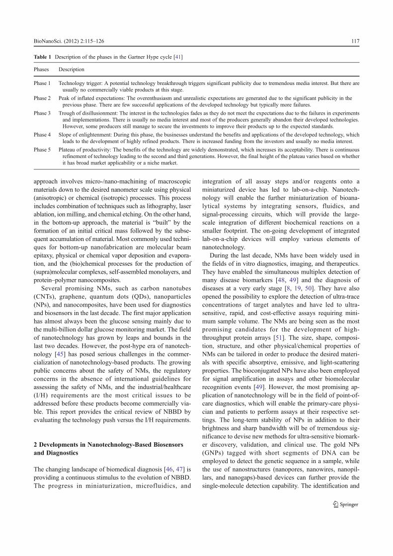
Table 1 Description of the phases in the Gartner Hype cycle [41]
Phases Description
Phase 1 Technology trigger: A potential technology breakthrough triggers significant publicity due to tremendous media interest. But there areusually no commercially viable products at this stage.
Phase 2 Peak of inflated expectations: The overenthusiasm and unrealistic expectations are generated due to the significant publicity in theprevious phase. There are few successful applications of the developed technology but typically more failures.
Phase 3 Trough of disillusionment: The interest in the technologies fades as they do not meet the expectations due to the failures in experimentsand implementations. There is usually no media interest and most of the producers generally abandon their developed technologies.However, some producers still manage to secure the investments to improve their products up to the expected standards.
Phase 4 Slope of enlightenment: During this phase, the businesses understand the benefits and applications of the developed technology, whichleads to the development of highly refined products. There is increased funding from the investors and usually no media interest.
Phase 5 Plateau of productivity: The benefits of the technology are widely demonstrated, which increases its acceptability. There is continuousrefinement of technology leading to the second and third generations. However, the final height of the plateau varies based on whetherit has broad market applicability or a niche market.
BioNanoSci. (2012) 2:115–126 117

characterization of single-stranded genomic DNA or RNAwithout amplification has already been shown.
NMs such as QDs and NPs are good imaging agents due totheir enhanced performance and functionality [52]. They canbe targeted to the specific disease sites in the body by conju-gating them to biomarker-specific vectors. The NMs-basedimaging agents provide additional information pertaining tothe physiology and function apart from the anatomical infor-mation, which enables more accurate and early disease diag-nosis, such as the highly sensitive detection of early stagecancer, thereby leading to better therapy. Similarly, the effec-tiveness of treatments can be monitored more rapidly andaccurately. The plasmonic NPs and drug delivery will beemployed for targeted therapeutics, where the first impactswill certainly be in treating cancer. The use of NPs improvesthe bioavailability and pharmacokinetics of therapeutics. Theytake the drugs directly to the target sites of disease in the bodyby avoiding exposure of healthy tissues, which increases theavailability of a drug at the target site and reduces the treat-ment dose. These developments in nanotechnology will behighly beneficial in shifting the late-stage diagnosis (involvingexpensive and socially burdensome treatment) to early-stagediagnosis (relatively less expensive and less invasive). Themost widely used NMs in NBBD are described below.
2.1 CNTs
During the past decade, CNTs have been one of the mostextensively used NMs in biosensors, diagnostics, tissueengineering, cell tracking and labeling, and delivery ofdrugs and biomolecules [15, 16, 53]. They are hollow cy-lindrical tubes composed of one, two, or several concentricgraphite layers capped by fullerenic hemispheres, which arereferred to as single-, double-, and multi-walled CNTs,respectively. They have unique structures, excellent electri-cal and mechanical properties, high thermal conductivity,high chemical stability, remarkable electrocatalytic activity,minimal surface fouling, low overvoltage, and high aspectratio (surface to volume). CNTs-based biosensors and diag-nostics have been employed for the highly sensitive detec-tion of analytes in healthcare, industries, environmentalmonitoring, and food quality analysis. They have beenpredominantly used in electrochemical sensing, mainly forglucose monitoring but also for the detection of fructose,galactose, neurotransmitters, neurochemicals, amino acids,immunoglobulin, albumin, streptavidin, insulin, human cho-rionic gonadotropin, C-reactive protein, cancer biomarkers,cells, microorganisms, DNA, and other biomolecules [54].
2.2 Graphene
Graphene, an atomically thin layer of sp2-hybridized carbon,is another most extensively used NM for diagnostics and
biosensors in the last few years due to its interesting andexciting properties, such as high mechanical strength, highthermal conductivity, high elasticity, tunable optical proper-ties, tunable band gap, very high room temperature electronmobility, and demonstration of the room temperature quantumHall effect. It is a transparent material with a very low pro-duction cost and low environmental impact. It has been ex-tensively employed in electrochemical, impedance,fluorescence, and electrochemiluminescence biosensors forthe detection of a wide range of analytes such as glucose,cytochrome c, NADH, hemoglobin, cholesterol, ascorbic ac-id, dopamine, uric acid, hydrogen peroxide, horseradish per-oxidase, catechol, DNA, heavy metal ions, and gases [55, 56].
2.3 QDs
QDs are inorganic nanocrystals, approximately 1–10 nm insize, with unique optical properties of broad excitation,narrow size-tunable emission spectra, high photochemicalstability, and negligible photobleaching. They have beenwidely used [57], mainly as alternatives to fluorophores,for the development of optical biosensors to detect ions,organic compounds, pharmaceutical analytes, and biomole-cules such as nucleic acids, proteins, amino acids, enzymes,carbohydrates, and neurotransmitters. They have also beenemployed for the in vivo detection of target sites in cancer.In fact, they are the ideal candidates for multiplexed opticalbioanalysis due to their ultra-high sensitivity, high specific-ity, cost effectiveness, miniaturized size, size-dependentemission wavelength, and rapid analyte detection [48].
2.4 NPs
NPs have also been extensively used in various bioanalyt-ical applications [58, 59], especially for the development ofbiosensors, diagnostics, imaging, drug delivery, and therapy,due to their unique optical and other properties. Theychange color in response to the binding of molecules totheir surface. The change in the properties of nanoparticlesby varying their size or shape has been exploited for variousbioanalytical applications.
The most widely used NPs are GNPs, which have a non-toxic, biocompatible, and inert core. The prominent plasmonabsorption and scattering properties of GNPs are highlyuseful for the early stage detection and photothermal therapyof cancer and other diseases. They have been used for thedevelopment of immunoassays, diagnostics, and biosensorsfor various analytes [60–66]. Based on their preferentialaccumulation at the tumor sites, they have been used forthe therapy of cancer and other diseases by acting as nano-carriers for the delivery of drugs, DNA, and genes. Themultivalent GNPs facilitate efficient drug delivery to thetarget sites by shielding the unstable drugs, while their
118 BioNanoSci. (2012) 2:115–126

strongly enhanced surface plasmon resonance absorptionenables the photothermal therapy of cancer. They have beenextensively used in imaging due to their enhancement of theRaman and Rayleigh signals that provide greater chemicalinformation. Therefore, it will be highly useful to combineall the benefits of GNPs, such as diagnostic, specific target-ing, and therapeutic, into a single multifunctional GNPs-based platform, which can be chemically tailored for aparticular disease.
Magnetic NPs are the second most widely used NPs,which have been extensively employed in biosensors anddiagnostics for the detection of proteins, enzymes, DNA,mRNA, drugs, metabolites, pathogens, and tumor cells.Various types of magnetic sensors based on different signaltransduction mechanisms, such as magnetic relaxationswitch assay sensors, magnetic particle relaxation sensors,and magnetoresistive sensors, have been developed [67].The diagnostic magnetic resonance (DMR) technology hasalso been employed extensively for magnetic biosensing[68]. The development of miniaturized chip-based nuclearmagnetic resonance detector (μNMR) has further enhancedthe capabilities of DMR for the highly sensitive analytedetection in microliter sample volumes, multiplex analysis,and development of cost-effective, portable, and high-throughput platforms for point-of-care diagnostics. Themagnetic NPs are being extensively used by industries suchas Phillips Research, Eindhoven, Netherlands for the devel-opment of immunoassays [69] and rapid integrated biosen-sor for multiplexed immunoassays [49].
2.5 Chitosan
Chitosan is one of the most promising NMs [70] for theintegration of biological components in medical devices[71] due to its excellent biocompatibility, complete biode-gradability, and non-toxic nature [72]. The degradationproducts of chitosan are harmless natural metabolites. It isobtained by the deacetylation of chitin, the second mostabundant natural polymer after cellulose, which is found inthe shells of crustaceans (crabs and shrimp), the cuticles ofinsects, and the cell walls of fungi. It is suitable for opticalsensors due to its transparent nature. It is also appropriate forelectrochemical sensors as the chitosan films are porous andhighly permeable to ions. The pH-dependent solubility ofchitosan enables the formation of stable films underneutral and basic pH conditions, whereas its aminegroups aid in the covalent binding of biomolecules andthe formation of nanocomposites with polymers or NPs.But it requires chemical modification such as carboxy-methylation to increase its solubility in water and othercommon solvents. It has been extensively used in bio-sensors, diagnostics, lab-on-a-chip devices, and other bio-medical or bioanalytical applications [70–72].
2.6 Dendrimers
Dendrimers are hyperbranched, monodispersed, star-shaped,and nanometer-scale three-dimensional macromolecules witha very high density of surface functional groups. They arecomposed of three distinct components, i.e., the core, theinterior dendron, and the exterior surface with terminal func-tional groups. They have been used extensively in variousbiosensors and diagnostics [73], such as those based on elec-trochemistry, fluorescence, surface enhanced Raman scatter-ing, impedimetry, and surface plasmon resonance, mainly asthey increase the analytical sensitivity, stability, and reproduc-ibility but reduce the non-specific interactions. They have alsobeen used for other bioanalytical applications [50, 74] such asdrug delivery, gene transfection, and catalysis.
2.7 Biological and Other NMs
Lipid vesicles, thin lipid films, and liposomes are biologicalNMs formed via the bottom-up nanotechnology approach.They have very similar composition to the cell membrane,being composed of phospholipids or other amphiphiles. Thebilayer lipid membrane structure provides a biomimeticenvironment for embedding the biocomponents, such asreceptors and proteins, under non-denaturing conditions.Due to their inherent biocompatibility, effective encapsula-tion of hydrophilic or hydrophobic drugs, and sensitivity topH and temperature, they have been used as drug-deliverycarriers for controlled drug release [75, 76] and for thedevelopment of biosensors and diagnostics [77]. The am-phiphilic nature allows them to spontaneously form orga-nized structures. They have been used for the amplificationof optical, electrochemical, and acoustic signals. Hybridnanoparticles composed of lipids and polydiacetylene(PDA) have been employed for the development of smartcolorimetric biosensors, where the externally induced con-formation change of PDA due to specific biomolecularinteractions results in remarkable blue-to-red chromatictransition [78]. This approach has been employed for therapid diagnosis of diseases, study of peptide–membraneinteractions, and the colorimetric screening of enzyme cata-lysts, antibacterial peptides, and physiological ions.
Besides these, other NMs (such as cellulose nanocrystals[79, 80]), biomolecules [81–83], and a wide range of nano-composites [84–86] with unique properties have also beenused. The nanoscale features of the bioanalytical platformshave also been modified for signal enhancement and betterassay sensitivity [87, 88]. Moreover, the tools and instrumentsbeing employed for nanoscale probing and manipulation havealso evolved. The Scanning Probe Microscope [89] that waspreviously used only for the topographical mapping/imagingof surfaces can now be employed to probe nanometer-localized electrical, optical, and nanomechanical properties
BioNanoSci. (2012) 2:115–126 119

[90], and to monitor interactions in real time. It has evolvedfrom a tool to a nanotechnology instrument for bottom-upnanofabrication [91] and for imaging biomolecule assem-blies, surfaces, and cells, both in ambient and liquid envi-ronment, with special modifications for the sensitivebiological surfaces [92]. Therefore, the last decade has seensignificant developments in nanotechnology and the contin-uously increased use of NMs in diagnostics and biosensors.
3 Technology Push versus Industrial/HealthcareRequirements
The numerous advances in NBBD have generated tremendoustechnology push, as evident from the exponentially increasednumber of publications (Fig. 1), patent applications, projects,and focused nanotechnology initiatives/themes. However, it isessential for the developed technologies to comply with I/Hrequirements in order to facilitate their market entry by gen-erating the desired market pull. Most of the nanotechnology-based products have only been demonstrated in the researchsettings and are devoid of extensive validation and trials inindustries and healthcare. There has been continuous decreasein the venture capital investment during the last few yearsmainly due to the relatively stagnant commercialization ofnanotechnology-based products and the growing public con-cerns about the safety of NMs. The critical I/H requirementsfor nanotechnology-based products are discussed here.
3.1 Reproducible and Cost-Effective Production
The reproducible and cost-effective production of NMs isthe most critical and in fact the preliminary requirement as itwill directly influence the reproducibility in biosensing anddiagnosis. The current state-of-the-art procedures have con-siderable variability in the production of NMs. However,there are substantial ongoing research efforts by researchersand industries in order to improve the production proceduresso that they can offer significant cost savings for some of theNMs. On the other hand, it is quite clear that the use of NMsin biosensors and diagnostics will certainly incur additionalcosts in comparison to the conventional procedures. There-fore, the cost-effective production of NMs and their analyt-ical benefits will be the determining factors in takingstrategic decisions pertaining to whether nanotechnology-based products can substantially score over the conventionalproducts in order to gain successful market entry.
3.2 Characterization
The NMs need to be more intensively characterized bydeveloping the right tools and techniques. The commerciallyavailable NMs such as CNTs, graphene, GNPs, and QDs are
characterized by routinely used analytical techniques suchas scanning electron microscopy, Raman spectroscopy,Fourier transform infrared spectroscopy, etc. But there is aneed for more stringent investigation of the properties ofNMs, which will provide highly useful information pertain-ing to the storage, functionalization, modification, and useof NMs under optimum conditions. Apart from the materialcharacterization, the nature of the metallic impurities, suchas those in the CNTs, also needs to be determined as it cansubstantially affect the properties as well as the toxicity ofNMs.
3.3 Material Safety
The material safety need to be evaluated individually for eachNM as they are unique [93]. When the NM is intended to beused for in vivo applications, the critical physiological param-eters such as absorption, distribution, metabolism, excretion,and toxicity should be determined. It has been demonstratedthat some NMs have prolonged tissue retention and may alsocontain heavy metals, which increases the risk of cytotoxicity.The toxicological studies of NMs also need to be done accord-ing to the established regulatory guidelines along with thedetermination of their efficacy so that the risk-to-benefitassessments could be done. However, in the absence of suchguidelines at the moment together with the lack of measure-ment tools and standard materials, the study of NMs’ toxicityand their environmental impact is quite challenging. The well-drafted guidelines by the National Institute for OccupationalSafety and Health for handling NPs should be followed for thedevelopment of new manufacturing processes to minimize theworkplace exposure risks. The risk assessment and risk man-agement paradigm for NPs, as described in Fig. 3, should alsobe considered.
3.4 Compliance with Regulatory Guidelines
The NBBD needs to adhere to strict health and safety pro-tocols, and regulatory guidelines so that the potential haz-ards of nanotechnology [94] can be effectively addressed.The field of nanotechnology has raised potential scientificand policy issues pertaining to the risk assessment andstandard setting, which has challenged the risk governanceand decision-making processes [95, 96]. Presently, there areintensive efforts in drafting the claims for the Nanotechnol-ogy Regulation [97], where the main objective is to makeclaims leading to the development of nanotechnology but atthe same time, evaluating critically the safety of nanotech-nology in terms of its effects on the public health and theenvironment. There have been many instances such as tet-raethyl lead and methyl tert-butyl ether, where the poten-tially hazardous technologies/products adversely affectingthe health and the environment were commercialized. The
120 BioNanoSci. (2012) 2:115–126

conventional direct regulation and self-regulation approaches,often referred to as hard and soft law, respectively, are beingexplored for making the claims of the Nanotechnology Reg-ulation. The conventional direct regulation is the “commandand control” regulation that involvesmaking prescriptive rulesto directly control the private sectors. On the other hand, self-regulation measures include the industrial codes of conduct,voluntary guidelines, or decision frameworks. The Environ-mental Defense–DuPont Nano Partnership Nano Risk Frame-work is an example of the self-regulation measure to evaluateand address the potential risk of NMs.
3.5 Correlation with Established Technology
The efficacy of the developed NBBD needs to be demonstrat-ed by correlating it with the established technologies, e.g.,their use as biosensor with the performance of state-of-the-artenzyme-linked assays, scanning probemicroscopy to standardimaging techniques such as electron microscopy (when itcomes to imaging NPs). This will enable the determinationof benefits (or drawbacks) of the developed nanotechnology-based products over the commercially available products,which will lend considerable support to their commercializa-tion. The requirement of technology correlation is becomingmore apparent to the researchers in nanotechnology as it hasbeen accepted as a norm by almost all scientific journals andinvestment/certification/regulatory agencies for evaluating thedeveloped technology. Thus, the researchers have already
started to address this concern and adopted it as a standardpractice in the development of NBBD.
3.6 Potential End-User Trials
Most of the NBBD are developed and evaluated in standardlaboratory settings using commercially available analytesamples. However, based on their intended applications,there is an absolute requirement for their validation andtesting in the actual end-user’s settings in industries andhealthcare using the “real world” samples. These end-usertrials will enhance the credibility and commercial appeal ofthe developed nanotechnology-based products by demon-strating their robustness under the actual conditions preva-lent at the end-user’s settings, where they will be finallyemployed. Presently, this is a serious limitation for most ofthe nanotechnology researchers as it requires significantfunding and efforts.
3.7 Toxicity Analysis
There are growing public concerns about the potential tox-icity of NMs, especially for in vivo biomedical applications,due to their ultra-small size and unique properties. Most ofthese concerns are fueled by fundamental misconceptionsabout NMs and nanotechnology, where the risk of nanotech-nology has been exaggerated. But these can be effectivelyaddressed with information outreach if the scientific
Fig. 3 Risk assessment and risk management paradigm for nanoparticles [99]. Reprinted with permission from Environmental Health Perspectives
BioNanoSci. (2012) 2:115–126 121

community can clearly demonstrate the safety of NMs fordiagnostics and biosensors before exploiting them commer-cially. This will abridge the information gap between thescientific community and public, which will lend consider-able support to the acceptance of developed technology andits commercialization. There are ongoing research efforts todetermine the toxicological profiles and potential adverseeffects of NMs [98–102], understand their biological inter-action mechanisms [103], develop robust and widely accept-able analytical tools and tests for characterizing NMs invarious environments [104], and to determine the safety ofNM throughout its life cycle, i.e., research and development,production, use, disposal, and/or recycle. The perceived andreal adverse impacts of NMs need to be effectivelyaddressed lest they become barriers for the future technolo-gy development.
The toxicity of NMs depends on numerous factors.The toxicity of CNTs depends on their dimensions, im-purities, surface chemistry, dispersion, type, dose, and theinteraction between various factors [105]. Similarly, theinherent toxicity of QDs that is made up of toxic materi-als combined with their clearance problems are the majorfactors why QDs have also not been approved for invivo applications. Although the biocompatible QDs havealso been demonstrated to prevent toxicity, they are veryexpensive. Despite the tremendous research efforts devot-ed to the use of chitosan during the last two decades, itis still not approved by the Food and Drug Administra-tion (FDA) for drug delivery, which has led to diminish-ing interest of biotech companies. The side effectsresulting from the toxicity of GNPs have also beendemonstrated. But these can be diminished by devisingnew procedures of functionalizing GNPs with compoundsthat enhance their biocompatibility and clearance. The
known and expected NPs exposure and clearance routesare shown in Fig. 4. A tiered testing system to assess NPtoxicity was suggested [106], where the physico-chemicalcharacterization needs to be done prior to and duringsubsequent testing in cell-free, cellular, and in vivoassays. However, it is difficult to predict the in vivotoxicity from the in vitro assays.
3.8 Evaluating the Need of NMs
A wide range of NMs have been employed in biosensorsand diagnostics mainly due to their demonstrated benefitssuch as increased signal, higher analytical sensitivity,lower limit of detection, and better analytical character-istics. As an example, most of the nanotechnology-basedconcepts have been initially applied to blood glucosemonitoring due to its enormous market potential. How-ever, it is well known to the industries and experts in thefield nowadays that the use of NMs does not provideany analytical advantage or cost effectiveness in compar-ison to the commercially available blood glucose moni-toring devices. It only leads to increased signal and, insome cases, greater detection range and/or no require-ment of external mediator. The increased detection rangebeyond the pathophysiological glucose range in diabeticsis of no real use. On the contrary, the use of NMsincreases the manufacturing cost, complexity, andhands-on time, while reducing the production and func-tional reproducibility. This is the main reason that despitethe numerous publications and patents pertaining to theuse of NMs for blood glucose monitoring [54, 56, 107],none of the nanotechnology-based concepts has actuallybeen taken up by the industries for commercialization.On the other hand, the simplified technologies that do
Fig. 4 Expected and known nanoparticle exposure and clearance routes [99]. Reprinted with permission from Environmental Health Perspectives
122 BioNanoSci. (2012) 2:115–126
not use NMs but increase the analytical performance andcost effectiveness are more readily adopted for glucose mon-itoring and other bioanalytical applications [108–110]. Therehas been lot of hype around the use of NMs in the last decadethat resulted in unrealistic hopes [111]. The academicresearchers have been forced to include elements of nanotech-nology for the sole reason of securing research funding [112].However, the recent years have seen a change in this trend asmore focus is now provided to the improvement of bioanalyt-ical applications. Therefore, the need of using NMs for aparticular application should be critically assessed by thetechnology and business experts.
4 Conclusions and Future Trends
The significant advances in field of NBBD in the last twodecades have generated tremendous technology push. Ini-tially, most of these developments were motivated by hype,which led to inflated expectations and the inclined trend toemploy nanotechnology-based concepts and devices. How-ever, the field of nanotechnology has now progressed pastthe peak of hype, where increased attention is being paid tothe toxicological and environmental effects of NMs. Thepost-hype era is mainly focused on determining the safety ofNMs, arriving at the international regulatory guidelines forassessing the safety of NMs, and determining the robustnessof NBBD in accordance with I/H requirements. The exten-sive benefits of employing NMs for biosensors and diag-nostics have been widely demonstrated and are well knownto the scientific community worldwide. The field of NBBDhas progressed to the “slope of enlightenment” phase of theGartner Hype cycle. However, extensive research efforts arestill required to critically investigate the production repro-ducibility, analytical parameters, and the safety of NMs. Thefinalization of international regulatory guidelines for assess-ing the safety of NMs, which is the topmost priority and infull swing at the moment, will provide the much-neededmomentum to this field. There is a critical need for thedeveloped technologies to meet I/H requirements in order
to become commercially viable. However, the commercialsuccess of the developed NBBD will be determined by thekey technology differentiators, cost effectiveness, reliability,and the generated market pull.
The interdisciplinary nature of nanotechnology is amajor challenge in itself as it is difficult to find theexpertise in all the fields at a particular setup or group.Therefore, the technical data pertaining to the applica-tions of nanotechnology-based products in a particularfield need to be critically reviewed by the experts inthat field. As an example, most of the published reportshave shown the intracellular delivery of nanoparticles tocells that were dead and permeable, while these studiesshould have been conducted in healthy cells to obtainaccurate results.
Presently, many companies are investing their time, mon-ey, and efforts on the development of procedures for theproduction of reproducible, stable, and biocompatible NMs.The researchers have also started the testing of NMs-basedbiosensors and diagnostics on “real world” samples, whichprovides much better understanding of sample matrix effectsand the highly useful information about the effects of phys-iological interferences. Similarly, various modifications ofNMs have been demonstrated to reduce the NM’s toxicityand make them biocompatible.
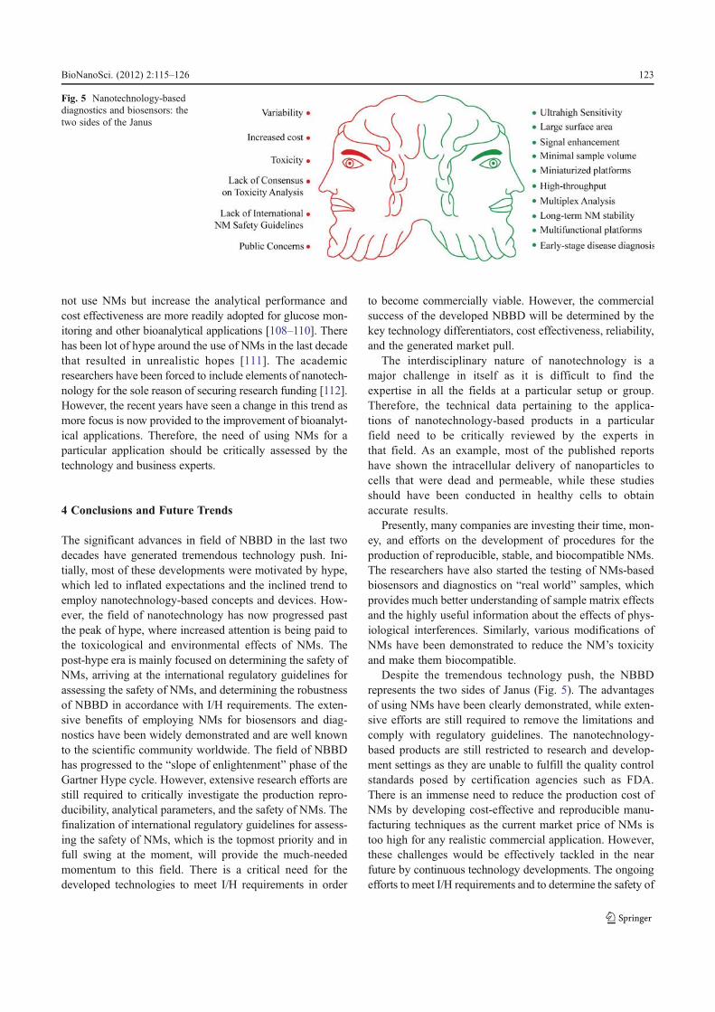
Despite the tremendous technology push, the NBBDrepresents the two sides of Janus (Fig. 5). The advantagesof using NMs have been clearly demonstrated, while exten-sive efforts are still required to remove the limitations andcomply with regulatory guidelines. The nanotechnology-based products are still restricted to research and develop-ment settings as they are unable to fulfill the quality controlstandards posed by certification agencies such as FDA.There is an immense need to reduce the production cost ofNMs by developing cost-effective and reproducible manu-facturing techniques as the current market price of NMs istoo high for any realistic commercial application. However,these challenges would be effectively tackled in the nearfuture by continuous technology developments. The ongoingefforts to meet I/H requirements and to determine the safety of
Fig. 5 Nanotechnology-baseddiagnostics and biosensors: thetwo sides of the Janus
BioNanoSci. (2012) 2:115–126 123

NMs will generate the desired market pull, which will boostthe commercialization of NBBD.
Acknowledgments We acknowledge the financial support receivedfrom EU-FP7 Health and EU-FP7 ICT for the project grant numbers258759 and 318408, respectively. K.M. would also like to acknowl-edge the financial support received from the Alexander von HumboldtFoundation.
References
1. Leary, J. F. (2010). Nanotechnology: what is it and why is smallso big? Canadian Journal of Ophthalmology, 45, 449–456.
2. Weiss, P. S. (2010). Nanoscience and nanotechnology: presentand future. ACS Nano, 4, 1771–1772.
3. Jiarong, C., Yuqing, M., Nongyue, H., Xiaohua, W., Sijiao, L.(2004). Nanotechnology and biosensors. Biotechnology Advan-ces, 22, 505–518.
4. Vaddiraju, S., Tomazos, I., Burgess, D. J., Jain, F. C.,Papadimitrakopoulos, F. (2010). Emerging synergy betweennanotechnology and implantable biosensors: a review. Biosen-sors and Bioelectronics, 25, 1553–1565.
5. Cheng, M. M. C., Cuda, G., Bunimovich, Y. L., et al. (2006).Nanotechnologies for biomolecular detection and medical diag-nostics. Current Opinion in Chemical Biology, 10, 11–19.
6. Hauck, T. S., Giri, S., Gao, Y., Chan, W. C. W. (2010). Nano-technology diagnostics for infectious diseases prevalent in devel-oping countries. Advance Drug Delaware Review, 62, 438–448.
7. Sosnik, A., & Amiji, M. (2010). Nanotechnology solutions forinfectious diseases in developing nations. Advance Drug Dela-ware Review, 62, 375–377.
8. Kim, P. S., Djazayeri, S., Zeineldi, R. (2011). Novel nanotech-nology approaches to diagnosis and therapy of ovarian cancer.Gynecologic Oncology, 120, 393–403.
9. Stylios, G. K., Giannoudis, P. V., Wan, T. (2005). Applications ofnanotechnologies in medical diagnostics. Injury, InternationalJournal of the Care of the Injured, 36S, S6–S13.
10. Fournier-Wirth, C., Coste, J. (2010). Nanotechnologies for path-ogen detection: future alternatives? Biologicals, 38, 9–13.
11. Ansari, A. A., Alhoshan, M., Alsalhi, M. S., Aldwayyan, A. S.(2010). Prospects of nanotechnology in clinical immunodiagnos-tics. Sensors, 10, 6535–6581.
12. Jain, K. K. (2005). Nanotechnology in clinical laboratory diag-nostics. Clinica Chimica Acta, 358, 37–54.
13. Thomas, C. R., George, S., Horst, A. M., et al. (2011). Nano-materials in the environment: from materials to high-throughputscreening to organisms. ACS Nano, 5, 13–20.
14. Shi, J., Votruba, A. R., Farokhzad, O. C., Langer, R. (2010).Nanotechnology in drug delivery and tissue engineering: fromdiscovery to applications. Nano Letters, 10, 3223–3230.
15. Vashist, S. K., Zheng, D., Pastorin, G., Al-Rubeaan, K., Luong, J.H. T., Sheu, F. S. (2011). Delivery of drugs and biomoleculesusing carbon nanotubes. Carbon, 49, 4077–4097.
16. Li, J., Yap, S. Q., Yoong, S. L., et al. (2012). Carbon nanotubebottles for incorporation, release and enhanced cytotoxic effect ofcisplatin. Carbon, 50, 1625–1634.
17. Moghimi, S. M., Peer, D., Langer, R. (2011). Reshaping thefuture of nanopharmaceuticals: ad ludicum. ACS Nano, 5,8454–8458.
18. Kim, K. Y. (2007). Nanotechnology platforms and physiologicalchallenges for cancer therapeutics. Nanomedicine: Nanotechnol-ogy, Biology, and Medicine, 3, 103–110.
19. Misra, R., Acharya, S., Sahoo, S. K. (2010). Cancer nanotech-nology: application of nanotechnology in cancer therapy. DrugDiscovery Today, 15, 842–850.
20. Kawasaki, E. S., & Player, A. (2005). Nanotechnology, nano-medicine, and the development of new, effective therapies forcancer. Nanomedicine: Nanotechnology, Biology, and Medicine,1, 101–109.
21. Farokhzad, O. C., & Langer, R. (2006). Nanomedicine: develop-ing smarter therapeutic and diagnostic modalities. Adv Drug DelRev, 58, 1456–1459.
22. Phan, J. H., Moffitt, R. A., Stokes, T. H., Liu, J., Young, A. N.,Nie, S., et al. (2009). Convergence of biomarkers, bioinformaticsand nanotechnology for individualized cancer treatment. Trendsin Biotechnology, 27, 350–358.
23. Yan, Y., Such, G. K., Johnston, A. P. R., Best, J. P., Caruso, F.(2012). Engineering particles for therapeutic delivery: prospectsand challenges. ACS Nano. doi:10.1021/nn3016162.
24. Fortina, P., Kricka, L. J., Bonnell, D., Kulkarni, A., Wang, J.,Miyahara, Y., et al. (2010). Nanotechnology: improving clinicaltesting? Clinical Chemistry, 56, 1384–1389.
25. Zarbin, M. A., Montemagno, C., Leary, J. F., Ritch, R. (2010).Nanotechnology in ophthalmology. Canadian Journal of Oph-thalmology, 45, 457–476.
26. Re, F., Gregori, M., Masserini, M. (2012). Nanotechnology forneurodegenerative disorders. Maturitas . doi:10.1016/j.maturitas.2011.12.015.
27. Sahoo, S. K., Parveen, S., Panda, J. J. (2007). The present andfuture of nanotechnology in human health care. Nanomedicine:Nanotechnology, Biology, and Medicine, 3, 20–31.
28. Brambilla, D., Droumaguet, B. L., Nicolas, J., et al. (2011).Nanotechnologies for Alzheimer’s disease: diagnosis, therapy,and safety issues. Nanomedicine: Nanotechnology, Biology, andMedicine, 7, 521–540.
29. Farrell, D., Alper, J., Ptak, K., Panaro, N. J., Grodzinski, P.,Barker, A. D. (2010). Recent advances from the National CancerInstitute Alliance for Nanotechnology in Cancer. ACS Nano, 4,589–594.
30. Retél, V. P., Hummel, M. J. M., Harten, W. H. V. (2009). Reviewon early technology assessments of nanotechnologies in oncolo-gy. Molecular Oncology, 3, 394–401.
31. Boisseau, P., & Loubaton, B. (2011). Nanomedicine, nanotech-nology in medicine. Comptes Rendus Physique, 12, 620–636.
32. Sawhney, A. P. S., Condon, B., Singh, K. V., Pang, S. S., Li, G.,Hui, D. (2008). Modern applications of nanotechnology in tex-tiles. Textile Research Journal, 78, 731–739.
33. Duncan, T. V. (2011). Applications of nanotechnology in foodpackaging and food safety: barrier materials, antimicrobials andsensors. Journal of Colloid and Interface Science, 363, 1–24.
34. Sakamoto, J. H., Ven, A. L. V. D., Godin, B., et al. (2010).Enabling individualized therapy through nanotechnology. Phar-macological Research, 62, 57–89.
35. Bonnell, D. (2010). The next decade of nanoscience and nano-technology. ACS Nano, 4, 6293–6294.
36. http://www.wtec.org/nano2/37. Gabellieri, C. (2011). Nanomedicine in the European Commis-
sion policy for nanotechnology. Nanomedicine: Nanotechnology,Biology, and Medicine, 7, 519–520.
38. Horton, M. A., & Khan, A. (2006). Medical nanotechnologyin the UK: a perspective from the London Centre for Nano-technology. Nanomedicine: Nanotechnology, Biology, andMedicine, 2, 42–48.
39. Pandza, K., Wilkins, T. A., Alfoldi, E. A. (2011). Collaborativediversity in a nanotechnology innovation system: evidence fromthe EU framework programme. Technovation, 31, 476–489.
40. Allarakhia, M., & Walsh, S. (2012). Analyzing and organizingnanotechnology development: application of the institutional
124 BioNanoSci. (2012) 2:115–126

analysis development framework to nanotechnology consortia.Technovation, 32, 216–226.
41. http://www.gartner.com/technology/research/methodologies/hype-cycle.jsp
42. Service RF. (2001). Breakthrough of the year: molecules getwired. Science, 294, 2442–2443.
43. Service RF. (2002). Bell Labs fires star physicist found guilty offorging data. Science, 298, 30–31.
44. http://www.sciencemag.org/content/298/5595/961.245. Hersam, M. (2011). Nanoscience and nanotechnology in the
posthype era. ACS Nano, 5, 1–2.46. Gubala, V., Harris, L. F., Ricco, A. J., Tan, M. X., Williams, D. E.
(2012). Point of care diagnostics: status and future. AnalyticalChemistry, 84, 487–515.
47. Rasooly, A. (2006). Moving biosensors to point-of-care cancerdiagnostics. Biosensors and Bioelectronics, 21, 1847–1850.
48. Frasco, M. F., & Chaniotakis, N. (2010). Bioconjugated quantumdots as fluorescent probes for bioanalytical applications. Analyt-ical and Bioanalytical Chemistry, 396, 229–240.
49. Bruls, D. M., Evers, T. H., Kahlman, J. A. H., et al. (2009). Rapidintegrated biosensor for multiplexed immunoassays based onactuated magnetic nanoparticles. Lab on a Chip, 9, 3504–3510.
50. Cheng, Y., Zhao, L., Li, Y., Xu, T. (2011). Design of biocompat-ible dendrimers for cancer diagnosis and therapy: current statusand future perspectives. Chemical Society Reviews, 40, 2673–2703.
51. Ghazani, A. A., Lee, J. A., Klostranec, J., et al. (2006). Highthroughput quantification of protein expression of cancer antigensin tissue microarray using quantum dot nanocrystals. Nano Let-ters, 6, 2881–2886.
52. Kairemo, K., Erba, P., Bergström, K., Pauwels, E. K. J. (2008).Nanoparticles in cancer. Current Radiopharmaceuticals, 1, 30–36.
53. Bianco A., Kostarelos K., Partidos C.D., Prato M. (2005). Bio-medical applications of functionalised carbon nanotubes. Chem-ical Communication, 571–577
54. Vashist, S. K., Zheng, D., Al-Rubeaan, K., Luong, J. H. T., Sheu,F. S. (2011). Advances in carbon nanotube based electrochemicalsensors for bioanalytical applications. Biotechnology Advances,29, 169–188.
55. Dresselhaus, M. S., & Araujo, P. T. (2010). The 2010 Nobel Prizein physics for graphene: some perspectives. ACS Nano, 4, 6297–6302.
56. Zheng, D., Vashist, S.K., Luong, J.H.T., Al-Rubeaan, K., Sheu,F.S. (2012). Amperometric glucose biosensing using 3-aminopropyltriethoxysilane functionalized graphene. Talanta,doi:10.1016/j.talanta.2012.05.014.
57. Azzazy, H. M. E., Mansour, M. M. H., Kazmierczak, S. C.(2007). From diagnostics to therapy: prospects of quantum dots.Clinical Biochemistry, 40, 917–927.
58. Parveen, S., Misra, R., Sahoo, S. K. (2012). Nanoparticles: aboon to drug delivery, therapeutics, diagnostics and imaging.Nanomedicine: Nanotechnology, Biology, and Medicine, 8,147–166.
59. Fan, Z., Shelton, M., Singh, A. K., Senapati, D., Khan, S. A.,Ray, P. C. (2012). Multifunctional plasmonic shell–magnetic corenanoparticles for targeted diagnostic, isolation, and photothermaldestruction of tumor cells. ACS Nano, 6, 1065–1073.
60. Boisselier, E., & Astruc, D. (2009). Gold nanoparticles in nano-medicine: preparations, imaging, diagnostics, therapies and tox-icity. Chemical Society Reviews, 38, 1759–1782.
61. Dreaden, E. C., Alkilany, A. M., Huang, X., Murphy, C. J., El-Sayed, M. A. (2012). The golden age: gold nanoparticles forbiomedicine. Chemical Society Reviews, 41, 2740–2779.
62. Dykman, L., & Khlebtsov, N. (2012). Gold nanoparticles inbiomedical applications: recent advances and perspectives.Chemical Society Reviews, 41, 2256–2282.
63. Misiakos, K., Kakabakos, S. E., Petrou, P. S., Ruf, H. H. (2004).A monolithic silicon optoelectronic transducer as a real-timeaffinity biosensor. Analytical Chemistry, 76, 1366–1373.
64. Weizmann, Y., Patolsky, F., Willner, I. (2001). Amplified detectionof DNA and analysis of single-base mismatches by the catalyzeddeposition of gold on Au-nanoparticles. Analyst, 126, 1502–1504.
65. Rosi, N. L., & Mirkin, C. A. (2005). Nanostructures in biodiag-nostics. Chemical Reviews, 105, 1547–1562.
66. Lee, K., Drachev, V. P., Irudayaraj, J. (2011). DNA–gold nano-particle reversible networks grown on cell surface marker sites:application in diagnostics. ACS Nano, 5, 2109–2117.
67. Koh, I., & Josephson, L. (2009). Magnetic nanoparticle sensors.Sensors, 9, 8130–8145.
68. Haun, J. B., Yoon, T.-J., Lee, H., Weissleder, R. (2010). Magneticnanoparticle biosensors. WIREs Nanomedicine Nanobiotechnol-ogy, 2, 291–304.
69. Dittmer, W. U., de Kievit, P., Prins, M. W. J., Vissers, J. L. M.,Mersch, M. E. C., Martens, M. F. W. C. (2008). Sensitive andrapid immunoassay for parathyroid hormone using magnetic par-ticle labels and magnetic actuation. Journal of ImmunologicalMethods, 338, 40–46.
70. Sashiwa, H., & Aiba, S.-I. (2004). Chemically modified chitinand chitosan as biomaterials. Progress in Polymer Science, 29,887–908.
71. Koev, S. T., Dykstra, P. H., Luo, X., Rubloff, G. W., Bentley, W.E., Payne, G. F., et al. (2010). Chitosan: an integrative biomaterialfor lab-on-a-chip devices. Lab on a Chip, 10, 3026–3042.
72. Kean, T., & Thanou, M. (2010). Biodegradation, biodistributionand toxicity of chitosan. Adv Drug Del Rev, 62, 3–11.
73. Satija, J., Sai, V. V. R., Mukherji, S. (2011). Dendrimers inbiosensors: concepts and applications. Journal of MaterialsChemistry, 21, 14367–14386.
74. Shen, M., & Shi, X. (2010). Dendrimer-based organic/inorganichybrid nanoparticles in biomedical applications. Nanoscale, 2,1596–1610.
75. Dennis, M., Vriezema, D. M., Aragonès, M. C., Elemans, J. A. A.W., Cornelissen, J. J. L. M., Rowan, A. E., et al. (2005). Self-assembled nanoreactors. Chemical Reviews, 105, 1445–1489.
76. Malam, Y., Loizidou, M., Seifalian, A. M. (2009). Liposomes andnanoparticles: nanosized vehicles for drug delivery in cancer.Trends in Pharmacological Sciences, 30, 592–599.
77. Christensen, S. M., & Stamou, D. (2007). Surface-based lipidvesicle reactor systems: fabrication and applications. Soft Matter,3, 828–836.
78. Jelinek, R., & Kolusheva, S. (2007). Biomolecular sensing withcolorimetric vesicles. Topics in Current Chemistry, 277, 155–180.
79. Leung, A. C. W., Hrapovic, S., Lam, E., Liu, Y., Male, K. B.,Mahmoud, K. A., et al. (2011). Characteristics and properties ofcarboxylated cellulose nanocrystals prepared from a novel one-step procedure. Small, 7, 302–305.
80. Lam, E., Male, K. B., Chong, J. H., Leung, A. C. W., Luong, J. H.T. (2012). Applications of functionalized and nanoparticle-modified nanocrystalline cellulose. Trends in Biotechnology, 30,283–290.
81. Shukla, G. C., Haque, F., Tor, Y., et al. (2011). A boost for theemerging field of RNA nanotechnology. ACS Nano, 5, 3405–3418.
82. Modi, S., Bhatia, D., Simmel, F. C., Krishnan, Y. (2010). Struc-tural DNA nanotechnology: from bases to bricks, from structureto function. Journal of Physical Chemistry Letters, 1, 1994–2005.
83. Campolongo, M. J., Tan, S. J., Xu, J., Luo, D. (2010). DNAnanomedicine: engineering DNA as a polymer for therapeutic anddiagnostic applications. Adv Drug Del Rev, 62, 606–616.
84. Xiao, Y., & Li, C. M. (2008). Nanocomposites: from fabricationsto electrochemical bioapplications. Electroanalytical, 20, 648–662.
BioNanoSci. (2012) 2:115–126 125
85. Hussain, F., Hojjati, M., Okamoto, M., Gorga, R. E. (2006).Polymer–matrix nanocomposites, processing, manufacturing,and application: an overview. Journal of Composite Materials,40, 1511–1575.
86. Rajesh, A. T., & Kumar, D. (2009). Recent progress in thedevelopment of nano-structured conducting polymers/nanocom-posites for sensor applications. Sensors and Actuators B: Chem-istry, 136, 275–286.
87. Dixit, C. K., & Kaushik, A. (2012). Nano-structured arrays formultiplex analyses and lab-on-a-chip applications. Biochemicaland Biophysical Research Communications, 419, 316–320.
88. Dixit, C.K., Kumar, A., Kaushik, A. (2012). Nanospherelithography-based platform for developing rapid and high sensi-tivity microarray systems. Biochemistry and Biophysics ResearchCommunications. doi:10.1016/j.bbrc.2012.05.144.
89. Binnig, G., Quate, C. F., Gerber, C. (1986). Atomic force micro-scope. Physical Review Letters, 56, 930–933.
90. http://www.ntmdt.com/spm-principles91. Mitsakakis, K., Sekula-Neuner, S., Lenhert, S., Fuchs, H., Gizeli,
E. (2012). Convergence of Dip-Pen Nanolithography and acous-tic biosensors towards a rapid-analysis multi-sample microsys-tem. Analyst, 137, 3076–3082.
92. Mitsakakis, K., Lousinian, S., Logothetidis, S. (2007). Earlystages of human plasma proteins adsorption probed by atomicforce microscope. Biomolecular Engineering, 24, 119–124.
93. Florence, A. T. (2004). The dangers of generalization in nano-technology. Drug Discovery Today, 9, 60–61.
94. Türk, V., Kaiser, C., Schaller, S. (2008). Invisible but tangible?Societal opportunities and risks of nanotechnologies. Journal ofCleaner Production, 16, 1006–1009.
95. Wiek, A., Lang, D. J., Siegrist, M. (2008). Qualitative systemanalysis as a means for sustainable governance of emergingtechnologies: the case of nanotechnology. Journal of CleanerProduction, 16, 988–999.
96. Novak, P. J., Arnold, W. A., Blazer, V. S., et al. (2011). On theneed for a national (U.S.) research program to elucidate thepotential risks to human health and the environment posed bycontaminants of emerging concern. Environmental Science andTechnology, 45, 3829–3830.
97. Malloy, T. F. (2011). Nanotechnology regulation: a study inclaims making. ACS Nano, 5, 5–12.
98. Sharifi, S., Behzadi, S., Laurent, S., Forrest, M. L., Stroeve, P.,Mahmoudi, M. (2012). Toxicity of nanomaterials. Chemical So-ciety Reviews, 41, 2323–2343.
99. Oberdörster, G., Oberdörster, E., Oberdörster, J. (2005). Nano-toxicology: an emerging discipline evolving from studies of
ultrafine particles. Environmental Health Perspectives, 113,823–839.
100. Oberdörster, G. (2005). Safety assessment for nanotechnologyand nanomedicine: concepts of nanotoxicology. Journal of Inter-nal Medicine, 267, 89–105.
101. Holl, M. M. B. (2009). Nanotoxicology: a personal perspective.WIREs Nanomedicine and Nanobiotechology, 1, 353–359.
102. Hutchison, J. E. (2008). Greener nanoscience: a proactive ap-proach to advancing applications and reducing implications ofnanotechnology. ACS Nano, 2, 395–402.
103. Leroueil, P. R., Hong, S., Mecke, A., Baker, J. R., Orr, B. G.,Holl, M. M. B. (2007). Nanoparticle interaction with biologicalmembranes: does nanotechnology present a Janus face? Accountsof Chemical Research, 40, 335–342.
104. Marquis, B. J., Love, S. A., Braun, K. L., Haynes, C. L. (2009).Analytical methods to assess nanoparticle toxicity. Analyst, 134,425–439.
105. Cui, H. F., Vashist, S. K., Al-Rubeaan, K., Luong, J. H. T., Sheu,F. S. (2010). Interfacing carbon nanotubes with living mammali-an cells and cytotoxicity issues. Chemical Research in Toxicolo-gy, 23, 1131–1147.
106. Oberdörster, G., Maynard, A., Donaldson, K., ILSI ResearchFoundation/Risk Science Institute Nanomaterial Toxicity Screen-ing Working Group, et al. (2005). Principles for characterizingthe potential human health effects from exposure to nanomateri-als: elements of a screening strategy. Particle and Fibre Toxicol-ogy, 2, 8. doi:10.1186/1743-8977-2-8.
107. Cash, K. J., & Clark, H. A. (2010). Nanosensors and nanomate-rials for monitoring glucose in diabetes. Trends in MolecularMedicine, 16, 584–593.
108. Zheng, D., Vashist, S. K., Al-Rubeaan, K., Luong, J. H. T., Sheu,F. S. (2012). Rapid and simple preparation of a reagentlessglucose electrochemical biosensor. Analyst. doi:10.1039/C2AN35128E.
109. Dixit, C. K., Vashist, S. K., O’Neill, F. T., O’Reilly, B., MacCraith,B. D., O’Kennedy, R. (2010). Development of a high sensitivityrapid sandwich ELISA procedure and its comparison with theconventional approach. Analytical Chemistry, 82, 7049–7052.
110. Dixit, C. K., Vashist, S. K., MacCraith, B. D., O’Kennedy, R.(2011). Multi-substrate compatible ELISA procedures for rapidand high sensitivity immunoassays. Nature Protocols, 6, 439–445.
111. Kostarelos, K., Bianco, A., Prato, M. (2008). Hype around nano-tubes creates unrealistic hopes. Nature, 453, 280.
112. Kotov, N. A. (2009). Politics and nanotechnology in the healthcare industry. ACS Nano, 3, 2855–2856.
126 BioNanoSci. (2012) 2:115–126