nano express open access nanomechanical characterization of
TRANSCRIPT

Lee et al. Nanoscale Research Letters 2012, 7:608http://www.nanoscalereslett.com/content/7/1/608
NANO EXPRESS Open Access
Nanomechanical characterization of chemicalinteraction between gold nanoparticles andchemical functional groupsGyudo Lee1,2, Hyungbeen Lee2, Kihwan Nam1,2, Jae-Hee Han3, Jaemoon Yang4, Sang Woo Lee2, Dae Sung Yoon2,Kilho Eom1,2 and Taeyun Kwon1,2*
Abstract
We report on how to quantify the binding affinity between a nanoparticle and chemical functional group usingvarious experimental methods such as cantilever assay, PeakForce quantitative nanomechanical property mapping,and lateral force microscopy. For the immobilization of Au nanoparticles (AuNPs) onto a microscale siliconsubstrate, we have considered two different chemical functional molecules of amine and catecholamine (here,dopamine was used). It is found that catecholamine-modified surface is more effective for the functionalization ofAuNPs onto the surface than the amine-modified surface, which has been shown from our various experiments.The dimensionless parameter (i.e., ratio of binding affinity) introduced in this work from such experiments is usefulin quantitatively depicting such binding affinity, indicating that the binding affinity and stability between AuNPsand catecholamine is approximately 1.5 times stronger than that between amine and AuNPs. Our study sheds lighton the experiment-based quantitative characterization of the binding affinity between nanomaterial and chemicalgroups, which will eventually provide an insight into how to effectively design the functional material usingchemical groups.
Keywords: Au nanoparticle, Dopamine, Surface chemistry, Atomic force microscopy, Lateral force microscopy
BackgroundSurface chemistry has played a critical role in designingfunctional nanomaterials for their biological or medicalapplications such as drug delivery, molecular therapeu-tics, and diagnostics [1,2]. In particular, the surfacemodification of a nanoparticle is of great importance toenhancing functionality in terms of target affinity [3-5],imaging contrast [3,4,6,7], and curative power [8]. Forinstance, magnetic nanoparticles chemically modifiedwith chemical functional groups or moieties (e.g., ligandand receptor) have been utilized for high-resolutionMRI, which is useful in cancer diagnostics since thechemical modification using chemical functional groupsor moieties leads to improved targetability and imagingcontrasts [3,6,7]. Moreover, gold nanoparticles (AuNPs)
* Correspondence: [email protected] for Molecular Sciences, Seoul 120-749, Republic of Korea2Department of Biomedical Engineering, Yonsei University, Wonju 220-710,Republic of KoreaFull list of author information is available at the end of the article
© 2012 Lee et al.; licensee Springer. This is an OAttribution License (http://creativecommons.orin any medium, provided the original work is p
functionalized with chemical functional groups or moi-eties have been recently used to enhance photocatalyticperformance [9], to form 3D networks of functionalizedAuNPs [10], and to sensitively detect specific biologicalmolecules (e.g., DNA) [11-13] and cancerous single cells[14,15].Dopamine hydrochloride (DOPA) has recently been
considered as a chemical linker that allows for efficientsurface chemistry useful in not only inorganic materials(e.g., nanoparticles) but also biological materials (e.g., tis-sue) due to its excellent adhesive property and biocom-patibility [16,17]. In particular, DOPA has been reportedas a chemical linker that is useful not only in the chem-ical modification of the surfaces of nanomaterials suchas nanoparticles [18,19], graphene oxide sheet [20], andcarbon nanotubes [21], but also in improving bindingaffinities such as protein-peptide cross-linking [22], cel-lular adhesion to substrate [23], osteoconduction [24],and hemostatic adhesive in segmentectomy [25]. Despitethe broad application of DOPA to surface chemistry
pen Access article distributed under the terms of the Creative Commonsg/licenses/by/2.0), which permits unrestricted use, distribution, and reproductionroperly cited.

Lee et al. Nanoscale Research Letters 2012, 7:608 Page 2 of 11http://www.nanoscalereslett.com/content/7/1/608
using mutual interaction between DOPA and nanomater-ials (e.g., nanoparticle), such an interaction has been poorlyunderstood and not yet studied thoroughly. Since the sur-face modification of nanomaterials using DOPA typicallyemploys a noncovalent conjugation (e.g., coordinate bond-ing, hydrophobic and electrostatic interactions, etc.) [6,26],it is essential to establish an experimental framework thatallows for measuring a weak binding affinity correspondingto such a noncovalent conjugation, which is useful in thedevelopment of drug carrier due to the fact that noncova-lent conjugation enables the excretion of waster matterfrom the human body after the drug carrier completes thefunction of drug delivery or bioimaging [6,7,27,28].In this work, we have quantitatively studied a chem-
ical interaction between nanoparticles and chemicalfunctional groups (e.g., DOPA and amine functionalgroup) using experimental toolkits such as cantileverbioassay [29-32], PeakForce Quantitative Nanomechani-cal Property Mapping (PeakForce QNM) [33,34], aswell as lateral force microscopy (LFM) [35-38]. In a re-cent decade, cantilever bioassays have been widely uti-lized for quantitative understanding of molecularinteractions on the surface by measuring the bendingdeflection change [39,40] and/or shifts in resonance[29,41]. Moreover, a cantilever has been also employedto measure physical quantities such as temperature[42], quantum state [43], and surface stress [29,44]. Wehave shown that a cantilever whose surface is functio-nalized with specific chemical functional groups (DOPAor amine functional group) allows us to quantitativelycharacterize the binding affinity between nanoparticlesand such chemical functional groups. Furthermore,LFM has recently been taken into account for decipher-ing the molecular interactions by estimating a frictionalforce that occurs due to breakage of such molecularinteractions [35,38]. In our study, we have employedLFM enabling the movement of a nanoparticle, whichis chemically interacting with chemical functionalgroups on the surface, in order to quantitatively under-stand the binding affinity between nanoparticle andchemical functional groups by measuring the frictionalforces required to break the binding between the nano-particle and chemical functional groups. In addition, wehave also measured the adhesion force between nano-particles and chemical functional groups using atomicforce microscopy (AFM), particularly the PeakForceQNM module. We have shown that the noncovalentinteraction between nanoparticles and specific chemicalfunctional groups can be quantitatively studied usingthe aforementioned experimental techniques (i.e., canti-lever assay, PeakForce QNM, and LFM) and that cat-echolamine (i.e., DOPA) is a chemical functional groupuseful in the surface modification of nanomaterials(e.g., nanoparticle) due to its excellent binding affinity.
MethodsMaterials and sample preparationAll materials including gold nanoparticle (G1652, ap-proximately 20 nm in size) and dopamine hydrochloride((HO)2C6H3CH2NH2·HCl) were purchased from Sigma-Aldrich (St. Louis, MO, USA). A silicon (Si) microcanti-lever (TESP, Bruker, Madison, WI, USA) was first rinsedby piranha solution (50% of sulfuric acid and 50% ofhydrogen peroxide). The cantilever was immersed for25 min into a 3-aminopropyltrimethoxysilane (APTMS)solution (200 μl/ethanol of 5 ml) for amine functionaliza-tion and then carefully washed by ethanol and purewater. The aminated surface of the cantilever (SA) wasimmersed into the AuNP suspension (approximately0.01% as HAuCl4) for 30 min for the preparation ofAuNP-SA (i.e., AuNP attached to amine-modified sur-face). In the case of DOPA-functionalized surface (SD),the aminated microcantilever was treated with glutaral-dehyde (GA, 10% in phosphate-buffered saline (PBS)) for30 min for surface activation and then immersed into theDOPA solution (65 mM in PBS at pH 7.4) for 10 h [16].Consequently, the DOPA-functionalized cantilever wasimmersed into the AuNP-dissolved solution for the prep-aration of AuNP-SD (i.e., AuNP bound to DOPA-functionalized surface). All experiment was conducted atroom temperature.
Analysis of surface chemistryScanning electron microscopy (SEM) imaging wasobtained using JSM-6500 F (JEOL, Tokyo, Japan). Thenumber of AuNPs in the SEM images was accuratelycounted by ImageJ software (NIH, Bethesda, MD, USA).X-ray photoelectron spectroscopy (XPS) analysis wasimplemented with Escalab 220i-XL (Thermo VG, Hastings,UK). The sampling area was 5 mm× 5 mm in a vacuum of1.0 × 10−9 mbar with calibration of C 1 s (285 eV). Tomeasure the resonant frequency shift of the cantilever dueto AuNP binding onto the cantilever surface, the sampleswere dried overnight in each fabrication process. The res-onant frequency of the cantilever is measured using theNanoscope V controller (Veeco, Santa Barbara, CA, USA).
Measurement of adhesion/friction forcesPeakForce QNM was used to measure the adhesion be-tween AuNPs and chemically functionalized surfaceusing the BioScope Catalyst (Veeco). For PeakForceQNM imaging, we have used a cantilever, particularlyScanAsyst Air probes (kN = 0.58 N/m; Bruker) in 22.2°Cand 38% humidity. For LFM imaging, we have employedvarious AFM cantilever tips (i.e., SNL-10, ScanAsyst Air,ScanAsyst Fluid, Bruker) with their stiffness in the rangeof 0.1 to 1 N/m. LFM images were obtained by scanningthe sample in contact mode with a scan size of2 × 2 μm2, scan rate of 0.5 Hz, and a set point of 1 V.
Lee et al. Nanoscale Research Letters 2012, 7:608 Page 3 of 11http://www.nanoscalereslett.com/content/7/1/608
The detached AuNPs from the surface was confirmed byusing PeakForce QNM imaging. All AFM, LFM, andPeakForce QNM images were analyzed with NanoScopeAnalysis software (Bruker).We have prepared the silicon surface onto which the
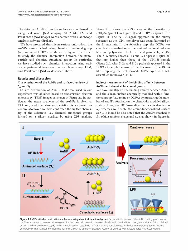
AuNPs were attached using chemical functional group(i.e., amine or DOPA), as shown in Figure 1, in orderto study the chemical interaction between the nano-particle and chemical functional group. In particular,we have studied such chemical interaction using vari-ous experimental tools such as cantilever assay, LFM,and PeakForce QNM as described above.
Results and discussionCharacterization of the AuNPs and surface chemistry ofSA and SDThe size distribution of AuNPs that were used in ourexperiment was obtained based on transmission electronmicroscopy (TEM) images as shown in Figure 2a. In par-ticular, the mean diameter of the AuNPs is given as19.4 nm, and the standard deviation is estimated as2.2 nm. Moreover, we have confirmed the surface chemis-try of the substrate, i.e., chemical functional groupsformed on a silicon surface, by using XPS analysis.
Figure 1 AuNPs attached onto silicon substrate using chemical functithe Si substrate and characterization regimes for the chemical interaction bon aminated surface (AuNP-SA); (II) AuNPs immobilized on catecholic surfacquantitatively characterized by experimental toolkits such as cantilever bioa
Figure 2b,c shows the XPS survey of the formation of-NH2-Si (panel I in Figure 1) and DOPA-Si (panel II inFigure 1). The N 1 s signal appeared in the surveyspectrum as the -NH2 monolayer was being fabricated onthe Si substrate. In the following step, the DOPA waschemically adsorbed onto the amine-functionalized sur-face and polymerized to form the dopamine layer [45].The XPS survey shows N 1 s and C 1 s peaks (Figure 2c)that are higher than those of the -NH2-Si sample(Figure 2b). Also, Si 2 s and Si 2p peaks disappeared in theDOPA-Si sample because of the thickness of the DOPAfilm, implying the well-formed DOPA layer with self-assembled monolayer [45-47].
Indirect measurement of the binding affinity betweenAuNPs and chemical functional groupsWe have investigated the binding affinity between AuNPsand the silicon surface chemically modified with a func-tional group (i.e., amine or DOPA) by measuring the num-ber of AuNPs attached on the chemically modified siliconsurface. Here, the DOPA-modified surface is denoted asSD, whereas we denote the amine-functionalized surfaceas SA. It should be also noted that the AuNPs attached toSD exhibit uniform shape and size, as shown in Figure 3a,
onal group. Schematic illustration of the AuNP-coating procedure onetween AuNPs and chemical functional groups. (I) AuNPs immobilizede (AuNP-SD) functionalized with dopamine (DOPA). Each sample isssay, PeakForce QNM, as well as lateral force microscopy (LFM).

Figure 2 Characteristics of the AuNPs and surface chemistry of SA and SD. Histogram of AuNP diameters (a) and XPS survey spectra of(b) the aminated Si substrate (SA) and (c) DOPA-functionalized Si substrate (SD). Inset in (a): TEM images of the AuNPs.
Figure 3 Characterization of the surface chemistry of the samples such as AuNP-SA and AuNP-SD. (a, b) SEM images of themicrocantilever functionalized with DOPA molecules used in the cantilever bioassay. The dimensions of the microcantilever such as stylus height,cantilever length, and width are shown. (c) The magnified image of the stylus vertex shows uniformly coated AuNPs on the entiremicrocantilever. (d, e) SEM images of the AuNPs immobilized on the Si substrate functionalized with amino groups and DOPA molecules,respectively. (f) Plot of the average number of AuNPs from the SEM images (n = 5). (g, h) Resonant frequency shift of the microcantilever in everystep of the fabrication procedure is measured in air (see Table 1). (i) Plot of the total mass of AuNPs bound to the chemically modified surfacemeasured from the frequency shift of a cantilever due to AuNP binding (n = 3).
Lee et al. Nanoscale Research Letters 2012, 7:608 Page 4 of 11http://www.nanoscalereslett.com/content/7/1/608

Table 1 The resonant frequency shift of AFM cantileversin cantilever assay
Bare SA SD AuNP coating
AuNP-SA (kHz) 300.2 ± 32.3 299.8 ± 31.7 298.4 ± 32.6
AuNP-SD (kHz) 279.8 ± 3.5 279.5 ± 3.8 276.9 ± 4.0 274.2 ± 4.0
Standard deviations are derived from three independent measurements.
Lee et al. Nanoscale Research Letters 2012, 7:608 Page 5 of 11http://www.nanoscalereslett.com/content/7/1/608
b,c. It is interestingly shown that the AuNPs attached toSA were locally aggregated, while the AuNPs immobilizedon SD were distributed with relatively high uniformity(Figure 3d,e), which suggests that the AuNPs were func-tionalized as a uniform monolayer onto SD. It is attributedto the fact that the local aggregation of AuNPs onto SA ishighly related to the molecular structure of APTMS,which leads to the formation of amine functional group asa disordered layer on the surface and, consequently, thedecrease in the uniformity of surface functionalization andlarge variation of surface density of AuNPs [48]. On theother hand, a uniform attachment of AuNPs onto SD isattributed to GA acting as a linker molecule between thesurface and DOPA such that the linker molecule allowsfor an ordered formation of DOPA on a silicon surface.The uniform distribution of the attached AuNPs on thesurface is due to the electrostatic repulsion between nano-particles. Meanwhile, the uniformity of the functionalizedmolecules is a critical factor in determining the bindingaffinity of AuNPs [48] because the uniform distributionof functional molecules is a priori requisite to optimizethe binding affinity between the surface and AuNPs. Al-though there are small aggregates of AuNPs locally evenin the AuNP-SD samples, the binding affinity between thesurface and AuNPs is clearly shown in the electronmicroscope imaging assay. The number of AuNPsattached onto either SA or SD (denoted as NA or ND,respectively) can be used as a quantity that representsthe binding affinity. Based on the SEM images ofAuNPs attached to either SA or SD, it is found thatNA = 503 ± 54 (mean ± standard deviation) per unit area of1 μm2, whereas ND = 798 ± 75 per unit area of 1 μm2
(Figure 3f). This clearly elucidates that SD exhibits higherbinding affinity to AuNPs than SA. For quantitative com-parison, we have introduced a dimensionless measuredefined as RN =ND/NA. This RN ratio can be used as adimensionless quantity useful in representing the bindingaffinity (for more detail, see below).Now, we have studied binding affinity using cantilever
assay that allows for measuring the total mass ofAuNPs attached to the surface of the microcantilever.For such a study, we have prepared cantilevers whosesurfaces are chemically modified by an amine group orDOPA, respectively. It is shown in Figure 3g,h that thesurface modification of cantilevers using amine groupor DOPA reduces their resonant frequencies, which isattributed to the weight of the functionalized chemicalgroups (i.e., amine or DOPA). We have observed thatthe binding of AuNPs onto the amine- or DOPA-immobilized surface of the cantilever significantlydecreases the resonant frequency of such a cantilever(Table 1). The total weight of AuNPs chemically boundto the cantilever can be estimated from the measuredfrequency shift due to AuNP binding to the cantilever.
In particular, the relationship between the total mass ofAuNPs bound to the cantilever and the frequency shiftis represented in the form Δω/ω0 = (1/2)(ΔM/Mc),where Δω is the resonant frequency shift due to AuNPbinding onto the cantilever, ΔM is the total mass ofAuNPs chemically attached to the cantilever, and ω0
and Mc represent the resonant frequency and mass, re-spectively, of the microcantilever whose surface ischemically modified. It is found that the total mass ofAuNPs attached to the amine-modified cantilever sur-face is estimated as ΔMA = 488 ± 10 pg, while the totalmass of AuNPs bound to the DOPA-functionalized can-tilever surface is measured as ΔMD = 630 ± 27 pg(Figure 3i). This clearly demonstrates that the DOPA-modified surface exhibits stronger binding affinity toAuNPs than the amine-modified surface. As in the pre-vious paragraph, we have introduced the dimensionlessquantity RM defined as RM =ΔMD/ΔMA, which allowsfor quantitative comparison. It is interestingly shownthat RM is very close to RN, as anticipated (i.e.,RM = approximately 1.3 and RN = approximately 1.6).This confirms that the dimensionless quantities RM
and RN are useful parameters that allow for quantify-ing the binding affinity between the chemically modifiedsurface and AuNPs.
Direct measurement of the binding affinity betweenAuNPs and chemical functional groupsWhile measurement of the number of attached AuNPson the surface or mass of the AuNPs bound to the sur-face is an indirect method to quantify the binding affinitybetween AuNPs and chemically functionalized surface,we have taken into account the direct method for quanti-tative characterization of such binding affinity. Here, wehave employed a novel scanning technique, namely Peak-Force QNM [49], that is useful in measuring the adhe-sion force between AuNPs and chemically modifiedsurface. Figure 4a,b shows the AFM topography imagesof AuNPs attached to either SA or SD. It is shown thatthe AFM height for the AuNPs attached to SD is mea-sured as approximately 14.2 nm, whereas the AFMheight for the AuNPs bound to SA is measured as ap-proximately 15.5 nm. This is attributed to the size of thefunctionalized molecules such that the chain length ofDOPA is much larger than that of the amine group [48].As shown in Figure 4g,h, the AuNP is more likely to beembedded in DOPA, that is, more number of DOPA

Figure 4 PeakForce QNM analysis of AuNP-immobilized surfaces. (a, b) Topographic AFM images of 20 nm of AuNPs immobilized onaminated surface and DOPA-functionalized surface, respectively. (c, d) Adhesion images of the samples show relative adhesion interactionbetween the bare Si AFM tip and AuNPs or other regions outside the AuNPs. All scale bars are 200 nm. The dashed line is a depiction of AFMstylus trajectory. (e, f) AFM deformation images show a larger deformation change near the edge than at the center of the nanoparticles. (g, h)Schematic diagram of the geometric design depicted AuNPs immobilized on the substrates functionalized with amino group and DOPAmolecules, respectively. The terms wtopology and htopology indicate the measured width and height, respectively, of the AuNPs in the aspect oftopology (a, b).
Lee et al. Nanoscale Research Letters 2012, 7:608 Page 6 of 11http://www.nanoscalereslett.com/content/7/1/608
molecules (than that of amine molecules) is likely to beinvolved in AuNP binding. This suggests that DOPAmolecules may allow for establishing the stable, reliableadhesion of nanoparticles. Notably, we found that thewidth of the AuNPs bound to SD (99.1 ± 12.3) in bothtopology and adhesion map is larger by the amount ofapproximately 7 nm than that bound to SA (92.2 ± 19.7),as shown in Figure 4a,b. This result seems to contradictthe fact that the AuNPs in SD are more deeply embeddedthan those in SA. It may be attributed to the fact that
AFM indentation may induce the significant motion ofAuNPs, which may distort the size of the AuNPs. In par-ticular, a previous study [50] reports that there is greaterenergy dissipation at the edge of a nanoparticle than atits center, implying that the nanoparticle would bewobbled during AFM indentation, whereas the nanopar-ticle would not be moved during tapping mode AFM im-aging. As shown in the AFM deformation images(Figure 4e,f ), there is a larger deformation change ofAuNPs in AuNP-SA than in AuNP-SD during PeakForce

Lee et al. Nanoscale Research Letters 2012, 7:608 Page 7 of 11http://www.nanoscalereslett.com/content/7/1/608
QNM. This indicates a stronger binding of AuNPs ontoSD rather than on SA, which is attributed to the narrowstructural dimension of AuNPs immobilized on SA incomparison with those on SD. Moreover, we have alsoconsidered the adhesion map for AuNPs attached to SAor SD. It is found that the adhesion force difference be-tween silicon nitride (Si3N4) AFM tip and the surface(i.e., SA or SD) is <5 nN and that the adhesion force is notsignificantly dependent on the type of surface chemistry(i.e., whether the surface is functionalized with aminegroup or DOPA). This indicates that the interaction be-tween the Si3N4 AFM tip and the surface is not criticalwhen we measure the adhesion force between AuNPsand surface. It is shown that the adhesion force betweenthe Si3N4 AFM tip and chemically modified surface towhich the AuNPs do not adhere is measured as approxi-mately 10 nN. Nevertheless, the adhesion force mapobtained from PeakForce QNM is insufficient to distin-guish the binding affinity between AuNPs and SD fromthat between AuNPs and SA, while the AFM height intopology and the width in the adhesion map for AuNPsbound to SA or SD allow for the distinction between suchbinding affinities as described earlier.Another way to directly measure the binding affinity
between AuNPs and chemically functionalized surfaceis to utilize LFM that enables the measurement of fric-tion force between the AFM tip and the sample surface(Figure 5). For LFM imaging, we have utilized atriangular-shaped microcantilever whose normal springconstant knorm is in the range of 0.16 to 1 N/m, suitablefor contact mode AFM imaging. In general, the normalspring constant of a cantilever depends on its shapeand material [51]. In our study, we have used Si3N4
cantilevers so that the normal spring constant of amicrocantilever is determined from its shape. The lateralspring constant klat is related to the normal springconstant given by the following equation [35]:
klat ¼ 2
6 cos2θ þ 3 1þ vð Þ sin2θLH
� �2
knorm ð1Þ
where θ is the angle between the base arms of the triangu-lar cantilever, v is the Poisson ratio for silicon nitride, L isthe length of the cantilever beam, and H is the tip verticalheight (see Table 2). With the estimation of klat fromEquation 1, the lateral force (Flat) can be calculated fromthe measurement of the lateral force signal in LFM ana-lysis such as [36]
Flat ¼ klat � Slat � ΔV : ð2ÞHere, Slat is the lateral sensitivity of the cantilever
defined as Slat = PHSnorm/aR*L [36], where P is a propor-tionality factor (≈2.5 for the triangular cantilever), Snormis the vertical deflection sensitivity of the cantilever, a is
the amplification factor of the lateral signal measured,and R* is the ratio of the beam height to the beam width(R* = 0.5) [36]. ΔV is the measured value in LFM ana-lysis, which is extracted from the LFM images. In gen-eral, the longer and larger the cantilever, the lower is itsnormal spring constant (i.e., more flexible in normal de-flection), but the larger is its lateral spring constant (klat).We can control the exerted applied force using differentspring constants of cantilevers (knorm) under an identicaldeflection set point (1 V) rather than a set point controlwith an identical AFM tip in order to avoid damage tothe samples and a subsidiary frictional noise.In LFM imaging for the measurement of friction force
between the two surfaces (i.e., surface of the AuNPs andthe substrate functionalized with chemical groups), onehas to be cautious in selecting a cantilever; in particular,a cantilever with a stiffness of <0.16 N/m is too flexibleto scan our sample, whereas a cantilever with a stiffnessof ≥1 N/m is too stiff to measure the friction force inour sample. As shown in Figure 6, the AFM imaging ofour sample using a cantilever with a stiffness of 1 N/mleads to the detachment of AuNPs from the surface dur-ing the imaging, which implies the difficulty in accur-ately measuring the friction force between AuNPs andsurface (i.e., SA or SD). Figure 5a,b shows the AFM/LFMimages of AuNPs attached to SD or SA. It is shown inFigure 5a that during AFM imaging using a cantileverwith a stiffness of knorm = 0.16 N/m, AuNPs are detachedfrom SA (i.e., AFM image shows a scratched pattern cor-responding to the imaged AuNPs), while the detachmentof AuNPs from SD does not occur. Moreover, it is foundthat AuNPs are still bound to SD even when AFM andLFM imaging were implemented using a cantilever witha stiffness of knorm = 0.7 N/m (Figure 5b). Figure 5cshows the section profile extracted from the AFM/LFMimages (as indicated by a white arrow). It is shown thatin the AFM height profile, as anticipated, the AFMheight of the AuNPs bound to SA is close to that of theAuNPs attached to SD. On the other hand, in the lateralforce profile extracted from the LFM image, we can findthe significant differences between the durability of twosamples, i.e., AuNPs bound to SA and SD, respectively.This is attributed to the fact that during imaging ofAuNPs bound to SA, the twist of the cantilever tip is notsignificant, which leads to low signals in the LFM image,while the binding between AuNPs and SD (stronger thanthat between AuNPs and SA) leads to more twist of thecantilever tip and consequently produce a large signal inLFM imaging [37] (Figure 5e). Based on the LFM imageswith Equations 1 and 2, we have measured the lateralforce between AuNPs and chemically modified surface.It is found that the mean lateral force between AuNPsand SA is measured as 660 nN, while the mean lateralforce between AuNPs and SD is estimated to be 1.2 μN

Figure 5 Lateral force microscopy analysis of binding affinity between AuNPs and chemically functionalized surfaces. (a, b) AFMtopographic images and lateral force images of AuNPs immobilized on Si substrates, respectively, functionalized with (a) amino groups (AuNP-SA)and (b) catecholamine molecules (AuNP-SD) by different normal spring constants (kN) of microcantilevers. The inset shows left embankment(indicated by a red arrowhead) due to swept AuNPs formed by scanning the surface in AFM contact mode with kN = 0.7 N/m of microcantilever.All scale bars including that of the inset are 250 nm. (c) Line profiles corresponding to the white arrow in each image of (a) and (b) show thecurves of scanner retracting distance versus the AuNP lateral displacement and the lateral force versus the lateral displacement, respectively. (d)Graph of the average lateral force of >100 AuNPs in (a) and (b) (the asterisk indicates p < 0.001) was extracted and calculated from the lineprofiles of lateral forces (c). (e) The model illustrates the physical interaction between the AFM tip and AuNPs attached on the chemicallyfunctionalized substrate (i.e., AuNP-SA and AuNP-SD).
Lee et al. Nanoscale Research Letters 2012, 7:608 Page 8 of 11http://www.nanoscalereslett.com/content/7/1/608
(Figure 5d). For quantitative comparison, we have intro-duced a dimensionless parameter defined as RF = FD/FA,where FA indicates the lateral force between AuNPs andSA, and FD represents the lateral force between AuNPs andSD. It is interestingly found that the dimensionless param-eter RF (=1.7) is very close to the aforementioned dimen-sionless parameters RN and RM (Figure 7). This suggeststhat the binding affinity between AuNPs and chemicallyfunctionalized surface can be quantitatively understood byusing either of the indirect experimental methods such ascantilever assay or direct force measurement such as LFMimaging. It should be noted that the binding force betweenAuNPs and chemically functionalized surface could be
measured using AFM pulling experiments [35,52], whichenables the measurement of the normal force required tobreak a chemical bond. In general, the normal adhesionforce driven by the mechanical detachment of AuNPs fromthe surface might be much lower than the shear adhesionforce between AuNPs and the surface. It is attributed to thefact that a shear force required to break chemical bonds ismuch larger than a normal force that leads to breakage ofchemical bonds [53,54]. This indicates that LFM imaging-based measurement allows for estimating the maximumstrength of chemical bonds between nanostructure andchemical functional group. Moreover, AFM pullingexperiment-based measurement of normal force required

Table 2 Summary of triangular microcantileverparameters (SNL and ScanAsyst Fluid) used in LFM study
ScanAsyst Fluid SNL (B)
L (μm) 70 205
w (μm) 10 40
t (μm) 0.6 0.6
H (μm) 8 8
E (GPa) 304 304
ν 0.24 0.24
θ 60° 64.5°
knorm (N/m) 0.74 0.16
Snorm (nm/V) 12.57 26.30
klat (N/m) 19.57 56.16
Slat (nm/V) 1.88 1.27
L, cantilever length; w, cantilever width; t, cantilever thickness; H, stylus height;E, Young's modulus of Si; v, Poisson's ratio of Si; θ, angle between the basearms of the triangular cantilever; knorm, measured cantilever's normal springconstant; Snorm, measured cantilever's normal sensitivity; klat, lateral springconstant calculated from the knorm; Slat lateral normal sensitivity calculatedfrom Snorm [35,36].
Figure 7 Plot of binding affinity ratio between AuNPs andchemically functionalized surfaces (AuNP-SA and AuNP-SD). Thebinding affinity ratios are obtained from experiments such as SEMimage, cantilever assay, and LFM. This analysis indicates that DOPAmolecules are approximately 1.5 times stronger than amino groupsin their adhesion property for the immobilization of AuNPs.
Lee et al. Nanoscale Research Letters 2012, 7:608 Page 9 of 11http://www.nanoscalereslett.com/content/7/1/608
to break chemical bonds requires statistical analysis (basedon repetitive experiments due to the effect of thermal fluc-tuation on force-driven bond rupture [55-57]), while LFMimaging-based measurement of shear force for breakingbonds does not require repetitive experiments becauseLFM imaging enables the parallel measurement of shearforces required to break chemical bonds in the scannedarea of a sample. In other words, LFM imaging enables thesimultaneous measurement of shear forces (with more than100 times) required to break chemical bonds, which resultsin an effective statistical analysis based on only a singleLFM image.
ConclusionsIn conclusion, we have demonstrated a quantitativecharacterization of the binding affinity between AuNPsand chemically modified surface using various experi-mental techniques such as SEM image analysis, canti-lever assay, PeakForce QNM, and LFM image analysis. It
Figure 6 The PeakForce QNM analysis of the AuNP-SD sample. AFM toembankments composed of AuNPs swept by the 2 × 2 μm2 scanning of a
is shown that the DOPA-modified surface is an effectiveconjugation method for functionalization of nanoparti-cles onto the surface when compared with amine-modified surface, as anticipated, from our variousexperiments. More remarkably, we have shown that di-mensionless parameters (i.e., RN, RM, and RF) introducedin this work are useful in quantifying the binding affinitybetween nanoparticle and chemical functional groups,and that these dimensionless parameters are consistentregardless of experiments, i.e., RN, RM, and RF are almostidentical to each other, implying that the binding affinitybetween nanostructure and chemical group can be quan-titatively studied using either indirect method (i.e., SEMimage analysis and cantilever assay) or direct method(i.e., lateral force measurement). Our study sheds lighton how to quantitatively study the binding affinity be-tween nanostructure and chemical functional group,which can provide the design principles for nanoparticle-based systems such as nanomedicine and nanobiosensor.
pology, peak force error, and adhesion images (3 × 3 μm2) of themicrocantilever with kN = 1 N/m in LFM.

Lee et al. Nanoscale Research Letters 2012, 7:608 Page 10 of 11http://www.nanoscalereslett.com/content/7/1/608
AbbreviationsAFM: Atomic force microscopy; APTMS: 3-aminopropyltrimethoxysilane;AuNP: Gold nanoparticle; DOPA: Dopamine hydrochloride; PeakForceQNM: PeakForce quantitative nanomechanical property mapping;LFM: Lateral force microscopy; SA: Amine-functionalized surface;SD: DOPA-modified surface; SEM: Scanning electron microscope;XPS: X-ray photoelectron spectroscopy.
Competing interestsThe authors declare that they have no competing interests.
Authors’ contributionsGL carried out the experiments and wrote the manuscript. HL performed theexperiments. KN and JHH participated in the XPS and SEM analyses and inthe interpretation of data. JY, SWL, and DSY participated in the discussionand interpretation of data. KE participated in the analysis of the experimentaldata, interpreted the result, and wrote the manuscript. TK conceived theresearch, analyzed the experimental data, interpreted the results, and wrotethe manuscript. All authors read and approved the final manuscript.
AcknowledgementsThis work is supported by the National Research Foundation (NRF) of Korea(under grant nos. NRF-2010-0009428, 2010–0027238, 2011–0009885, and2012R1A2A2A04047240).
Author details1Institute for Molecular Sciences, Seoul 120-749, Republic of Korea.2Department of Biomedical Engineering, Yonsei University, Wonju 220-710,Republic of Korea. 3Department of Energy IT, Gachon University, Seongnam,Gyeonggi-do 461-701, Republic of Korea. 4Department of Radiology, Collegeof Medicine, Yonsei University, Seoul 120-749, Republic of Korea.
Received: 15 September 2012 Accepted: 19 October 2012Published: 31 October 2012
References1. Rosi NL, Mirkin CA: Nanostructures in biodiagnostics. Chem Rev 2005,
105:1547.2. Schroeder A, Heller DA, Winslow MM, Dahlman JE, Pratt GW, Langer R, Jacks
T, Anderson DG: Treating metastatic cancer with nanotechnology. Nat RevCancer 2012, 12:39.
3. Lee J-H, Huh Y-M, Jun Y-w, Seo J-w, Jang J-t, Song H-T, Kim S, Cho E-J,Yoon H-G, Suh J-S, Cheon J: Artificially engineered magneticnanoparticles for ultra-sensitive molecular imaging. Nat Med 2007,13:95.
4. Chen J, Saeki F, Wiley BJ, Cang H, Cobb MJ, Li Z-Y, Au L, Zhang H, KimmeyMB, Li X, Xia Y: Gold nanocages: bioconjugation and their potential useas optical imaging contrast agents. Nano Lett 2005, 5:473.
5. Yang P-H, Sun X, Chiu J-F, Sun H, He Q-Y: Transferrin-mediated goldnanoparticle cellular uptake. Bioconjug Chem 2005, 16:494.
6. Ling D, Park W, Park YI, Lee N, Li F, Song C, Yang S-G, Choi SH, Na K, HyeonT: Multiple-interaction ligands inspired by mussel adhesive protein:synthesis of highly stable and biocompatible nanoparticles. Angew ChemInt Ed 2011, 50:11360.
7. Lim E-K, Huh Y-M, Yang J, Lee K, Suh J-S, Haam S: pH-triggereddrug-releasing magnetic nanoparticles for cancer therapy guided bymolecular imaging by MRI. Adv Mater 2011, 23:2436.
8. Lee J-H, Jang J-t, Choi J-s, Moon SH, Noh S-h, Kim J-w, Kim J-G, Kim I-S, ParkKI, Cheon J: Exchange-coupled magnetic nanoparticles for efficient heatinduction. Nat Nanotechnol 2011, 6:418.
9. Mingce L, Jingjing J, Yan L, Ruqiong C, Liying Z, Weimin C: Effect of goldnanoparticles on the photocatalytic and photoelectrochemicalperformance of Au modified BiVO4. Nano-Micro Lett 2011, 3:171.
10. Vitale F, Fratoddi I, Battocchio C, Piscopiello E, Tapfer L, Russo M, PolzonettiG, Giannini C: Mono- and bi-functional arenethiols as surfactants for goldnanoparticles: synthesis and characterization. Nanoscale Res Lett 2011,6:103.
11. Jin R, Wu G, Li Z, Mirkin CA, Schatz GC: What controls the meltingproperties of DNA-linked gold nanoparticle assemblies? J Am Chem Soc2003, 125:1643.
12. Storhoff JJ, Elghanian R, Mucic RC, Mirkin CA, Letsinger RL: One-potcolorimetric differentiation of polynucleotides with single baseimperfections using gold nanoparticle probes. J Am Chem Soc 1959,1998:120.
13. Zhang Y, Li B, Chen X: Simple and sensitive detection of dopamine in thepresence of high concentration of ascorbic acid using goldnanoparticles as colorimetric probes. Microchimica Acta 2010, 168:107.
14. Yang J, Eom K, Lim E-K, Park J, Kang Y, Yoon DS, Na S, Koh EK, Suh J-S, HuhY-M, Kwon TY, Haam S: In situ detection of live cancer cells by usingbioprobes based on au nanoparticles. Langmuir 2008, 24:12112.
15. Li J-L, Wang L, Liu X-Y, Zhang Z-P, Guo H-C, Liu W-M, Tang S-H: In vitrocancer cell imaging and therapy using transferrin-conjugated goldnanoparticles. Cancer Lett 2009, 274:319.
16. Lee H, Dellatore SM, Miller WM, Messersmith PB: Mussel-inspired surfacechemistry for multifunctional coatings. Science 2007, 318:426.
17. Lee H, Lee BP, Messersmith PB: A reversible wet/dry adhesive inspired bymussels and geckos. Nature 2007, 448:338.
18. Lu C-C, Zhang M, Li A-J, He X-W, Yin X-B: 3,4-Dihydroxy-l-phenylalanine forpreparation of gold nanoparticles and as electron transfer promoter inH2O2 biosensor. Electroanalysis 2011, 23:2421.
19. Black KCL, Liu Z, Messersmith PB: Catechol redox induced formation ofmetal core − polymer shell nanoparticles. Chem Mater 2011, 23:1130.
20. Kim S, Ku SH, Lim SY, Kim JH, Park CB: Graphene–biomineral hybridmaterials. Adv Mater 2011, 23:29.
21. Ryu S, Lee Y, Hwang J-W, Hong S, Kim C, Park TG, Lee H, Hong SH: High-strength carbon nanotube fibers fabricated by infiltration and curing ofmussel-inspired catecholamine polymer. Adv Mater 1911, 23:1971.
22. Burdine L, Gillette TG, Lin H-J, Kodadek T: Periodate-triggered cross-linkingof dopa-containing peptide − protein complexes. J Am Chem Soc 2004,126:11442.
23. Ku SH, Ryu J, Hong SK, Lee H, Park CB: General functionalization route forcell adhesion on non-wetting surfaces. Biomaterials 2010, 31:2535.
24. Yang HS, Park J, La WG, Jang H-k, Lee M, Kim B-S: 3,4-Dihydroxyphenylalanine-assisted hydroxyapatite nanoparticle coating onpolymer scaffolds for efficient osteoconduction. Tissue Eng Part C Methods2011, 18:1.
25. Baik SH, Kim JH, Cho HH, Park S-N, Kim YS, Suh H: Development andanalysis of a collagen-based hemostatic adhesive. J Surg Res 2010,164:e221.
26. Bishop KJM, Wilmer CE, Soh S, Grzybowski BA: Nanoscale forces and theiruses in self-assembly. Small 2009, 5:1600.
27. Cheng YC, Samia A, Meyers JD, Panagopoulos I, Fei B, Burda C: Highlyefficient drug delivery with gold nanoparticle vectors for in vivophotodynamic therapy of cancer. J Am Chem Soc 2008, 130:10643.
28. Kim C-k, Ghosh P, Rotello VM: Multimodal drug delivery using goldnanoparticles. Nanoscale 2009, 1:61.
29. Eom K, Park HS, Yoon DS, Kwon T: Nanomechanical resonators and theirapplications in biological/chemical detection: nanomechanics principles.Phys Rep 2011, 503:115.
30. Buchapudi KR, Huang X, Yang X, Ji H-F, Thundat T: Microcantileverbiosensors for chemicals and bioorganisms. Analyst 2011, 136:1539–1556.
31. Anja B, Søren D, Stephan Sylvest K, Silvan S, Maria T: Cantilever-likemicromechanical sensors. Rep Prog Phys 2011, 74:036101.
32. Datar R, Kim S, Jeon S, Hesketh P, Manalis S, Boisen A, Thundat T: Cantileversensors: nanomechanical tools for diagnostics. MRS Bull 2009, 34:449.
33. Sweers K, van der Werf K, Bennink M, Subramaniam V: Nanomechanicalproperties of alpha-synuclein amyloid fibrils: a comparative study bynanoindentation, harmonic force microscopy, and Peakforce QNM.Nanoscale Res Lett 2011, 6:270.
34. Young TJ, Monclus MA, Burnett TL, Broughton WR, Ogin SL, Smith PA: Theuse of the PeakForce TM quantitative nanomechanical mappingAFM-based method for high-resolution Young's modulus measurementof polymers. Meas Sci Technol 2011, 22:125703.
35. Noy A, Frisbie CD, Rozsnyai LF, Wrighton MS, Lieber CM: Chemical forcemicroscopy: exploiting chemically-modified tips to quantify adhesion,friction, and functional group distributions in molecular assemblies.J Am Chem Soc 1995, 117:7943.
36. Tocha E, Schönherr H, Vancso GJ: Quantitative nanotribology by AFM: anovel universal calibration platform. Langmuir 2006, 22:2340.
37. Song J, Wang X, Riedo E, Wang ZL: Elastic property of vertically alignednanowires. Nano Lett 1954, 2005:5.

Lee et al. Nanoscale Research Letters 2012, 7:608 Page 11 of 11http://www.nanoscalereslett.com/content/7/1/608
38. Landherr LJT, Cohen C, Agarwal P, Archer LA: Interfacial friction andadhesion of polymer brushes. Langmuir 2011, 27:9387.
39. Lang HP, Hegner M, Gerber C: Cantilever array sensors. Mater Today 2005,8:30.
40. Wu G, Ji H, Hansen K, Thundat T, Datar R, Cote R, Hagan MF, ChakrabortyAK, Majumdar A: Origin of nanomechanical cantilever motion generatedfrom biomolecular interactions. Proc Natl Acad Sci 2001, 98:1560.
41. Hamdy Y, Khaled E: State-space approach to vibration of gold nano-beaminduced by ramp type heating. Nano-Micro Lett 2010, 2:139.
42. Lin YH, McConney ME, LeMieux MC, Peleshanko S, Jiang C, Singamaneni S,Tsukruk VV: Trilayered ceramic–metal–polymer microcantilevers withdramatically enhanced thermal sensitivity. Adv Mater 2006, 18:1157.
43. Chan J, Alegre TPM, Safavi-Naeini AH, Hill JT, Krause A, Groblacher S,Aspelmeyer M, Painter O: Laser cooling of a nanomechanical oscillatorinto its quantum ground state. Nature 2011, 478:89.
44. Haiss W: Surface stress of clean and adsorbate-covered solids. Rep ProgPhys 2001, 64:591.
45. Ou J, Wang J, Liu S, Zhou J, Ren S, Yang S: Microtribological andelectrochemical corrosion behaviors of polydopamine coating onAPTS-SAM modified Si substrate. Appl Surf Sci 2009, 256:894.
46. Graf N, Yeğen E, Lippitz A, Treu D, Wirth T, Unger WES: Optimization ofcleaning and amino-silanization protocols for Si wafers to be used asplatforms for biochip microarrays by surface analysis (XPS, ToF-SIMS andNEXAFS spectroscopy). Surf Interface Anal 2008, 40:180.
47. Liu Y, Yu B, Hao J, Zhou F: Amination of surfaces via self-assembly ofdopamine. J Colloid Interface Sci 2011, 362:127.
48. Aureau D, Varin Y, Roodenko K, Seitz O, Pluchery O, Chabal YJ: Controlleddeposition of gold nanoparticles on well-defined organic monolayergrafted on silicon surfaces. J Phys Chem C 2010, 114:14180.
49. Adamcik J, Berquand A, Mezzenga R: Single-step direct measurement ofamyloid fibrils stiffness by peak force quantitative nanomechanicalatomic force microscopy. Appl Phys Lett 2011, 98:193701.
50. Duner G, Thormann E, Dedinaite A, Claesson PM, Matyjaszewski K, Tilton RD:Nanomechanical mapping of a high curvature polymer brush graftedfrom a rigid nanoparticle. Soft Matter 2012, 8:8312.
51. Palacio MLB, Bhushan B: Normal and lateral force calibration techniquesfor AFM cantilevers. Crit Rev Solid State Mater Sci 2010, 35:73.
52. Stroh C, Wang H, Bash R, Ashcroft B, Nelson J, Gruber H, Lohr D, LindsaySM, Hinterdorfer P: Single-molecule recognition imaging microscopy.Proc Natl Acad Sci USA 2004, 101:12503.
53. Kufer SK, Puchner EM, Gumpp H, Liedl T, Gaub HE: Single-moleculecut-and-paste surface assembly. Science 2008, 319:594.
54. Qu L, Dai L, Stone M, Xia Z, Wang ZL: Carbon nanotube arrays with strongshear binding-on and easy normal lifting-off. Science 2008, 322:238.
55. Eom K, Makarov DE, Rodin GJ: Theoretical studies of the kinetics ofmechanical unfolding of cross-linked polymer chains and theirimplications for single-molecule pulling experiments. Phys Rev E 2005,71:021904.
56. Evans E: Probing the relation between force—lifetime—and chemistry insingle molecular bonds. Annu Rev Biophys Biomol Struct 2001, 30:105.
57. Evans E, Ritchie K: Dynamic strength of molecular adhesion bonds.Biophys J 1997, 72:1541.
doi:10.1186/1556-276X-7-608Cite this article as: Lee et al.: Nanomechanical characterization ofchemical interaction between gold nanoparticles and chemicalfunctional groups. Nanoscale Research Letters 2012 7:608.
Submit your manuscript to a journal and benefi t from:
7 Convenient online submission
7 Rigorous peer review
7 Immediate publication on acceptance
7 Open access: articles freely available online
7 High visibility within the fi eld
7 Retaining the copyright to your article
Submit your next manuscript at 7 springeropen.com