n103-chap 48 outline - columbiana county career and ... management/nursing interventions • ......
TRANSCRIPT
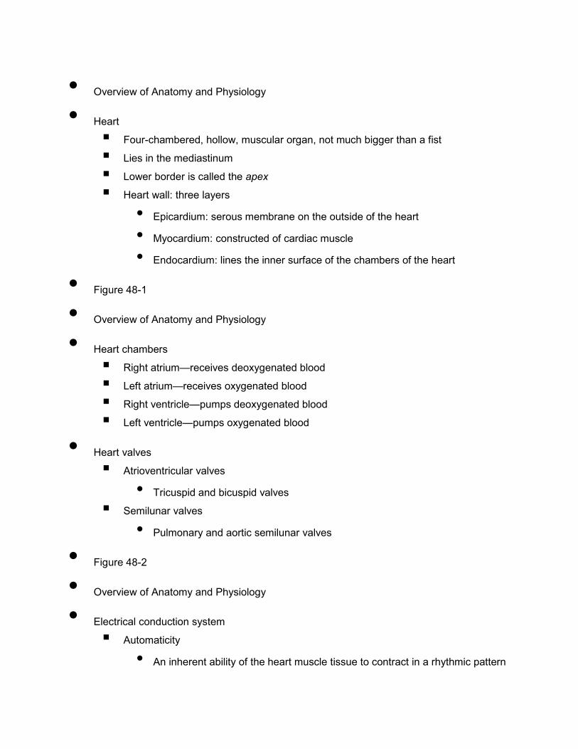
• Overview of Anatomy and Physiology
• Heart
� Four-chambered, hollow, muscular organ, not much bigger than a fist
� Lies in the mediastinum
� Lower border is called the apex
� Heart wall: three layers
• Epicardium: serous membrane on the outside of the heart • Myocardium: constructed of cardiac muscle • Endocardium: lines the inner surface of the chambers of the heart
• Figure 48-1
• Overview of Anatomy and Physiology
• Heart chambers
� Right atrium—receives deoxygenated blood
� Left atrium—receives oxygenated blood
� Right ventricle—pumps deoxygenated blood
� Left ventricle—pumps oxygenated blood
• Heart valves
� Atrioventricular valves
• Tricuspid and bicuspid valves � Semilunar valves
• Pulmonary and aortic semilunar valves
• Figure 48-2
• Overview of Anatomy and Physiology
• Electrical conduction system
� Automaticity
• An inherent ability of the heart muscle tissue to contract in a rhythmic pattern
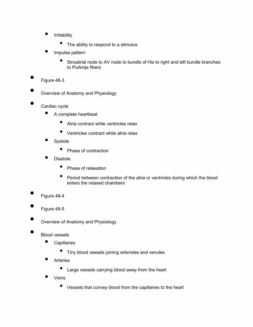
� Irritability
• The ability to respond to a stimulus � Impulse pattern
• Sinoatrial node to AV node to bundle of His to right and left bundle branches to Purkinje fibers
• Figure 48-3
• Overview of Anatomy and Physiology
• Cardiac cycle
� A complete heartbeat
• Atria contract while ventricles relax • Ventricles contract while atria relax
� Systole
• Phase of contraction � Diastole
• Phase of relaxation • Period between contraction of the atria or ventricles during which the blood
enters the relaxed chambers
• Figure 48-4
• Figure 48-5
• Overview of Anatomy and Physiology
• Blood vessels
� Capillaries
• Tiny blood vessels joining arterioles and venules � Arteries
• Large vessels carrying blood away from the heart � Veins
• Vessels that convey blood from the capillaries to the heart
• Circulation
• Coronary blood supply
� Right and left coronary arteries
• Branch off of the aorta • Encircle the heart like a crown • Supply the myocardium with blood
� Coronary veins
• Return the unoxygenated blood to the coronary sinus, then to the right atrium
• Figure 48-6
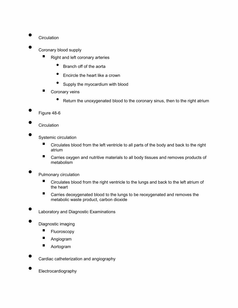
• Circulation
• Systemic circulation
� Circulates blood from the left ventricle to all parts of the body and back to the right atrium
� Carries oxygen and nutritive materials to all body tissues and removes products of metabolism
• Pulmonary circulation
� Circulates blood from the right ventricle to the lungs and back to the left atrium of the heart
� Carries deoxygenated blood to the lungs to be reoxygenated and removes the metabolic waste product, carbon dioxide
• Laboratory and Diagnostic Examinations
• Diagnostic imaging
� Fluoroscopy
� Angiogram
� Aortogram
• Cardiac catheterization and angiography
• Electrocardiography
• Cardiac monitors
• Thallium scanning
• Laboratory tests: CBC, blood cultures, coagulation studies, ESR electrolytes, lipids, arterial blood gases, cardiac markers
• Figure 48-7
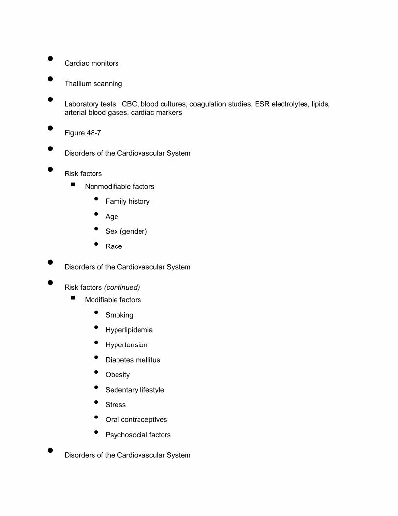
• Disorders of the Cardiovascular System
• Risk factors
� Nonmodifiable factors
• Family history • Age • Sex (gender) • Race
• Disorders of the Cardiovascular System
• Risk factors (continued)
� Modifiable factors
• Smoking • Hyperlipidemia • Hypertension • Diabetes mellitus • Obesity • Sedentary lifestyle • Stress
• Oral contraceptives • Psychosocial factors
• Disorders of the Cardiovascular System
• Cardiac dysrhythmias
� Any cardiac rhythm that deviates from normal sinus rhythm
• Sinus tachycardia • Sinus bradycardia • Supraventricular tachycardia • Atrial fibrillation • Atrioventricular block • Premature ventricular contractions • Ventricular tachycardia • Ventricular fibrillation
• Disorders of the Cardiovascular System
• Cardiac Arrest
� The sudden cessation of cardiac output and circulatory process
� Cause: ventricular tachycardia, ventricular fibrillation, and ventricular asystole
� Signs and symptoms: abrupt loss of consciousness with no response to stimuli; gasping respirations followed by apnea; absence of pulse and blood pressure; pupil dilation; pallor and cyanosis
� Treatment: cardiopulmonary resuscitation (CPR) and advanced cardiac life support (ACLS)
• Disorders of the Heart
• Coronary atherosclerotic heart disease
� Coronary artery disease (CAD)
• A variety of conditions that obstruct blood flow in the coronary arteries � Atherosclerosis
• A common arterial disorder characterized by yellowish plaques of cholesterol, lipids, and cellular debris in the inner layers of the walls of the arteries; the primary cause of atherosclerotic heart disease (ASHD)
• Figure 48-10
• Disorders of the Heart
• Angina pectoris
� Etiology/pathophysiology
• Cardiac muscle is deprived of oxygen • Increased workload on the heart
� Clinical manifestations/assessment
• Pain (usually relieved by rest) • Dyspnea • Anxiety; apprehension • Diaphoresis • Nausea
• Disorders of the Heart
• Angina pectoris (continued)
� Medical management/nursing interventions
• Correct cardiovascular risk factors • Avoid precipitating factors • Pharmacological management
� Dilate coronary arteries and decrease workload of heart
o Nitroglycerin
o Beta-adrenergic blocking agents
o Calcium channel blockers
• Disorders of the Heart
• Angina pectoris (continued)
� Medical management/nursing interventions
• Surgical interventions � Coronary artery bypass graft (CABG)
� Percutaneous transluminal coronary angioplasty (PTCA)
� Stent placement

• Disorders of the Heart
• Myocardial infarction
� Etiology/pathophysiology
• Occlusion of a major coronary artery or one of its branches with subsequent necrosis of myocardium
• Most common cause is atherosclerosis • Ability of the cardiac muscle to contract and pump blood is impaired
• Figure 48-16
• Disorders of the Heart
• Myocardial infarction (continued)
� Clinical manifestations/assessment
• Asymptomatic (silent MI) • Pain (not relieved by rest, position, or nitroglycerin) • Nausea • SOB; dizziness; weakness • Diaphoresis • Pallor—ashen color
• Sense of impending doom
• Figure 48-11
• Disorders of the Heart
• Myocardial infarction (continued)
� Medical management/nursing interventions
• Oxygen • Fibrinolytic agents • Percutaneous transluminal coronary angioplasty (PTCA) • Coronary artery bypass graft surgery
• Pharmacological management � Vasopressors, analgesics, nitrates, beta-adrenergic blockers, calcium
channel blockers, antidysrhythmics, diuretics, inotropic agents, diuretics, stool softeners
• Figure 48-12
• Figure 48-13
• Disorders of the Heart
• Heart failure
� Etiology/pathophysiology
• Abnormal condition characterized by circulatory congestion resulting from the heart’s inability to act as an effective pump
• Left ventricular failure � Most common
• Right ventricular failure � Usually caused by left ventricular failure
• Disorders of the Heart
• Heart failure (continued)
� Clinical manifestations/assessment
• Decreased cardiac output � Fatigue
� Angina
� Anxiety; restlessness
� Oliguria
� Decreased GI motility
� Pale, cool skin
� Weight gain
• Disorders of the Heart
• Heart failure (continued)
� Clinical manifestations/assessment (continued)
• Left ventricular failure

� Pulmonary congestion
o Dyspnea
o Paroxysmal nocturnal dyspnea
o Cough; frothy, blood-tinged sputum
o Orthopnea
o Pulmonary crackles
o Pleural effusion (x-ray)
• Disorders of the Heart
• Heart failure (continued)
� Clinical manifestations/assessment (continued)
• Right ventricular failure � Distended jugular veins
� Anorexia, nausea, and abdominal distention
� Liver enlargement
� Ascites
� Edema in feet, ankles, sacrum; may progress up the legs into thighs, external genitalia, and lower trunk
• Disorders of the Heart
• Heart failure (continued)
� Medical management/nursing interventions
• Pharmacological management � Increase cardiac efficiency
o Digitalis
o Vasodilators
o ACE inhibitors (decrease blood pressure)
• Bed rest, HOB elevated • Oxygen • Treat edema and pulmonary congestion • Monitor fluid retention (weigh daily; strict I&O)
• Disorders of the Heart
• Pulmonary edema
� Etiology/pathophysiology
• Accumulation of fluid in lung tissues and alveoli

• Complication of congestive heart failure (CHF) � Clinical manifestations/assessment
• Restlessness • Agitation • Disorientation • Diaphoresis • Dyspnea and tachypnea
• Disorders of the Heart
• Pulmonary edema (continued)
� Clinical manifestations/assessment (continued)
• Tachycardia • Pallor or cyanosis • Cough—large amounts of blood-tinged, frothy sputum
• Wheezing, crackles • Cold extremities
• Disorders of the Heart
• Pulmonary edema (continued)
� Medical management/nursing interventions
• Pharmacological management � Morphine sulfate
� Nitroglycerin
� Diuretics
� Inotropic agents
� Vasodilators
• High Fowler’s or orthopneic position • Oxygen
• Disorders of the Heart
• Valvular heart disease
� Etiology/pathophysiology
• Heart valves are compromised and do not open and close properly � Stenosis
� Insufficiency
• Causes may be: � Congenital
� Rheumatic fever
• Disorders of the Heart
• Valvular heart disease (continued)
� Clinical manifestations/assessment
• Fatigue • Angina • Oliguria • Pale, cool skin • Weight gain • Restlessness • Abnormal breath sounds • Edema
• Disorders of the Heart
• Valvular heart disease (continued)
� Medical management/nursing interventions
• Pharmacological management � Diuretics
� Digoxin
� Antidysrhythmics
• Restrict activities • Sodium-restricted diet • Surgery

� Open mitral commissurotomy
� Valve replacement
• Disorders of the Heart
• Rheumatic heart disease
� Etiology/pathophysiology
• Rheumatic fever � Inflammatory disease that is a delayed childhood reaction to
inadequately treated childhood upper respiratory tract infection of beta-hemolytic streptococci
� Causes scar tissue in the heart
• Disorders of the Heart
• Rheumatic heart disease (continued)
� Clinical manifestations/assessment
• Elevated temperature • Elevated heart rate • Epistaxis • Anemia • Joint pain and stiffness • Nodules on the joints • Specific to valve affected • Heart murmur
• Disorders of the Heart
• Rheumatic heart disease (continued)
� Medical management/nursing interventions
• Pharmacological management � NSAIDs
• Prevention � Treat infections rapidly and completely
• Bed rest

• Application of heat • Dietary recommendations
� Well-balanced diet
� Supplement with vitamins B and C
• Encourage fluids • Commissurotomy or valve replacement
• Disorders of the Heart
• Pericarditis
� Etiology/pathophysiology
• Inflammation of the membranous sac surrounding the heart • May be acute or chronic • Bacterial, viral, or fungal • Noninfectious conditions
� Azotemia, MI, neoplasms, scleroderma, trauma, systemic lupus erythematosus (SLE), radiation, drugs
• Disorders of the Heart
• Pericarditis (continued)
� Clinical manifestations/assessment
• Debilitating pain • Dyspnea • Fever • Chills • Diaphoresis • Leukocytosis • Pericardial friction rub • Pericardial effusion
• Disorders of the Heart
• Pericarditis (continued)
� Medical management/nursing interventions
• Pharmacological management � Analgesics
� Salicylates
� Antibiotics
� Anti-inflammatory agents
� Corticosteroids
• Oxygen • IV fluids • Surgery: pericardial window, pericardial tap
• Disorders of the Heart
• Endocarditis
� Etiology/pathophysiology
• Infection or inflammation of the inner membranous lining of the heart � Clinical manifestations/assessment
• Influenza-like symptoms • Petechiae on the conjunctiva, mouth, and legs • Anemia • Splinter hemorrhages under nails • Weight loss • Heart murmur
• Disorders of the Heart
• Endocarditis (continued)
� Medical management/nursing interventions
• Bed rest • Antibiotics
� IV for 1 to 2 months
• Prophylactic antibiotics for “high-risk” patients

• Surgical repair of diseased valves or valve replacement
• Disorders of the Heart
• Myocarditis
� Etiology/pathophysiology
• Inflammation of the myocardium • Rheumatic heart disease • Viral, bacterial, or fungal infection • Endocarditis • Pericarditis
• Disorders of the Heart
• Myocarditis (continued)
� Medical management/nursing interventions
• Bed rest • Oxygen • Antibiotics; anti-inflammatory agents • Assessment and correction of dysrhythmias
� Clinical manifestations/assessment
• Vary according to site of infection • Cardiac enlargement • Murmur; gallop; tachycardia
• Disorders of the Heart
• Cardiomyopathy
� Etiology/pathophysiology
• A group of heart muscle diseases that primarily affects the structural or functional ability of the myocardium
• Not associated with CAD, hypertension, vascular disease, or pulmonary disease
• Primary—unknown cause
• Secondary—infective, metabolic, nutritional, alcohol, peripartum, drugs, radiation, SLE, rheumatoid arthritis
• Disorders of the Heart
• Cardiomyopathy (continued)
� Clinical manifestations/assessment
• Angina • Syncope • Fatigue • Dyspnea on exertion • Severe exercise intolerance • Signs and symptoms of left- and right-sided CHF
• Disorders of the Heart
• Cardiomyopathy (continued)
� Medical management/nursing interventions
• Pharmacological management � Diuretics
� ACE inhibitors
� Beta-adrenergic blocking agents
• Treat underlying cause • Internal defibrillator • Cardiac transplant
• Disorders of the Peripheral Vascular System
• Arterial assessment
� PATCHES
• P = Pulses • A = Appearance
• T = Temperature • C = Capillary refill • H = Hardness • E = Edema • S = Sensation
• Venous assessment
� First symptom is usually edema
� Dark pigmentation
� Dryness and scaling
� Ulcerations
� Pain, aching, and cramping
• Usually relieved by rest or elevation
• Diagnostic tests
� Noninvasive procedures
• Treadmill test • Plethysmography • Digital subtraction angiography (DSA) • Doppler ultrasound
� Invasive procedures
• Phlebography or venography • 125I-fibrinogen uptake test
• Angiography
• Arteriosclerosis
� Thickening, loss of elasticity, and calcification of arterial walls, resulting in decreased blood supply
• Atherosclerosis
� Narrowing of the artery due to yellowish plaques of cholesterol, lipids, and cellular debris in the inner layers of the walls of large- and medium-sized arteries
� A type of arteriosclerosis
• Hypertension
� Etiology/pathophysiology
• A sustained elevated systolic blood pressure greater than 140 mm Hg and/or a sustained elevated diastolic blood pressure greater than 90 mm Hg.
• Vasoconstriction (increases blood pressure ) • Essential (primary) hypertension
� 90% to 95% of all diagnosed cases
• Secondary hypertension � Attributed to an identifiable medical diagnosis
• Hypertension (continued)
� Clinical manifestations/assessment
• Headache; blurred vision • Epistaxis • Angina
� Medical management/nursing interventions
• Pharmacological management � Antihypertensive medications; diuretics
• Dietary recommendations � Weight control, reduction of saturated fats, and low sodium
• No smoking
• Arteriosclerosis obliterans
� Etiology/pathophysiology
• Narrowing or occlusion of the blood vessel with plaque formation—little or no blood flow to the affected extremity
� Clinical manifestations/assessment
• Pain—intermittent claudication
• Pulselessness • Pallor • Paresthesia • Paralysis
• Arteriosclerosis obliterans (continued)
� Medical management/nursing interventions
• Anticoagulants • Fibrinolytics • Surgery
� Embolectomy
� Endarterectomy
� Arterial bypass
� Percutaneous transluminal angioplasty
� Amputation
• Arterial embolism
� Etiology/pathophysiology
• Blood clots in the arterial bloodstream • May originate in the heart • Foreign substances
� Clinical manifestations/assessment
• Pain • Absent distal pulses • Pale, cool, and numb extremity • Necrosis
• Arterial embolism (continued)
� Medical management/nursing interventions
• Pharmacological management � Anticoagulants
� Fibrinolytics
• Endarterectomy • Embolectomy
• Arterial aneurysm
� Etiology/pathophysiology
• Enlarged, dilated portion of an artery • Causes: arteriosclerosis; trauma; congenital
� Clinical manifestations/assessment
• Asymptomatic • Large pulsating mass • Pain, if large enough to press on other structures
• Figure 48-20
• Arterial aneurysm (continued)
� Medical management/nursing interventions
• Assess for signs and symptoms of rupture, thrombi, ischemia • Control hypertension • Surgery
� Ligation
� Grafts
• Thromboangitis obliterans (Buerger’s disease)
� Etiology/pathophysiology
• Occlusive vascular condition in which the small and medium-sized arteries become inflamed and thrombotic
� Clinical manifestations/assessment
• Pain; sensitivity to cold • Skin cold and pale • Ulcerations on feet or hands; gangrene • Superficial thrombophlebitis
• Thromboangitis obliterans (Buerger’s disease) (continued)
� Medical management/nursing interventions
• No smoking • Exercise to develop collateral circulation • Surgery
� Amputation of gangrenous fingers and toes

� Sympathectomy
• Raynaud’s disease
� Etiology/pathophysiology
• Intermittent arterial spasms • Primarily affects fingers, toes, ears, and nose • Exposure to cold or emotional stress
� Clinical manifestations/assessment
• Chronically cold hands and feet • Pallor, coldness, numbness, cyanosis, and pain during spasms; erythema
following a spasm
• Ulcerations on the fingers and toes
• Raynaud’s disease (continued)
� Medical management/nursing interventions
• Pharmacological management � Vasodilators
� Calcium antagonists
� Muscle relaxants
• Surgery: sympathectomy • No smoking • Avoid exposure to cold • Amputation for gangrene
• Thrombophlebitis
� Etiology/pathophysiology
• Inflammation of a vein in conjunction with the formation of a thrombus • Risk factors: venous stasis, hypercoagulability, trauma of a blood vessel,
immobilization after surgery
� Clinical manifestations/assessment
• Pain • Edema • Positive Homans’ sign

• Erythema, warmth, and tenderness along the vein
• Figure 48-23
• Thrombophlebitis (continued)
� Medical management/nursing interventions
• Superficial � Pharmacological management
o NSAIDs
� Bed rest
� Moist heat
� Elevate extremity
• Thrombophlebitis (continued)
� Medical management/nursing interventions
• Deep � Pharmacological management
o Anticoagulants
o Fibrinolytics
� Bed rest
� Elevate extremity
� Antiembolism stockings
� Surgery: thrombectomy; vena cava umbrella (Greenfield filter)
• Varicose veins
� Etiology/pathophysiology
• Tortuous, dilated vein with incompetent valves � Clinical manifestations/assessment
• Dark, raised, tortuous veins • Fatigue; dull aches • Cramping of the muscles • Heaviness or pressure of extremity • Edema, pain, changes in skin color, and ulcerations with venous stasis
• Varicose veins (continued)
� Medical management/nursing interventions
• Elastic stockings • Rest • Elevate legs • Sclerotherapy • Surgery
� Vein ligation and stripping
• Venous stasis ulcers
� Etiology/pathophysiology
• Ulcerations of the legs from chronic deep vein insufficiency and stasis of blood in the venous system of the legs
• Open necrotic lesion due to an inadequate supply of oxygen-rich blood to the tissue
• Causes � Varicose veins, burns, trauma, sickle cell anemia, diabetes mellitus,
neurogenic disorders, and hereditary factors
• Venous stasis ulcers (continued)
� Clinical manifestations/assessment
• Pain • Ulceration with dark pigmentation • Edema
� Medical management/nursing interventions
• Diet: increased protein; vitamins A and C and zinc • Debridement of necrotic tissue • Antibiotics • Unna boot
• Figure 48-17
• Nursing Process
• Nursing diagnoses
� Activity intolerance
� Anxiety
� Decreased cardiac output
� Ineffective coronary tissue perfusion
� Fluid volume excess
� Impaired gas exchange
� Knowledge, deficient
� Pain