mutant enpp1 mice as a model for generalized arterial...
TRANSCRIPT
INTRODUCTIONSeveral mendelian genetic disorders have recently been shown toresult in vascular mineralization, with profound phenotypicmanifestations (Li and Uitto, 2013; Nitschke and Rutsch, 2012b).The prototype of such conditions is generalized arterial calcificationof infancy (GACI), an autosomal recessive disorder characterizedby early mineralization of blood vessels, often diagnosed prenatallythrough ultrasound (Rutsch et al., 2011). The newborns manifestwith severe hypertension, cardiomyopathy and heart failure,resulting in demise of the affected individuals in most cases duringthe first year of life. GACI is caused by loss-of-function mutationsin the ENPP1 gene, which codes for ectonucleotidepyrophosphatase/phosphodiesterase 1 (ENPP1), an enzyme thathydrolyzes ATP to AMP and inorganic pyrophosphate (PPi) (Rufet al., 2005). Because PPi is a powerful local inhibitor of ectopicmineralization, in the absence of the ENPP1 activity, progressivevascular mineralization ensues.
A number of mouse models have been identified to recapitulatethe features of human diseases with vascular mineralization (Li and Uitto, 2013; Mackenzie et al., 2012). A mutant mouse with a missense mutation (p.V246D) in the Enpp1 gene was recently identified by the neuromutagenesis program at The Jackson Laboratory as a result of ENU treatment
(http://mousemutant.jax.org/articles/mmrmutantasj.html). Thesemice demonstrated stiff posture, abnormalities in the front legs andstiffening of the joints. The standard pathological screen performedat 7 months of age revealed very stiff and unbendable joints withsevere osteoarthritis; hence, this mutation was named ‘ages withstiffened joints’ (asj). An interesting histopathological observationin these mice was mineralization of the dermal sheath of vibrissae,an observation that we had previously made in Abcc6tm1Jfk knockoutmice, a model for pseudoxanthoma elasticum (PXE), which developlate-onset mineralization of the dermis, arterial blood vessels andBruch’s membrane in the eye (Klement et al., 2005). Consideringthe apparent overlap of aberrant mineralization between GACI andPXE, we have now carefully characterized the Enpp1asj mouse asa potential model for GACI.
RESULTSPhenotypic manifestations of Enpp1asj miceThe Enpp1asj mice were obtained from The Jackson Laboratory,and by ~2 months of age they were noted to have stiffening of thejoints, particularly the forepaws, which resulted in a slow, hobblinggait that worsened as they aged (Fig. 1A). This process was clearlyaccelerated when the mice were placed on an ‘acceleration diet’,rich in phosphorus and low in magnesium (Jiang and Uitto, 2012).
In spite of the limited locomotion, the asj mice that were kepton a normal laboratory diet had a normal lifespan. However, if themothers were placed on the acceleration diet during pregnancy andthe pups were placed on the same diet at weaning at 4 weeks ofage, the lifespan of the mice was drastically reduced (Fig. 2).Specifically, more than 50% of the asj mice died spontaneouslybefore the age of 6 weeks, and the average age of demise was 6.4±0.6weeks (mean ± s.e.m.; n=15). There was no difference in the ageof death between males and females (P=0.47; Student’s t-test). Only4 out of 28 asj mice survived to 12 weeks of age; these mice werethen sacrificed for analysis.
We also looked for evidence of embryonic lethality in the asjmice by genotyping a total of 136 newborn pups representing 35litters from heterozygous mating pairs. The distribution between
Disease Models & Mechanisms 1227
Disease Models & Mechanisms 6, 1227-1235 (2013) doi:10.1242/dmm.012765
1Department of Dermatology and Cutaneous Biology, Jefferson Medical College,Thomas Jefferson University, Philadelphia, PA 19107, USA2Division of Pulmonary, Allergy and Critical Care Medicine, Department ofMedicine, University of Pittsburgh, PA 15213, USA3The Jackson Laboratory, Bar Harbor, ME 04609, USA4Department of Biochemistry and Molecular Biology, Jefferson Medical College,Thomas Jefferson University, Philadelphia, PA 19107, USA5Jefferson Institute of Molecular Medicine, Thomas Jefferson University,Philadelphia, PA 19107, USA*Author for correspondence ([email protected])
Received 17 April 2013; Accepted 17 June 2013
© 2013. Published by The Company of Biologists LtdThis is an Open Access article distributed under the terms of the Creative Commons AttributionLicense (http://creativecommons.org/licenses/by/3.0), which permits unrestricted use, distributionand reproduction in any medium provided that the original work is properly attributed.
SUMMARY
Generalized arterial calcification of infancy (GACI), an autosomal recessive disorder, is characterized by early mineralization of blood vessels, oftendiagnosed by prenatal ultrasound and usually resulting in demise during the first year of life. It is caused in most cases by mutations in the ENPP1gene, encoding an enzyme that hydrolyzes ATP to AMP and inorganic pyrophosphate, the latter being a powerful anti-mineralization factor. Recently,a novel mouse phenotype was recognized as a result of ENU mutagenesis – those mice developed stiffening of the joints, hence the mutant mousewas named ‘ages with stiffened joints’ (asj). These mice harbor a missense mutation, p.V246D, in the Enpp1 gene. Here we demonstrate that themutant ENPP1 protein is largely absent in the liver of asj mice, and the lack of enzymatic activity results in reduced inorganic pyrophosphate (PPi)levels in the plasma, accompanied by extensive mineralization of a number of tissues, including arterial blood vessels. The progress of mineralizationis highly dependent on the mineral composition of the diet, with significant shortening of the lifespan on a diet enriched in phosphorus and lowin magnesium. These results suggest that the asj mouse can serve as an animal model for GACI.
Mutant Enpp1asj mice as a model for generalized arterialcalcification of infancyQiaoli Li1, Haitao Guo1, David W. Chou1, Annerose Berndt2, John P. Sundberg3 and Jouni Uitto1,4,5,*
RESEARCH ARTICLED
iseas
e M
odel
s & M
echa
nism
s
DM
M
wild-type, heterozygous and homozygous asj mice was 37:71:28.This distribution did not differ from the expected mendeliandistribution of 34:68:34 (χ2=1.456; P=0.483). Thus, the asj mutantmice have a significantly shortened lifespan when placed on aspecial diet, but there is no evidence of embryonic lethality.
Evidence of aberrant mineralizationHistopathological examination of mice whose mothers were on theacceleration diet during pregnancy demonstrated extensivemineralization in the dermal sheath of vibrissae as well as in anumber of internal organs when examined by hematoxylin andeosin (H&E) or Alizarin red stains (Fig. 1). Specifically, the dermal
sheath of vibrissae was noted to be mineralized as early as at 4weeks of age, and the mineralization progressed up to 12 weeks ofage, the latest time point available for examination owing to earlydemise of the affected mice (Fig. 1B). The extent of mineralizationwas quantitated by chemical assay for calcium in the biopsies ofmuzzle skin containing the vibrissae in mice in the range of 4- to12-weeks old, which showed a marked, up to 17.7-fold, increase inmineral content in asj mice compared with wild-type littermates(Fig. 3A). The heterozygotes were phenotypically andhistopathologically normal, and the calcium content of the muzzleskin was low, the same as in normal wild-type mice (Fig. 3A).
The mineralization of the dermal sheath of vibrissae was alsomonitored noninvasively by computed tomography. At 7 weeks ofage, the asjmice on the acceleration diet showed evidence of severemineralization in the muzzle area, a finding that was not presentin the wild-type mice (Fig. 4). The composition of mineral wasfurther analyzed by energy dispersive X-ray (EDAX) of the depositsin the asj mouse vibrissae from histopathological sections(Kavukcuoglu et al., 2012). The analysis revealed calcium andphosphorus as the principal ions in ~2:1 ratio, similar to that inendochondral bone (Fig. 5A). Topographic ‘radar’ mappingcolocalized calcium and phosphorus, suggesting the presence ofhydroxyapatite (Fig. 5B).
In addition to mineralization of the dermal sheath of vibrissae,extensive mineralization was noted in the aorta, as well as in thecoronary and carotid arteries, and in the retina of the eye (Fig. 1B;Table 1). Also, blood vessels in the liver were mineralized, but nomineralization was noted in the liver parenchyma of the asj mice.No mineralization was noted in the dermal sheath of vibrissae, aortaor eyes in the heterozygote mice, but 2 of 13 heterozygotes showedmineralization in the heart. Only one wild-type mouse (1/13)showed evidence of mineralization in the eyes (Table 1). Aninteresting observation was that there was extensive mineralizationin the kidneys of the asjmice kept on the acceleration diet (Fig. 1C).The mineralization affected primarily the medullary tubules as wellas arcuate and renal arteries. Similar mineralization was noted inEnpp1+/asj heterozygous mice, as visualized by histopathology(Fig. 1C) and quantitated by direct chemical assay of calcium(Fig. 3B). Evidence of mineralization of the kidney of wild-type micewas also noted when kept on an acceleration diet, but to a muchlower extent than in homozygous and heterozygous asj mice(Fig. 1C; Fig. 3B).
The asj mice on normal laboratory diet had a normal lifespan,and these asj mice had much less mineralization in the dermalsheath of vibrissae, as determined by histopathology, as comparedwith mice on the acceleration diet. In addition, the vascularmineralization phenotype was delayed until ~5 months of age, ascompared with early onset at 4 weeks when the mice were kept onthe acceleration diet. Thus, the asj mice manifest with extensivemineralization of a number of connective tissues, and the extentof mineralization is clearly modulated by the diet.
Genetic and molecular characterizationSequencing of the Enpp1asj mice confirmed that they werehomozygous for a p.V246D substitution as a result of a T-to-Atransversion mutation in position 771 within exon 7 of the Enpp1gene (Fig. 6A). This nucleotide substitution allows distinction ofthe corresponding wild-type and mutant asj alleles by restriction
dmm.biologists.org1228
A mouse model for GACIRESEARCH ARTICLE
TRANSLATIONAL IMPACT
Clinical issueA number of heritable disorders manifest with aberrant mineralization of theskin and vascular connective tissues. These conditions are characterized by abroad spectrum of phenotypic variability and, in some cases, considerablemorbidity and mortality. One such condition is generalized arterial calcificationof infancy (GACI), which is diagnosed on the basis of prenatal or perinatalcalcification of arterial blood vessels. Affected children usually die within the firstyear of life. Most individuals with GACI harbor mutations in the ENPP1 gene,which encodes the enzyme ectonucleotide pyrophosphatase/phosphodiesterase 1 that hydrolyzes ATP to AMP and inorganic pyrophosphate(PPi). PPi is a powerful local inhibitor of ectopic mineralization; thus, in theabsence of ENPP1 activity, progressive vascular mineralization ensues. Newanimal models of GACI are needed to investigate the mechanisms involved invascular mineralization and its associated disorders.
ResultsA novel mouse model for GACI was recently identified as a result of ENUmutagenesis. A characteristic feature of these mice is stiffening of the jointsthat worsened with age; hence, the mouse phenotype was designated as ‘ageswith stiffened joints’, asj. These mice develop progressive mineralization of theskin, aorta, coronary arteries, and arterial blood vessels in a number of tissues.In the present study, the asj mice were characterized further. The developmentof the mineralization phenotype could be noted soon after birth when themice were placed on an ‘acceleration diet’, rich in phosphorus and low inmagnesium. The underlying molecular defect was demonstrated to be ahomozygous T-to-A transversion mutation in position 771 of the Enpp1 generesulting in homozygous p.V246D substitution. The mutant allele resulted innormal levels of the corresponding mRNA transcript, but the level of ENPP1protein was below the detection limit by western analysis, and the ENPP1enzymatic activity was reduced to <20% of the corresponding wild-typemouse. As a result of reduced ENPP1 enzymatic activity, the PPi:Pi ratio wasmarkedly reduced. The heterozygous Enpp1+/asj mice did not demonstrate themineralization phenotype in their arterial blood vessels, and their enzymaticactivity as well as plasma PPi levels were between that of the wild-type andthe asj mutant mouse.
Implications and future directionsThis study shows that Enpp1asj mutant mice recapitulate the genetic, molecularand phenotypic features of humans with GACI, including autosomal recessiveinheritance, inactivating mutations in the Enpp1 gene, and profoundmineralization of arterial blood vessels noted shortly after birth and resultingin early demise. The age of onset of the mineralization phenotype is notablyearlier, and thereby closer to that in affected humans, than reported previouslyin mouse models of vascular mineralization. This is attributable, at least in part,to the administration of a special diet. Overall, the asj mouse serves as a modelto study the pathomechanistic features of GACI, and provides a means to testpharmacologic approaches, such as bisphosphonates, towards treatment ofthis currently intractable disease.
Dise
ase
Mod
els &
Mec
hani
sms
D
MM

enzyme digestion with TaqαI, forming the basis of genotyping ofthese mice (Fig. 6B).
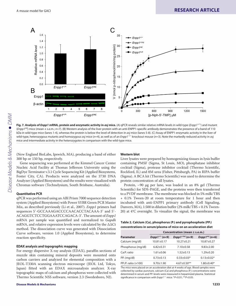
To examine the consequences of the missense mutation inEnpp1 at the mRNA and protein levels, quantitative PCR (qPCR)and western analyses were performed on four asj and four wild-type mice sacrificed at 4-12 weeks of age. The mRNA levels werenot different in the liver of asj and wild-type mice by qPCR (Fig. 7A).However, isolation of the protein from liver with subsequentwestern analysis with an anti-ENPP1 specific antibody clearlyrevealed the presence of a band of the appropriate size, 110 kDa,in wild-type mice, but the level of protein was below the detectionlimit of the western analysis in asjmice (Fig. 7B). It should be notedthat, although the antibody used is clearly specific for wild-typeENPP1, its precise epitope is not known and, consequently, ourresults do not exclude the possibility that it does not recognize themutant protein. This possibility, however, is unlikely because theantibody used is polyclonal. The enzyme kinetics revealed that theENPP1 isolated from the liver of asj mice was markedly reducedin activity (Fig. 7C). The Michaelis constant (Km) for the wild-typeEnpp1 mouse, as determined by a Hanes-Woolf plot, was213.6±14.0 μM (mean ± s.e.m.), as compared with a Km of 192.0±6.7μM for asj mice (Fig. 7C) (P>0.05). However, the maximum rateof reaction (Vmax) for the wild-type enzyme was 33.4±2.1 nmol p-nitrophenol released/minute/mg protein versus 8.1±0.3 nmol in asjmice (P<0.01).
Because the asj mice demonstrated residual enzyme activity, yetwestern analysis showed the presence of little, if any, ENPP1 protein,
two critical experiments were performed. First, the ENPP1enzymatic activity was measured in heterozygous asj mice incomparison with wild-type and homozygous mutant mice. Theresults showed that the Km in heterozygous mice was 203.8±13.1μM. The Vmax was 20.8±0.8 nmol, a 38% reduction compared withwild-type enzyme measured at linear range of reaction (P<0.05).Second, liver from Enpp1tm1Gdg knockout mice, developed bytargeted ablation of the gene, was used for the correspondingenzyme assay (Sali et al., 1999). These mice showed a very low Vmax,2.9±0.4 nmol, even less than in asj mice (P<0.05), suggesting thatthe asj mice might have some residual activity, and not becompletely null (Fig. 7C).
To examine the consequences of reduced ENPP1 activity in asjmice, the concentrations of PPi in plasma of wild-type, heterozygousmutant and homozygous mutant mice were determined in a three-step enzymatic reaction. As expected, the asjmice showed markedlyreduced PPi levels, ~20% from the wild-type controls, withconcomitant reduction in the PPi:Pi ratio, and the heterozygousmice showed intermediate levels (Table 2). The serum calcium andphosphorus levels and the corresponding Ca:P ratio were notstatistically different in these three groups of mice (Table 2).
DISCUSSIONIn this study, we have extensively characterized a mutant Enpp1asjmouse as a model for GACI. This autosomal recessive disordermanifests with profound arterial mineralization; results of prenatalultrasound can often suggest the possibility of this disease, which
Disease Models & Mechanisms 1229
A mouse model for GACI RESEARCH ARTICLE
Fig. 1. Phenotypic presentation and aberrant mineralization in asj mice at 12 weeks of age. (A)The Enpp1asj mice develop progressive stiffening of thejoints leading to contractures as shown on the front paws (lower panel) in comparison with a corresponding wild-type mouse (upper panel). (B)Histopathologyreveals extensive mineralization in the heart, aorta, carotid artery, retina of the eye and dermal sheath of vibrissae, but not in the liver parenchyma of asj mice.(C)Extensive aberrant mineralization in the kidneys of heterozygous (middle panel) and homozygous (lower panel) asj mice. Focal areas of mineralization arealso noted in the kidneys of wild-type mice (upper panel). Alizarin red stain; original magnifications: heart, carotid artery, eye, vibrissae, liver, 150×; aorta, kidney,100×.
Dise
ase
Mod
els &
Mec
hani
sms
D
MM

is then commonly diagnosed in the early postnatal period (Nitschkeand Rutsch, 2012b; Rutsch et al., 2011). In most cases, the affectedchildren die within the first 6 months of life as a consequence ofvascular insufficiency causing end-organ damage. This disease inits classic form is caused by mutations in the ENPP1 gene, whichencodes ectonucleotide pyrophosphatase/phosphodiesterase 1(ENPP1), also known as plasma cell membrane glycoprotein 1 (PC-1) (Ruf et al., 2005). This enzyme converts ATP to AMP and PPi,and this pathway is the main source of inorganic pyrophosphate.The extracellular PPi plays a crucial role as an inhibitor ofhydroxyapatite formation, whereas Pi promotes the formation ofhydroxyapatite crystals. Thus, the mineralization is controlled bythe PPi:Pi ratio as a result of activities of a number of enzymes,including ENPP1 and tissue nonspecific alkaline phosphatase(TNAP), as well as transporter proteins mediating the extracellulartransport of Pi and PPi, including ankylosis protein (ANK) and typeIII sodium-dependent Pi co-transporter 1 (PiT1) (Mackenzie et al.,2012). Thus, a reduced PPi:Pi ratio in individuals with GACI as aresult of reduced ENPP1 activity mechanistically leads to aberrantmineralization of extracellular connective tissues.
The asj mice examined in this study have several features thatrecapitulate GACI in humans. The hallmark of the disease,extensive arterial calcification, can be demonstrated in the asjmiceas early as 4 weeks of age when the mice are on the accelerationdiet, and the homozygous mice often die most likely as aconsequence of aberrant mineralization of the aorta as well as of
blood vessels in other tissues, including heart and carotid arteries.Notably, these mice also demonstrated mineral deposits in theretina of the eye as well as in the dermal sheath of vibrissae in themuzzle skin. Similar to individuals with GACI, the plasmaconcentration of PPi was markedly reduced (<20% of the controlmice) and the heterozygotes demonstrated intermediate levels. Itshould be noted that the heterozygous littermates did notdemonstrate vascular mineralization, with the exception of thekidneys, and were indistinguishable from the wild-type controls,attesting to the autosomal recessive mode of the asj mutant allele.In this context, it should be noted that currently there is no evidencefor modulation of the Enpp1asj mutation by other genes, such asAbcc6 in PXE mice (Klement et al., 2005) or in some inbred strainsof mice (KK/HlJ, DBA/2J and C3H/HeJ) (Berndt et al., 2013)associated with vascular mineralization.
The asjmouse was developed at The Jackson Laboratory as partof the ENU mutagenesis program (http://mousemutant.jax.org/articles/mmrmutantasj.html). The phenotypic features of thesemice include stiffening of the joints [hence ‘ages with stiffenedjoints’ (asj)], and these mice were shown to harbor a missensemutation, p.V246D, in the Enpp1 gene. Initial necropsies of thesemice at 7 months of age revealed periarticular mineralization ofthe ligaments and mineralization of the dermal sheath of vibrissae.Previously, mutations affecting the Enpp1 gene have been describedin a mutant mouse, ‘tip toe walking’ (ttw/ttw), shown to harbor astop codon mutation in the Enpp1 coding sequence (Okawa et al.,
dmm.biologists.org1230
A mouse model for GACIRESEARCH ARTICLE
Fig. 2. Kaplan-Meier survival curves of asj mice on theacceleration diet. Note that >50% of asj mice died spontaneouslyprior to age 6 weeks, whereas the heterozygous mice (Enpp1+/asj)had a survival similar to wild-type controls.
Fig. 3. Quantitation of mineralization by chemical assayof calcium in the dermal sheath of vibrissae and in thekidneys of mice in the range of 4-12 weeks of age kepton acceleration diet. (A)Marked increase in mineralizationis noted in the vibrissae of homozygous asj mice incomparison with wild-type or heterozygous animals (n=8).(B)Markedly increased mineralization in the kidneys ofheterozygous and homozygous asj mice is noted (n=7-9).Statistical significance: *P<0.05 versus wild type; **P<0.001versus wild type.
Dise
ase
Mod
els &
Mec
hani
sms
D
MM

1998). Similarly, Enpp1tm1Gdg knockout mice exhibit abnormalitiessimilar to those in ttw/ttw mice (Sali et al., 1999). Furthermore,another Enpp1 mutant mouse, with a p.C397S missense mutation,has been characterized by low bone mineral density, crystal-relatedarthropathy and vascular calcification (Babij et al., 2009).Characterization of these mice has largely focused on alterationsin bone mineralization in long bones, the calvariae and periarticular,as well as perispinal soft tissue mineralization. Although arterialcalcification was documented in some of these previously describedmice, the changes were frequently not noted to be present until16-22 weeks of age (Babij et al., 2009; Mackenzie et al., 2012). Inasj mice, the mineralization was noted to occur as early as 4 weeks
of age, with an early demise at ~6 weeks of birth when on theacceleration diet. Consequently, this mouse model accuratelyrecapitulates features of GACI, a disease that is usually lethal withinthe first 6 months of life. This difference in the age of onset ofmineralization can be attributable, at least in part, to the specialdiet that was used in our study to accelerate the mineralizationprocess. This ‘acceleration diet’ consists of increased phosphate andreduced magnesium in comparison with the standard mouselaboratory diet (Jiang and Uitto, 2012; Li and Uitto, 2010).Specifically, the phosphate concentration was increased twofold(8.5 mg/g of food), and the magnesium content was reduced to20% of the control (0.4 mg/g of food). All the mice were placed onthis experimental diet at weaning at 4 weeks of age but, in addition,the mothers during the pregnancy and lactation were on this specialdiet. We previously showed that this diet also accelerates theaberrant mineralization noted in Abcc6tm1Jfk-null mice, a model ofanother aberrant mineralization disorder, pseudoxanthomaelasticum (Jiang and Uitto, 2012). It should be noted, however, thatthis diet does not cause any mineralization in the arterial vessels,the eyes or in the vibrissae in wild-type control mice or inheterozygotes of mutations in either Enpp1 or Abcc6.
An interesting observation was extensive mineralization in thekidneys of homozygous and heterozygous asj mice, and wild-typecontrol mice also showed some evidence of aberrant mineralizationwhen the mice were placed on the experimental acceleration diet.The observed mineralization of kidneys in the heterozygous micediffers from that in humans in that heterozygous carriers of ENPP1mutations do not show any evidence of mineralization. Similarfindings of nephrocalcinosis have previously been noted in micewith a high phosphorus-containing diet, similar to individuals withhyperphosphatemia (Markowitz and Perazella, 2009). Theincreased mineralization in the kidney suggests that this might bea process determined by a more complex genetic background inwhich ENPP1 plays a role but is not the sole contributor.
In their classic forms, GACI and PXE are two clinically distinctconditions, GACI manifesting with extensive arterial calcification
Disease Models & Mechanisms 1231
A mouse model for GACI RESEARCH ARTICLE
Fig. 4. Computed tomography demonstrating extensive mineralization ofdermal sheath of vibrissae in a 7-week-old asj mouse in comparison witha wild-type littermate. Single slices demonstrate evidence of mineralization(arrowheads in B), and computerized reconstruction reveals the mineraldeposits in association with dermal sheath of vibrissae (upper left panel in B,arrowheads).
Fig. 5. Energy dispersive X-ray analysis of the mineral deposits in the dermal sheath of vibrissae in asj mice. (A)Elemental composition analysis reveals thepresence of calcium and phosphorus in ~2:1 ratio. (B)X-ray topography of the distribution of phosphorus and calcium reveals colocalization, as demonstrated bymerging the images.
Dise
ase
Mod
els &
Mec
hani
sms
D
MM

at birth leading to early demise of the affected individuals,whereas the clinical manifestations and tissue mineralization inPXE is of late onset and slowly progressive (Nitschke and Rutsch,2012b; Uitto et al., 2010). However, recently, individuals withfeatures of early GACI with development of PXE, withcharacteristic skin findings, have been noted (Le Boulanger et al.,2010; Li et al., 2012). In addition, a subset of individuals with GACIhas been recently shown to harbor mutations in the ABCC6 gene,instead of ENPP1 (Nitschke and Rutsch, 2012b). Theseobservations suggest the presence of common pathomechanisticpathways leading to aberrant tissue mineralization (Nitschke andRutsch, 2012a). Although the pathomechanistic details of tissuemineralization particularly in the case of PXE are currentlyunknown, it should be noted that the PPi levels in plasma ofAbcc6−/− mice are not altered (Q.L. et al., unpublished). Finally,there are a number of additional heritable aberrant mineralizationdisorders resulting in calcium deposits in the skin, including
normophosphatemic and hyperphosphatemic familial tumoralcalcinosis, and arterial calcification with CD73 deficiency, eachdue to mutations in different genes (Li and Uitto, 2013). Thepresence of hydroxyapatite crystal deposition in thesephenotypically diverse conditions attest to the complexmineralization/anti-mineralization network required for normalhomeostasis. Dissection of the pathomechanistic details leadingto aberrant mineral deposition in these single-gene disorders willassist us in the development and testing of efficient treatmentmodalities. In this context, the asjmice could well serve as a modelsystem to study potential treatment modalities for GACI undergenetically and environmentally controlled conditions. In thisregard, a few studies have reported improvement in some patientswith GACI with treatment with bisphosphonates, but controlledstudies attesting to the efficacy of this treatment strategy arelacking (Edouard et al., 2011; Ramjan et al., 2009). Furthermore,this mouse model could be used to test other compounds, suchas pyrophosphate and sodium thiosulfate, which have beensuggested to counteract vascular mineralization (Hayden andGoldsmith, 2010; Ning et al., 2013; O’Neill et al., 2011).Collectively, the ability to carefully dissect and analyze asj micecould provide an enhanced clinical understanding of GACI andcontribute towards improved treatment.
MATERIALS AND METHODSAnimals and dietEnpp1asj mice on a C57BL/6J background were obtained from TheJackson Laboratory (Bar Harbor, ME) (http://mousemutant.jax.org/articles/mmrmutantasj.html). Enpp1+/+ and Enpp1asj mice weregenerated from heterozygous matings. Mice were maintainedeither on standard laboratory diet (Laboratory Autoclavable RodentDiet 5010; PMI Nutritional International, Brentwood, MO) or fedan ‘acceleration diet’ (Harlan Teklad, Rodent diet TD.00442,Madison, WI), which we have previously shown to accelerate theectopic mineralization in Abcc6−/− mice; this diet is enriched inphosphorus and has reduced magnesium content (Jiang and Uitto,2012). The mice were maintained under standard conditions at theAnimal Facility of Thomas Jefferson University, and all protocolswere approved by the Institutional Animal Care and Use Committeeof Thomas Jefferson University. Proper handling and care werefollowed according to the Animal Welfare Policies of the PublicHealth Service.
Genotyping and gene sequencingA primer pair with sequences 5�-TGATCTGCATCCTGGGATAA-3� and 5�-TAAGGAAAGACCAATTGCAGA-3� was used toamplify exon 7 of the Enpp1 gene. To identify the wild-type andEnpp1asj mutant alleles, PCR products were digested with TaqαI
dmm.biologists.org1232
A mouse model for GACIRESEARCH ARTICLE
Table 1. Aberrant tissue mineralization in Enpp1+/+, Enpp1+/asj and Enpp1asj mice on an acceleration diet
Genotype No. mice examined
Soft tissue mineralization (%)
Vibrissae Liver Kidneys Heart Aorta Eyes Carotid artery Enpp1+/+ 13 0 0 85 0 0 8 0
Enpp1+/asj 9 0 8 89 15 0 0 0
Enpp1asj 9 100** 56* 100 67** 56* 44 78**
Mice were placed on acceleration diet at 4 weeks of age and tissues were collected for histopathology at 3 months or earlier at the time of demise of the Enpp1asj mice. The values
represent the percent of mice with tissues affected by mineralization as examined by H&E stain on one section. Statistical analyses were performed with Fisher’s exact test; *P<0.01;
**P<0.001.
Fig. 6. Genotyping and mutation analysis of asj mice. (A)Sequence analysisreveals a homozygous T-to-A nucleotide substitution (arrows) in asj mice,which results in substitution of valine 246 for an aspartic acid (p.V246D).(B)TaqaI restriction enzyme digestion of PCR products corresponding to exon7 of the Enpp1 gene reveals a 300 bp fragment representing the wild-typeallele and a 150 bp fragment corresponding to the mutant allele.
Dise
ase
Mod
els &
Mec
hani
sms
D
MM
(New England BioLabs, Ipswich, MA), producing a band of either300 bp or 150 bp, respectively.
Gene sequencing was performed at the Kimmel Cancer CenterNucleic Acid Facility at Thomas Jefferson University using theBigDye Terminator v.3.1 Cycle Sequencing Kit (Applied Biosystems,Foster City, CA). Products were analyzed on the 3730 DNAAnalyzer (Applied Biosystems) and the results were visualized withChromas software (Technelysium, South Brisbane, Australia).
Quantitative PCRqPCR was performed using an ABI Prism 7000 sequence detectionsystem (Applied Biosystems) with Power SYBR Green PCR MasterMix, as described previously (Li et al., 2007). Enpp1 primers hadsequences 5�-GCCAAAGACCCCAACACCTACAAA-3� and 5�-ACAGGTCTCCTGGAAATCCAGACA-3�. The amount of Enpp1mRNA per sample was quantified and normalized to GapdhmRNA, and relative expression levels were calculated by the ΔΔCtmethod. The dissociation curve was generated with DissociationCurve software, version 1.0 (Applied Biosystems), to determinereaction specificity.
EDAX analysis and topographic mappingFor energy dispersive X-ray analysis (EDAX), paraffin sections ofmuzzle skin containing mineral deposits were mounted ontocarbon carriers and analyzed for elemental composition with aJEOL-T330A scanning electron microscope (JEOL Ltd, Tokyo,Japan) fitted with an EDAX microanalysis analyzer. X-raytopographic maps of calcium and phosphorus were collected withThermo Scientific NSS software, version 2.3 (Swedesboro, NJ).
Western blotLiver lysates were prepared by homogenizing tissues in lysis buffercontaining PMSF (Sigma, St Louis, MO), phosphatase inhibitorcocktail (Sigma), protease inhibitor cocktail (Thermo Scientific,Rockford, IL) and 8M urea (Fisher, Pittsburgh, PA) in RIPA buffer(Sigma). A BCA kit (Thermo Scientific) was used to determine theprotein concentration of all lysates.
Protein, ~90 μg per lane, was loaded in an 8% gel (ThermoScientific) for SDS-PAGE, and the proteins were then transferredto a PVDF membrane. The membrane was blocked in 5% milk/TBS+ 0.1% Tween-20 at room temperature for 1 hour and thenincubated with anti-ENPP1 primary antibody (Cell Signaling,Danvers, MA), 1:500 in dilution buffer (2% milk/TBS + 0.1% Tween-20) at 4°C overnight. To visualize the signal, the membrane was
Disease Models & Mechanisms 1233
A mouse model for GACI RESEARCH ARTICLE
Table 2. Calcium (Ca), phosphorus (Pi) and pyrophosphate (PPi) concentrations in serum/plasma of mice on an acceleration diet
Parameter
Concentration (mean ± s.e.m.)
Enpp1+/+ (n=9) Enpp1+/asj (n=9) Enpp1asj (n=6) Calcium (mg/dl) 10.81±0.17 10.27±0.21 10.87±0.27
Phosphorus (mg/dl) 6.82±0.31 7.10±0.58 9.83±2.00
Ca:Pi ratio 1.61±0.06 1.52±0.13 1.29±0.20
PPi (mg/dl) 0.73±0.13 0.33±0.03* 0.13±0.02*
PPi:Pi ratio (×100) 9.70±1.90 4.67±0.50** 1.80±0.40*
The mice were placed on an acceleration diet at 4 weeks of age. Blood samples were
collected by cardiac puncture, calcium (Ca) and phosphorus (Pi) concentrations were
determined in serum and PPi levels were measured in heparinized plasma. Statistical
significance in comparison with Enpp1+/+ mice: *P<0.01; **P<0.05.
Fig. 7. Analysis of Enpp1 mRNA, protein and enzymatic activity in asj mice. (A)qPCR reveals similar relative mRNA levels in wild-type (Enpp1+/+) and mutant(Enpp1asj) mice (mean ± s.e.m.; n=7). (B)Western analysis of the liver protein with an anti-ENPP1-specific antibody demonstrates the presence of a band of 110kDa in wild-type mice (lanes 1-4), whereas the protein is below the level of detection in asj mice (lanes 5-8). (C)Assay of ENPP1 enzymatic activity in the liver ofwild-type, heterozygous mutants and homozygous asj mice (n=4), as well as of an Enpp1−/− knockout mouse (n=3). Note the markedly reduced activity in asjmice and intermediate activity in the heterozygotes in comparison with the wild-type mice.
Dise
ase
Mod
els &
Mec
hani
sms
D
MM
incubated in anti-rabbit secondary antibody (LI-COR, Lincoln, NE)in dilution buffer for 1 hour at room temperature and then scannedwith an Odyssey Infrared Imager (LI-COR). The membrane wasthen stripped and reprobed with anti-β-actin antibody (Bioorbyt,San Francisco, CA) 1:750 in dilution buffer.
Enzyme activity assayENPP1 activity in wild-type, heterozygous and asj mouse liver, aswell as in Enpp1tm1Gdg knockout mice (kindly provided by Dr RobertTerkeltaub, University of California, San Diego, CA), wasdetermined using the substrate thymidine 5�-monophosphate p-nitrophenyl ester (p-Nph-5�-TMP, Sigma). Total proteins wereextracted from whole liver with lysis buffer containing 50 mMHEPES, 0.1 mM EGTA, 0.1 mM EDTA, 120 mM NaCl, 0.5% NP-40, pH 7.5, PMSF, and complete protease inhibitor (Roche) (Babijet al., 2009). A BCA kit (Pierce) was used to determine the proteinconcentration of lysates. Protein lysates were diluted with assaybuffer (50 mM Tris-HCl, pH 9.5, and 250 mM NaCl in water) to100 ng/μl. In 96-well plates, 50 μl of p-Nph-5�-TMP (diluted withassay buffer to nine different concentrations) was added to 50 μlof protein lysate. All samples were tested in duplicate. The sampleswere then incubated at 37°C, and absorbance (400 nm) wasmeasured with a microplate reader (Bio-Rad 800) every 5 minutesfor up to 30 minutes to ensure linearity of the reaction. A molarextinction coefficient of the reaction product, p-nitrophenol, of18.4×103 M−1 cm−1 was used in determination of enzyme kinetics.Enzyme activity was expressed as nmol p-nitrophenol released perminute per mg of protein.
Quantification of calcium and phosphateTo quantify the calcium deposition in the dermal sheath of mousevibrissae, and the kidneys, muzzle skin and kidney were harvestedand decalcified with 0.15 N HCl for 48 hours (skin) or with 0.6 NHCl for 1 week (kidney) at room temperature. The calcium contentin these samples as well as in serum was determinedcolorimetrically by the o-cresolphthalein complexone method[Calcium (CPC) Liquicolor; Stanbio Laboratory, Boerne, TX]. Thephosphate content of serum was determined with Malachite GreenPhosphate Assay kit (BioAssay Systems, Hayward, CA). The valuesfor calcium and phosphate were normalized to tissue weight.
Inorganic pyrophosphate assayPPi was measured by an enzymatic assay using uridine-diphosphoglucose (UDPG) pyrophosphorylase as previouslydescribed (Lomashvili et al., 2005; O’Neill et al., 2010), withmodifications. Heparinized plasma samples (20 μl; 1:4 dilution)were heated at 65°C for 10 minutes, followed by three differentassays performed on each sample: (1) no addition of PPi standard,(2) pre-incubation with 0.35 U pyrophosphatase at 37°C for 1 hour,and (3) addition of 3 μM PPi. Samples were then added to 100 μlof reaction buffer that contained 5.2 mM Mg Acetate, 57 mM TrisAcetate (pH 7.8), 4 μM NADP, 7.5 μM UDPG, 18.6 μM glucose-1,6-diphosphate, 0.14 U UDPG pyrophosphorylase, 2.5 Uphosphoglucomutase, 0.4 U glucose-6-phosphate dehydrogenase,and 0.02 μCi [3H]UDPG. After a 30-minute incubation at 37°C,200 μl of 4% activated charcoal was added to each sample withoccasional stirring to bind residual UDPG. After centrifugation,
the radioactivity in 100 μl of supernatant was counted. The plasma[PPi] was determined as (CPM1-CPM2)/(CPM3-CPM1)×3 μM.
Histopathological analysisMuzzle skin and internal organs from euthanized mice were fixedin 10% phosphate-buffered formalin, routinely processed, andembedded in paraffin. Tissues were sectioned (6 μm) and stainedwith H&E and Alizarin red using standard procedures. Slides wereexamined under light microscopy for mineralization and otherlesions by an experienced veterinary pathologist (J.P.S.).
Small-animal computed tomography (CT scan)Enpp1 wild-type and asj mice were examined for mineralizationat 7 weeks of age by CT scan, as described (Le Corre et al., 2012).Briefly, mice were anesthetized with a xylazine-ketamine-acetopromazine cocktail (160 μl per 25 g body weight of 10 mg/kgxylazine, 200 mg/kg ketamine, 2 mg/kg acetopromazine) and thenscanned with a MicroCAT II (ImTek Inc., Oak Ridge, TN). A 3-dimensional facial rendering was created for each mouse usingAmira software, version 3.1 (Visualization Sciences Group,Burlington, MA).
Statistical analysisThe comparisons in different groups of mice were completed usingtwo-sided Kruskal-Wallis nonparametric tests. The Kruskal-Wallistest is comparable to one-way analysis of variance, but without theparametric assumptions. Fisher’s exact test was used to determinethe difference between proportions in mineralization phenotypesin mice. All statistical computations were completed using SPSSversion 15.0 software (SPSS Inc., Chicago, IL).ACKNOWLEDGEMENTSThe authors thank Adele Donahue, Alix Grand-Pierre and Dian Wang for technicalhelp; Mark Pawlowski for assistance with histopathology; and Carol Kelly inmanuscript preparation. Gerald Harrison assisted in EDAX analysis, and DrMadhukar Thakur in CT scan. Dr Robert Terkeltaub, UCSD, generously providedliver samples from Enpp1tm1Gdg mice.
COMPETING INTERESTSThe authors declare that they do not have any competing or financial interests.
AUTHOR CONTRIBUTIONSQ.L., H.G. and D.W.C. performed the experiments; A.B. and J.P.S. performedhistopathologic analyses; J.U. developed the concept, interpreted the data andprepared the manuscript.
FUNDINGThis was supported by NIH/NIAMS grants R01 AR28450 and R21 AR063781. Q.L.and A.B. were the recipients of the North American Hair Research SocietyMentorship Award.
REFERENCESBabij, P., Roudier, M., Graves, T., Han, C. Y., Chhoa, M., Li, C. M., Juan, T., Morony,
S., Grisanti, M., Li, X. et al. (2009). New variants in the Enpp1 and Ptpn6 genescause low BMD, crystal-related arthropathy, and vascular calcification. J. Bone Miner.Res. 24, 1552-1564.
Berndt, A., Li, Q., Potter, C. S., Liang, Y., Silva, K. A., Kennedy, V., Uitto, J. andSundberg, J. P. (2013). A single-nucleotide polymorphism in the Abcc6 geneassociates with connective tissue mineralization in mice similar to targeted modelsfor pseudoxanthoma elasticum. J. Invest. Dermatol. 133, 833-836.
Edouard, T., Chabot, G., Miro, J., Buhas, D. C., Nitschke, Y., Lapierre, C., Rutsch, F.and Alos, N. (2011). Efficacy and safety of 2-year etidronate treatment in a child withgeneralized arterial calcification of infancy. Eur. J. Pediatr. 170, 1585-1590.
Hayden, M. R. and Goldsmith, D. J. (2010). Sodium thiosulfate: new hope for thetreatment of calciphylaxis. Semin. Dial. 23, 258-262.
dmm.biologists.org1234
A mouse model for GACIRESEARCH ARTICLED
iseas
e M
odel
s & M
echa
nism
s
DM
M

Jiang, Q. and Uitto, J. (2012). Restricting dietary magnesium accelerates ectopicconnective tissue mineralization in a mouse model of pseudoxanthoma elasticum(Abcc6–/–). Exp. Dermatol. 21, 694-699.
Kavukcuoglu, N. B., Li, Q., Pleshko, N. and Uitto, J. (2012). Connective tissuemineralization in Abcc6-/- mice, a model for pseudoxanthoma elasticum. Matrix Biol.31, 246-252.
Klement, J. F., Matsuzaki, Y., Jiang, Q. J., Terlizzi, J., Choi, H. Y., Fujimoto, N., Li, K.,Pulkkinen, L., Birk, D. E., Sundberg, J. P. et al. (2005). Targeted ablation of theabcc6 gene results in ectopic mineralization of connective tissues. Mol. Cell. Biol. 25,8299-8310.
Le Boulanger, G., Labrèze, C., Croué, A., Schurgers, L. J., Chassaing, N., Wittkampf,T., Rutsch, F. and Martin, L. (2010). An unusual severe vascular case ofpseudoxanthoma elasticum presenting as generalized arterial calcification ofinfancy. Am. J. Med. Genet. A. 152A, 118-123.
Le Corre, Y., Le Saux, O., Froeliger, F., Libouban, H., Kauffenstein, G., Willoteaux,S., Leftheriotis, G. and Martin, L. (2012). Quantification of the calcificationphenotype of Abcc6-deficient mice with microcomputed tomography. Am. J. Pathol.180, 2208-2213.
Li, Q. and Uitto, J. (2010). The mineralization phenotype in Abcc6 (-/-) mice is affectedby Ggcx gene deficiency and genetic background – a model for pseudoxanthomaelasticum. J. Mol. Med. (Berl.) 88, 173-181.
Li, Q. and Uitto, J. (2013). Mineralization/anti-mineralization networks in the skin andvascular connective tissues. Am. J. Pathol. 183, 10-18.
Li, Q., Jiang, Q., Larusso, J., Klement, J. F., Sartorelli, A. C., Belinsky, M. G., Kruh, G.D. and Uitto, J. (2007). Targeted ablation of Abcc1 or Abcc3 in Abcc6(-/-) mice doesnot modify the ectopic mineralization process. Exp. Dermatol. 16, 853-859.
Li, Q., Schumacher, W., Jablonski, D., Siegel, D. and Uitto, J. (2012). Cutaneousfeatures of pseudoxanthoma elasticum in a patient with generalized arterialcalcification of infancy due to a homozygous missense mutation in the ENPP1 gene.Br. J. Dermatol. 166, 1107-1111.
Lomashvili, K. A., Khawandi, W. and O’Neill, W. C. (2005). Reduced plasmapyrophosphate levels in hemodialysis patients. J. Am. Soc. Nephrol. 16, 2495-2500.
Mackenzie, N. C., Huesa, C., Rutsch, F. and MacRae, V. E. (2012). New insights intoNPP1 function: lessons from clinical and animal studies. Bone 51, 961-968.
Markowitz, G. S. and Perazella, M. A. (2009). Acute phosphate nephropathy. KidneyInt. 76, 1027-1034.
Ning, M. S., Dahir, K. M., Castellanos, E. H. and McGirt, L. Y. (2013). Sodiumthiosulfate in the treatment of non-uremic calciphylaxis. J. Dermatol. [Epub ahead ofprint] doi: 10.1111/1346-8138.12139.
Nitschke, Y. and Rutsch, F. (2012a). Generalized arterial calcification of infancy andpseudoxanthoma elasticum: two sides of the same coin. Front. Genet. 3, 302.
Nitschke, Y. and Rutsch, F. (2012b). Genetics in arterial calcification: lessons learnedfrom rare diseases. Trends Cardiovasc. Med. 22, 145-149.
O’Neill, W. C., Sigrist, M. K. and McIntyre, C. W. (2010). Plasma pyrophosphate andvascular calcification in chronic kidney disease. Nephrol. Dial. Transplant. 25, 187-191.
O’Neill, W. C., Lomashvili, K. A., Malluche, H. H., Faugere, M. C. and Riser, B. L.(2011). Treatment with pyrophosphate inhibits uremic vascular calcification. KidneyInt. 79, 512-517.
Okawa, A., Nakamura, I., Goto, S., Moriya, H., Nakamura, Y. and Ikegawa, S. (1998).Mutation in Npps in a mouse model of ossification of the posterior longitudinalligament of the spine. Nat. Genet. 19, 271-273.
Ramjan, K. A., Roscioli, T., Rutsch, F., Sillence, D. and Munns, C. F. (2009).Generalized arterial calcification of infancy: treatment with bisphosphonates. Nat.Clin. Pract. Endocrinol. Metab. 5, 167-172.
Ruf, N., Uhlenberg, B., Terkeltaub, R., Nürnberg, P. and Rutsch, F. (2005). Themutational spectrum of ENPP1 as arising after the analysis of 23 unrelated patientswith generalized arterial calcification of infancy (GACI). Hum. Mutat. 25, 98.
Rutsch, F., Nitschke, Y. and Terkeltaub, R. (2011). Genetics in arterial calcification:pieces of a puzzle and cogs in a wheel. Circ. Res. 109, 578-592.
Sali, A., Favaloro, J. M., Terkeltaub, R. and Goding, J. W. (1999). Germline deletion ofthe nucleoside triphosphate pyrophosphohydrolase (NTPPPH) plasma cellmembrnae glycoprotein-1 (PC-1) produces abnormal calcification of periarticulartissues. In Ecto-ATPases and Related Ectoenzymes, (ed. L. R. Vanduffel), pp. 267-282.Maastricht, The Netherlands: Shaker Publishing.
Uitto, J., Li, Q. and Jiang, Q. (2010). Pseudoxanthoma elasticum: molecular geneticsand putative pathomechanisms. J. Invest. Dermatol. 130, 661-670.
Disease Models & Mechanisms 1235
A mouse model for GACI RESEARCH ARTICLED
iseas
e M
odel
s & M
echa
nism
s
DM
M