muscle tissue & muscles - los angeles mission college 7... · comparison of the three types of...
TRANSCRIPT

Muscle Tissue & Muscles
Dr. Ali Ebneshahidi
ebneshahidi

Functions of the muscular system
1. Locomotion – all body movements are the results of skeletal
muscle contraction.
2. Vasoconstriction and Vasodilatation- constriction and dilation of
blood vessel walls are the results of smooth muscle contraction.
3. Peristalsis – wavelike motion along the digestive tract is produced
by the smooth muscle.
4. Cardiac motion – heart chambers are able to pump blood to the
lungs and the body because of cardiac muscle contraction.
5. Posture maintenance- contraction of skeletal muscles maintains
body posture and muscle tone.
6. Heat generation – about 75% of ATP energy used in muscle
contraction is released as heat to help maintain a constant body
temperature (since skeletal muscles are the most abundant‚ they
release the largest amount of heat in muscle tissue).
ebneshahidi
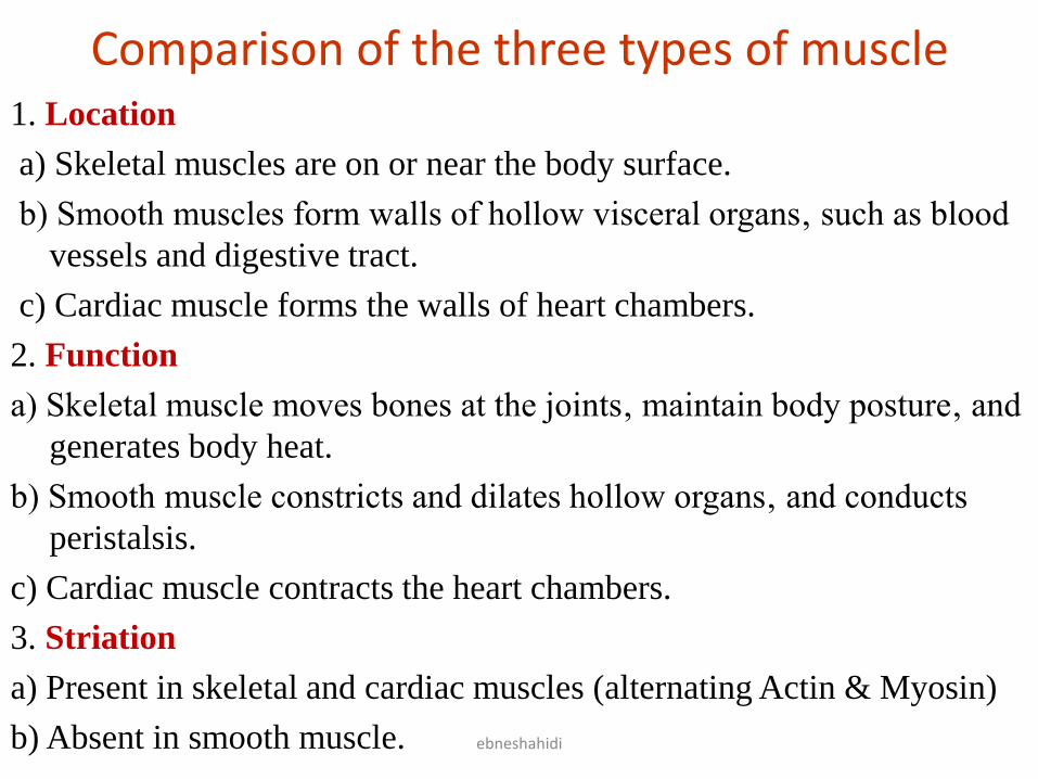
Comparison of the three types of muscle 1. Location
a) Skeletal muscles are on or near the body surface.
b) Smooth muscles form walls of hollow visceral organs‚ such as blood
vessels and digestive tract.
c) Cardiac muscle forms the walls of heart chambers.
2. Function
a) Skeletal muscle moves bones at the joints‚ maintain body posture‚ and
generates body heat.
b) Smooth muscle constricts and dilates hollow organs‚ and conducts
peristalsis.
c) Cardiac muscle contracts the heart chambers.
3. Striation
a) Present in skeletal and cardiac muscles (alternating Actin & Myosin)
b) Absent in smooth muscle.
ebneshahidi

ebneshahidi

4. Nucleus
a) smooth and cardiac are uninucleated (one nucleus per cell).
b) skeletal muscle is multinucleated (several nuclei per cell).
5. Transverse tubule ( T tubule )
a) well developed in skeletal and cardiac muscles to transport
calcium.
b) absent in smooth muscle.
6. Intercalated disk
a) specialized intercellular junction that only occurs in cardiac
muscle.
b) skeletal and smooth muscles mainly rely on desmosomes.
7. Mode of control
a) skeletal muscle is always under voluntary control‚ with some
exceptions (the diaphragm, and pili arrector muscles in the
dermis).
b) smooth and cardiac muscles are under involuntary control.
ebneshahidi

ebneshahidi

8. Contraction
a) Skeletal muscle contracts and relaxes rapidly with powerful force.
b) Smooth muscle contracts and relaxes slowly but at longer duration‚
and is self-exciting and rthymic.
c) Cardiac muscle contracts and relaxes as a unit (syncytium), and is also
self –exciting.
ebneshahidi

Smooth Muscle
a) Actin and myosin filaments are
randomly arranged in the myofibrils
(resulting in the lack of striations)‚ thus
smooth muscle contracts slowly and with
less force involved.
b) Sarcoplasmic reticulum (SR) is not
well developed and lacks transverse
tubules (T- tubules)‚ thus calcium ion
cannot be released rapidly.
ebneshahidi

ebneshahidi
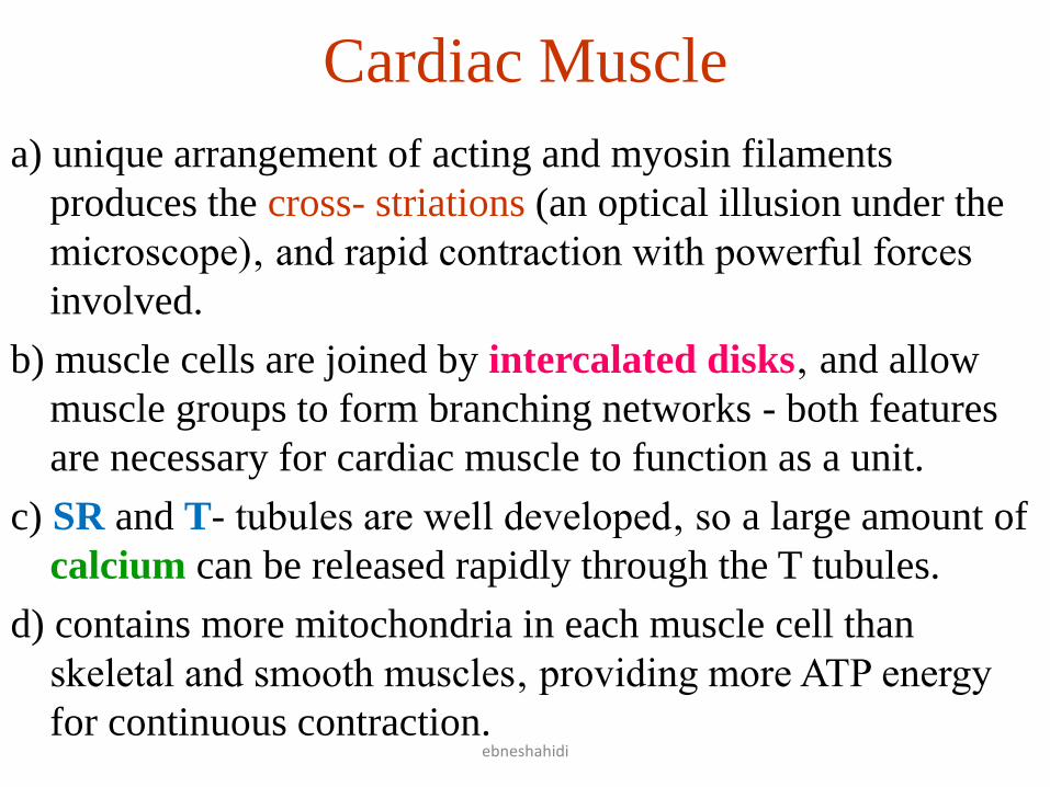
Cardiac Muscle
a) unique arrangement of acting and myosin filaments
produces the cross- striations (an optical illusion under the
microscope)‚ and rapid contraction with powerful forces
involved.
b) muscle cells are joined by intercalated disks‚ and allow
muscle groups to form branching networks - both features
are necessary for cardiac muscle to function as a unit.
c) SR and T- tubules are well developed‚ so a large amount of
calcium can be released rapidly through the T tubules.
d) contains more mitochondria in each muscle cell than
skeletal and smooth muscles‚ providing more ATP energy
for continuous contraction.
ebneshahidi

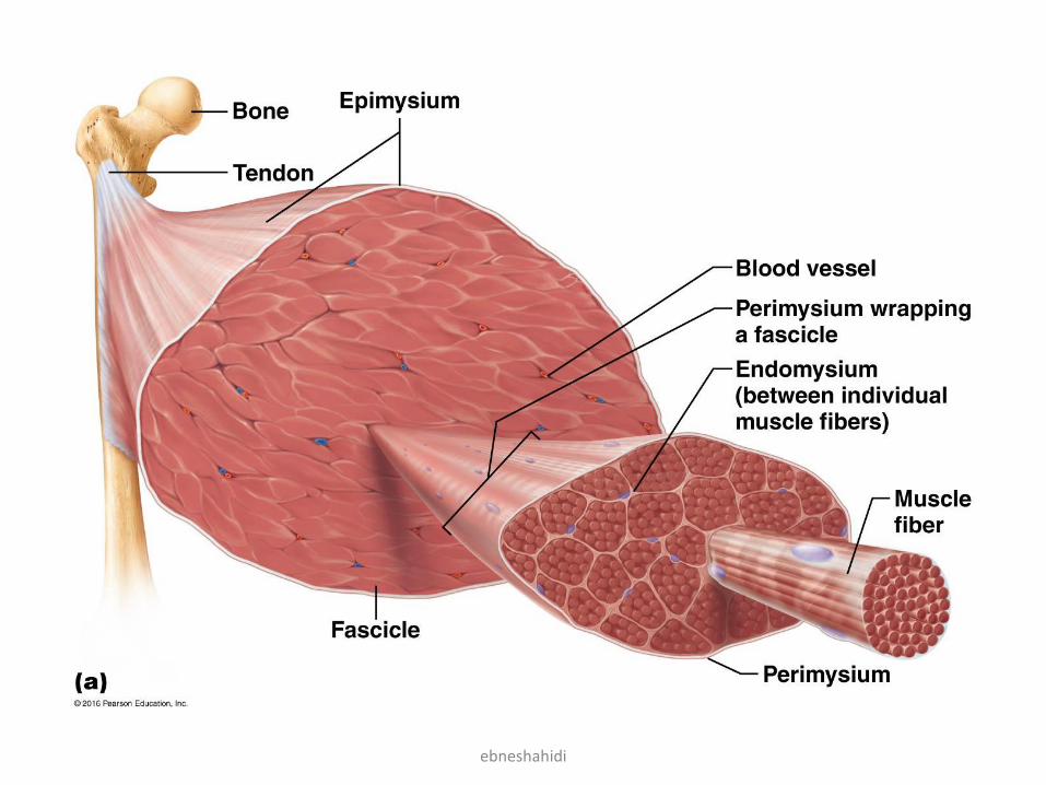
Skeletal Muscle
1. Gross anatomy
a) actin and myosin filaments → sarcomeres →
myofibrils → muscle fibers (muscle cells) →
fascicles → skeletal muscle.
b) coverings:
• Sarcolemma surrounds the cell (plasmalemma).
• Endomysium surrounds muscle fibers (between).
• Perimysium surrounds fascicles.
• Epimysium surrounds skeletal muscle.
ebneshahidi

ebneshahidi
ebneshahidi
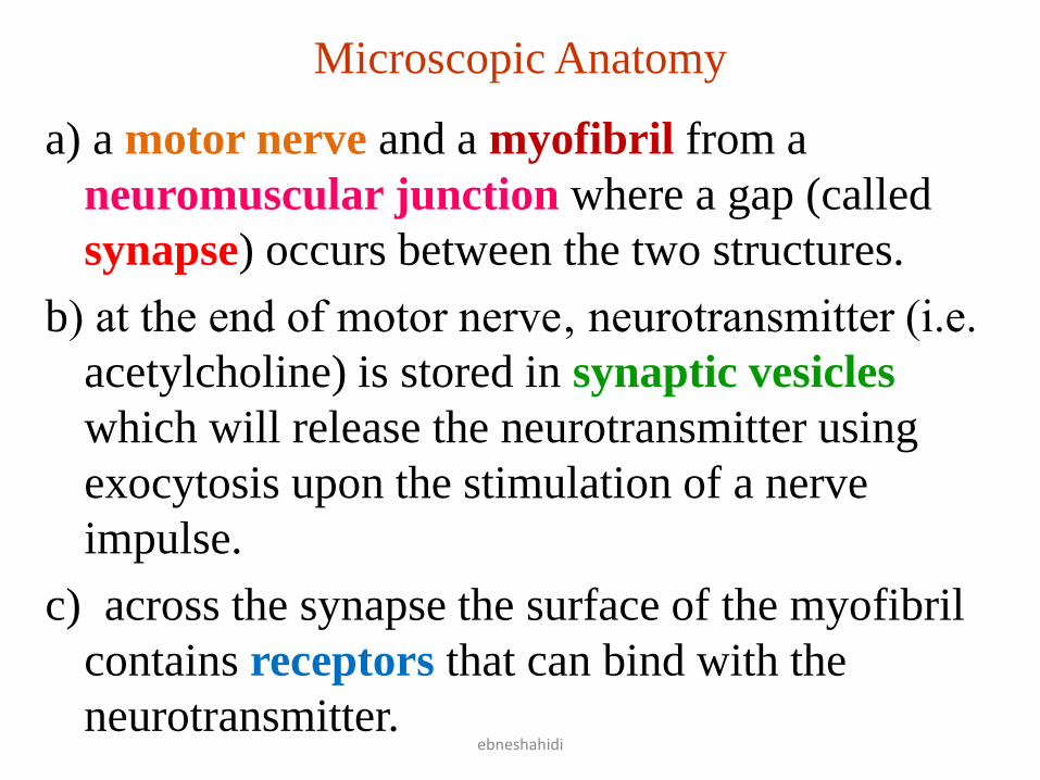
Microscopic Anatomy
a) a motor nerve and a myofibril from a
neuromuscular junction where a gap (called
synapse) occurs between the two structures.
b) at the end of motor nerve‚ neurotransmitter (i.e.
acetylcholine) is stored in synaptic vesicles
which will release the neurotransmitter using
exocytosis upon the stimulation of a nerve
impulse.
c) across the synapse the surface of the myofibril
contains receptors that can bind with the
neurotransmitter.
ebneshahidi

ebneshahidi
The Neuromuscular Junction

Parts of Skeletal Muscle
• a) origin- immovable end of the
muscle; connects to a bone by tendon
or to another muscle by fascia.
• b) belly middle portion of the muscle
where most contraction occurs.
• c) insertion – movable end of the
muscle; connects to a bone by tendon
or to another muscle by fascia.
• Example: during flexing motion of
the arm, the origin of biceps brachii
(at the coracoid process of scapula)
does not move, while the insertion
(at the radial tuberosity of radius)
does which pulls radius toward
humerus.
ebneshahidi

Interaction of skeletal muscles
a) prime mover – the muscle that initiates a movement and produces most of the force.
b) synergist – muscles that assist the prime mover.
c) antagonist – muscles that resist the prime mover.
• Example: during motion of the arm, biceps brachii serves as the prime mover and is assisted by brachialis (the synergist for this action), but is resisted by triceps brachii (the antagonist that tries to conduct extension to resist the prime mover).
ebneshahidi

Naming of skeletal muscles
a) action – some skeletal muscles are named according to their actions (e.g. adductor femoris brings the thigh toward the midline).
b) size – some skeletal muscles are named by their size (e.g. gluteus maximus is the largest muscle in the buttock).
c) shape- some skeletal muscles are named according their shapes (e.g. trapezius is trapezoid shaped, deltoid is triangular shaped).
d) location –some skeletal muscles are named by their locations (e.g. external oblique is a slanted muscle at the outermost surface of the abdomen).
e) attachment- some other skeletal muscles are named according to their attachments to bones (e.g. sternocleido-mastoid attaches from the sternum through the clavicle to the mastoid process of temporal bone).
ebneshahidi

Principal actions of skeletal muscles
Flexor- muscles that decrease the anterior angle at a
joint (e.g. flexor carpi radialis).
Extensor- muscles that increase the anterior angle at
a joint (e.g. extensor carpi ulnaris).
Abductor- muscles that move body parts away from
the midline (e.g. abductor pollicis brevis).
Adductor- muscles that move body parts closer to
the midline (e.g. adductor longus).
Levator – muscles that produce an upward
movement (e.g. levator scapulae ventralis).
ebneshahidi

Depressor- muscles that produce a downward
movement (e.g. depressor labii inferioris).
Pronator- muscles that turn the palm downward
(e.g. pronator teres).
Sphincter- muscles that decrease the size of an
opening (e.g. external anal sphincter).
Tensor- muscles that make a body part more rigid
(e.g. tensor fascia lata).
Rotator- muscles that move a body part around
its longitudinal axis (e.g. obturator externus).
ebneshahidi

Unique characteristics of skeletal muscle
Excitability- the ability to respond to a stimulus.
Contractility- the ability to undergo shortening.
Extensibility- the ability to be stretched.
Elasticity- the ability to resume its resting length after contraction.
Muscle tone- the ability to be partially contracted at all times.
• Isotonic contraction- muscle contracts while the tension within the muscle remains the same (e.g. lifting an object).
• Isometric contraction- muscle is unable to contract despite the increase of tension within the muscle (e.g. pushing an immovable object).
ebneshahidi

Connective tissue Covering of muscles
• Fascia: which separate
muscles is a layer of
dense connective tissue,
that may project beyond
the muscle fiber to from
a cord like tendon. It
may also form broad
fibrous sheets called
aponeuroses.
ebneshahidi

Major Skeletal Muscles of The Body Muscles of facial Expression:
1) The epicranius – covers the
upper part of cranium,
consists of 2 parts.
a) occipitalis – covers the
occipital bone and pulls scalp
posteriorly, innervated by
facial nerve (cranial nerve
VII).
b) frontails – covers the
frontal bone and raises the
eyebrows.
2) orbicularis oculi – Ring like
muscle that surrounds the eye.
it closes or blinks the eyelid,
innervated by facial nerve.
ebneshahidi

3) orbicularis oris – a muscle that encircle the mouth and is also known as kissing muscle, closes lips (cranial nerve VII).
4) buccinators: located in the wall of cheek. Draws mouth laterally, and compresses cheek as in whistling and sucking. Holds food in contact with teeth when chewing (cranial nerve VII).
5) Zygomaticus – Extends from zygomatic arch to the corner of mouth and is for smiling or laughing (cranial nerve VII).
ebneshahidi

6) The platysma: thin sheet of muscle extending from the chest up to
neck to face. It helps lower the mandible ( cranial nerve VII).
ebneshahidi

Muscles of Mastication
• Masseter: Extends
from zygomatic arch
to mandible. It raises
the jaw (cranial
nerve VII).
• Temporalis: fan
shaped muscle
located on the side
of the skull above
and in front of the
ear. It raises the jaw
& is innervatad by
trigeminal nerve
(cranial nerve V).
ebneshahidi

• Medial Pterygoid : Extend
back and downward from
the sphenoid, palatine, and
maxillary bones to the
ramus of mandible. Moves
jaw from side to side ,
elevates mandible, and is
innervated by trigeminal
nerve.
• Lateral Pterygoid: Extends
from mandiblular condyle
to the sphenoid bone.
Moves jaw from side to
side, protracts the
mandible, and is
innervated by trigeminal
nerve.
ebneshahidi
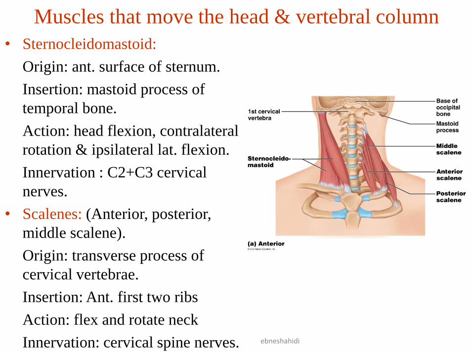
Muscles that move the head & vertebral column
• Sternocleidomastoid:
Origin: ant. surface of sternum.
Insertion: mastoid process of
temporal bone.
Action: head flexion, contralateral
rotation & ipsilateral lat. flexion.
Innervation : C2+C3 cervical
nerves.
• Scalenes: (Anterior, posterior,
middle scalene).
Origin: transverse process of
cervical vertebrae.
Insertion: Ant. first two ribs
Action: flex and rotate neck
Innervation: cervical spine nerves.
ebneshahidi
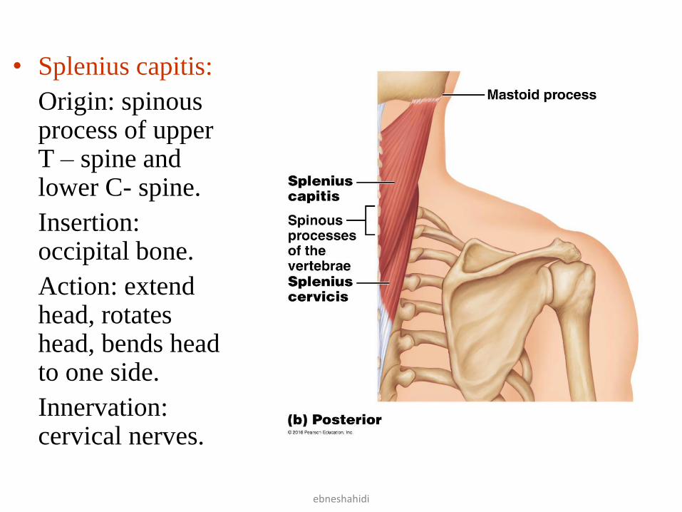
• Splenius capitis:
Origin: spinous process of upper T – spine and lower C- spine.
Insertion: occipital bone.
Action: extend head, rotates head, bends head to one side.
Innervation: cervical nerves.
ebneshahidi

• Semispinalis capitis:
Origin: transverse processes of C-spine and upper T-spine.
Insertion: occipital bone.
Action: extend head.
Innervation: cervical & thoracic nerves.
• Erector spinae: group of muscles consisting of iliocostalis, longissimus , and spinalis muscles.
Action: extend head, extend back, and maintain erect position of vertebral column.
Innervation: spinal
nerves.
ebneshahidi

Muscles that move the pectoral girdle
• Trapezius (upper, middle , lower):
Origin: occipital bone and spines
of C + T vertebrae.
Insertion: clavicle, spine and
acromion process of scapula.
Action: rotates scapula, pulls
shoulder & scapula downward.
Innervation: Accessory nerve.
• Rhomboid major:
Origin: spine of upper T- vertebra
Insertion: medial border of
scapula.
Action: raises and adducts scapula
Innervation: dorsal scapular nerve
ebneshahidi

• Levator scapulae:
Origin: transverse process of C-spine vertebras.
Insertion: medial margin of scapula.
Action: raises scapula.
Innervation: dorsal scapular and cervical nerves.
ebneshahidi

• Serratus anterior:
Origin: outer surface of upper ribs.
Insertion: venteral surface of scapula.
Action: pulls scapula Ant. & downward.
Innervation: long thoracic nerve.
• Pectoralis minor:
Origin: sternal ends of upper ribs.
Insertion: coracoid process of scapula.
Action: pulls scapula forward, downward and raises ribs.
Innervation: pectoral nerves.
ebneshahidi

Muscles That Move the arm Flexors
• Coracobrachialis:
Origin: coracoid process of scapula.
Insertion: shaft of humerus.
Action: flexes and adducts the arm
Innervation: musculocutaneous
nerve.
• Pectoralis major:
Origin: clavicle, sternum, and
costal cartilages of upper ribs.
Insertion: intertubercular groove of
humerus.
Action: flexes, adducts and rotates
arm medially.
Innervation: pectoral nerve.
ebneshahidi
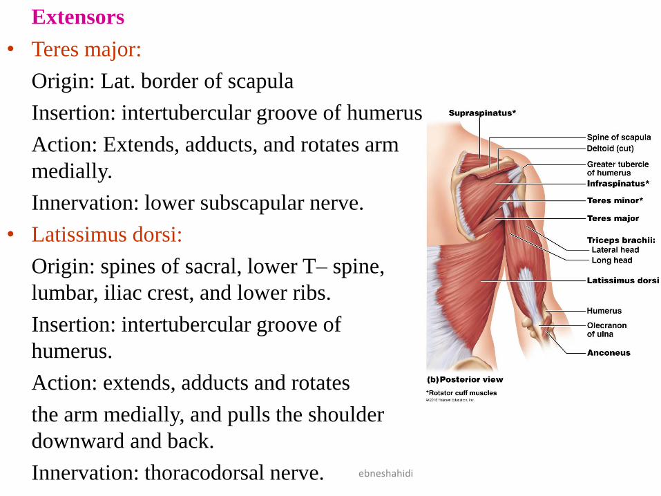
Extensors
• Teres major:
Origin: Lat. border of scapula
Insertion: intertubercular groove of humerus
Action: Extends, adducts, and rotates arm
medially.
Innervation: lower subscapular nerve.
• Latissimus dorsi:
Origin: spines of sacral, lower T– spine,
lumbar, iliac crest, and lower ribs.
Insertion: intertubercular groove of
humerus.
Action: extends, adducts and rotates
the arm medially, and pulls the shoulder
downward and back.
Innervation: thoracodorsal nerve.
ebneshahidi

Abductors
• Supraspinatus :
Origin: posterior surface of the scapula above spine.
Insertion: Greater tubercle of humerus.
Action : Abducts the arm
Innervation: subscapular nerve.
• Deltoid:
Origin: Acromion process, spine of scapula, and clavicle.
Insertion: Deltoid tuberosity of humerus.
Action: Abduct, extend, and flexes arm.
Innervation: Axillary nerve.
ebneshahidi

ebneshahidi

Rotators
• Infraspinatus:
Origin: post. surface of scapula below spine.
Insertion: Greater tubercle of humerus.
Action: rotates arm laterally.
Innervation: suprascapular nerve.
• Teres minor:
Origin: Lat. border of scapula.
Insertion: Greater tubercle of humerus.
Action: rotates arm laterally.
Innervations: Axillary nerve.
ebneshahidi
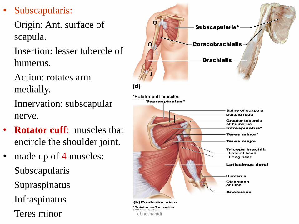
• Subscapularis:
Origin: Ant. surface of
scapula.
Insertion: lesser tubercle of
humerus.
Action: rotates arm
medially.
Innervation: subscapular
nerve.
• Rotator cuff: muscles that
encircle the shoulder joint.
• made up of 4 muscles:
Subscapularis
Supraspinatus
Infraspinatus
Teres minor
ebneshahidi

Muscle that move the forearm
Flexors
• Biceps brachii:
Origin: coracoid
process and tubercle
above glenoid cavity
of scapula.
Insertion: radial
tuberostiy of radius.
Action: flexes forearm
at elbow & rotates
hand laterally.
Innervation:
musculocutaneous
nerve.
ebneshahidi
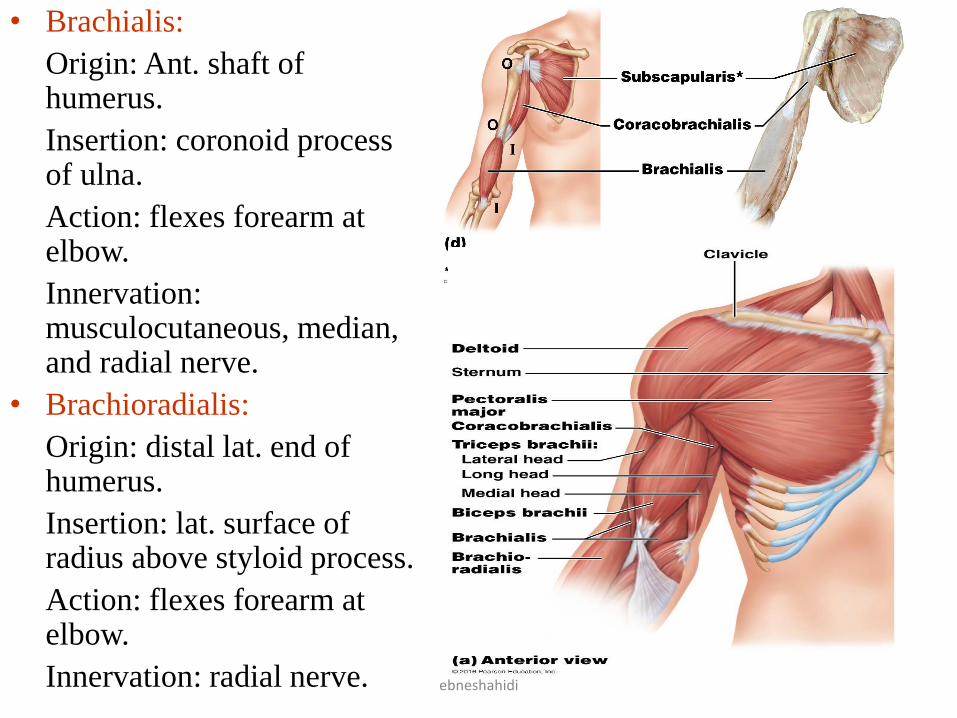
• Brachialis:
Origin: Ant. shaft of humerus.
Insertion: coronoid process of ulna.
Action: flexes forearm at elbow.
Innervation: musculocutaneous, median, and radial nerve.
• Brachioradialis:
Origin: distal lat. end of humerus.
Insertion: lat. surface of radius above styloid process.
Action: flexes forearm at elbow.
Innervation: radial nerve.
ebneshahidi

ebneshahidi

Extensors
• Triceps brachii:
Origin: tubercle below glenoid
cavity, lat. & medial humerus.
Insertion: olecranon process of
ulna.
Action: Extends forearm at
elbow.
Innervation: radial nerve.
Rotators
• Supinator:
Origin: lat. epicondyle of
humerus and crest of ulna.
Insertion: lat. surface of radius.
Action: rotates arm laterally.
Innervation: Radial nerve.
ebneshahidi

• Pronator teres:
Origin: medial epicondyle of humerus and coronoid process of ulna.
Insertion: lat. surface of radius.
Action: rotates forearm medially.
Innervation: median nerve.
• Pronator quadratus:
Origin: Ant. distal end of ulna.
Insertion: ant. distal end of radius.
Action: rotates forearm medially.
Innervation: median nerve.
ebneshahidi
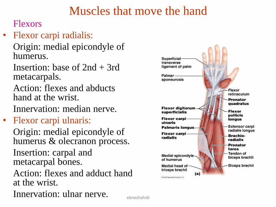
Muscles that move the hand Flexors
• Flexor carpi radialis:
Origin: medial epicondyle of humerus.
Insertion: base of 2nd + 3rd metacarpals.
Action: flexes and abducts hand at the wrist.
Innervation: median nerve.
• Flexor carpi ulnaris:
Origin: medial epicondyle of humerus & olecranon process.
Insertion: carpal and metacarpal bones.
Action: flexes and adduct hand at the wrist.
Innervation: ulnar nerve.
ebneshahidi

• Flexor digitorum superficialis:
Origin: medial epicondyle of humerus, coronoid process of ulna & radius.
Insertion: tendons of fingers.
Action: flexes fingers and hand.
Innervation: median nerve.
ebneshahidi

• Palmaris longus:
Origin: medial epicondyle of humerus.
Insertions: fascia of palm.
Action: flexes hand at wrist.
Innervation: median nerve.
• Flexor digitorum profundus:
Origin: ant. surface of ulna.
Insertion: bases of distal phalanges in fingers 2-5.
Action: flexes distal joints of fingers.
Innervation: median and ulnar nerve.
ebneshahidi

Extensors
Extensor carp radialis longus:
insertion: base of 2nd metacarpal.
Action: extends and abducts hand at wrist.
innervation: radial nerve.
Extensor carpi radials bravis:
origin: lat. epicondyle of humerus.
insertion: base of 2nd & 3rd metacarpals.
Action: extends and abducts hand at wrist.
innervation: radial nerve.
ebneshahidi

Extensor carpi ulnaris:
origin: lat. epicondyle of
humerus.
insertion: base of 5th
metacarpal.
Action: extends and
adducts hand at wrist.
innervation: radial nerve.
Extensor digitorum:
origin: lat. epicondyle of
humerus.
insertion: post. surface of
phalanges 2-5.
Action: extend fingers.
Innervation: radial nerve.
ebneshahidi

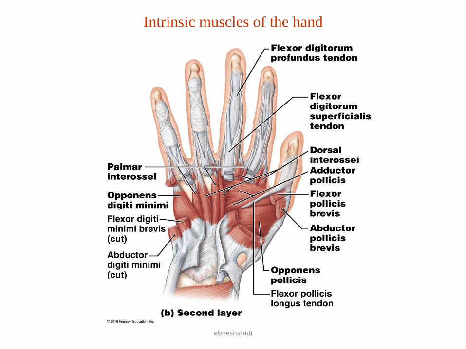
Intrinsic muscles of the hand
ebneshahidi
ebneshahidi
Intrinsic muscles of the hand

Diaphragm • Diaphragm:
Origin: inferior border of rib cage & sternum.
Insertion: central tendon.
Action: aid in expiration by depressing ribcage, prime mover of inspiration since it flattens and moves inferiorly on contraction.
Innervation: intercostal nerves 7-12.
ebneshahidi

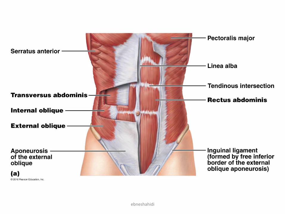
Muscles of the abdominal wall • External oblique:
Origin: outer surface of lower
ribs.
Insertion: iliac crest & linea alba
Action: tenses abdominal wall.
Innervation: intercostal nerves 7-
12.
• Internal oblique:
Origin: crest of ilium & inguinal
ligament.
Insertion: crtilages of lower ribs,
inea alba, and crest of pubis.
Action: tenses abdominal wall.
Innervation: intercostal nerves 7-
12.
ebneshahidi

• Transversus abdominis:
Origin: costal cartilage of lower ribs, iliac crest, inguinal ligaments and processes of lumbar fascia.
Insertion: linea alba & crest of pubis.
Action: tenses abdominal wall.
Innervation: intercostal nerves 7-12.
• Rectus abdominis:
Origin: crest of pubis & symphysis pubis.
insertion: xiphoid process of sternum and costal cartilages.
Action: tenses abdominal wall , and flexes back.
Innervation: intercostal nerves 7-12.
ebneshahidi
ebneshahidi

Muscles of the pelvic outlet
• Levator ani:
Origin: pubic bone and ischial spine.
Insertion: coccyx.
Action: sphincter like action in anal canal & vagina.
innervation: pudendal nerve.
• Sphincter urethrae:
Origin: margin of pubis and ischium.
Insertion: midline fibers.
Action: opens and closes urethra.
Innervation: pudendal nerve.
ebneshahidi

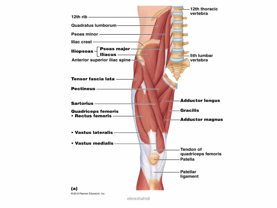
Muscles that move the thigh Anterior group
• Psoas major:
Origin: lumbar vertebrae.
Insertion: lesser trochanter of femur.
Action: flexes thigh.
Innervation: branches of L1-L3 nerves.
• Iliacus:
Origin: iliac fossa of ilium.
Insertion: lesser trochanter of femur.
Action: flexes thigh.
Innervation: femoral nerve.
ebneshahidi

Posterior group
• Hamstrings: (Biceps femoris, Semitendinosus, and semimembranosus).
• Gluteus maximums:
Origin: sacrum, coccyx, and post. surface of ilium.
Insertion: post. surface of femur and fascia.
Action: extends thigh.
Innervation: gluteal nerve (inferior).
• Gluteus medius:
Origin: lat. surface of ilium
Insertion: greater trochanter of femur.
Action: abducts and rotates thigh medially.
Innervation: superior gluteal nerve.
ebneshahidi

• Gluteus minimus:
Origin: lat. surface of ilium.
Insertion: greater trochanter of femur.
Action: abducts and rotates thigh medially.
Innervation: superior gluteal nerve.
• Tensor fascia lata:
Origin: Ant. iliac crest.
Insertion: fascia of thigh.
Action: Abducts, rotates thigh medially, flexes thigh.
Innervation: sup. gluteal nerve.
ebneshahidi

Thigh Adductors
• Pectineus:
Origin: spine of pubis.
Insertion: femur distal to lesser trochanter.
Action: adducts and flexes thigh.
Innervation: obturator and femoral nerves.
• Adductor longus:
Origin: pubic bone near symphysis pubis.
Insertion: posterior surface of femur.
Action: adducts, flexes, and rotate thigh laterally.
Innervation: obturator nerve.
ebneshahidi

• Adductor magnus :
Origin: Ischial tuberosity.
Insertion: post. surface of
femur.
Action: adducts, extends,
and rotate thigh laterally.
Innervation: obturator nerve.
• Gracillis:
Origin: symphysis pubis.
Insertion: medial surface of
tibia.
Action: adduct thigh, and
flexes leg at the knee.
Innervation: obturator nerve.
ebneshahidi

Muscles that move the leg
Flexors: Biceps femoris,
semitendinosus,
semimembranosus
(hamstring group), and
sartorius.
• Biceps femoris:
Origin: Ischial tuberosity
and femur.
Insertion: Head of fibula
and lat. condyle of tibia.
Action: Flexes and rotates
leg laterally and extends
thigh.
Innervation: Tibial nerve.
ebneshahidi

• Semitendinosus:
Origin: Ischial tuberosity.
Insertion: Medial surface of tibia.
Action: Flexes and rotates leg medially and extends thigh.
Innervation: Tibial nerve.
• Semimembranosus:
Origin: Ischial tuberosity.
Insertion: medial condyle of tibia.
Action: flexes and rotates leg medially and extends thigh.
Innervation: Tibial nerve. ebneshahidi

ebneshahidi

• Sartorius:
Origin: anterior
superior iliac
spine.
Insertion: medial
surface of tibia.
Action: flexes leg
and thigh,
abducts, and
rotates thigh
laterally.
Innervation:
femoral nerve.
ebneshahidi

Extensors: Rectus femoris, Vastus lateralis, vastus intermedius, and
vastus medialis (Quadriceps femoris group).
• Rectus femoris:
Origin: spine of ilium and margin of acetabulum.
Insertion: patella and tibial tuberosity via patellar tendon.
Action: extends leg at knee.
Innervation: femoral nerve.
• Vastus lateralis:
Origin: greater trochanter and post. surface of femur.
Insertion: patella and tibial tuberosity
via patellar tendon.
Action: extends leg at knee.
Innervation: femoral nerve.
; ebneshahidi
ebneshahidi

• Vastus medialis:
Origin : medial surface of
femur.
Insertion: patella & tibial
tuberosity via patella
ligament.
Action: extends knee.
Innervation: femoral nerve.
• Vastus intermedius:
Origin: Ant. and Lat. surfaces
of femur.
Insertion: patella & tibial
tuberosity via patellar
ligament.
Action: extends knee.
Innervation: femoral nerve.
ebneshahidi

Muscles that move the foot Dorsal Flexors
• Tibialis anterior:
Origin: Lat. condyle and lat. surface of tibia.
Insertion: Tarsal bone (cuneiform) & 1st metatarsal.
Action: dorsiflexion and inversion of foot.
Innervation: deep peroneal nerve.
• Peroneus (Fibularis) teritus:
Origin: Ant. surface of fibula.
Insertion: Dorsal surface of 5th metatarsal.
Action: Dorsiflexion & eversion of foot.
Innervation: deep peroneal nerve.
ebneshahidi

ebneshahidi

• Extensor digitorum
longus:
Origin: lat. condyle of
tibia & ant. surface of
fibula.
Insertion: dorsal surface
of 2nd & 3rd phalanges
of 4 lat. toes.
Action: dorsiflexion and
eversion of foot &
extension of toes.
Innervation: deep
peroneal nerve.
ebneshahidi

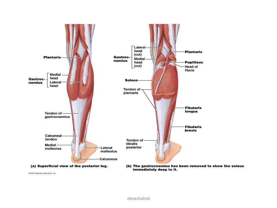
Plantar flexors
• Gastrocnemius:
Origin: Lat. & medial condyles of femur (two bellies).
Insertion: post. surface of calcaneus.
Action: plantar flexion of foot and flexion of leg at knee.
Innervation: Tibial nerve.
• Soleus:
Origin: Head & shaft of fibula, post. surface of tibia.
Insertion: post. surface of calcaneus.
Action: plantar flexion of foot.
Innervation: Tibial nerve.
ebneshahidi
ebneshahidi

• Flexor digitorum longus:
Origin: post. surface of tibia.
Insertion: distal phalanges of 4 lat. toes.
Action: plantar flexion and inversion of foot, and flexion of 4 lat. toes.
Innervation: tibial nerve.
ebneshahidi

Invertor
• Tibialis posterior:
Origin: Lat. condyle and post. surfaces of tibia and fibula.
Insertion: tarsal and metatarsal bones.
Action: plantar flexion and inversion of foot.
Innervation: tibial nerve.
Evertor
• Peroneus (Fibularis) longus:
Origin: Lat. condyle of tibia, head, and shaft of fibula.
Insertion: tarsal and metatarsal bones.
Action: plantar flexion and eversion of foot and supports arch.
Innervation: superficial peroneal nerve.
ebneshahidi

Intrinsic muscles of the foot
ebneshahidi

ebneshahidi
Intrinsic muscles of the foot

ebneshahidi
Intrinsic muscles of the foot

ebneshahidi

ebneshahidi
Clinical Terms
Fibrillation: contraction of muscle fibers producing rapid,
uncoordinated activity within a muscle.
Torticollis: neck muscles like SCM muscle contract involuntarily;
also called wry neck causing chronic rotation and tilting of head to
one side.
Muscular dystrophy: progressive muscle weakness and atrophy
caused by a deficient protein called dystrophin.
Myasthenia gravis: chronic disease caused by muscles that are
weak and easily fatigue. It results from immune system’s attack on
neuromuscular junctions so that stimuli are not transmitted from
motor neurons to muscle fibers.
Shin splint: sorness of the front of leg due to straining of flexor
digitorum longus.
Myositis: inflammation of skeletal muscle tissue.
ebneshahidi