muscle-derived proteins as serum biomarkers for...
TRANSCRIPT

This work is licensed under a Creative Commons Attribution-NonCommercial 3.0 Unported License
Newcastle University ePrints - eprint.ncl.ac.uk
Burch PM, Pogoryelova O, Goldstein R, Bennett D, Guglieri M, Straub V,
Bushby K, Lochmüller H, Morris C. Muscle-Derived Proteins as Serum
Biomarkers for Monitoring Disease Progression in Three Forms of Muscular
Dystrophy. Journal of Neuromuscular Diseases 2015, 2(3), 241-255.
Copyright:
© 2015 – IOS Press and the authors. This article is published online with Open Access and distributed
under the terms of the Creative Commons Attribution Non-Commercial License.
DOI link to article:
http://dx.doi.org/10.3233/JND-140066
Date deposited:
20/10/2015

Journal of Neuromuscular Diseases 2 (2015) 241–255DOI 10.3233/JND-140066IOS Press
241
Research Report
Muscle-Derived Proteins as SerumBiomarkers for Monitoring DiseaseProgression in Three Forms of MuscularDystrophy
Peter M. Burcha,1,∗, Oksana Pogoryelovac,1, Richard Goldsteina, Donald Bennettb, Michela Guglieric,Volker Straubc, Kate Bushbyc, Hanns Lochmullerc and Carl Morrisb
aWorldwide Research & Development, Pfizer Inc., Groton, CT, USAbWorldwide Research & Development, Pfizer Inc., Cambridge, MA, USAcJohn Walton Muscular Dystrophy Research Centre, Institute of Genetic Medicine, Newcastle University,Newcastle upon Tyne, UK
Abstract.Background: Identifying translatable, non-invasive biomarkers of muscular dystrophy that better reflect the disease pathologythan those currently available would aid the development of new therapies, the monitoring of disease progression and the responseto therapy.Objective: The goal of this study was to evaluate a panel of serum protein biomarkers with the potential to specifically detectskeletal muscle injury.Method: Serum concentrations of skeletal troponin I (sTnI), myosin light chain 3 (Myl3), fatty acid binding protein 3 (FABP3)and muscle-type creatine kinase (CKM) proteins were measured in 74 Duchenne muscular dystrophy (DMD), 38 Becker musculardystrophy (BMD) and 49 Limb-girdle muscular dystrophy type 2B (LGMD2B) patients and 32 healthy controls.Results: All four proteins were significantly elevated in the serum of these three muscular dystrophy patient populations whencompared to healthy controls, but, interestingly, displayed different profiles depending on the type of muscular dystrophy.Additionally, the effects of patient age, ambulatory status, cardiac function and treatment status on the serum concentrations ofthe proteins were investigated. Statistical analysis revealed correlations between the serum concentrations and certain clinicalendpoints including forced vital capacity in DMD patients and the time to walk ten meters in LGMD2B patients. Serumconcentrations of these proteins were also elevated in two preclinical models of muscular dystrophy, the mdx mouse and thegolden-retriever muscular dystrophy dog.Conclusions: These proteins, therefore, are potential muscular dystrophy biomarkers for monitoring disease progression andtherapeutic response in both preclinical and clinical studies.
Keywords: Muscular dystrophy, biomarker, skeletal troponin I, myosin light chain 3, creatine kinase, fatty acid bindingprotein 3
1These authors contributed equally to this work.∗Correspondence to: Peter M. Burch, Ph.D., Pfizer, Inc., Eastern
Point Road, Groton, CT 06340, USA. Tel.: +1 860 686 0947; Fax:+1 860 715 1251; E-mail: [email protected].
ISSN 2214-3599/15/$35.00 © 2015 – IOS Press and the authors. All rights reserved
This article is published online with Open Access and distributed under the terms of the Creative Commons Attribution Non-Commercial License.

242 P.M. Burch et al. / Serum Biomarkers of Muscular Dystrophy
INTRODUCTION
The muscular dystrophies are a heterogeneous groupof genetic disorders that result in progressive skeletalmuscle weakness and wasting [1]. There are nine majorforms of muscular dystrophy that differ clinically inmany respects including age of onset, spectrum ofmuscle groups affected, severity of muscle injury andlethality [1]. Duchenne muscular dystrophy (DMD) isone of the most severe and common forms of mus-cular dystrophy with a prevalence of approximately1 in 3,500–6,000 live male births [2]. DMD resultsfrom mutations in the DMD gene that results in anabsence of dystrophin protein expression [3]. Beckermuscular dystrophy (BMD) is caused by mutationsthat lead to partial dystrophin deficiency resulting in amilder form of the disease with later onset and greaterlife expectancy [3]. Limb-girdle muscular dystrophytype 2B (LGMD2B) is an autosomal recessive disorderresulting from mutations in the DYSF gene that lead todysferlin deficiency [4]. Clinically, LGMD2B resultsin progressive proximal muscle weakness, diagnosedwhen patients are in their 20’s and 30’s, but, unlikeDMD and BMD, typically does not affect the cardiacand respiratory muscles [4, 5].
There are very few pharmacological treatmentoptions for muscular dystrophy [6]. For DMD andBMD patients corticosteroid treatment is the onlyintervention consistently shown to delay disease pro-gression, but with potentially treatment limiting sideeffects of weight gain, loss of bone density and behav-ioral problems with long-term use [7, 8]. For LGMD2Bcorticosteroid treatment has proven ineffective [5].Fortunately, a number of therapeutics with differentmodes of action are currently in late pre-clinical devel-opment or early clinical trials offering hope that newtreatment options for muscular dystrophy will becomeavailable [6, 9].
The progression of new therapeutics toward clinicaltesting has highlighted the need for improved out-come measures and biomarkers to support regulatoryapproval [10–12]. A number of clinical assessmentsare available for evaluating functional improvement inmuscular dystrophy patients including the 6 minutewalk test, timed functional tests, forced vital capac-ity (FVC) and the North Star Ambulatory Assessment(NSAA), among others [11]. Total serum creatinekinase (CK) is the standard clinical chemistry test thatis used to diagnose muscular dystrophy [13]. This testis based on measuring the enzymatic activity of CKpresent in the serum after release from damaged mus-cle tissue [13]. While this widely available test is useful
for detecting muscle injury, serum CK activity doesnot correlate well with disease severity in musculardystrophy patients [11]. This is due to a number of con-founding factors affecting a patient’s serum CK levelincluding age, the level of physical activity and theamount of muscle mass [11, 14], as well as factors thataffect CK enzyme activity such as serum glutathionelevels and treatment with certain drugs [15].
Numerous research teams have described novelpotential biomarkers for monitoring disease progres-sion and drug efficacy in muscular dystrophy patients,particularly DMD, including circulating microRNA[16–19] and proteins identified in serum and urine[20–22]. Two recent reports utilized proteomicsapproaches to comprehensively profile the proteomeof serum from DMD and BMD patients [14, 23]. Bothgroups identified a number of proteins involved in mus-cle function and metabolism that were significantlyelevated in the serum of muscular dystrophy patientswhen compared to healthy controls. Promisingly, anumber of these identified muscle-derived proteinswere common to both studies and correlated withdisease severity in DMD and BMD patients. Whilethe experimental approaches used in these reportswere semi-quantitative it suggested that the strategyof measuring serum concentrations of muscle-derivedproteins could provide useful biomarkers for monitor-ing disease progression in muscular dystrophy patients.
Here we report on the results of a multiplexed elec-trochemiluminescent enzyme-linked immunosorbentassay (ELISA) approach to quantitatively measure theserum concentrations of four proteins, abundant inskeletal muscle tissue, in serum from DMD, BMDand LGMD2B patients. The four proteins measured arethe myofibrillar proteins skeletal troponin I (sTnI) andmyosin light chain 3 (Myl3), creatine kinase muscle-type (CKM) and the lipid transport protein fatty acidbinding protein 3 (FABP3). Using clinical assess-ments, such as FVC, NSAA score, ambulatory statusand cardiac function, we sought to determine if mea-suring serum concentrations of these muscle-derivedproteins better reflected the patients’ disease state thanCK activity.
MATERIALS AND METHODS
Subjects
Serum samples from 38 BMD and 49 LGMD2Bpatients and 100 serum samples from 74 DMD patientswere obtained from Newcastle University and the Jain

P.M. Burch et al. / Serum Biomarkers of Muscular Dystrophy 243
Foundation through the MRC Centre for Neuromus-cular Diseases Biobank. Collection of samples frompatients and their use in research have been ethicallyapproved by the NRES Committee North East – New-castle and North Tyneside 1. For the healthy controls18 serum samples were purchased from Bioreclama-tionIVT (Nassau, NY) and 14 serum samples wereobtained from the Pfizer Research Support Program(Groton, CT). Collection of samples through the PfizerResearch Support Program was ethically approved byPfizer’s Institutional Review Board and conducted bythe Pfizer Global Occupational Health and WellnessClinic.
Muscle protein immunoassay and clinicalchemistry
sTnI, FABP3, Myl3 was quantified in serum sam-ples using the Meso Scale Discovery (MSD, Rockville,MD) Muscle Injury Panel 1 reagent kit (catalog #K15181C). CKM was measured using the MSD Mus-cle Injury Panel 2 reagent kit (catalog # K15180C).Both assays were run and measured as per the manu-facturer’s instructions. Subject samples were dilutedto be within the dynamic range of the assay. ForsTnI, FABP3, Myl3 testing, the muscular dystrophypatients’ samples were diluted 1:8 while control sub-ject samples were diluted 1:4. CKM measurement forthe DMD, BMD and LGMD2B patients required sam-ples to be diluted from 1:200 to 1:800 to be withinthe dynamic range of the assay while normal controlsubjects required dilution of 1:25. ALT, AST and totalserum CK were analyzed by standard clinical chem-istry techniques on the Siemens Advia 2400 platform.
Statistical analysis
Data were transformed and tested for the normalityassumption using the Shapiro-Wilk Test. Comparisonsbetween groups were made using ANOVA (para-metric) and Kruskal-Wallis (nonparametric) Tests.
Correlation analyses were performed using Spear-man’s Correlation (order statistics). Data from asubject with two serial samples were evaluated withANOVA on paired differences and repeated measuresANOVA. All analyses were performed using SAS ver-sion 9.2, Cary, N. Carolina. All graphs were generatedusing Graphpad Prism version 6.03.
RESULTS
Validation of assay performance
Numerous recent reports have demonstrated thepotential utility of measuring the serum concentrationsof muscle-derived proteins as biomarkers of muscleinjury [14, 23, 24]. We have extensively used a mul-tiplexed immunoassay to monitor serum and plasmaconcentrations of the muscle-derived proteins sTnI,Myl3, FABP3 and CKM in preclinical muscle dis-ease and muscle injury studies in rodents. While theseassays were developed and validated using rat mus-cle proteins and homogenates, the highly conservedamino acid sequence of the target proteins betweenall species, including human, suggested these assaycould be applied to other species without modifi-cation (Table S1). To validate the assays for usewith human serum samples, purified recombinant full-length FLAG-tagged human sTnI, CKM, Myl3 andFABP3 were tested. SDS-PAGE analysis followed bysilver staining (Figure S1A) and Western blot analysis(Figure S1B) to detect the FLAG tag confirmed boththe purity and expected molecular weights of the tar-get proteins. These proteins were subsequently testedin the muscle protein immunoassays as part of the assayvalidation.
Dilution linearity of the recombinant human pro-teins was observed between the concentrations of 160and 5 ng/ml with FABP3, 320 and 5 ng/ml with Myl3,250 and 2 ng/ml with sTnI and 200 and 6 ng/ml withCKM (Figure S1C). No cross-reactivity of the assayswith the other purified proteins tested was observed
Table 1Characteristics of muscular dystrophy patients and healthy controls
Control (n = 32) BMD (n = 38) DMD (n = 74) LGMD2B (n = 49)
Age (years) 21.1 ± 15.2 29.6 ± 20.4 12.6 ± 5.8 37.3 ± 10.5Age range (years) 5–53 4–85 3–25 18–66Sex - Male 32 (100%) 38 (100%) 74 (100%) 20 (41%)Non-ambulant 9 (24%) 29 (39%) 16 (33%)Treated with corticosteroids 62 (84%)Treated for cardiomyopathy 28 (38%)Follow-up sample available 26 (35%)
Data are mean ± standard deviation, age range or number of individuals (% of the cohort).

244 P.M. Burch et al. / Serum Biomarkers of Muscular Dystrophy
(data not shown). The CKM assay did not detect thehuman creatine kinase B-type (CKB) protein (datanot shown). Taken together this data confirms thatthese assays can be used to accurately and specif-ically measure human proteins sTnI, Myl3, FABP3and CKM. All serum samples were diluted into thelinear detection range of each assay for subsequentanalysis.
Circulating biomarker levels in musculardystrophy
To begin to assess the utility of the muscle injurypanel for monitoring disease progression in muscu-lar dystrophy serum samples from 74 DMD, 38 BMDand 49 LGMD2B patients and 32 healthy controlswere obtained (Table 1). While the control populationincluded juveniles as well as younger and older adultsto cover the range of the muscular dystrophy cohorts,
it should be noted that the average age of the DMDand BMD cohorts is significantly different (P < 0.05)from the healthy controls while the LGMD2B cohortwas not (data not shown). Standard clinical chem-istry testing for total serum CK revealed significantlyelevated average total CK levels for all three mus-cular dystrophy patient groups compared to healthycontrols for this assay (Fig. 1A). The DMD patientsshowed the highest mean total serum CK (7443 ± 6357U/L). This is consistent with the severe muscle damageand more rapid disease progression observed in DMDpatients [1, 25]. This was followed by the LGMD2B(4342 ± 3518 U/L) and the BMD (3108 ± 4007 U/L)patient groups, whereas the healthy volunteers grouphad an average serum CK activity of 109 ± 57.61 U/L.Compared to the healthy control group the mean totalserum CK activity was elevated 28 fold over controlsin BMD patients, 40 fold in LGMD2B patients and68 fold in DMD patients. Also, as has previously been
Fig. 1. Scatter plots of the serum clinical chemistry and protein serum concentrations in muscular dystrophy patients. (A) Total serum CK,AST and ALT levels and (B) serum concentrations of sTnI, Myl3, FABP3 and CKM for the DMD, BMD, LGMD2B patient groups and healthycontrols are shown. The line and error bars represent the mean and standard deviation of each group. ∗∗∗P < 0.001; ∗∗∗∗P < 0.0001.

P.M. Burch et al. / Serum Biomarkers of Muscular Dystrophy 245
reported, average serum AST and ALT levels were alsoelevated above the healthy volunteer controls for theseanalytes in all three patient populations [26, 27].
The serum concentrations of sTnI, Myl3, CKM andFABP3 were determined in the muscular dystrophypatients’ samples and healthy controls. While measur-able amounts of FABP3 and CKM were detected in theserum of all the healthy controls, the serum concentra-tions were below the lower limit of quantitation for allof the subjects for sTnI and 22 of the subjects for Myl3.In these cases, the concentration for the lower limit ofquantitation of the assays was reported for the samples.The mean serum concentrations of all four proteinswere significantly elevated in the muscular dystrophypatient cohorts when compared to controls (Fig. 1B).Consistent with the total serum CK levels, the serumconcentrations of all four markers were most elevatedin the DMD patient group, followed by LGMD2B andBMD. Because the sTnI and Myl3 levels in many ofthe controls were below the limit of quantitation ofthe assay we could not accurately calculate the foldchange over controls for those markers. For CKM thefold-change in mean serum levels over controls were17-fold for BMD, 88-fold for DMD and 37-fold forLGMD2B. Similarly FABP3 levels were ninefold overmean controls levels in BMD, 13-fold in DMD and 12-fold in LGMD2B. Comparisons among the differenttypes of muscular dystrophy (Table S2) revealed sig-nificantly elevated mean serum concentrations of sTnI(P < 0.0001) and CKM (P < 0.01) in DMD patientsrelative to the levels measured in BMD patients, butwhen comparing the levels between the DMD andLGMD2B cohorts only sTnI showed a statisticallysignificant (P < 0.05) difference. With the LGMD2Bpatients, however, both sTnI and FABP3 levels weresignificantly elevated (P < 0.05) over that measured inBMD patients.
As noted previously the amino acid sequences ofsTnI, Myl3, FABP3 and CKM are highly conservedbetween rodent, canine and human. This suggestedthese biomarker assays may also be directly applica-ble to the most commonly used preclinical models ofmuscular dystrophy, the mdx mouse and the goldenretriever muscular dystrophy (GRMD) canine model[28]. In the mdx model all four biomarker concentra-tions were significantly elevated (P < 0.001) in serumfrom adult mdx mice (N = 10) when compared to age-matched wild-type controls (N = 9, Figure S2). sTnIhad the greatest fold increase in the mean serumconcentration in the mdx mouse when compared wild-type controls (124 fold), followed by Myl3 (9.7-fold),CKM (8.8-fold) and FABP3 (3-fold,). All marker
serum levels were significantly increased (P < 0.0001)in GRMD affected dogs (Supplementary Figure S3),but a comparison of the mean biomarker serum con-centrations in GRMD canines (N = 5) to unaffectedcontrols (N = 7) revealed that CKM had the greatestfold increase (1,331-fold), followed by sTnI (727-fold), Myl3 (98.7-fold) and FABP3 (3.1-fold).
Effect of disease progression and treatment inDMD patients
In DMD patients loss of ambulation typically occursbetween 7 and 12 years old [1]. The effect of ambula-tory status of the DMD patients on the biomarker levelswas investigated. Twenty-nine of the DMD patients(39%) in this study were non-ambulant. The non-ambulant DMD patients had significantly lower levelsof total serum CK activity and sTnI, Myl3, FABP3 andCKM concentrations when compared to the 46 ambu-lant DMD patients (Fig. 2A). BMD and LGMD2Bpatients can remain ambulant into late adulthood [1,5] so the number of non-ambulant BMD (N = 9) andLGMD2B (N = 16) patients was small, but like theDMD patients, the non-ambulant patients had signif-icantly lower biomarker levels and total serum CKactivity when compared to the ambulant patients (datanot shown).
As DMD patients get older the majority eventu-ally develops cardiomyopathy [29]. Twenty-eight ofthe DMD patients (38%) were classified as having car-diac symptoms based on a reduction in ejection fraction(<55%) or ongoing treatment for cardiomyopathy. Themean serum levels of all four muscle protein biomark-ers and total serum CK levels were significantly lowerin the patients with cardiac symptoms when com-pared to those without (Fig. 2B). It should be notedthat 82% (N = 23) of the patients with cardiac symp-toms were also non-ambulant. Also, for those patientswith cardiac symptoms where respiratory function wasmeasured at the time of blood draw (N = 26) the meanFVC was 46.9 ± 25.4% compared to 84.2 ± 23.0% forthe DMD patients with no noted cardiac symptoms(N = 35).
Corticosteroids are the most commonly prescribeddrugs for the treatment of DMD and BMD [30]. Sixty-two of the DMD patients (83%) in the sample set werebeing treated with either deflazacort or prednisolone.There was no significant difference in the mean serumlevels of the muscle protein biomarkers or serum CKactivity between the patients being treated with steroidsand those not taking steroids at the time of evalua-tion (Figure S4). This data suggests steroid treatment

246 P.M. Burch et al. / Serum Biomarkers of Muscular Dystrophy
Fig. 2. Scatter plots of the protein serum concentrations in DMD patients based on clinical status. Serum concentrations of sTnI, Myl3, FABP3,CKM and total CK in (A) ambulant (N = 46) and non-ambulant (N = 29) DMD patients or (B) those classified as with (N = 25) and without(N = 50) cardiomyopathy are shown. The line and error bars represent the mean and standard deviation of each group. ∗∗∗∗P < 0.0001.
does not have a strong effect on the circulating lev-els of the investigated muscle proteins, but it shouldbe noted that the number of DMD patients in thisstudy not actively being treated with steroids was small(N = 11).
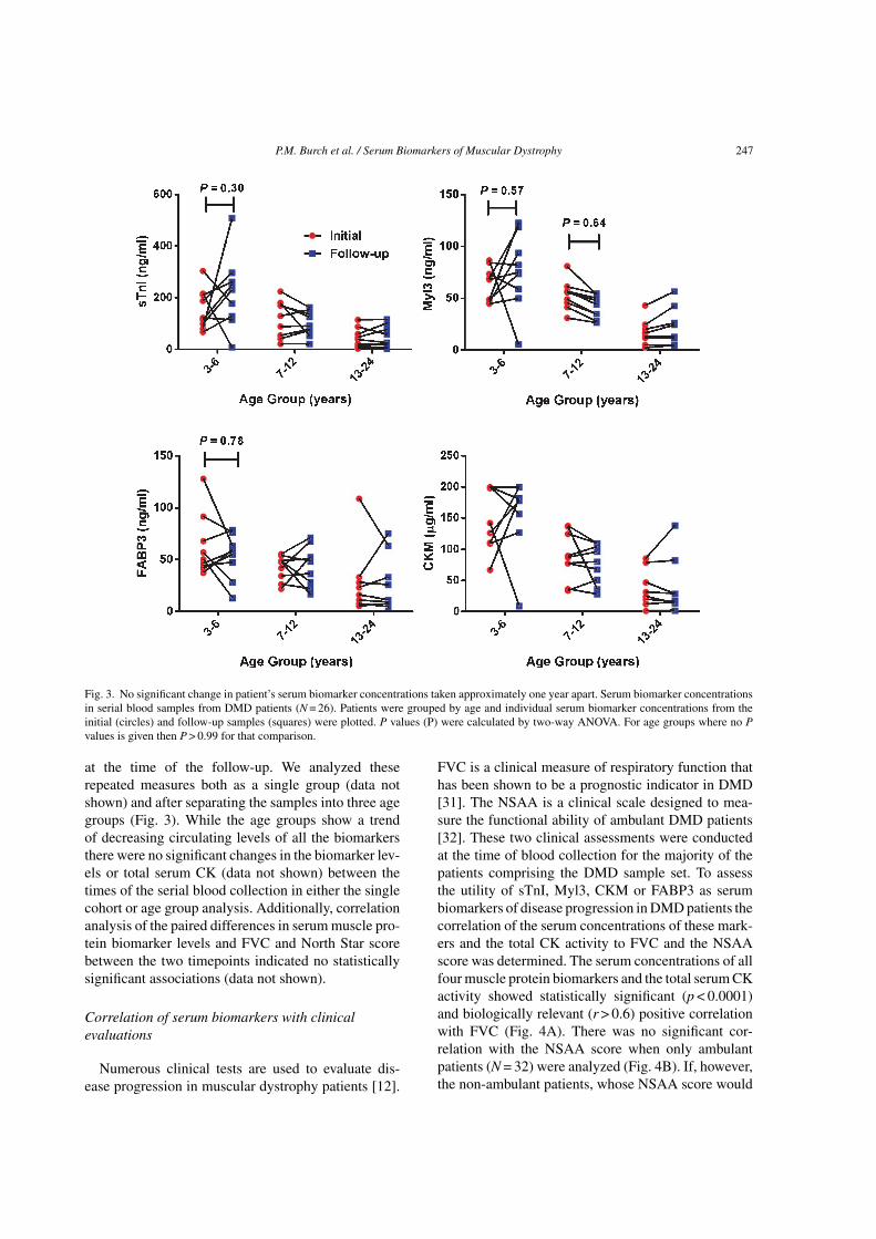
A subset of the DMD patients (N = 26) in this studyhad a follow-up evaluation an average of 11.2 ± 3.3months after the initial examination and serum sam-ples were taken again. Of these patients 20 had FVCmeasurements and 24 had NSAA scores measured
P.M. Burch et al. / Serum Biomarkers of Muscular Dystrophy 247
Fig. 3. No significant change in patient’s serum biomarker concentrations taken approximately one year apart. Serum biomarker concentrationsin serial blood samples from DMD patients (N = 26). Patients were grouped by age and individual serum biomarker concentrations from theinitial (circles) and follow-up samples (squares) were plotted. P values (P) were calculated by two-way ANOVA. For age groups where no Pvalues is given then P > 0.99 for that comparison.
at the time of the follow-up. We analyzed theserepeated measures both as a single group (data notshown) and after separating the samples into three agegroups (Fig. 3). While the age groups show a trendof decreasing circulating levels of all the biomarkersthere were no significant changes in the biomarker lev-els or total serum CK (data not shown) between thetimes of the serial blood collection in either the singlecohort or age group analysis. Additionally, correlationanalysis of the paired differences in serum muscle pro-tein biomarker levels and FVC and North Star scorebetween the two timepoints indicated no statisticallysignificant associations (data not shown).
Correlation of serum biomarkers with clinicalevaluations
Numerous clinical tests are used to evaluate dis-ease progression in muscular dystrophy patients [12].
FVC is a clinical measure of respiratory function thathas been shown to be a prognostic indicator in DMD[31]. The NSAA is a clinical scale designed to mea-sure the functional ability of ambulant DMD patients[32]. These two clinical assessments were conductedat the time of blood collection for the majority of thepatients comprising the DMD sample set. To assessthe utility of sTnI, Myl3, CKM or FABP3 as serumbiomarkers of disease progression in DMD patients thecorrelation of the serum concentrations of these mark-ers and the total CK activity to FVC and the NSAAscore was determined. The serum concentrations of allfour muscle protein biomarkers and the total serum CKactivity showed statistically significant (p < 0.0001)and biologically relevant (r > 0.6) positive correlationwith FVC (Fig. 4A). There was no significant cor-relation with the NSAA score when only ambulantpatients (N = 32) were analyzed (Fig. 4B). If, however,the non-ambulant patients, whose NSAA score would

248 P.M. Burch et al. / Serum Biomarkers of Muscular Dystrophy
be classified as zero, were included in the analysis(N = 61) there was a strong and statistically signifi-cant positive correlation (r > 0.6, p < 0.0001) betweenthe NSAA score and the serum concentrations of thebiomarkers (Figure S5).
FVC was also measured for the BMD patients at thetime of blood sampling. Analysis of this data, however,revealed no statistically significant correlation betweenFVC and sTnI, CKM or total serum CK activity(Figure S6). There was a weak correlation betweenFVC and Myl3 (r = –0.35, p = 0.04) and FABP3(r = –0.36, p = 0.033), but, interestingly, this correla-tion was negative, in contrast to the strong positivecorrelation observed in the DMD patients.
For a majority of the LGMD2B patients numerousclinical evaluations were conducted at the time of bloodsampling evaluation, including FVC (N = 47) and thetime required to walk ten meters (N = 31). Like for theBMD patients, none of the serum concentrations of thebiomarkers or the serum CK activity correlated withthe FVC in the LGMD2B patients (Fig. 5A). Simi-larly, sTnI, FABP3 and CKM serum concentrationsand serum CK activity did not correlate with the timeto walk 10 m in these patients (Fig. 5B). The corre-lation Myl3 with the time to walk 10 m was stronger(r = 0.36, p = 0.045) than those determined for the othermarkers, but was still a weak association.
Effect of age on biomarker levels
Previous reports have noted a consistent declinein serum CK activity with age in dystrophinopathypatients [25, 33, 34]. Serum CK levels peak in earlychildhood for both DMD and BMD patients and thendecline at a rate of approximately 18% per year inDMD patients and 6% per year in BMD patients [25].Analysis of CK activity versus age for the DMD, BMDand LGMD2B patient populations assayed here closelymatch these previously reported observations (Fig. 6A, B and C). It should be noted that age or birth-date information was not available for thirteen DMDpatients, leaving sixty-one subjects for this analysis.All four muscle protein biomarkers also followed asimilar trend of steady decline that significantly cor-related with age (data not shown) in DMD, BMDand LGMD2B patients. Two possible exceptions tothis trend were Myl3 and FABP3 in BMD patientsover sixty years of age, where serum concentrationsof these proteins remained elevated, but the numberof patients in this age range was very small (N = 4).Finally, the approximate annual rates of decline foreach biomarker were calculated (Table S3). The annual
rate of decrease in total serum CK activity of approx-imately 18.2 ± 1.4% per year in the DMD patientsclosely matched that reported previously [25]. All thebiomarkers and CK showed the most rapid rate ofdecline in DMD patients followed by LGMD2B andBMD patients. Serum CK activity and CKM had themost rapid rate of decline in all three patient pop-ulations. FABP3 showed the slowest annual rate ofdecline in DMD and BMD patients, at 10.2 ± 1.2%and 0.1 ± 0.5%, respectively. In LGMD2B patients,however, Myl3 levels declined at the slowest rate atapproximately 2.0 ± 0.5% per year.
DISCUSSION
To aid the development of the next generation oftherapies for muscular dystrophy new clinical tools areneeded to monitor disease progression and the responseto treatment. Recent reports have used proteomicsapproaches to identify serum biomarkers of muscu-lar dystrophy [14, 23]. Interestingly, but, perhaps notsurprisingly, most of the proteins identified by theseprevious studies as significantly different betweenhealthy control and muscular dystrophy patient cohortswere of muscle origin. Here we have investigated theserum concentrations of four proteins that are abun-dantly expressed in skeletal muscle in DMD, BMD andLGMD2B patients as potential biomarkers of musculardystrophy.
Serum concentrations of sTnI, Myl3, FABP3 andCKM were all significantly elevated in DMD, BMDand LGMD2B patients when compared to serum fromhealthy controls. The fold increase in average serumconcentrations for all markers when comparing themuscular dystrophy patients to the healthy controlssuggests they would provide a good dynamic range fornon-invasively measuring the outcome of a therapeuticintervention. The fact that significantly elevated serumconcentrations were also observed in the mdx mouseand GRMD canine suggest these biomarkers could beused as translational biomarkers for drug development.We did not have ready access to an animal model ofdysferlinopathy for this study, but future testing ofthese biomarkers in the SJL or A/J mouse could extendthese observations to a relevant preclinical model ofLGMD2B [35].
Total serum CK activity was also significantlyelevated in all three muscular dystrophy patient popula-tions, but there are a number of potential advantages themarkers presented here afford. First, based on the pat-tern of tissue expression, CKM and sTnI, in particular,

P.M. Burch et al. / Serum Biomarkers of Muscular Dystrophy 249
Fig. 4. Correlations of protein serum concentration and clinical measures in DMD patients. A graph of (A) FVC measurement and (B) NSAAscore versus the serum concentrations of sTnI, Myl3, FABP3, CKM and total serum CK for each DMD patient. The Spearman’s rank-ordercorrelation coefficient (r), P value (P) and number of patients in the sample set (N) is shown for each analysis.

250 P.M. Burch et al. / Serum Biomarkers of Muscular Dystrophy
Fig. 5. Correlations of protein serum concentrations and clinical measures in LGMD2B patients. A graph of the (A) FVC measurement and(B) the time to walk 10 m test versus serum concentrations of sTnI, Myl3, FABP3, CKM and total serum CK for each LGMD2B patients isshown. The Spearman’s correlation coefficient (r), P value (P) and number of patients in the sample set (N) is shown for each analysis.
are potentially more specific markers of skeletal mus-cle injury than total serum CK. This is becausecytosolic CK is a dimer composed of either the cre-atine kinase, muscle type (CKM) or creatine kinase,brain type (CKB). The total serum CK assay measures
the enzymatic activity of all CK isoenzymes includingthe CK-MM homodimer, predominately expressed inskeletal muscle, the CK-MB homodimer, principallyreleased from cardiomyocytes, and CK-BB, expressedin the brain and at lower levels in numerous other

P.M. Burch et al. / Serum Biomarkers of Muscular Dystrophy 251
Fig. 6. Scatter plots of protein serum concentrations and age in DMD, BMD and LGMD2B patients. A graph of the serum concentrationsof sTnI, Myl3, FABP3, CKM and total serum CK are shown versus the age of the patient for the (A) DMD (N = 61), (B) BMD (N = 38) and(C) LGMD2B (N = 49) samples.
tissues [13, 36]. Therefore, as has been noted by otherresearchers and clinicians, total serum CK cannot beused to independently distinguish between skeletalmuscle and cardiac damage or can be elevated dueto medical conditions not directly related to muscledisease [37]. Additionally, whereas serum CK activ-ity can be affected by low serum glutathione levels
and interactions with certain classes of drugs [15, 38],these factors would, most likely, have less of an impacton an ELISA-based assay.
The clinical information available for the DMDpatients allowed further analysis of the effect of dis-ease progression and treatment on the biomarker levels.Ambulant patients had significantly higher serum

252 P.M. Burch et al. / Serum Biomarkers of Muscular Dystrophy
concentrations of all four biomarkers and total serumCK activity than non-ambulant patients. This is con-sistent with the long-standing observation that serumCK level increase with physical activity in ambulantDMD patients [39]. This could be due to an increasein the rate of release due to contraction or the presenceof relatively more muscle mass in ambulant versusnon-ambulant patients [25]. DMD patients withoutcardiomyopathy also had significantly higher serumconcentrations of all four biomarkers and serum CK.But since the majority of patients with cardiomyopathywere also non-ambulant it wasn’t possible to deter-mine if cardiomyopathy alone was a factor in the serumbiomarker levels observed. Finally, there was no signif-icant difference in the serum biomarkers or CK activityof DMD patients on or off corticosteroid therapy,which is perhaps surprising given the improvementin muscle pathology and rate of disease progressionobserved with treatment [30]. A limiting factor inthis analysis, however, is the fact that the number ofDMD patients not being treated with corticosteroidswas small (N = 11). Also, the multiple mechanisms ofaction proposed for corticosteroid therapy [40] couldconceivably be expected to both increase (i.e. increasedmuscle mass) and decrease (i.e. reduced inflammation)the release of muscle-derived proteins into circulation.Ultimately, the utility of these biomarkers for monitor-ing the response to a therapy will have to be determinedon a case-by-case basis.
While it was a bit surprising there was no significantchange detected in the biomarker levels in the subsetof DMD patients where serial samples were collected,there are a number of possible reasons for this. First,although the average time between blood samples wasapproximately a year the range was wide (3.9–18.4months). Also, although our data indicates the aver-age serum concentrations of the biomarkers decreaseapproximately 10–19% per year there is probably awide range in this rate of decline in individual patientsand, as our data suggests, at different stages of thedisease. A follow-up longitudinal study where bloodsamples are consistently collected a year or more apartwill be needed to address this aspect of the study morethoroughly.
FVC is a clinical measure that provides a quantita-tive assessment of weakness in the respiratory musclesand is a prognostic biomarker in DMD patients [31].We showed that sTnI, Myl3, FABP3 and CKM havea significant positive correlation with FVC in theDMD patient population, supporting their utility asa non-invasive biomarker for monitoring respiratorymuscle weakness. Interestingly, this correlation with
FVC did not extend to the BMD, indicating the serumbiomarkers may not have utility in monitoring pro-gressive muscle weakness in this patient population.It should be noted, though, that disease progressionand the involvement of the respiratory muscles is muchslower and more variable in BMD than DMD [41]. Thesame lack of correlation between the biomarker lev-els and FVC was also seen in the LGMD2B patients,but this result fits with the clinical observation thatLGMD2B does not typically affect the respiratorymuscles [4].
Correlation analysis of other functional clinical end-points also gave mixed results. The NSAA score is aclinical test that measures the ability of DMD patientsto independently perform a series of mobility tests [32].In the DMD patients, there was no significant corre-lation of the biomarker levels or total serum CK withthe NSAA score, but this only allowed the analysis of aminority (N = 32) of the DMD patients in the study. It’sworth noting that a similar lack of correlation betweenthis NSAA and other proposed muscular dystrophyserum biomarkers has been reported including multi-ple miRNA [18], MMP-9 and TIMP-1 [21], which mayindicate that this clinical assessment has limited utilitywhen evaluating the value of serum biomarkers. Simi-larly, sTnI, CKM and FABP3 did not correlate with theclinical test of the time to walk 10 m in the LGMD2Bpatients. The correlation of Myl3 with the 10 m walktest, though weak, suggests it might have utility asa non-invasive biomarker of muscle performance inLGMD2B patients. Future studies with a larger sam-ple size and, ideally, longitudinal data would be neededto confirm this observation.
A person’s muscle mass is one significant factorinfluencing total serum CK levels [36], in that the totalmass would impact the amount of creatine kinase pro-tein available for release into circulation upon muscleinjury. Although we have not specifically analyzed theeffect of muscle mass on sTnI, CKM, FABP3 and Myl3serum concentrations here it would be expected to havea similar impact. It is indirectly reflected in the obser-vation that the serum concentrations of the biomarkersdecrease with age within the three muscular dystro-phy populations, since muscle mass decreases withdisease progression [1]. Age is also a potential con-founding factor for these biomarkers. We evaluatedage as a covariate and found it collinear with the clini-cal endpoints. The clinical endpoints are more relevantto disease status and biomarker changes so age wasexcluded from the models to avoid confounding. Theseobservations have implications for the interpretation ofchanges in the serum concentrations of the biomarkers
P.M. Burch et al. / Serum Biomarkers of Muscular Dystrophy 253
in monitoring disease progression or the response totherapeutic intervention since a reduction in the levelsin a particular patient could reflect improved muscleintegrity and the slowing of disease progression or,conversely, the continued loss of muscle mass dueto disease progression or aging. Also, recent physi-cal activity can rapidly and dramatically increase totalserum CK activity [36]. The observation that ambulantpatients have significantly higher serum concentrationsof all four biomarkers suggests that physical activity isa factor for them as well, although further work will beneeded to understand the magnitude and timing of therelease of the proteins into circulation after exercise.
There were some notable differences in the charac-teristics of the serum biomarker in the different formsof muscular dystrophy. First, in DMD patients CKMshowed the greatest fold increase in serum concentra-tion relative to the healthy controls, whereas in BMDand LGMD2B it was Myl3. The strong correlationbetween the four biomarkers and FVC in DMD, butthe lack of correlation in BMD, while puzzling, suggestdisease-dependent differences in the release or elimi-nation of the biomarker proteins in circulation despitea decline in muscle function. Also, the analysis of theannual rate of decline of the biomarkers serum con-centrations with age showed that while CKM declinedthe most rapidly and FABP3 the least rapidly in DMDand BMD, in the LGMD2B patient population FABP3declined at a rate slightly greater than CKM. A follow-up longitudinal study would be needed to investigatethis observation further.
The different biomarker profiles in the threemuscular dystrophies studied here suggest differentmechanisms regulating the release of the proteinsinto circulation. As has been suggested by otherresearchers, the release of myofibrillar proteins, suchas myosin light chain and the troponins, likely involvesproteolytic cleavage during inflammation and necro-sis, whereas cytoplasmic proteins, such as FABP3and CK, could be more directly released into cir-culation through a rupture in the sarcolemma [23].Also, the common assumption is that muscle-derivedproteins are released into circulation from membranetears caused by mechanical injury during muscle con-traction. It follows, then, that the contents of muscletissue with diminished integrity due to a lack of dys-trophin or dysferlin expression would passively leakinto circulation in greater amounts than healthy mus-cle. This “leaky” muscle argument has, however, beenchallenged by a number of researchers [13, 42]. Infact, studies comparing the repair of mdx and con-trol wild-type muscle fibers showed no difference in
their ability to reseal sarcolemma damage, whereasmembrane resealing in muscle fibers from dysferlindeficient mice was delayed [43]. Recently it has beenshown that myosin light chain 1 and 3 (MLC1-3), isexported from skeletal muscle via LAMP-1 positivevesicles in dystrophic muscle [44]. Taken together, thisindicates the rate and mechanism by which muscle-derived proteins are released into circulation may beboth biomarker and disease dependent.
Here we investigated the utility of measuring serumconcentrations of sTnI, Myl3, FABP3 and CKMin DMD, BMD and LGMD2B patients. While thisanalysis revealed that these serum biomarkers stilldisplay some of the potential disadvantages of totalserum CK activity, the potentially increased specificityfor skeletal muscle injury and the correlation withclinical end-points in DMD and LGMD2B patientssuggested that these biomarkers could be a valuablenon-invasive tool for monitoring disease progressionand therapeutic response, particularly when cardiomy-opathy is present. Future work will be directed atdetermining if one or a panel of these biomarkers isparticularly valuable for monitoring clinical outcomesin a subset of patients or for a particular therapeuticintervention.
ACKNOWLEDGMENTS
We thank Dr. H. Lee Sweeney for the contributionof the GRMD serum samples. We also thank the Jainfoundation for providing the LGMD2B patient sam-ples and their helpful comments. We would also liketo thank Dr. Lori Fitz, Dr. Peter Bialek, Dr. MichaelBinks and Richard Giovanelli for guidance and help-ful discussions. We would also like to thank Pfizer’sWorldwide Comparative Medicine Group and ShelliSchomaker for technical and logistical support forthe mouse serum collection. The EU funded projectsBIO-NMD (No.241665), Neuromics (No. 305121)and RD-Connect (305444) supported this work. Thestudy was supported by the Medical Research Council(MRC) Centre for Neuromuscular Diseases Biobanks(Newcastle) which are part of EuroBioBank; we aregrateful to Dan Cox and Mojgan Reza for technicalsupport at the biobank and Karen Bettinson for supportwith phenotype data.
CONFLICTS OF INTEREST
The authors have no conflicts of interest to report.

254 P.M. Burch et al. / Serum Biomarkers of Muscular Dystrophy
ETHICAL STANDARDS
All human and animal studies have been approvedby the appropriate ethics committees. All persons gavetheir informed consent prior to inclusion in the studyand all information about the participants was providedas anonymized data.
SUPPLEMENTARY MATERIAL
The supplementary material is available in theelectronic version of this article: http://dx.doi.org/10.3233/JND-140066.
REFERENCES
[1] Flanigan KM. The muscular dystrophies. Seminars in Neu-rology 2012;32(3):255-63.
[2] Mendell JR, Shilling C, Leslie ND, Flanigan KM, al-DahhakR, Gastier-Foster J, et al. Evidence-based path to newbornscreening for Duchenne muscular dystrophy. Annals of Neu-rology 2012;71(3):304-13.
[3] Davies KE. Challenges in Duchenne muscular dystrophy.Neuromuscular disorders : NMD 1997;7(8):482-6.
[4] Laval SH, Bushby KM. Limb-girdle muscular dystrophies–from genetics to molecular pathology. Neuropathology andApplied Neurobiology 2004;30(2):91-105.
[5] Walter MC, Reilich P, Thiele S, Schessl J, Schreiber H,Reiners K, et al. Treatment of dysferlinopathy withdeflazacort: A double-blind, placebo-controlled clinical trial.Orphanet Journal of Rare Diseases 2013;8:26.
[6] Leung DG, Wagner KR. Therapeutic advances in musculardystrophy. Annals of Neurology 2013;74(3):404-11.
[7] McDonald CM, Henricson EK, Abresch RT, Han JJ, EscolarDM, Florence JM, et al. The cooperative international neu-romuscular research group Duchenne natural history study–alongitudinal investigation in the era of glucocorticoid therapy:Design of protocol and the methods used. Muscle & Nerve2013;48(1):32-54.
[8] Ricotti V, Ridout DA, Scott E, Quinlivan R, Robb SA, ManzurAY, et al. Long-term benefits and adverse effects of inter-mittent versus daily glucocorticoids in boys with Duchennemuscular dystrophy. Journal of Neurology, Neurosurgery, andPsychiatry. 2013;84(6):698-705.
[9] Malik V, Rodino-Klapac LR, Mendell JR. Emerging drugs forDuchenne muscular dystrophy. Expert Opinion on EmergingDrugs. 2012;17(2):261-77.
[10] Bushby K, Connor E. Clinical outcome measures fortrials in Duchenne muscular dystrophy: Report from Inter-national Working Group meetings. Clinical Investigation2011;1(9):1217-35.
[11] Govoni A, Magri F, Brajkovic S, Zanetta C, Faravelli I, CortiS, et al. Ongoing therapeutic trials and outcome measures forDuchenne muscular dystrophy. Cellular and molecular lifesciences : CMLS 2013;70(23):4585-602.
[12] Lynn S, Aartsma-Rus A, Bushby K, Furlong P, GoemansN, De Luca A, et al. Measuring clinical effectiveness ofmedicinal products for the treatment of Duchenne musculardystrophy. Neuromuscular Disorders : NMD. 2014.
[13] Ozawa E, Hagiwara Y, Yoshida M. Creatine kinase, cellmembrane and Duchenne muscular dystrophy. Molecular andCellular Biochemistry 1999;190(1-2):143-51.
[14] Ayoglu B, Chaouch A, Lochmuller H, Politano L, Bertini E,Spitali P, et al. Affinity proteomics within rare diseases: ABIO-NMD study for blood biomarkers of muscular dystro-phies. EMBO Molecular Medicine 2014;6(7):918-36.
[15] Gunst JJ, Langlois MR, Delanghe JR, De Buyzere ML,Leroux-Roels GG. Serum creatine kinase activity is not areliable marker for muscle damage in conditions associ-ated with low extracellular glutathione concentration. ClinicalChemistry 1998;44(5):939-43.
[16] Cacchiarelli D, Legnini I, Martone J, Cazzella V, D’AmicoA, Bertini E, et al. miRNAs as serum biomarkers forDuchenne muscular dystrophy. EMBO Molecular Medicine2011;3(5):258-65.
[17] Hu J, Kong M, Ye Y, Hong S, Cheng L, Jiang L. Serum miR-206 and other muscle-specific microRNAs as non-invasivebiomarkers for Duchenne muscular dystrophy. Journal ofNeurochemistry. 2014;129(5):877-83.
[18] Zaharieva IT, Calissano M, Scoto M, Preston M, Cirak S, FengL, et al. Dystromirs as serum biomarkers for monitoring thedisease severity in Duchenne muscular Dystrophy. PloS One2013;8(11):e80263.
[19] Li X, Li Y, Zhao L, Zhang D, Yao X, Zhang H, et al.Circulating Muscle-specific miRNAs in Duchenne Muscu-lar Dystrophy Patients. Molecular Therapy Nucleic Acids2014;3:e177.
[20] Cynthia Martin F, Hiller M, Spitali P, Oonk S, DaleboutH, Palmblad M, et al. Fibronectin is a serum biomarker forDuchenne muscular dystrophy. Proteomics Clinical Applica-tions 2014;8(3-4):269-78.
[21] Nadarajah VD, van Putten M, Chaouch A, Garrood P, StraubV, Lochmuller H, et al. Serum matrix metalloproteinase-9(MMP-9) as a biomarker for monitoring disease progressionin Duchenne muscular dystrophy (DMD). NeuromuscularDisorders : NMD 2011;21(8):569-78.
[22] Rouillon J, Zocevic A, Leger T, Garcia C, Camadro JM, UddB, et al. Proteomics profiling of urine reveals specific titinfragments as biomarkers of Duchenne muscular dystrophy.Neuromuscular Disorders : NMD 2014;24(7):563-73.
[23] Hathout Y, Marathi RL, Rayavarapu S, Zhang A, BrownKJ, Seol H, et al. Discovery of serum protein biomarkersin the mdx mouse model and cross-species comparison toDuchenne muscular dystrophy patients. Human MolecularGenetics 2014.
[24] Dowling P, Holland A, Ohlendieck K. Mass Spectrometry-Based Identification of Muscle-Associated and Muscle-Derived Proteomic Biomarkers of Dystrophinopathies.Journal of Neuromuscular Diseases 2014;1(1):15-40.
[25] Zatz M, Rapaport D, Vainzof M, Passos-Bueno MR, BortoliniER, Pavanello Rde C, et al. Serum creatine-kinase (CK) andpyruvate-kinase (PK) activities in Duchenne (DMD) as com-pared with Becker (BMD) muscular dystrophy. Journal of theNeurological Sciences 1991;102(2):190-6.
[26] McMillan HJ, Gregas M, Darras BT, Kang PB. SerumTransaminase Levels in Boys With Duchenne and BeckerMuscular Dystrophy. Pediatrics 2011;127(1):e132-e6.
[27] Veropalumbo C, Del Giudice E, Esposito G, MaddalunoS, Ruggiero L, Vajro P. Aminotransferases and musculardiseases: A disregarded lesson. Case reports and reviewof the literature. Journal of Paediatrics and Child Health2012;48(10):886-90.

P.M. Burch et al. / Serum Biomarkers of Muscular Dystrophy 255
[28] Willmann R, Possekel S, Dubach-Powell J, Meier T, RueggMA. Mammalian animal models for Duchenne musculardystrophy. Neuromuscular Disorders : NMD 2009;19(4):241-9.
[29] Spurney C, Shimizu R, Morgenroth LP, Kolski H,Gordish-Dressman H, Clemens PR. Cooperative InternationalNeuromuscular Research Group Duchenne Natural HistoryStudy demonstrates insufficient diagnosis and treatment ofcardiomyopathy in Duchenne muscular dystrophy. Muscle &Nerve 2014;50(2):250-6.
[30] Angelini C, Peterle E. Old and new therapeutic developmentsin steroid treatment in Duchenne muscular dystrophy. Actamyologica : Myopathies and cardiomyopathies : Official jour-nal of the Mediterranean Society of Myology / edited by theGaetano Conte Academy for the Study of Striated MuscleDiseases 2012;31(1):9-15.
[31] Phillips MF, Quinlivan RC, Edwards RH, CalverleyPM. Changes in spirometry over time as a prognosticmarker in patients with Duchenne muscular dystrophy.American Journal of Respiratory and Critical Care Medicine2001;164(12):2191-4.
[32] Scott E, Eagle M, Mayhew A, Freeman J, Main M,Sheehan J, et al. Development of a functional assess-ment scale for ambulatory boys with Duchenne musculardystrophy. Physiotherapy research international : The Jour-nal for Researchers and Clinicians in Physical Therapy2012;17(2):101-9.
[33] Brooke MH, Fenichel GM, Griggs RC, Mendell JR, MoxleyR, Miller JP, et al. Clinical investigation in Duchenne dystro-phy: 2. Determination of the “power” of therapeutic trialsbased on the natural history. Muscle & Nerve 1983;6(2):91-103.
[34] Pernice W, Guggolz MA, Guggolz M, Beckmann R, WaisU. A mathematical analysis of creatine kinase activity in the
course of Duchenne muscular dystrophy. Muscle & Nerve1986;9(4):333-40.
[35] Kobayashi K, Izawa T, Kuwamura M, Yamate J. Dysferlinand animal models for dysferlinopathy. Journal of ToxicologicPathology 2012;25(2):135-47.
[36] Brancaccio P, Maffulli N, Limongelli FM. Creatine kinasemonitoring in sport medicine. British Medical Bulletin2007;81-82:209-30.
[37] Walker DB. Serum chemical biomarkers of cardiacinjury for nonclinical safety testing. Toxicologic Pathology2006;34(1):94-104.
[38] Rosalki SB. Low serum creatine kinase activity. ClinicalChemistry. 1998;44(5):905.
[39] Florence JM, Fox PT, Planer GJ, Brooke MH. Activity, crea-tine kinase, and myoglobin in Duchenne muscular dystrophy:A clue to etiology? Neurology. 1985;35(5):758-61.
[40] Angelini C. The role of corticosteroids in muscular dystrophy:A critical appraisal. Muscle & Nerve 2007;36(4):424-35.
[41] Shahrizaila N, Kinnear WJM, Wills AJ. Respiratory involve-ment in inherited primary muscle conditions. Journalof Neurology, Neurosurgery, and Psychiatry 2006;77(10):1108-15.
[42] Allen DG, Whitehead NP. Duchenne muscular dystrophy–what causes the increased membrane permeability in skeletalmuscle? The International Journal of Biochemistry & CellBiology 2011;43(3):290-4.
[43] Bansal D, Miyake K, Vogel SS, Groh S, Chen CC, WilliamsonR, et al. Defective membrane repair in dysferlin-deficientmuscular dystrophy. Nature 2003;423(6936):168-72.
[44] Duguez S, Duddy W, Johnston H, Laine J, Le Bihan MC,Brown KJ, et al. Dystrophin deficiency leads to disturbanceof LAMP1-vesicle-associated protein secretion. Cellular andMolecular Life Sciences : CMLS 2013;70(12):2159-74.