muscarinic inhibition of hippocampal and striatal adenylyl cyclase is mainly due to the m4 receptor
TRANSCRIPT

ORIGINAL PAPER
Muscarinic Inhibition of Hippocampal and Striatal AdenylylCyclase is Mainly Due to the M4 Receptor
Gonzalo Sanchez Æ Natalia Colettis Æ Pablo Vazquez ÆCarlos Cervenansky Æ Alejandra Aguirre ÆJorge A. Quillfeldt Æ Diana Jerusalinsky Æ Edgar Kornisiuk
Accepted: 15 January 2009 / Published online: 4 February 2009
� Springer Science+Business Media, LLC 2009
Abstract The five muscarinic acetylcholine receptors
(M1–M5) are differentially expressed in the brain. M2 and
M4 are coupled to inhibition of stimulated adenylyl
cyclase, while M1, M3 and M5 are mainly coupled to the
phosphoinositide pathway. We studied the muscarinic
receptor regulation of adenylyl cyclase activity in the rat
hippocampus, compared to the striatum and amygdala.
Basal and forskolin-stimulated adenylyl cyclase activity
was higher in the striatum but the muscarinic inhibition
was much lower. Highly selective muscarinic toxins MT1
and MT2—affinity order M1 C M4 [[ others—and
MT3—highly selective M4 antagonist—did not show sig-
nificant effects on basal or forskolin-stimulated cyclic
AMP production but, like scopolamine, counteracted oxo-
tremorine inhibition. Since MTs have negligible affinity for
M2, M4 would be the main subtype responsible for mus-
carinic inhibition of forskolin-stimulated enzyme.
Dopamine stimulated a small fraction of the enzyme (3.1%
in striatum, 1.3% in the hippocampus). Since MT3 fully
blocked muscarinic inhibition of dopamine-stimulated
enzyme, M4 receptor would be responsible for this
regulation.
Keywords Muscarinic acetylcholine receptor �Adenylyl cyclase � Muscarinic toxins � Hippocampus �Striatum
Introduction
The modulation of excitatory transmission by muscarinic
acetylcholine receptors (mAChRs) seems particularly rel-
evant to learning and memory processing in the
hippocampal formation, where the diversity and differen-
tial localization of the receptors are likely to account for
the complex cholinergic modulation in this structure [1, 2].
The non-selective muscarinic antagonist scopolamine
causes amnesia for many behavioral tasks in rats when
directly administered into the hippocampus immediately
after training [3], supporting the idea of the involvement of
hippocampal mAChRs in memory consolidation.
There are five subtypes of mAChRs (M1–M5); all of
them are expressed in the brain with differential localiza-
tion and more than one subtype is often expressed in the
same cell [4].
It was previously reported that in the hippocampus non-
M4 receptors mainly involving the M1 subtype, would
amount around 60%, whereas the M4 subtype would be
about 24%. In the striatum M1 would amount about 57% and
M4 27%, and about 54 and 15%, respectively, in the
Diana Jerusalinsky and Edgar Kornisiuk contributed equally to this
paper.
G. Sanchez � N. Colettis � P. Vazquez � A. Aguirre �D. Jerusalinsky � E. Kornisiuk (&)
Instituto de Biologıa Celular & Neurociencia ‘‘Prof. Eduardo De
Robertis’’, Facultad de Medicina, Universidad de Buenos
Aires-CONICET, Paraguay 2155, 2do piso, 1121 Buenos Aires,
Argentina
e-mail: [email protected]
G. Sanchez � D. Jerusalinsky
CBC, Universidad de Buenos Aires, Buenos Aires, Argentina
C. Cervenansky
Unidad de Bioquımica Analıtica, Institut Pasteur de Montevideo,
Montevideo, Uruguay
J. A. Quillfeldt
Laboratorio de Psicobiologia e Neurocomputacao, Dept.
Biofısica, Instituto de Biociencias, Univ. Federal do RGS,
POA, Brazil
123
Neurochem Res (2009) 34:1363–1371
DOI 10.1007/s11064-009-9916-9
amygdala [5]. There is a conspicuous expression of mAChR
in both hippocampus and striatum, where many interneurons
are cholinergic, compared to the amygdala. In spite of the
striatal origin of many interneurons in this structure, most of
them are GABAergic [2, 6]. It is interesting to note that
pyramidal neurons, granule cells and interneurons in the
hippocampal formation are immuno-positive for M1 and M4
receptors, while exhibiting M2 weak staining [7].
The role of discrete receptor subtypes in vivo has been
poorly understood due to the lack of pharmacological tools
selective enough to discriminate between them. M1, M3
and M5 subtypes participate in intracellular signaling
mainly by coupling to the phosphoinositide pathway
through Gq/11 proteins, while M2 and M4 receptors are
preferentially coupled to the inhibition of stimulated
adenylyl cyclase through Gi/0 [8, 9]. In addition, there are
evidence that M1 and M3 could modulate cyclic AMP
production in reconstituted systems in transfected cell lines
[10, 11]. Most of the previous studies were carried out with
antagonists of limited selectivity for receptor subtypes;
hence, none of them allowed a clear discrimination
between M2 and M4 receptors. Mistry and co-workers [12]
have shown that at both M2 and M4 receptors, various
agonists tested were more potent in mediating Gi/0 versus
Gs-coupled responses and that there were no significant
differences for the agonist oxotremorine at inhibiting cyclic
AMP production. Most of the literature reports the effect of
agonists on cloned receptors expressed in cell lines and
there are few reports on native receptors.
Muscarinic toxins (MTs) are small proteins in Dendro-
aspis snake venom that show very high selectivity for some
mAChR subtypes and have been used both in vitro and in
vivo [13–15]. MT1 and MT2 selectively bind to M1 and M4
receptor subtypes; MT1 shows similar affinities for both
receptors, while MT2 has a 4-fold higher affinity for M1
than for M4. In pharmacological assays, MT1 behaved as
an M1 agonist in isolated nerve-muscle preparations [13,
15, 16]. In previous assays it was shown that MT2
enhanced carbachol-induced phosphatidylinositol turnover
in homogenates of rat cerebral cortex [17]. Both MT1 and
MT2 caused facilitation of memory consolidation when
injected immediately after training into the rat hippocam-
pus; this effect was attributed to their agonist activity at M1
receptors [18].
On the other hand, another toxin, MT3, exhibits
214-fold higher affinity for M4 than for M1 receptors [19].
When injected into the dorsal hippocampus of rats imme-
diately after training in an inhibitory avoidance task, MT3
caused amnesia on the retrieval tested 24 h later, whereas
both MT1 and MT2 were facilitatory [19, 20].
All three toxins show negligible binding to muscarinic
receptor subtypes other than M1 and M4; in particular, they
do not bind M2 receptors [21].
It has been previously reported that MT3 antagonized
the acetylcholine inhibition of adenylyl cyclase activity in
certain brain regions [22]. Both in the rat olfactory tubercle
and the striatum, MT3 appeared to completely antagonize
the acetylcholine induced inhibition of adenylyl cyclase,
stimulated either by forskolin in the olfactory tubercle or
by a D1 dopamine agonist in the striatum. In contrast, in
both the frontal cortex and the hippocampus the cholinergic
inhibition appeared to be only partially reversed (about
40%) by MT3 [22].
Although the binding properties of both MT1 and MT2
to M1 and M4 receptors and their agonist action at M1 are
already known, there is no previous report on the effect of
these toxins on cyclic AMP production, nor on their ago-
nist/antagonist actions at M4 receptors either. Therefore,
we intended to clarify this point.
The main goal in the present study was to shed more
light on the regulation of adenylyl cyclase activity by
mAChR subtypes in the hippocampus. Taking into account
the participation of the hippocampal cholinergic musca-
rinic transmission in learning and memory [1] and the
reported effects of MTs on memory in rats [13, 19, 20], this
work would also contribute to further understand the par-
ticipation of mAChR subtypes in cognitive functions.
The amygdala is relevant for fear-related learning and
muscarinic influences are essential for the amygdala-med-
iated modulation of memory [23]. In the striatum, the
cholinergic interneurons are relevant to circuits that par-
ticipate in motor control [24]. The mAChR are
conspicuously expressed in both hippocampus and stria-
tum, while they are relatively scarce in the amygdala [2, 6].
We have studied the action of muscarinic agonists and
antagonists on adenylyl cyclase activity stimulated either
by forskolin or dopamine in the hippocampal formation,
compared to the striatum and amygdala of the rat, and we
have determined the effects of the subtype selective com-
pounds MT1, 2 and 3.
Experimental Procedure
Materials
Muscarinic toxins were purified from Dendroaspis
angusticeps snake venom (J. Leakey Ltd., Kenya,
East Africa) [25]. Forskolin (7-deacetyl-7-(O-N-methylpi-
perazine)-c-butyryl-) dihydrochloride was from Calbiochem-
Novabiochem Corporation (La Jolla, CA, USA). [a-32P]-ATP
(3000 Ci mmol-1) and [2,8-3H]-cyclic AMP (31.7 Ci
mmol-1) were purchased from Dupont-New England
Nuclear (Boston, MA, USA). Oxotremorine sesquifuma-
rate, scopolamine methylbromide and other reagents were
from Sigma (St Louis, MO, USA).
1364 Neurochem Res (2009) 34:1363–1371
123

Experiments with rats were performed in strict accor-
dance to the Review Committee of the Veterinary School,
University of Buenos Aires, the Brazilian law to the rec-
ommendations of the Brazilian Society for Neurosciences,
and the International Brain Research Organization (IBRO),
and are in compliance with the National Institutes of Health
Guide for Care and Use of Laboratory Animals (publica-
tion No 85-23, revised 1985).
Tissue Dissection and Membrane Preparation for
Adenylyl Cyclase Assays
Male Wistar rats (180–250 g) were killed by decapitation,
their brains were removed, washed twice in 0–4�C 0.32 M
sucrose, and hippocampi, striata and amigdalae were
dissected.
The structures were homogenized in 10 volumes (w/v)
of ice-cold hypotonic buffer: 10 mM N-[2-hydroxyethyl]
piperazine-N0-[2-ethanesulfonic acid] (HEPES)/NaOH,
0.3 mM ethylene glycol-bis(ß-aminoethyl ether)-N,N,
N0,N0-tetraacetic acid (EGTA), 2.3 mM MgCl2, pH 7.4,
plus 0.32 M sucrose; the homogenates were centrifuged for
10 min at 1,0009g at 4�C. The supernatants were centri-
fuged at 11,0009g for 20 min at 4�C. The pellets were
resuspended, incubated for 20 min in 20 ml of hypotonic
buffer, and centrifuged at 27,0009g for 20 min at 4�C. The
final pellets were resuspended in the incubation buffer:
50 mM HEPES/NaOH, 0.3 mM EGTA, 0.1 mM guanosine
50-triphosphate (GTP), 2.3 mM MgCl2, 2 mM 3-isobutyl-
1-methylxanthine, 0.2 mM adenosine 50-triphosphate
(ATP), 5 mM phosphocreatine, 50 U/ml creatine phos-
phokinase, 0.5 mg/ml bovine serum albumin, 0.1 mg/ml
bacitracin, 25 lg/ml aprotinin, pH 7.4, and were left for
20 min on ice.
Adenylyl Cyclase Assays
Adenylyl cyclase assays were performed according to
Olianas and co-workers [26], with slight modifications. The
enzyme activity was measured in 100 ll of incubation
buffer containing 50–100 cpm/pmol [a-32P]-ATP and each
of the several agonists and antagonists used. The reaction
was started by addition of 30 ll (30–40 lg protein) of
membrane preparation, immediately incubated at 35�C for
10 min, and stopped with 100 ll of 40 mM ATP, 2%
sodium dodecyl sulfate, pH 7.5. To estimate the adenosine
30:50-cyclic monophosphate (cyclic AMP) recovery,
0.2 lCi [2,8-3H]-cyclic AMP were added and the volume
was adjusted to 1 ml with water.
The cyclic AMP produced during the reaction was iso-
lated by sequential chromatography on Dowex and alumina
columns according to Salomon et al. [27], with slight
modifications. The samples were run in Dowex 50WX8
columns; the first elution volume plus two rinses of 1 ml
were discarded. Then, 3 ml of water were added. The
eluate was collected and 200 ll of 1.5 M imidazole buffer,
pH 7.2, were added; it was run through the alumina column
and the eluate was collected. Once the elution has finished,
the columns were washed with 1 ml of 0.1 M imidazole
buffer, pH 7.5.
The eluates were collected in glass vials and mixed with
16 ml of liquid scintillation cocktail Optiphase HiSafe 2
(Wallack) in order to measure the radioactivity.
Curve Fitting
Data were analysed by non-linear regression using
GraphPad Prism version 4.00 for Windows (GraphPad
Software, San Diego, CA, USA, www.graphpad.com).
Equations for either one site or two sites—sigmoidal dose–
response curves—were fitted to data from enzyme activity
assays; the software was used to compare the results to find
out the best regression according to an F-test, by balancing
the change in sum of squares and the degrees of freedom
for each experimental data-set. For the two-sites regression
model, two percentages were calculated, one for the pro-
portion of high affinity sites and the other for the low
affinity sites; and two EC50 values were also calculated,
one for high and another for low affinity populations.
Results
Muscarinic Action on Forskolin-stimulated Cyclic-
AMP Production
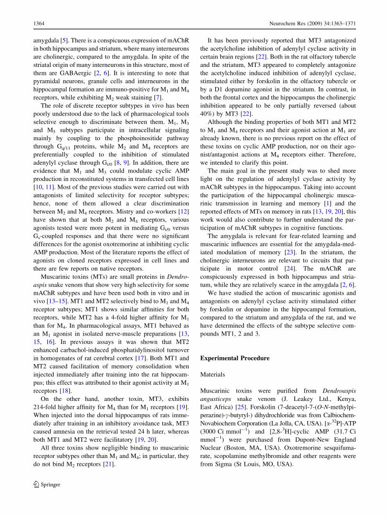
Hippocampal adenylyl cyclase activity was stimulated by
forskolin in a dose-dependent manner and fitted to a sig-
moidal dose–response curve, with a maximal stimulation
reached at 0.5 mM forskolin (Fig. 1a). At 100 lM for-
skolin, the percentage of stimulated adenylyl cyclase
activity in hippocampal membranes was 978.4 ± 102.3%
(n = 38) with respect to the basal level of cyclic AMP
produced in the absence of forskolin. The cyclic AMP
production are shown in Table 1; the basal level in the
hippocampus was 3.8 ± 0.9 nmol min-1 mg protein-1.
In the striatum, the basal cyclic AMP production was
33.0 ± 1.2 nmol min-1 mg protein-1 (Table 1). With
100 lM forskolin adenylyl cyclase activity increased to
2281.0 ± 124.4% (n = 42) respect to the basal level
(Fig. 1b). As expected, the stimulation increased with
forskolin concentration, but the enzyme activity did not
reach a plateau at the same forskolin concentrations it did
in the hippocampus. Since 100 lM forskolin produced a
significant stimulation in both structures without reaching
Neurochem Res (2009) 34:1363–1371 1365
123
saturation levels, we chose this concentration for the fol-
lowing assays with muscarinic toxins MT1, MT2 and MT3.
Hippocampal membranes stimulated by 100 lM for-
skolin (considered as 100%) showed a 110.7 ± 9.5%
(n = 13) cyclic AMP production in the presence of 10 lM
MT1, while in the striatum it was 96.4 ± 8.8% (n = 6).
Therefore, MT1 does not significantly modify forskolin-
stimulated cyclic AMP production under these conditions.
The addition of 10 lM MT3 to striatal membranes did
not produce statistically significant difference in basal
adenylyl cyclase activity (97.2 ± 3.9%, n = 5). Likewise,
MT3 up to 25 lM did not produce any significant change
in the basal level of enzyme activity (99.8 ± 2.4%, n = 2)
and did not significantly affect the forskolin stimulated
activity in hippocampal synaptosomal membranes (10 lM
MT3: 118.9 ± 15.5%, n = 11).
Figure 2 corresponds to the inhibition curves of cyclic
AMP synthesis—forskolin induced—by the specific
muscarinic agonist oxotremorine in hippocampal synapto-
somal membranes, either with or without the antagonist
scopolamine (Fig. 2a) or the different muscarinic toxins
(Fig. 2b–d). The agonist oxotremorine caused a biphasic
inhibition on forskolin-stimulated adenylyl cyclase activ-
ity. The curves were better fitted to a two-step, rather than a
one-step transition equation. In general, the inhibition was
apparent at 10-8 M, and the first step follows up to
10-5 M. In the hippocampus, this inhibition by the lower
concentrations of oxotremorine (corresponding to ‘‘high-
affinity’’ inhibition) amounted 13.5 ± 2.8% of the total
forskolin-stimulated adenylyl cyclase activity, with an IC50
of 2.3 nM. This first step of the curve was fully blocked by
10 lM scopolamine, a rather specific muscarinic antago-
nist, indicating that this was actually mediated by
muscarinic receptors (Fig. 2a). The second step, a steeper
decrease in activity corresponding to the ‘‘low-affinity’’
inhibition fraction, was obtained with oxotremorine con-
centrations higher than 10-5 M. The dose-dependent
portion of the curve corresponding to this low-affinity step
reached a 44.7 ± 4.4% further inhibition of forskolin-
stimulated cyclic AMP production. In contrast to the first
part of the curve, this inhibition was not affected by sco-
polamine (Fig. 2a).
Both MT1 and MT3 were used in concentrations much
higher than their respective Ki at M4 receptors (126 nM for
MT1 and 1.2 nM for MT3) [19, 21] to investigate their
effects on the inhibition caused by oxotremorine. Scopol-
amine, as well as both toxins MT1 (1 lM) (Fig. 2b) and
MT3 (100 nM) (Fig. 2d) eliminated the high-affinity inhi-
bition by oxotremorine, showing similar monophasic
curves, without affecting the low-affinity inhibition step
(where the estimated IC50 value for oxotremorine was
about 1 mM). As can be seen in Fig. 2c, the curve for
oxotremorine inhibition in the presence of MT2 (1 lM)
showed a slightly different profile: there was a rightward
shift of the high-affinity portion of the curve. This con-
centration of the toxin did not significantly affect forskolin-
stimulated cyclic AMP production (100.9 ± 4.5%). Hence,
the MTs have counteracted the inhibition caused by low
Fig. 1 Dose–response curve of stimulation of adenylyl cyclase by
forskolin in rat hippocampal membranes (a) and bar diagram
representing the forskolin stimulation in striatal membranes (b). Data
are represented as mean ± SEM (as percentages). 100% corresponds
to [32P]-cyclic AMP produced in basal conditions, without forskolin.
Basal production of [32P]-cyclic AMP was 3.8 ± 0.9 nmol min-1 mg
protein-1 (n = 6) for the hippocampus and 33.0 ± 1.2 nmol -
min-1 mg protein-1 (n = 6) for the striatum
Table 1 Effects of forskolin and dopamine on [32P]-cyclic AMP
production in hippocampal, striatal and amygdala membranes
Hippocampus Striatum Amygdala
Basal 3.8 ± 0.9 33.0 ± 1.2 3.3 ± 0.8
Forskolin
10-7 M 5.8 ± 0.5 50.6 ± 13.2 –
10-5 M 25.1 ± 3.8 129.1 ± 27.4 13.4 ± 1.6
10-4 M 37.2 ± 3.9 752.7 ± 40.9 22.0 ± 2.4
Dopamine
10-6 M 4.0 ± 0.1 33.1 ± 3.7 –
10-4 M 4.1 ± 0.1 48.5 ± 2.9 –
10-3 M 4.4 ± 0.2 54.9 –
Values in nmol of [32P]-cyclic AMP min-1 mg protein-1 (med-
ia ± SEM; except dopamine 10-3 M in striatum, with no replicates)
1366 Neurochem Res (2009) 34:1363–1371
123

concentrations of the muscarinic agonist oxotremorine
(high-affinity) upon forskolin-stimulated adenylyl cyclase
activity. However, neither scopolamine nor the MTs were
able to block the inhibition caused by oxotremorine at
concentrations higher than 10-5 M.
In assays carried out in striatal membranes, the inhibi-
tion of the forskolin-stimulated cyclic AMP production by
oxotremorine showed dose–response curves rather similar
to those found in the hippocampal membranes and which
were also well fitted by a two site model. However, the
high-affinity inhibition by oxotremorine (10-8–10-4 M)
amounted only 6.8 ± 4.2% of the total forskolin-stimu-
lated adenylyl cyclase activity with an IC50 of about
0.3 nM; this decrease was not statistically significant. The
second step, a steeper decrease corresponding to the low-
affinity inhibition fraction obtained with oxotremorine
concentrations between 10-4 and 10-3 M, reached a
34.5 ± 8.2% further inhibition of forskolin-stimulated
cyclic AMP production. This latest inhibition was not
modified by scopolamine either. Like in the hippocampal
membranes, 1 lM MT1 as well as scopolamine, were not
able to reverse the oxotremorine inhibition at agonist
concentrations higher than 10-4 M.
In amygdala membranes, the stimulation of adenylyl
cyclase activity by 100 lM forskolin reached about
670% with respect to the basal activity (3.3 ± 0.8 nmol of
[32P]-cyclic AMP min-1 mg protein-1, Table 1). But in
this structure, the muscarinic agonist oxotremorine up to
10-4 M did not appear to inhibit the forskolin-stimulated
adenylyl cyclase activity.
Fig. 2 Inhibition curves by oxotremorine, of adenylyl cyclase
activity stimulated by forskolin (100 lM) in hippocampal mem-
branes, either in the presence (s) or absence (d) of a 10 lM
scopolamine; b 1 lM MT1; c 1 lM MT2; d 100 nM MT3. Data are
represented as mean ± SEM (as percentages). 100% corresponds to
[32P]-cyclic AMP produced in the presence of 100 lM forskolin. The
inhibition curves by oxotremorine alone are better fitted to a two-step
transition equation. Inhibition by low concentrations of oxotremorine
amounted 13.5% of total forskolin-stimulated adenylyl cyclase
activity, IC50 of 2.3 nM. The inhibition curves in the presence of
10 lM scopolamine (a), 1 lM MT1 (b) and 100 nM MT3 (d) were
monophasic, showing only the oxotremorine low-affinity inhibition,
with IC50 of 1.0, 0.5 and 1.2 mM respectively (n = 3–10). The
inhibition curve in the presence of 1 lM of MT2 was biphasic (c),
with a high-affinity inhibition by oxotremorine of 13.5%, IC50 1 of
402.8 nM and IC50 2 16.1 mM
Neurochem Res (2009) 34:1363–1371 1367
123

Muscarinic Action on Dopamine-stimulated
Cyclic-AMP Production
The dose–response curves for the stimulation of cyclic AMP
production by dopamine in hippocampal and striatal syn-
aptosomal membranes are shown in Fig. 3. The increase in
cyclic AMP production stimulated by 1 mM dopamine
corresponds to 166% in the striatum and to 116% in the
hippocampus. As shown in Table 1, cyclic AMP production
stimulated by dopamine in the striatum was about 12-fold
that in the hippocampus. As we could not performed reliable
assays in the hippocampus due to the low increase reached by
dopamine stimulation, we were only able to study the mus-
carinic effects on dopamine stimulation on striatal tissue.
The inhibition by oxotremorine of dopamine-stimulated
adenylyl cyclase in the striatum is represented by the bar
diagram in Fig. 4. The adenylyl cyclase activity stimulated
by 100 lM dopamine reached 140.9 ± 5.0% of the basal
level. This stimulated activity was inhibited to
121.2 ± 3.9% by 10 lM oxotremorine. Thus, the agonist
was able to inhibit up to 48% of the activity increase by
100 lM dopamine in the striatum. This inhibition by
10 lM oxotremorine was completely blocked by 10 lM
MT3 (144.2 ± 5.4%, Fig. 4).
On the other hand, we did not find any significant
muscarinic effect on adenylyl cyclase activity stimulated
by dopamine in amygdala membranes.
Discussion
In this paper we report results of experiments carried out in
rat synaptosomal membranes of rat hippocampus, striatum
and amygdala. The basal activity of adenylyl cyclase in the
rat striatum membranes was about 8.7 fold of the activity in
the hippocampus and 10-fold that in the amygdala. The
stimulation of synaptosomal membranes from striatum by
forskolin was about 20-fold that in the hippocampus and
34-fold that in the amygdala. It was shown that in both the
striatum and hippocampus the enzyme activity increased
with forskolin concentration but did not reach a steady state
in striatum at the same concentrations it did in the hippo-
campus (Fig. 1; Table 1).
It is widely accepted that M2 and M4 [8] mAChR pref-
erentially interact with Gi/0 proteins causing inhibition of
previously stimulated adenylyl cyclase [28]. If this were the
case in the hippocampus, both M2 and M4 receptor subtypes
should be recruited for the inhibition of adenylyl cyclase by
the relatively non-selective agonist oxotremorine.
Fig. 3 Dose–response curve of stimulation of adenylyl cyclase by
dopamine in rat hippocampal (d) and striatal (s) membranes. Data
are represented as mean ± SEM (as percentages). 100% corresponds
to [32P]-cyclic AMP produced in basal conditions, without forskolin.
Basal production of [32P]-cyclic AMP was 3.8 ± 0.9 nmol min-1 mg
protein-1 for hippocampal membranes (n = 6) and 33.0 ± 1.2
nmol min-1 mg protein-1 for striatal membranes (n = 6)
Fig. 4 Bar diagram representing the effect on striatal membranes of
oxotremorine (Oxo) and MT3 over [32P]-cyclic AMP produced under
100 lM dopamine (Dopa) stimulation. Data are represented as
mean ± SEM (as percentages). 100% corresponds to [32P]-cyclic
AMP produced in basal conditions, without dopamine. Basal
production was 33.0 ± 1.2 nmol min-1 mg protein-1 (n = 6).
*Mean ± SEM was significantly different compared to the cyclic
AMP production in the presence of 100 lM dopamine alone
(P \ 0.05, ANOVA followed by Dunnet0s multiple comparison test)
1368 Neurochem Res (2009) 34:1363–1371
123

Here we show that the inhibition of adenylyl cyclase by
oxotremorine in the hippocampus appeared biphasic, with
both high-affinity and low-affinity inhibition steps (Fig. 2).
The first part of the agonist inhibition curve was fully
blocked by the non-selective muscarinic antagonist sco-
polamine, which did not affect the second low-affinity step.
Therefore, these results are consistent with the muscarinic
modulation of adenylyl cyclase in the hippocampus being
mainly—if not only—represented by the high-affinity
oxotremorine inhibition. Although some minor muscarinic
contribution to the low-affinity oxotremorine inhibition can
not be completely discarded, it did not appear significant in
this case, since there was not any detectable antagonism by
scopolamine. Hence, this also corroborates that the mus-
carinic inhibition affected just a small portion of all the
forskolin-stimulated adenylyl cyclase in the hippocampus
(13.5%, Fig. 2). Of course, there are more adenylyl cyclase
variants which are targets for forskolin stimulation—i.e.,
different isoforms, different location [29]—than those
regulated by mAChR.
The muscarinic toxins used did not produce significant
direct effects on either basal or forskolin-stimulated aden-
ylyl cyclase activity. MT2 has lower affinity than MT1 at
both M1 and M4 receptors, but discriminates better between
them (4-fold higher affinity for M1 than for M4; 21). MT3
is highly selective for M4 receptors [19, 30]. None of the
muscarinic toxins used have any detectable binding to M2
receptors [14, 19, 21].
The full blockade of the muscarinic inhibition of hip-
pocampal adenylyl cyclase by both MT1 and MT3
(Fig. 2b, d) was rather similar to that by scopolamine
(Fig. 2a). On the other side, the slightly different effect of
MT2—a rightward shift—on the high-affinity oxotremo-
rine inhibition of hippocampal adenylyl cyclase might be
due to the lower affinity this toxin exhibits at M4 receptor,
compared to the other muscarinic toxins used (Fig. 2c).
Hence, the muscarinic toxins antagonism at M4 receptors
would counteract the oxotremorine inhibition of stimu-
lated adenylyl cyclase. Both MT1 and MT2 acted as M1
agonists in several physiological paradigms, including
central and peripheral nervous system [13, 15, 18, 21, 31]
and in cell lines [16, 32]. In our assays, MT1, MT2 and
MT3 behaved as M4 antagonists, in agreement with the
reported data by Olianas et al. [22]. MT3 was reported to
also behave as an M4 antagonist at the rabbit anococ-
cygeus muscle preparation [16] and in memory
consolidation in the rat [19].
It had previously been suggested that neither M1 nor M2
receptors were responsible for cholinergic inhibition of
adenylyl cyclase in the striatum [33] or in the cerebral
cortex of the rat [34], whereas adenylyl cyclase muscarinic
inhibition would be mediated by M2 receptors in the brain
stem [35].
Oxotremorine inhibited the forskolin-stimulated aden-
ylyl cyclase in the striatum (though not significantly in low
concentrations) following a biphasic inhibition curve, as it
did in the hippocampus. Again, the oxotremorine low-
affinity inhibition step was not blocked by scopolamine. In
contrast, it has been reported that cyclic AMP production
stimulated by forskolin (10 lM) was inhibited to around
25% by acetylcholine and 24% by carbachol in the rat
striatum following a monophasic curve [36].
According to our estimations from the data reported by
Olianas et al. [22], 10 lM forskolin stimulated cyclic AMP
synthesis to around 390 pmol mg-1 min-1 in dentate gyrus,
and to approximately 255 pmol mg-1 min-1 in CA regions
of rat hippocampal formation. These values are much lower
than the data here reported in Table 1. It is difficult to
interpret why the results are so different, but the discrep-
ancies could be due to the different membrane preparation.
These authors reported that acetylcholine (20 lM) inhibited
forskolin stimulation of adenylyl cyclase by 21–25% in the
hippocampal regions, and that the toxin MT3 antagonized
this effect of acetylcholine by partially reversing that inhi-
bition to about 33–48%. Although we were not able to
calculate from the published data the ratio of adenylyl
cyclase stimulated respect to the basal level, the percentages
of reversion of the inhibition by 20 lM acetylcholine would
be comparable to the inhibition we obtained with lower
oxotremorine concentrations. However, in our assays MT3
did fully block only that adenylyl cyclase inhibition which
was also antagonized by scopolamine.
As it was already mentioned, neither MT3 nor scopol-
amine did fully block the oxotremorine inhibition, but they
acted in a similar way, reversing a similar proportion of
that inhibition.
It was previously reported that acetylcholine inhibited
the forskolin-stimulated adenylyl cyclase in the rat hippo-
campus with much higher EC50 than those we found with
oxotremorine and that MT3 blocked just a fraction of that
inhibition, whilst most of the response was not affected
[22]. Thus, it was suggested that M2 receptors were also
involved in this regulation. In contrast, we have shown that
the high-affinity inhibition by oxotremorine, the only that
can be considered as pure muscarinic since it was blocked
by scopolamine, was also fully blocked by muscarinic
toxins in the hippocampus. Therefore, we strongly suggest
that M4 receptors are responsible for either all or most of
the muscarinic inhibition of forskolin-stimulated adenylyl
cyclase in hippocampus. The discrepancy may be due to
the fraction of adenylyl cyclase inhibition that was con-
sidered as modulated by muscarinic receptors. We believe
that part of the inhibition by either high oxotremorine
concentration or by other agonists should not be consider
as muscarinic since it was caused by very high agonist
concentrations and was not blocked by scopolamine;
Neurochem Res (2009) 34:1363–1371 1369
123

therefore, it should not be expected to be antagonized by
any of the toxins.
Olianas et al. [26, 37] reported that acetylcholine
inhibited the basal level of striatal adenylyl cyclase activity
as well as that stimulated either by forskolin or by a D1
receptor agonist. It was reported that carbachol also caused
an inhibition in basal cyclic AMP production [38]. Since
this inhibition was blocked by MT3, the authors proposed
that M4 receptors could be responsible for the inhibition of
dopamine-sensitive adenylyl cyclase in the striatum.
However, we were not able to detect any significant effect
on basal cyclic AMP production of either oxotremorine or
carbachol in the striatum. It is evident that just a small
fraction of adenylyl cyclase was stimulated by dopamine,
both in the striatum and hippocampus, though (at 1 mM
dopamine) that fraction was higher in the striatum (166 vs.
116% with respect to each basal level). If we consider the
forskolin-stimulated adenylyl cyclase as total enzyme
activity, we may conclude that a fraction of about 1.3% of
the total cyclase activity would be regulated by dopamine
in hippocampal membranes; and this fraction would be
about 3.1% in striatal membranes. As a consequence, in the
conditions of our experiments, it was not feasible to per-
form reliable assays to analyze the effect of selective
agonists and antagonists in the hippocampus. Although it
was rather difficult in the striatum, the results were more
reliable than in the hippocampus. Oxotremorine inhibited
by 49% the dopamine stimulated adenylyl cyclase activity
in the striatum, respect to the basal value (see Table 1).
That inhibition was fully reversed by MT3 (Fig. 4).
Although the concentration of the toxin used was much
higher than any receptor subtype selective concentration,
MT3 has proved to have negligible or very low affinity for
M2 receptors [19], the subtype also proposed to be involved
in adenylyl cyclase inhibition. Therefore some minor M2
receptor contribution to the observed adenylyl cyclase
inhibition could not be discarded.
On the other hand, we did not find any significant
muscarinic effect on adenylyl cyclase activity in amygdala
membranes, which could mean that the localization of the
enzyme is different and/or there is not significant coupling
with mAChR.
It has been reported by Sanchez-Lemus and Arias-
Montano that M1 receptors would also be involved in
regulation of cyclic AMP production through protein
kinase C activation [39]. However, we did not find any
cyclic AMP increase by either MT1 or MT2 addition. An
explanation for this would be that those authors carried out
their study in miniprisms while our membrane preparation
did not preserve the biochemical and micro-architectural
integrity, i.e., the corresponding substrates and effectors.
It was previously shown that MT1 and MT2—agonists
at M1 receptors—given into the rat hippocampus facilitated
memory consolidation of an inhibitory avoidance task in
the rat [18, 31]. MT3, highly selective for M4, injected into
the hippocampus was amnesic for the same task [19, 20].
Therefore, previous and present results—MT1 and MT3
antagonism at M4—support the hypothesis that the M4
receptor could be a positive modulator of memory.
Acknowledgments We thank to the University of Buenos Aires,
ANPCyT/FONCyT, CONICET (Argentina), and to CNPq (Brazil) for
financial support.
References
1. Jerusalinsky D, Kornisiuk E, Izquierdo I (1997) Cholinergic
neurotransmission and synaptic plasticity concerning memory
processing. Neurochem Res 22:507–515. doi:10.1023/A:1027
376230898
2. Van Der Zee E, Luiten PG (1999) Muscarinic acetylcholine
receptors in the hippocampus, neocortex and amygdala: a review
of immunocytochemical localization in relation to learning and
memory. Prog Neurobiol 58:409–471. doi:10.1016/S0301-0082
(98)00092-6
3. Izquierdo I, Da Cunha C, Rosat R et al (1992) Neurotransmitter
receptors involved in post-training memory processing by the
amygdala, medial septum, and hippocampus of the rat. Behav
Neural Biol 58:16–26. doi:10.1016/0163-1047(92)90847-W
4. Levey AI, Kitt CA, Simonds WF et al (1991) Identification and
localization of muscarinic acetylcholine receptor proteins in brain
with subtype-specific antibodies. J Neurosci 11:3218–3226
5. Sanchez G, Alvares de Oliveira L, Oberholzer MV et al (2008)
M4 muscarinic receptors are involved in modulation of neuro-
transmission at synapses of Schaffer collaterals on CA1
hippocampal neurons in rats. J Neurosci Res (in press, published
on line 24 Sept)
6. Sah P, Faber ESL, Lopez de Armentia M et al (2003) The
amygdaloid complex: anatomy and physiology. Physiol Rev
83:803–834
7. Levey AI, Edmunds SM, Koliatsos V et al (1995) Expression of
M1–M4 muscarinic acetylcholine receptor proteins in rat hippo-
campus and regulation by cholinergic innervation. J Neurosci
15:4077–4092
8. Peralta EG, Ashkenazi A, Winslow JW (1988) Differential reg-
ulation of PI hydrolysis and adenylyl cyclase by muscarinic
receptor subtypes. Nature 334:434–437. doi:10.1038/334434a0
9. Hulme EC, Birdsall NJ, Buckley NJ (1990) Muscarinic receptor
subtypes. Annu Rev Pharmacol Toxicol 30:633–673. doi:
10.1146/annurev.pa.30.040190.003221
10. Felder CC, Kanterman RY, Ma AL et al (1989) A transfected M1
muscarinic acetylcholine receptor stimulates adenylate cyclase via
phosphatidylinositol hydrolysis. J Biol Chem 264:20356–20362
11. Burford NT, Nahorski SR (1996) Muscarinic M1 receptor-stim-
ulated adenylate cyclase activity in chinese hamster ovary cells is
mediated by Gs alpha and is not a consequence of phosphoinos-
itidase C activation. Biochem J 315:883–888
12. Mistry R, Dowling MR, Challis RAJ (2005) An investigation of
whether agonist-selective receptor conformations accur with
respect to M2 and M4 muscarinic acetylcholine receptor signal-
ling via Gi/0 and Gs proteins. Br J Pharmacol 144:566–575. doi:
10.1038/sj.bjp.0706090
13. Jerusalinsky D, Harvey A (1994) Toxins from mamba venoms:
small proteins with selectivities for different subtypes of mus-
carinic acetylcholine receptors. Trends Pharmacol Sci 15:424–
430. doi:10.1016/0165-6147(94)90092-2
1370 Neurochem Res (2009) 34:1363–1371
123

14. Jerusalinsky D, Kornisiuk E, Alfaro P et al (2000) Muscarinic
toxins: novel pharmacological tools for the muscarinic choliner-
gic system. Toxicon 38:747–761. doi:10.1016/S0041-0101(99)
00196-8
15. Harvey A, Kornisiuk E, Bradley KN et al (2002) Effects of
muscarinic toxins MT1 and MT2 from green mamba on different
muscarinic cholinoceptors. Neurochem Res 27:1543–1554. doi:
10.1023/A:1021660708187
16. Bradley KN (2000) Muscarinic toxins from the green mamba.
Pharmacol Ther 85:87–109. doi:10.1016/S0163-7258(99)
00064-9
17. Jerusalinsky D, Raskovsky S, Kornisiuk E (1994) Muscarinic
toxins from the venom of elapid snakes. In: Tipton K, Dajas F
et al (eds) Neurotoxins in neurobiology. Ellis Hodwood Ltd,
Chichester, pp 89–102
18. Jerusalinsky D, Cervenansky C, Walz R et al (1993) A peptide
muscarinic toxin from the green mamba venom shows agonist-
like action in an inhibitory avoidance learning task. Eur J Phar-
macol 240:103–105. doi:10.1016/0014-2999(93)90554-U
19. Jerusalinsky D, Kornisiuk E, Alfaro P et al (1998) Muscarinic
toxin selective for M4 receptors impairs memory in the rat. Neu-
roReport 9:1407–1411. doi:10.1097/00001756-199805110-00029
20. Ferreira AR, Furstenau L, Blanco C et al (2003) Role of hippo-
campal M1 and M4 muscarinic receptor subtypes in memory
consolidation in the rat. Pharmacol Biochem Behav 74:411–415.
doi:10.1016/S0091-3057(02)01007-9
21. Kornisiuk E, Jerusalinsky D, Cervenansky C et al (1995) Binding
of muscarinic toxins MT 9 1 and MT 9 2 from the venom of the
green mamba Dendroaspis angusticeps to cloned human mus-
carinic cholinoceptors. Toxicon 33:11–18 (Corrigendum Toxicon
33:1111)
22. Olianas MC, Adem A, Karlsson EI et al (1998) Identification of
rat brain muscarinic M4 receptors coupled to cyclic AMP using
the selective antagonist muscarinic toxin 3. Eur J Pharmacol
357:235–242. doi:10.1016/S0014-2999(98)00553-6
23. Vazdarjanova A, McGaugh JL (1999) Basolateral amygdala is
involved in modulating consolidation of memory for classical
fear conditioning. J Neurosci 19:6615–6622
24. Gomeza J, Zhang L, Kostenis E et al (1999) Enhancement of D1
dopamine receptor-mediated locomotor stimulation in M(4)
muscarinic acetylcholine receptor knockout mice. Proc Natl Acad
Sci USA 96:10483–10488. doi:10.1073/pnas.96.18.10483
25. Jerusalinsky D, Cervenansky C, Pena C et al (1992) Two poly-
peptides from Dendroaspis angusticeps venom selectively inhibit
the binding of central muscarinic cholinergic receptor ligands.
Neurochem Int 20:237–246. doi:10.1016/0197-0186(92)90173-O
26. Olianas MC, Adem A, Karlsson E et al (1996) Rat striatal mus-
carinic receptors coupled to the inhibition of adenylyl cyclase
activity: potent block by the selective M4 ligand muscarinic toxin
3 (MT3). Br J Pharmacol 118:283–288
27. Salomon Y, Londos D, Rodbell M (1974) A highly sensitive
adenylate cyclase assay. Anal Biochem 58:541–548. doi:10.1016/
0003-2697(74)90222-X
28. Caulfield MP, Birdsall NJM (1998) International union of phar-
macology. XVII. Classification of muscarinic acetylcholine
receptors. Pharmacol Rev 50:279–290
29. Visel A, Alvarez-Bolado G, Thaller C (2006) Comprehensive
analysis of the expression patterns of the adenylate cyclase gene
family in the developing and adult mouse brain. J Comp Neurol
496:684–697. doi:10.1002/cne.20953
30. Liang JS, Carsi-Gabrenas J, Krajewski JL et al (1996) Anti-
muscarinic toxins from Dendroaspis angusticeps. Toxicon
34:1257–1267
31. Jerusalinsky D, Kornisiuk E, Bernabeu R et al (1995) Muscarinic
toxins from the venom of Dendroaspis snakes with agonis-like
actions. Toxicon 33:389–397. doi:10.1016/0041-0101(94)
00103-F
32. Bradley KN, Rowan EG, Harvey AL (2003) Effects of musca-
rinic toxins MT2 and MT7, from green mamba venom, on M1,
M3 and M5 muscarinic receptors expressed in chinese hamster
ovary cells. Toxicon 41:207–215. doi:10.1016/S0041-0101(02)
00278-7
33. Ehlert FJ, Delen FM, Yun SH et al (1989) Coupling of subtypes
of the muscarinic receptor to adenylate cyclase in the corpus
striatum and heart. J Pharmacol Exp Ther 251:660–671
34. McKinney M, Anderson DJ, Vella-Rountree L et al (1991)
Pharmacological profiles for rat cortical M1 and M2 muscarinic
receptors using selective antagonists: comparison with N1E-115
muscarinic receptors. J Pharmacol Exp Ther 257:1121–1129
35. McKinney M, Anderson D, Forray C et al (1989) Characteriza-
tion of the striatal M2 muscarinic receptor mediating inhibition of
cyclic AMP using selective antagonists: a comparison with the
brainstem M2 receptor. J Pharmacol Exp Ther 250:565–572
36. Olianas MC, Maullu C, Onali P (1997) Effects of clozapine on rat
striatal muscarinic receptors coupled to inhibition of adenylyl
cyclase activity and on the human cloned M4 receptor. Br J
Pharmacol 122:401–408. doi:10.1038/sj.bjp.0701357
37. Olianas MC, Onali P, Neff NH et al (1983) Adenylate cyclase
activity of synaptic membranes from rat striatum. Inhibition by
muscarinic receptor agonists. Mol Pharmacol 23:393–398
38. Onali P, Olianas MC (2002) Muscarinic M4 receptor inhibition of
dopamine D1-like receptor signalling in rat nucleus accumbens.
Eur J Pharmacol 448:105–111. doi:10.1016/S0014-2999(02)
01910-6
39. Sanchez-Lemus E, Arias-Montano JA (2006) M1 muscarinic
receptors contribute to, whereas M4 receptors inhibit, dopamine
D1 receptor-induced [3H]-cyclic AMP accumulation in rat striatal
slices. Neurochem Res 31:555–561. doi:10.1007/s11064-006-
9052-8
Neurochem Res (2009) 34:1363–1371 1371
123