inhibition of adenylyl cyclase type 5 increases longevity ...€¦ · healthful aging through...
TRANSCRIPT
Review ArticleInhibition of Adenylyl Cyclase Type 5 Increases Longevity andHealthful Aging through Oxidative Stress Protection
Stephen F. Vatner, Ronald E. Pachon, and Dorothy E. Vatner
Departments of Cell Biology & Molecular Medicine and Medicine, New Jersey Medical School, Rutgers University,Newark, NJ 07103, USA
Correspondence should be addressed to Stephen F. Vatner; [email protected]
Received 2 January 2015; Revised 10 March 2015; Accepted 13 March 2015
Academic Editor: Vincent Pialoux
Copyright © 2015 Stephen F. Vatner et al. This is an open access article distributed under the Creative Commons AttributionLicense, which permits unrestricted use, distribution, and reproduction in any medium, provided the original work is properlycited.
Mice with disruption of adenylyl cyclase type 5 (AC5 knockout, KO) live a third longer than littermates. The mechanism, in part,involves the MEK/ERK pathway, which in turn is related to protection against oxidative stress. The AC5 KO model also protectsagainst diabetes, obesity, and the cardiomyopathy induced by aging, diabetes, and cardiac stress and also demonstrates improvedexercise capacity. All of these salutary features are also mediated, in part, by oxidative stress protection. For example, chronic betaadrenergic receptor stimulation induced cardiomyopathy was rescued by AC5 KO. Conversely, in AC5 transgenic (Tg) mice, whereAC5 is overexpressed in the heart, the cardiomyopathy was exacerbated and was rescued by enhancing oxidative stress resistance.Thus, the AC5 KO model, which resists oxidative stress, is uniquely designed for clinical translation, since it not only increaseslongevity and exercise, but also protects against diabetes, obesity, and cardiomyopathy. Importantly, inhibition of AC5’s action toprolong longevity and enhance healthful aging, as well as its mechanism through resistance to oxidative stress, is unique among allof the nine AC isoforms.
1. Introduction
Adenylyl cyclase (AC) is a ubiquitous enzymewhich regulatesall organs and catalyzes the conversion of ATP to cAMP.There are nine major mammalian AC isoforms; types 5 and6 are the major isoforms in the heart. Since our laboratoryhas been primarily involved in cardiovascular regulation, ourinterest was in disrupting one of the twomajor isoforms in theheart, type 5 AC (AC5 knock out (KO)), to see how regulationof cardiac function is altered. We found that the AC5 KOheart was protected against the stresses of chronic pressureoverload [1] and chronic catecholamine stimulation [2, 3].However, even more interesting and potentially importantwere our other findings of the salutary effects of AC5 inhi-bition, not necessarily related to the heart. We also studiedthese mice for 3 years and found that they lived a third longerthan wild type (WT) [4]. Although longevity models areimportant, the translational value for these studies is limited,unless the model also improves healthful aging. In fact olderindividuals often are not interested in expanding life span,
if it is accompanied by many of the limitations observedin the elderly, for example, exercise intolerance, diabetes,and obesity. Importantly the AC5 KO model also promoteshealthful aging, as it enhances exercise capacity, and protectsagainst diabetes and obesity and diabetic cardiomyopathy [5].Indeed the AC5 KO model shares many of the same featuresof themost commonly studiedmodel of longevity and protec-tion against obesity, caloric restriction [6]. However, caloricrestriction does not improve exercise performance, but, onthe contrary, it actually diminishes exercise capacity [7].There is also one major limitation to the translation of caloricrestriction to longevity and protection against diabetes andobesity in patients, that is, compliance due to difficulty inmaintaining low calorie or low fat diets. Accordingly, thereis considerable need for a novel therapy that might mimic thephenotype of caloric restriction, without the negative aspectsof limiting caloric intake. One such model is inhibition ofAC5, which has a similar phenotype to caloric restriction [6],but the mice actually eat more than their wild type controls,while weighing less.
Hindawi Publishing CorporationOxidative Medicine and Cellular LongevityVolume 2015, Article ID 250310, 13 pageshttp://dx.doi.org/10.1155/2015/250310

2 Oxidative Medicine and Cellular Longevity
production
scavenging enzymes
Mitochondria
Oxidative stress
PTP activationcytochrome c release
(apoptosis)
Defective electron transport chain
Cell dysfunction
Cell death Aging
SIRT1/FoxO3a/MnSODby AC5KO
Raf/MEK/ERK/MnSODby AC5 KO
↑ ROS
↓ ROS
Figure 1: Mitochondrial dysfunction and oxidative stress. During the aging process, ROS accumulation and DNAmutation in mitochondriainduce oxidative stress, which results in themitochondrial permeability transition pore (PTP) opening and electron transport chain deficiency,thereby leading to cell dysfunction and cell death. Inhibition of AC5 activates SIRT1/FoxO3a andRaf/MEK/ERKpathways, and both pathwaysupregulate the antioxidant, MnSOD, resulting in resistance to oxidative stress during aging.
Table 1: Longevity and oxidative stress resistance.
Longevity models related to oxidativestress resistance
Longevity models not relatedto oxidative stress resistance
Longevity models with relationship to oxidativestress resistance controversial or not studied
AC5 KO [3–6] Gpx4 +/− KO [28] Ames dwarf [29–31]Caloric restriction [8, 32–35] GHR/BP KO [36, 37] FIRKO [38, 39]Snell dwarf [40–42] MIF KO [43, 44] PAPPA−/− KO [45, 46]GHR KO [9, 40] 𝛼MUPA OE [47, 48] RII𝛽−/− KO [49]Igf-1r+/− KO [10, 50, 51] S6K1−/− KO [52, 53]Klotho OE [11, 12, 54] Hct-UCP2 OE [55]p66shc−/− [13, 56] PEPCK-C OE [57]TRX OE [14, 58] PtenTg [59]MCAT [15, 60–62] SIRT6 Tg [63]MT OE [16]Agtr1 𝛼−/− KO [17]Irs1−/− KO [18]R6/2 Irs2+/− IRS2 Btg KO [19]Surf1−/− KO [21, 22, 64]Mclk1+/− KO [65, 66]Data are shown as mice models followed by the supporting bibliography in parenthesis [].
One common mechanism that mediates longevity andhealthful aging is protection against oxidative stress [4,6, 8–22]. Table 1 summarizes the most commonly studiedlongevity models, showing that the majority of these models,as well as the AC5 KO model, have one common theme,that is, protection against oxidative stress. Accordingly, inorder to understand themechanismsmediating the beneficialeffects of the AC5 KO model, it is important to examine itsprotection against oxidative stress.
2. Oxidative Stress in Aging and inthe AC5 KO (Figures 1 and 2)
The AC5 KO model of aging protects against oxidative stressby reducing cAMP and protein kinase A (PKA), which inturn activates the Raf/MEK/ERK pathway, which increasesMnSOD and protects against oxidative stress [4] (Figures 1and 2). As noted above, protection against oxidative stress is acommonmechanism in longevitymodels (Table 1). Oxidative

Oxidative Medicine and Cellular Longevity 3
Nor
mal
ized
leve
ls
0.0
0.3
0.6
0.9
1.2
1.5
1.8
WT
MnSOD
AC5 KO
∗
(a)
0
10
20
30
40
50
60
Surv
ival
(%)
WT
Cell viability
AC5 KO
∗
(b)
WTAC5 KO
Phospho-ERK
0
2
4
6
8
10
p-ER
K/ER
K
∗
(c)
AC5
cAMP
PKA
Raf-1
p-MEK
p-ERK
MnSOD
Oxidative stress
Exercise diabetes
catecholamine stress
Longevity Cardiomyopathy
AC5 KO
(d)
Figure 2: ERK pathway and oxidative stress in aging AC5 KO. (a) Western blotting of MnSOD in the hearts of aging WT and AC5 KOmice.The levels of MnSOD are significantly greater in AC5 KO mice compared to WT mice (∗𝑃 < 0.05). Data are expressed as mean ± SEM. (b)Cell viability was tested in response to oxidative stress in neonatal cardiacmyocytes fromAC5KO andWT.Myocardial cells were treated withH2
O2
(25 𝜇M) and evaluated for cell viability using Cell Titer-Blue Cell Viability Assay. AC5 neonatal myocytes showed resistance to oxidativestress andDNAdamage (∗𝑃 < 0.05 versusWT). Data are presented asmean ± SEM. (c) By western blotting, the level of ERK phosphorylationwas significantly increased in AC5 KO mice compared with WT (∗𝑃 < 0.05). Data are expressed as mean ± SEM. (d) Signaling diagram forAC5 regulation of oxidative stress through the ERK pathway is shown. Data is redrawn from Yan et al. [4].
stress is enhanced during the aging process; mitochondriaproduce less energy (ATP) and also increase the productionof reactive oxygen species (ROS) as a product of aerobicmetabolism (Figure 1). Furthermore the activity of free-radical scavenging enzymes changes with aging. One mecha-nism by which oxidative stress is increased in the aging tissueis through increased ROS induced apoptosis and necrosisby opening of the mitochondrial membrane permeabilitytransition pore and release of apoptotic inducing factors, for
example, cytochrome c [23] (Figure 1). Oxidative stress hasbeen linked to aging and senescence through the tumor-suppressor p53 and transcriptional responses, mediated byp44/p53 and p66 [24]. There is also considerable supportfor the regulation of oxidative stress by the Raf/MEK/ERKpathway [25]. In summary, the mechanism of oxidative stressinduced aging seems to be multifactorial, where mitochon-drial damage and impaired function play a significant role.The final common pathway for premature cell death has been

4 Oxidative Medicine and Cellular Longevity
reported to be apoptosis, necrosis, and control of the cell cycle[26, 27] (Figure 1), specifically an increase in the percentageof cells stopped at the G0/G1 phase of the cell cycle [26].
To further understand the importance of oxidative stressin aging, the extent to which the major diseases limit longe-vity must be examined for their relation to oxidative stress.Based on the statistics of the CDC in 2010, the leading causesof death in humans in order of frequency are heart disease,cancer, chronic lower respiratory diseases, stroke, accidents,Alzheimer’s disease, diabetes, influenza and pneumonia,nephritis, nephrotic syndrome, and nephrosis. It is importantto recognize that most of these diseases are related to oxida-tive stress. Such is the case for heart disease [67], cancer [68],COPD [69], stroke [70], Alzheimer’s [71], diabetes [72], andchronic kidney disease [73]. In mice, the causes of mortalityalso share oxidative stress mechanisms. The most commoncause of death in C57BL/6J mice is neoplasia, includinglymphoma and other hematological and nonhematologicalcancers, followed by chronic kidney and heart diseases [74].As previously described, these entities have been linked tooxidative stress, further supporting the importance of oxida-tive stress in limiting longevity.
Perhaps themost convincing evidence for oxidative stressin aging is Progeria, a unique medical condition character-ized by premature aging [75], such that teenagers often sufferfrom atherosclerosis, cardiomyopathy, and coronary arterydisease and death. Interestingly, accumulation of oxidizedproteins causing DNA damage has been described as thecausative effect in this disease [76, 77]. One abnormallyformed protein is called laminA. It is an autosomal dominantmutation involving (LMNA) gene and/or abnormal posttran-slational processing (ZMPSTE24) [75, 78].
3. Oxidative Stress in Exercise
The AC5 KO model is one of the few aging models knownto also improve exercise performance [5]. This is importantfor two reasons. First, maintained exercise performance iscommon to longevity, as reduced exercise performance iscommon to many of the diseases that limit longevity, forexample, cardiopulmonary diseases. Secondly, exercise train-ing and conditioning is recognized to be therapeutic to mostdiseases and is one intervention that can extend longevity[79]. It is therefore important and well established thatboth resting and contracting skeletal muscles produce ROS.Exercise has been also related to induction of oxidative stresswhen it is performed at high intensity. However, moderateintensity aerobic exercise enhances endothelium dependentvasodilation through the increased production of nitric oxide[80]. This could be explained by the fact that while highlevels of free radicals can damage cellular components, lowtomoderate level of oxidants playmultiple regulatory roles incells such as the control of gene expression, regulation of cellsignaling pathways, and modulation of skeletal muscle forceproduction [80].
There are multiple cellular mechanisms mediating resist-ance to oxidative stress, by regular moderate exercise. Theseinclude reduction of basal formation of oxidants, improve-ment of the antioxidant defense system, and increased
resistance of tissues against ROS damage [81]. Furthermore,in a study using rats, exercise increased total serum antiox-idant substances with an additional beneficial effect on lipidprofile [82]. More specifically, another study evaluating therelationship of oxidative stress, endothelial dysfunction, andatherosclerosis with physical inactivity in mice showed thatdecreased physical activity increases vascular NADPH oxi-dase activity and enhances vascular ROS production, whichcontributes to endothelial dysfunction and atherosclerosis asopposed to physically active animals [83].
4. Oxidative Stress in Diabetes and Obesity
As noted above the AC5 KO mouse shares a common phe-notype and genotype with caloric restriction [6], a frequentlystudiedmodel demonstrating protection against diabetes andaging. The AC5 KO protection against glucose intoleranceand insulin resistance and obesity is observed in the animalson a standard diet, but it is even more pronounced whenstressed with a high fat diet [84]. The AC5 KO mice weighless and have less obesity and better serum lipids as well asglucose tolerance and insulin resistance compared to theWTmice [84]. Glucose intolerance and reduced insulin sensi-tivity have also been linked to oxidative stress mechanisms.Chronic hyperglycemia leads to generation of ROS resultingin increased oxidative stress and destruction of pancreaticcells, critical to insulin secretion [72].
The AC5 KO is also protected from obesity [6], whichis also linked to oxidative stress mechanisms. There areseveral mechanisms by which obesity produces oxidativestress. First, adipose tissue produces certain bioactive sub-stances called adipokines such as IL-6 and leptin, whichinduce the production of ROS. A second mechanism is thatmitochondrial and peroxisomal oxidation of fatty acids canproduce ROS in oxidation reactions. And third, there is anoverconsumption of oxygen in the mitochondrial respiratorychain [85]. Additionally, the pattern of food consumption inobese patients can potentially exacerbate the cellular damage;lipid rich diets also produce ROS due to modificationsin oxygen metabolism contributing to the cell dysfunctioninduced by obesity [85]. Interestingly, the persistence ofobesity in humans has been shown to decrease the activityof antioxidant enzymes in the adipose tissue such as catalase,superoxide dismutase, and glutathionine peroxidase [85, 86].This could represent another mechanism involved in theprogression of the disease and the development of obesityrelated complications. Finally, a study in humans confirmedthe increased oxidative stress state seen in patients withobesity and diabetes, measuring urinary creatinine-8-epi-PGF2𝛼 as amarker of systemic oxidative stress [87]. Taking allthe previous evidence together, it is clear that oxidative stressis a common factor in obese and diabetic patients.
5. Oxidative Stress in Cardiomyopathy andHeart Failure
There is accumulating evidence that increased oxidative stressis involved in the pathogenesis of various types of cardio-myopathy [88], including dilated [89, 90], diabetic [91, 92],

Oxidative Medicine and Cellular Longevity 5
ischemic [93, 94], hypertensive [95], adriamycin-induced[96], and pressure overload-induced cardiomyopathy [97–99], as well as beta adrenergic receptor overexpressioninduced cardiomyopathy [3, 100]. It is important to note thatoxidative stress affects different cell types involved in cardio-myopathy, not just cardiac myocytes. Dysfunction in othercells, such as endothelial cells and fibroblasts, plays animportant role in the development of cardiomyopathies, aswell.
The AC5 KO model is also protected against cardiomy-opathy and heart failure through oxidative stressmechanisms[3] (Figures 3–5). For example, chronic beta adrenergicreceptor (𝛽-AR) stimulation induces cardiomyopathy andheart failure by increasingmarkers of oxidative stress damageincluding myocyte necrosis and apoptosis [100]. We foundthat augmenting oxidative stress by mating the AC5 KOmicewith MnSOD KO mice resulted in loss of the protectionagainst the decreased cardiac function and increased cardiacfibrosis in response to chronic catecholamine stimulation inthe double knockouts (Figure 5) [3, 5]. Conversely, whenAC5 is overexpressed in the heart, as occurs in the cardiacspecific AC5 Tg mouse, the cardiomyopathy induced bychronic catecholamine stimulation is exacerbated (Figure 5).In this model mating the AC5 Tgmice withMnSODTgmicerescues the cardiomyopathy (Figure 5). Furthermore, AC5KO can also prevent the cardiomyopathy induced by chron-ically enhanced 𝛽-AR signaling in mice with overexpressed𝛽2-AR also, potentially, through enhancing resistance tooxidative stress [100].These findings confirm the importanceof oxidative stress in the pathogenesis of heart failure ingeneral and in the protection afforded by the AC5 KOmodelin particular. The AC5 KO model is also protected againstcardiomyopathies induced by chronic pressure overload [1],diabetes [5], and aging [4].
6. Mechanisms of AC5 KO Induced Longevityand Oxidative Stress Resistance
6.1. ERK Signaling Pathway Related to AC5 KO and Oxida-tive Stress (Figure 2). We previously found that AC5 KOincreases longevity and stress resistance via activation of theRaf/MEK/ERK signaling pathway [4]. This finding, based onthe reduction in cAMP byAC5, is supported by studies show-ing that the AC/cAMP/PKA pathway negatively regulates theMEK-ERK signaling pathway [101]. TheMEK/ERK signalingpathway is also one of the main stress signaling pathways andcentral mediators activated in response to oxidative damage[102, 103]. Previous findings suggested that a decrease in theactivation of the Raf/MEK/ERK pathway is associated withaging [104–109]. For example, decreased levels of ERK phos-phorylation were observed in senescent fibroblasts [104, 110]and hepatocytes from aging rats [111], whereas caloric restric-tion, a well-recognized mechanism mediating longevity andstress resistance, significantly reduced the age-related declinein ERK activation [111]. The long-lived Snell dwarf micealso exhibits an elevated level of ERK phosphorylation [112].We have shown an activation of the MEK/ERK signalingpathway in various tissues from long-lived AC-5 KO mice,which is consistent with that found in caloric restricted mice
and long-lived Snell dwarf mice. Interestingly, recent studiesdemonstrated a slower and more prolonged activation ofERK with longevity [113]. Furthermore, the activation of theERK pathway in response to oxidative stress is reduced withage, while loss of oxidative stress resistance with aging isassociated with decreased ERK activity, implying that ERKactivation exerts a prosurvival signal against aging inducedoxidative stress [111].
In addition, superoxide dismutase (SOD) has beenreported as the downstream target of the Cyr1 (AC)/cAMP/PKA pathway in yeast, which induces protection [114]. Wehave shown that MnSOD, which is a major molecule protect-ing against oxidative stress, is upregulated in AC5 KO mice(Figure 2(a)) [4] but downregulated, when AC5 is upregu-lated, as in the AC5 Tg heart (Figure 3(c)) [3]. A deficiency ofSOD is able to induce senescence; for example, homozygousSOD2−/− mice show significant damage to mitochondrialDNA in lung and liver compared toWTmice or heterozygousmice, resulting in a survival rate of only up to seven daysafter birth due to cardiomyopathy and liver disease [115]. Thelinkage between SOD and ERK activation is controversial,however. Although EGF-induced phosphorylation of ERK1/2is attenuated by overexpression of Cu/ZnSOD in vascularsmooth muscle cells, several other studies have shown thatthe ERK signaling cascade plays a positive role in overex-pression of MnSOD, which protects murine fibrosarcomacells from apoptosis [116] and suppresses tumor growth inthemac25/IGFBP-rP1-transfected human breast and prostatecancer cell lines [117]. Our findings have proven that MnSODis the downstream enzyme involved in the ERK signalingcascades [3, 4] mediating longevity and stress resistance inAC5 KO mice.
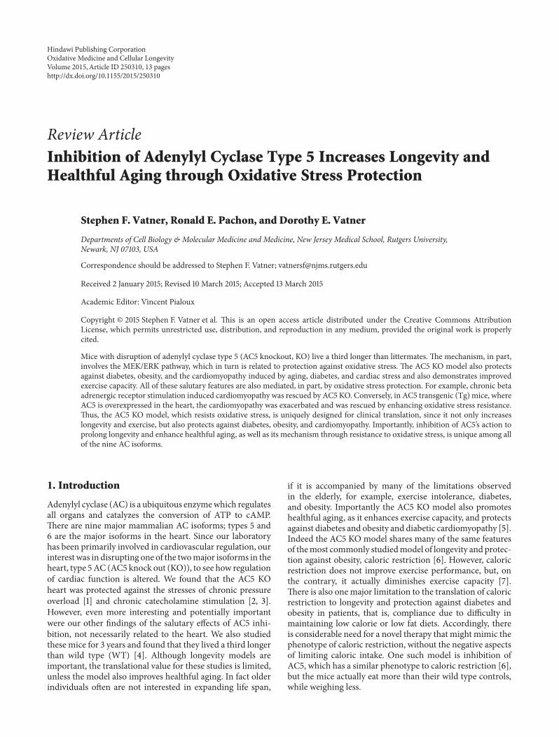
6.2. AC5, SIRT1, FoxO3a, and MnSOD (Figures 3 and 4).As noted above, we found that AC5 KO increases life spanand protects against oxidative stress though upregulatingthe antioxidant, MnSOD [4], whereas MnSOD regulated thecardiomyopathy induced by chronic catecholamine stimula-tion through the AC5, SIRT1, FoxO3a, and MnSOD pathway[3]. MnSOD is regulated transcriptionally by several tran-scription factors, such as NF-𝜅B, p53, and FoxO3a [118–120].Among them, FoxO3a is most closely related to the antiagingeffects of MnSOD. It is known that FoxO3a is essential for theparticipation of MnSOD in antiaging mechanisms of variousspecies. In C. elegans, FoxO3a transcriptionally upregulatedMnSOD, which induced life span extension [121]. In rats,aging induced downregulation of MnSOD is due to the inac-tivation of FoxO3a [122]. In human quiescent cells, FoxO3abinds directly to the promoter of MnSOD and protects thecells from oxidative stress [123]. The transcriptional activityof FoxO factor could be activated by deacetylation. SIRT1is a deacetylase which is able to activate FoxO3a (Figure 4)[118]. Interestingly, we found that the SIRT1/FoxO3/MnSODpathway is only activated by AC5 and not by AC6 (anothermajor AC isoform in the heart and brain), indicating a uniqueregulation of this pathway by AC5 (Figure 3(e)) [3]. ThePuigserver lab reported a new short-term SIRT1 activationpathway that involved𝛽-AR/AC/cAMP/PKA [124]. However,the AC5 KOmodel is quite different, since it increases NAD+

6 Oxidative Medicine and Cellular Longevity
0
40
80
120
160
200
WT AC5 Tg
cAM
P (p
mol
e/m
in/m
g)
Baseline AC activity
(a)
0
400
800
1200
1600
WT AC5 Tg
cAM
P (p
mol
e/m
in/m
g)
AC activity with forskolin
(b)
0.0
0.2
0.4
0.6
0.8
1.0
1.2
Rela
tive A
DU
MnSOD∗
WTAC5 Tg
(c)
00.5
11.5
22.5
33.5
44.5
Nor
mal
ized
leve
ls
SIRT1 MnSOD
WTAC5 Tg
∗ ∗
∗
FoxO3a
(d)
0
0.2
0.4
0.6
0.8
1
1.2
1.4
Rela
tive A
DU
SIRT1
0
0.2
0.4
0.6
0.8
1
1.2
1.4
Rela
tive A
DU
MnSOD
WTAC6 KO
WTAC6 KO
(e)
Figure 3: SIRT1/FoxO3a/MnSOD pathway in AC5 KO, AC5 Tg, and AC6 KO. (a) and (b) AC activity at baseline and in response to forskolinstimulation was enhanced in AC5 Tg mice compared to WT. (c) AC5 regulated MnSOD expression. Downregulation of MnSOD in AC5Tg mice hearts is shown (𝑛: 6 per group) (∗𝑃 < 0.05). (d) Expression of SIRT1 by western blotting in the AC5 myocardial cells. SIRT1 washighly expressed compared to WT. (∗𝑃 < 0.05). Expression of FoxO3a by western blotting in AC5 KO mouse hearts is shown. More FoxO3awas expressed in the nucleus of AC5 KO myocytes compared to WT. MnSOD expression increased in AC5 KO hearts (∗𝑃 < 0.05). Data areexpressed as mean ± SEM. (e) SIRT1 andMnSOD expression levels in AC6 KO did not show any difference compared toWT. Data is redrawnfrom Lai et al. [3].
Oxidative Medicine and Cellular Longevity 7
AC5
SIRT1
MnSODFoxO3a
Oxidative stress
Cardiac dysfunction
Exercisediabetes
catecholamine stress
Figure 4: Signaling diagram for AC5 regulation of oxidative stressthrough the SIRT1/FoxO3a pathway.
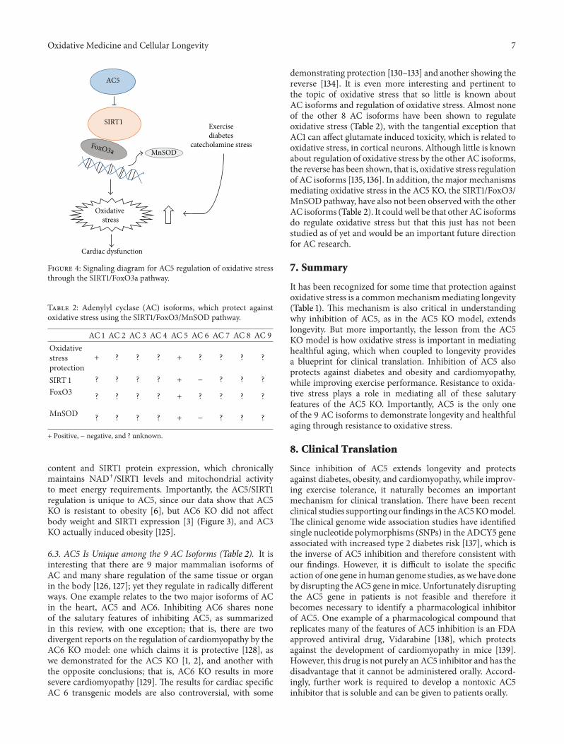
Table 2: Adenylyl cyclase (AC) isoforms, which protect againstoxidative stress using the SIRT1/FoxO3/MnSOD pathway.
AC 1 AC 2 AC 3 AC 4 AC 5 AC 6 AC 7 AC 8 AC 9Oxidativestressprotection
+ ? ? ? + ? ? ? ?
SIRT 1 ? ? ? ? + − ? ? ?FoxO3 ? ? ? ? + ? ? ? ?
MnSOD ? ? ? ? + − ? ? ?
+ Positive, − negative, and ? unknown.
content and SIRT1 protein expression, which chronicallymaintains NAD+/SIRT1 levels and mitochondrial activityto meet energy requirements. Importantly, the AC5/SIRT1regulation is unique to AC5, since our data show that AC5KO is resistant to obesity [6], but AC6 KO did not affectbody weight and SIRT1 expression [3] (Figure 3), and AC3KO actually induced obesity [125].
6.3. AC5 Is Unique among the 9 AC Isoforms (Table 2). It isinteresting that there are 9 major mammalian isoforms ofAC and many share regulation of the same tissue or organin the body [126, 127]; yet they regulate in radically differentways. One example relates to the two major isoforms of ACin the heart, AC5 and AC6. Inhibiting AC6 shares noneof the salutary features of inhibiting AC5, as summarizedin this review, with one exception; that is, there are twodivergent reports on the regulation of cardiomyopathy by theAC6 KO model: one which claims it is protective [128], aswe demonstrated for the AC5 KO [1, 2], and another withthe opposite conclusions; that is, AC6 KO results in moresevere cardiomyopathy [129]. The results for cardiac specificAC 6 transgenic models are also controversial, with some
demonstrating protection [130–133] and another showing thereverse [134]. It is even more interesting and pertinent tothe topic of oxidative stress that so little is known aboutAC isoforms and regulation of oxidative stress. Almost noneof the other 8 AC isoforms have been shown to regulateoxidative stress (Table 2), with the tangential exception thatAC1 can affect glutamate induced toxicity, which is related tooxidative stress, in cortical neurons. Although little is knownabout regulation of oxidative stress by the other AC isoforms,the reverse has been shown, that is, oxidative stress regulationof AC isoforms [135, 136]. In addition, the major mechanismsmediating oxidative stress in the AC5 KO, the SIRT1/FoxO3/MnSOD pathway, have also not been observed with the otherAC isoforms (Table 2). It couldwell be that other AC isoformsdo regulate oxidative stress but that this just has not beenstudied as of yet and would be an important future directionfor AC research.
7. Summary
It has been recognized for some time that protection againstoxidative stress is a commonmechanismmediating longevity(Table 1). This mechanism is also critical in understandingwhy inhibition of AC5, as in the AC5 KO model, extendslongevity. But more importantly, the lesson from the AC5KO model is how oxidative stress is important in mediatinghealthful aging, which when coupled to longevity providesa blueprint for clinical translation. Inhibition of AC5 alsoprotects against diabetes and obesity and cardiomyopathy,while improving exercise performance. Resistance to oxida-tive stress plays a role in mediating all of these salutaryfeatures of the AC5 KO. Importantly, AC5 is the only oneof the 9 AC isoforms to demonstrate longevity and healthfulaging through resistance to oxidative stress.
8. Clinical Translation
Since inhibition of AC5 extends longevity and protectsagainst diabetes, obesity, and cardiomyopathy, while improv-ing exercise tolerance, it naturally becomes an importantmechanism for clinical translation. There have been recentclinical studies supporting our findings in theAC5KOmodel.The clinical genome wide association studies have identifiedsingle nucleotide polymorphisms (SNPs) in the ADCY5 geneassociated with increased type 2 diabetes risk [137], which isthe inverse of AC5 inhibition and therefore consistent withour findings. However, it is difficult to isolate the specificaction of one gene in human genome studies, as we have doneby disrupting theAC5 gene inmice. Unfortunately disruptingthe AC5 gene in patients is not feasible and therefore itbecomes necessary to identify a pharmacological inhibitorof AC5. One example of a pharmacological compound thatreplicates many of the features of AC5 inhibition is an FDAapproved antiviral drug, Vidarabine [138], which protectsagainst the development of cardiomyopathy in mice [139].However, this drug is not purely an AC5 inhibitor and has thedisadvantage that it cannot be administered orally. Accord-ingly, further work is required to develop a nontoxic AC5inhibitor that is soluble and can be given to patients orally.

8 Oxidative Medicine and Cellular Longevity
−20
−30
−40
−10
0
∗
†
ΔLV
EF
Decrease in LVEF with chronic ISO
AC5 Tg
AC5 Tg × MnSOD TgAC5 Tg
AC5 Tg × MnSOD Tg
0
2
4
6
8
Fibr
osis
(%)
(a)
(c) (d)
(b)
Fibrosis
∗
†
−25
−20
−15
−10
−5
0
∗
†
ΔLV
EF
AC5 KOWT
AC5 KO × MnSOD+/−
Decrease in LVEF with chronic ISO
0
1
2
3
4
Fibr
osis
(%)
AC5 KOWT
∗
†
AC5 KO × MnSOD+/−
Fibrosis
Inhibiting oxidative stress rescues enhanced ISO cardiomyopathy in AC5 Tg
Increasing oxidative stress blocks the rescue of ISO cardiomyopathy in AC5 KO
Figure 5: The effects of chronic isoproterenol (ISO) on AC5 Tg hearts ((a) and (b)). Chronic ISO exacerbated cardiomyopathy in AC5 Tgcompared withWT, as reflected by a greater decrease in left ventricular ejection fraction (LVEF) (a) andmore fibrosis (b) (∗𝑃 < 0.05). Matingthe AC5 Tg mice with MnSOD Tg (AC5 Tg ×MnSOD Tg) mice rescued ISO cardiomyopathy. The effects of chronic ISO on AC5 KO heartsare shown ((c) and (d)). Chronic ISO reduced cardiomyopathy in AC5 KO compared with WT, as reflected by less of a decrease in LVEF (a)and less fibrosis (b). Mating the AC5 KO mice with MnSOD heterozygous mice (AC5 KO ×MnSOD+/−) eliminated the protective effects ofAC5 KO with chronic ISO. ISO: isoproterenol. ∗𝑃 < 0.05. Data is redrawn from Lai et al. [3].
Conflict of Interests
The authors declare that there is no conflict of interestsregarding the publication of this paper.
Authors’ Contribution
Stephen F. Vatner and Ronald E. Pachon contributed equallyto this study.
Oxidative Medicine and Cellular Longevity 9
References
[1] S. Okumura, G. Takagi, J.-I. Kawabe et al., “Disruption of type5 adenylyl cyclase gene preserves cardiac function against pres-sure overload,” Proceedings of the National Academy of Sciencesof the United States of America, vol. 100, no. 17, pp. 9986–9990,2003.
[2] S. Okumura, D. E. Vatner, R. Kurotani et al., “Disruption of type5 adenylyl cyclase enhances desensitization of cyclic adenosinemonophosphate signal and increases Akt signal with chroniccatecholamine stress,”Circulation, vol. 116, no. 16, pp. 1776–1783,2007.
[3] L. Lai, L. Yan, S. Gao et al., “Type 5 adenylyl cyclase increasesoxidative stress by transcriptional regulation of manganesesuperoxide dismutase via the SIRT1/FoxO3a pathway,” Circula-tion, vol. 127, no. 16, pp. 1692–1701, 2013.
[4] L. Yan, D. E. Vatner, J. P. O’Connor et al., “Type 5 adenylylcyclase disruption increases longevity and protects againststress,” Cell, vol. 130, no. 2, pp. 247–258, 2007.
[5] S. F. Vatner, M. Park, L. Yan et al., “Adenylyl cyclase type 5 incardiac disease, metabolism, and aging,” The American Journalof Physiology—Heart and Circulatory Physiology, vol. 305, pp.H1–H8, 2013.
[6] L. Yan, J. Y. Park, J.-G. Dillinger et al., “Common mechanismsfor calorie restriction and adenylyl cyclase type 5 knockoutmodels of longevity,”Aging Cell, vol. 11, no. 6, pp. 1110–1120, 2012.
[7] E. P. Weiss, S. B. Racette, D. T. Villareal et al., “Lower extremitymuscle size and strength and aerobic capacity decrease withcaloric restriction but not with exercise-induced weight loss,”Journal of Applied Physiology, vol. 102, no. 2, pp. 634–640, 2007.
[8] S. C. Faulks, N. Turner, P. L. Else, and A. J. Hulbert, “Calorierestriction in mice: effects on body composition, daily activity,metabolic rate, mitochondrial reactive oxygen species produc-tion, and membrane fatty acid composition,” The Journals ofGerontology Series A: Biological Sciences and Medical Sciences,vol. 61, no. 8, pp. 781–794, 2006.
[9] K. Flurkey, J. Papaconstantinou, R.A.Miller, andD. E.Harrison,“Lifespan extension and delayed immune and collagen agingin mutant mice with defects in growth hormone production,”Proceedings of the National Academy of Sciences of the UnitedStates of America, vol. 98, no. 12, pp. 6736–6741, 2001.
[10] M. Holzenberger, J. Dupont, B. Ducos et al., “IGF-1 receptorregulates lifespan and resistance to oxidative stress in mice,”Nature, vol. 421, no. 6919, pp. 182–187, 2003.
[11] H. Kurosu, M. Yamamoto, J. D. Clark et al., “Suppression ofaging in mice by the hormone Klotho,” Science, vol. 309, no.5742, pp. 1829–1833, 2005.
[12] M. Yamamoto, J. D. Clark, J. V. Pastor et al., “Regulation of oxi-dative stress by the anti-aging hormone klotho,” The Journal ofBiological Chemistry, vol. 280, no. 45, pp. 38029–38034, 2005.
[13] E. Migliaccio, M. Giogio, S. Mele et al., “The p66𝑠ℎ𝑐 adaptorprotein controls oxidative stress response and life span inmammals,” Nature, vol. 402, no. 6759, pp. 309–313, 1999.
[14] A. Mitsui, J. Hamuro, H. Nakamura et al., “Overexpression ofhuman thioredoxin in transgenic mice controls oxidative stressand life span,” Antioxidants and Redox Signaling, vol. 4, no. 4,pp. 693–696, 2002.
[15] S. E. Schriner, N. J. Linford, G. M. Martin et al., “Extensionof murine life span by overexpression of catalase targeted tomitochondria,” Science, vol. 308, no. 5730, pp. 1909–1911, 2005.
[16] X. Yang, T. A.Doser, C. X. Fang et al., “Metallothionein prolongssurvival and antagonizes senescence-associated cardiomyocyte
diastolic dysfunction: role of oxidative stress,”The FASEB Jour-nal, vol. 20, no. 7, pp. 1024–1026, 2006.
[17] A. Benigni, D. Corna, C. Zoja et al., “Disruption of the Ang IItype 1 receptor promotes longevity inmice,”The Journal of Clini-cal Investigation, vol. 119, no. 3, pp. 524–530, 2009.
[18] C. Selman, S. Lingard, A. I. Choudhury et al., “Evidence for life-span extension and delayed age-related biomarkers in insulinreceptor substrate 1 null mice,” The FASEB Journal, vol. 22, no.3, pp. 807–818, 2008.
[19] M. Sadagurski, Z. Cheng, A. Rozzo et al., “IRS2 increases mito-chondrial dysfunction and oxidative stress in a mouse model ofHuntington disease,” Journal of Clinical Investigation, vol. 121,no. 10, pp. 4070–4081, 2011.
[20] R. A. Miller, G. Buehner, Y. Chang, J. M. Harper, R. Sigler, andM. Smith-Wheelock, “Methionine-deficient diet extendsmouselifespan, slows immune and lens aging, alters glucose, T4, IGF-I and insulin levels, and increases hepatocyte MIF levels andstress resistance,” Aging Cell, vol. 4, no. 3, pp. 119–125, 2005.
[21] C. Dell’Agnello, S. Leo, A. Agostino et al., “Increased longevityand refractoriness to Ca2+-dependent neurodegeneration inSurf1 knockout mice,” Human Molecular Genetics, vol. 16, no.4, pp. 431–444, 2007.
[22] A. Bartke, “New findings in gene knockout, mutant and trans-genic mice,” Experimental Gerontology, vol. 43, no. 1, pp. 11–14,2008.
[23] Y.-H.Wei andH.-C. Lee, “Oxidative stress, mitochondrial DNAmutation, and impairment of antioxidant enzymes in aging,”Experimental Biology and Medicine, vol. 227, no. 9, pp. 671–682,2002.
[24] V. Gambino, G. de Michele, O. Venezia et al., “Oxidative stressactivates a specific p53 transcriptional response that regulatescellular senescence and aging,”Aging Cell, vol. 12, no. 3, pp. 435–445, 2013.
[25] S. Ikeyama, G. Kokkonen, S. Shack, X.-T. Wang, and N. J.Holbrook, “Loss in oxidative stress tolerance with aging linkedto reduced extracellular signal-regulated kinase and Akt kinaseactivities,”The FASEB Journal, vol. 16, no. 1, pp. 114–116, 2002.
[26] J. Sastre, F. V. Pallardo, and J. Vina, “The role of mitochondrialoxidative stress in aging,” Free Radical Biology & Medicine, vol.35, no. 1, pp. 1–8, 2003.
[27] A. F. G. Slater, C. Stefan, I. Nobel, D. J. Van Den Dobbelsteen,and S. Orrenius, “Signallingmechanisms and oxidative stress inapoptosis,” Toxicology Letters, vol. 82-83, pp. 149–153, 1995.
[28] Q. Ran, H. Liang, Y. Ikeno et al., “Reduction in glutathione per-oxidase 4 increases life span through increased sensitivity toapoptosis,” The Journals of Gerontology, Series A: BiologicalSciences and Medical Sciences, vol. 62, no. 9, pp. 932–942, 2007.
[29] A. Csiszar, N. Labinskyy, V. Perez et al., “Endothelial functionand vascular oxidative stress in long-lived GH/IGF-deficientAmes dwarf mice,” The American Journal of Physiology—Heartand Circulatory Physiology, vol. 295, no. 5, pp. H1882–H1894,2008.
[30] M. L. Heiman, F. C. Tinsley, J. A. Mattison, S. Hauck, and A.Bartke, “Body composition of prolactin-, growth hormone-,and thyrotropin-deficient Ames dwarfmice,” Endocrine, vol. 20,no. 1-2, pp. 149–154, 2003.
[31] A. Sanz, A. Bartke, and G. Barja, “Long-lived ames dwarf mice:oxidative damage to mitochondrial DNA in heart and brain,”Journal of the American Aging Association, vol. 25, no. 3, pp. 119–122, 2002.

10 Oxidative Medicine and Cellular Longevity
[32] X. Qiu, K. Brown, M. D. Hirschey, E. Verdin, and D. Chen,“Calorie restriction reduces oxidative stress by SIRT3-mediatedSOD2 activation,” Cell Metabolism, vol. 12, no. 6, pp. 662–667,2010.
[33] D. A. Sinclair, “Toward a unified theory of caloric restrictionand longevity regulation,” Mechanisms of Ageing and Develop-ment, vol. 126, no. 9, pp. 987–1002, 2005.
[34] J. M. Dhahbi, P. L. Mote, J. Wingo et al., “Caloric restrictionalters the feeding response of key metabolic enzyme genes,”Mechanisms of Ageing and Development, vol. 122, no. 10, pp.1033–1048, 2001.
[35] R. Weindruch, R. L. Walford, S. Fligiel, and D. Guthrie, “Theretardation of aging in mice by dietary restriction: longevity,cancer, immunity and lifetime energy intake,” The Journal ofNutrition, vol. 116, no. 4, pp. 641–654, 1986.
[36] K. T. Coschigano, D. Clemmons, L. L. Bellush, and J. J.Kopchick, “Assessment of growth parameters and life span ofGHR/BPgene-disruptedmice,”Endocrinology, vol. 141, no. 7, pp.2608–2613, 2000.
[37] S. J. Hauck, J. M. Aaron, C.Wright, J. J. Kopchick, andA. Bartke,“Antioxidant enzymes, free-radical damage, and response toparaquat in liver and kidney of long-living growth hormonereceptor/binding protein gene-disrupted mice,” Hormone andMetabolic Research, vol. 34, no. 9, pp. 481–486, 2002.
[38] M. Bluher, B. B. Kahn, and C. R. Kahn, “Extended longevity inmice lacking the insulin receptor in adipose tissue,” Science, vol.299, no. 5606, pp. 572–574, 2003.
[39] H. M. Brown-Borg, “Longevity in mice: is stress resistance acommon factor?” Age, vol. 28, no. 2, pp. 145–162, 2006.
[40] A. B. Salmon, S. Murakami, A. Bartke, J. Kopchick, K. Yasu-mura, and R. A. Miller, “Fibroblast cell lines from young adultmice of long-livedmutant strains are resistant tomultiple formsof stress,” The American Journal of Physiology—Endocrinologyand Metabolism, vol. 289, no. 1, pp. E23–E29, 2005.
[41] K. Flurkey, J. Papaconstantinou, and D. E. Harrison, “TheSnell dwarf mutation 𝑃𝑖𝑡𝑑𝑤 can increase life span in mice,”Mechanisms of Ageing and Development, vol. 123, no. 2-3, pp.121–130, 2002.
[42] S. Murakami, A. Salmon, and R. A. Miller, “Multiplex stressresistance in cells from long-lived dwarf mice,” The FASEBJournal, vol. 17, no. 11, pp. 1565–1566, 2003.
[43] J. M. Harper, J. E. Wilkinson, and R. A. Miller, “Macrophagemigration inhibitory factor-knockout mice are long lived andrespond to caloric restriction,” The FASEB Journal, vol. 24, no.7, pp. 2436–2442, 2010.
[44] K. Koga, A. Kenessey, S. R. Powell, C. P. Sison, E. J.Miller, andK.Ojamaa, “Macrophagemigration inhibitory factor provides car-dioprotection during ischemia/reperfusion by reducing oxida-tive stress,” Antioxidants & Redox Signaling, vol. 14, no. 7, pp.1191–1202, 2011.
[45] C. A. Conover and L. K. Bale, “Loss of pregnancy-associatedplasma protein A extends lifespan in mice,” Aging Cell, vol. 6,no. 5, pp. 727–729, 2007.
[46] C. A. Conover, L. K. Bale, M. T. Overgaard et al., “Metallopro-teinase pregnancy-associated plasma protein A is a criticalgrowth regulatory factor during fetal development,” Develop-ment, vol. 131, no. 5, pp. 1187–1194, 2004.
[47] R. Miskin, O. Tirosh, M. Pardo et al., “AlphaMUPA mice: atransgenic model for longevity induced by caloric restriction,”Mechanisms of Ageing and Development, vol. 126, no. 2, pp. 255–261, 2005.
[48] O. Tirosh, M. Pardo, B. Schwartz, and R. Miskin, “Long-lived 𝛼MUPA transgenic mice show reduced SOD2 expression,enhanced apoptosis and reduced susceptibility to the carcino-gen dimethylhydrazine,” Mechanisms of Ageing and Develop-ment, vol. 126, no. 12, pp. 1262–1273, 2005.
[49] L. C. Enns, J. F. Morton, P. R. Treuting et al., “Disruption of pro-tein kinase A in mice enhances healthy aging,” PLoS ONE, vol.4, no. 6, Article ID e5963, 2009.
[50] A. F. Bokov, N. Garg, Y. Ikeno et al., “Does reduced IGF-1Rsignaling in igf1r+/- mice alter aging?” PLoS ONE, vol. 6, no.11, Article ID e26891, 2011.
[51] H. Liang, E. J. Masoro, J. F. Nelson, R. Strong, C. A. McMahan,and A. Richardson, “Genetic mouse models of extended lifes-pan,” Experimental Gerontology, vol. 38, no. 11-12, pp. 1353–1364,2003.
[52] C. Selman, J. M. A. Tullet, D. Wieser et al., “Ribosomal proteinS6 kinase 1 signaling regulates mammalian life span,” Science,vol. 326, no. 5949, pp. 140–144, 2009.
[53] S. H. Um, F. Frigerio, M. Watanabe et al., “Absence of S6K1protects against age- and diet-induced obesity while enhancinginsulin sensitivity,” Nature, vol. 431, pp. 200–205, 2004.
[54] A. Bartke, “Long-lived Klotho mice: new insights into the rolesof IGF-1 and insulin in aging,” Trends in Endocrinology andMetabolism, vol. 17, no. 2, pp. 33–35, 2006.
[55] B. Conti, M. Sanchez-Alavez, R. Winsky-Sommerer et al.,“Transgenic mice with a reduced core body temperature havean increased life span,” Science, vol. 314, no. 5800, pp. 825–828,2006.
[56] M. Trinei, M. Giorgio, A. Cicalese et al., “A p53-p66Shc sig-nalling pathway controls intracellular redox status, levels ofoxidation-damaged DNA and oxidative stress-induced apopto-sis,” Oncogene, vol. 21, no. 24, pp. 3872–3878, 2002.
[57] P. Hakimi, J. Yang, G. Casadesus et al., “Overexpression of thecytosolic form of phosphoenolpyruvate carboxykinase (GTP)in skeletal muscle repatterns energy metabolism in the mouse,”The Journal of Biological Chemistry, vol. 282, no. 45, pp. 32844–32855, 2007.
[58] M. Hotta, F. Tashiro, H. Ikegami et al., “Pancreatic beta cell-spe-cific expression of thioredoxin, an antioxidative and anti-apoptotic protein, prevents autoimmune and streptozotocin-induced diabetes,” The Journal of Experimental Medicine, vol.188, no. 8, pp. 1445–1451, 1998.
[59] A.Ortega-Molina, A. Efeyan, E. Lopez-Guadamillas et al., “Ptenpositively regulates brown adipose function, energy expendi-ture, and longevity,” Cell Metabolism, vol. 15, no. 3, pp. 382–394,2012.
[60] D.-F. Dai, L. F. Santana, M. Vermulst et al., “Overexpressionof catalase targeted to mitochondria attenuates murine cardiacaging,” Circulation, vol. 119, no. 21, pp. 2789–2797, 2009.
[61] D. L. Johannsen and E. Ravussin, “Can increased muscle rosscavenging keep older animals young and metabolically fit?”Cell Metabolism, vol. 12, no. 6, pp. 557–558, 2010.
[62] P. M. Treuting, N. J. Linford, S. E. Knoblaugh et al., “Reductionof age-associated pathology in old mice by overexpression ofcatalase in mitochondria,”The Journals of Gerontology, Series A,Biological Sciences and Medical Sciences, vol. 63, no. 8, pp. 813–824, 2008.
[63] Y. Kanfi, S. Naiman, G. Amir et al., “The sirtuin SIRT6 regulateslifespan in male mice,” Nature, vol. 483, no. 7388, pp. 218–221,2012.

Oxidative Medicine and Cellular Longevity 11
[64] A. L. Lin, D. A. Pulliam, S. S. Deepa et al., “Decreased invitro mitochondrial function is associated with enhanced brainmetabolism, blood flow, and memory in Surf1-deficient mice,”Journal of Cerebral Blood Flow and Metabolism, vol. 33, no. 10,pp. 1605–1611, 2013.
[65] J. Lapointe and S. Hekimi, “Early mitochondrial dysfunction inlong-lived Mclk1+/- mice,” Journal of Biological Chemistry, vol.283, no. 38, pp. 26217–26227, 2008.
[66] J. Lapointe, Z. Stepanyan, E. Bigras, and S. Hekimi, “Reversalof the mitochondrial phenotype and slow development of oxi-dative biomarkers of aging in long-lived Mclk1+/- mice,” TheJournal of Biological Chemistry, vol. 284, no. 30, pp. 20364–20374, 2009.
[67] N. S. Dhalla, R. M. Temsah, and T. Netticadan, “Role of oxida-tive stress in cardiovascular diseases,” Journal of Hypertension,vol. 18, no. 6, pp. 655–673, 2000.
[68] M. Valko, C. J. Rhodes, J. Moncol, M. Izakovic, and M. Mazur,“Free radicals, metals and antioxidants in oxidative stress-induced cancer,” Chemico-Biological Interactions, vol. 160, no. 1,pp. 1–40, 2006.
[69] I. Rahman, “The role of oxidative stress in the pathogenesisof COPD: implications for therapy,” Treatments in RespiratoryMedicine, vol. 4, no. 3, pp. 175–200, 2005.
[70] M. M. H. El Kossi and M. M. Zakhary, “Oxidative stress in thecontext of acute cerebrovascular stroke,” Stroke, vol. 31, no. 8, pp.1889–1892, 2000.
[71] W. R. Markesbery, “Oxidative stress hypothesis in Alzheimer’sdisease,” Free Radical Biology &Medicine, vol. 23, no. 1, pp. 134–147, 1997.
[72] U. Karunakaran and K.-G. Park, “A systematic review of oxida-tive stress and safety of antioxidants in diabetes: focus on isletsand their defense,”Diabetes &Metabolism Journal, vol. 37, no. 2,pp. 106–112, 2013.
[73] B. P. Oberg, E. McMenamin, F. L. Lucas et al., “Increased pre-valence of oxidant stress and inflammation in patients withmoderate to severe chronic kidney disease,” Kidney Interna-tional, vol. 65, no. 3, pp. 1009–1016, 2004.
[74] C. F. Brayton, P. M. Treuting, and J. M. Ward, “Pathobiologyof aging mice and GEM: background strains and experimentaldesign,” Veterinary Pathology, vol. 49, no. 1, pp. 85–105, 2012.
[75] J. K. Sinha, S. Ghosh, andM. Raghunath, “Progeria: a rare gene-tic premature ageing disorder,” The Indian Journal of MedicalResearch, vol. 139, pp. 667–674, 2014.
[76] B. S. Berlett andE. R. Stadtman, “Protein oxidation in aging, dis-ease, and oxidative stress,” The Journal of Biological Chemistry,vol. 272, no. 33, pp. 20313–20316, 1997.
[77] G. Lattanzi, S. Marmiroli, A. Facchini, and N. M. Maraldi,“Nuclear damages and oxidative stress: new perspectives forlaminopathies,” European Journal of Histochemistry, vol. 56,article e45, 2012.
[78] M. S. Kane, M. E. Lindsay, D. P. Judge et al., “LMNA-associatedcardiocutaneous progeria: an inherited autosomal dominantpremature aging syndrome with late onset,” American Journalof Medical Genetics Part A, vol. 161, no. 7, pp. 1599–1611, 2013.
[79] V. Gremeaux, M. Gayda, R. Lepers, P. Sosner, M. Juneau, andA. Nigam, “Exercise and longevity,”Maturitas, vol. 73, no. 4, pp.312–317, 2012.
[80] C. Goto, Y. Higashi, M. Kimura et al., “Effect of different inten-sities of exercise on endothelium-dependent vasodilation inhumans: role of endothelium-dependent nitric oxide and oxida-tive stress,” Circulation, vol. 108, no. 5, pp. 530–535, 2003.
[81] J. Kruk and E. Duchnik, “Oxidative stress and skin diseases:possible role of physical activity,”Asian Pacific Journal of CancerPrevention, vol. 15, no. 2, pp. 561–568, 2014.
[82] R. C.M. Burneiko, Y. S. Diniz, C.M.Galhardi et al., “Interactionof hypercaloric diet and physical exercise on lipid profile,oxidative stress and antioxidant defenses,” Food and ChemicalToxicology, vol. 44, no. 7, pp. 1167–1172, 2006.
[83] U. Laufs, S. Wassmann, T. Czech et al., “Physical inactivityincreases oxidative stress, endothelial dysfunction, and athero-sclerosis,” Arteriosclerosis, Thrombosis, and Vascular Biology,vol. 25, no. 4, pp. 809–814, 2005.
[84] D. Ho, X. Zhao, L. Yan et al., “Adenylyl cyclase type 5 deficiencyprotects against diet-induced obesity and insulin resistance,”Diabetes, 2015.
[85] A. Fernandez-Sanchez, E.Madrigal-Santillan, M. Bautista et al.,“Inflammation, oxidative stress, and obesity,” International Jour-nal of Molecular Sciences, vol. 12, no. 5, pp. 3117–3132, 2011.
[86] M. Ozata, M. Mergen, C. Oktenli et al., “Increased oxidativestress and hypozincemia in male obesity,” Clinical Biochemistry,vol. 35, no. 8, pp. 627–631, 2002.
[87] J. F. Keaney Jr., M. G. Larson, R. S. Vasan et al., “Obesity andsystemic oxidative stress: clinical correlates of oxidative stressin the Framingham study,” Arteriosclerosis, Thrombosis, andVascular Biology, vol. 23, no. 3, pp. 434–439, 2003.
[88] D. Romero-Alvira, E. Roche, and L. Placer, “Cardiomyopathiesand oxidative stress,”Medical Hypotheses, vol. 47, no. 2, pp. 137–144, 1996.
[89] D. Cesselli, I. Jakoniuk, L. Barlucchi et al., “Oxidative stress-mediated cardiac cell death is a major determinant of ventric-ular dysfunction and failure in dog dilated cardiomyopathy,”Circulation Research, vol. 89, no. 3, pp. 279–286, 2001.
[90] J. Narula, P. Pandey, E. Arbustini et al., “Apoptosis in heartfailure: release of cytochrome c from mitochondria and activa-tion of caspase-3 in human cardiomyopathy,” Proceedings of theNational Academy of Sciences of the United States of America,vol. 96, no. 14, pp. 8144–8149, 1999.
[91] P. K. Singal, A. Bello-Klein, F. Farahmand, and V. Sandhawalia,“Oxidative stress and functional deficit in diabetic cardiomy-opathy,” Advances in Experimental Medicine and Biology, vol.498, pp. 213–220, 2001.
[92] L. E. Wold, A. F. Ceylan-Isik, and J. Ren, “Oxidative stressand stress signaling: menace of diabetic cardiomyopathy,” ActaPharmacologica Sinica, vol. 26, no. 8, pp. 908–917, 2005.
[93] D. Bandyopadhyay, A. Chattopadhyay, G. Ghosh, and A. G.Datta, “Oxidative stress-induced ischemic heart disease: protec-tion by antioxidants,” Current Medicinal Chemistry, vol. 11, no.3, pp. 369–387, 2004.
[94] N. S. Dhalla, A. B. Elmoselhi, T. Hata, and N. Makino, “Statusof myocardial antioxidants in ischemia-reperfusion injury,”Cardiovascular Research, vol. 47, no. 3, pp. 446–456, 2000.
[95] A.Gonzalez,M.A. Fortuno, R.Querejeta et al., “Cardiomyocyteapoptosis in hypertensive cardiomyopathy,” CardiovascularResearch, vol. 59, no. 3, pp. 549–562, 2003.
[96] T. Li, I. Danelisen, A. Bello-Klein, and P. K. Singal, “Effects ofprobucol on changes of antioxidant enzymes in adriamycin-induced cardiomyopathy in rats,” Cardiovascular Research, vol.46, no. 3, pp. 523–530, 2000.
[97] G. Condorelli, C. Morisco, G. Stassi et al., “Increased cardiomy-ocyte apoptosis and changes in proapoptotic and antiapoptoticgenes bax and bcl-2 during left ventricular adaptations tochronic pressure overload in the rat,” Circulation, vol. 99, no.23, pp. 3071–3078, 1999.

12 Oxidative Medicine and Cellular Longevity
[98] M.H.V.M. Jacob,M. R.N. Pontes, A. S. R. Araujo et al., “Aortic-banding induces myocardial oxidative stress and changes inconcentration and activity of antioxidants in male Wistar rats,”Life Sciences, vol. 79, no. 23, pp. 2187–2193, 2006.
[99] E. Teiger, T.-V. Dam, L. Richard et al., “Apoptosis in pressureoverload-induced heart hypertrophy in the rat,” The Journal ofClinical Investigation, vol. 97, no. 12, pp. 2891–2897, 1996.
[100] L. Yan, S. F. Vatner, and D. E. Vatner, “Disruption of type 5adenylyl cyclase prevents 𝛽-adrenergic receptor cardiomyopa-thy: a novel approach to𝛽-adrenergic receptor blockade,”Amer-ican Journal of Physiology—Heart and Circulatory Physiology,vol. 307, no. 10, pp. H1521–H1528, 2014.
[101] W. Kolch, “Meaningful relationships: the regulation of the Ras/Raf/MEK/ERK pathway by protein interactions,” BiochemicalJournal, vol. 351, no. 2, pp. 289–305, 2000.
[102] T. Finkel and N. J. Holbrook, “Oxidants, oxidative stress and thebiology of ageing,”Nature, vol. 408, no. 6809, pp. 239–247, 2000.
[103] S.-O. Yoon, C.-H. Yun, and A.-S. Chung, “Dose effect of oxi-dative stress on signal transduction in aging,” Mechanisms ofAgeing & Development, vol. 123, no. 12, pp. 1597–1604, 2002.
[104] A. Lorenzini, M. Tresini, M. Mawal-Dewan et al., “Role of theRaf/MEK/ERK and the PI3K/Akt(PKB) pathways in fibroblastsenescence,” Experimental Gerontology, vol. 37, no. 10-11, pp.1149–1156, 2002.
[105] D. Hutter, Y. Yo, W. Chen et al., “Age-related decline in Ras/ERK mitogen-activated protein kinase cascade is linked to areduced association between Shc and EGF receptor,” Journals ofGerontology—Series A. Biological Sciences and Medical Sciences,vol. 55, no. 3, pp. B125–B134, 2000.
[106] C. Torres, M. K. Francis, A. Lorenzini, M. Tresini, and V. J.Cristofalo, “Metabolic stabilization of MAP kinase phosphat-ase-2 in senescence of human fibroblasts,” Experimental CellResearch, vol. 290, no. 2, pp. 195–206, 2003.
[107] S. Meloche, K. Gopalbhai, B. G. Beatty, S. W. Scherer, and J.Pellerin, “Chromosome mapping of the human genes encodingthe MAP kinase kinase MEK1 (MAP2K1) to 15q21 and MEK2(MAP2K2) to 7q32,” Cytogenetics and Cell Genetics, vol. 88, no.3-4, pp. 249–252, 2000.
[108] X. Zhen, K. Uryu, G. Cai, G. P. Johnson, and E. Friedman, “Age-associated impairment in brain MAPK signal pathways andthe effect of caloric restriction in Fischer 344 rats,” Journals ofGerontology, Series A: Biological Sciences and Medical Sciences,vol. 54, no. 12, pp. B539–B548, 1999.
[109] R. A. Miller, G. Garcia, C. J. Kirk, and J. M. Witkowski, “Earlyactivation defects in T lymphocytes from aged mice,” Immu-nological Reviews, vol. 160, pp. 79–90, 1997.
[110] M. Tresini, A. Lorenzini, L. Frisoni, R. G. Allen, and V. J.Cristofalo, “Lack of Elk-1 phosphorylation and dysregulation ofthe extracellular regulated kinase signaling pathway in senes-cent human fibroblast,” Experimental Cell Research, vol. 269, no.2, pp. 287–300, 2001.
[111] S. Ikeyama, G. Kokkonen, S. Shack, X.-T. Wang, and N. J.Holbrook, “Loss in oxidative stress tolerance with aging linkedto reduced extracellular signal-regulated kinase and Akt kinaseactivities.,”The FASEB journal, vol. 16, no. 1, pp. 114–116, 2002.
[112] M. A. Madsen, C.-C. Hsieh, W. H. Boylston, K. Flurkey, D.Harrison, and J. Papaconstantinou, “Altered oxidative stress res-ponse of the long-lived Snell dwarf mouse,” Biochemical & Bio-physical Research Communications, vol. 318, no. 4, pp. 998–1005,2004.
[113] N. Elbourkadi, S. N. Austad, and R. A. Miller, “Fibroblasts fromlong-lived species of mammals and birds show delayed, but
prolonged, phosphorylation of ERK,” Aging Cell, vol. 13, no. 2,pp. 283–291, 2014.
[114] P. Fabrizio, L.-L. Liou, V. N. Moy et al., “SOD2 functions down-stream of Sch9 to extend longevity in yeast,” Genetics, vol. 163,no. 1, pp. 35–46, 2003.
[115] B. S. Mandavilli, J. H. Santos, and B. Van Houten, “Mitochon-drial DNA repair and aging,” Mutation Research/Fundamentaland Molecular Mechanisms of Mutagenesis, vol. 509, no. 1-2, pp.127–151, 2002.
[116] Y. Zhao, K. K. Kiningham, S.-M. Lin, and D. K. St. Clair, “Over-expression of MnSOD protects murine fibrosarcoma cells (FSa-II) fromapoptosis andpromotes a differentiation programupontreatment with 5-azacytidine: involvement ofMAPK andNF𝜅Bpathways,”Antioxidants&Redox Signaling, vol. 3, no. 3, pp. 375–386, 2001.
[117] S. R. Plymate, K. H. Haugk, C. C. Sprenger et al., “Increasedmanganese superoxide dismutase (SOD-2) is part of the mech-anism for prostate tumor suppression by Mac25/insulin-likegrowth factor binding-protein-related protein-1,”Oncogene, vol.22, no. 7, pp. 1024–1034, 2003.
[118] A. Brunet, L. B. Sweeney, J. F. Sturgill et al., “Stress-dependentregulation of FOXO transcription factors by the SIRT1 deacety-lase,” Science, vol. 303, no. 5666, pp. 2011–2015, 2004.
[119] D. A. Salih and A. Brunet, “FoxO transcription factors in themaintenance of cellular homeostasis during aging,” CurrentOpinion in Cell Biology, vol. 20, no. 2, pp. 126–136, 2008.
[120] A. Sengupta, J. D.Molkentin, andK. E. Yutzey, “FoxO transcrip-tion factors promote autophagy in cardiomyocytes,” Journal ofBiological Chemistry, vol. 284, no. 41, pp. 28319–28331, 2009.
[121] A. Barthel, D. Schmoll, and T. G. Unterman, “FoxO proteinsin insulin action and metabolism,” Trends in Endocrinology andMetabolism, vol. 16, no. 4, pp. 183–189, 2005.
[122] M. Li, J.-F. Chiu, B. T. Mossman, and N. K. Fukagawa, “Down-regulation of manganese-superoxide dismutase through phos-phorylation of FOXO3a by Akt in explanted vascular smoothmuscle cells from old rats,”The Journal of Biological Chemistry,vol. 281, no. 52, pp. 40429–40439, 2006.
[123] G. J. P. L. Kops, T. B. Dansen, P. E. Polderman et al., “Forkheadtranscription factor FOXO3a protects quiescent cells fromoxidative stress,” Nature, vol. 419, no. 6904, pp. 316–321, 2002.
[124] Z. Gerhart-Hines, J. E. Dominy Jr., S. M. Blattler et al., “ThecAMP/PKA pathway rapidly activates SIRT1 to promote fattyacid oxidation independently of changes in NAD+,” MolecularCell, vol. 44, no. 6, pp. 851–863, 2011.
[125] Z. Wang, V. Li, G. C. K. Chan et al., “Adult type 3 adenylylcyclase-deficient mice are obese,” PLoS ONE, vol. 4, no. 9,Article ID e6979, 2009.
[126] T. Onda, Y. Hashimoto, M. Nagai et al., “Type-specific regula-tion of adenylyl cyclase: selective pharmacological stimulationand inhibition of adenylyl cyclase isoforms,” The Journal ofBiological Chemistry, vol. 276, no. 51, pp. 47785–47793, 2001.
[127] N. Defer, M. Best-Belpomme, and J. Hanoune, “Tissue speci-ficity and physiological relevance of various isoforms of adeny-lyl cyclase,”The American Journal of Physiology—Renal Physiol-ogy, vol. 279, no. 3, pp. F400–F416, 2000.
[128] T. Tang, N. C. Lai, H. K. Hammond et al., “Adenylyl cyclase 6deletion reduces left ventricular hypertrophy, dilation, dysfunc-tion, and fibrosis in pressure-overloaded female mice,” Journalof the American College of Cardiology, vol. 55, no. 14, pp. 1476–1486, 2010.
Oxidative Medicine and Cellular Longevity 13
[129] T. Tang,M.H.Gao,N. C. Lai et al., “Adenylyl cyclase type 6 dele-tion decreases left ventricular function via impaired calciumhandling,” Circulation, vol. 117, no. 1, pp. 61–69, 2008.
[130] T. Takahashi, T. Tang, N. C. Lai et al., “Increased cardiac ade-nylyl cyclase expression is associated with increased survivalafter myocardial infarction,”Circulation, vol. 114, no. 5, pp. 388–396, 2006.
[131] N. C. Lai, T. Tang, M. H. Gao et al., “Activation of cardiac ade-nylyl cyclase expression increases function of the failing ische-mic heart in mice,” Journal of the American College of Cardiol-ogy, vol. 51, no. 15, pp. 1490–1497, 2008.
[132] D. M. Roth, H. Bayat, J. D. Drumm et al., “Adenylyl cyclaseincreases survival in cardiomyopathy,” Circulation, vol. 105, no.16, pp. 1989–1994, 2002.
[133] D. M. Roth, M. H. Gao, N. C. Lai et al., “Cardiac-directed ade-nylyl cyclase expression improves heart function in murinecardiomyopathy,” Circulation, vol. 99, no. 24, pp. 3099–3102,1999.
[134] A. Guellich, S. Gao, C. Hong et al., “Effects of cardiac overex-pression of type 6 adenylyl cyclase affects on the responseto chronic pressure overload,” The American Journal of Phy-siology—Heart and Circulatory Physiology, vol. 299, no. 3, pp.H707–H712, 2010.
[135] G. Lappas, G. B. Daou, andM. B. Anand-Srivastava, “Oxidativestress contributes to the enhanced expression of Gi𝛼 proteinsand adenylyl cyclase signaling in vascular smooth muscle cellsfrom spontaneously hypertensive rats,” Journal of Hypertension,vol. 23, no. 12, pp. 2251–2261, 2005.
[136] C. M. Tan, S. Xenoyannis, and R. D. Feldman, “Oxidant stressenhances adenylyl cyclase activation,” Circulation Research, vol.77, no. 4, pp. 710–717, 1995.
[137] S.D. Rees,M. Z. I.Hydrie, J. P.O’Hare et al., “Effects of 16 geneticvariants on fasting glucose and type 2 diabetes in South Asians:ADCY5 and GLIS3 variants may predispose to type 2 diabetes,”PLoS ONE, vol. 6, no. 9, Article ID e24710, 2011.
[138] K. Iwatsubo, S. Minamisawa, T. Tsunematsu et al., “Direct inhi-bition of type 5 adenylyl cyclase prevents myocardial apoptosiswithout functional deterioration,” Journal of Biological Chem-istry, vol. 279, no. 39, pp. 40938–40945, 2004.
[139] K. Iwatsubo, C. Bravo, M. Uechi et al., “Prevention of heartfailure in mice by an antiviral agent that inhibits type 5 cardiacadenylyl cyclase,” American Journal of Physiology—Heart andCirculatory Physiology, vol. 302, no. 12, pp. H2622–H2628, 2012.