murraya koenigii against lead-induced hepatotoxicity in ... · hepatoprotective activity of aqueous...
TRANSCRIPT

Research Article
HEPATOPROTECTIVE ACTIVITY OF AQUEOUS LEAF EXTRACT OF MURRAYA KOENIGII AGAINST LEAD-INDUCED HEPATOTOXICITY IN MALE WISTAR RAT
DEBOSREE GHOSH1, SYED BENAZIR FIRDAUS1, ELINA MITRA1, MONALISA DEY1, AINDRILA CHATTOPADHYAY2, SANJIB K. PATTARI3, SANTANU DUTTA4, KULADIP JANA5 AND DEBASISH BANDYOPADHYAY1*
1Department of Physiology, University of Calcutta, University College of Science and Technology, 92 APC Road, Kolkata 700 009 India, 2Department of Physiology, Vidyasagar College, 39, Sankar Ghosh Lane, Kolkata 700009, India, 3RN Tagore International Institute of
Cardiac Sciences, Kolkata, India, 4Department of Cardiothoracic Surgery, SSKM Hospital, Kolkata, India, 5Division of Molecular Medicine, Centenary Building, Bose Institute, Kolkata 700054, India. Email: [email protected]
Received: 19 Dec 2012, Revised and Accepted: 01 Feb 2013
ABSTRACT
Objectives: Aim of the study is to find therapeutic potentials of aqueous Curry Leaf (Murraya koenigii) Extract (CuLE) against lead induced oxidative damage in hepatic tissue. The objectives are to study the alterations of various stress parameters in lead induced hepatotoxicity and amelioration of the same with CuLE.
Methods: Rats were intraperitoneally injected with lead acetate (15mg/kg body weight). Another group was pre-treated with CuLE (50 mg / kg, fed orally).The positive control group was fed CuLE (50 mg / kg), and the control animals received vehicle treatment i.p. for 7 consecutive days. Concentration of lead in liver was estimated by AAS study. The alterations in the activity of the different bio-markers of hepatic damage, biomarkers of oxidative stress, activities of the antioxidant and some of the mitochondrial enzymes were studied. Histomorphology and alteration in tissue collagen level was studied through H-E staining and Sirius red staining respectively. Quantification of tissue collagen content was evaluated using confocal microscopy.
Results: Lead caused alterations in all the parameters studied. All these changes were mitigated when the rats were pre-treated with CuLE. Concentration of lead in liver tissue was also decreased following pre-treatment with CuLE.
Conclusions: The results indicate that the CuLE ameliorates lead-induced hepatic damage in experimental rats by antioxidants present in the extract. CuLE may have future therapeutic relevance in the prevention of lead-induced hepatotoxicity in humans exposed occupationally or environmentally to this toxic heavy metal and may be used for development of new hepatoprotective drugs of herbal origin with less cytotoxic effects.
Keywords: Antioxidants, Curry leaves, Hepatotoxicity, Rat
INTRODUCTION
Liver is the largest gland in our body, the vital metabolic organ which metabolizes various xenobiotics daily. In the process the organ is affected by various chemicals and toxins. Exposure to different organic and inorganic elements and compounds including several environmental toxins, pollutants and drugs can cause induction of generation of highly reactive substances such as reactive oxygen species (ROS) who in turn can bring about oxidative stress mediated cellular damages. Identification of a successful hepatoprotective agent without any cytotoxic side effect will be very useful for treating hepatic diseases and protect this vital organ [1]. Lead is a well-known heavy metal and widely distributed environmental toxin. Exposure to lead induces broad range of physiological, biochemical and behavioural dysfunctions [2]. Studies have proposed that one possible mechanism of lead toxicity is through generation of reactive oxygen species (ROS) [3, 4,] which disturbs the prooxidant and antioxidant balance of the body. This generation of oxidative stress leads to oxidative damage of critical biomolecules such as lipids, proteins and DNA [5]. It has been also reported that lead exposure has effect on the antioxidant enzyme levels and their activities [6]. These enzymes include superoxide dismutase (SOD), catalase (CAT ), glutathione peroxidase (GPx), expressions of which were found to be altered in animals and in workers exposed to lead [7,8,9].
Curry leaf is well known for its extensive use as a popular spice herb in Indian dishes and also for its therapeutic uses in Ayurveda. The bark and leaf extracts have been therapeutically used in folk medicine to control diabetes, pancreatitis, asthma, stomach-ache etc. Murraya koenigii also possess hypoglycaemic activity and antiulcer activity. Murraya koenigii is an extensively investigated spice for various organic constituents such as coumarins, terpenoids and carbazole alkaloids [10].
The current work demonstrates that CuLE protects against lead acetate induced hepatotoxicity and this protection is exerted possibly through the antioxidants present in CuLE
MATERIALS AND METHODS
Plant Material
Fresh, green Curry leaves [Murraya koenigii (L.) Spreng] were collected from different parts of West Bengal, i.e., from the districts of Burdwan, Hoogly, South 24 Parganas and Kolkata metropolitan area throughout the year during the course of the study. The identity of the plant was confirmed by Mr.P. Venu , Scientist ‘F’ , Botanical Survey of India, Central National Herbarium (Government of India, Ministry of Environment and Forests), Botanic Garden, Howrah 711 103,West Bengal. The Herbarium of the plant was deposited in the BSI against voucher specimen no. CNH/1-1/41/2010/Tech.II/232.
Preparation of aqueous extract of the Curry leaves
The Curry leaves were separated, washed thoroughly in normal tap water and kept at room temperature in Borosil tray for one hour with its bottom covered with a piece of blotting paper to soak any excess water. The leaves were then dried in a hot air oven at 50 o C for two hours till they were dry and crispy and crushed into a coarse dust with mortar and pestle. Then they were grinded in a mechanical grinder to fine dusts and were stored in air tight Tarson bottles at -20 0C until further use.
For the preparation of the aqueous extract, the dried leaf dusts were soaked over night in double distilled water (7.5g per 100 ml), filtered through loin cloth and the filtrate was centrifuged at 5000 rpm for 10 min (using a REMI cold-centrifuge) .The supernatant, thus obtained, was filtered again through loin cloth, collected in sterile polypropylene tubes and frozen at -20 0 C. The contents of the tubes were then
International Journal of Pharmacy and Pharmaceutical Sciences
ISSN- 0975-1491 Vol 5, Suppl 1, 2013
AAccaaddeemmiicc SScciieenncceess

Bandyapadhyay et al. Int J Pharm Pharm Sci, Vol 5, Suppl 1, 285-295
286
lyophilized and the resulting lyophilized material (a dry powdery material) [herein after referred to as the aqueous extract] was stored at -20 o C until further use. A definite amount of the aqueous extract was always freshly dissolved in double distilled water to give a particular concentration and the resulting solution was used in our in vivo studies. Any leftover of this solution was always discarded.
Chemicals used
All chemicals used in the present studies were of analytical grade. Anhydrous DTNB, Folin Ciocalteu phenol reagent and Hematoxylin were procured from SRL, India Limited. TEP was procured from SIGMA, ALDRICH, MO, USA. Sodium carbonate (Na2CO3), cupric sulfate pentahydrate (CuSO4.5H2O), hydrochloric acid (HCl) was obtained from Merck (Darmstadt, Germany).
Animals
Male Wistar rats of body weight 160-180 gm were used throughout the experiments. The animals were handled as per the guidelines of institutional animal ethics committee (IAEC) of department of Physiology, university of Calcutta in accordance with the committee for the purpose of control and supervision of experiment on animals (CPCSEA), Ministry of Environment and Forest, Government of India. All the experimental protocols had the approval of Institutional Animal Ethics Committee (IAEC) of the Department of Physiology, University of Calcutta. Prof. P. K. Samanta, M. Sc. (Vet.), Ph. D., Professor and Veterinary Surgeon and CPCSEA Nominee to Department of Physiology, University of Calcutta, acted as the advisor for animal care and handling.
Phytochemical screening
The aqueous extract obtained was subjected to preliminary phytochemical screening, to identify the chemical constituents [11].
Pb induced hepatotoxicity in vivo and hepatoprotection with CuLE
After acclimatization to laboratory conditions, the rats were divided into four groups, with 6 rats in each group:
Group I: Control
Group II: CuLE
Groups III: Lead Treated
Groups IV: CuLE + Lead Treated
30 minutes after CuLE was fed, the animals of the lead acetate and the CuLE +lead acetate treated groups were injected with lead acetate solution, intraperitoneally, at a dose of 15 mg kg-1 body weight (LD 50 is 150 mg/ kg BW) for 7 consecutive days. The animals of the control group received the vehicle only. Each day the body weight of the animals were measured and recorded.
Collection of blood and tissues, and preparation of the serum
After the treatment period, animals of each group were kept fasted overnight. The body weight of the animals of each group were measured and recorded. The animals were sacrificed through cervical dislocation and the abdominal cavity was carefully opened and blood was immediately collected from hepatic vein in two different sets of tubes, one was used for blood analysis and the other for measurement of serum parameters. The blood in the latter tube was allowed to clot for serum to separate out and then centrifuged at 2500 rpm for 15 minutes. Serum was collected carefully with auto pipette in individual microfuge tube and stored at –20oC. The liver was excised carefully and washed several times in ice cold saline, and bottled dry, immediately weighed and stored at -20 o C until analysis [11].
Biochemical estimation
Measurement of the activities of serum glutamate pyruvate transaminase (SGPT)
Serum GPT activity was measured by standard routine methods. The activities of the enzyme were expressed as IU/L [12].
Measurement of serum total LDH and lactate dehydrogenase 5 (LDH 5) activity
Total serum lactate dehydrogenase activity was measured from the oxidation of NADH (0.1mM) to NAD+ at 340 nm using 1.0 mM sodium pyruvate as substrate according to the method of Strittmatter (1965) [13] with some modifications (Varcoe, 2001) [14].
The activity of lactate dehydrogenase 5 (LDH 5) in the serum from the same rat was measured from the oxidation of NADH (0.1 mM) to NAD+ at 340 nm using 1.0 mM sodium pyruvate as the substrate, after incubating the serum samples at 57o C for 30 min which destroys all isoforms except LDH 5 according to the method of Strittmatter (1965) [13] with some modifications [14].The enzyme activity was expressed as IU/L.
Determination of serum bilirubin level
Serum bilirubin (total) level was measured using the Diazo method [15]. The method is based on van den Bergh reaction. When bilirubin reacts with diazo reagent, purple colored azobilirubin is formed. Serum (0.5ml) was taken in a centrifuge tube and to it 0.25ml of Diazo reagent (containing Diazo A + Diazo B) and 0.25ml saturated ammonium sulfate solution were added, mixed well followed by addition of 4 ml of acetic acid, mixed and kept in slanting position for 30 minutes and then centrifuged for 5 minutes at 2000 rpm and the optical density of the supernatant, thus obtained, was recorded colorimetrically using green filter at 530 nm [16].
Estimation of the lead (Pb) content in the rat liver and kidney tissues by Atomic Absorption Spectrophotometry (AAS)
The tissue samples were prepared and the lead content was measured as per the protocol mentioned in the cook book of the Varian AA240 Atomic Absorption Spectrophotometer, GTA 120 (Graphite tube atomizer) available at the Chemical Engineering Department of University College of Science and Technology, University of Calcutta. The tissue samples were incubated overnight at 370C and their respective dry weight was recorded. Then the tissue was placed in a conical flask containing measured volume of double distilled water. Concentrated nitric acid was carefully added to it and the conical flask with its contents were placed on the hot plate and heated at 65–700C for digestion of the tissues. Then, perchloric acid was added for the precipitation of the protein and heated until white fumes come out. The contents of the conical flasks were then carefully and quantitatively transferred into 25 ml volumetric flasks, and, finally the volume was made up to 25 ml with double distilled water. The lead content of the samples was then measured using an atomic absorption spectrophotometer. The lead content was expressed in ug/g of rat tissue.
Histological studies
Immediately following sacrifice of the animals, liver was surgically extirpated and fixed in 10% formalin and embedded in paraffin following routine procedure as described earlier [11]. Tissue sections (5 μM thick) were prepared and stained with hematoxylin-eosin.
Besides, a small portion of the hepatic tissue was fixed in 10% neutral buffered formalin acetic acid alcohol fixative and processed further for Per-iodic Acid Schiff (PAS) staining for glycogen.
Another set of the tissue sections were stained with Sirius red (Direct Red 80) and the stained tissue sections were examined under Olympus BX51 (Olympus Corporation, Tokyo, Japan) microscope and images were captured with a digital camera attached to it. The same tissue sections were further imaged with laser scanning confocal system (Leica TCS, SP2, and Germany) and the stacked images through multiple slices were captured. The digitized images were then analyzed using image analysis system (Image J, NIH Software, Bethesda, MI) and the total collagen area fraction of each image was measured and expressed as the % collagen volume.

Bandyapadhyay et al. Int J Pharm Pharm Sci, Vol 5, Suppl 1, 285-295
287
Preparation of homogenate, measurement of lipid peroxidation level
The liver tissues were separately homogenized (10%) in ice-cold 0.9%saline (pH 7.0) with a Potter Elvehjem glass homogenizer for 30 s and lipid peroxides in the homogenate were determined as thiobarbituric acid reactive substances (TBARS) according to the method of Buege and Aust (1978) [17] with some modification as adopted by Bandyopadhyay et al. (2004) [18]. In brief, the homogenate was added to thiobarbituric acid–trichloro acetic acid (TBA–TCA) reagent with thorough shaking and was heated for 20 min at 800C. The samples were then cooled to room temperature. The absorbance of the pink chromogen present in the clear supernatant after centrifugation at 1200 g for 10 min at room temperature was measured at 532 nm using a UV–Vis spectrophotometer (Bio-Rad, Hercules, CA, USA). Tetraethoxypropane (TEP) was used as standard. Values were expressed as nmoles of TBARS/mg protein.
Measurement of protein carbonyl content, reduced GSH level, GSSG level, GSSG:GSH ratio and total sulfhydryl group content
Protein carbonyl content was estimated by DNPH assay [19]. About 0.1 g of liver was rinsed in 10 mM PBS buffer (pH 7.4) and homogenized and centrifuged at 10,000g for 10 min at 4ºC. After centrifugation, 0.5 ml of tissue supernatant was taken in each tube and 0.5 ml DNPH in 2.0 M HCl was added to the tubes. The tubes were vortexed every 10 min in the dark for 1 h. Proteins were then precipitated with 30% TCA and centrifuged at 4000g for 10 min. The pellet was washed three times with 1.0 ml of ethanol: ethyl acetate (1:1, v/v). The final pellet was dissolved in 1.0 ml of 6.0 M guanidine HCl in 20 mM potassium dihydrogen phosphate (pH 2.3). The absorbance was determined at 370 nm. The protein carbonyl content was calculated using a molar absorption coefficient of 2.2 X10⁴ M -1 cm-1. The values were expressed as nmoles of carbonyl/mg protein.
GSH content (as acid soluble sulfhydryl) of the liver was estimated by its reaction with DTNB (Ellman’s reagent) following the method of Sedlak and Lindsey (1968) [20] with some modifications [11]. The tissues were homogenized (10%) in 2 mM ice-cold ethylenediaminetetraacetic acid (EDTA). The homogenate was mixed with Tris–HCl buffer ( pH 9.0) followed by addition of DTNB for colour development. Using a UV–Vis spectrophotometer (BIORAD, Smart Spec Plus), the absorbance was recorded at 412 nm and the values were expressed as nmoles/mg protein.
GSSG content was measured by the method of Sedlak and Lindsay, 1968 [20] with some modifications. Hepatic tissue was homogenized (10%) in 2 mM ice-cold ethylenediaminetetraacetic acid (EDTA).The reaction mixture contained 0.1 mM sodium phosphate buffer, EDTA, NADPH and 0.14 units per ml glutathione reductase. The absorbance was measured at 340 nm using a UV-VIS spectrophotometer to determine the GSSG content. The values were expressed as nmoles GSSG/mg protein. GSSG:GSH ratio was evaluated.
Total sulfhydryl group content was measured following the method as described by Sedlak and Lindsay (1968) [20]. The values were expressed as nmoles TSH/ mg protein.
Measurement of the activities of cytosolic (Cu-Zn type) and mitochondrial (Mn-type) superoxide dismutase (SOD), catalase (CAT), Glutathione reductase, Glutathione peroxidase, and Glutathione-S-transferase
Copper-Zinc superoxide dismutase (Cu-Zn SOD or SOD1) activity was measured by hematoxylin autooxidation method of Martin et al. (1987) with some modifications as adopted by Mukherjee et al. (2010) [21] .In brief, the tissues were homogenized (10%) in ice-cold 50 mM phosphate buffer containing 0.1 mM EDTA, pH 7.4. The homogenate was centrifuged at 12,000 g for 15 min.The supernatant was collected. Inhibition of hematoxylin auto-oxidation by the cell free supernatant was measured at 560 nm using a UV–Vis spectrophotometer. The enzyme activity was expressed as U / mg of tissue protein.
Manganese superoxide dismutase (Mn-SOD or SOD2) activity was estimated by pyrogallol autooxidation method [22]. A weighed amount of tissue was homogenized (10%) in ice-cold 50 mM Tris–HCl buffer containing 0.1 mM EDTA, pH 7.4.Centrifuged at 2000 rpm for 5 min.The supernatant was carefully collected and centrifuged again at 10,000 rpm in cold for 20 min. The supernatant was discarded and the pellet was suspended in 50 mM Tris–HCl buffer, pH 7.4. One ml of assay mixture contains 50 mM of Tris–HCl buffer (pH 8.2), 30 mM EDTA, 2 mM of pyrogallol and suitable volume of the mitochondrial preparation as the source of enzyme. An increase in absorbance was recorded at 420 nm for 3 min in a UV/VIS spectrophotometer. The enzyme activity was expressed as units/min/mg of tissue protein.
Catalase was assayed by the method of Beers and Sizer (1952) [23] with some modifications as adopted by Chattopadhyay et al.,(2003) [24]. Briefly, weighed amounts of the tissues were homogenized (5%) in ice-cold 50 mM phosphate buffer, pH 7.0. The homogenate was centrifuged in cold at 12,000 g for 12 min. The aliquots of the supernatant serving as the source of enzyme were incubated with 0.01 ml of absolute ethanol at 4oC for 30 min.Then10% Triton X-100 was added to have a final concentration of 1%. The sample, thus obtained, was used to determine the catalase activity by measuring the breakdown of H2O2 spectrophotometrically at 240 nm. The enzyme activity was expressed as m H2O2 consumed / min / mg protein.
Glutathione reductase activity was estimated using the method of Krohne-Ehrich et al. (1977) [25]. The final volume of 3 ml assay mixture contained 50mM phosphate buffer, 200 mM KCl, 1mM EDTA and water.0.1 mM NADPH was added together with suitable amount of homogenate (enzyme) into the assay mixture. The reaction was initiated with 1mM oxidized glutathione (GSSG). The decrease in NADPH absorption was recorded at 340 nm. The specific activity of the enzyme was calculated as units/min/mg tissue protein.
Glutathione peroxidase activity was measured as per the method of Paglia and Valentine (1967) [26] with some modifications as adopted by Chattopadhyay et al. (2000). Hepatic tissue was homogenized (10%) in ice-cold 50mM phosphate buffer containing 2mM EDTA ( pH 7.0.) A volume of 1ml of the assay mixture contained 0.05 M phosphate buffer with 2 mM EDTA, pH 7.0, 0.025 mM sodium azide, 0.15 mM glutathione, and 0.25 mM NADPH. The reaction was started by the addition of 0.36 mM H2O2. Linear decrease of absorbance at 340 nm was recorded using a UV /VIS spectrophotometer. The specific activity was expressed as nmoles of NADPH produced/min/mg tissue protein.
Glutathione-S-transferase activity of was measured according to the method of Habig et al. (1974) [27]. The enzymatic activity was recorded observing the conjugation of 1-chloro, 2,4-dinitrobenzene (CDNB) with reduced glutathione (GSH). One unit of enzyme conjugates 10.0 nmoles of CDNB with reduced glutathione per minute at 25ºC. The rate where the reaction is linear is noted at 340nm. The molar extinction of CDNB is 0.0096 μM-1/cm. The enzyme activity was expressed as Units/min/ mg of tissue protein.
Measurement of xanthine oxidase and xanthine dehydrogenase activities
Xanthine oxidase activity was estimated by the conversion of xanthine to uric acid following the method of Greenlee and Handler (1964) [28]. The tissues were homogenized in cold (10%) in 50 mM phosphate buffer, pH 7.8. Then centrifuged at 500 g for 10 min. The supernatant thus obtained was again centrifuged at 12,000 g for 20 min. The supernatant was used for spectrophotometric assay at 295 nm, using 0.1 mM xanthine in 50 mM phosphate buffer, pH 7.8, as the substrate. The enzyme activity was expressed as milli units/min/mg protein.
Xanthine dehydrogenase activity was measured by following the reduction of NAD+ to NADH according to the method of Strittmatter (1965) [13] with some modifications [11]. The weighed amounts of rat liver and kidney tissues were homogenized in cold (10%) in 50 mM phosphate buffer with 1 mM EDTA, pH 7.2. The homogenates were centrifuged in cold at 500g for 10 min. The supernatant was

Bandyapadhyay et al. Int J Pharm Pharm Sci, Vol 5, Suppl 1, 285-295
288
further centrifuged in cold at 12,000g for 20 min. The final supernatant was used as the source of the enzyme, and the activity of the enzyme was measured spectrophotometrically at 340 nm with 0.3 mM xanthine as the substrate (in 50 mM phosphate buffer, pH 7.5) and 0.7 mM NAD+ as an electron donor. The enzyme activity was expressed as milli units/min/mg tissue protein.
Measurement of the activities of the pyruvate dehydrogenase and some of the key mitochondrial Kreb’s cycle enzymes
The liver tissues were homogenized (10%) in ice-cold 50 mM phosphate buffer, pH 7.4, with a Potter Elvehjem glass homogenizer (Belco Glass Inc., Vineland, NJ, USA) for 30s. The homogenate was then centrifuged at 500 g for 10 min.The supernatant was again centrifuged at 12,000 g for 15 min to obtain the mitochondrial fraction. The mitochondrial pellet, thus obtained, was re-suspended in the buffer and used for assaying the mitochondrial enzymes.
Pyruvate dehydrogenase (PDH) activity was measured spectrophotometrically according to the method of Chretien et al., (1995) [29], with some modifications as adopted by Mitra et al., [11], following the reduction of NAD+ to NADH at 340nm using 50mM phosphate buffer, ph 7.4, 0.5mM sodium pyruvate as the substrate, and 0.5mM NAD+ in addition to enzyme. The enzyme activity was expressed as Units / mg protein.
Isocitrate dehydrogenase (ICDH) activity was measured according to the method of Duncan et al.,(1979) [30] by measuring the reduction of NAD+ to NADH at 340nm with the help of a UV-VIS spectrophotometer. One ml assay volume contained 50mM phosphate buffer, pH 7.4, 0.5mM isocitrate, 0.1mM MnSO4, 0.1mM NAD+ and enzyme. The enzyme activity was expressed as units/mg protein.
Alpha-ketoglutarate dehydrogenase (α-KGDH) activity was measured spectrophotometrically according to the method of Duncan et. al (1979) [30] by measuring the reduction of 0.35mM NAD+ to NADH at 340nm using 50mM phosphate buffer, pH 7.4, as the assay buffer and 0.1mM α-ketoglutarate as the substrate. The enzyme activity was expressed as units/mg protein.
Succinate dehydrogenase (SDH) activity was measured spectrophotometrically by following the reduction of potassium ferricyanide (K3FeCN6) at 420nm according to the method of Veeger et.al.(1969) [31] with some modifications. One ml assay mixture contained 50mM phosphate buffer, pH 7.4, 2% (w/v) BSA, 4mM succinate, 2.5mM K3FeCN6 and the enzyme. The enzyme activity was expressed as units/mg protein
Measurement of some of the mitochondrial respiratory chain enzymes
The NADH-cytochrome c oxidoreductase activity was measured spectrophotometrically by following the reduction of oxidized cytochrome c at 565nm according to the method of Goyal and Srivastava (1995) [32]. 50mM phosphate buffer, 0.1 mg BSA, 20mM oxidized cytochrome c, and 0.5 mM NADH was contained in 1.0 ml assay mixture along with enzyme. The enzyme activity was expressed as Units / mg protein.
Cytochrome c oxidase activity was determined spectrophotometrically by following the oxidation of reduced cytochrome c at 550nm according to the method of Goyal and Srivastava (1995) [32]. 50 mM phosphate buffer, pH 7.4, 40 mM reduced cytochrome c, and a suitable aliquot of the enzyme was contained in 1 ml assay mixture. The enzyme activity was expressed as Units / mg protein.
Measurement of tissue protein content
Protein was estimated by the method of Lowry et al., (1951) [33] using bovine serum albumin (BSA) as the standard.
Histological studies
A portion of the rat liver was fixed immediately in 10% formalin and embedded in paraffin following routine procedure as used earlier by Mitra et al., [11]. Sections (5 μm thick) were prepared and stained with hematoxylin–eosin (Sigma). The tissue sections were examined
under Leica microscope and the images were captured with a digital camera attached to it.
Quantification of fibrosis by confocal microscopy
The rat liver tissue sections (5 µm thick) were stained with Sirius red (Direct Red 80; Sigma Chemical Co., St. Louis, MO, USA) [11] and imaged with a laser scanning confocal system (Zeiss LSM 510 META, Germany) and the stacked images through multiple slices were captured. Four slides were prepared for each rat from each group and only the representative images are presented. The digitized images were then analyzed using image analysis system (ImageJ, NIH Software, Bethesda, MI) and the total collagen area fraction of each image was measured and expressed as the % collagen volume.
Statistical evaluation
Each experiment was repeated at least three times with different rats. Data are presented as means ± S.E.M. Significance of mean values of different parameters between the treatments groups were analyzed using one way analysis of variances (ANOVA) after ascertaining the homogeneity of variances between the treatments. Pairwise comparisons were done by calculating the least significance. Statistical tests were performed using Microcal Origin version 7.0 for Windows.
RESULTS
Phytochemical analysis
The freshly prepared extracts were subjected to preliminary phytochemical screening test for various constituents. This revealed the presence of alkaloids, tannins, flavonoids, glycosides, terpenoids and steroids.
Survival and Mortality
No mortality occurred in any of the treatment groups during the treatment period.
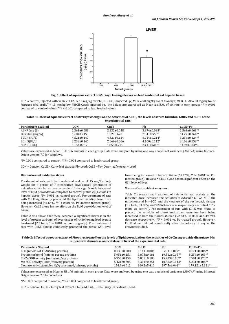
Status of tissue lead content
Fig. 1demonstrates accumulation of lead in liver tissue following treatment of rats with lead acetate at a dose of 15 mg / kg bw (i.p.) for a period of seven consecutive days. However, when the rats were pre-treated with CuLE at a dose of 10 mg / kg bw (fed orally), the tissue lead content was found to be reduced significantly in liver (48.78%, p<0.001 vs. Pb-treated group).
Biomarkers of organ functions
Table 1 demonstrates a 13% increase in the level of serum bilirubin (p<0.001 vs. control) following treatment of rats with the present dose of lead acetate for seven consecutive days. Likewise, in the lead acetate treated rats, serum alkaline phosphatse level was also found to be significantly increased (56.52%, p< 0.001 vs. control) compared to control. When the rats were pre-treated with CuLE at a dose of 50 mg /kg bw (fed orally) for the similar time period, both serum bilirubin and alkaline phosphatse levels were found to be almost completely protected from being increased. CuLE alone, however, was found to have no effect on serum level of bilirubin or serum alkaline phosphatse.
Biomarkers of organ damage
Table 1 further shows that treatment of rats with lead acetate caused a significant elevation in the level of activities of SGPT (1.2 folds, p<0.001 vs. control) and serum TLDH (83.64%, p<0.001 vs. control) as well as LDH 5 (83.61% folds, p<0.001 vs. control; a specific marker enzyme of liver damage). However, when the rats were pre-treated with the present dose of CuLE, the activities of all the three enzymes in serum were found to be significantly protected from being increased (TLDH:36.01%, p< 0.001 vs. Pb-treated group ; LDH 5: 32.25 %, p< 0.001 vs. Pb-treated group; SGPT:35.50%, p< 0.001 vs. Pb-treated group). CuLE alone was found to have no significant effect on the activities of these marker enzymes for hepatic damage.
Bandyapadhyay et al. Int J Pharm Pharm Sci, Vol 5, Suppl 1, 285-295
289
CON MUR LEAD MUR+LEAD
0
20
40
60
80
*
LIVER
CO
NC
EN
TR
AT
ION
OF
LE
AD
(ug
g-1)
tissu
e
Animal groups
Fig. 1: Effect of aqueous extract of Murraya koenigii leaves on lead content of rat hepatic tissue.
CON = control, injected with vehicle; LEAD= 15 mg/kg bw Pb (CH3COO)2 injected i.p.; MUR = 50 mg/kg bw of Murraya; MUR+LEAD= 50 mg/kg bw of Murraya (fed orally) + 15 mg/kg bw Pb(CH3COO)2 injected i.p.; the values are expressed as Mean ± S.E.M. of six rats in each group; *P < 0.001 compared to control values. **P < 0.001 compared to lead treated values.
Table 1: Effect of aqueous extract of Murraya koenigii on the activities of ALKP, the levels of serum bilirubin, LDH5 and SGPT of the experimental rats.
Parameters Studied CON CuLE Pb CuLE+Pb ALKP (mg %) 2.361±0.083 2.432±0.058 3.674±0.088* 2.563±0.063** Bilirubin (mg %) 12.8±0.715 13.2±0.620 21.4±0.550* 14.27±0.764** TLDH (IU/L) 4.521±0.147 4.321±0.124 8.214±0.214* 5.256±0.124** LDH 5(IU/L) 2.233±0.145 2.066±0.066 4.100±0.112* 3.103±0.058** SGPT (IU/L) 10.5± 0.617 10.5± 0.711 23.1±0.608* 14.9±0.583**
Values are expressed as Mean ± SE of 6 animals in each group. Data were analyzed by using one way analysis of variances (ANOVA) using Microcal Origin version 7.0 for Windows.
*P<0.001 compared to control; **P< 0.001 compared to lead treated group;
CON = Control; CuLE = Curry leaf extract; Pb=Lead; CuLE +Pb= Curry leaf extract + Lead.
Biomarkers of oxidative stress
Treatment of rats with lead acetate at a dose of 15 mg/Kg body weight for a period of 7 consecutive days caused generation of oxidative stress in rat liver as evident from significantly increased level of lipid peroxidation compared to control [Table 2] (1.2 folds in hepatic tissue *P< 0.001 vs. control group). Pre-treatment of rats with CuLE significantly protected the lipid peroxidation level from being increased (41.64%, **P< 0.001 vs. Pb acetate-treated group). However, CuLE alone has no effect on the lipid peroxidation level of tissue.
Table 2 also shows that there occurred a significant increase in the level of protein carbonyl of liver tissues of rat following lead acetate treatment (2.2 folds, *P< 0.001 vs. control group). Pre-treatment of rats with CuLE almost completely protected the tissue GSH level
from being increased in hepatic tissue (57.26%, **P< 0.001 vs. Pb-treated group). However, CuLE alone has no significant effect on the GSH level of liver.
Status of antioxidant enzymes
Table 2 reveals that treatment of rats with lead acetate at the indicated dose increased the activities of cytosolic Cu–Zn–SOD, the mitochondrial Mn–SOD and the catalase of the rat hepatic tissues (1.2 folds, 94.85% and 92.06% increase respectively vs control, * P < 0.001 vs. control). Pre-treatment of rats with CuLE was found to protect the activities of these antioxidant enzymes from being increased in both the tissues studied (52.23%, 41.01% and 39.79% decrease respectively, **P < 0.001 vs. Pb-treated group). However, CuLE alone, did not significantly alter the activity of any of the enzymes studied.
Table 2: Effect of aqueous extract of Murraya koenigii on the levels of lipid peroxidation, the activities of Cu-Zn superoxide dismutase, Mn superoxide dismutase and catalase in liver of the experimental rats.
Parameters Studied CON CuLE Pb CuLE+Pb LPO (nmoles of TBARS/mg protein) 0.133±0.008 0.111±0.006 0.293±0.007* 0.171±0.006** Protein carbonyl (nmoles per mg protein) 5.951±0.151 5.873±0.181 19.312±0.187* 8.254±0.165** Cu-Zn SOD activity (units/min/mg protein) 6.950±0.150 6.033±0.188 15.703±0.187* 7.501±0.173** Mn SOD activity (units/min/mg protein) 5.421±0.205 5.301±0.251 10.563±0.143* 6.231±0.106** Catalase activity(µmoles H2O2 consumed/min/mg protein) 154.9±4.012 160.2±5.410 297.5±6.041* 179.121±5.321**
Values are expressed as Mean ± SE of 6 animals in each group. Data were analyzed by using one way analysis of variances (ANOVA) using Microcal Origin version 7.0 for Windows.
*P<0.001 compared to control; **P< 0.001 compared to lead treated group;
CON = Control; CuLE = Curry leaf extract; Pb=Lead; CuLE +Pb= Curry leaf extract +Lead.

Bandyapadhyay et al. Int J Pharm Pharm Sci, Vol 5, Suppl 1, 285-295
290
Status of GSH, GSSG, GSSG: GSH and TSH
Fig. 2 (A, C & D) shows that there occurred a significant increase in GSH, GSSH level and in the GSSG: GSH ratio of liver tissues of rat following lead acetate treatment (61.29%, 2.85 and 1 fold respectively,*P< 0.001 vs. control group). Pre-treatment of rats with CuLE almost completely protected the tissue GSH and GSSG levels and thus the GSSG: GSH ratio also from being increased in hepatic tissue (38.01 %, 65% and 54% respectively, **P< 0.001 vs. Pb-
treated group). However, CuLE alone has no significant effect on the GSH and GSSG levels of liver.
Treatment with lead decreased the TSH level significantly (13.33%,*P< 0.001 vs. control group). Pre-treatment of rats with CuLE almost completely protected the TSH from being decreased in hepatic tissue (15.38%, **P< 0.001 vs. Pb-treated group). However, CuLE alone has no significant effect on the TSH level of liver (fig.2 B).
Fig. 2: Effect of aqueous extract of Murraya koenigii against lead-induced alteration in the value of GSH (A), TSH (B), GSSG (C) and GSSG:GSH (D) in rat hepatic tissue.
Status of pro-oxidant enzymes
The activities of hepatic xanthine oxidase (XO) (Table 3) and xanthine dehydrogenase (XDH) as well as the total enzyme activity, i.e., XO plus XDH and, XO : XDH ratio, all increased significantly following treatment of rats with lead acetate (3.3 folds, 1.5 folds, 1.7 folds and 73.33% increase respectively in hepatic tissue vs. control, *P < 0.001 vs. control). All these
parameters were significantly protected from being increased when the rats were pre-treated with CuLE indicating CuLE’s ability to neutralize free radicals in vivo (74.74%, 60.86%, 62.73% and 35.26% decrease respectively in hepatic tissue vs. lead acetate treated groups, **P < 0.001 vs. Lead acetate-treated group). However, CuLE alone has no effect on the activities of xanthine oxidase (XO) xanthine dehydrogenase (XDH), the total enzyme activity, i.e., XO plus XDH and XO: XDH ratio.
Table 3: Effect of aqueous extract of Murraya koenigii on the activities of XO, XDH, XO+XDH and XO/XDH in liver of the experimental rats.
Parameters Studied CON CuLE Pb CuLE+Pb XO (milliunits/min/mg protein) 0.023±0.008 0.021±0.006 0.099±0.007* 0.025±0.006** XDH (milliunits/min/mg protein) 0.255±0.0820 0.246±0.0764 0.631±0.085* 0.247±0.072** XO+XDH 0.273±0.095 0.267±0.082 0.730±0.082* 0.272±0.879** XO/XDH 0.090±0.042 0.085±0.052 0.156 ±0.042* 0.101±0.042 **
Values are expressed as Mean ± SE of 6 animals in each group. Data were analyzed by using one way analysis of variances (ANOVA) using Microcal Origin version 7.0 for Windows.
*P<0.001 compared to control; **P< 0.001 compared to lead treated group;
CON = Control; CuLE = Curry leaf extract; Pb=Lead; CuLE +Pb= Curry leaf extract +Lead.

Bandyapadhyay et al. Int J Pharm Pharm Sci, Vol 5, Suppl 1, 285-295
291
Status of the activities of pyruvate dehydrogenase and some of the mitochondrial Kreb’s cycle enzymes:
Table 4 reveals that treatment of rats with lead acetate inhibits rat hepatic pyruvate dehydrogenase activity (74.30% decrease, *P < 0.001 vs. their control for both the tissues). Pre-treatment of rats with CuLE significantly protected the enzyme activity from being decreased in hepatic tissues (2.09 folds increase, **P < 0.001 vs. Lead acetate -treated group). However, CuLE alone was found to have no effect on the activity of this enzyme in the tissue.
Table 4 further reveals that treatment of rats with lead acetate significantly decreased the activity of isocitrate dehydrogenase in hepatic tissue (77.36%, *P < 0.001 vs. their respective control). Isocitrate dehydrogenase is a key enzyme in cellular defence against oxidative damage as it provides NADPH in the mitochondria, which is needed for the regeneration of mitochondrial GSH or thioredoxin. The activity of the enzyme in the liver was found to be protected significantly from being decreased when the rats were pre-treated with CuLE (2.4 folds, **P < 0.001 vs. Lead acetate -treated group). However, CuLE alone has no effect on the activity of isocitrate dehydrogenase in the hepatic tissue.
Treatment of rats with lead acetate inhibits alpha keto glutarate dehydrogenase (α-KGDH) activity in hepatic tissue (78.94%, *P < 0.001 vs. control) (Table 4). This enzyme was found to be able to generate ROS during its catalytic function, which is regulated by the NADH/NAD+ ratio (Tretter and Adam-Vizi, 2005). The activity of the enzyme was found to be significantly protected from being decreased in both the organs studied when the rats were pre-treated with 10 mg/kg body weight of CuLE (4.1 folds, **P < 0.001 vs. Lead acetate -treated group). However, CuLE alone has no significant effect on the activity of α-KGDH in hepatic tissue.
Treatment of rats with lead acetate for seven consecutive days, inhibited the activity of succinate dehydrogenase (SDH) in hepatic tissues significantly (73.74%, *P < 0.001 vs. control) (Table 4). This might result in interference of the metal in electron transport chain (ETC) and thus generate copious amounts of superoxide anion free radicals in the tissue mitochondria. However, pre-treatment of rats with CuLE significantly protected the SDH activity from being decreased in hepatic tissue (2.3 folds, **P < 0.001 vs. Lead acetate -treated group). CuLE alone has no effect on the activity of this enzyme in any of the tissue studied.
Table 4: Effect of aqueous extract of Murraya koenigii on the activities of PDH, ICDH, α-KGDH and SDH in liver of the experimental rats.
Parameters Studied Control CuLE Pb CuLE+Pb PDH (units/min/mg protein) 1.603±0.028 1.602±0.029 0.412±0.022* 1.276±0.025** ICDH(units/min/mg protein) 0.053±0.002 0.057±0.002 0.012±0.003* 0.041±0.002** α-KGDH (units/min/mg protein) 0.057±0.0024 0.054±0.0032 0.012±0.0041* 0.061±0.0038** SDH(units/min/mg protein) 1.603±0.029 1.612±0.029 0.421±0.022* 1.276±0.026**
Values are expressed as Mean ± SE of 6 animals in each group. Data were analyzed by using one way analysis of variances (ANOVA) using Microcal Origin version 7.0 for Windows.
*P<0.001 compared to control; **P< 0.001 compared to lead treated group;
CON = Control; CuLE = Curry leaf extract; Pb=Lead; CuLE +Pb= Curry leaf extract +Lead.
Status of the activities of mitochondrial respiratory chain enzymes
Treatment of rats with lead acetate for similar period of time also decreased cytochrome c oxidase activity in hepatic tissue (84.01% decrease, *P< 0.001 vs. control group). The activity of this enzyme was found to be significantly protected from being decreased compared to lead acetate treated group when rats were pre-treated with CuLE (2.6 folds, **P< 0.001 vs. Lead acetate -treated group). CuLE alone, however, has no effect on the activity of this enzyme in the hepatic tissue.
Treatment of rats with lead acetate for seven consecutive days at a dose of 15 mg / kg body weight inhibits NADH cytochrome c oxido-reductase activity (70.99%, *P< 0.001 vs. control group). However, the enzyme activity was found to be completely protected when the rats were pre-treated with CuLE at a dose of 10 mg/kg body weight
for the similar period of time (2.4 folds, **P< 0.001 vs. Lead acetate -treated group). However, CuLE alone has no significant effect on the activity of this enzyme in hepatic tissue.
Status of the activities of glutathione peroxidase, glutathione reductase and glutathione –S- transferase
Treatment of rats with lead acetate for seven consecutive days at a dose of 15 mg / kg body weight increased the activities of glutathione peroxidise (fig. 3A), glutathione reductase (fig.3B) and glutathione –S- transferase (3 C) (84.71%, 98.24% and 87.50% respectively,*P< 0.001 vs. control group). However, the enzyme activities were found to be completely protected when the rats were pre-treated with CuLE at a dose of 50mg/kg body weight for the similar period of time (46.15%, 82.22% and 43.33%,**P< 0.001 vs. Lead acetate -treated group). However, CuLE alone has no significant effect on the activities of these enzymes in hepatic tissue.
Table 5: Effect of aqueous extract of Murraya koenigii on the activities of Cytochrome c oxidase and NADH cytochrome c oxido-reductase in liver of the experimental rats.
Parameters Studied Control CuLE Pb CuLE+Pb Cytochrome c oxidase activity(units/min/mg protein) 1.541±0.0023 1.498±0.0053 0.245±0.0058* 1.544±0.0065** NADH cytochrome c oxido-reductase activity(units/min/mg protein) 7.121±0.036 8.072±0.045 2.066±0.012* 7.013±0.0058**
Values are expressed as Mean ± SE of 6 animals in each group. Data were analyzed by using one way analysis of variances (ANOVA) using Microcal Origin version 7.0 for Windows.
*P<0.001 compared to control; **P< 0.001 compared to lead treated group;
CON = Control; CuLE = Curry leaf extract; Pb=Lead; CuLE +Pb= Curry leaf extract +Lead.
Histological studies
Fig. 4A (first panel) documents H and E stained sections of hepatic tissue (magnification 400X) showing intact portal veins, scattered dead hepatocytes, focal hepatic necrosis with dilated sinusoids in lead acetate treated rats compared to control. However, pre-treatment of rats with CuLE protected the tissue from being
damaged and found to have normal sinusoids. CuLE alone, however, has no effect on hepatic tissue morphology.
Fig. 4B demonstrates a reduction of hepatic glycogen content following treatment of rats with lead acetate for seven consecutive days. However, pre-treatment of rats with CuLE significantly protected the tissue glycogen content from being reduced compared

Bandyapadhyay et al. Int J Pharm Pharm Sci, Vol 5, Suppl 1, 285-295
292
to lead acetate group. However, CuLE alone was found to have no effect on hepatic tissue glycogen content compared to control.
Fig. 4A (second panel) represents the status of hepatic glycogen studied by PAS staining of the tissue sections. Here also, pre-treatment of rats with CuLE was found to have a protective effect on tissue glycogen content compared to lead acetate treated rats. CuLE alone, however, has no effect on tissue glycogen content.
Fig. 4A (third panel) shows Sirius red stained hepatic tissue section (magnification 400X) with deposition of collagen around the central
vein region following treatment of rats with lead acetate. Pre-treatment of rats with CuLE prevented the deposition of hepatic tissue collagen. CuLE alone was found to have no effect on tissue collagen content.
Fig. 4A (fourth panel) shows similar images (magnification 400X) captured by confocal laser scanning microscope. Figure 2C represents quantification of fibrosis as percent collagen volume. The results further indicate a protective effect of CuLE against Pb-induced damage in rat hepatic tissue.
Fig. 3: Effect of aqueous extract of Murraya koenigii against lead-induced lead-induced alteration in the activities of glutathione peroxidises (A), glutathione reductase (B), glutathione S transferase (C) in rat hepatic tissue.
DISCUSSION
The accumulation of significant amount of Pb in liver tissue (AAS study) resulted in the induction of an oxidative stress response in the liver. Oxidative stress can be defined as a situation of an imbalance toward the pro-oxidant side of the pro-oxidant/antioxidant balance [11].
Bilirubin is the primary bile pigment that is formed from the breakdown of heme of hemoglobin in red blood cells. It gets transported to the liver and is secreted by the liver into the bile. Conjugation of bilirubin is a prerequisite for its excretion into the bile [34]. Structural damage to hepatic cells leads to functional compromisation of the organ. This leads to increased level of serum bilirubin indicating hepatic damage and functional compromisation. Increased level of serum lactate dehydrogenase and lactate dehydrogenase 5 is indicative of hepatic damage. Increased level of SGPT and alkaline phosphatase activities also indicate hepatocyte damage. LDH 5 is a specific damage marker of liver tissue. Thus the serum levels of ALKP, LDH 5 and SGPT are main indices of liver
injury [11,36]. We observed increased level of serum bilirubin, TLDH, LDH 5, SGPT and ALKP on treatment with lead indicating hepatic damage. All these were prevented from being increased on pre-treatment with CuLE. Thus we can conclude the hepatoprotective activity of CuLE.
The levels of LPO, Protein carbonyl, GSH and GSSG can be used as indices of oxidative stress [11, 37].
Membrane lipids are highly susceptible to free radical damage. Lipids when reacted with free radicals can undergo the highly damaging chain reaction of lipid peroxidation.
Increased serum bilirubin level in the lead treated rats can be considered as a compensatory/ retaliatory phenomenon in response to cellular peroxidative changes [38]. This is because bilirubin functions in vivo as a powerful antioxidant, anti-mutagen, and an endogenous tissue protector [37]. This thus shows a positive correlation between increased level of LPO and serum bilirubin level which supports the report of Pratibha et al., (2004) [38]. Protein CO

Bandyapadhyay et al. Int J Pharm Pharm Sci, Vol 5, Suppl 1, 285-295
293
is used as a biomarker of oxidative stress. The usage of protein CO groups as biomarkers of oxidative stress is advantageous compared to the measurement of other oxidation products because of the relative early formation and the relative stability of carbonylated
proteins. We found in our model an enhanced level of protein CO as well as level of lipid peroxidation in hepatic tissues of lead treated rats. Both were prevented from being increased on pre-treatment with CuLE.
Fig. 4: Histopathological Studies of Liver.
A, first panel: Changes in the rat hepatic tissue morphology (Hematoxylin and Eosin stained, 400 X magnifications).
A, second panel: Changes in the glycogen content in rat liver (PAS stained, 400 X magnifications).
A, third panel: Changes in the collagen content in rat liver (Sirius red stained sections, 400 X magnifications).
A, fourth panel: Images captured by confocal laser scanning microscope for quantification of fibrosis (Sirius red stained sections, 400X magnification).
B. Graph showing glycogen content of the hepatic tissues.
C. Graph showing collagen volume % of the hepatic tissues.
Con = control, injected with vehicle; Lead = 15 mg/kg bw Pb(CH3COO)2 injected i.p.;CuLE= 50 mg/kg bw of Murraya; CuLE+Lead= 50 mg/kg bw of Murraya + 15 mg/kg bw Pb(CH3COO)2 injected i.p.; the values are expressed as Mean ± S.E.M. of six rats in each group; *P < 0.001 compared to control values. **P < 0.001 compared to lead treated values.
Glutathione and glutathione-related enzymes play a key role in protecting the cell against oxidative stress. Glutathione is tripeptide composed of L-cysteine, L- glutamic acid and glycinecysteinyl moiety. Reactive Oxygen Species (ROS) are reduced by GSH in the presence of GSH peroxidase. GSH is oxidized to GSSG, which in turn is rapidly reduced back to GSH by GSSG reductase at the expense of NADPH. The GSH redox cycle consist of GSH, GPx, GR and GST, which are the major components of the antioxidant defence system. Coordinated activities of these enzymes maintain intracellular thiol status. GSH plays a role in the detoxification of a variety of electrophilic compounds and peroxides via catalysis by glutathione S-transferases (GST) and glutathione peroxidases (GPx). Lipid peroxidation can generate large amounts of electrophilic and
oxidizing reactive species which can lead to a variety of DNA and tissue damage [39].GST are a family of ubiquitous enzymes that can catalyze the formation of GSH electrophile thioether conjugates and the GSH-linked reduction of lipid hydroperoxides [39].
Glutathione reductase (GR) reduces oxidized glutathione (GSSG) to biologically active GSH.NADPH is the cofactor of GR. GPx detoxifies peroxides using GSH as an electron donor, producing GSSG as an end product. We observed increased activities of all the three enzymes in rat hepatic tissues on treatment with lead. Increased level of GSH is probably the inducer for enhanced activity of GR. On the other hand increased oxidation of GSH to GSSG leads to increased level of GSSG and the ratio of GSSG:GSH in lead treated animals compared to

Bandyapadhyay et al. Int J Pharm Pharm Sci, Vol 5, Suppl 1, 285-295
294
control. GSH is increased to meet the increased demand of the same for combatting the situation of increased lipid peroxidation. This increase in the level of lipid peroxidation is caused by lead induced generation of ROS. All these were prevented from being increased on pre-treatment with CuLE.
Cells produce reactive oxygen species and the protection against them is also an intrinsic property of every living cell. To minimize oxidative damage, organisms developed antioxidative mechanisms. Often it is observed that these antioxidative defense systems of cells are triggered by increased ROS production. Oxidants such as ROS are balanced against this antioxidative defense system that consists of enzymes and metabolites in all sub cellular compartments [39]. In conditions of oxidative stress, however, normal capacities of these mechanisms are insufficient, triggering cells to increase and expand their antioxidative network [40].SOD and CAT are two key antioxidant enzymes in oxidative stress induced pathogenesis. Super oxide anion radicals are generated in vivo and are increased with condition of oxidative stress. Increased level of super oxide anion radical causes enhanced activity of the enzyme SOD while increased SOD activity leads to increased level of hydrogen peroxide. Thus the level of Catalase activity is also increased in response to the increased hydrogen peroxide.
Xanthine oxidoreductase, under normal conditions, exists in dehydrogenase form and uses NAD+ and there is no or very little production of superoxide anion. Under ischemic conditions, there is depletion of ATP and subsequent loss of membrane Ca2+ gradient. Increased Ca2+ levels activates Ca2+ dependent proteases which cause selective proteolysis of the dehydrogenase to convert it into xanthine oxidase (XO) which acts both on hypoxanthine and xanthine at the expense of molecular oxygen to produce superoxide anion free radical [41]. Thus, XO in oxidative stress conditions may play an important role in contributing free radical mediated damage. A significant increase in the activity of xanthine oxidase: xanthine dehydrogenase, and also increase in their individual activity in the tissue confirms lead induced stress mediated generation of reactive oxygen species.
Mitochondria are the major source of ROS production in cells [42]. In our study we found that there has been considerable decrease in activities of pyruvate dehydrogenase and the Kreb’s cycle enzymes like Isocitrate dehydrogenase, alpha-keto glutarate dehydrogenase and succinate dehydrogenase following treatment of rats with lead for seven consecutive days. The activities of all these enzymes were protected when the rats were pre-treated with the CuLE. Earlier researchers have reported that other heavy metals markedly inhibit uncoupler-stimulated oxidation on various NADH-linked substrates as well as that of succinate [11,42]. Heavy metals are also known to affect respiratory chain complexes and there is substrate specificity [43].The impairment of electron transfer through NADH: ubiquinone oxidoreductase (complex I) and ubiquinol: cytochrome c oxidoreductase (complex III) may induce superoxide formation. Mitochondrial production of ROS is thought to play an adverse role in many pathologic states of organs, including liver. Lead induced formation of ROS in mitochondria does not depend on the inhibition of complex I or II. Complex III is the only site in the ETC complex where ROS are produced in the presence of heavy metals [43, 44]. In our present study, lead treatment inhibits NADH cytochrome c oxidoreductase and cytochrome c oxidase enzymes of ETC, and succinate dehydrogenase of mitochondria of rat hepatic tissue. The activities of these enzymes were found to be protected when the rats were pre-treated with CuLE. This strongly indicates that the extract possesses either some chelating property or is simply able to prevent mitochondria from ROS production by itself being a quencher of reactive oxygen species.
Histological examination of haematoxylin-eosin stained sections of hepatic tissues of lead treated animals showed some significant alterations. However, the hepatic tissue sections from the rats pre-treated with CuLE did not show any such changes. The results indicate the ability of the aqueous extract to provide protection against lead induced tissue injury. Picrosirius stain of the tissue sections show that there was increased deposition of collagen around the central hepatic vein in lead treated animals. Pre-
treatment of rats with CuLE was found to limit the collagen content indicating a protective role of the extract in maintaining the tissue integrity. There was not much difference between collagen content of the hepatic tissues of the control and the CuLE only treated group. The lead induced hepatic damage in our experimental situation, is due to generation of oxidative stress as is evident from elevated levels of tissue LPO and protein carbonyl content and GSSG level and the bio-markers of oxidative stress.
CONCLUSION
From these studies it is concluded that the aqueous extract of the leaves of Murraya koenigii protects rat liver against lead induced oxidative damage. Curry leaves may find its extensive use against lead induced hepatotoxic situation at a specific pharmacological dose. It may as well find its place in alternative medicine or integrative medicinal interventions also. Gathering hints from our preliminary CuLE dose response studies [46] we carried out this detailed investigation to invent the fact that CuLE can not only protect the liver tissue from lead induced oxidative stress and stress mediated structural and functional damage of the hepatocytes but also it can provide a very strong and complete hepatoprotection against lead induced hepatotoxicity. Till date there has been no report on the side effects of CuLE. Hence, we may conclude from this investigation that CuLE is a potent hepatoprotective agent and can be used as an effective protector against lead induced hepatic damage. Further works are needed to identify the active principle (s) present in the leaves of the plant and elucidate its possible mode of action [47]. The CuLE appears to provide hepato-protection through its antioxidant activity and these may be attributed to the presence of phenolics and flavonoids [48, 49].
ACKNOWLEDGEMENT
Debosree Ghosh gratefully acknowledges the receipt of a Junior Research Fellow (JRF) under INSPIRE program of Department of Science and Technology, Government of India. SBF is a URF of UGC, under University of Calcutta. EM is a Project Fellow under a major research project of UGC awarded to Dr. D.B., Government of India. MD is a UPE Project Fellow of UGC, under University of Calcutta. Dr. A.C. is supported from the funds available to her from a Minor UGC project, Govt. of India. Dr. S.K.P., Dr. S.D. and Dr.K.J. are supported from the funds available to them from their respective institutes. This work is also partially supported by UGC Major Research Project Grant awarded to Dr. DB [F. No. 37-396/2009 (SR)]. Technical help from Parthabrata Roy and Sumanta Ghoshal is also gratefully acknowledged.
REFERENCES
1. Gnanaprakash K, Madhusudhana Chetty C, Ramkanth S, Alagusundaram M, Tiruvengadarajan VS, Angala Parameswari S, Mohamed Saleem TS. Aqueous Extract of Flacourtia indica Prevents Carbon Tetrachloride Induced Hepatotoxicity in Rat. Int J Biol life Sci. 2010; 6:51-55.
2. Koller LD.The immunotoxic effect of lead in lead exposed laboratory animals. Ann N Y acad Sci.1990; 587:160-167.
3. Gurer H, Ercal N. Can antioxidants be beneficial in the treatment of lead poisoning? Free Radic Biol Med. 2000; 29: 927-945.
4. Wang HP, Qian SY, Schafer FQ, Domann FE, Oberley LW, Buettner GR. Phospholipid hydroperoxide glutathione peroxidase protects against singlet oxygen-induced cell damage of photodynamic therapy. Free Radic Biol Med. 2001; 30: 825-835.
5. Alghazal MA, Lenártová V, Holovská K, Sobeková A, Falis M, Legáth J. Activities of Antioxidant and Detoxifying Enzymes in Rats after Lead Exposure. Acta Vet Brno. 2008; 77: 347–354.
6. Adonaylo VN, Oteiza PI: Lead intoxication. Antioxidant defenses and oxidative damage in rat brain.Toxicology .1999; 135: 77-85.
7. McGowan C, Donaldson WE. Changes in organ nonprotein sulfhydryl and glutathione concentrations during acute and chronic administration of inorganic lead to chicks. Biol Trace Elem Res. 1986;10: 37-46.

Bandyapadhyay et al. Int J Pharm Pharm Sci, Vol 5, Suppl 1, 285-295
295
8. Bechara EJ, Medeiros MH, Monteiro HP, Hermes-lima M, Pereira B, Demasi M. A free radical hypothesis of lead poisoning and inborn porphyrias associated with 5-aminolevulinic acid overload. Quim Nova. 1993 ; 16: 385-392.
9. Sugawara E, Nakamura K, Miyake T, Fukumura A, Seki Y. Lipid peroxidation and concentration of glutathione in erythrocytes from workers exposed to lead. Br J Ind Med. 1991; 48: 239-242.
10. Adeshina GO, Onaolapo JA, Ehinmidu JO and Odama LE. Phytochemical and antimicrobial studies of the ethyl acetate extract of Alchornea cordifolia leaf found in Abuja, Nigeria. J Med Plants Res.2010; 4: 649-658.
11. Mitra E, Ghosh AK, Ghosh D, Mukherjee D, Chattopadhyay A, Dutta S, Pattari S K, Bandyopadhyay D. Protective Effect Of Aqueous Curry Leaf (Murraya Koenigii) Extract Against Cadmium-Induced Oxidative Stress In Rat Heart. Food Chem Toxicol. 2012; 50:1340–1353.
12. Reitman S, Frankel S. Determination of serum glutamic oxaloacetic and glutamic pyruvic transaminase. Am J Clin Pathol.1957; 28: 56–63.
13. Strittmatter C. Studies on avian xanthine dehydrogenases: properties and patterns of appearance during development. J Biol Chem.1965; 240: 2557–2564.
14. Varcoe J S. Clinical Biochemistry: Techniques and Instrumentation-A practical approach, first ed. World Scientific Publishing Company. 2001; 40–43.
15. Royden N R, Di Pasqua, A A. New Diazo Method for the Determination of Bilirubin. Clin Chem.1962; 8: 570-578.
16. Singh M, Batish M K, Singh M, Singh K, Lochan K K, Garg R. Correlation of Plasma Color Index with Serum Bilirubin in Neonatal Jaundice. Indian Pediatr. 2001; 38: 278-280.
17. Buege JA, Aust S G. Microsomal Lipid Peroxidation. Methods. Enzymol.1978; 52:302–310.
18. Bandyopadhyay D, Ghosh G, Bandyopadhyay A, Reiter R J. Melatonin protects against piroxicam-induced gastric ulceration. J Pineal Res 2004; 36:195–203.
19. Levine RL, Williams JA, Stadtman ER, Shacter E.Carbonyl assays for determination of oxidatively modified proteins. Methods Enzymol.1994; 233: 346–357.
20. Sedlak J, Lindsay RH. Estimation of total, protein-bound, nonprotein sulfhydryl groups in tissue with Ellman’s reagent. Anal Biochem.1968; 25: 192–205.
21. Mukherjee D, Roy SG, Bandyopadhyay A, Chatyopadhyay A, Basu A, Mitra E, Ghosh AK, Reiter R, Bandyopadhyay B. Melatonin protects against isoproterenol-induced myocardial injury in the rat: antioxidative mechanisms. J Pineal Res. 2010; 48: 251–262.
22. Marklund S, Marklund G. Involvement of the superoxide anione radical in the autoxidation of pyragallol and a convenient assay for superoxide dismutase.Eur J Biochem.1974; 47: 469–474.
23. Beers Jr, RF, Sizer IW. A spectrophotometric method for measuring the breakdown of hydrogen peroxide by catalase. J Biol Chem.1952.195: 133–140.
24. Chattopadhyay A, Biswas S, Bandyopadhyay D, Sarkar C, Datta AG. Effect of isoproterenol on lipid peroxidation and antioxidant enzymes of myocardial tissue of mice and protection by quinidine. Mol Cell Biochem. 2003; 245: 43–49.
25. Krohne-Ehrich G, Schirmer RH, Untucht-Grau R. Glutathione reductase from human erythrocytes. Isolation of the enzyme and sequence analysis of the redox-active peptide. Eur J Biochem. 1977; 80: 65–71.
26. Paglia D E, Valentine WN. Studies on the quantitative and qualitative characterization of erythrocyte glutathione peroxidase. J Lab Clin Med.1967; 70:158–169.
27. Habig WH, Pabst MJ, Jakoby WB. Glutathione-S-transferases, the first enzymatic step in mercapturic acid formation. J Biol Chem.1974; 249:7130–7139.
28. Greenlee L, Handler P. Xanthine oxidase. IV. Influence of pH on substrate specificity. J Biol Chem.1964. 239: 1090–1095.
29. Chretien D, Pourrier M, Bourgeron T, Séné M, Rötig A, Munnich A, Rustin P. An improved spectrophotometric assay of pyruvate dehydrogenase in lactate dehydrogenase contaminated mitochondrial preparations from human skeletal muscles. Clin Chim Acta. 1995; 240: 129–136.
30. Duncan MJ, Fraenkel DG. Alpha-ketoglutarate dehydrogenase mutant of Rhizobium meliloti. J Bacteriol.1979; 137: 415–419.
31. Veeger C, DerVartanian DV, Zeylemaker WP. Succinate dehydrogenase. Methods Enzymol.1969;13: 81–90.
32. Goyal N, Srivastava VM. Oxidation and reduction of cytochrome c by mitochondrial enzymes of Setaria cervi. J. Helminthol.1995; 69: 13–17.
33. Lowry OH, Rosebrough NJ, Farr AL, Randall RJ. Protein measurement with the Folin phenol reagent. J Biol Chem 1951; 193: 265–275.
34. Dalle-Donne I, Rossi R, Giustarini D, Milzani A, Colombo R. Protein carbonyl groups as biomarkers of oxidative stress.Clin Chim Acta. 2003; 329:23-38.
35. Nelson DC, Cox MM). Lehninger Principles of Biochemistry. 3rd edition. Worth Publishers, USA. 2000; 842.
36. Giboney PT. Mildly elevated liver transaminase levels in the asymptomatic patient. Am Fam Physician. 2005; 71: 1105-1110.
37. Acharya M, Lau-Cam CA. Comparison of the protective actions of N acetylcysteine, hypotaurine and taurine against acetaminophen-induced hepatotoxicity in the rat. J Biomed Sci. 2010; 17: 35.
38. Pratibha K, Usha A, Rajni A. Serum adenosine deaminase 51- nucleotidase and malondialdehyde in acute infective hepatitis. Indian J Clin Biochem. 2004; 19:128-131.
39. Awasthi YC, Zimniak P, Singhal SS, Awasthi S.Physiological role of ghlutathione S- transferases in protection mechanisms against lipid peroxidation : A commentary . Biochem Arch.1995; 11: 47-54.
40. Halliwell B. Reactive species and antioxidants. Redox biology is a fundamental theme of aerobic life. Plant Physiol. 2006; 141: 312–322.
41. Reghuvanshi R, Kaul A, Bhakuni P, Mishra A, Mishra MK. Xanthine oxidase as a marker of myocardial infarction. Indian J. Clin Biochem. 2007;22: 90–92.
42. Miccadei S, Floridi A. Sites of inhibition of mitochondrial electron transport by cadmium. Chem Biol Interact.1993; 89:159–167.
43. Belyaeva EA, Korotkov SM, Saris NE. In vitro modulation of heavy metal induced rat liver mitochondria dysfunction: a comparison of copper and mercury with cadmium. J. Trace. Elem Med Biol. 2011; 25: 63–73.
44. Wang Y, Fang J, Leonard SS, Rao KM. Cadmium inhibits the electron transfer chain and induces reactive oxygen species. Free Radic Biol Med. 2004; 36: 1434–1443.
45. Chen Q, Vazquez EJ, Moghaddas S, Hoppel CL, Lesnefsky EJ. Production of reactive oxygen species by mitochondria: central role of complex III. J Biol Chem. 2003;278: 36027–36031.
46. Ghosh D, Firdaus SB, Mitra E, Dey M, Bandyopadhyay D. Protective effect of aqueous leaf extract of Murraya koenigi against lead induced oxidative stress in rat liver, heart and kidney: a dose response study. Asian J Pharm Clin Res. 2012;5 (4):54-58.
47. Parida P, Yadav RNS. Comparative docking study of M1 Protein (Influenza virus) to check drug efficacy. Int J Pharm Pharm Sci. 2012; 4 (3): 243-246.
48. Mathews LA, Dhanyaraj D, Prathibhakumari P V, Prasad G. Hepatoprotective and antioxidant potential of Sphaeranthus indicus [Linn] on liver damage in Wistar rats. Int J Pharm Pharm Sci. 2012; 4 (3): 222-225.
49. Anusuya N, Sellamuthu M. Antioxidant and free radical scavenging potential of different solvent extracts of Indigifera tinctoria L. leaves. Int J Pharm Pharm Sci. 2013; 5 (1): 142-147.