multiple myeloma learning program - oncology nursing in
TRANSCRIPT

Multiple Myeloma Learning Program


3
Co
nte
nts
Dear Colleague
It is with great pleasure that we present the learning program “An Introduction to Multiple Myeloma: A resource for healthcare professionals” on behalf of the Haematology Nurses and Healthcare Professionals Group.
A faculty of specialist nurses working in the field of haematology/oncology, haematologists/oncologists, and patient advocates have collaborated to develop this program dedicated to learning about myeloma.
This program features topics relevant to the multidisciplinary team approach to caring for patients with myeloma and their relatives. Nurses, other allied health care professionals and patient organizations play an important role in this process and the group is excited to share with you the most current information and up-to-date recommendations for addressing both short-term and long-term management of patient and family needs.
The Multiple Myeloma Learning Program was made possible by grants from Amgen, Bristol-Myers Squibb, Celgene, Janssen Pharmaceutical Companies, Novartis Oncology and Takeda Pharma AG Switzerland.
On behalf of the faculty and the Haematology Nurses and Healthcare Professionals Group who developed this resource, we hope that the Multiple Myeloma Learning Program will be of value to you in your care of patients with myeloma.
Sincerely,
Erik Aerts
President
Haematology Nurses and Healthcare Professionals Group

The Haematology Nurses and Healthcare Professionals Group gratefully acknowledges the following individuals for their review and contributions to this learning program.
Faculty:
Erik Aerts (CH)
Ruth Bähler (CH)
Corien Eeltink (NL)
Andrea Guy (UK)
Sarah Liptrott (IT)
Matthias Nägele (DE)
Panagiotis Samaras (CH)
Reviewers:
Niccolò Frungillo (IT)
Nicolaus Kröger (DE)
Mairéad Ni Chonghaile (IRL)
Markus Rubeli (CH)
Acknowledgement
The completion of this Multiple Myeloma Learning Program would not have been possible without the editorial support of Carol Krcmar.
The Multiple Myeloma Learning Program is also available online at the
www.hemcare.org
5
Co
nte
nts
Contents
Foreword ....................................................................................... 3
Module I: Understanding Multiple Myeloma.............................. 7
Module II: Multiple Myeloma: Diagnosis and Staging............... 19
Module III: Treatment of Multiple Myeloma............................... 29
Module IV: Comprehensive Managment of the Patient with Multiple Myeloma................................................................ 47
Multiple Myeloma Learning Program - Glossary......................... 63


Module I: Understanding Multiple Myeloma
7Module I: Understanding Multiple Myeloma
Mo
du
le I
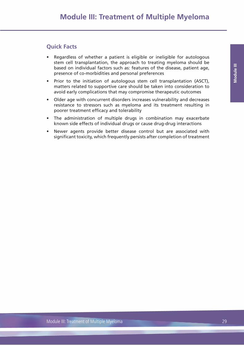
Quick Facts
• Multiple myeloma is an incurable malignant disease arising from plasma cells, the most mature form of B lymphocytes
• B lymphocytes, a type of cell of the immune system, mature in the bone marrow and at a later stage become plasma cells; abnormalities in the bone marrow microenvironment cause an uncontrolled proliferation of clonal plasma cells, the hallmark of myeloma
• Myeloma is typically preceded by an asymptomatic premalignantperiod that, if detected, is termed either monoclonal gammopathy ofundetermined significance (MGUS) or smoldering multiple myeloma (SMM), depending on the extent of bone marrow involvement andmonoclonal protein levels
• Through innate (non-specific, natural or native immunity) and adaptive (acquired immunity) immunity, the immune system recognizes and eliminates pathogens
• Myeloma is rarely diagnosed before age 40 after which the incidence increases rapidly peaking at age 84; the majority of patients are older than 70 years at the time of diagnosis
• Unraveling the molecular subgroups of multiple myeloma may provide valuable information to improve patient outcomes

Module I: Understanding Multiple Myeloma
Module I: Understanding Multiple Myeloma8
A. Understanding Multiple Myeloma
1. Overview of the immune system and the immune response
a. Innate immunity
b. Adaptive immunity
c. Humoral and cellular immunity
B. Pathophysiology and Epidemiology
1. The pathophysiology of multiple myeloma
a. The role of genetics
2. Etiology
a. Risk factors
3. Epidemiology
4. Future perspectives
C. Resources
D. Review Questions
E. References

Module I: Understanding Multiple Myeloma
9Module I: Understanding Multiple Myeloma
Mo
du
le I
Understanding Multiple Myeloma
Multiple myeloma, or myeloma, is a cancer arising from plasma cells, the most mature form of B lymphocytes (see Figures 1 and 2, Table 1). Myeloma belongs to a group of related paraprotein anemias characterized by an abnormal clonal plasma cell infiltration in the bone marrow (Morgan 2012). The first case of multiple myeloma was reported as early as 1844. The discovery of the replacement of bone marrow with a red substance was followed by the identification of Bence-Jones protein in the urine of patients with myeloma.
The typical disease course in multiple myeloma is characterized by periods of active disease in which patients require treatment, followed by periods of remission and then eventual relapse. This pattern is repeated with remissions becoming progressively shorter over time until the disease eventually becomes refractory to further treatment (NCCN 2016).
Three classic features of multiple myeloma are present at diagnosis:
• monoclonal plasma cells
• monoclonal protein
• myeloma-related organ and tissue impairment including bone lesions (Durie 2003).
The most common presenting symptoms are:
• fatigue
• bone pain
• recurrent infections
• renal impairment
Figure 1. Development of blood cells. A stem cell moves through several phases to become either a red blood cell, white blood cell or platelet. In multiple myeloma, mutations deregulate the development of plasma cells causing an abnormal proliferation of plasma cells in the bone marrow.
Multiple myeloma accounts for approximately 0.8% of all newly diagnosed cancer cases worldwide. The global incidence is approximately 120,000 cases per year. Because the median age at diagnosis is about 70 years, the rapidly aging world population means the incidence of myeloma is likely to rise significantly to about 350,000 cases by the year 2050 (Ludwig 2013). In a review of 1027 patients with multiple myeloma, 38% were 70 years of age or older at diagnosis while 2% were 40 years or younger (Kyle 2003). Rates for new cases of myeloma have been rising on average 0.8% each year over the last 10 years. By contrast, death rates have been falling on average 0.8% each year from 2004 to 2013.
To date, available therapeutic measures have not yet provided a cure for myeloma. However, advances in understanding the etiology of multiple myeloma, including knowledge of the genetic abnormalities underlying myeloma, and more effective therapeutic options available to patients have resulted in improved patient survival and patients are now dying with their disease instead of from their disease. New therapeutic options with unique modes of action and impact on disease outcome have also helped to aid the quality of life of patients with myeloma.
Overview of the immune system and the immune response
The primary function of the immune system is to defend the body against pathogenic microorganisms. These organisms may be infectious microbes, such as viruses, bacteria, fungi, protozoa and parasites, or innocuous environmental substances, such as pollens or foods. The immune system differentiates self from nonself; foreign substances recognized as being nonself act as a stimulus to trigger the immune response.
There are two mechanisms used by the immune system to recognize and eliminate pathogens:
• innate immunity (also known as non-specific, natural or native immunity): encompassing more primitive elements of the immune system including macrophages, natural killer cells (NK) and antigen-presenting cells (APC)
• adaptive immunity (or acquired immunity): encompassing T- and B-lymphocytes
Innate immunity
The innate immune system is activated immediately or within hours of detecting the presence of an intruding pathogen and is the body’s first line of defense. The innate immune response is an antigen-independent (or non-specific) defense mechanism. As such, it is unable to recognize or “memorize” the same pathogen should

Module I: Understanding Multiple Myeloma
Module I: Understanding Multiple Myeloma10
a second exposure occur. Recently, scientists have proposed that innate immune responses include adaptive characteristics comparable to immunologic memory.
The primary function of innate immunity is to recruit immune cells to sites of infection and inflammation through the production of cytokines (small proteins involved in cell-to-cell communication). In immunity, there are several categories of cytokines important for immune cell growth, activation and function.
Cytokine production causes a release of antibodies and other proteins and glycoproteins that then activate the complement system, a biochemical cascade that functions to identify and coat (opsonize) foreign antigens making them susceptible to phagocytosis (Warrington 2011).
Innate immune protection involves cells of both hematopoietic and non-hematopoietic origin. Hematopoietic cells include macrophages, dendritic cells, mast cell, neutrophils, eosinophils, natural killer (NK) cells and natural killer T cells (Table 1, Figure 2) (Turvey 2010). Non-hematopoietic cells include epithelial cells of the skin, and respiratory and gastrointestinal tracts.
Adaptive immunity
Adaptive, or acquired immunity, in contrast to innate immunity, is a slower response to pathogens and produces long-lived memory cells existing in a dormant state until the foreign substance is reintroduced. Adaptive immunity develops when innate immunity is ineffective in eliminating pathogens and infection is established (Warrington 2011). The primary functions of the adaptive immune system are:
• recognize specific “non-self” antigens
• generate pathogen-specific immunologic effector pathways to eliminate specific pathogens or pathogen-infected cells
• develop an immunologic memory to eliminate specific pathogens (Bonilla 2010)
Cells of the adaptive immune system include: T and B cells (or lymphocytes) (Table 1, Figure 2). T cells derive from hematopoietic stem cells in bone marrow and mature in the thymus, they stimulate cellular immune responses by which their major role in the immune response is to identify and destroy infected cells. T cells have a unique antigen-binding receptor on their membrane, known as the TCR (T-cell receptor), which requires activation through APCs to be able to recognize a specific antigen. APCs are found in the epithelium, skin and gastrointestinal and respiratory tracts. APCs are essential in recognizing specific antigens.
The surfaces of APCs express major histocompatibility complex (MHC). MHC (or human leukocyte antigen [HLA]) proteins serve two general roles:
• MHC proteins function as carriers to present antigens on cell surfaces. MHC class I proteins are essential for presenting viral antigens and are expressed by nearly all cell types, except red blood cells. MHC class II proteins are important for presenting antigens to T helper cells (also known as CD4 cells)
• MHC proteins also signal if a cell is a host cell or a foreign cell. In organ transplantation, MHC proteins are matched to lower rejection risk
T cells are activated when they encounter an APC that has digested an antigen and subsequently displays antigen fragments bound to its MHC molecules (Warrington 2011). Once activated, the T cell secretes cytokines, which in turn stimulates T cells to differentiate into either cytotoxic T or T helper cells. The major role of T cells is to recognize cells infected by viruses, intracellular bacteria or other intracellular parasites and destroy them (Chaplin 2010).
B cells develop from hematopoietic stem cells in the bone marrow. Once matured, they leave the marrow expressing a unique antigen-binding receptor on their membrane (Warrington 2011). Approximately 1% of B cells develop into plasma cells; one activated B cell can generate up to 4,000 plasma cells. B cell proliferation and differentiation into antibody-secreting plasma cells is activated by foreign antigens. B cells also aid in the activation, anergy (inactivation of T cell response after encounter with an antigen), differentiation and expansion of T cells (Noonan 2015). Activated B lymphocytes produce proinflammatory cytokines, such as Il-1 and IL-6, and granulocyte macrophage colony stimulating factor and tumor necrosis factor (TNF).
Humoral and cellular immunity
As mentioned above, the principle function of B cells is the production of antibodies against foreign antigens:
Categories of Cytokines
Colony-stimulating factors (CSF): essential for cell development and differentiation
Interferons: necessary for immune-cell activation. Type I interferons mediate antiviral immune responses, type II interferon is important for antibacterial responses
Interleukins: provide context-specifi c instructions, with activating or inhibitory responses
Chemokines: produced in specifi c locations in the body or at a site of infection to attract immune cells. Different chemokines will recruit different immune cells to the site of infection
Tumor necrosis factor (TNF): family of cytokines, stimulates immune-cell proliferation and activation; critical for activating infl ammatory responses
Module I: Understanding Multiple Myeloma
11Module I: Understanding Multiple Myeloma
Mo
du
le I
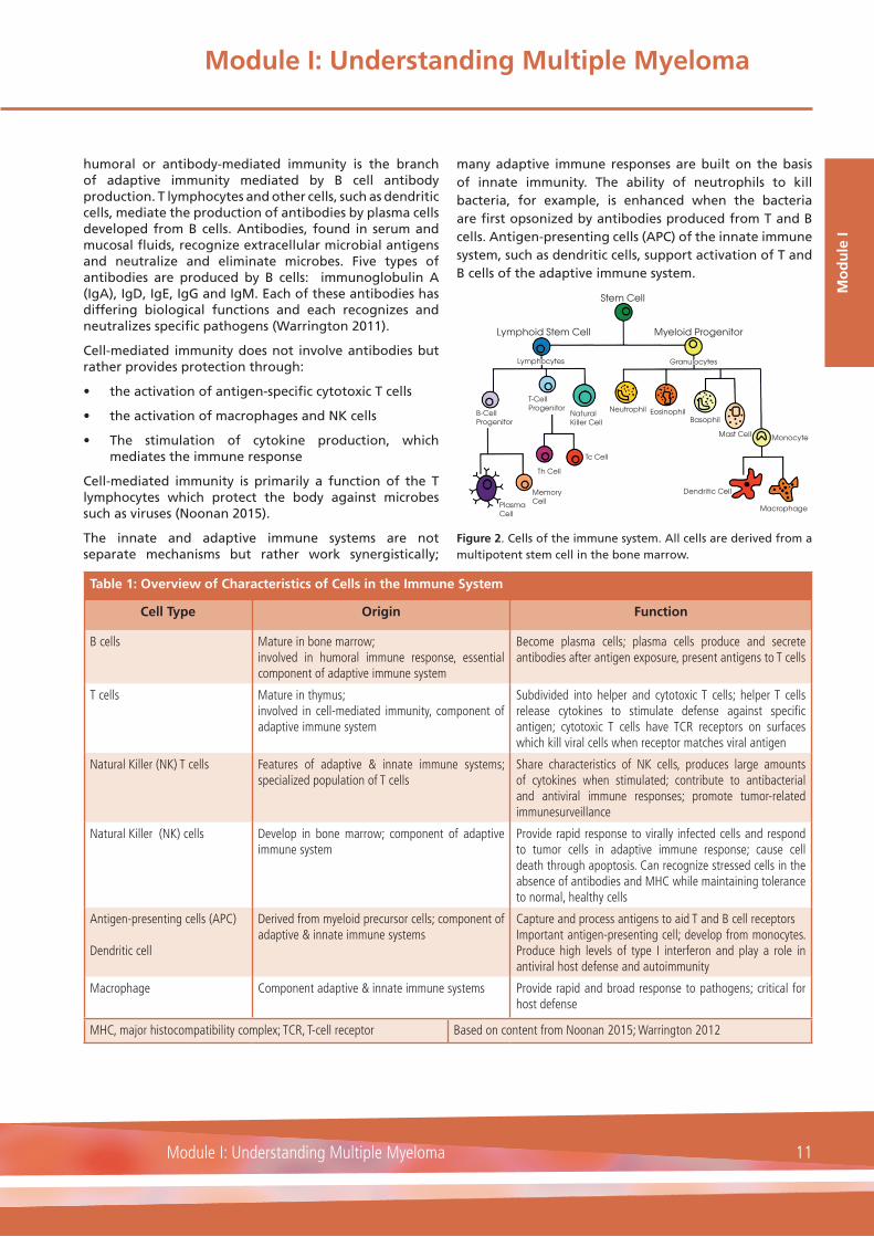
humoral or antibody-mediated immunity is the branch of adaptive immunity mediated by B cell antibody production. T lymphocytes and other cells, such as dendritic cells, mediate the production of antibodies by plasma cells developed from B cells. Antibodies, found in serum and mucosal fluids, recognize extracellular microbial antigens and neutralize and eliminate microbes. Five types of antibodies are produced by B cells: immunoglobulin A (IgA), IgD, IgE, IgG and IgM. Each of these antibodies has differing biological functions and each recognizes and neutralizes specific pathogens (Warrington 2011).
Cell-mediated immunity does not involve antibodies but rather provides protection through:
• the activation of antigen-specific cytotoxic T cells
• the activation of macrophages and NK cells
• The stimulation of cytokine production, which mediates the immune response
Cell-mediated immunity is primarily a function of the T lymphocytes which protect the body against microbes such as viruses (Noonan 2015).
The innate and adaptive immune systems are not separate mechanisms but rather work synergistically;
many adaptive immune responses are built on the basis of innate immunity. The ability of neutrophils to kill bacteria, for example, is enhanced when the bacteria are first opsonized by antibodies produced from T and B cells. Antigen-presenting cells (APC) of the innate immune system, such as dendritic cells, support activation of T and B cells of the adaptive immune system.
Figure 2. Cells of the immune system. All cells are derived from a multipotent stem cell in the bone marrow.
Stem Cell
Myeloid ProgenitorLymphoid Stem Cell
Lymphocytes
B-CellProgenitor
T-CellProgenitor
NaturalKiller Cell
Neutrophil EosinophilBasophil
Mast Cell Monocyte
Macrophage
Dendritic Cell
Tc Cell
Th Cell
MemoryCellPlasma
Cell
Granulocytes
Table 1: Overview of Characteristics of Cells in the Immune System
Cell Type Origin Function
B cells Mature in bone marrow;involved in humoral immune response, essential component of adaptive immune system
Become plasma cells; plasma cells produce and secrete antibodies after antigen exposure, present antigens to T cells
T cells Mature in thymus;involved in cell-mediated immunity, component of adaptive immune system
Subdivided into helper and cytotoxic T cells; helper T cells release cytokines to stimulate defense against specifi c antigen; cytotoxic T cells have TCR receptors on surfaces which kill viral cells when receptor matches viral antigen
Natural Killer (NK) T cells Features of adaptive & innate immune systems; specialized population of T cells
Share characteristics of NK cells, produces large amounts of cytokines when stimulated; contribute to antibacterial and antiviral immune responses; promote tumor-related immunesurveillance
Natural Killer (NK) cells Develop in bone marrow; component of adaptive immune system
Provide rapid response to virally infected cells and respond to tumor cells in adaptive immune response; cause cell death through apoptosis. Can recognize stressed cells in the absence of antibodies and MHC while maintaining tolerance to normal, healthy cells
Antigen-presenting cells (APC)
Dendritic cell
Derived from myeloid precursor cells; component of adaptive & innate immune systems
Capture and process antigens to aid T and B cell receptorsImportant antigen-presenting cell; develop from monocytes. Produce high levels of type I interferon and play a role in antiviral host defense and autoimmunity
Macrophage Component adaptive & innate immune systems Provide rapid and broad response to pathogens; critical for host defense
MHC, major histocompatibility complex; TCR, T-cell receptor Based on content from Noonan 2015; Warrington 2012

Module I: Understanding Multiple Myeloma
Module I: Understanding Multiple Myeloma12
Pathophysiology and Epidemiology
The pathophysiology of multiple myeloma
Multiple myeloma is a malignancy of plasma cells that results in an overproduction of light and heavy chain monoclonal immunoglobulins. The disease is frequently characterized by plasmacytosis in bone marrow, production of monoclonal proteins, osteolytic bone lesions, renal disease, anemia, hypercalcemia and/or immunodeficiency.
Figure 3. Presentation of a healthy plasma cell and a myeloma cell.
While the pathophysiology of multiple myeloma is a complicated process, it is also one which is well-organized comprising sequential interactions. Symptomatic myeloma is typically preceded by an asymptomatic premalignant period that, if detected, is termed either monoclonal gammopathy of undetermined significance (MGUS) or an asymptomatic phase known as smoldering multiple myeloma (SMM) depending on the extent of bone marrow involvement and monoclonal protein levels (Morgan 2012; Rajkumar 2013). SMM is considered an intermediate stage between MGUS and myeloma. The risk of progression from MGUS to myeloma is about 1% per year, and the risk of progression to myeloma from SMM is about 10% per year (Figure 4). The disease process begins with the appearance of a small number of monoclonal plasma cells.
The pathophysiologic changes in multiple myeloma relate to abnormalities within the bone marrow microenvironment, bone marrow stromal cells and cytokine interactions, which cause disease progression and treatment resistance (Noonan 2015). Normally, plasma cells comprise about 4% of the composition of the bone marrow: in myeloma, plasma cell concentrations can be greater than 10%. The basic premise underlying the progression of myeloma is that multiple mutations in different pathways deregulate the biology of the plasma cell causing it to change in ways that generate the features of myeloma. While many of the genes and pathways underlying this transformation have been characterized, there appears to be no single genetic change underlying the process that can be targeted therapeutically (Morgan 2012).
Later in disease progression, myeloma plasma cells are no longer restricted to growth within the bone marrow and can be found at extramedullary sites and as circulating leukemic cells. It seems that transition through these different states requires the acquisition of genetic abnormalities leading to the development of the biological hallmarks of myeloma (Figure 5).
Figure 5. The effects on the body caused by the displacement of bone marrow with plasma cells: biological hallmarks of myeloma.
Role of genetics in multiple myeloma
It is now known that chromosomal abnormalities are extremely common and occur early in multiple myeloma (Fonesca 2004). In a study of 1,064 patients, chromosomal abnormalities were identified in 90% (Avet-Loiseau 2007). Chromosomal abnormalities in newly diagnosed patients with myeloma have been studied using fluorescence in situ hybridization (FISH or iFISH). Using this technique, several overlapping and non-overlapping genetic abnormalities have been identified in patients with myeloma. Based on
Figure 4. Progression to symptomatic multiple myeloma. A strategy to secure better patient outcomes is to identify patients with a high risk of progression and institute early treatment before organ damage occurs.
Module I: Understanding Multiple Myeloma
13Module I: Understanding Multiple Myeloma
Mo
du
le I
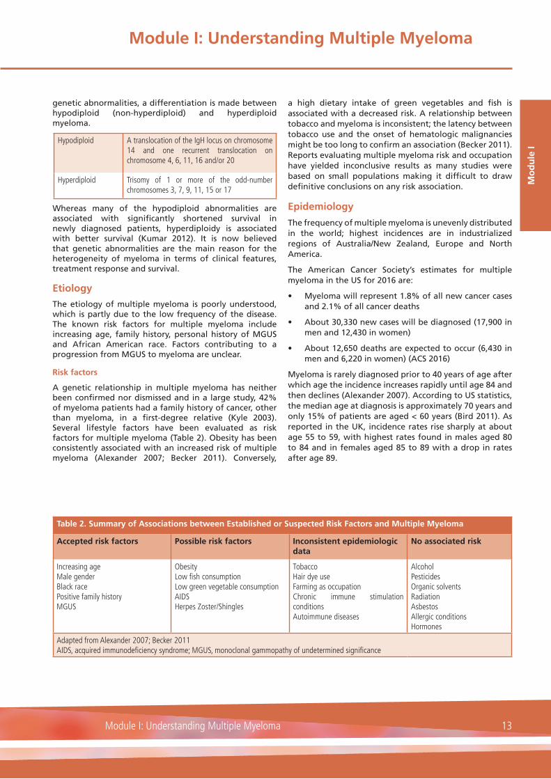
genetic abnormalities, a differentiation is made between hypodiploid (non-hyperdiploid) and hyperdiploid myeloma.
Whereas many of the hypodiploid abnormalities are associated with significantly shortened survival in newly diagnosed patients, hyperdiploidy is associated with better survival (Kumar 2012). It is now believed that genetic abnormalities are the main reason for the heterogeneity of myeloma in terms of clinical features, treatment response and survival.
Etiology
The etiology of multiple myeloma is poorly understood, which is partly due to the low frequency of the disease. The known risk factors for multiple myeloma include increasing age, family history, personal history of MGUS and African American race. Factors contributing to a progression from MGUS to myeloma are unclear.
Risk factors
A genetic relationship in multiple myeloma has neither been confirmed nor dismissed and in a large study, 42% of myeloma patients had a family history of cancer, other than myeloma, in a first-degree relative (Kyle 2003). Several lifestyle factors have been evaluated as risk factors for multiple myeloma (Table 2). Obesity has been consistently associated with an increased risk of multiple myeloma (Alexander 2007; Becker 2011). Conversely,
a high dietary intake of green vegetables and fish is associated with a decreased risk. A relationship between tobacco and myeloma is inconsistent; the latency between tobacco use and the onset of hematologic malignancies might be too long to confirm an association (Becker 2011). Reports evaluating multiple myeloma risk and occupation have yielded inconclusive results as many studies were based on small populations making it difficult to draw definitive conclusions on any risk association.
Epidemiology
The frequency of multiple myeloma is unevenly distributed in the world; highest incidences are in industrialized regions of Australia/New Zealand, Europe and North America.
The American Cancer Society’s estimates for multiple myeloma in the US for 2016 are:
• Myeloma will represent 1.8% of all new cancer cases and 2.1% of all cancer deaths
• About 30,330 new cases will be diagnosed (17,900 in men and 12,430 in women)
• About 12,650 deaths are expected to occur (6,430 in men and 6,220 in women) (ACS 2016)
Myeloma is rarely diagnosed prior to 40 years of age after which age the incidence increases rapidly until age 84 and then declines (Alexander 2007). According to US statistics, the median age at diagnosis is approximately 70 years and only 15% of patients are aged < 60 years (Bird 2011). As reported in the UK, incidence rates rise sharply at about age 55 to 59, with highest rates found in males aged 80 to 84 and in females aged 85 to 89 with a drop in rates after age 89.
Hypodiploid A translocation of the IgH locus on chromosome 14 and one recurrent translocation on chromosome 4, 6, 11, 16 and/or 20
Hyperdiploid Trisomy of 1 or more of the odd-number chromosomes 3, 7, 9, 11, 15 or 17
Table 2. Summary of Associations between Established or Suspected Risk Factors and Multiple Myeloma
Accepted risk factors Possible risk factors Inconsistent epidemiologic data
No associated risk
Increasing ageMale genderBlack racePositive family historyMGUS
ObesityLow fi sh consumptionLow green vegetable consumptionAIDSHerpes Zoster/Shingles
TobaccoHair dye useFarming as occupationChronic immune stimulation conditionsAutoimmune diseases
AlcoholPesticidesOrganic solventsRadiationAsbestosAllergic conditionsHormones
Adapted from Alexander 2007; Becker 2011AIDS, acquired immunodefi ciency syndrome; MGUS, monoclonal gammopathy of undetermined signifi cance
Module I: Understanding Multiple Myeloma
Module I: Understanding Multiple Myeloma14
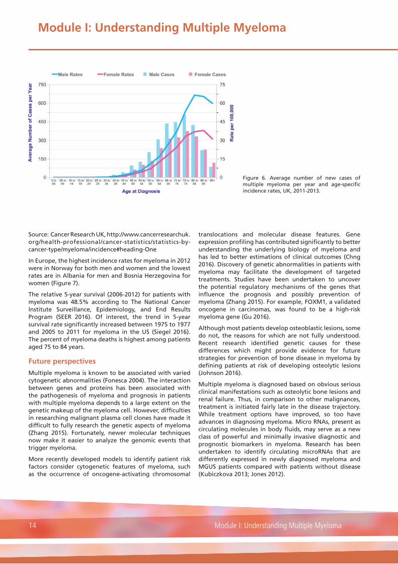
Source: Cancer Research UK, http://www.cancerresearchuk.org/health-professional/cancer-statistics/statistics-by-cancer-type/myeloma/incidence#heading-One
In Europe, the highest incidence rates for myeloma in 2012 were in Norway for both men and women and the lowest rates are in Albania for men and Bosnia Herzegovina for women (Figure 7).
The relative 5-year survival (2006-2012) for patients with myeloma was 48.5% according to The National Cancer Institute Surveillance, Epidemiology, and End Results Program (SEER 2016). Of interest, the trend in 5-year survival rate significantly increased between 1975 to 1977 and 2005 to 2011 for myeloma in the US (Siegel 2016). The percent of myeloma deaths is highest among patients aged 75 to 84 years.
Future perspectives
Multiple myeloma is known to be associated with varied cytogenetic abnormalities (Fonesca 2004). The interaction between genes and proteins has been associated with the pathogenesis of myeloma and prognosis in patients with multiple myeloma depends to a large extent on the genetic makeup of the myeloma cell. However, difficulties in researching malignant plasma cell clones have made it difficult to fully research the genetic aspects of myeloma (Zhang 2015). Fortunately, newer molecular techniques now make it easier to analyze the genomic events that trigger myeloma.
More recently developed models to identify patient risk factors consider cytogenetic features of myeloma, such as the occurrence of oncogene-activating chromosomal
translocations and molecular disease features. Gene expression profiling has contributed significantly to better understanding the underlying biology of myeloma and has led to better estimations of clinical outcomes (Chng 2016). Discovery of genetic abnormalities in patients with myeloma may facilitate the development of targeted treatments. Studies have been undertaken to uncover the potential regulatory mechanisms of the genes that influence the prognosis and possibly prevention of myeloma (Zhang 2015). For example, FOXM1, a validated oncogene in carcinomas, was found to be a high-risk myeloma gene (Gu 2016).
Although most patients develop osteoblastic lesions, some do not, the reasons for which are not fully understood. Recent research identified genetic causes for these differences which might provide evidence for future strategies for prevention of bone disease in myeloma by defining patients at risk of developing osteolytic lesions (Johnson 2016).
Multiple myeloma is diagnosed based on obvious serious clinical manifestations such as osteolytic bone lesions and renal failure. Thus, in comparison to other malignances, treatment is initiated fairly late in the disease trajectory. While treatment options have improved, so too have advances in diagnosing myeloma. Micro RNAs, present as circulating molecules in body fluids, may serve as a new class of powerful and minimally invasive diagnostic and prognostic biomarkers in myeloma. Research has been undertaken to identify circulating microRNAs that are differently expressed in newly diagnosed myeloma and MGUS patients compared with patients without disease (Kubiczkova 2013; Jones 2012).
Figure 6. Average number of new cases of multiple myeloma per year and age-specific incidence rates, UK, 2011-2013.

Module I: Understanding Multiple Myeloma
15Module I: Understanding Multiple Myeloma
Mo
du
le I
Discussion related to treating smoldering multiple myeloma, in place of continuing to observe patients with the disease, as an early intervention is ongoing. Because a large proportion of patients remain free of progression for long periods of time, should practice change from
observation to active treatment, evidence demonstrating any benefit of this approach, including prolonged survival, drug safety and limitation of development of resistant plasma cell clones, would need to be provided (Salem 2015).
Figure 7. Estimated incidence and mortality of multiple myeloma and immunoproliferative diseases in both sexes in European countries by incidence rank, 2012. Age standardized rate (European) per 100,000.EUCAN. http://eco.iarc.fr/eucan/Cancer.aspx?Cancer=39. Accessed June 2016

Module I: Understanding Multiple Myeloma
Module I: Understanding Multiple Myeloma16
ResourcesAmerican Cancer Society (ACS)
www.cancer.org
National non-profit organization providing cancer resources online and community services
American Society for Blood and Marrow Transplantation (ASBMT)
www.asbmt.org
International professional association promoting education, clinical standards and research
European Myeloma Network (EMN)
myeloma-europe.org.linux9.curanetserver.dk/index.php?index
Support the development of novel diagnostics and therapies for multiple myeloma
European Oncology Nursing Society (ONS)
www.cancernurse.eu
Pan-European organization dedicated to the support and development of cancer nurses
European Society for Blood and Marrow Transplantation (EBMT)
www.ebmt.org
European professional association involved in promoting all aspects of transplantation of hematopoietic stem cells
European Society for Blood and Marrow Transplantation – Nursing Section
www.ebmt.org/Contents/Nursing/Pages/default.aspx
Promote excellence in the provision of blood and marrow transplantation and hematology care
International Myeloma Foundation (IMF)
www.myeloma.org
Information about myeloma, treatment, research efforts, support available in several languages
International Myeloma Working Group (IMWG)
myeloma.org/PortalPage.action?tabId=8&menuId=125&portalPageId=8
A division of IMF. Conduct basic, clinical and translational research to improve outcomes in myeloma
Multiple Myeloma Research Foundation (MMRF)
www.themmrf.org
Information about myeloma, research efforts, support
Myeloma UK
www.myeloma.org.uk
Professional and patient information, professional education
National Cancer Institute
www.cancer.gov
Information on disease types and research

Module I: Understanding Multiple Myeloma
17Module I: Understanding Multiple Myeloma
Mo
du
le I
Review Questions
1. Multiple myeloma is characterized by (please tick any/all that apply):
A. The presence of abnormal T cells in peripheral blood
B. Abnormal clonal plasma cell infiltration of the bone marrow
C. The presence of B cell infiltrates in the liver
D. The production of cytokines by natural killer cells
2. True or false:
In adaptive or acquired immunity, memory cells exist in a dormant state until a foreign substance is reintroduced in the body.
A. True
B. False
3. Cell-mediated immunity provides protection through (please tick any/all that apply):
A. The production of colony-stimulating factors
B. The activation of antigen-specific cytotoxic T cells
C. The activation of macrophages and NK cells
D. The stimulation of cytokine production
4. Following antigen exposure, B cells produce (please tick any/all that apply):
A. Cytokines
B. Pathogens
C. Antibodies
D. Immunoglobulins
5. Accepted risk factors for multiple myeloma include (please tick any/all that apply):
A. tobacco, alcohol, asbestos
B. allergic conditions, autoimmune disease, tobacco
C. pesticides, autoimmune diseases, radiation exposure
D. MGUS, increasing age, positive family history
6. True or false:
The discovery of genetic abnormalities in patients with multiple myeloma may facilitate the development of targeted treatment.
A. True
B. False
Answers available online at www.hemcare.org
Module I: Understanding Multiple Myeloma
Module I: Understanding Multiple Myeloma18
References Alexander DD, Mink PJ, Adam H-O, Cole P, Mandel JS, Oken MM, Trichopoulos D. Multiple myeloma: a review of the epidemiologic literature. International Journal of Cancer 2007; 120: 40-61
American Cancer Society (ACS). Cancer Facts & Figures 2016. Atlanta: American Cancer Society 2016
Avet-Loiseau H, Attal M, Moreau P, et al. Genetic abnormalities and survival in multiple myeloma: the experience of the Intergroupe Francophone du Myélome. Blood 2007; 109: 3489-3495
Becker N. Epidemiology of multiple myeloma. Recent Results Cancer Res 2011; 183: 25-35
Bird JM, Owen RG, Snowden JA, et al. Guidelines for the diagnosis and management of multiple myeloma 2011. Br J Haematol 2011; 154: 32-75
Bonilla FA, Oettgen HC. Adaptive immunity. Journal of Allergy and Clinical Immunology 2010; 125(Suppl 2): S33-40
Cancer Research UK, http://www.cancerresearchuk.org/health-professional/cancer-statistics/statistics-by-cancer-type/myeloma/incidence#heading-One. Accessed June 2016
Chaplin DD. Overview of the immune response. Journal of Allergy and Clinical Immunology 2010; 125(Suppl 2): S3-S23
Chng WJ, Chung T-H, Kumar S, et al. Gene signature combinations improve prognostic stratification of multiple myeloma patients. Leukemia 2016; 30: 1071-1078
Durie BG, Kyle RA, Belch A, et al. Myeloma management guidelines: a consensus report from the scientific advisors of the International Myeloma Foundation. Hematol J 2003; 4: 379–398.
Fonesca R, Barloqie B, Bataile R, et al. Genetics and cytogenetics of multiple myeloma: a workshop report. Cancer Research 2004; 64: 1546-1558
Gu C, Yang Y, Sompallae R, et al. FOXM1 is a therapeutic target for high-risk multiple myeloma. Leukemia 2016; 30: 873-882
Johnson DC, Weinhold N, Mitchen J, et al. Genetic factors influencing the risk of multiple myeloma bone disease. Leukemia 2016; 30: 883-888
Jones CI, Zabolotskaya MV, King AJ, Stewart HJS, Horne GA, Chevassult TJ, Newbury SF. Identification of circulating microRNAs as diagnostic biomarkers for use in multiple myeloma. British Journal of Cancer 2012; 107:1987-1996
Kubiczkova L, Kryukov F, Slaby O, et al. Circulating serum microRNAs as novel diagnostic and prognostic biomarkers for multiple myeloma and monoclonal gammopathy of undetermined significance. Journal of the European Hematology Association 2013; haematol.2013.093500; Doi:10.3324/haematol.2013.093500
Kumar S, Fonesca R, Ketterling RP, et al. Trisomies in multiple myeloma: impact on survival in patients with high-risk cytogenetics. Blood 2012; 119: 2100-2105
Kyle RA, Remstein ED, Themeau TM, et al. Clinical course and prognosis of smoldering (asymptomatic) multiple myeloma. New England Journal of Medicine 2007; 356: 2282-2290
Kyle, RA, Gertz, MA, Witzig, TE, Lust JA, Lacy MQ, Dispenzieri A, et al. Review of 1027 patients with newly diagnosed multiple myeloma. Mayo Clinic Proceedings 2003; 78: 21–33
Ludwig H, Miquel JS, Dimopoulos MA, et al. International Myeloma Working Group recommendations for global myeloma care. Leukemia 2013; 1-12
Morgan GJ, Walker BA, Davies FE. The genetic architecture of multiple myeloma. Nature Reviews Cancer 2012; 12: 335-348
National Comprehensive Cancer Network (NCCN) Guidelines Version 1.2017: Multiple Myeloma. Available at: https://www.nccn.org/professionals/physician_gls/pdf/myeloma.pdf. Accessed October 2016
Noonan KA, Huff CA, Davis J, et al. Adoptive transfer of activated marrow-infiltrating lymphocytes induces measurable antitumor immunity in the bone marrow in multiple myeloma. Science Translational Medicine 2015; 7(288):288ra78
Rajkumar SV, Dimopoulos MA, Palumbo A, et al. International Myeloma Working Group updated criteria for the diagnosis of multiple myeloma. The Lancet Oncology 2014; 15: e538-e548
Rajkumar SV, Gupta V, Fonseca R, et al. Impact of primary molecular cytogenetic abnormalities and risk of progression in smoldering multiple myeloma. Leukemia 2013; 27: 1738-1744
Salem KZ, Ghobrial IM. The road to cure in multiple myeloma starts with smoldering disease. Expert Opinion on Orphan Drugs 2015; 3: 653-661
SEER Cancer Statistics Factsheets: Myeloma. National Cancer Institute. Bethesda, MD,http://seer.cancer.gov/statfacts/html/mulmy.html. Accessed June 2016
Siegel RL, Miller KD, Jemal A. Cancer statistics, 2016; CA: A Cancer Journal for Clinicians 2016; 66: 7-30
Turvey SE, Broide DH. Innate immunity. Journal of Clinical Immunology 2010; 125(Suppl. 2): S24-S32
Warrington R, Watson W, Kim HL, Antonetti FR. An introduction to immunology and immunopathology. Alergy, Asthma and Clinical Immunology 2011; 7(Suppl. 1): S1
Zhang K, Xu Z, Sun Z. Identification of the key genes connected with plasma cells of multiple myeloma using expression profiles. Onco Targets and Therapy 2015; 8: 1795-1803

Module II: Multiple Myeloma: Diagnosis and Staging
19Module II: Multiple Myeloma: Diagnosis and Staging
Mo
du
le II
Quick Facts
• The typical clinical manifestations of multiple myeloma, known as CRAB symptoms, are: increased Calcium level, Renal dysfunction, Anemia, destructive Bone lesions
• Many clinical features of multiple myeloma are related to proliferation of plasma cells in the bone marrow
• Approximately 15% of patients present with hypercalcemia; signs and symptoms include confusion, muscle weakness, constipation, thirst
• The frequency of bone lesions in myeloma, approximately 80%-90% of patients, is unique among hematologic malignancies
• Cytogenetic abnormalities are becoming increasingly important as a means of unraveling the different disease categories within multiple myeloma

Module II: Multiple Myeloma: Diagnosis and Staging
Module II: Multiple Myeloma: Diagnosis and Staging20
A. Introduction
B. Presentation and Physical Findings
1. Laboratory
2. Radiographic and Imaging Studies
3. Biopsies
4. Differential Diagnosis
C. Staging and Survival
D. Prognostic Factors
E. Clinical manifestations of myeloma at diagnosis and sequelae
1. Increased serum calcium
2. Renal insufficiency
3. Anemia
4. Bone lesions
F. Resources
F. Review Questions
G. References

Module II: Multiple Myeloma: Diagnosis and Staging
21Module II: Multiple Myeloma: Diagnosis and Staging
Mo
du
le II
IMWG definition of multiple myeloma:
Clonal bone marrow plasma cells ≥10% or biopsy-proven bony or extramedullary plasmacytoma and any one or more of the following myeloma defining events:
Myeloma defining events: evidence of end organ damage attributed to the underlying plasma cell proliferative disorder as characterized by the CRAB acronym:
• Hypercalcemia: serum calcium > 0.25 mmol/L (>1mg/dl) higher than the upper limit of normal or > 2.75 mmol/L (>11mg/dl)
• Renal insufficiency: creatinine clearance <40 mL/min or serum creatinine >177 µmol/L (>2 mg/dL)
• Anemia: hemoglobin of >20 g/L below the lower limit of normal, or a hemoglobin <100 g/L
• Bone lesions: ≥1 osteolytic lesion on skeletal radiography, CT or PET-CT
Any one or more of the following biomarkers of malignancy:
• ≥ 60% clonal plasma cells on bone marrow examination
• Involved:uninvolved serum free light chain assay ratio ≥100
• > 1 focal lesions on MRI that is at least 5 mm or greater in size
CT, computed tomography; PET, PET, positron emission tomography; MRI, magnetic resonance imaging
Adapted from: Rajkumar 2014
Figure 1. Revised International Myeloma Working Group (IMWG) criteria for diagnosis of multiple myeloma
Introduction
Proliferation of plasma cells in the bone marrow results in the classic symptoms of myeloma including anemia and bone destruction with lytic lesions. Malignant plasma cells produce osteoclast-activating factors, such as tumor necrosis factor and interleukin-6, which enhance osteoclast activity which, in turn, enhances bone resorption causing hypercalcemia. The large amount of immunoglobulins produced by the malignant plasma cells overloads the kidneys with proteins that cannot be reabsorbed or filtered leading to tubular damage, proteinuria and eventual kidney failure (Dvorak 2006). The typical clinical manifestations of multiple myeloma are summarized by the CRAB symptoms (also known as myeloma defining events):
• increased Calcium level
• Renal dysfunction
• Anemia
• destructive Bone lesions
Typically, myeloma is preceded by monoclonal gammopathy of undetermined significance (MGUS), an asymptomatic condition. Similarly, smoldering multiple myeloma (SMM), or asymptomatic multiple myeloma, also has a high risk of progression to symptomatic, or
active multiple myeloma. It is now believed that patients at high risk of progression to symptomatic disease may benefit from therapy with an increase in survival time if treatment is initiated before serious organ damage occurs. To diagnose patients at risk of developing symptomatic or active disease, the International Myeloma Working Group (IMWG) now proposes to add three biomarkers of malignancy to the established myeloma defining CRAB events; the presence of at least one of these markers is considered sufficient for a diagnosis of multiple myeloma, regardless of the presence or absence of symptoms or of CRAB events (Figure 1). Each of these markers is associated with an approximately 80% or higher risk of developing myeloma-related organ damage within two years.
Presentation and Physical Findings
Initial investigations in patients with suspected myeloma (Table 1) are undertaken to screen for the disease, establish a diagnosis, estimate the tumor burden and prognosis, and assess myeloma-related organ impairment (Bird 2014).
Assessment of past medical history should include information on comorbid conditions, such as coronary artery disease, congestive heart failure, hypertension, renal and liver disorders and lung diseases as these

Module II: Multiple Myeloma: Diagnosis and Staging
Module II: Multiple Myeloma: Diagnosis and Staging22
conditions could affect treatment options. The patient should be asked about first-degree relatives with a diagnosis of hematologic malignancies, especially lymphoma, chronic lymphocytic leukemia and plasma cell dyscrasia (Dimopoulos 2011).
Clinical findings will vary from totally asymptomatic presentation, in patients whose disease is discovered incidentally, to life-threatening symptoms. Multiple myeloma should be suspected in older adults presenting with back pain (back or ribs), and constitutional symptoms such as sweating and weight loss.
Non-CRAB manifestations of myeloma are extremely diverse in nature (Talamo 2010). The most common non-CRAB manifestation is back pain. Because multiple myeloma originates in the bone marrow, many of its clinical manifestation derive from:
• proliferation of plasma cells in the bone marrow causing anemia, leukopenia, thrombocytopenia and their associated symptoms
• macroscopic destruction of the bones caused by lytic lesions , hypercalcemia
• mechanical pressure from tumor masses in the bones leading to spinal cord compression and nerve root compression (Talamo 2010)
In newly diagnosed patients, skeletal abnormalities are present on conventional radiography in approximately 60% to 80% of patients, anemia is present in 70%, hypercalcemia in 15%, and elevated serum creatinine in 20%. Macroscopic destruction of the bones is commonly seen at presentation; areas most often affected are the back, ribs and hips. Approximately 25% of patients present without symptoms and are identified incidentally by laboratory results, such as an elevated total protein, encountered during routine testing or in evaluation of other health problems (Katzel 2007).
Laboratory
The hallmark sign of myeloma is the detection of monoclonal protein (M protein) produced by the abnormal plasma cells and found in blood and/or urine. Therefore, both blood and urine are assessed to detect and characterize monoclonal immunoglobulin. A serum protein electrophoresis, a urine protein electrophoresis from a 24-hour urine specimen to detect Bence-Jones protein, immunofixation in serum and urine, and determination of serum free light-chains and their ratio should be performed (Table 1). To assess the extent and level of activity of myeloma, albumin and �2-microglobulin are needed for International Staging System (ISS). Further, analysis of complete blood count, and calcium, creatinine
and lactate dehydrogenase levels as well as cytogenetic assessment of high-risk features such as del17p are recommended. If infection is suspected, a determination of C-reactive protein levels is helpful.
Radiographic and imaging studies
Standard work-up for multiple myeloma includes whole skeletal bone X-rays including radiography of the spine, skull, shoulders, thoracic cage, pelvis and long bones of the arms and legs. Whole skeletal bone X-rays are still the radiological gold standard for myeloma, but there is an international consensus to use whole body magnetic resonance imaging (WB-MRI), positron emission tomography (PET) or a low dose computed tomography (LD-CT) for bone study in place of conventional X-ray to improve the positive predictive value on bone disease (Harousseau 2010). Pathologic fractures of long bones are especially common in newly diagnosed patients taking corticosteroids and are often the reason the patient seeks medical attention (Melton 2005).
Biopsies
Monoclonal plasma cell proliferation is detected via bone marrow aspiration and/or bone marrow biopsy (Ludwig 2014). A bone marrow aspirate and biopsy is essential in establishing the diagnosis of multiple myeloma (Bird 2014).
Differential diagnosis
A differential diagnosis should be made between smoldering and active multiple myeloma. Both of the following criteria must be met to establish a diagnosis of smoldering (asymptomatic) multiple myeloma:
• Serum monoclonal protein (IgG or IgA) ≥30 g/L or urinary monoclonal protein ≥500 mg per 24 h and/or clonal bone marrow plasma cells 10% to 60%
• Absence of myeloma-defining events or amyloidosis
• It is also important to distinguish between MGUS and active multiple myeloma. Some of the clinical findings indicative of MGUS include:
• Absence of end-organ damage such as hypercalcemia, renal insufficiency, anemia and bone lesions (CRAB criteria)
• Clonal bone marrow plasma cells <10%
• Serum monoclonal protein (IgM and non-IgM) <30% (Rajkumar 2014)
Other diseases with a similar clinical presentation to multiple myeloma include solitary plasmacytoma and other B-cell lymphoproliferative disorders.

Module II: Multiple Myeloma: Diagnosis and Staging
23Module II: Multiple Myeloma: Diagnosis and Staging
Mo
du
le II
Table 1. Procedures for the Diagnosis of Multiple Myeloma
Parameter of interest Information provided
Monoclonal plasma cells
Unilateral bone marrow aspiration and/or bone biopsy BMPC infi ltration, enables FISH cytogenetics, immunophenotyping, immunocytochemistry, conventional karyotyping, gene arrays
Monoclonal protein
Serum protein electrophoresis (SPEP) M-component, possible suppression of non-paraprotein immunoglobulins; emergence of a new M-component (rare)
Urine protein electrophoresis (UPEP) (24-hour urine) M-component, indicates glomerular damage when albumin present (amyloidosis)
Nephelometry of serum immunoglobulins
Measurement of IgA, overestimates the M-component concentration in patients with IgG and IgM myeloma. Provides information about suppression of non-involved immunoglobulins
Immunofi xationelectrophoresis
Identifi es isotype and light chain type, confi rms CR at baseline in serum and in urine in those with proteinuria
Serum free light chain measurement (serum) Detects mildly elevated levels of free light chains, which indicates presence of abnormal monoclonal protein (M-protein); supports disease monitoring and response to treatment; greater sensitivity than SPEP or UPEP
Bone lesions specifi c to myeloma
Skeletal bone survey byconventional radiography
Assessment of extent of bone disease, and of progressive bone disease
CT, MRI, PET, PET/CT,PET/MRI
Higher sensitivity for myeloma specifi c bone lesions, assessmentof extramedullary disease, PET provides information aboutactivity of the disease
Additional laboratory parameters
Albumin, ß2-microglobulin, lactate dehydrogenase (LDH), CRP, complete blood count and differential, peripheral blood smear, chemistry screen (with calcium and creatinine)
Provides information about organ function and aggressiveness of the disease (LDH), bacterial infections (CRP)
BMPC, bone marrow plasma cell; CT, computed tomography; CR, complete response; CRP, C-reactive protein; FISH, interphase fl uorescence in situ hybridization; FU, follow-up; LDH, lactate dehydrogenase; MRI, magnetic resonance imaging; PD, progressive disease; PET, positron emission tomography.Adapted from: Ludwig 2014; Dimopoulos 2011

Module II: Multiple Myeloma: Diagnosis and Staging
Module II: Multiple Myeloma: Diagnosis and Staging24
Staging and Survival
The International Staging System (ISS) is a simple risk stratification algorithm based on the important biological parameters serum beta2-microglobulin (�2M) and serum albumin (Greipp 2005). The score derived from the ISS identifies three patient groups with different prognoses (Table 2).
Biomarkers, such as cytogenetic abnormalities, are becoming increasingly important as a means of unraveling the different disease categories within multiple myeloma. Chromosomal abnormalities, detected by FISH, are key to defining biologic features of myeloma and to providing prognostic and predictive information (Ross 2012). Serum lactate dehydrogenase (LDH) is also a relevant serum marker in myeloma. Elevated LDH indicates an increased disease aggressiveness and suggests a high proliferation rate of plasma cells and/or the presence of tumor mass. The IMWG staging system incorporates the ISS, chromosomal abnormalities and LDH data to define subgroups of patients with different prognoses (Table 2) (Palumbo 2015).
Prognostic Factors
Patient survival depends on the stage of the disease. However, there is general consensus that while staging provides prognostic information, it is not useful for making therapeutic decisions. Patients with suspected myeloma require urgent referral to an oncology specialist. Spinal cord compression, hypercalcemia and renal failure are
medical emergencies requiring immediate investigation and treatment (Bird 2014).
Several cytogenetic and molecular genetic abnormalities have been shown to affect outcome in multiple myeloma (Table 3).
Clinical Manifestations of Myeloma at Initial Presentation: Sequelae and Management
Because multiple myeloma is a cancer of the bone, many of its clinical manifestations derive from microscopic diffuse infiltration of the bone marrow, macroscopic destruction
Table 2: Staging Systems for Multiple Myeloma
International Staging System (ISS) Revised ISS (R-ISS)
Stage I ß2M < 3.5 mg/L and serum albumin > 3.5 g/dL R-ISS Stage I ISS stage I and standard-risk CA by iFISHa and serum LDH < upper limit of normal
Stage II ß2M < 3.5 mg/L and serum albumin > 3.5 g/dL o rß2M 3.5-5.5 mg/L
R-ISS Stage II Not R-ISS stage I or III
Stage III ß2M > 5.5 mg/L R-ISS Stage III ISS stage III and either high-risk CA by iFISH or serum LDH > upper limit of normal
ß2M, ß2-microglobulin; iFISH, interphase fl uorescent in situ hybridizationa Standard-risk: No high-risk chromosomal abnormality. High-risk: Presence of del(17p) and/or translocation t(4;14) and/or t(14;16)Adapted from: Palumbo 2015; NCCN 2016
Table 3: Factors associated with Standard and Higher Risk Outcomes
Factors associated with standard risk Factors associated with higher risk/poorer outcome
Presence of hyperdiploidy, t(11;14), t(6;14)Normal levels of serum ß2-microglobulinNormal levels of lactate dehydrogenaseNormal karyotypeNone of the high-risk factors
Any chromosomal abnormality detected on standard cytogenetic analysisImmunoglobulin heavy chain gene translocations t(4;14), t(14;16) and t(14;20), or 17p13 depletion or chromosome 1 abnormalitiesHigh levels of serum ß2-microglobulinHigh levels of lactate dehydrogenaseInternational Staging System stage III
Adapted from: Rajkumar 2011; Bird 2014

Module II: Multiple Myeloma: Diagnosis and Staging
25Module II: Multiple Myeloma: Diagnosis and Staging
Mo
du
le II
of the bones and mechanical pressure from tumor masses arising from the bones (Talamo 2010). Evidence of tissue or organ impairment is a critical finding for deciding if treatment should be initiated. The most common clinical features of myeloma-related organ or tissue impairment, characterized by the acronym CRAB are presented in Figure 1.
Malignant plasma cells secrete para-proteins, which can be directly responsible for a spectrum of manifestations. Other possible clinical manifestations of myeloma at diagnosis include: symptomatic hyperviscosity (rare), amyloidosis, recurrent infections, neurologic impairment from spinal cord compression, peripheral neuropathy and extramedullary plasmacytomas (Blade 2010; Talamo 2010).
Increased serum calcium
Approximately 15% of patients present with hypercalcemia (Katzel 2007), which occurs most often in the context of symptomatic disease. Signs and symptoms of hypercalcemia can include:
• nervous system dysfunction (confusion, coma and obtundation)
• muscle weakness
• pancreatitis
• constipation
• thirst
• polyuria
• shortening of the Q-T interval on electrocardiogram
• acute renal insufficiency
Treatment of myeloma should be initiated immediately if the patient presents with hypercalcemia. Active treatment of hypercalcemia should be initiated to minimize long-term renal damage (Bird 2014).
Mild hypercalcemia (corrected calcium 2.6-2.9 mmol/l) can be treated with oral and/or intravenous rehydration. Moderate to severe hypercalcemia (corrected calcium ≥2.9 mmol/l) should be treated with intravenous normal saline. Adequate urinary output should be ensured as well as administration of a loop diuretic to avoid volume overload and promote urinary calcium excretion.
Management of hypercalcemia is discussed in Module IV.
Renal insufficiency
Impairment of renal function is a common and potentially serious complication of myeloma. Approximately 20% to 25% of patients present with renal insufficiency (Bird 2014) and symptoms can be reversed in most patients during the course of the disease. The remainder of patients have some degree of persistent renal impairment which may
require renal replacement therapy. Renal failure is a result of damage caused to renal tubules by free light chains (known as cast nephropathy or “myeloma kidney”). Other physiologic problems, such as dehydration, hypercalcemia and infection, can contribute to renal impairment. Patients with renal insufficiency at presentation have a higher risk of early death.
Early diagnosis of both new and relapsed myeloma aids in starting early treatment for renal impairment and can prevent further renal damage. Hydration with at least 3 liters/day can optimize renal function; patients should be provided information on the importance of increasing fluid intake throughout the disease course.
The management and sequela of renal dysfunction are discussed in Module IV.
Anemia
Anemia is present in 70% of newly diagnosed patients (Katzel 2007) and occurs in almost all myeloma patients during their disease course. At diagnosis, anemia is most often due to osteolytic suppression of erythropoiesis by tumor-related cytokines, renal insufficiency and/or vitamin or iron deficiency (Katzel 2007). At presentation, the patient may have symptoms of anemia including dyspnea, fatigue or dizziness. Treatment of myeloma will most often improve erythropoiesis. Symptomatic anemia is often improved by administration of exogenous erythropoietin.
The management of anemia is discussed in Module IV.
Bone lesions
The frequency of bone lesions in myeloma is unique among hematologic malignancies with bone lesions occurring in 80% to 90% of patients. Unlike the bone loss in other malignancies, where bone destruction is followed by new bone formation, myeloma bone lesions are purely osteolytic (Silbermann 2010). Bone disease due to lytic bone lesions can be either focal or diffuse and can cause pain, pathological fractures/spinal cord compression and hypercalcemia. Bone pain is present in up to 60% of patients at disease presentation and pathologic fractures develop in about 60% of patients during the course of the disease (Melton 2005). Bone lesions and their sequelae can compromise mobility, activities of daily living and quality of life (Roodman 2009).
Bone lesions located in vertebrae, pelvis, femur or humerus place the patient at risk for bone fracture. Bone fractures require stabilization and subsequent radiotherapy: radiotherapy is helpful in improving pain and promoting healing (Bird 2014).
The management and sequela of bone lesions are discussed in Module IV.

Module II: Multiple Myeloma: Diagnosis and Staging
Module II: Multiple Myeloma: Diagnosis and Staging26
ResourcesAmerican Cancer Society (ACS)
www.cancer.org
National non-profit organization providing cancer resources online and community services
American Society for Blood and Marrow Transplantation (ASBMT)
www.asbmt.org
International professional association promoting education, clinical standards and research
European Myeloma Network (EMN)
myeloma-europe.org.linux9.curanetserver.dk/index.php?index
Support the development of novel diagnostics and therapies for multiple myeloma
European Oncology Nursing Society (ONS)
www.cancernurse.eu
Pan-European organization dedicated to the support and development of cancer nurses
European Society for Blood and Marrow Transplantation (EBMT)
www.ebmt.org
European professional association involved in promoting all aspects of transplantation of hematopoietic stem cells
European Society for Blood and Marrow Transplantation – Nursing Section
www.ebmt.org/Contents/Nursing/Pages/default.aspx
Promote excellence in the provision of blood and marrow transplantation and hematology care
International Myeloma Foundation (IMF)
www.myeloma.org
Information about myeloma, treatment, research efforts, support available in several languages
International Myeloma Working Group (IMWG)
myeloma.org/PortalPage.action?tabId=8&menuId=125&portalPageId=8
A division of IMF. Conduct basic, clinical and translational research to improve outcomes in myeloma
Multiple Myeloma Research Foundation (MMRF)
www.themmrf.org
Information about myeloma, research efforts, support
Myeloma UK
www.myeloma.org.uk
Professional and patient information, professional education
National Cancer Institute
www.cancer.gov
Information on disease types and research

Module II: Multiple Myeloma: Diagnosis and Staging
27Module II: Multiple Myeloma: Diagnosis and Staging
Mo
du
le II
Review Questions
1. Common clinical manifestations of multiple myeloma at the time of diagnosis include (please tick any/all that apply):
A. Liver dysfunction
B. Anemia
C. Renal dysfunction
D. Hypercalcemia
2. The clinical features of multiple myeloma can generally be attributed to the proliferation of plasma cells in the bone marrow
A. True
B. False
3. Factors associated with higher risk and poorer outcomes include (please tick any/all that apply):
A. Normal karyotype
B. Chromosomal abnormality
C. Stage I per ISS
D. High levels of serum ß2-microglobulin
E. High levels of lactate dehydrogenase
4. As a hematologic malignancy, multiple myeloma is unique due to the frequency of what symptom at the time of diagnosis (please tick any/all that apply):
A. Hypercalcemia
B. Renal dysfunction
C. Thrombocytopenia
D. Bone lesions
5. Anemia, present in about 70% of newly diagnosed patients with myeloma, is characterized by which of the following three symptoms (please tick any/all that apply):
A. Fatigue
B. Dizziness
C. Bleeding
D. Dyspnea
Answers available online at www.hemcare.org
Module II: Multiple Myeloma: Diagnosis and Staging
Module II: Multiple Myeloma: Diagnosis and Staging28
References
Bird, JM, Owen RG, D’Sa S, et al. Guidelines for the diagnosis and management of multiple myeloma 2014. Available at: http://www.bcshguidelines.com/documents/MYELOMA_GUIDELINE_Feb_2014_for_BCSH.pdf. Accessed: July 2016
Blade J, Cibeira MT, de Larrea CF, Rosinol L. Multiple myeloma. Ann Onc 2010; 21(Suppl 7): vii 313-vii319
Dimopoulos M, Kyle R, Fermand JP, et al. Consensus recommendations for standard investigative workup: report of the International Myeloma Workshop Consensus Panel 3. Blood 2011; 117: 4701–4705
Dvorak C. Common complaints, difficult diagnosis: multiple myeloma. Journal of the American Academy of Nurse Practitioners 2006; 18: 190-194
Greipp PR, San Miguel J, Durie BG, et al. International staging system for multiple myeloma. Journal of Clinical Oncology 2005; 23: 3412-3420
Harousseau J-L, Dreyling M. Multiple myeloma: ESMO Clinical Practice Guidelines for diagnosis, treatment and follow-up. Annals of Oncology 2010; 21(Suppl 5): v155-v157
Katzel JA, Parameswaran H, Vesole DH. Multiple myeloma: charging toward a bright future. CA A Cancer Journal for Clinicians 2007; 57: 301-318
Ludwig H, Miguel JS, Dimopoulos MA, et al. International Myeloma Working Group recommendations for global myeloma care. Leukemia 2014; 28: 981-992
Melton III LJ, Kyle RA, Achenbach SJ, Oberg AL, Rajkumar SV. Fracture risk with multiple myeloma: a population-based study. J Bone Miner Res 2005; 20: 487–493
National Comprehensive Cancer Network (NCCN) Guidelines Version 3.2016: Multiple Myeloma
Palumbo A, Avet-Loiseau H, Oliva S, et al. Revised International Staging System for multiple myeloma: a report from International Myeloma Working Group. Journal of Clinical Oncology 2015; 33: 2863-2869
Rajkumar SV, Dimopoulos MA, Palumbo A, et al. International Myeloma Working Group updated criteria for the diagnosis of multiple myeloma. Lancet Oncology 2014; 15: e538-548
Rajkumar SV. Multiple myeloma: 2011 update on diagnosis, risk-stratification, and management. American Journal of Hematology 2011; 86: 57-65
Roodman GD. Pathogenesis of myeloma bone disease. Leukemia 2009; 23: 435-441.
Ross, FM, Avet-Loiseau H, Ameye G, et al. Report from the European Myeloma Network on interphase FISH in multiple myeloma and related disorders. Haematologica 2012; 97: 1272-1277
Silbermann R, Roodman GD. Clinical Presentation of Myeloma Bone Disease. In: Roodman GD (Ed): Myeloma Bone Disease. Humana Press, Pittsburgh, USA 2010
Talamo G, Farooq U, Zangari M, Liao J, Dolloff NG, Loughran TP, Epner E. Beyond the CRAB symptoms: a study of presenting clinical manifestations of multiple myeloma. Clinical Lymphoma, Myeloma & Leukemia 2010; 10: 464-468
Module III: Treatment of Multiple Myeloma
29Module III: Treatment of Multiple Myeloma
Mo
du
le II
I
Quick Facts
• Regardless of whether a patient is eligible or ineligible for autologous stem cell transplantation, the approach to treating myeloma should be based on individual factors such as: features of the disease, patient age, presence of co-morbidities and personal preferences
• Prior to the initiation of autologous stem cell transplantation (ASCT), matters related to supportive care should be taken into consideration to avoid early complications that may compromise therapeutic outcomes
• Older age with concurrent disorders increases vulnerability and decreases resistance to stressors such as myeloma and its treatment resulting in poorer treatment efficacy and tolerability
• The administration of multiple drugs in combination may exacerbate known side effects of individual drugs or cause drug-drug interactions
• Newer agents provide better disease control but are associated with significant toxicity, which frequently persists after completion of treatment

Module III: Treatment of Multiple Myeloma
Module III: Treatment of Multiple Myeloma30
A. Treatment
1. Autologous Stem Cell Transplantation for Newly Diagnosed, Trans- plant Eligible Patients
a. Autologous transplantation process
b. Consolidation and maintenance treatment
c. Treatment of relapsed and refractory myeloma
2. Allogeneic Stem Cell Transplantation for Newly Diagnosed, Trans- plant Eligible Patients
3. Treatment of Newly Diagnosed, Transplant Ineligible Patients
a. Maintenance treatment
4. Treatment of Relapsed Disease
5. Role of Radiation Therapy in Multiple Myeloma Treatment
6. Treatment in Special Populations
a. Older and frail patients
b. Patients with co-morbidities
c. In pregnancy
7. Nursing Measures Related to Commonly used Drugs in Multiple Myeloma Treatment
8. Complementary Therapies
9. Future Treatment Perspectives
B. Resources
C. Review Questions
D. References
Module III: Treatment of Multiple Myeloma
31Module III: Treatment of Multiple Myeloma
Mo
du
le II
I
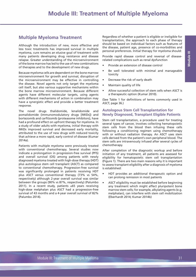
Multiple Myeloma Treatment
Although the introduction of new, more effective and less toxic treatments has improved survival in multiple myeloma, cure remains an elusive goal of treatment with many patients developing drug resistance and disease relapse. Greater understanding of the microenvironment of the bone marrow has led to the use of new combinations of therapies and to the development of new drugs.
Because myeloma cells are dependent on the bone marrow microenvironment for growth and survival, disruption of the microenvironment may be effective in controlling the disease. Novel agents not only target the myeloma cell itself, but also various supportive mechanisms within the bone marrow microenvironment. Because different agents have different molecular targets, using agents with different mechanisms of action in combination may have a synergistic effect and provide a better treatment response.
The novel drugs thalidomide, lenalidomide and pomalidomide (immunomodulatory drugs [IMiDs]) and bortezomib and carfilzomib (proteasome inhibitors), have had a profound effect on upfront therapy for myeloma. In a study of older adults with myeloma, initial therapy with IMiDs improved survival and decreased early mortality, attributed to the use of new drugs with reduced toxicity that achieve a more rapid, early control of disease (Kumar 2014a).
Patients with multiple myeloma were previously treated with conventional chemotherapy. Several studies now indicate a prolongation in progression-free survival (PFS) and overall survival (OS) among patients with newly diagnosed myeloma treated with high-dose therapy (HDT) plus autologous stem cell transplant (ASCT) as compared to conventional chemotherapy. Progression-free survival was significantly prolonged in patients receiving HDT plus ASCT versus conventional therapy (73% vs 54%, respectively) although 2-year overall survival was similar between the groups (90% vs 87%, respectively) (Palumbo 2011). In a recent study, patients ≤65 years receiving high-dose melphalan plus ASCT had a progression-free survival of 43 months and a 4-year overall survival of 82% (Palumbo 2014).
Regardless of whether a patient is eligible or ineligible for transplantation, the approach to each phase of therapy should be based on individual factors such as features of the disease, patient age, presence of co-morbidities and personal preferences. Initial therapy for myeloma should:
Provide rapid disease control and reversal of disease-related complications such as renal dysfunction
• Provide an extension of disease control
• Be well tolerated with minimal and manageable toxicity
• Decrease the risk of early death
• Maintain quality of life
• Allow successful collection of stem cells when ASCT is a therapeutic option (Kumar 2010).
(See Table 1 for definitions of terms commonly used in ASCT, page 36.)
Autologous Stem Cell Transplantation for Newly Diagnosed, Transplant Eligible Patients
Stem cell transplantation, a procedure used for treating several types of cancer, involves collecting hematopoietic stem cells from the blood then infusing these cells following a conditioning regimen using chemotherapy with or without radiation therapy. An ASCT uses stem cells derived from the patient’s own peripheral blood. The stem cells are intravenously infused after several cycles of chemotherapy.
After completion of the diagnostic workup and before initiation of any treatment, all patients are assessed for eligibility for hematopoietic stem cell transplantation (Figure 1). There are two main reasons why it is important to assess transplant eligibility after a diagnosis of myeloma is established:
• HDT provides an additional therapeutic option and can prolong remission in most patients
• ASCT eligibility must be established before beginning any treatment which might affect pluripotent bone marrow stem cells; for example, alkylating agents (e.g. melphalan), can interfere with stem cell mobilization (Eberhardt 2014; Kumar 2014b)
Module III: Treatment of Multiple Myeloma
Module III: Treatment of Multiple Myeloma32
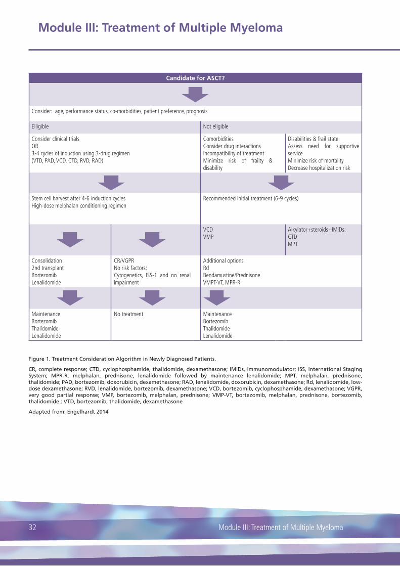
Candidate for ASCT?
Consider: age, performance status, co-morbidities, patient preference, prognosis
Elligible Not eligible
Consider clinical trialsOR3-4 cycles of induction using 3-drug regimen(VTD, PAD, VCD, CTD, RVD, RAD)
ComorbiditiesConsider drug interactionsIncompatibility of treatmentMinimize risk of frailty & disability
Disabilities & frail stateAssess need for supportive serviceMinimize risk of mortalityDecrease hospitalization risk
Stem cell harvest after 4-6 induction cyclesHigh-dose melphalan conditioning regimen
Recommended initial treatment (6-9 cycles)
VCDVMP
Alkylator+steroids+IMiDs:CTDMPT
Consolidation2nd transplantBortezomibLenalidomide
CR/VGPRNo risk factors:Cytogenetics, ISS-1 and no renal impairment
Additional optionsRdBendamustine/PrednisoneVMPT-VT, MPR-R
Maintenance BortezomibThalidomideLenalidomide
No treatment MaintenanceBortezomibThalidomideLenalidomide
Figure 1. Treatment Consideration Algorithm in Newly Diagnosed Patients.
CR, complete response; CTD, cyclophosphamide, thalidomide, dexamethasone; IMiDs, immunomodulator; ISS, International Staging System; MPR-R, melphalan, prednisone, lenalidomide followed by maintenance lenalidomide; MPT, melphalan, prednisone, thalidomide; PAD, bortezomib, doxorubicin, dexamethasone; RAD, lenalidomide, doxorubicin, dexamethasone; Rd, lenalidomide, low-dose dexamethasone; RVD, lenalidomide, bortezomib, dexamethasone; VCD, bortezomib, cyclophosphamide, dexamethasone; VGPR, very good partial response; VMP, bortezomib, melphalan, prednisone; VMP-VT, bortezomib, melphalan, prednisone, bortezomib, thalidomide ; VTD, bortezomib, thalidomide, dexamethasone
Adapted from: Engelhardt 2014
Module III: Treatment of Multiple Myeloma
33Module III: Treatment of Multiple Myeloma
Mo
du
le II
I
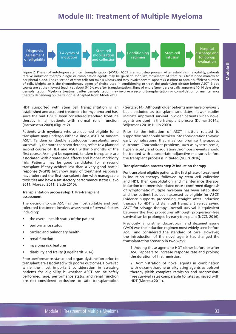
Diagnosis/ Assessment
of elligibility
3-4 cycles of induction
Stem cell mobilization
and collection
Conditioning regimen
Stem cell infusion
Hospital discharge and
follow-up evaluation
HDT supported with stem cell transplantation is an established and accepted treatment for myeloma and has, since the mid 1990’s, been considered standard frontline therapy in all patients with normal renal function (Harousseau 2009) (Figure 2).
Patients with myeloma who are deemed eligible for a transplant may undergo either a single ASCT or tandem ASCT. Tandem or double autologous transplants, used successfully for more than two decades, refers to a planned second course of HDT and ASCT within 6 months of the first course. As might be expected, tandem transplants are associated with greater side effects and higher morbidity risk. Patients may be good candidates for a second transplant if they achieve less than a very good partial response (VGPR) but show signs of treatment response, have tolerated the first transplantation with manageable toxicities and have a satisfactory performance status (Cavo 2011; Moreau 2011; Bladé 2010).
Transplantation process step 1: Pre-transplant assessment
The decision to use ASCT as the most suitable and best tolerated treatment involves assessment of several factors including:
• the overall health status of the patient
• performance status
• cardiac and pulmonary health
• renal function
• myeloma risk features
• disability and frailty (Engelhardt 2014)
Poor performance status and organ dysfunction prior to transplant are associated with poorer outcomes. However, while the most important consideration in assessing patients for eligibility is whether ASCT can be safely performed: age, performance status and renal function are not considered exclusions to safe transplantation
(Gertz 2014). Although older patients may have previously been excluded as transplant candidates, newer studies indicate improved survival in older patients when novel agents are used in the transplant process (Kumar 2014a; Wijermans 2010; Hulin 2009).
Prior to the initiation of ASCT, matters related to supportive care should be taken into consideration to avoid early complications that may compromise therapeutic outcomes. Concomitant problems, such as hypercalcemia, hyperviscosity and coagulation/thrombosis events should be treated with appropriate adjunctive measures before the transplant process is initiated (NCCN 2016).
Transplantation process step 2: Induction therapy
For transplant eligible patients, the first phase of treatment is induction therapy followed by stem cell collection and HDT, then consolidation and maintenance therapy. Induction treatment is initiated once a confirmed diagnosis of symptomatic multiple myeloma has been established and the patient has been assessed as eligible for ASCT. Evidence supports proceeding straight after induction therapy to HDT and stem cell transplant versus saving ASCT for salvage therapy: overall survival is equivalent between the two procedures although progression-free survival can be prolonged by early transplant (NCCN 2016).
Previously, vincristine, doxorubicin and dexamethasone (VAD) was the induction regimen most widely used before ASCT and considered the standard of care. However, the introduction of the novel agents has changed the transplantation scenario in two ways:
1. Adding these agents to HDT either before or after ASCT appears to increase response rate and prolong the duration of first remission.
2. Administration of novel agents in combination with dexamethasone or alkylating agents as upfront therapy yields complete remission and progression-free survival rates comparable to rates achieved with HDT (Moreau 2011).
Figure 2. Phases of autologous stem cell transplantation (ASCT). ASCT is a multistep process. After establishing eligibility, patients receive induction therapy. Single or combination agents may be given to mobilize movement of stem cells from bone marrow to peripheral blood. The collection of stem cells can take 4-6 hours and may involve several apheresis sessions to obtain sufficient number of cells. Melphalan is the chemotherapy agent of choice used in conditioning to treat the underlying disease before ASCT. Blood counts are at their lowest (nadir) at about 5-10 days after transplantation. Signs of engraftment are usually apparent 10-14 days after transplantation. Myeloma treatment after transplantation may involve a second transplantation or consolidation or maintenance therapy depending on the response. Adapted from: Miceli 2013
Module III: Treatment of Multiple Myeloma
Module III: Treatment of Multiple Myeloma34
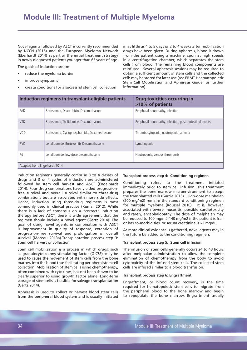
Induction regimens in transplant-eligible patients Drug toxicities occurring in >10% of patients
PAD Bortezomib, Doxorubicin, Dexamethasone Peripheral neuropathy, infection
VTD Bortezomib, Thalidomide, Dexamethasone Peripheral neuropathy, infection, gastrointestinal events
VCD Bortezomib, Cyclophosphamide, Dexamethasone Thrombocytopenia, neutropenia, anemia
RVD Lenalidomide, Bortezomib, Dexamethasone Lymphopenia
Rd Lenalidomide, low-dose dexamethasone Neutropenia, venous thrombosis
Adapted from: Engelhardt 2014
Novel agents followed by ASCT is currently recommended by NCCN (2016) and the European Myeloma Network (Eberhardt 2014) as part of the initial treatment strategy in newly diagnosed patients younger than 65 years of age.
The goals of induction are to:
• reduce the myeloma burden
• improve symptoms
• create conditions for a successful stem cell collection
Induction regimens generally comprise 3 to 4 classes of drugs and 3 or 4 cycles of induction are administered followed by stem cell harvest and ASCT (Engelhardt 2014). Four-drug combinations have yielded progression-free survival and overall survival similar to three-drug combinations but are associated with more side effects. Hence, induction using three-drug regimens is most commonly used in clinical practice (Kumar 2012). While there is a lack of consensus on a “correct” induction therapy before ASCT, there is wide agreement that the regimen should include a novel agent (Gertz 2014). The goal of using novel agents in combination with ASCT is improvement in quality of response, extension of progression-free survival and prolongation of overall survival (Moreau 2013a).Transplantation process step 3: Stem cell harvest or collection
Stem cell mobilization is a process in which drugs, such as granulocyte colony stimulating factor (G-CSF), may be used to cause the movement of stem cells from the bone marrow into the blood thus facilitating peripheral stem cell collection. Mobilization of stem cells using chemotherapy, often combined with cytokines, has not been shown to be clearly superior to using growth factor alone. Long-term storage of stem cells is feasible for salvage transplantation (Gertz 2014).
Apheresis is used to collect or harvest blood stem cells from the peripheral blood system and is usually initiated
in as little as 4 to 5 days or 2 to 4 weeks after mobilization drugs have been given. During apheresis, blood is drawn from the patient using a machine, spun at high speeds in a centrifugation chamber, which separates the stem cells from blood. The remaining blood components are reinfused. Several apheresis sessions may be required to obtain a sufficient amount of stem cells and the collected cells may be stored for later use (see EBMT Haematopoietic Stem Cell Mobilisation and Apheresis Guide for further information).
Transplant process step 4: Conditioning regimen
Conditioning refers to the treatment initiated immediately prior to stem cell infusion. This treatment prepares the bone marrow microenvironment to accept the transplanted cells (Garcia 2015). High-dose melphalan (200 mg/m2) remains the standard conditioning regimen for multiple myeloma (Roussel 2010). It is, however, associated with severe mucositis, possible cardiotoxicity and rarely, encephalopathy. The dose of melphalan may be reduced to 100 mg/m2-140 mg/m2 if the patient is frail or has co-morbidities, or serum creatinine is ≥2 mg/dL.
As more clinical evidence is gathered, novel agents may in the future be added to the conditioning regimen.
Transplant process step 5: Stem cell infusion
The infusion of stem cells generally occurs 24 to 48 hours after melphalan administration to allow the complete elimination of chemotherapy from the body to avoid cytotoxicity of the infused stem cells. The collected stem cells are infused similar to a blood transfusion.
Transplant process step 6: Engraftment
Engraftment, or blood count recovery, is the time required for hematopoietic stem cells to migrate from the peripheral blood to the bone marrow and begin to repopulate the bone marrow. Engraftment usually

Module III: Treatment of Multiple Myeloma
35Module III: Treatment of Multiple Myeloma
Mo
du
le II
I
occurs starting 10 days after stem cell infusion and is defined as the first of 3 days with neutrophil count >0.5 x 10⁹/L and platelet count >20 x 109/L (without transfusions) (Ruutu 2011).
Follow-up
The International Myeloma Working Group (IMWG) uniform response criteria are the preferred criteria to determine response to treatment (Kyle 2009). Assessment for treatment response is usually done at about 2 to 3 months after ASCT; patients are then followed every 3 to 4 months thereafter. Tests performed at follow-up assessment often include
• Analysis of serum and/or urine for M-protein
• Serum free light chain assay
• Bone marrow biopsy in patients with no measurable disease
• Assessment of minimal residual disease using multiparametric flow cytometry to identify patients at risk for poorer outcomes (Engelhardt 2014; Shah 2015)
Consolidation and maintenance therapy
The previous approach to consolidate a favorable response with a first ASCT was to implement a second or tandem transplantation. Evidence from prospective trials and meta-analyses on the advantage of tandem ASCT on reducing relapse and prolonging survival are conflicting (Shah 2015). Novel agents are now being administered soon after ASCT to further improve the quantity and quality of the response (Moreau 2011).
Consolidation is defined as a planned course of full or intermediate dose cycles (Shah 2015) aimed at increasing the quantity and depth of responses achieved with HDT and ASCT (Moreau 2013a). The American Society for Blood and Marrow Transplantation does not recommend consolidation on a routine basis but it can be considered in the setting of a clinical trial (Shah 2015), the ESMO guidelines are ambivalent as to the benefit of consolidation following ASCT (Harousseau 2010). A second transplant following bortezomib and lenalidomide administration is recommended by the European Myeloma Network (Eberhardt 2014).
Several strategies have been undertaken to extend disease response following ASCT. Despite the implementation of these strategies, the role of maintenance therapy in myeloma remains controversial (Blade 2010).
The goals of consolidation and maintenance therapy are to:
• prevent disease relapse
• prolong the duration of remission after ASCT
• extend overall survival
• maintain quality of life (Matsui, 2012; Moreau 2011)
Several regimens for maintenance therapy using corticosteroids, thalidomide and interferon-� have been implemented with mixed or unsatisfactory results. Maintenance thalidomide was associated with improved overall survival (OS) (Attal 2006) but is associated with toxicities and inferior outcomes in terms of quality of life (Stewart 2013). Maintenance lenalidomide has demonstrated improved progression-free survival (PFS), but some trials have shown an increase in second primary malignancies in patients treated with maintenance lenalidomide (Shah 2015). Various trials are assessing bortezomib and bortezomib plus thalidomide or bortezomib plus prednisone. The European Myeloma Network recommends maintenance with thalidomide or lenalidomide post ASCT as these agents increase PFS and possibly OS, and a bortezomib-based regimen for patients who failed VGPR or complete response (CR) after ASCT (Engelhardt 2014).
For longer-term management of myeloma after ASCT, bisphosphonate therapy and prophylactic anticoagulation or antiplatelet therapy for patients receiving thalidomide or lenalidomide therapy is recommended. Patients should be followed closely due to the risk of second malignancy following lenalidomide maintenance therapy (Shah 2015).
Treatment of relapsed and refractory disease
There is a high likelihood of disease relapse after conventional chemotherapy and ASCT. Although prospective clinical evidence on outcomes is lacking and recommendations from professional organizations are conflicting, a second ASCT (also known as salvage ASCT) may be a viable option for relapsed and refractory disease (patients with less than a partial response to induction therapy) (Shah 2015).
The NCCN recommends repeat ASCT for relapsed disease depending on the time interval between the preceding ASCT and documented disease progression. Based on evidence, the NCCN suggests 2 to 3 years as the minimum length of remission for consideration of a second ASCT for relapsed disease (NCCN 2016).

Module III: Treatment of Multiple Myeloma
Module III: Treatment of Multiple Myeloma36
Table 1. Definitions of Terms Commonly Used in Stem Cell Transplantation in Multiple Myeloma
Allogeneic transplant A procedure in which bone marrow or peripheral blood stem cells from a donor (usually related) are collected, stored and infused into a recipient following high-dose chemotherapy and/or radiation therapy
Autologous transplant A procedure in which the patient’s own bone marrow or peripheral blood stem cells are collected and infused
Collection Collection or harvesting of stem cells through apheresis. Sessions can last 4 to 6 hours, the number of sessions needed to collect a specifi ed quantity of cells is variable. Collected cells are cryopreserved in DMSO to prevent cell breakdown. Cells may be stored for an indefi nite period of time. The dose of peripheral blood stem cells infused is critical to the success and rate of hematopoietic recovery after transplantation.
Conditioning Chemotherapeutic regimen administered to treat the underlying disease prior to ASCT and prepare the bone marrow microenvironment to accept the transplanted cells. Melphalan 200 mg/m2 is typically used in myeloma
Engraftment Recovery of blood count, often seen starting 10 days after stem cell infusion. Defi ned as the fi rst of 3 days with neutrophil count > 0.5 x 10⁹/L, platelets > 20 x 109/L (without transfusion)
Hematopoietic stem cell An immature cell that can develop into all types of blood cells, including white blood cells, red blood cells, and platelets. Hematopoietic stem cells are found in the peripheral blood and the bone marrow. Also called blood stem cell.
Induction Treatment initiated once a confi rmed diagnosis of symptomatic multiple myeloma has been established.
Nadir The lowest point or lowest value of blood cell count; occurs at different times for different cells but usually between day +5 and day +10 after ASCT
Stem cell infusion Infusion or transplantation of collected stem cells. Infusion time varies depending on the amount of stem cells. The DMSO preservative causes patients to have a distinct odor emanating from the mouth and skin.
Stem cell mobilization Stimulation and movement of stem cells from the bone marrow into the peripheral blood. Agents used alone or in combination to enhance stem cell mobilization include G-CSF and chemotherapy agents or plerixafor. May take 1-2 weeks depending on agents used.
ASCT, autologous stem cell transplantation; DMSO, dimethyl sulfoxide; G-CSF, granulocyte-colony-stimulating factor
Duarte 2011; Faiman 2013; Ruutu 2016
Allogeneic Transplantation for Newly Diagnosed, Transplant Eligible Patients
The role of allogeneic hematopoietic stem cell
transplantation (allo-SCT) remains controversial and is not
routinely recommended. Allo-SCT may be considered for
young patients with high-risk myeloma who are willing to
accept treatment-related adverse effects of this procedure
(Eberhardt 2014).
Treatment of Newly Diagnosed, Transplant Ineligible Patients
While treatment options for patients assessed as transplant ineligible were rather limited several years ago, there are now numerous treatments available providing increasingly better drug response rates.
The treatment schema for patients with symptomatic myeloma ineligible for ASCT generally comprises induction therapy followed by maintenance treatment and
Induction regimens in transplant-eligible patients Drug toxicities occurring in >10% of patients
VMP Bortezomib, Melphalan, Prednisone Neutropenia, thrombocytopenia, anemia, peripheral neuropathy
MPT Melphalan, Prednisone, Thalidomide Neutropenia, venous thrombosis, peripheral neuropathy, infection
MPR Melphalan, Prednisone, Lenalidomide Neutropenia, anemia, thrombocytopenia, infection
Adapted from: Engelhardt 2014

Module III: Treatment of Multiple Myeloma
37Module III: Treatment of Multiple Myeloma
Mo
du
le II
I
observation then salvage therapy if necessary (Mehta 2010). The European Myeloma Network recommends induction with either bortezomib, melphalan and prednisone or melphalan, prednisone and thalidomide (Engelhardt 2014). The advantages of weekly bortezomib administration schedules, especially in older or frail patients, is better tolerability, less risk of polyneuropathy severity and longer therapy endurance (Engelhardt 2014).
In Europe, melphalan-prednisone-thalidomide (MPT) and melphalan-prednisone-bortezomib (VMP) are considered standard treatments for patients older than 65 or those not eligible for ASCT. Recent large phase 3 trials have shown the benefit of lenalidomide-containing regimens over standard regimens (Palumbo 2012; Benboubker 2014).Maintenance therapy
The role of maintenance therapy in newly diagnosed transplant-ineligible patients is controversial. While the goal of therapy after induction is to maintain a favorable result, there is no clear consensus as to the length of therapy. The need for and type of maintenance treatment will depend on the individual patients’ response to induction therapy.
Treatment of Relapse
The choice of therapy in relapsed myeloma will depend on several parameters including age, performance status, comorbidities, the type, efficacy and tolerance of previous treatment, the number of previous treatments, the available remaining treatment options and the interval since the last therapy (Moreau 2013b). Regimens identical to those used as initial treatment can induce a second remission, when relapse occurs off therapy (Harousseau 2010). The European Society of Medical Oncologists (ESMO) recommends thalidomide in combination with dexamethasone and/or chemotherapy for treatment of relapsed/refractory myeloma (Harousseau 2010).
Other drug regimens recommended by ESMO include: bortezomib either alone or in combination with dexamethasone or with chemotherapy; lenalidomide in combination with dexamethasone. Recently, panobinostat (an HDAC-inhibitor) and daratumumab and elotuzumab (monoclonal antibodies) were approved by the European Medicines Agency for use in previously treated patients whose myeloma has relapsed or is refractory to approved treatments. The three agents are currently being investigated as single agents or in combination with other novel agents in clinical trials.
Role of Radiation Therapy in Myeloma
Plasma cells are generally sensitive to the effects of radiation. Hence, approximately two-thirds of patients will require radiation therapy at some stage during the course of their disease (Table 2). The traditional indications for radiation therapy in myeloma are pain control for large osteolytic lesions, prophylactic treatment of impending pathological fractures, post-fracture pain, spinal cord compression and treatment of extramedullary disease (Talamo 2015). Radiation may be administered to patients who are not candidates for systemic treatment
or as an adjunct to systemic treatment (Palumbo 2014).In patients with refractory disease who are candidates for a second ASCT, preservation of bone marrow should be considered when planning a course of radiation. A recent study, however, indicated no significant decrease in the median number of peripheral blood stem cells collected for autologous transplant with previous radiation therapy to the spine and pelvis (Talamo 2015). This study also reported that despite the wide-spread use of novel agents, radiation therapy was a major therapeutic modality in 34% of patients included in the study (Talamo 2015).
Table 2. Side Effects of Radiation Therapy to Specific Fields
Radiation field Potential side effects
Skin Redness, irritation, swelling, blistering, discolorationDryness, itchiness, peeling
Head & neck Mouth soresSwallowing diffi culties
Mediastinal area NauseaLoss of appetitePainful swallowing
Any fi eld Fatigue
Adapted from: Brigle 2015
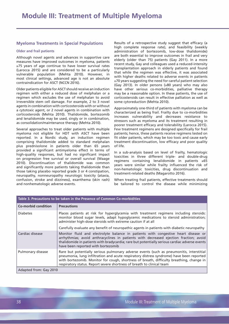
Module III: Treatment of Multiple Myeloma
Module III: Treatment of Multiple Myeloma38
Myeloma Treatments in Special Populations
Older and frail patients
Although novel agents and advances in supportive care measures have improved outcomes in myeloma, patients ≥75 years of age continue to have lower survival rates (Larocca 2015) and are considered to be a particularly vulnerable population (Mehta 2010). However, in most clinical settings, advanced age is not an absolute contraindication for ASCT (NCCN 2016).
Older patients eligible for ASCT should receive an induction regimen with either a reduced dose of melphalan or a regimen which excludes the use of melphalan to avoid irreversible stem cell damage. For example, 2 to 3 novel agents in combination with corticosteroids with or without a cytotoxic agent, or 2 novel agents in combination with corticosteroids (Mehta 2010). Thalidomide, bortezomib and lenalidomide may be used, singly or in combination, as consolidation/maintenance therapy (Mehta 2010).
Several approaches to treat older patients with multiple myeloma not eligible for HDT with ASCT have been reported. In a Nordic study, an induction regimen comprising thalidomide added to standard melphalan plus prednisolone in patients older than 65 years provided a significant antimyeloma effect in terms of high-quality responses, but had no significant impact on progression free survival or overall survival (Waage 2010). Discontinuation of thalidomide was common and significantly more patients taking thalidomide than those taking placebo reported grade 3 or 4 constipation, neuropathy, nonneuropathy neurologic toxicity (ataxia, confusion, stroke and dizziness), exanthema (skin rash) and nonhematologic adverse events.
Results of a retrospective study suggest that efficacy (a high complete response rate), and feasibility (weekly administration of bortezomib, low-dose thalidomide) are both essential to improve outcomes in frail and very elderly (older than 75) patients (Gay 2011). In a more recent study, Gay and colleagues used a reduced-intensity transplantation approach in elderly patients and found that while the regimen was effective, it was associated with higher deaths related to adverse events in patients ≥70 years suggesting the need for careful patient selection (Gay 2013). In older persons (≥80 years) who may also have other serious co-morbidities, palliative therapy may be a reasonable option. In these patients, the use of corticosteroids can result in effective palliation as well as some cytoreduction (Mehta 2010).
Approximately one-third of patients with myeloma can be characterized as being frail. Frailty due to co-morbidities increases vulnerability and decreases resistance to stressors such as myeloma and its treatment resulting in poorer treatment efficacy and tolerability (Larocca 2015). Few treatment regimens are designed specifically for frail patients; hence, these patients receive regimens tested on fit older patients, which may be too toxic and cause early treatment discontinuation, low efficacy and poor quality of life.
In a sub-analysis based on level of frailty, hematologic toxicities in three different triple- and double-drug regimens containing lenalidomide in patients ≥65 years were similar while frailty influenced the risk of non-hematologic toxicities, drug discontinuation and treatment-related deaths (Magarotto 2016).
When treating frail patients, effective treatments should be tailored to control the disease while minimizing
Table 3. Precautions to be taken in the Presence of Common Co-morbidities
Co-morbid condition Precautions
Diabetes Places patients at risk for hyperglycemia with treatment regimens including steroids: monitor blood sugar levels, adapt hypoglycemic medications to steroid administration; administer high-dose steroids with extreme caution if at all
Carefully evaluate any benefit of neuropathic agents in patients with diabetic neuropathy
Cardiac disease Monitor fluid and electrolyte balance in patients with congestive heart disease or arrhythmias; avoid anthracyclines in patients with decreased ejection fraction; avoid thalidomide in patients with bradycardia; rare but potentially serious cardiac adverse events have been reported with bortezomib
Pulmonary disease Rare but potentially serious pulmonary adverse events (such as pneumonitis, interstitial pneumonia, lung infiltration and acute respiratory distress syndrome) have been reported with bortezomib. Monitor for cough, shortness of breath, difficulty breathing, change in respiratory status. Report severe shortness of breath to clinical team
Adapted from: Gay 2010

Module III: Treatment of Multiple Myeloma
39Module III: Treatment of Multiple Myeloma
Mo
du
le II
I
toxicity and treatment discontinuation: the goal of therapy should be to keep patients asymptomatic as long as possible, preserve functional status and independence, and improve quality of life (Larocca 2015; Mehta 2010).
Patients with co-morbidities
The presence of one or more diseases co-occurring with myeloma may affect treatment decisions and outcomes. Because interactions between the co-existing illnesses can worsen the course of the disease, several precautions should be undertaken (Table 3).
A co-morbidity index was developed by physicians at the University of Freiburg to estimate the prognosis and possible therapy-associated risks for patients with myeloma. This easy to use assessment is available at: http://www.myelomacomorbidityindex.org/en_calc.html
In pregnancy
Multiple myeloma, usually considered a disease of older age, has recently become more commonly encountered in women of child-bearing age who are pregnant, most likely due to the rising median age at pregnancy (Mahmoud 2016; Lavi 2014)). Generally, core needle or excisional biopsies and bone marrow biopsy are considered safe procedures to be performed during pregnancy. Computed tomography (CT) scans and positron emission tomography (PET) scans are not advised due to the risk of radiation exposure to the fetus, while with adequate abdominal shielding, plain chest X-ray can be used. The effect of MRI exposure in the prenatal period has not been fully assessed but these should most probably be avoided during the first trimester.
Prompt therapy is advocated in pregnant patients (Mahmoud 2016). Thalidomide, lenalidomide and pomalidomide may induce birth defects and should not be taken by women of child-bearing age. Because of
the lack of data on bortezomib use in pregnancy, it too should not be used. Corticosteroids are the safest therapy and can be administered as monotherapy in women with mildly symptomatic disease until delivery. Should a more intensive therapy be required due to more aggressive disease conditions, women may be advised to terminate the pregnancy, if in the first trimester, to undertake intensive combination therapy. In rapidly progressive cases later in pregnancy, chemotherapy is advisable although standard of care has not been established (Lavi 2014).
The normal physiological changes occurring during pregnancy may influence the pharmacokinetics and pharmacodynamics of chemotherapeutic agents.
Nursing Measures Related to Commonly used Drugs in Multiple Myeloma Treatment
Single agents are infrequently used to treat myeloma. The administration of multiple drugs in combination may exacerbate known side effects of individual drugs or cause drug-drug interactions. Furthermore, while newer agents provide better disease control, none of them are free of significant toxicity, which frequently persists after completion of treatment (Boland 2013). In regards to any and all medications and chemotherapeutic agents administered, both patients and their care-givers should be provided information on:
• mechanism of action
• route and duration of administration
• possible and expected side effects
• self-care measures
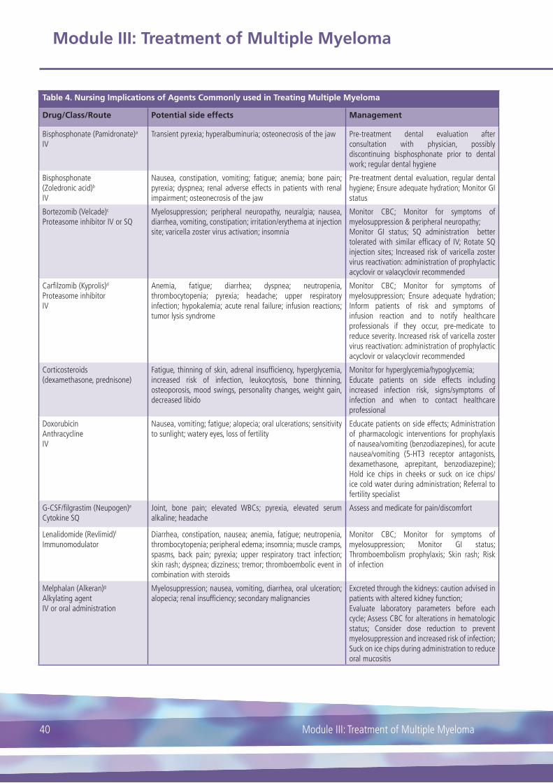
Module III: Treatment of Multiple Myeloma
Module III: Treatment of Multiple Myeloma40
Table 4. Nursing Implications of Agents Commonly used in Treating Multiple Myeloma
Drug/Class/Route Potential side effects Management
Bisphosphonate (Pamidronate)a
IVTransient pyrexia; hyperalbuminuria; osteonecrosis of the jaw Pre-treatment dental evaluation after
consultation with physician, possibly discontinuing bisphosphonate prior to dental work; regular dental hygiene
Bisphosphonate(Zoledronic acid)b
IV
Nausea, constipation, vomiting; fatigue; anemia; bone pain; pyrexia; dyspnea; renal adverse effects in patients with renal impairment; osteonecrosis of the jaw
Pre-treatment dental evaluation, regular dental hygiene; Ensure adequate hydration; Monitor GI status
Bortezomib (Velcade)c
Proteasome inhibitor IV or SQMyelosuppression; peripheral neuropathy, neuralgia; nausea, diarrhea, vomiting, constipation; irritation/erythema at injection site; varicella zoster virus activation; insomnia
Monitor CBC; Monitor for symptoms of myelosuppression & peripheral neuropathy; Monitor GI status; SQ administration better tolerated with similar effi cacy of IV; Rotate SQ injection sites; Increased risk of varicella zoster virus reactivation: administration of prophylactic acyclovir or valacyclovir recommended
Carfi lzomib (Kyprolis)d
Proteasome inhibitorIV
Anemia, fatigue; diarrhea; dyspnea; neutropenia, thrombocytopenia; pyrexia; headache; upper respiratory infection; hypokalemia; acute renal failure; infusion reactions; tumor lysis syndrome
Monitor CBC; Monitor for symptoms of myelosuppression; Ensure adequate hydration; Inform patients of risk and symptoms of infusion reaction and to notify healthcare professionals if they occur, pre-medicate to reduce severity. Increased risk of varicella zoster virus reactivation: administration of prophylactic acyclovir or valacyclovir recommended
Corticosteroids(dexamethasone, prednisone)
Fatigue, thinning of skin, adrenal insuffi ciency, hyperglycemia, increased risk of infection, leukocytosis, bone thinning, osteoporosis, mood swings, personality changes, weight gain, decreased libido
Monitor for hyperglycemia/hypoglycemia;Educate patients on side effects including increased infection risk, signs/symptoms of infection and when to contact healthcare professional
DoxorubicinAnthracyclineIV
Nausea, vomiting; fatigue; alopecia; oral ulcerations; sensitivity to sunlight; watery eyes, loss of fertility
Educate patients on side effects; Administration of pharmacologic interventions for prophylaxis of nausea/vomiting (benzodiazepines), for acute nausea/vomiting (5-HT3 receptor antagonists, dexamethasone, aprepitant, benzodiazepine); Hold ice chips in cheeks or suck on ice chips/ice cold water during administration; Referral to fertility specialist
G-CSF/fi lgrastim (Neupogen)e
Cytokine SQJoint, bone pain; elevated WBCs; pyrexia, elevated serum alkaline; headache
Assess and medicate for pain/discomfort
Lenalidomide (Revlimid)f
ImmunomodulatorDiarrhea, constipation, nausea; anemia, fatigue; neutropenia, thrombocytopenia; peripheral edema; insomnia; muscle cramps, spasms, back pain; pyrexia; upper respiratory tract infection; skin rash; dyspnea; dizziness; tremor; thromboembolic event in combination with steroids
Monitor CBC; Monitor for symptoms of myelosuppression; Monitor GI status; Thromboembolism prophylaxis; Skin rash; Risk of infection
Melphalan (Alkeran)g
Alkylating agentIV or oral administration
Myelosuppression; nausea, vomiting, diarrhea, oral ulceration; alopecia; renal insuffi ciency; secondary malignancies
Excreted through the kidneys: caution advised in patients with altered kidney function;Evaluate laboratory parameters before each cycle; Assess CBC for alterations in hematologic status; Consider dose reduction to prevent myelosuppression and increased risk of infection; Suck on ice chips during administration to reduce oral mucositis

Module III: Treatment of Multiple Myeloma
41Module III: Treatment of Multiple Myeloma
Mo
du
le II
I
Table 4. Nursing Implications of Agents Commonly used in Treating Multiple Myeloma
Plerixafor (Mozobil)h
Chemokine inhibitor SQIn conjunction with G-CSF: diarrhea, nausea, vomiting; fatigue; injection site reactions; headache, arthralgia; dizziness
Monitor GI status, bowel management
Pomalidomide (Pomalyst)i
ImmunomodulatorOral
In combination with steroids: thromboembolic events, myelosuppression, dizziness/confusion, neuropathy. Upper respiratory infection; pyrexia; diarrhea, constipation; back pain; peripheral edema; secondary malignancies; tumor lysis syndrome
Monitor for myelosuppression especially in combination with steroids; monitor GI status; monitor cardiac status; Avoid co-administration with strong CYP1A2 inhibitors
Thalidomide(Thalomid) jImmunomodulatorOral
Myelosuppression; thromboembolic events in combination with steroids; hypocalcemia; peripheral neuropathy (late effect); sleepiness, fatigue; constipation, anorexia, nausea; edema
Monitor CBC; Monitor GI status; Thromboembolism prophylaxis; Assess for peripheral neuropathy
CBC, complete blood count; GI, gastrointestinal; IV, intravenous; SQ, subcutaneous; WBCs, white blood cells;
Adapted from Miceli 2013. a Pamidronate 2009; b Zoledronic acid 2016; c Velcade 2015; d Kyprolis 2012; e Neupogen 2016; f Revlimid 2015; g Alkeran 2008; h Mozobil 2015; i Pomalyst 2015; j Thalomid 2015
Complementary Therapies
Complementary therapy can be defined as therapies used alongside, or integrated with, conventional health care (Tavares 2003). By contrast, alternative therapies are generally those used in place of conventional therapy. A study conducted in the UK estimated the use of complementary therapies by patients with hematological malignancies including myeloma to be >25% (Molassiotis 2005a). Complementary therapy has a role in managing myeloma when used as adjunct to conventional therapy and can improve quality of life and coping with the effects of the disease (Snowden 2011).
While there is a lack of clinical evidence on complementary therapy in myeloma management, some studies indicate that complementary therapy can help patients to:
• better manage symptoms
• live with altered body image
• promote relaxation
• alleviate anxiety
• reduce chemotherapy side effects
• improve sleep pattern
• reduce stress and tension
• improve well-being (Molassiotis 2005b)
The most commonly used complementary therapies by myeloma patients include acupuncture, homoeopathy, touch therapies (aromatherapy, massage and reflexology), healing and energy therapies (reiki), spiritual healing and therapeutic touch, hypnosis and hypnotherapy, herbal medicines and dietary interventions (Molassiotis 2005a). Green tea and cumin are gaining recognition as complementary therapies for myeloma (Snowden 2011).
Patients should be asked about their use of any complementary therapy, including herbal teas. Patients,
caregivers and healthcare professionals should have access to high-quality information on the role of complementary therapy in myeloma. Further, healthcare professionals should maintain updated information on complementary therapies and carefully consider these therapies before recommending them.
Future Treatment Perspectives
With the development and implementation of novel drugs, some investigators have begun to consider using novel agents without the upfront administration of ASCT as an alternative to early transplantation; the role of ASCT has become a matter of debate: should it be used upfront or as salvage treatment at the time of progression for patients initially treated with novel agents? (Moreau 2013a).
One of the most promising strategies in treatment is immunotherapy. Although interferon had been used in myeloma, its use was discontinued due to low tolerability, some benefit has been demonstrated with interferon used as maintenance therapy. Agents and combinations of agents that channel the body’s own immune system to generate an antitumor response have been evaluated in pre-clinical studies. Various drugs based on immunological mechanisms, such as monoclonal antibodies that target surface molecules of the malignant plasma cell, are currently being tested (Ocio 2014).
Greater emphasis is being placed on more personalized therapy. Biomarkers for sensitivity/resistance to particular drugs are under investigation. It could be proposed that therapeutic options should be chosen depending on the results of serial clonal evaluations, comparing the disease genome at the time of diagnosis and at relapse. The timing and the choice of a specific therapy could also be important in order to reduce the clonal diversity at diagnosis or at the time of relapse in case of the emergence of a new clone, or, on the contrary, in case of a stable clone that remains sensitive to a former regimen. (Moreau 2013a).
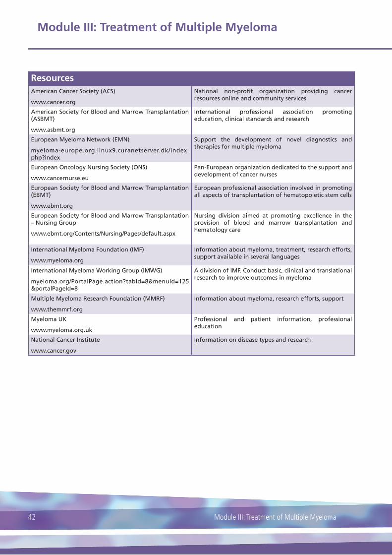
Module III: Treatment of Multiple Myeloma
Module III: Treatment of Multiple Myeloma42
ResourcesAmerican Cancer Society (ACS)
www.cancer.org
National non-profit organization providing cancer resources online and community services
American Society for Blood and Marrow Transplantation (ASBMT)
www.asbmt.org
International professional association promoting education, clinical standards and research
European Myeloma Network (EMN)
myeloma-europe.org.linux9.curanetserver.dk/index.php?index
Support the development of novel diagnostics and therapies for multiple myeloma
European Oncology Nursing Society (ONS)
www.cancernurse.eu
Pan-European organization dedicated to the support and development of cancer nurses
European Society for Blood and Marrow Transplantation (EBMT)
www.ebmt.org
European professional association involved in promoting all aspects of transplantation of hematopoietic stem cells
European Society for Blood and Marrow Transplantation – Nursing Group
www.ebmt.org/Contents/Nursing/Pages/default.aspx
Nursing division aimed at promoting excellence in the provision of blood and marrow transplantation and hematology care
International Myeloma Foundation (IMF)
www.myeloma.org
Information about myeloma, treatment, research efforts, support available in several languages
International Myeloma Working Group (IMWG)
myeloma.org/PortalPage.action?tabId=8&menuId=125&portalPageId=8
A division of IMF. Conduct basic, clinical and translational research to improve outcomes in myeloma
Multiple Myeloma Research Foundation (MMRF)
www.themmrf.org
Information about myeloma, research efforts, support
Myeloma UK
www.myeloma.org.uk
Professional and patient information, professional education
National Cancer Institute
www.cancer.gov
Information on disease types and research

Module III: Treatment of Multiple Myeloma
43Module III: Treatment of Multiple Myeloma
Mo
du
le II
I
Review Questions
1. The goals of initial therapy for myeloma are (please tick any/all that apply):
A. To provide rapid disease control and reversal of disease-related complications
B. To be well tolerated with minimal and manageable toxicity
C. Decrease the risk of early death
D. Allow successful collection of stem cells when ASCT is a therapeutic option
2. The newly developed novel agents (thalidomide, lenalidomide and bortezomib) are more effective and less toxic than conventional chemotherapeutic agents.
A. True
B. False
3. Older and frail patients may be more vulnerable to the side effects and toxicities of myeloma treatment due to (please tick any/all that apply):
A. Advanced age alone
B. Presence of co-morbidities
C. More severe disease
D. Increased risk of toxicities
4. Commonly reported side effects of thalidomide treatment include (please tick any/all that apply):
A. Peripheral neuropathy
B. Myelosuppression
C. GI complications
D. Secondary malignancies
Answers available online at www.hemcare.org

Module III: Treatment of Multiple Myeloma
Module III: Treatment of Multiple Myeloma44
References
Alkeran 2008. Available at: https://dailymed.nlm.nih.gov/dailymed/archives/fdaDrugInfo.cfm?archiveid=13952. Accessed: July 2016
Attal M, Harousseau JL, Leyvraz S et al. Maintenance therapy with thalidomide improves survival in patients with multiple myeloma. Blood 2006; 108: 3289-3294
Benboubker L, Dimopoulos MA, Dispenzieri A, et al. Lenalidomide and dexamethasone in transplant-ineligible patients with myeloma. New England Journal of Medicine 2014; 371: 906-917
Bladé J, Rosinol L, Cibeira MT, Rovira M, Carreras E. Hematopoietic stem cell transplantation for multiple myeloma beyond 2010. Blood 2010; 115: 3655-3663
Boland E, Eiser C, Ezaydi Y, Greenfield DM, Ahmedzai SH, Snowden JA. Living with advanced but stable multiple myeloma: a study oft he symptom burden and cumulative effects of disease and intensive (hematopoietic stem cell transplant-based) treatment on health-related quality of life. Journal of Pain and Symptom Management 2013; 46: 671-680
Brigle K. Treatment of Relapsed and Refractory Multiple Myeloma. In: Tariman JD and Faiman B, eds. Multiple Myeloma: a textbook for nurses, second edition. Oncology Nursing Society, 2015
Cavo, M, Rajkumar SV, Palumbo A, et al. International Myeloma Working Group consensus approach to the treatment of multiple myeloma patients who are candidates for autologous stem cell transplantation. Blood 2011; 117: 6063-6073
Duarte RF, Shaw BE, Marin P et al. Plerixafor plus granulocyte CSF can mobilize hematopoietic stem cells from multiple myeloma and lymphoma patients failing previous mobilization attempts: EU compassionate use data. Bone Marrow Transplantation 2011; 46: 52-58
Engelhardt M, Terpos E, Kleber M et al. European Myeloma Network recommendations on the evaluation and treatment of newly diagnosed patients with multiple myeloma. Haematologica 2014; 99: 232-242
European Group for Blood and Marrow Transplantation - Nurses Group (EBMT). Haematopoietic Stem Cell Mobilisation and Apheresis: A Practical Guide for Nurses and Other Allied Health Care Professionals. Available at: http://www.ebmt-swiss-ng.org/practical-guides-for-nurses.html. Accessed October 2016.
Faiman B, Miceli T, Noonan K, Lilleby K. Clinical updates in blood and marrow transplantation in multiple myeloma. Clinical Journal of Oncology Nursing 2013; 17(Suppl): 33-41
Garcia IN. High-dose therapy and stem cell transplantation. In: Tariman JD and Faiman B, eds. Multiple Myeloma: a textbook for nurses, second edition. Oncology Nursing Society, 2015
Gay F, Magarotto V, Crippa C et al. Bortezomib induction, reduced-intensity transplantation, and lenalidomide consolidation-maintenance for myeloma: updated results. Blood 2013; 122: 1376-1383
Gay F, Larocca A, Wijermans P et al. Complete response correlates with long-term progression-free and overall survival in elderly myeloma treated with nove agents: analysis of 1175 patients. Blood 2011; 117: 3025-3031
Gay F, Palumbo A. Management of disease- and treatment-related complications in patients with multiple myeloma. Medical Oncology 2010; 27: S43-S52
Gertz MA, Dingli D. How we manage autologous stem cell transplantation for patients with multiple myeloma. Blood 2014; 124: 882-890
Harousseau J-L, Dryling M. Multiple myeloma: ESMO Clinical Practice Guidelines for diagnosis, treatment and follow-up. Ann Onc 2010; 21 (Suppl 5): v155-157
Harousseau JL, Moreau P. Autologous hematopoietic stem-cell transplantation for multiple myeloma. New England Journal of Medicine 2009; 360: 2645-2654
Hulin C, Facon T, Rodon P et al. Efficacy of Melphalan and Prednisone Plus Thalidomide in Patients Older Than 75 Years With Newly Diagnosed Multiple Myeloma: IFM 01/01 Trial. Journal of Clinical Oncology 2009; 27: 3664-3670
Kumar SK, Dispenzieri A, Rajkumar SV. Continued improvement in survival in multiple myeloma: changes in early mortality and outcomes in older patients. Leukemia 2014a; 28: 1122-1128
Kumar SK, Mikhael J, Laplant B, et al. Phase 2 trial of intravenously administered plerixafor for stem cell mobilization in patients with multiple myeloma following lenalidomide-based initial therapy. Bone Marrow Transplantation 2014b; 49: 201-205
Kumar S, Flinn I, Richardson PG et al. Randomized, multicenter, phase 2 study (EVOLUTION) of combinations of bortezomib, dexamethasone, cyclophosphamide, and lenalidomide in previously untreated multiple myeloma. Blood 2012; 119: 4375-4382
Kumar S. Multiple myeloma – current issues and controversies. Cancer Treatment Reviews 2010; 36(Suppl 2): S3-S11
Kyle RA, Rajkumar SV. Criteria for diagnosis, staging, risk stratification and response assessment of multiple myeloma. Leukemia 2009; 23: 3-9
Kyprolis 2012. Available at: http://pi.amgen.com/united_
Module III: Treatment of Multiple Myeloma
45Module III: Treatment of Multiple Myeloma
Mo
du
le II
I
states/kyprolis/kyprolis_pi.pdf. Accessed: July 2016
Larocca A, Palumbo A. How I treat fragile myeloma patients. Blood 2015; 126: 2179-2185
Lavi N, Horowitz NA, Brenner B. An update on the management of hematologic malignancies in pregnancy. Women’s Health (Lond) 2014; 10: 255-266
Magarotto V, Bringhen S, Offidani M et al. Triplet vs doublet lenalidomide-containing regimens for the treatment of elderly patients with newly diagnosed multiple myeloma. Blood 2016; 127: 1102-1108
Mahmoud HK, Samra MA, Fathy GM. Hematologic malignancies during pregnancy: a review. J Adv Res 2016; 7: 589-596
Matsui W, Borrello I, Mitsiades C. Autologous stem cell transplantation and multiple myeloma cancer stem cells. Biology of Blood and Marrow Transplantation 2012; 18(Suppl 1): S27-S32
Mehta J, Cavo M, Singhal S. How I treat elderly patients with myeloma. Blood 2010; 116: 2215-2223
Miceli T, Lilleby K, Noonan K, Kurtin S, Faiman B, Mangan PA. Autologous hematopoietic stem cell transplantation for patients with multiple myeloma: an overview for nurses in community practice. Clinical Journal of Oncology Nursing 2013: 17(Suppl): 13-24
Molassiotis A, Margulies A, Fernandez-Ortega P, et al. Complementary and alternative medicine use in patients with haematological malignancies in Europe. Complementary Therapies in Clinical Practice 2005a; 11: 105–110
Molassiotis A, Fernandez-Ortega P, Pud D, et al. (2005b) Complementary and alternative medicine use in colorectal cancer patients in seven European countries. Complementary Therapies in Medicine 2005b; 13: 251–257
Moreau P, Minvielle S. Multiple myeloma: so much progress, but so many unsolved questions. Haematologica 2013a; 98; 487-489
Moreau P, Miguel JS, Ludwig H, Schouten H, Mohty M, Dimopoulos M, Dreyling M. Multiple myeloma: ESMO Clinical Practice Guidelines for diagnosis, treatment and follow-up. Ann Onc 2013b; 24 (Suppl6): vi133-vi137
Moreau P, Avet-Loiseau H, Harousseau J-L, Attal M. Current trends in autologous stem-cell transplantation for myeloma in the era of novel therapies. Journal of Clinical Oncology 2011; 29: 1898-1906
Mozobil 2015. Available at: http://products.sanofi.us/Mozobil/mozobil.html. Accessed July 2016
National Comprehensive Cancer Network (NCCN) Guidelines Version 1.2017: Multiple Myeloma. Available
at: https://www.nccn.org/professionals/physician_gls/pdf/myeloma.pdf. Accessed October 2016
Neupogen 2016. Available at: http://pi.amgen.com/united_states/neupogen/neupogen_pi_hcp_english.pdf. Accessed July 2016
Ocio EM, Richardson PG, Rajkumar SV et al. New drugs and novel mechanisms of action in multiple myeloma in 2013: a report from the International Myeloma Working Group (IMWG). Leukemia 2014; 28: 525-542
Palumbo A, Cavallo F, Gay F et al. Autologous transplantation and maintenance therapy in multiple myeloma. New England Journal of Medicine 2014; 371; 895-905
Palumbo A, Hajek R, Delforge M, et al; MM-015 Investigators. Continuous lenalidomide treatment for newly diagnosed multiple myeloma. New England Journal of Medicine 2012; 366:1759-1769
Palumbo A, Cavallo F, Hardan I et al. Melphalan/prednisone/lenidomide (MPR) versus high-dose melphalan and autologous transplantation (MEL200) in newly diagnosed multiple myeloma (MM) patients <65 years: Results of a randomized phase III study (Abstract). 53rd ASH Annual Meeting and Exposition 2011. Available at: https://ash.confex.com/ash/2011/webprogram/Paper36073.html. Accessed: July 2016
Pamidronate 2009. Available at: http://www.accessdata.fda.gov/drugsatfda_docs/label/2009/021113s008lbl.pdf. Accessed: July 2016
Pomalyst 2015. Available at: http://ir.celgene.com/releasedetail.cfm?releaseid=908418. Accessed July 2016
Revlimid 2015. Available at: http://www.revlimid.com/wp-content/uploads/full-prescribing-information.pdf. Accessed: July 2016
Roussel M, Moreau P, Huynh A et al. Bortezomib and high-dosemelphalan as conditioning regimen before autologous stem cell transplantation in patients with de novo multiple myeloma: a phase 2 study of the Intergroupe Francophone du Myelome (IFM). Blood 2010; 115: 32-37
Ruutu T. Engraftment. European Group for Blood and Marrow Transplantation (EMBT). Retrieved from: https://www.ebmt.org/Contents/Resources/Library/Slidebank/Documents /EBMT%202011%20SC%20S l ide%20Bank/1439%20Ruutu.pdf. Accessed: July 2016
Shah N, Callander N, Ganguly S, et al. Hematopoietic stem cell transplantation for multiple myeloma: gudelines from the American Society for Blood and Marrow Transplantation. Biology of Blood and Marrow Transplantation 2015; 21: 1155-1166
Module III: Treatment of Multiple Myeloma
Module III: Treatment of Multiple Myeloma46
Snowden JA, Ahmedzai SH, Ashcroft J, et al. Guidelines for supportive care in multiple myeloma 2011. British Journal of Haematology 2011; 154: 76-103
Stewart AK, Trudel S, Bahlis NJ, et al. A randomized phase 3 trial of thalidomide and prednisone as maintenance therapy after ASCT in patients with MM with a quality-of-life assessment: the National Cancer Institute of Canda Clinical Trials Group Myeloma 10 Trial. Blood 2013; 121: 1517-1523
Talamo G, Dimaio C, Abbi KKS, et al. Current role of radiation therapy for multiple myeloma. Frontiers in Oncology 2015; 5: 1-6
Tavares, M. National Guidelines for the use of complementary therapies in supportive palliative care. Available at: http://www.fih.org.uk/information_library/publications/health_guidelines/complementary.html. Accessed: July2016
Thalomid 2015. Available at: http://www.celgene.com/content/uploads/thalomid-pi.pdf. Accessed: July 2016
Velcade 2015. Available at: http://www.velcade.com/files/pdfs/velcade_prescribing_information.pdf.
Accessed: July 2016
Waage A, Gimsing P, Fayers P, et al. Melphalan and prednisone plus thalidomide or placebo in elderly patients with multiple myeloma. Blood 2010; 116: 1405-1412
Wijermans P, Schaafsma M, Termorshuizen F et al. Phase III study of the value of thalidomide added to melphalan plus prednisone in elderly patients with newly diagnosed multiple myeloma: the HOVON 49 Study. Journal of Clinical Oncology 2010;28:3160–3166
Zoledronic acid 2016. Available at: https://www.pharma.us.novartis.com/sites/www.pharma.us.novartis.com/files/Zometa.pdf. Accessed July 2016
Module IV: Comprehensive Managment of the Patient with Multiple Myeloma
47Module IV: Comprehensive Managment of the Patient with Multiple Myeloma
Mo
du
le IV
Quick Facts
• Novel therapies thalidomide and bortezomib as well as myeloma itself can cause peripheral neuropathy, a challenging adverse event that can affect quality of life and compromise optimal treatment
• Anemia, neutropenia, and thrombocytopenia are expected side effects of novel therapies; patients should be monitored closely and educated about the signs and symptoms of these side effects
• Thromboembolic events or pulmonary embolism are significant side effects of thalidomide, pomalidomide or lenalidomide when these agents are used in combination with corticosteroids or chemotherapy
• Identifying strategies tailored to individual patient needs and aimed at preventing a compromise in health-related quality of life (HRQoL) is essential to maintaining and improving HRQoL
• For caregivers, providing care is often stressful and caregivers should be assessed if their general well-being may be negatively affected by their activities
• The interdisciplinary team should recognize when myeloma is advanced and untreatable and provide discussions with the patient and caregiver regarding accepting or refusing further treatment and management of symptoms
Module IV: Comprehensive Managment of the Patient with Multiple Myeloma
Module IV: Comprehensive Managment of the Patient with Multiple Myeloma48
A. Management of the Patient with Multiple Myeloma
1. Common problems associated with myeloma treatment
2. Common problems associated with myeloma
a. Anemia
b. Bone disease
c. Renal dysfunction
B. Comorbid Conditions and late effects of treatment
1. Co-morbid conditions
2. Late effects of treatment
C. Special considerations in managing the elderly myeloma patient
D. Psychosocial Issues related to Myeloma and its Treatment
1. Health-related quality of life
2. Adherence issues
E. Supportive Care
1. Caregivers
F. Survivorship
G. End of Life Care
H. Resources
I. Review Questions
J. References

Module IV: Comprehensive Managment of the Patient with Multiple Myeloma
49Module IV: Comprehensive Managment of the Patient with Multiple Myeloma
Mo
du
le IV
Management of the Patient with Multiple Myeloma
The treatment of myeloma has dramatically changed in recent years now providing a significant improvement in response and survival rates in comparison to earlier treatment options. With few exceptions, it is difficult to categorize problems experienced by myeloma patients as being related strictly to the disease or to treatment. Providing the supportive treatment necessitated by these problems is an essential part of the overall management of myeloma.
The type and severity of problems experienced by the patient will vary based on personal and disease characteristics, the type and length of administered treatments and the patient’s history of adverse events (Kurtin 2015).
One of the challenges in addressing patient problems is reaching and maintaining a balance between alleviation of symptoms and not causing further complications through interventions. For example, safely providing relief of pain through the administration of narcotics while closely monitoring the patient for common side effects of these agents such as constipation and nausea. This means the management of patients with myeloma is complex and multifaceted. The provision of optimal care requires a comprehensive approach, which integrates healthcare professionals from a variety of clinical settings as well as caregivers and patients (Garcia 2015).
Common problems associated with multiple myeloma treatment
Alopecia can occur after the administration of certain chemotherapy agents and is a common occurrence after transplantation. Alopecia, involving the loss of head and
body hear, is a temporary condition and hair will grow back after chemotherapy is completed.
Gastrointestinal (GI) problems are common side effects of myeloma therapy. Some degree of GI toxicity following ASCT is likely to occur and can include
• oral mucositis
• esophagitis
• nausea
• vomiting
• diarrhea
Constipation is a common side effect of thalidomide and diarrhea frequently occurs in conjunction with lenalidomide (Gay 2010). Both GI complaints have been reported with bortezomib-based regimens. Oral mucositis, which results from damage to the mucosal epithelium caused by melphalan administration, can be extremely painful and lead to other problems such as weight loss, anorexia, dehydration and infection (Pallera 2004; Sonis 2004; Brown 2004). Lower incidences of grades 3 to 4 mucositis were reported in patients who held ice chips in the pockets of their cheeks for two hours following melphalan infusion (Lilleby 2006) and in patients who sucked on ice chips or rinsed with ice-cold water during chemotherapy administration (Svanberg 2010).
Myelosuppression, manifested as a reduction in red blood cells (anemia), white blood cells (neutropenia) and platelets (thrombocytopenia), is a common and expected side effect of the novel therapies used in myeloma treatment as well as a consequence of the conditioning regimen for ASCT. The severity of the side effects of anemia, neutropenia and thrombocytopenia will depend on how low the actual blood count of red blood cells, neutrophils and platelets is and the duration of the lowered blood count (Table 1).
Table 1: Severity Grading of Anemia, Neutropenia and Thrombocytopenia
Adverse Event/Measurement
Grade 1: Mild
Grade 2: Moderate
Grade 3: Severe
Grade 4: Life-threatening
Grade 5
Anemia/Hemoglobin
<LLN–10g/dL<LLN–6.2mmol/L<LLN–100g/L
<10–8g/dL<6.2–4.9mmol/L<100–80g/L
<8–6.5g/dL<4.9–4mmol/L<80–65g/L
<6.5g/dL<4mmol/L<65g/L
Death
Neutropenia/Neutrophils
<LLN–1500/mm3
<LLN–1.5x10⁹/L<1500–1000/mm3
<1.5–1.0x10⁹/L<1000–500/ mm3
<1.0–0.5x10⁹/L<500/ mm3
<0.5x10⁹/LDeath
Thrombocytopenia/Platelets
<LLN–75000/mm3
<LLN–75x109/L<75000–50000/mm3
<75–50x109/L<50000–25000/mm3
<50–25x109/L<25000/ mm3
<25x109/LDeath
LLN, lower limit of normalSource: CTCAE 2006

Module IV: Comprehensive Managment of the Patient with Multiple Myeloma
Module IV: Comprehensive Managment of the Patient with Multiple Myeloma50
Anemia can be caused by myeloma or by treatment whereas neutropenia and thrombocytopenia are more frequently caused by treatment with bortezomib (thrombocytopenia) and lenalidomide and alkylating agents (neutropenia and thrombocytopenia). Anemia and thrombocytopenia are generally treated using transfusion support (Table 2). Erythropoiesis-stimulating agents can be used in the treatment of anemia. However, these agents increase the risk of thromboembolic events. In patients with a high risk of thromboembolic events including patients previously treated with thalidomide or lenalidomide in combination with doxorubicin and corticosteroids, the use of erythropoiesis-stimulating agents should be carefully reconsidered (Schrijvers 2010).
Fatigue occurs in the majority of patients with myeloma and can be a major cause of reduced functioning and lowered quality of life (Snowden 2011). Unfortunately, fatigue is often under-recognized by healthcare professionals. The causes of fatigue are multifactorial and include treatable causes (anemia, low levels of the hormone testosterone) as well as psychological causes and treatment-related causes (sedation medications).
Osteonecrosis of the jaw, characterized by necrotic-exposed bone in the maxilla-facial region, is uncommon but potentially serious. Risk increases with prolonged bisphosphonate administration and the disorder tends to be a chronic condition. Typical features are pain and localized infection, loosening of teeth and spontaneous avulsion and soft tissue ulceration with sinus formation (Snowden 2011). Patients should receive a comprehensive dental examination and appropriate preventive dentistry before beginning bisphosphonate therapy. While on therapy, patients should maintain excellent oral hygiene and avoid invasive dental procedures (Kyle 2007).
Pain is often one of the reasons why patients with myeloma seek medical care (Snowden 2011) and it rarely occurs in isolation of other disease- or treatment-related problems. Most often, pain is accompanied by fatigue and depression. The experience and sensation of pain is highly subjective. Several measurement tools are available to better assess the location, intensity, type and experience of pain as reported by the patient (Eaton 2009; EONS 2012a; Snowden 2011).
While an impaired immune function is an important characteristic of myeloma that increases the risk of infections, neutropenia also places the patient at risk of developing infection (Kurtin 2015; Gay 2010). Prolonged use of high-dose steroids can compromise host defenses against fungal and viral infections. The risk intensity for infection varies depending on the underlying disease, the myelotoxicity of the agents administered, co-morbidities, age, prior infections and environmental exposure to micro-organisms (Bevans 2009).
Peripheral neuropathy (PN), a neurologic dysfunction of peripheral, motor, sensory and autonomic neurons (EONS 2012b), is associated with the use of both bortezomib and thalidomide and can be debilitating in some patients (Ludwig 2010). The PN associated with bortezomib is mainly sensory and painful neuropathy and is reversible in the majority of patients (Gay 2010; Richardson 2009). The PN caused by thalidomide is mainly sensory neuropathy. Generally, peripheral neuropathy is a late complication in myeloma (Tariman 2008); the risk for developing treatment-related PN increases with prolonged administration of thalidomide and doses should be decreased or thalidomide should be discontinued if symptoms worsen (Palumbo 2008).
PN can also be caused by myeloma or by co-morbidities such as diabetes mellitus or nerve compression syndromes (Snowden 2011; Terpos 2015). Other medications or conditions possibly contributing to the development of PN include: alcohol use, vitamin B12 deficiency, paraneoplastic syndrome, vascular insufficiency (Garcia 2015).
PN can impact quality of life due to physical, social and psychological effects of unrelieved neuropathic pain (Tariman 2008). There are currently no effective medications to relieve neuropathic symptoms. Assessment tools, such as the Total Neuropathy Score, are available to measure the severity of PN and should be used to provide an objective assessment of PN (Snowden 2011).
Some chemotherapy agents are known to cause pulmonary complications, which are estimated to occur in 30% to 60% of ASCT recipients (Faiman 2013). Diffuse alveolar hemorrhage, characterized by the acute onset of alveolar infiltrates and hypoxemia, is a potentially life-threatening complication. Risk factors include older age, allogeneic transplant and myeloablative conditioning (Majhail 2006). Treatment consists of corticosteroids and supportive care
Dermatologic adverse events can be a side effect of thalidomide and lenalidomide treatments. These events are generally mild to moderate and can be easily managed (Gay 2010). In rare cases, more serious toxic epidermic necrolysis and Stevens-Johnson syndrome can occur: both are potentially life-threatening conditions and require specialized interventions.
Thromboembolic events (deep vein thrombosis [DVT], or pulmonary embolism [PE]) are one of the most significant side effects associated with the use of IMiDs (immunomodulatory drugs) such as thalidomide, pomalidomide and lenalidomide, when these agents are used in combination with corticosteroids or chemotherapy (Ludwig 2010). The risk of developing thromboembolic events appears to be increased when erythropoiesis-stimulating agents are added to IMiDs. General risk factors for thromboembolic events include: myeloma
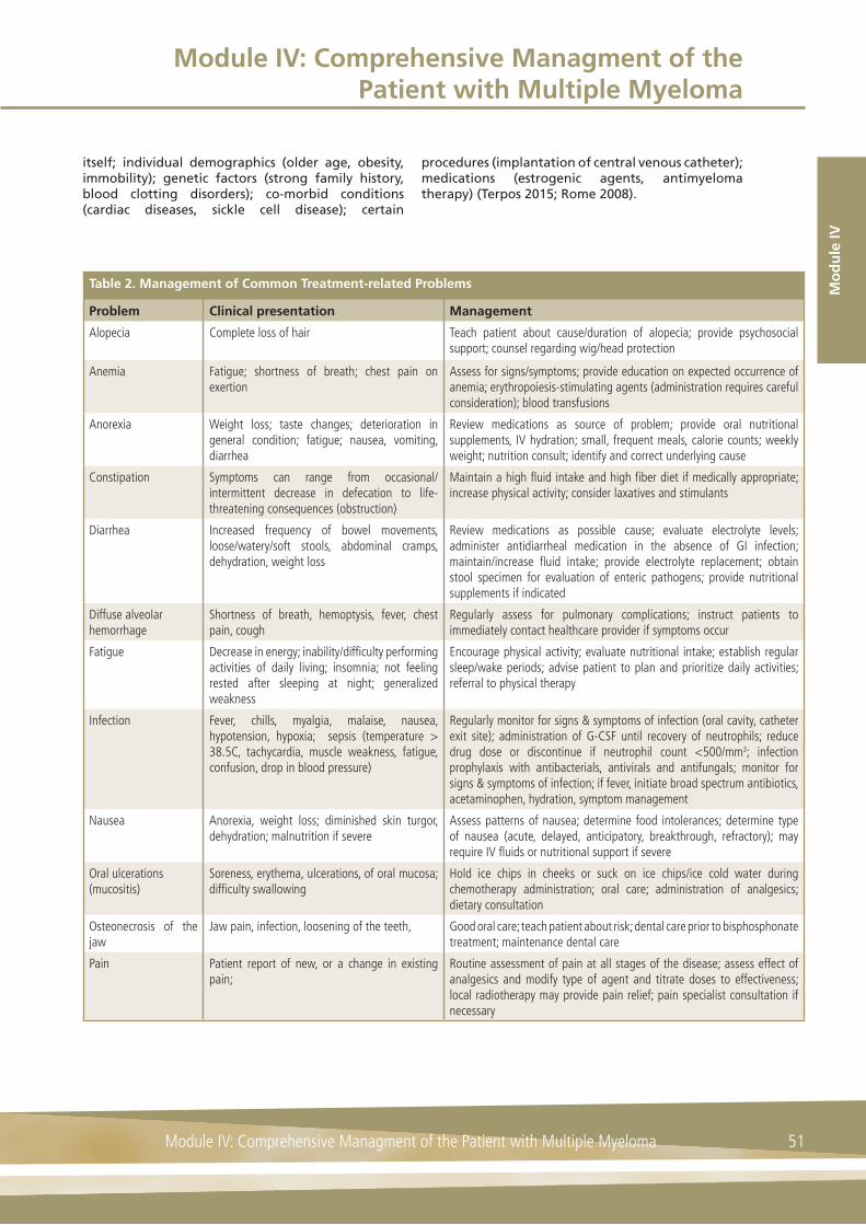
Module IV: Comprehensive Managment of the Patient with Multiple Myeloma
51Module IV: Comprehensive Managment of the Patient with Multiple Myeloma
Mo
du
le IV
itself; individual demographics (older age, obesity, immobility); genetic factors (strong family history, blood clotting disorders); co-morbid conditions (cardiac diseases, sickle cell disease); certain
procedures (implantation of central venous catheter); medications (estrogenic agents, antimyeloma therapy) (Terpos 2015; Rome 2008).
Table 2. Management of Common Treatment-related Problems
Problem Clinical presentation Management
Alopecia Complete loss of hair Teach patient about cause/duration of alopecia; provide psychosocial support; counsel regarding wig/head protection
Anemia Fatigue; shortness of breath; chest pain on exertion
Assess for signs/symptoms; provide education on expected occurrence of anemia; erythropoiesis-stimulating agents (administration requires careful consideration); blood transfusions
Anorexia Weight loss; taste changes; deterioration in general condition; fatigue; nausea, vomiting, diarrhea
Review medications as source of problem; provide oral nutritional supplements, IV hydration; small, frequent meals, calorie counts; weekly weight; nutrition consult; identify and correct underlying cause
Constipation Symptoms can range from occasional/intermittent decrease in defecation to life-threatening consequences (obstruction)
Maintain a high fl uid intake and high fi ber diet if medically appropriate; increase physical activity; consider laxatives and stimulants
Diarrhea Increased frequency of bowel movements, loose/watery/soft stools, abdominal cramps, dehydration, weight loss
Review medications as possible cause; evaluate electrolyte levels; administer antidiarrheal medication in the absence of GI infection; maintain/increase fl uid intake; provide electrolyte replacement; obtain stool specimen for evaluation of enteric pathogens; provide nutritional supplements if indicated
Diffuse alveolar hemorrhage
Shortness of breath, hemoptysis, fever, chest pain, cough
Regularly assess for pulmonary complications; instruct patients to immediately contact healthcare provider if symptoms occur
Fatigue Decrease in energy; inability/diffi culty performing activities of daily living; insomnia; not feeling rested after sleeping at night; generalized weakness
Encourage physical activity; evaluate nutritional intake; establish regular sleep/wake periods; advise patient to plan and prioritize daily activities; referral to physical therapy
Infection Fever, chills, myalgia, malaise, nausea, hypotension, hypoxia; sepsis (temperature > 38.5C, tachycardia, muscle weakness, fatigue, confusion, drop in blood pressure)
Regularly monitor for signs & symptoms of infection (oral cavity, catheter exit site); administration of G-CSF until recovery of neutrophils; reduce drug dose or discontinue if neutrophil count <500/mm3; infection prophylaxis with antibacterials, antivirals and antifungals; monitor for signs & symptoms of infection; if fever, initiate broad spectrum antibiotics, acetaminophen, hydration, symptom management
Nausea Anorexia, weight loss; diminished skin turgor, dehydration; malnutrition if severe
Assess patterns of nausea; determine food intolerances; determine type of nausea (acute, delayed, anticipatory, breakthrough, refractory); may require IV fl uids or nutritional support if severe
Oral ulcerations (mucositis)
Soreness, erythema, ulcerations, of oral mucosa; diffi culty swallowing
Hold ice chips in cheeks or suck on ice chips/ice cold water during chemotherapy administration; oral care; administration of analgesics; dietary consultation
Osteonecrosis of the jaw
Jaw pain, infection, loosening of the teeth, Good oral care; teach patient about risk; dental care prior to bisphosphonate treatment; maintenance dental care
Pain Patient report of new, or a change in existing pain;
Routine assessment of pain at all stages of the disease; assess effect of analgesics and modify type of agent and titrate doses to effectiveness; local radiotherapy may provide pain relief; pain specialist consultation if necessary
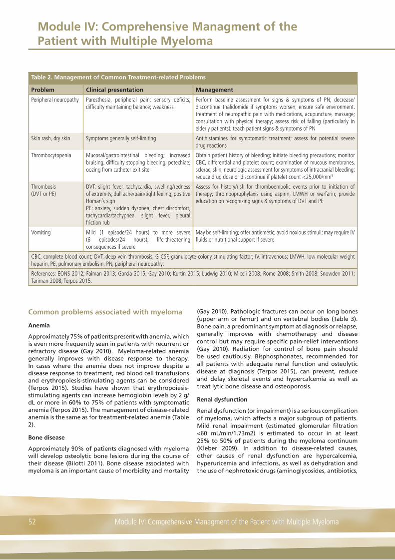
Module IV: Comprehensive Managment of the Patient with Multiple Myeloma
Module IV: Comprehensive Managment of the Patient with Multiple Myeloma52
Table 2. Management of Common Treatment-related Problems
Problem Clinical presentation Management
Peripheral neuropathy Paresthesia, peripheral pain; sensory defi cits; diffi culty maintaining balance; weakness
Perform baseline assessment for signs & symptoms of PN; decrease/discontinue thalidomide if symptoms worsen; ensure safe environment. treatment of neuropathic pain with medications, acupuncture, massage; consultation with physical therapy; assess risk of falling (particularly in elderly patients); teach patient signs & symptoms of PN
Skin rash, dry skin Symptoms generally self-limiting Antihistamines for symptomatic treatment; assess for potential severe drug reactions
Thrombocytopenia Mucosal/gastrointestinal bleeding; increased bruising, diffi culty stopping bleeding; petechiae; oozing from catheter exit site
Obtain patient history of bleeding; initiate bleeding precautions; monitor CBC, differential and platelet count; examination of mucous membranes, sclerae, skin; neurologic assessment for symptoms of intracranial bleeding; reduce drug dose or discontinue if platelet count <25,000/mm3
Thrombosis (DVT or PE)
DVT: slight fever, tachycardia, swelling/redness of extremity, dull ache/pain/tight feeling, positive Homan’s signPE: anxiety, sudden dyspnea, chest discomfort, tachycardia/tachypnea, slight fever, pleural friction rub
Assess for history/risk for thromboembolic events prior to initiation of therapy; thromboprophylaxis using aspirin, LMWH or warfarin; provide education on recognizing signs & symptoms of DVT and PE
Vomiting Mild (1 episode/24 hours) to more severe (6 episodes/24 hours); life-threatening consequences if severe
May be self-limiting; offer antiemetic; avoid noxious stimuli; may require IV fl uids or nutritional support if severe
CBC, complete blood count; DVT, deep vein thrombosis; G-CSF, granulocyte colony stimulating factor; IV, intravenous; LMWH, low molecular weight heparin; PE, pulmonary embolism; PN, peripheral neuropathy;
References: EONS 2012; Faiman 2013; Garcia 2015; Gay 2010; Kurtin 2015; Ludwig 2010; Miceli 2008; Rome 2008; Smith 2008; Snowden 2011; Tariman 2008; Terpos 2015.
Common problems associated with myeloma
Anemia
Approximately 75% of patients present with anemia, which is even more frequently seen in patients with recurrent or refractory disease (Gay 2010). Myeloma-related anemia generally improves with disease response to therapy. In cases where the anemia does not improve despite a disease response to treatment, red blood cell transfusions and erythropoiesis-stimulating agents can be considered (Terpos 2015). Studies have shown that erythropoiesis-stimulating agents can increase hemoglobin levels by 2 g/dL or more in 60% to 75% of patients with symptomatic anemia (Terpos 2015). The management of disease-related anemia is the same as for treatment-related anemia (Table 2).
Bone disease
Approximately 90% of patients diagnosed with myeloma will develop osteolytic bone lesions during the course of their disease (Bilotti 2011). Bone disease associated with myeloma is an important cause of morbidity and mortality
(Gay 2010). Pathologic fractures can occur on long bones (upper arm or femur) and on vertebral bodies (Table 3). Bone pain, a predominant symptom at diagnosis or relapse, generally improves with chemotherapy and disease control but may require specific pain-relief interventions (Gay 2010). Radiation for control of bone pain should be used cautiously. Bisphosphonates, recommended for all patients with adequate renal function and osteolytic disease at diagnosis (Terpos 2015), can prevent, reduce and delay skeletal events and hypercalcemia as well as treat lytic bone disease and osteoporosis.
Renal dysfunction
Renal dysfunction (or impairment) is a serious complication of myeloma, which affects a major subgroup of patients. Mild renal impairment (estimated glomerular filtration <60 mL/min/1.73m2) is estimated to occur in at least 25% to 50% of patients during the myeloma continuum (Kleber 2009). In addition to disease-related causes, other causes of renal dysfunction are hypercalcemia, hyperuricemia and infections, as well as dehydration and the use of nephrotoxic drugs (aminoglycosides, antibiotics,

Module IV: Comprehensive Managment of the Patient with Multiple Myeloma
53Module IV: Comprehensive Managment of the Patient with Multiple Myeloma
Mo
du
le IV
Table 3. Management of Common Disease-related Problems
Problem Clinical presentation Management
Bone disease Pathologic fractures of the long bones or vertebral bodies; bone pain
Monitor for side effects of bisphosphonates (renal dysfunction, GI complications, hypocalcemia, osteonecrosis of the jaw), obtain pre-treatment dental evaluation. For impending fracture, cord compression, plasmacytoma: physical therapy, orthopedic consultation; evaluate safety in the home; accurate and continual pain assessment, provide pain management; use spinal support if indicated; calcium and vitamin D supplements; weight-bearing exercise as tolerated
Renal dysfunction Serum creatinine ≥2 mg/dL OR creatinine clearance <30 ml/min ORe-GFR < 60 ml/min (mild dysfunction)
In newly diagnosed patients start thalidomide + bortezomib or lenalidomide; avoid aggravating factors such as contrast dye, non-steroidal anti-infl ammatory agents, dehydration; closely monitor bisphosphonatesEnsure adequate hydration; urine alkalization; treat hypercalcemia
e-GFR, estimated glomerular fi ltration rate GI, gastrointestinal;
Source: Majhail 2012;Terpos 2015
antihypertensive, lenalidomide-based regimens and non-steroidal anti-inflammatory agents).
Fast-acting treatment of myeloma using agents whose known adverse effects do not further impair renal function is required to reduce tumor burden. Bortezomib, for example, has a rapid onset of action and elimination of the agent is independent of renal clearance so that dose adjustments are not necessary in the presence of renal insufficiency (Terpos 2015). Bortezomib in combination with doxorubicin, and dexamethasone was found to result in renal responses in 62% and complete renal responses (GFR >60 ml/min) in 31% of patients (Ludwig 2009) and is recommended by the European Myeloma Network (Terpos 2015). Lenalidomide is also a feasible option for treating renal impairment with good response rates, both to disease and recovery of renal function (Terpos 2015).
Comorbid Conditions and Late Effects of Treatment
Comorbid conditions
All patients with co-morbid conditions, such as diabetes, renal failure and cardiopulmonary disease, have a higher risk of infections and should receive antibiotic prophylaxis. Diabetes, cardiac disease and several other co-morbidities can increase the risk of thrombosis and these patients should receive anti-thrombotic prophylaxis. Comorbid conditions may worsen during the cancer survivorship continuum
Late effects of treatment
Little evidence-based literature exists on late effects of treatment specific to the treatment regimens used in multiple myeloma, including hematopoietic stem cell transplantation. Patients are at risk for developing delayed complications of chemotherapy and, if applicable, from radiotherapy (Table 4).
A secondary malignancy is a devastating late complication of myeloma treatment. A dose-dependent risk of therapy-related acute myeloid leukemia and myelodysplastic syndrome has been reported after almost all alkylating agents including melphalan (Morton 2014). In the UK and US, it is now recommended that screening of cancer survivors should be started earlier (8 years after treatment), occur more frequently (annually) and involve more diagnostic modalities (Morton 2014).

Module IV: Comprehensive Managment of the Patient with Multiple Myeloma
Module IV: Comprehensive Managment of the Patient with Multiple Myeloma54
Table 4. Common Late Effects of Cancer Treatment*
System/organ Complication General Risk Factors
Immune system Infection Prolonged immunosuppressionVenous access devices
Ocular Cataracts, visual changes, retinopathySicca syndrome, xerostomiaMicrovascular retinopathy
Prolonged corticosteroid useRadiation exposure
Oral Sicca syndrome, xerostomiaCaries
Radiation exposure to head & neck
Pulmonary PneumonitisPulmonary fi brosisRestrictive lung disease
Pre-existing pulmonary diseaseRadiation exposure to chest/TBITobacco useInfectious agents
Cardiovascular CardiomyopathyCongestive heart failureArrhythmiasCoronary artery diseaseThromboembolism
Cumulative dose and combinations of cardiotoxic drugs (anthracyclines)Radiation exposure to chestOlder age at transplantPre-existing cardiovascular risk factorsChronic kidney diseaseMetabolic syndromeObesityLonger survival duration
Liver Hepatitis B and C Cumulative transfusion exposure
Renal and genitourinary
Chronic kidney diseaseBladder dysfunctionUrinary tract infectionsIncontinence
Drug exposure (calcineurin inhibitors, amphotericin, aminoglycosides)CMVHemorrhagic cystitis
Muscle and connective tissue
Myopathy, atrophyFasciitis/sclerodermaPolymyositis
Corticosteroids
Skeletal Osteonecrosis (joints)Osteoporosis
Pre-existing bone diseaseLong-term steroid useInactivity
Nervous system Peripheral neuropathyLeukoencephalopathyNeuropsychological and cognitive defi cits
Radiation exposure to headExposure to fl udarabineIntrathecal chemotherapy
Endocrine HypothyroidismAdrenal insuffi ciencyHypogonadism
Radiation exposure to head & neckLong-term steroid useStem cell transplantationRadioimmunotherapySystemic therapies: vascular endothelial growth factor inhibitors, IMiDs, retinoid inhibitors
Second cancers Solid tumorsHematologic malignanciesPTLD
Radiation exposureT-cell depletionExposure to alkylating agents or etoposide
Psychosocial and sexual
DepressionAnxietyFatigueSleep disturbancesPosttraumatic stress disorderSexual dysfunction, loss of libido
Prior psychiatric disordersHypogonadismCancer experience
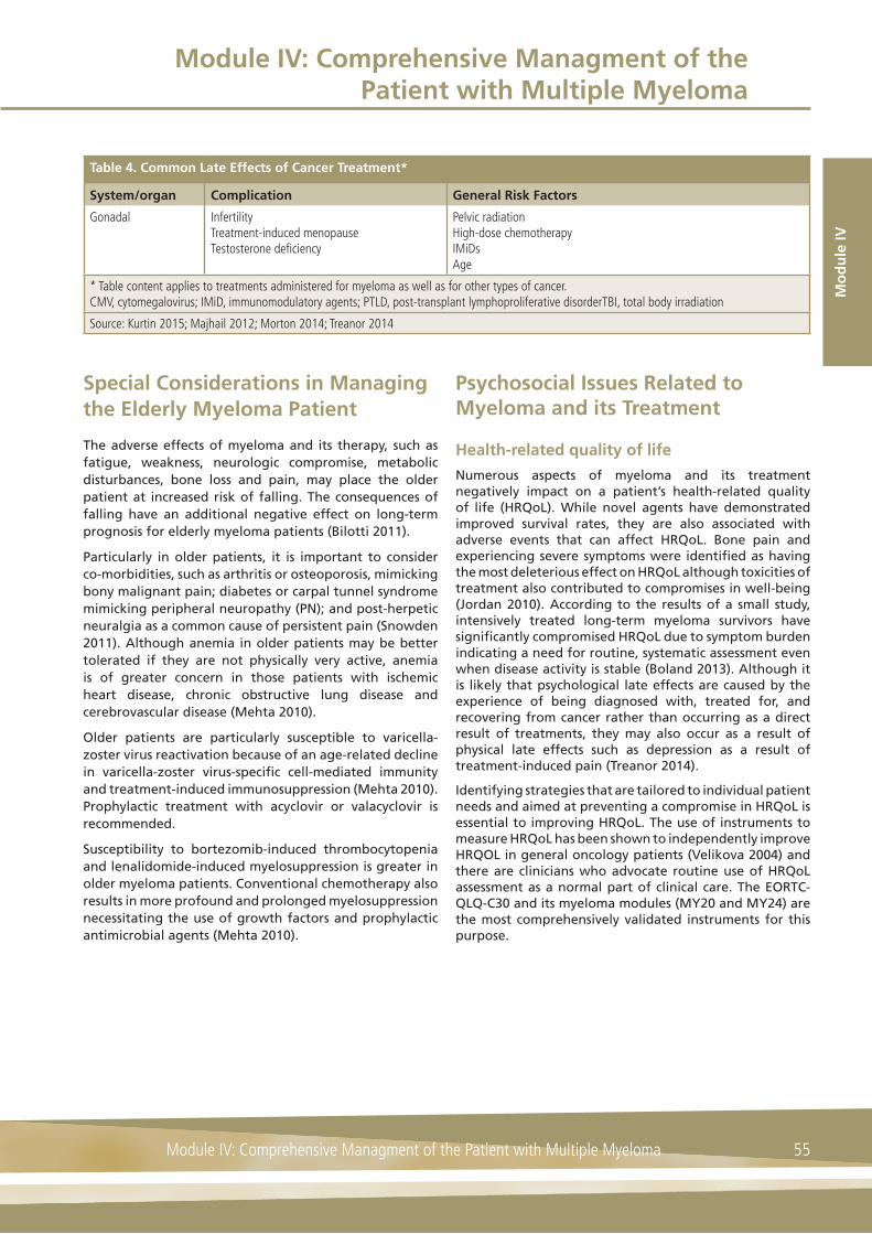
Module IV: Comprehensive Managment of the Patient with Multiple Myeloma
55Module IV: Comprehensive Managment of the Patient with Multiple Myeloma
Mo
du
le IV
Table 4. Common Late Effects of Cancer Treatment*
System/organ Complication General Risk Factors
Gonadal InfertilityTreatment-induced menopauseTestosterone defi ciency
Pelvic radiationHigh-dose chemotherapyIMiDsAge
* Table content applies to treatments administered for myeloma as well as for other types of cancer.CMV, cytomegalovirus; IMiD, immunomodulatory agents; PTLD, post-transplant lymphoproliferative disorderTBI, total body irradiation
Source: Kurtin 2015; Majhail 2012; Morton 2014; Treanor 2014
Special Considerations in Managing the Elderly Myeloma Patient
The adverse effects of myeloma and its therapy, such as fatigue, weakness, neurologic compromise, metabolic disturbances, bone loss and pain, may place the older patient at increased risk of falling. The consequences of falling have an additional negative effect on long-term prognosis for elderly myeloma patients (Bilotti 2011).
Particularly in older patients, it is important to consider co-morbidities, such as arthritis or osteoporosis, mimicking bony malignant pain; diabetes or carpal tunnel syndrome mimicking peripheral neuropathy (PN); and post-herpetic neuralgia as a common cause of persistent pain (Snowden 2011). Although anemia in older patients may be better tolerated if they are not physically very active, anemia is of greater concern in those patients with ischemic heart disease, chronic obstructive lung disease and cerebrovascular disease (Mehta 2010).
Older patients are particularly susceptible to varicella-zoster virus reactivation because of an age-related decline in varicella-zoster virus-specific cell-mediated immunity and treatment-induced immunosuppression (Mehta 2010). Prophylactic treatment with acyclovir or valacyclovir is recommended.
Susceptibility to bortezomib-induced thrombocytopenia and lenalidomide-induced myelosuppression is greater in older myeloma patients. Conventional chemotherapy also results in more profound and prolonged myelosuppression necessitating the use of growth factors and prophylactic antimicrobial agents (Mehta 2010).
Psychosocial Issues Related to Myeloma and its Treatment
Health-related quality of life
Numerous aspects of myeloma and its treatment negatively impact on a patient’s health-related quality of life (HRQoL). While novel agents have demonstrated improved survival rates, they are also associated with adverse events that can affect HRQoL. Bone pain and experiencing severe symptoms were identified as having the most deleterious effect on HRQoL although toxicities of treatment also contributed to compromises in well-being (Jordan 2010). According to the results of a small study, intensively treated long-term myeloma survivors have significantly compromised HRQoL due to symptom burden indicating a need for routine, systematic assessment even when disease activity is stable (Boland 2013). Although it is likely that psychological late effects are caused by the experience of being diagnosed with, treated for, and recovering from cancer rather than occurring as a direct result of treatments, they may also occur as a result of physical late effects such as depression as a result of treatment-induced pain (Treanor 2014).
Identifying strategies that are tailored to individual patient needs and aimed at preventing a compromise in HRQoL is essential to improving HRQoL. The use of instruments to measure HRQoL has been shown to independently improve HRQOL in general oncology patients (Velikova 2004) and there are clinicians who advocate routine use of HRQoL assessment as a normal part of clinical care. The EORTC-QLQ-C30 and its myeloma modules (MY20 and MY24) are the most comprehensively validated instruments for this purpose.

Module IV: Comprehensive Managment of the Patient with Multiple Myeloma
Module IV: Comprehensive Managment of the Patient with Multiple Myeloma56
Psychological late effects may also occur as a result of physical late effects such as depression as a result of treatment-induced pain (Treanor 2014). Following ASCT, patients often describe feeling “let down” and may express anxiety regarding “what comes next” (Garcia 2015). Symptoms of depression are reported by approximately 20% to 25% of patients undergoing cancer treatment (Brown 2009). The symptoms of depression are often overlooked because they sometimes mirror symptoms of cancer treatment. Depression may adversely affect physical health, may increase symptom-related fatigue and distress, and has been associated with a higher incidence of suicide (Garcia 2015).
Treatment and treatment side effects, stress, fatigue, changes in body image, co-morbidities and a variety of other disease- or treatment-related factors can lead to sexual dysfunction.
Cancer survivors may experience social effects relating to their cancer experience such as changes in relationships, and/or employment or financial status (Treanor 2014). Cancer survivors are more likely than the general population to be unemployed and these patients have more difficulty reintegrating into work life, experience
discrimination, fear losing benefits and experience disease-associated stigma (Treanor 2014)
Adherence issues
To achieve maximal benefit from most treatments, patients need to initiate and continue the treatment as prescribed. The reasons for nonadherence are multifactorial and include patient-, physician-, medication and system-related variables (Hershman 2016).
The most common reason for nonadherence is toxicity of the prescribed treatment. The novel agents (lenalidomide, thalidomide and bortezomib) have contributed to increased response rates and increased survival time but cause side effects that, although predictable and manageable, can be life-threatening and interfere with adherence (Bertolotti 2008). Several methods have been attempted, such as phone consultations by a nurse, daily text messaging reminders and written communication, to increase medication adherence. Although studies have indicated the effectiveness of these interventions, the extent and the duration of the improvements in adherence have not been shown.
While adherence rates were reportedly high in one study of myeloma patients taking an oral chemotherapy regimen (cyclophosphamide, thalidomide and dexamethasone), there was potential for non-intentional nonadherence due to deficits in knowledge on the drug, such as the reason for taking the drug and how to take the drug (Arber 2015). Strategies to improve adherence with oral chemotherapy agents include:
Supportive Care
Myeloma is a chronic disease with no known and effective curative treatment. The disease trajectory involves multiple periods of remission and relapse and treatment is likely to be administered from the time of diagnosis until the time of death. As functional decline is associated with a loss of independence and decreased quality of life, the maintenance of independence is a primary goal for myeloma survivors (Kurtin 2015). Preserving quality of life and independent functioning requires maintaining mobility, effective pain control, preventing falls or injuries, optimal sleep and rest, adequate nutrition and medication support (Kurtain 2015) among other factors.
Caregivers
Both patients and caregivers need to adapt to a diagnosis of myeloma, how it affects the individual patient and what changes in lifestyle will be necessary to increase the success in living with the disease. Caregivers are challenged to assimilate complex information, often very rapidly, and develop skills to provide assistance with activities of daily living, with activities typically considered to be within the realm of nursing care or medical treatment, and to provide emotional support during a difficult period (Table 5). Caregivers may be relatives of the patient, friends, acquaintances or volunteers (Kurtin 2013) and their number and presence will vary depending on the patient’s condition.
A caregiver plays an essential role in attaining and maintaining optimal outcomes throughout the disease process. While providing support, the caregiver also struggles with her or his own feelings about the diagnosis and the uncertainty about future events and how she or he will cope with them. Healthcare professionals need to understand the role of the caregiver, the dynamics of the caregiver-patient relationship and causes of real and potential caregiver stress (Kurtin 2013).
Reminder triggers Pill diaries, pill boxes, patient calendars or spreadsheets, blister packs, cellular phones/alarms, electronic pill bottles, medication electronic monitoring system
Education Provide education on when and how to take the medication, indications, potential side effects, drug interactions
Adapted from: Schneider 2011

Module IV: Comprehensive Managment of the Patient with Multiple Myeloma
57Module IV: Comprehensive Managment of the Patient with Multiple Myeloma
Mo
du
le IV
Table 5. Key elements of the caregiver role
Direct care activities Monitor and report treatment side effectsProcure and administer medicationsMake decisions on when to call a healthcare providerMake decisions on administering “as needed” medicationsPerform technical procedures (dressing changes, IV and pump care)
Indirect care activities Serve as contact person for healthcare providerServe as contact person for family, friendsServe as patient advocateManage household Manage medical and insurance forms and billsOrganize transportation
Emotional support Balance medical expectations while maintaining hopeActive listenerProvide reassurance, emotional comfort
Adapted from: Kurtin 2013
Self-management in the home setting is becoming increasingly more prevalent as the length of time in hospital decreases. Hence, providing patients and their caregivers with clear instructions on recognizing and managing treatment side effects is important to optimize outcomes.
Caregivers are particularly vulnerable to the high demands of caring for someone with myeloma (Molassiotis 2011). The demands of providing care produce changes in role, emotional well-being, social activities and employment. The level of care required by the patient strongly influences the caregiver’s life and, possibly, health effects. Caregivers often require, but do not receive, the respite, health care, psychosocial and financial assistance they need to meet the many needs of the patient.
Providing care is a stressful undertaking; in terms of preventative care, assessment should be made of the degree to which the caregiver’s life and health may be negatively affected and recommendations provided on interventions to reduce any negative repercussions of caretaking (Bevans 2012).
Interventions to support caregivers
• Individualize caregiver education
• Provide consistent and clear information, reinforce important concepts
• Provide written material
• Suggest maintaining a diary or log of treatments, blood counts, transfusions and side effects, the treatment administered and the outcome
• Encourage respite from caring for the patient and continuation of hobbies
• Encourage stress management practices such as walking and meditating
• Suggest hospital and community resources to support coping
• Provide criteria and procedure for emergency situations
• Encourage caregiver to seek help and/or assistance if needed
Survivorship
The 5-year relative survival for myeloma was 26.3% in 1975 versus 49.6% in 2008 with a 0.8% decline in death rates over 2004 to 2013 (SEER 2016).
Cancer survivorship is now defined as the period from the time of diagnosis until the end of life (NCI 2016). An Institute of Medicine report states, “Optimal survivorship care is characterized by an organized plan for follow up that is shared with patients so they can take responsibility for their care” (Hewitt 2006, p. 194). According to this report, the essential components of survivorship care are:
• Prevention and detection of new cancers and recurrent cancer
• Surveillance for cancer spread, recurrence or second cancers
• Intervention for consequences of cancer and its treatment
Coordination between specialists and primary care providers to ensure that all of the survivor’s health needs are met (Hewitt 2006)
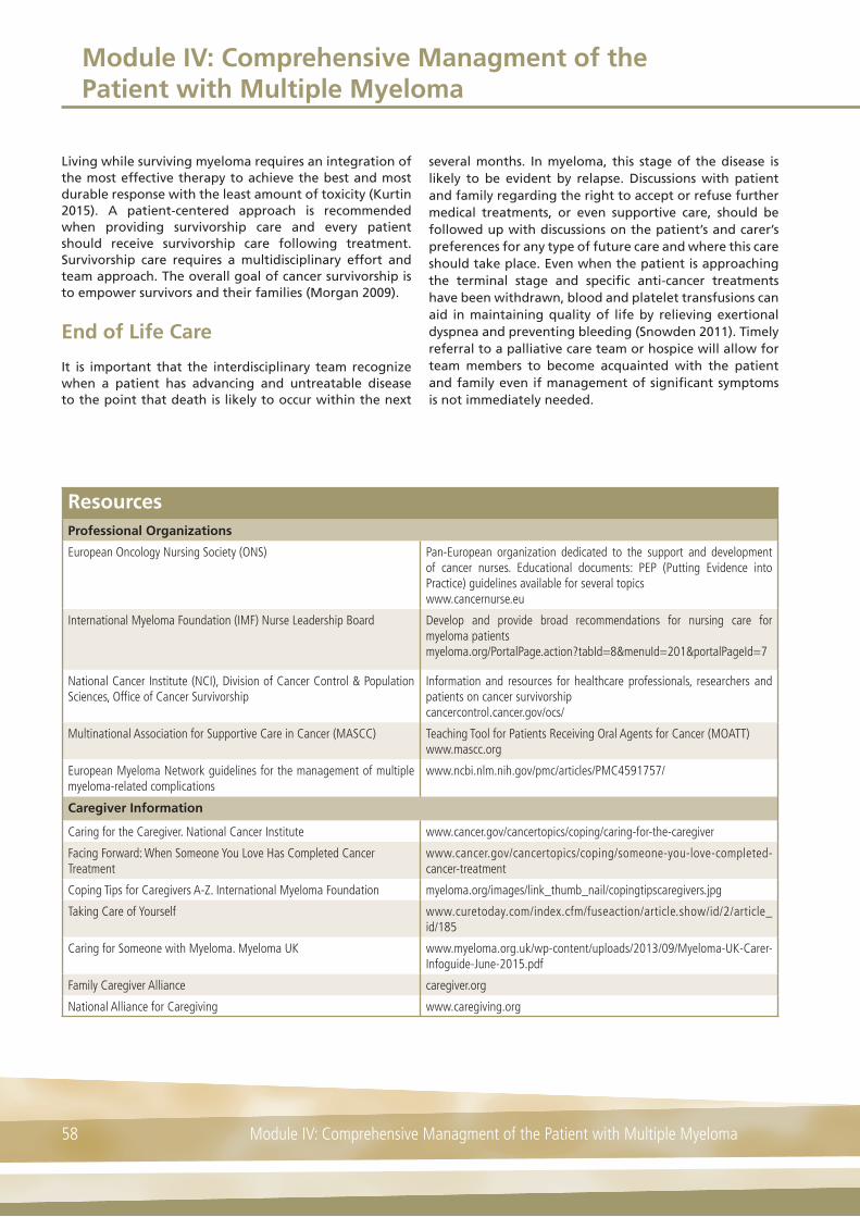
Module IV: Comprehensive Managment of the Patient with Multiple Myeloma
Module IV: Comprehensive Managment of the Patient with Multiple Myeloma58
Living while surviving myeloma requires an integration of the most effective therapy to achieve the best and most durable response with the least amount of toxicity (Kurtin 2015). A patient-centered approach is recommended when providing survivorship care and every patient should receive survivorship care following treatment. Survivorship care requires a multidisciplinary effort and team approach. The overall goal of cancer survivorship is to empower survivors and their families (Morgan 2009).
End of Life Care
It is important that the interdisciplinary team recognize when a patient has advancing and untreatable disease to the point that death is likely to occur within the next
several months. In myeloma, this stage of the disease is likely to be evident by relapse. Discussions with patient and family regarding the right to accept or refuse further medical treatments, or even supportive care, should be followed up with discussions on the patient’s and carer’s preferences for any type of future care and where this care should take place. Even when the patient is approaching the terminal stage and specific anti-cancer treatments have been withdrawn, blood and platelet transfusions can aid in maintaining quality of life by relieving exertional dyspnea and preventing bleeding (Snowden 2011). Timely referral to a palliative care team or hospice will allow for team members to become acquainted with the patient and family even if management of significant symptoms is not immediately needed.
ResourcesProfessional Organizations
European Oncology Nursing Society (ONS) Pan-European organization dedicated to the support and development of cancer nurses. Educational documents: PEP (Putting Evidence into Practice) guidelines available for several topicswww.cancernurse.eu
International Myeloma Foundation (IMF) Nurse Leadership Board Develop and provide broad recommendations for nursing care for myeloma patientsmyeloma.org/PortalPage.action?tabId=8&menuId=201&portalPageId=7
National Cancer Institute (NCI), Division of Cancer Control & Population Sciences, Offi ce of Cancer Survivorship
Information and resources for healthcare professionals, researchers and patients on cancer survivorshipcancercontrol.cancer.gov/ocs/
Multinational Association for Supportive Care in Cancer (MASCC) Teaching Tool for Patients Receiving Oral Agents for Cancer (MOATT)www.mascc.org
European Myeloma Network guidelines for the management of multiple myeloma-related complications
www.ncbi.nlm.nih.gov/pmc/articles/PMC4591757/
Caregiver Information
Caring for the Caregiver. National Cancer Institute www.cancer.gov/cancertopics/coping/caring-for-the-caregiver
Facing Forward: When Someone You Love Has Completed CancerTreatment
www.cancer.gov/cancertopics/coping/someone-you-love-completed-cancer-treatment
Coping Tips for Caregivers A-Z. International Myeloma Foundation myeloma.org/images/link_thumb_nail/copingtipscaregivers.jpg
Taking Care of Yourself www.curetoday.com/index.cfm/fuseaction/article.show/id/2/article_id/185
Caring for Someone with Myeloma. Myeloma UK www.myeloma.org.uk/wp-content/uploads/2013/09/Myeloma-UK-Carer-Infoguide-June-2015.pdf
Family Caregiver Alliance caregiver.org
National Alliance for Caregiving www.caregiving.org

Module IV: Comprehensive Managment of the Patient with Multiple Myeloma
59Module IV: Comprehensive Managment of the Patient with Multiple Myeloma
Mo
du
le IV
ResourcesPatient Information
Living well with myeloma: Your essential guide. Myeloma UK www.myeloma.org.uk/wp-content/uploads/2013/09/Myeloma-UK-Living-well-with-Myeloma-Essential-Guide.pdf
Myeloma: Your essential guide. Myeloma UK www.myeloma.org.uk/wp-content/uploads/2013/09/Myeloma-UK-Myeloma-Essential-Guide.pdf
Myeloma Patients Europe (MPE). An umbrella organization of multiple myeloma patient groups and associations from across Europe. Information available in several languages
www.mpeurope.org/
National Coalition for Cancer Survivorship www.canceradvocacy.org
OncoLink OncoLife Survivorship Care Plan www.oncolink.com/oncolife
Stupid Cancer www.stupidcancer.org
Symptom Assessment Tools
Tool Source
Peripheral Neuropathy Questionnaire Colson K, Doss DS, Swift R, Tariman J. Thomas TE. Bortezomib, a newly approved proteasome inhibitor for the treatment of multiple myeloma: nursing implications. Clinical Journal of Oncology Nursing 2004; 8: 473-480
Grading System for Adverse Effects of Cancer Treatment Trotti A, Colevas AD, Setser A, et al. CTCAE v3.0: development of a comprehensive grading system for the adverse effects of cancer treatment. Seminars in Radiation Oncology 2003; 13: 176–181.
Grading System for Mucositis World Health Organization (WHO)www.researchgate.net/fi gure/264009927_fi g2_Table-1-World-Health-Organization-Oral-Mucositis-Assessment-Scale
Oral Mucositis Guidelines European Oncology Nursing Society. Guidelines incorporate the latest developments in oral mucositis into standardized patient care.www.cancernurse.eu/documents/EONSClinicalGuidelinesSection4-en.pdf
Baseline Assessment for Peripheral Neuropathy European Oncology Nursing Society. Peripheral Neuropathy: Improving symptom management in cancer care through evidence based practice. Euro PEP (Putting Evidence into Practice) Program. Available in several languageswww.cancernurse.eu/documents/EONSPEPPeripheralNeuropathyEnglish.pdf
Leeds Assessment of Neuropathic Symptoms and Signs (LANSS) Scale (neuropathic pain)
www.endoexperience.com/documents/Apx4_LANSS.pdfBennett MI, Attal N, Backonja MM, et al. Using screening tools to identify neuropathic pain.Pain 2007: 127: 199–203
Brief Pain Inventory (short form) www.npcrc.org/fi les/news/briefpain_short.pdf
National Initiative on Pain ControlPain Assessment Scales
www.painedu.org/Downloads/NIPC/Pain%20Assessment%20Scales.pdf
Numeric Pain Intensity Scale www.partnersagainstpain.com/printouts/A7012AS2.pdf.
Impact of Cancer Scale Zebrack BJ, Ganz PA, Bernaards CA, Petersen L, Abraham L. Assessing the impact of cancer: development of a new instrument for long-term survivors. Psychooncology. 2006; 15: 407-421
Concerns Checklist National Cancer Survivorship Initiative- Concerns Checklist; http://www.ncsi.org.uk/wp-content/uploads/MAC13689_Identifyingconcerns_Pad_v3.pdf
Module IV: Comprehensive Managment of the Patient with Multiple Myeloma
Module IV: Comprehensive Managment of the Patient with Multiple Myeloma60
Review Questions
1. Signs and symptoms of peripheral neuropathy include (please tick any/all that apply):
A. Paresthesia
B. Peripheral pain
C. Infection
D. Difficulty maintaining balance
2. The risk of developing thromboembolic events appears to be increased when erythropoiesis-stimulating agents are given with some novel agents.
Answer:
A. True
B. False
3. Anemia, neutropenia, and thrombocytopenia are expected side effects of novel therapies; patients should be monitored closely and educated about the signs and symptoms of these side effects which include (please tick any/all that apply):
A. Fatigue
B. Fever, chills, malaise
C. Mucosal/gastrointestinal bleeding
D. Shortness of breath
4. Patients should be made aware of potential changes in health-related quality of life including those caused by treatment or by the psychologic effects of myeloma
Answers:
A. True
B. False
5. Caregivers often experience stress related to their caregiving activities; interventions to support caregivers are (please tick any/all that apply):
A. Individualize education provided to caregivers
B. Encourage respite from caring activities
C. Suggest hospital and community resources to support coping
D. Encourage measures for stress reduction
Answers available online at www.hemcare.org

Module IV: Comprehensive Managment of the Patient with Multiple Myeloma
61Module IV: Comprehensive Managment of the Patient with Multiple Myeloma
Mo
du
le IV
References
Arber A, Williams P, Lemanska A, Faithfull S. Do patients on oral chemotherapy have sufficient knowledge for optimal adherence? A mixed methods study. European Journal of Cancer Care 2015; doi: 10.1111/ecc.12413
Bertolotti P, Bilotti E, Colson K, et al. Management of side effects of novel therapies for multiple myeloma: consensus statements developed by the International Myeloma Foundation’s Nurse Leadership Board. Clinical Journal of Oncology Nursing 2008; 12: 9-12
Bevans MF, Sternberg EM. Caregiving burden, stress, and health effects among family caregivers of adult cancer patients. Journal of the American Medical Association 2012; 307: 398-403
Bevans M, Tierney, DK, Bruch, et al. Hematopoietic stem cell transplantation nursing: A practice variation study [Online exclusive]. Oncology Nursing Forum 2009, 36, E317–E325. doi:10.1188/09.ONF.E317-E325
Bilotti E, Faiman BM, Richards TA, Tariman JD, Miceli TS, Rome SI. Survivorship care guidelines for patients living with multiple myeloma: consensus statements of the International Myeloma Foundation Nurse Leadership Board. Clinical Journal of Oncology Nursing 2011; 15(Suppl): 5-8
Boland E, Eiser C, Ezaydi Y, Greenfield DM, Ahmedzai SH, Snowden JA. Living with advanced but stable multiple myeloma: a study of the symptom burden and cumulative effects of disease and intensive (hematopoietic stem cell transplant-based) treatment on health-related quality of life. Journal of Pain and Symptom Management 2013; 46: 671-680
Brown C, Wingard J. Clinical consequences of oral mucositis. Seminars in Oncology Nursing 2004; 20: 16-21
Common Terminology Criteria for Adverse Events v3.0 (CTCAE). Cancer Therapy Evaluation Program, 2006. Available at: https://ctep.cancer.gov/protocoldevelopment/electronic_applications/docs/ctcaev3.pdf. Accessed: October 2016
Eaton, LH. Pain. In L.H. Eaton & J.M. Tipton (Eds.), Putting evidence into practice: Improving oncology patient outcomes. 2009, (pp. 215–221). Pittsburgh, PA: Oncology Nursing Society
European Oncology Nursing Society (EONS). Peripheral Neuropathy: Improving symptom management in cancer care through evidence based practice. 2012. Available at: http://www.cancernurse.eu/documents/EONSPEPPeripheralNeuropathyEnglish.pdf. Accessed: July 2016
European Oncology Nursing Society (EONS). Pain: Improving symptom management in cancer care through
evidence based practice. 2012. Available at: http://www.cancernurse.eu/documents/EONSPEPPainEnglish.pdf. Accessed: July 2016
Faiman B, Miceli T, Noonan K, Lilleby K. Clinical updates in blood and marrow transplantation in multiple myeloma. Clinical Journal of Oncology Nursing 2013; 17(Suppl): 33-41
Garcia IN. High-dose therapy and stem cell transplantation. In: Tariman JD and Faiman B, eds. Multiple Myeloma: a textbook for nurses, second edition. Oncology Nursing Society, 2015
Gay F, Palumbo A. Management of disease- and treatment-related complications in patients with multiple myeloma. Medical Oncology 2010; 27: S43-S52
Hershman DL. Sticking to it: improving outcomes by increasing adherence. Journal of Clinical Oncology 2016; doi: 10.1200/JCO.2016.67.7336
Hewitt M, Greenfield S, Stovall E. (Eds.). From cancer patient to cancer survivor: Lost in transition. Available at: http://www.nap.edu/catalog/11468/from-cancer-patient-to-cancer-survivor-lost-in-transition. Accessed: July 2016
Jordan K, Ishak JK, Lewis P, et al. Determinants of global QOL and physical and social functionality in multiple myeloma. Proceedings of the 52nd Annual Meeting of the American Society of Hematology (ASH), Orlando, FL; USA. Blood 2010; 116: Abstract 934
Kleber M, Ihorst G, Deschler B, et al. Detection of renal impairment as one specific comorbidity factor in multiple myeloma: multicenter study in 198 consecutive patients. European Journal of Haematology 2009; 83:519-527
Kurtin S. Living with Multiple Myeloma. In: Tariman JD and Faiman B, eds. Multiple Myeloma: a textbook for nurses, second edition. Oncology Nursing Society, 2015
Kurtin S, Lilleby K, Spong J. Caregivers of multiple myeloma survivors. Clinical Journal of Oncology Nursing 2013; 17 (Suppl): 25-30
Kyle RA, Yee GC, Somerfield MR, et al. American Society of Clinical Oncology 2007 Clinical Practice Guideline Update on the role of bisphosphonates in multiple myeloma. Journal of Clinical Oncology 2007; 25: 2464-2472
Lilleby K, Garcia P, Gooley T, et al. A prospective, randomized study of cryotherapy during administration of high-dose melphalan to decrease the severity and duration of oral mucositis in patients with multiple myeloma undergoing autologous peripheral blood stem cell transplantation. Bone Marrow Transplantation 2006; 37: 1031-1035
Ludwig H, Beksac M, Blade J, et al. Current multiple myeloma treatment strategies with novel agents: a European perspective. The Oncologist 2010; 15: 6-25
Module IV: Comprehensive Managment of the Patient with Multiple Myeloma
Module IV: Comprehensive Managment of the Patient with Multiple Myeloma62
Ludwig H, Adam Z, Greil R, et al. Reversal of acute renal impairment by bortezomib-doxorubicin-dexamethasone (BDD) in multiple myeloma (MM). Results from a phase II study [abstract 385]. Haematologica 2009; 94 (Suppl 2):154
Majhail NS, Rizzo D, Lee SJ, et al. Recommended screening and preventive practices flor long-term survivors after hematopoietic cell transplantation. Hematology/Oncology and Stem Cell Therapy 2012; 5: 1-30
Majhail NS, Parks K, Defor TE, Weisdorf DF. Diffuse alveolar hemorrhage and infection-associated alveolar hemorrhage following hematopoietic stem cell transplantation: related and high-risk clinical syndromes. Biology of Blood and Marrow Transplantation 2006; 12: 1038-1046
Mehta J, Cavo M, Singhal S. How I treat elderly patients with myeloma. Blood 2010; 116: 2215-2223.
Miceli T, Colson K, Gavino M, Lilleby K. Myelosuppression associated with novel therapies in patients with multiple myeloma: consensus statement of the IMF Nurse Leadership Board. Clinical Journal of Oncology Nursing 2008; 12 (Suppl): 13-20
Molassiotis A, Wilson B, Blair S, Howe T, Cavet J. Living with multiple myeloma: experiences of patients and their informal caregivers. Supportive Care in Cancer 2011; 19: 101-111
Morgan MA. Cancer survivorship: history, quality-of-life issues, and the evolving multidisciplinary approach to implementation of cancer survivorship care plans. Oncology Nursing Forum 2009; 36: 429-436
Morton LM, Swerdlow AJ, Schaapveld M, Ramadan S, Hodgson DC, Radford J, van Leeuwen FE. Current knowledge and future research directions in treatment-related second primary malignancies. European Journal of Cancer 2014; 12 (Suppl): 5-17
National Cancer Institute (NCI). Office of Cancer Survivorship. Survivorship Definitions. Available at: http://cancercontrol.cancer.gov/ocs/statistics/definitions.html. Accessed: July 2016
Pallera, AM, Schwartzberg, LS. Managing the toxicity of hematopoietic stem cell transplant. Journal of Supportive Oncology 2004; 2: 223–237
Palumbo A, Facon T, Sonneveld P et al. Thalidomide for treatment of multiple myeloma: 10 years later. Blood 2008; 111: 3968 –3977
Richardson PG, Sonneveld P, Schuster MW et al. Reversibility of symptomatic peripheral neuropathy with bortezomib in the phase III APEX trial in relapsed multiple myeloma: Impact of a dose-modification guideline. British Journal of Haematology 2009; 144: 895–903
Rome S, Doss D, Miller K, Westphal J. Thromboembolic events associated with novel therapies in patients with multiple myeloma: consensus statement of the IMF Nurse Leadership Board. Clinical Journal of Oncology Nursing 2008; 12 (Suppl): 21-28
Schneider SM, Hess K, Gosselin T. Interventions to promote adherence with oral agents. Seminars in Oncology Nursing 2011; 27: 133-141
Schrijvers D, De Samblanx H, Roila F. Erythropoiesis-stimulating agents in the treatment of anemia in cancer patients: ESMO Clinical Practice Guidelines for use. Annals of Oncology 2010; 21(Suppl 5): v244-v247
SEER Cancer Statistics Factsheets: Myeloma. National Cancer Institute. Bethesda, MD,http://seer.cancer.gov/statfacts/html/mulmy.html
Smith LC, Bertolotti P, Curran K, Jenkins B. Gastrointestinal side effects associated with novel therapies in patients with multiple myeloma: consensus statement of the IMF Nurse Leadership Board. Clinical Journal of Oncology Nursing 2008; 12 (Suppl): 37-51
Snowden JA, Ahmedzai SH, Ashcroft J, et al. Guidelines for supportive care in multiple myeloma 2011. British Journal of Haematology 2011; 154: 76-103
Sonis ST, Elting LS, Keefe D, et al. Perspectives on cancer therapy-induced mucosal injury: pathogenesis, measurement, epidemiology and consequences for patients. Cancer 2004; 100: 1995–2025
Svanberg A, Öhrn K, Birgegård G. Oral cryotherapy reduces mucositis and improves nutrition – a randomised controlled trial. Journal of Clinical Nursing 2010; 19: 2146-2151
Tariman JD, Love G, McCullagh E, Sandifer S. Peripheral neuropathy associated with novel therapies in patients with multiple myeloma: consensus statement of the IMF Nurse Leadership Board. Clinical Journal of Oncology Nursing 2008; 12: 29-36
Terpos E, Kleber M, Engelhardt M, et al. European Myeloma Network Guidelines for the management of multiple myeloma-related complications. Haematologica 2015; 100: 1254-1266
Treanor CJ, Donnelly M. The late effects of cancer and cancer treatment: a rapid review. The Journal of Community and Supportive Oncology 2014; 12: 137-148
Velikova G, Booth L, Smith AB, Brown PM, Lynch P, Brown JM, Selby PJ. Measuring quality of life in routine oncology practice improves communication and patient wellbeing: a randomized controlled trial. Journal of Clinical Oncology 2004; 22: 714-724

Multiple Myeloma Learning Program
Multiple Myeloma Learning Program 63
Glo
ssar
y
Glossary of Terms*
Term Abbreviation Definition
Adjunct therapy Another treatment used together with the primary treatment intended to assist the primary treatment. Also called adjunctive therapy
Antibody A molecule (also called an immunoglobulin) produced by a mature B cell (plasma cell) in response to an antigen. When an antibody attaches to an antigen, it helps the body destroy or inactivate the antigen
Antigen Any substance capable of inducing a specifi c immune response and reacting with the products of that response; that is, with specifi c antibody or specifi cally sensitized T lymphocytes or both. Antigens may be soluble substances, such as toxins and foreign proteins, or particulates, such as bacteria and tissue cells.
Apoptosis Process of programd cell death
Autologous stem cell transplant ASCT A procedure in which blood-forming stem cells (cells from which all blood cells develop) are removed, stored, and later reinfused to the same person after high-dose chemotherapy with/without radiotherapy
B cell or B lymphocyte A small white blood cell crucial to the immune defenses. B cells come from bone marrow and develop into plasma cells, the source of antibodies
Biomarker Any substance, structure or process that can be measured in the body or its products and infl uence or predict the incidence of disease or treatment outcome. Also called molecular marker and signature molecule
Cancer incidence The number of new cancers of a specifi c site/type occurring in a specifi ed population during a year, usually expressed as the number of cancers per 100,000 population
Cancer prevalence The number of people alive on a certain date who have been diagnosed with cancer. Includes patients newly diagnosed, receiving active treatment, completed treatment, living with progressive disease symptoms
Cancer survivor An individual is considered a cancer survivor from the time of diagnosis, through the balance of her or his life
Colony stimulating factors CSF A substance that stimulates the production of blood cells. Colony-stimulating factors include granulocyte colony-stimulating factor (G-CSF), granulocyte-macrophage colony-stimulating factor (GM-CSF), and promegapoietin
Complete response/Complete remission
CR The disappearance of all signs of cancer in response to treatment; does not always mean the cancer has been cured
CRAB CRAB Criteria used for defi ning start of treatment for multiple myeloma. C=elevated serum calcium; R=renal insuffi ciency; A=anemia; B=bone disease. Any one of these factors indicates need for systemic therapy.
Cytokines Powerful chemical substances secreted by cells enabling cell-to-cell communication. Cytokines include lymphokines produced by lymphocytes and monokines produced by monocytes and macrophages
Cytogenetics A branch of genetics concerned with the study of the structure and function of the cell, especially chromosomes
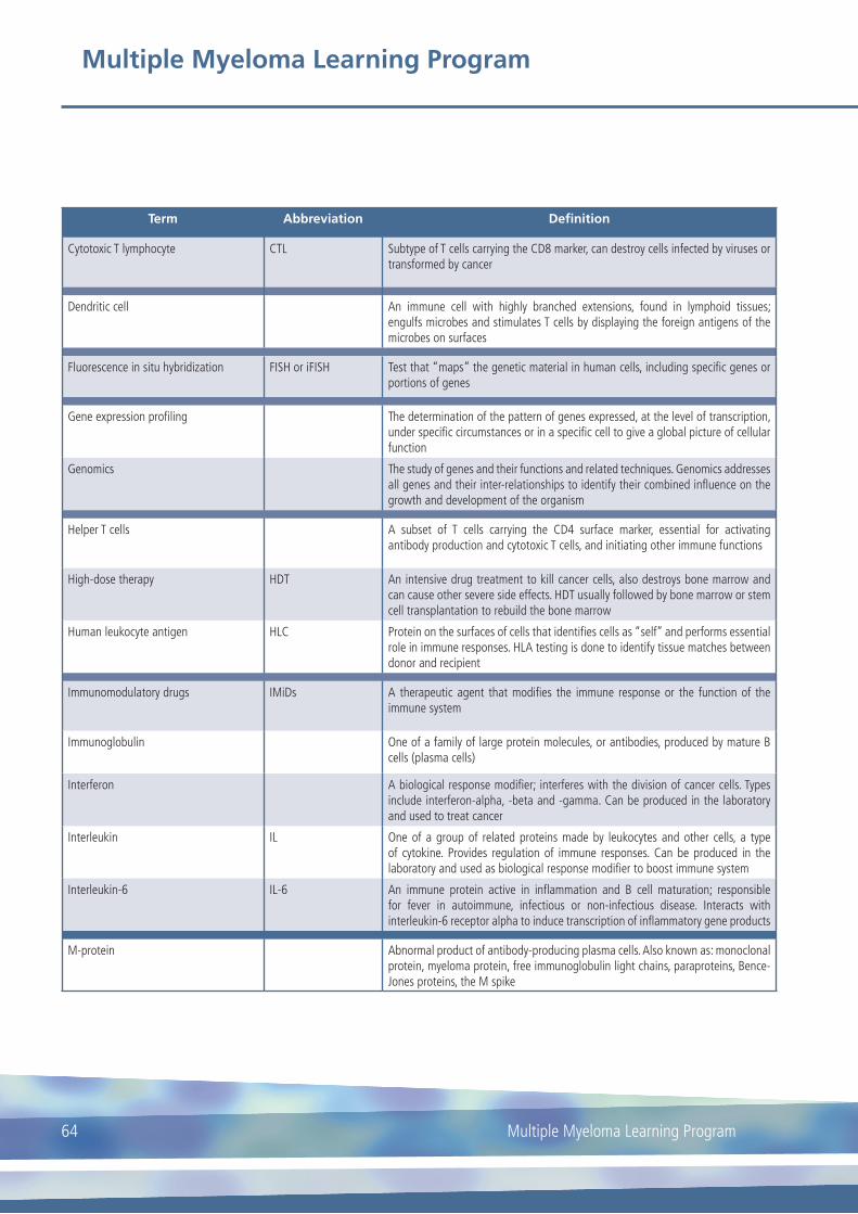
Multiple Myeloma Learning Program
Multiple Myeloma Learning Program64
Term Abbreviation Definition
Cytotoxic T lymphocyte CTL Subtype of T cells carrying the CD8 marker, can destroy cells infected by viruses or transformed by cancer
Dendritic cell An immune cell with highly branched extensions, found in lymphoid tissues; engulfs microbes and stimulates T cells by displaying the foreign antigens of the microbes on surfaces
Fluorescence in situ hybridization FISH or iFISH Test that “maps” the genetic material in human cells, including specifi c genes or portions of genes
Gene expression profi ling The determination of the pattern of genes expressed, at the level of transcription, under specifi c circumstances or in a specifi c cell to give a global picture of cellular function
Genomics The study of genes and their functions and related techniques. Genomics addresses all genes and their inter-relationships to identify their combined infl uence on the growth and development of the organism
Helper T cells A subset of T cells carrying the CD4 surface marker, essential for activating antibody production and cytotoxic T cells, and initiating other immune functions
High-dose therapy HDT An intensive drug treatment to kill cancer cells, also destroys bone marrow and can cause other severe side effects. HDT usually followed by bone marrow or stem cell transplantation to rebuild the bone marrow
Human leukocyte antigen HLC Protein on the surfaces of cells that identifi es cells as “self” and performs essential role in immune responses. HLA testing is done to identify tissue matches between donor and recipient
Immunomodulatory drugs IMiDs A therapeutic agent that modifi es the immune response or the function of the immune system
Immunoglobulin One of a family of large protein molecules, or antibodies, produced by mature B cells (plasma cells)
Interferon A biological response modifi er; interferes with the division of cancer cells. Types include interferon-alpha, -beta and -gamma. Can be produced in the laboratory and used to treat cancer
Interleukin IL One of a group of related proteins made by leukocytes and other cells, a type of cytokine. Provides regulation of immune responses. Can be produced in the laboratory and used as biological response modifi er to boost immune system
Interleukin-6 IL-6 An immune protein active in infl ammation and B cell maturation; responsible for fever in autoimmune, infectious or non-infectious disease. Interacts with interleukin-6 receptor alpha to induce transcription of infl ammatory gene products
M-protein Abnormal product of antibody-producing plasma cells. Also known as: monoclonal protein, myeloma protein, free immunoglobulin light chains, paraproteins, Bence-Jones proteins, the M spike

Multiple Myeloma Learning Program
Multiple Myeloma Learning Program 65
Glo
ssar
y
Term Abbreviation Definition
Major histocompatibility complex MHC A group of genes controlling several aspects of the immune response. MHC genes code for “self” markers on all body cells
Monoclonal gammopathy of undetermined signifi cance
MGUS A condition in which an abnormal protein, monoclonal protein or M protein produced by plasma cells in bone marrow, is found in blood by electrophoresis and/or immunofi xation. May progress to multiple myeloma
Multiple myeloma MM Malignant disease of plasma cells
Near complete response nCR Response to therapy in which paraprotein is no longer detectable by electrophoresis but by immunofi xation
Oncogene A mutated (changed) form of a gene involved in normal cell growth. Oncogenes may cause the growth of cancer cells. Mutations in genes that become oncogenes can be inherited or caused by exposure to cancer-causing substances in the environment
Opsonization The process by which bacteria and other cells are altered to become more readily/more effi ciently engulfed by phagocytes
Osteoclast-activating factor A lymphokine that stimulates bone resorption and inhibits bone-collagen synthesis
Osteolysis The dissolution of bone, especially the loss of calcium from bone
Osteolytic lesion A ‘punched out” area of severe bone loss. Also called osteoclastic lesions
Overall survival OS The length of time from either the date of diagnosis or the start of treatment during which a patient is still alive
Pancytopenia A disorder in which all three cell lines in peripheral blood (red blood cells, white blood cells and platelets) are decreased in number. Usually occurs 10-14 days after marrow ablative therapy
Partial response PR Treatment outcome where there is a greater than 50% decrease in M protein; also referred to as partial remission
Progression-free survival PFS The length of time during and after cancer treatment that a patient lives with the disease but the cancer does not worsen
Refractory When a disease or condition does not respond to treatment
Relapse Return of a disease or signs and symptoms of a disease after a period of improvement
Remission Period of time when symptoms improve or subside; can be temporary or permanent
Renal response Positive change in renal function, usually measured by estimated glomerular fi ltration rate (e-GFR), following treatment
Salvage therapy Treatment given after the cancer has not responded to other treatments
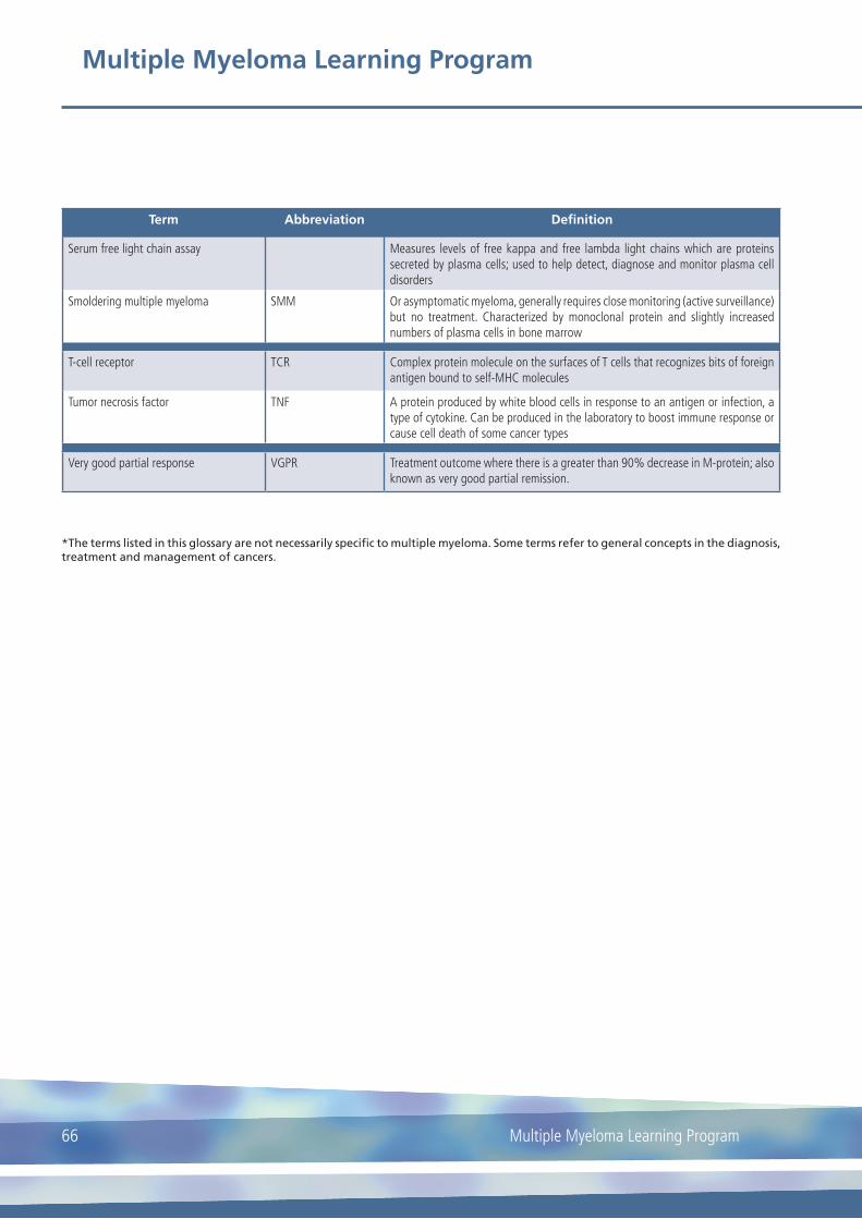
Multiple Myeloma Learning Program
Multiple Myeloma Learning Program66
Term Abbreviation Definition
Serum free light chain assay Measures levels of free kappa and free lambda light chains which are proteins secreted by plasma cells; used to help detect, diagnose and monitor plasma cell disorders
Smoldering multiple myeloma SMM Or asymptomatic myeloma, generally requires close monitoring (active surveillance) but no treatment. Characterized by monoclonal protein and slightly increased numbers of plasma cells in bone marrow
T-cell receptor TCR Complex protein molecule on the surfaces of T cells that recognizes bits of foreign antigen bound to self-MHC molecules
Tumor necrosis factor TNF A protein produced by white blood cells in response to an antigen or infection, a type of cytokine. Can be produced in the laboratory to boost immune response or cause cell death of some cancer types
Very good partial response VGPR Treatment outcome where there is a greater than 90% decrease in M-protein; also known as very good partial remission.
*The terms listed in this glossary are not necessarily specific to multiple myeloma. Some terms refer to general concepts in the diagnosis, treatment and management of cancers.

Notes
Participants Working Group:
Erik Aerts (CH)
Ruth Bähler (CH)
Corien Eeltink (NL)
Andrea Guy (UK)
Sarah Liptrott (IT)
Matthias Nägele (DE)
Panagiotis Samaras (CH)
This brochure was supported by Amgen, Bristol-Meyers Squibb, Celgene, Janssen, Novartis, Takeda
Copyright® 2017 by HNHCP - Haematology Nurses & Healthcare Professionals Group