multifaceted roles of tunneling nanotubes in intercellular ... · have highlighted here the latest...
TRANSCRIPT

HAL Id: pasteur-00716379https://hal-pasteur.archives-ouvertes.fr/pasteur-00716379
Submitted on 16 Jul 2012
HAL is a multi-disciplinary open accessarchive for the deposit and dissemination of sci-entific research documents, whether they are pub-lished or not. The documents may come fromteaching and research institutions in France orabroad, or from public or private research centers.
L’archive ouverte pluridisciplinaire HAL, estdestinée au dépôt et à la diffusion de documentsscientifiques de niveau recherche, publiés ou non,émanant des établissements d’enseignement et derecherche français ou étrangers, des laboratoirespublics ou privés.
Multifaceted roles of tunneling nanotubes inintercellular communication.
Ludovica Marzo, Karine Gousset, Chiara Zurzolo
To cite this version:Ludovica Marzo, Karine Gousset, Chiara Zurzolo. Multifaceted roles of tunneling nanotubes in in-tercellular communication.. Front Physiol, 2012, 3, pp.72. <10.3389/fphys.2012.00072>. <pasteur-00716379>

REVIEW ARTICLEpublished: 10 April 2012
doi: 10.3389/fphys.2012.00072
Multifaceted roles of tunneling nanotubes in intercellularcommunication
Ludovica Marzo1,2†, Karine Gousset 1† and Chiara Zurzolo1,2*
1 Unité de traffic membranaire et pathogenèse, Institut Pasteur, Paris, France2 Dipartimento di Biologia e Patologia Cellulare e Molecolare, Università Federico II, Napoli, Italy
Edited by:
Claudia Verderio, Consiglio Nazionale
delle Ricerche, Italy
Reviewed by:
Hans-Hermann Gerdes, University of
Bergen, Norway
Yan Zhang, Peking University, China
*Correspondence:
Chiara Zurzolo, Unité de traffic
membranaire et pathogenèse, Institut
Pasteur, 25 rue du docteur Roux,
75074 Paris Cedex 15, France.
e-mail: [email protected]
†Ludovica Marzo and Karine Gousset
have contributed equally to this work.
Cell-to-cell communication and exchange of materials are vital processes in multicel-
lular organisms during cell development, cell repair, and cell survival. In neuronal and
immunological cells, intercellular transmission between neighboring cells occurs via dif-
ferent complex junctions or synapses. Recently, long distance intercellular connections in
mammalian cells called tunneling nanotubes (TNTs) have been described.These structures
have been found in numerous cell types and shown to transfer signals and cytosolic mate-
rials between distant cells, suggesting that they might play a prominent role in intercellular
trafficking. However, these cellular connections are very heterogeneous in both structure
and function, giving rise to more questions than answers as to their nature and role as
intercellular conduits. To better understand and characterize the functions of TNTs, we
have highlighted here the latest discoveries regarding the formation, structure, and role of
TNTs in cell-to-cell spreading of various signals and materials.We first gathered information
regarding their formation with an emphasis on the triggering mechanisms observed, such
as stress and potentially important proteins and/or signaling pathways. We then describe
the various types of transfer mechanisms, in relation to signals and cargoes that have been
shown recently to take advantage of these structures for intercellular transfer. Because a
number of pathogens were shown to use these membrane bridges to spread between
cells we also draw attention to specific studies that point toward a role forTNTs in pathogen
spreading. In particular we discuss the possible role thatTNTs might play in prion spreading,
and speculate on their role in neurological diseases in general.
Keywords: tunneling nanotubes, intercellular communication, long-range connections, vesicular transport, signal
spreading, pathogen spreading, organelle transfer
INTRODUCTION
The ability of cells to communicate with each other is essential for
the life of a multicellular organism and is evolutionarily conserved
between species (Gurke et al., 2008). Without cell-to-cell commu-
nication,processes such as remodeling of tissues and organs,differ-
entiation during development, growth, cell division, and responses
to stimuli could not take place. Therefore, a great number of
cellular genes and their products are implicated in intercellular
communication and their misregulation leads to the establishment
of pathological conditions associated with many diseases.
Chemical signaling by secretion of small molecules toward dis-
tant cells is the classical form of cell-to-cell communication and
does not involve physical contact. It includes chemical media-
tors with paracrine effects on cells nearby, release of synaptic
vesicles containing neurotransmitters between neurons (chemi-
cal synapses; Süudhof, 2008), and hormones, which travel in the
blood stream after their release and can reach and stimulate distant
target cells.
In cases of close proximity, cells can interact with each other
through gap junctions or synapses. Gap junctions connect the
cytoplasm of two neighboring cells by clustering tens to thou-
sands of intercellular channels, allowing the transfer of ions
and small, hydrosoluble molecules (Maeda and Tsukihara, 2011).
They mediate electrical and metabolic coupling of cells and are
implicated in a wide range of biological processes such as mus-
cle contraction or electrical synapses in neurons (Connors and
Long, 2004). Immunological synapses, established at the interface
between a T-cell and an antigen-presenting cell (APC), are rather
mediated by membrane receptors (Rechavi et al., 2007; Tarakanov
and Goncharova, 2009) and are essential for the adaptive immune
response (Dustin et al., 2010). Structurally similar to the immuno-
logical synapse are the virological synapses. These supramole-
cular structures are cytoskeleton-dependent adhesive junctions
induced by virus-infected cells and used by these pathogens to
directly transfer to non-infected cells (Jolly and Sattentau, 2004).
Human immunodeficiency virus type 1 (HIV-1) and human T-
cell leukemia virus type 1 (HTLV-1) can spread using virological
synapses between T cells (Tarakanov and Goncharova, 2009).
Recently, long-range forms of intercellular communication
consisting of different types of membrane bridges have been
described in a wide variety of cell types in in vitro cell culture sys-
tems (Gerdes et al., 2007; Abounit and Zurzolo, 2012; Figure 1).
Similar connections have also been found in vivo and in tis-
sue explants (Wolpert and Gustafson, 1961; Miller et al., 1995;
Ramírez-Weber and Kornberg, 1999; Demontis and Dahmann,
2007; Chinnery et al., 2008). The discovery of these new types of
www.frontiersin.org April 2012 | Volume 3 | Article 72 | 1
Marzo et al. TNTs in intercellular communication
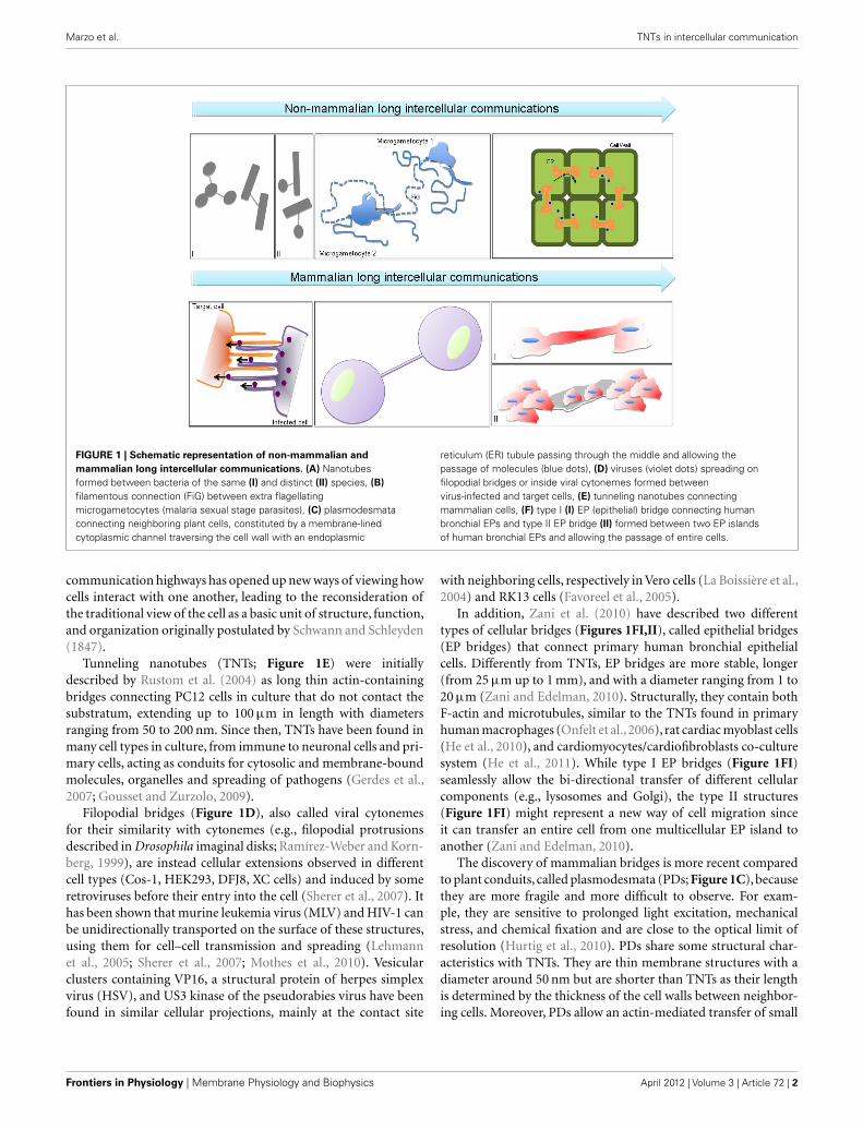
FIGURE 1 | Schematic representation of non-mammalian and
mammalian long intercellular communications. (A) Nanotubes
formed between bacteria of the same (I) and distinct (II) species, (B)
filamentous connection (FiG) between extra flagellating
microgametocytes (malaria sexual stage parasites), (C) plasmodesmata
connecting neighboring plant cells, constituted by a membrane-lined
cytoplasmic channel traversing the cell wall with an endoplasmic
reticulum (ER) tubule passing through the middle and allowing the
passage of molecules (blue dots), (D) viruses (violet dots) spreading on
filopodial bridges or inside viral cytonemes formed between
virus-infected and target cells, (E) tunneling nanotubes connecting
mammalian cells, (F) type I (I) EP (epithelial) bridge connecting human
bronchial EPs and type II EP bridge (II) formed between two EP islands
of human bronchial EPs and allowing the passage of entire cells.
communication highways has opened up new ways of viewing how
cells interact with one another, leading to the reconsideration of
the traditional view of the cell as a basic unit of structure, function,
and organization originally postulated by Schwann and Schleyden
(1847).
Tunneling nanotubes (TNTs; Figure 1E) were initially
described by Rustom et al. (2004) as long thin actin-containing
bridges connecting PC12 cells in culture that do not contact the
substratum, extending up to 100 µm in length with diameters
ranging from 50 to 200 nm. Since then, TNTs have been found in
many cell types in culture, from immune to neuronal cells and pri-
mary cells, acting as conduits for cytosolic and membrane-bound
molecules, organelles and spreading of pathogens (Gerdes et al.,
2007; Gousset and Zurzolo, 2009).
Filopodial bridges (Figure 1D), also called viral cytonemes
for their similarity with cytonemes (e.g., filopodial protrusions
described in Drosophila imaginal disks; Ramírez-Weber and Korn-
berg, 1999), are instead cellular extensions observed in different
cell types (Cos-1, HEK293, DFJ8, XC cells) and induced by some
retroviruses before their entry into the cell (Sherer et al., 2007). It
has been shown that murine leukemia virus (MLV) and HIV-1 can
be unidirectionally transported on the surface of these structures,
using them for cell–cell transmission and spreading (Lehmann
et al., 2005; Sherer et al., 2007; Mothes et al., 2010). Vesicular
clusters containing VP16, a structural protein of herpes simplex
virus (HSV), and US3 kinase of the pseudorabies virus have been
found in similar cellular projections, mainly at the contact site
with neighboring cells, respectively in Vero cells (La Boissière et al.,
2004) and RK13 cells (Favoreel et al., 2005).
In addition, Zani et al. (2010) have described two different
types of cellular bridges (Figures 1FI,II), called epithelial bridges
(EP bridges) that connect primary human bronchial epithelial
cells. Differently from TNTs, EP bridges are more stable, longer
(from 25 µm up to 1 mm), and with a diameter ranging from 1 to
20 µm (Zani and Edelman, 2010). Structurally, they contain both
F-actin and microtubules, similar to the TNTs found in primary
human macrophages (Onfelt et al., 2006), rat cardiac myoblast cells
(He et al., 2010), and cardiomyocytes/cardiofibroblasts co-culture
system (He et al., 2011). While type I EP bridges (Figure 1FI)
seamlessly allow the bi-directional transfer of different cellular
components (e.g., lysosomes and Golgi), the type II structures
(Figure 1FI) might represent a new way of cell migration since
it can transfer an entire cell from one multicellular EP island to
another (Zani and Edelman, 2010).
The discovery of mammalian bridges is more recent compared
to plant conduits, called plasmodesmata (PDs; Figure 1C), because
they are more fragile and more difficult to observe. For exam-
ple, they are sensitive to prolonged light excitation, mechanical
stress, and chemical fixation and are close to the optical limit of
resolution (Hurtig et al., 2010). PDs share some structural char-
acteristics with TNTs. They are thin membrane structures with a
diameter around 50 nm but are shorter than TNTs as their length
is determined by the thickness of the cell walls between neighbor-
ing cells. Moreover, PDs allow an actin-mediated transfer of small
Frontiers in Physiology | Membrane Physiology and Biophysics April 2012 | Volume 3 | Article 72 | 2

Marzo et al. TNTs in intercellular communication
molecules, transcription factors, and also spreading of viruses, cre-
ating a sort of continuity between the cytoplasm of connected
cells (symplast; Lucas et al., 2009). Even more similarities between
the mammalian TNTs and the plant PDs are found regarding the
mechanisms of formation and transfer (e.g., passive diffusion of
small molecules and gated-mechanisms for bigger components)
although the nature of the transported molecules can vary (Rus-
tom, 2009; Abounit and Zurzolo, 2012). This highlights a possible
common origin during evolution of TNTs and PDs that can allow a
better understanding of the newly discovered mammalian bridges
by comparing them with the better-known PDs.
Interestingly, along the same line of thinking, recent find-
ings of bacterial networks (Figure 1A) and parasite protrusions
(Figure 1B) make us wonder how evolutionally conserved these
kinds of intercellular communications can be. Indeed, Dubey
and Ben-Yehuda (2011) have recently shown that Bacillus sub-
tilis grown on a solid surface can establish nanotube-mediated
networks with neighboring bacteria of the same or different
species (Figures 1AI,II), as Staphylococcus aureus or Escherichia
coli, pointing toward a common way of communication shared
between phylogenetically distant bacterial species. These linking
structures and their mammalian or plant counterparts facilitate
transfer of cytoplasmic components and non-conjugative plas-
mids, allowing the exchange of hereditary traits for the acquisition
of new features between connected bacteria (Dubey and Ben-
Yehuda,2011). Sometimes in nature similarities in structure do not
reflect related functions. This could be the case of the cell-to-cell
connections formed by the malaria pathogen during reproduc-
tion in the mosquito midgut (Rupp et al., 2011; Figure 1B). In
this paper, the authors have described the presence of filamen-
tous structures containing F-actin, that they called “filaments of
gametes” or FiGs, in the activated gametocytes. Multiple FiGs are
generated on the surface of the cell a few minutes after activation
and can extend up to 180 µm. A closer look at these structures
revealed that they possess closed-ends and they do not transfer
material. Interestingly, FiGs exhibit adhesion molecules on their
surface that can instead mediate contact and recognition with the
right mating partners for the Plasmodium, allowing clustering of
gametocytes and facilitating the process of reproduction (Rupp
et al., 2011).
This and the other examples of intercellular contacts established
by different types of cells reported here reveal a high heterogeneity
in both structure and functions of these fascinating new routes of
communication that need further characterization and classifica-
tion (Figure 1). Furthermore in order to better understand their
physiological relevance more efforts will be needed to identify
these structures in vivo. To this aim the identification of spe-
cific TNT markers by using in vitro models is of fundamental
importance.
This review will focus on mammalian TNTs, their possible
mechanism of formation and their various functions, giving
particular attention to their implication in prion spreading.
MECHANISMS OF TNT FORMATION AND PROTEINS
INVOLVED
In two-dimensional cultures, TNT-like structures were first dis-
criminated from filopodia from their structural space. Contrary
to filopodia, they formed long bridges between cells and were
not attached to the substratum (Rustom et al., 2004). In addi-
tion to their spatial differences, TNTs, and filopodia appear to
serve different purposes. While filopodia act as important envi-
ronmental sensors and play key roles in cell motility, the main role
of TNTs appears to be as a direct conduit for cell-to-cell com-
munication, specifically in the transport of material from one cell
to another. As stated above, numerous membrane bridges have
been described in a multitude of cell types. Even within TNT-like
structures, it became quickly evident that these various struc-
tures were distinct from one another both in their structures and
functions.
TNT FORMATION AND STRUCTURAL COMPONENTS
Tunneling nanotube-like structures were first described in PC12
neuronal cells (Rustom et al., 2004). In these cells, de novo actin-
driven formation of TNTs was observed. Further examination of
PC12 cells and TNT formation suggested that while the majority
of tubes formed via directed filopodia-like protrusions, a small
subset (7%) were also able to form after cells previously in con-
tact detached from one another (Bukoreshtliev et al., 2009) (for
review, see Abounit and Zurzolo, 2012). In the mouse neuronal
CAD cell line, we were also able to observe both types of TNT
formation (data not shown). However, the significance and the
differences between these two modes of formation and whether
they lead to various structures remain unclear. Similar to other
cell types, we observed a high degree of heterogeneity in the diam-
eters of TNT-like structures (Gousset et al., 2009). Furthermore, as
previously described in PC12 cells (Rustom et al., 2004), neuronal
TNTs formed between CAD cells contained actin filaments but
no microtubules, even in the tubes with larger diameters (Gousset
et al., 2009). The fact that most TNTs in neuronal cells arise from
the extension of filopodia-like protrusions toward neighboring
cells suggested that actin polymerization plays an important role
in this type of TNT formation. Rustom et al. (2004) demonstrated
that using the F-actin depolymerizing drug latrunculin, no TNTs
were detected in PC12 treated cells. This type of treatment could
thus be used to selectively block TNT formation and look at the
effect of the presence or absence of nanotubes in various cultures.
In our lab, we took advantage of this treatment to highlight the
importance of the presence of TNTs in the transfer of infectious
prion aggregates in neuronal cells (Gousset et al., 2009). Using
nanomolar concentrations of Cytochalasin D (CytoD), another
actin-depolymerizing drug, Bukoreshtliev et al. (2009) went fur-
ther and examined the effects of this drug during the lifetime of
TNTs. They showed that as expected, low levels of CytoD abro-
gated both filopodia formation and TNT formation. Interestingly,
they also demonstrated that once formed, CytoD had little effects
on the stability of these tubes or their ability to transfer material
from one cell to another. Thus, most neuronal TNTs arise from
filopodia-like structures, detached from the substratum. Once
formed however, they are no longer sensitive to low levels of actin-
depolymerizing drugs, demonstrating that functional TNTs are
distinct from filopodia in both structure and function. Interest-
ingly, recent experiments with primary rat astrocytes and neurons
also showed actin to be the major cytoskeleton component of
TNTs formed between these cells (Wang et al., 2011). Indeed, these
www.frontiersin.org April 2012 | Volume 3 | Article 72 | 3

Marzo et al. TNTs in intercellular communication
authors showed that treatment with latrunculin or CytoD abro-
gated their formation, thus further validating the use of neuronal
cell lines as models for neuronal TNTs.
Tunneling nanotube-like structures have also been described in
immune cells, such as B-cells, Natural killer cells, and macrophages
(Onfelt et al., 2004). In macrophages, two types of nanotubes
were described (Onfelt et al., 2006). The thin nanotubes were
found to contain actin filament only, whereas thicker nanotubes,
with diameters larger than 0.7 µm, contained both F-actin and
microtubules. These different structures appeared to have dis-
tinct functions, with the thicker structures being able to transport
in a bi-directional manner vesicles and various organelles in a
microtubule dependent mechanism. Similarly, long nanotube con-
nections between Jurkat T cells and primary T cells were also
described (Sowinski et al., 2008). In these cells, F-actin but no
microtubules were detected in TNTs. In addition, while these
tubes were not open-ended, they still allowed for the transfer of
HIV-1 via a receptor-dependent mechanism. Finally, numerous
networks of TNT-like structures were observed between dendritic
cells and THP-1 monocytes (Watkins and Salter, 2005). These con-
nections varied greatly in length and diameter but were able to
quickly transfer calcium fluxes and small dyes to interconnected
cells.
Thus, while numerous TNT-like structures have been described
in immune cells, these tubes are clearly distinct from one another
both in their structural components as well as in their means of
transfer. The one characteristic consistent for all types of immune
cells is their formation that appears to rely primarily on cell-to-cell
attachment and formation of immunological synapses prior to cell
separation and tube formation (Sowinski et al., 2008).
In urothelial cell lines, two types of TNT-like structures were
observed (Veranic et al., 2008). The shorter but more dynamic
structures, described as Type I nanotubes, were found to con-
tain actin and to connect with neighboring cells by an anchoring
type of intercellular junctions. By using time-lapse phase-contrast
microscopy the authors observed that these structures did not col-
lapse after micromolar concentrations of CytoD suggesting that
after anchoring actin was no longer necessary (Veranic et al.,
2008). On the other hand, the longer and more stable structures,
or type II nanotubes, no longer contain actin filaments but were
composed of cytokeratin filaments. Although the authors have
observed vesicles on both these types of structures, further inves-
tigation is necessary to understand if these structures are involved
in transferring materials, thus fulfilling the TNT definition.
These examples show the disparity in the various cytoskeleton
requirements and formation mechanisms in naturally occurring
TNT-like structures in neuronal, immunological, or epithelial
cells. The type of formation however (de novo actin-driven vs
detachment after cell-to-cell contact) might arise from the nature
and role that these cells play in vivo. Indeed, mobile cells, which
can more easily come into contact with other cells, might be
more prone to form tubes from a previous cell-to-cell contact,
whereas more immobile cells might be more adept at creating
and extending tubes de novo toward distant cells. Because of
the increasing number of studies on different and highly hetero-
geneous TNT-like structures in several in vitro systems a more
systematic classification is needed.
SIGNALS AND MOLECULES INVOLVED IN TNT FORMATION: IS STRESS
A MAJOR PLAYER?
In order to better understand the role that TNTs may play in
intracellular transfer of materials, a better characterization of
the initiation steps of TNT formation, the signals that guide the
extension of these structures toward a neighboring cell and the
mechanisms of binding and fusion need to be elucidated.
Recently, the effects of stress on TNT formation have been ana-
lyzed in different cell types (Wang et al., 2011). In their studies,
Wang and colleagues have shown that stress induced by hydrogen
peroxide (H2O2) treatment led to an increase in TNT formation
in both astrocytes and neurons. They also observed the transfer
of various organelles, such as ER, Golgi, endosomes, and mito-
chondria via TNTs in astrocyte cultures. For both astrocytes and
neurons, it was always the cells undergoing stress that developed
TNTs and transferred cellular materials in a unidirectional fashion
to the non-activated cells, suggesting that TNT formation might be
directly induced by stress and may represent a defense mechanism
of the stressed cells. Interestingly, they found that p53 activation,
which is critical in apoptosis, led to an increase in TNT forma-
tion. Conversely, down regulation of p53 blocked TNT formation
(Wang et al., 2011). Subsequently, they showed that EGF receptor
up-regulation was also necessary for TNT initiation using different
conditioned media and that the initiation of TNT formation was
likely dependent on the initiating cells and not the receiving cells.
Finally, since the EGF receptor can activate the Akt/PI3K/mTOR
pathway, they used various mutants and inhibitors to selectively
block or activate each protein and found that this pathway was
indeed up regulated in H2O2 activated cells, leading to an increase
in TNT development (Wang et al., 2011). In another study, using
a macrophage cell line and HeLa cells, it was demonstrated that
the interaction between m-Sec and the Ral/exocyst complex was
also critical for TNT formation (Hase et al., 2009). Therefore, to
understand if m-Sec might also be important for TNT formation
in astrocytes, Wang et al. (2011) analyzed by RT-PCR the levels
of m-Sec in astrocytes and found a positive relationship between
H2O2 treatment and the levels of m-Sec expression. Interestingly,
their data indicated that m-Sec might be regulated by p53 acti-
vation. Thus, the authors suggest that the initiating cells control
TNT formation in a p53 and Akt/PI3K/mTOR pathway activation-
dependent manner, but they do not exclude that some guidance
cues might be originating from the receptor cells (Wang et al.,
2011). Further studies are required in order to explore other poten-
tial molecular targets downstream of p53 and Akt/PI3K/mTOR
pathways that might represent key elements involved in TNT
formation.
In another study Yasuda et al. (2010) analyzed the transfer of
mitotracker labeled vesicles via TNTs between endothelial prog-
enitor cells (EPC) and human umbilical vein endothelial cells
(HUVEC). They observed both TNT formation between the two-
cell types and transfer of mitochondrial material from the EPC to
the HUVEC. Upon treatment of the HUVEC with adriamycin,
they observed a large increase in the transfer of mitotracker
particles from the non-stressed EPC to the adriamycin-stressed
HUVEC. In addition, the transfer was unidirectional since the
reverse loading and transfer experiments were not significant
(Yasuda et al., 2010). While it was not clear in these experiments
Frontiers in Physiology | Membrane Physiology and Biophysics April 2012 | Volume 3 | Article 72 | 4
Marzo et al. TNTs in intercellular communication
which cell type initiated the formation of the nanotubes, contrary
to what was found in neuronal and astrocyte cultures (Wang et al.,
2011), the transfer of material occurred from the non-stressed cells
to the stressed cells. These observations raised the question of how
these cells initiated TNTs. Further characterization in these co-
cultures could determine whether the stressed cells might release
some signals that might attract filopodia-like protrusions from the
EPC to the HUVEC or whether the HUVEC cells might initiate
formation and allow for a reverse transfer of material from the
receptor cell to the initiator cell.
This is exactly what the authors next set out to demonstrate.
Indeed, in a follow-up study, they analyzed more precisely the
TNT formation mechanisms between these cells. First they showed
that co-cultures of EPC with stressed HUVEC led to a rescue of
HUVEC viability. However, when the EPC were pre-treated with
nanomolar levels of CytoD to block TNT formation prior to co-
culture with the HUVEC, the rescue effects were almost entirely
abrogated, pointing toward the importance of TNT formation
from EPC to HUVEC for cell survival. Using both fluorescence
microscopy and FACS analyses they observed basal levels of trans-
fer of lysosomes between the two-cell types in a bi-directional
manner under non-stressed conditions. However, the transfer was
much more efficient as it increased in speed and frequency and was
found preferentially between non-stressed EPC and GC-stressed
HUVEC, suggesting that the stressed cells were able to signal and
guide filopodia-like protrusions for the formation of de novo TNTs
to occur (Gerdes et al., 2007; Yasuda et al., 2010, 2011). Further
examination suggested that surface-exposed phosphatidylserines
(PS) in HUVEC might be able to guide TNT formation from the
EPC to the stressed HUVEC. Indeed, when PS on HUVEC were
blocked by binding of Annexin V, the selective TNT formation
and transfer from EPC to HUVEC was also blocked (Yasuda et al.,
2011).
Overall, these studies suggest that transfer of materials via TNTs
in most cell types occurred from the cell type that initiated TNT
formation to the receptor cell. However, while certain stress condi-
tions might increase the formation of TNTs between cells, it does
not affect all cells the same way. Indeed, while in astrocytes and
neurons, stress appears to increase TNT formation in the stressed
cells leading to an increase in transfer of material, in endothe-
lial cells stress increase the guidance signals from the stressed
cells leading to an increased formation of TNTs from the non-
stressed cells. Thus, once more the analysis of these two studies
brings forward the disparities that exist in formation and nature
of TNTs between different cell types. It suggests that even within
an identical type of TNT formation (i.e., de novo extension of
filopodia-like protrusions) the mechanisms might be very distinct
from one another (activation of attractive guidance signals vs acti-
vation of initiation of filopodia-like protrusions). However, these
studies implicate the involvement of more general signaling path-
ways in TNT formation. For example, the role of m-Sec, which
was found to be important in macrophages, HeLa cells, and astro-
cytes (Hase et al., 2009), could be of general importance in TNT
formation, independent of cell type. In addition, since filopodia-
like protrusions are critical for TNT formation in neuronal cells
(Bukoreshtliev et al., 2009), our lab, has turned its attention to
the role that the actin molecular motor protein Myosin-X might
play in both the formation of TNT-like structures and its function
in transfer of materials in neuronal cells. We found that over-
expression of Myosin-X (Berg and Cheney, 2002) increased the
number of TNTs observed in our cell cultures (data not shown).
In addition, similar to what Wang and colleagues (Wang et al.,
2011) have found with stress signals, we observed a unidirectional
transfer of vesicles occurring from the cells over-expressing Myo-X
to the acceptor cells (data not shown).
Finally the search for guidance signals and the role that lipids
might play in TNT formation might provide further information
about TNT formation.
MECHANISMS INVOLVED IN OPEN-ENDEDNESS OF TNTs
As previously stated, in T cells no membrane continuity or transfer
of cytosolic material have been observed (Sowinski et al., 2008),
suggesting different types of tubular structures between T cells and
other cell types that allowed for the transfer of cytosolic materi-
als such as neuronal cells, astrocytes, myeloid cells, or endothelial
cells. Recently, however, Arkwright et al. (2010) have shown that
specific stimulation could lead to an increase of TNTs in T cells
along with the transfer of cytosolic material. First, they showed
that FAS activation resulted in an increase in TNT formation and
that both toxin B of Clostridium difficile (an inhibitor of actin
Rho-GTPases) and secramine A (an inhibitor of CDC42) specif-
ically blocked FAS stimulated TNT formation in T cells. They
also analyzed the bi-directional exchange of labeled membranes
in T-cell co-cultures. As expected, they only found a negligible
number of TNTs with both markers in control cells, whereas upon
FAS stimulation they observed a 20-fold increase in the num-
ber of TNTs labeled with both membrane markers. The transfer
of cytosolic materials, including fluorescent cytosolic proteins as
well as labeled vesicles, was also observed upon FAS-stimulation
between T cells. These experiments demonstrated that the nan-
otubular structures initiated by FAS-stimulation were different
from the TNTs previously described in non-activated T cells and
did not contain an immunological synapse (Sowinski et al., 2008).
These connections were similar to the connections observed in
other cell types and demonstrate the complexity and dynamism
of the various TNT-like structures that have been described to
date. While this study demonstrates that within the same cells,
different activation can quickly lead to the formation of different
types of TNTs with distinct functions; the mechanisms involved
in the gating of these tubular structures remain undetermined.
Overall, these recent studies on TNTs have shown the diversity of
these structures but also their ability to transfer numerous signals
upon specific activation.
CAUSE AND CONSEQUENCES OF TNT-MEDIATED TRANSFER,
FROM SIGNAL TO ORGANELLES AND PATHOGENS
Tunneling nanotubes have revealed a high degree of heterogene-
ity also from a functional point of view, as different components
seems to be selectively transferred by different cell types. What
determines this selectivity remains unknown.
First, further investigation is needed to understand why some
cargoes are unidirectionally or bi-directionally transported. Uni-
lateral transfer occurs in the case where a donor cell transfers
material to an acceptor cell, whereas bi-lateral transfer happens
www.frontiersin.org April 2012 | Volume 3 | Article 72 | 5
Marzo et al. TNTs in intercellular communication
when both cells mutually exchange materials. The reasons for
these different transport mechanisms can depend on the struc-
tural components (actin only vs actin + microtubules containing
TNTs) or on specific signals that stimulate nanotube formation
and are responsible for directing the traffic in one or two ways.
As already mentioned above, bi-directional transfer is found
when both actin and microtubules are present (Onfelt et al., 2006;
Arkwright et al., 2010; He et al., 2010, 2011), while it appears to be
unidirectional when TNTs contain actin only (Rustom et al., 2004;
Koyanagi et al., 2005; Gurke et al., 2008; Eugenin et al., 2009; Gous-
set et al., 2009; Domhan et al., 2011). A recent work by Plotnikov
et al. (2010) shows that unidirectional transfer from rat renal tubu-
lar cells (RTC) to bone marrow multipotent mesenchymal stromal
cells (MMSC) was observed in this co-culture system (Plotnikov
et al., 2010). However, passage of molecules in the opposite direc-
tion was also detected, albeit at a lower rate. Additionally, it
has been shown that lysosome exchange (Lysotracker-labeled)
between endothelial progenitor cells (EPC) and endothelial cells
(HUVEC) in co-cultures occurs at a basal level and that this trans-
fer selectively increases in one direction, from EPC to HUVEC
cells, upon injury of the latter (Yasuda et al., 2010). These two
reports suggest that a shift from a bi-directional basal level of
transfer to a selective unidirectional transfer toward a specific cell
population might take place by means of intercellular thin con-
nections resembling TNTs between cells upon specific treatment,
as is the case for differentiation signal flow toward MMSC cells
(Plotnikov et al., 2010) and stress signal deriving from damaged
organelles (Yasuda et al., 2010). What remains to be determined
is how transfer occurs within TNTs and whether common molec-
ular motors might be involved during this process. Furthermore,
the fact that TNT structures contain F-actin as backbone suggests
that an acto-myosin-dependent mechanism could be responsible
for organelles or pathogens transfer mediated by TNTs (Rustom
et al., 2004; Gerdes et al., 2007; Hurtig et al., 2010). It has been
reported that organelle transfer through TNTs is an active process
that depends on actin and ATP (Onfelt et al., 2006; Gurke et al.,
2008; Bukoreshtliev et al., 2009; Gousset et al., 2009). Indeed the
use of F-actin depolymerizing drugs and ATP-depletion experi-
ments resulted in an almost complete block of organelle transfer
(Onfelt et al., 2006; Gurke et al., 2008; Bukoreshtliev et al., 2009;
Gousset et al., 2009). Furthermore by measuring the trajectory of
the organelles transferring from one cell to another Gurke et al.
(2008) demonstrated that the vesicle movement inside TNTs of
NRK cells was due to active transport and not to free diffusion.
Similar conclusions were obtained by measuring the mean square
displacement of PrP containing vesicles in TNTs (Gousset et al.,
2009). In addition, vesicular traffic on actin- and microtubules-
containing TNTs in macrophages was shown to be sensitive to
ATP-depletion, indicating that independently of the cytoskeleton
components transfer through TNTs occurs as an active process
(Onfelt et al., 2006). Finally the actin-binding motor Myosin Va is
present in TNTs and partially localizes with endocytic organelles
(Rustom et al., 2004; Gerdes et al., 2007). A more detailed analy-
sis on the role of Myosin Va and the screening of myosin motors
involved in endocytic vesicles traffic or pathogens spreading will be
necessary to further dissect the mechanism of transfer occurring
via TNTs.
SIGNAL TRANSFER
Up to now several reports have shown that calcium signals could
propagate between remote cells through TNTs (Watkins and Salter,
2005; Hase et al., 2009; Smith et al., 2011; Table 1). This is espe-
cially important for remote cells that are unable to propagate
calcium-mediated signaling to cells in close proximity using gap
junctions (Wang and Gerdes, 2011). Initially, Watkins and Salter
(2005) demonstrated that myeloid cells can respond to stimula-
tion through soluble factors or mechanical stress and are able to
amplify the cellular response by calcium signaling through mem-
brane connections. Since then, propagation of calcium flux has
been shown in many other cell types able to make connections
between each other (Hase et al., 2009; Smith et al., 2011; Table 1).
More recently, the transfer of IP3 receptor (IP3R) and endoplasmic
reticulum has been described along TNTs in SH-SY5Y neuroblas-
toma and HEK cell lines (Smith et al., 2011). The authors made
a comparison between the current produced at the end of a TNT
(typically 30 µm in length and 200 nm in diameter) and single
inositol trisphosphate receptor (IP3R)-channels. While the first
produces a current <1 fA, corresponding to calcium flux prop-
agated from an activated cell, the opening of a single channel
results in ∼100 fA. Considering that a single opened IP3R-channel
generally fails to induce Ca2+ signaling, the passive diffusion of
Ca2+ within TNTs appears quite inefficient. However, since IP3R
is able to transfer along TNTs, it could overcome the limit of pas-
sive diffusion of calcium by amplifying calcium signaling within a
population. Finally, a recent study has reported the formation of
electrically coupled nanotubes that do not allow diffusion of cal-
cium or IP3R, but are instead involved in the bi-directional spread
of electrical current between distant cells through gap junctions
(Wang et al., 2010). These type of TNTs are immuno-positive for
connexin-43, at one end of the connection and allow the passage
of electrical signals which in turn leads to the activation of low
voltage gated channels that allow a local influx of calcium in the
connected cell. Electrical coupling-competent TNTs,distinguished
from those that do not possess gap junctions, have been found in
different cell types and represent a selective way for transferring
electrical signals compared to gap junctions coupling (Wang et al.,
2010; Wang and Gerdes, 2011; Abounit and Zurzolo, 2012).
Overall, calcium spreading through nanotubes appears to be a
good option for different types of cells to quickly spread calcium
signals under physiological conditions, leading to fast responses in
connected neighboring cells (for review see Abounit and Zurzolo,
2012; Table 1).
Particularly fascinating and newly discovered is the spreading
of death signals by nanotubes occurring in Jurkat and primary
T cells (Arkwright et al., 2010; Table 1). Fas-mediated signaling
is important for peripheral deletion of activated T lymphocytes
(Green et al., 2003). Mutations in the cytoplasmic domain of the
Fas receptor are responsible for a rare genetic disease, the autoim-
mune lymphoproliferative syndrome (the type Ia ALPS; Martin
et al., 1999). As stated previously, Arkwright et al. (2010) have
shown that stimulation of the Fas receptor leads to an increase in
the number of TNT-connected cells and this is critically dependent
on Rho GTPase activation. Accordingly, the authors also demon-
strated that primary T cells deriving from ALPS patients were
not able to form networks of TNTs. This points toward a pivotal
Frontiers in Physiology | Membrane Physiology and Biophysics April 2012 | Volume 3 | Article 72 | 6

Marzo et al. TNTs in intercellular communication
Table 1 | Overview of the different cargos found inTNT-like structures.
Functions ofTNTs Cargo detection Cell type References
SPREADING OF SIGNALS
Calcium signaling IP3R SH-SY5Y neuroblastoma, HEK cells Smith et al. (2011)
Ca2+; Fura-2 THP-1 monocytes and dendritic cells Watkins and Salter (2005)
Ca2+ Raw264.7 macrophages, HeLa cells Hase et al. (2009)
Electrical coupling through gap junction at
the TNT end
Normal rat kidney (NRK), HEK, HUVEC, NCC
and rat pheochromocytoma (PC12) cells
Wang et al. (2010)
Death signals FasL, caspase-3 Jurkat and primary T cells Arkwright et al. (2010)
Cytotoxicity NK cells Chauveau et al. (2010)
ORGANELLE EXCHANGE
Endosomes Purified mouse anti-EEA1 antibodies CMs and FBs co-culture system* He et al. (2010)
DiD (1,1′-dioctadecyl-3,3,3′,3′-tetramethylin
dodicarbocyanine perchlorate)
NRK cells Gurke et al. (2008)
Qtracker® Human renal proximal tubular epithelial cells
(RPTEC)
Domhan et al. (2011)
DiD (1,1′-dioctadecyl-3,3,3′,3′-tetramethylin
dodicarbocyanine perchlorate)
Human monocyte-derived macrophages Onfelt et al. (2006)
Endosomes-related organelles (DiI and DiO) PC12 cells Rustom et al. (2004)
Lysosomes Lysotracker® PC12 cells Rustom et al. (2004)
Qtracker® Human renal proximal tubular epithelial cells
(RPTEC)
Domhan et al. (2011)
Lysotracker® Mouse catecholaminergic neuronal cell line,
Cath.a-Differentiated (CAD)
Gousset et al. (2009)
Lysotracker® EPC and HUVEC co-culture system (rescue
from injuries)*
Yasuda et al. (2011)
Mouse anti-LAMP1 antibodies Human monocyte-derived macrophages Onfelt et al. (2006)
Mitochondria Mitotracker® EPC or CD34+ cells and neonatal rat
cardiomyocytes co-culture system
(Differentiation)*
Koyanagi et al. (2005)
Mitotracker® MMSC and RTC* Plotnikov et al. (2010)
Mitotracker® H9c2 Cardiomyoblasts and MMSC (rescue
from injuries)*
He et al. (2011)
TMRE Jurkat and primary T cells Arkwright et al. (2010)
MitoTracker Human monocyte-derived macrophages Onfelt et al. (2006)
Membrane components CD81, CD59 Jurkat and primary T cells Arkwright et al. (2010)
c-HA-Ras PC12 Rustom et al. (2004)
Surface receptors (HLA-A,B,C class I MHC) Myeloid cells Watkins and Salter (2005)
DiO MMSC and RTC* Plotnikov et al. (2010)
GPI-anchored GFP, TM-proteins (ICAM-I,
HLA-Cw7)
Jurkat T cells, primary mouse T cells Sowinski et al. (2008)
GFP-PrP CAD neuronal cells Gousset et al. (2009)
MHC-I Immune cells Onfelt et al. (2004)
Golgi and Endoplasmic
reticulum
Bodipy FL glibenclamide (ER-tracker)
Bodipy FL C5-ceramide (Golgi-tracker)
Human monocyte-derived macrophages
(MDM)
Kadiu and Gendelman
(2011b)
Cytoplasmic components Cytosolic GFP CMs and FBs co-culture system* He et al. (2010)
Calcein MMSC and RTC Plotnikov et al. (2010)
Cytosolic GFP EPC or CD34+ cells and neonatal rat
cardiomyocytes co-culture system*
Koyanagi et al. (2005)
Cytosolic stain CFSE Jurkat and primary T cells Arkwright et al. (2010)
Lucifer yellow Myeloid cells Watkins and Salter (2005)
Nanoparticles Nanoparticles quantum dots (CdSe/ZnS) CMs and FBs co-culture system* He et al. (2010)
(Continued)
www.frontiersin.org April 2012 | Volume 3 | Article 72 | 7
Marzo et al. TNTs in intercellular communication
Table 1 | Continued
Functions ofTNTs Cargo detection Cell type References
PATHOGENS SPREADING
Bacteria Mycobacterium bovis BCG Human monocyte-derived macrophages Onfelt et al. (2006)
Virus Gag and Env (antibodies), GFP-Gag Jurkat T cells, activated primary human or
primary mouse T cells
Sowinski et al. (2008)
HIV particles, HIV-p24 Primary human macrophages infected by HIV Eugenin et al. (2009)
Env and Gag proteins Human monocyte-derived macrophages Kadiu and Gendel-
man (2011a), Kadiu and
Gendelman (2011b)
Proteinaceous
aggregates
PrPSc CAD neuronal cells, GCN and DC co-culture
system
Gousset et al. (2009)
A–b fusion proteins Astrocytes and neurons Wang et al. (2010)
The table summarizes all the cargo detected in TNT-like structures by classifying them according to their nature (signals, organelle, and pathogens) and the cell type
in which they were found. *Exchange of cargos observed in co-culture of different cell type.
EPC, endothelial progenitors; HUVEC, stressed endothelial cells; MMSC, bone marrow multipotent mesenchymal stromal cells; RTC, rat renal tubular cells; CM, rat
ventricular cardiomyocytes; FB, cardiofibroblasts.
role of the Fas-mediated pathway in promoting TNT formation
and transfer in T cells. Additionally, transfer of both membrane
(detected by CD59 and CD81 staining) and cytoplasmic compo-
nents was detected in Fas-induced TNTs. Interestingly, FasL and
active caspase-3 passage from Fas-activated cells in neighboring
non-activated ones was detected, thus resulting in the spreading
of apoptosis through fratricide, highlighting that this might be
an efficient way to shut down cellular responses (Arkwright et al.,
2010). Moreover, it has been reported that FasL is upregulated in
cancer cells (O’Connell et al., 1996) and this could confer a double
advantage to these cells in “counterattacking” the immune system
and stimulating their own proliferation. In this light, TNTs could
act as conduits for diverse signals between tumor cells (for their
own survival) and from tumor cells to immune cells (for death),
thus leading to opposite effects.
Finally, Chauveau et al. (2010) have recently observed that also
Natural Killer immune cells (NK cells) can easily form intercellu-
lar nanotubes, particularly upon activation. NK cells are important
immune cells implicated in defense against a range of infections
(Herberman and Ortaldo, 1981). The authors demonstrated that
human primary NK cells are able to connect with different cell
types by intercellular bridges and use them to mediate cytotoxicity
(Table 1) and, therefore, help lyse remote target cells leading to cell
death (Chauveau et al., 2010).
ORGANELLE TRANSFER
Tunneling nanotubes can in certain cases be highways for diverse
organelle transfer (Table 1). Labeling with membrane-specific
dyes, markers of the endo-lysosomal pathway, or other dyes
specific to organelles such as mitochondria, has revealed sub-
cellular organelles traveling between cells along these connections
(Table 1). A range of cell types, including T cells, macrophages,
NRK, stem cells, epithelial cells, myocardial cells have exhibited
transfer of mitochondria (Table 1). Differentiation of embryonic
endothelial progenitor cells (EPC) in myocyte-like phenotype was
observed when EPC were co-cultured with neonatal rat cardiomy-
ocytes suggesting that TNT-mediated transfer of mitochondria
could have a reprogramming function in these cells (Koyanagi
et al., 2005). Moreover, Spees et al. (2006) have observed the pas-
sage of mitochondria from adult non-hematopoietic stem cells
(from human bone marrow hMSCs) or skin fibroblasts to A549
ρ˚ epithelial cells that were defective or deleted in mtDNA rescue
aerobic respiration. However, the authors could only hypothe-
size an involvement of tubular connections between the two-cell
types without demonstrating it. A closer look at some recent
work involving the use of co-culture systems shows that TNT-
mediated mitochondrial transfer could indeed rescue injured cells
for pathological conditions (Cselenyák et al., 2010). For exam-
ple, Cselenyak and coworkers set up a co-culture system of H9c2
cardiomyoblasts and mesenchymal stem cells (MSC) mimicking
ischemic damage in H9c2 cells by using oxygen glucose depri-
vation (OGD). They were able to show passage of functionally
active mitochondria (labeled with Mitotracker dye) in the dam-
aged cells specifically when nanotubular connections between the
cells were present (Cselenyák et al., 2010). In addition, selective
bi-directional transfer of mitochondria in between connected rat
ventricular cardiomyocytes (CMs) and cardiofibroblasts (FBs) was
observed in tubular structures (He et al., 2011). These connections
were enriched in actin and microtubules and allowed for the traf-
fic of soluble cytosolic dyes as well, suggesting continuity between
the membranes. The authors also explored a possible physiologi-
cal significance of the nanotubular structures found in CMs–FBs
co-culture system in vitro by culturing mouse heart tissue slices.
By labeling CMs and FBs with WGA and other specific markers,
the authors were able to detect thin structures between the two-cell
types, reminiscent of the connections observed in vitro (He et al.,
2011).
A rescue function of TNT-mediated organelle transfer might
be associated with other cell types that undergo injuries as well
(Table 1). Accordingly, the observation cell-to-cell contacts estab-
lished between RTC and MMSC leads to the hypothesis that
the exchange of cytoplasmic and organelle components could be
involved in restoring functions of damaged cells following acute
renal failure (Plotnikov et al., 2010). Indeed, endothelial cells
Frontiers in Physiology | Membrane Physiology and Biophysics April 2012 | Volume 3 | Article 72 | 8
Marzo et al. TNTs in intercellular communication
presenting lysosomal dysfunction after exposure to AGE-modified
collagen I (Yasuda et al., 2010) appeared to be rescued by trans-
ferring normal lysosomal pool from endothelial progenitors to
stressed cells (Yasuda et al., 2011) This suggests a role for organelle
TNT-mediated transfer in restoring functions and tissue repair,
which needs to be further characterized (Table 1).
Smaller particles, named nanoparticles, have also been
shown to travel within nanotubes (He et al., 2011). Particu-
larly, Streptavidin-coated CdSe/ZnS Quantum Dots (QDs) were
detected along membrane nanotubes of rat cardiac myoblast cells
(H9c2) at a speed compatible with movement of DiD-labeled
vesicles associated with dynein/kinesin motors walking on micro-
tubules (Onfelt et al., 2006), thus suggesting that nanoparticles
can be transported inside vesicles within these structures (He
et al., 2011). In fact, when WGA was used to label membrane
vesicles, QDs colocalized with it inside TNTs, confirming the vesic-
ular transport of these molecules. Moreover, like thicker TNTs
described in macrophages (Onfelt et al., 2006) the nanotubes of
H9c2 cells contained both actin and microtubules and allowed
a bi-directional transfer of membrane vesicles (He et al., 2010).
Use of nanoparticles, such as QDs, is an emerging research field
for diverse medical applications, such as therapies and diagnostics
(Youns et al., 2011). For example, these small compounds could
be used to selectively deliver drugs to cancer cells or for other
infectious diseases (Singh and Nalwa, 2011). The fact that cells
can establish membrane nanotubes together with the new finding
that nanoparticles could pass from one cell to another by these
means of communication open up new ways for diffusing small
therapeutics inside target “cell communities.”
PATHOGEN SPREADING
Tunneling nanotubes could be either actively hijacked from dif-
ferent pathogens or transport them as “Trojan horses,” along
the membrane or inside, leading to the spreading of infection
(Table 1). Hijacking of these structures can be preceded by induc-
tion of TNT formation, thus optimizing pathogen transfer, as has
been shown for HIV particles spreading, both surfing on or inside
TNTs in primary macrophages (Eugenin et al., 2009). The HIV
virus can use these highways to spread as an alternative to the
other means already mentioned above.
Recently, a more detailed characterization of HIV-carriers
mediating the transfer of the virus along TNTs bridging
macrophages has been made that the authors called bridging
conduits (BCs; Kadiu and Gendelman, 2011b). In this work, the
authors first observed an increase in the number of connections
in macrophages, as previously described (Sowinski et al., 2008).
They then identified the composition of BCs by proteomic analy-
sis following isolation from cell bodies. Although the approach
used to isolate intercellular connections could not totally exclude
the presence of other cellular protrusions, the work gives some
insights on the possible compositions of BCs in the context of HIV
spread. Indeed, they found several organelle markers including
endo-lysosomal compartment (14%), ER (9%), and Golgi (4%)
inside BCs, the majority of which were regulators of different
steps within the HIV life cycle. They were also able to confirmed
by confocal microscopy that 72% of Golgi and 32% of ER colo-
calize in TNTs with the viral protein Env; similar results were also
obtained for the viral protein Gag, suggesting a role for these intra-
cellular compartments in HIV intracellular trafficking (Kadiu and
Gendelman, 2011a). Indeed, Golgi and ER represent sorting sta-
tions for the virus prior to reaching endosomal vesicles and before
spreading. Additionally, they observed that Golgi and ER undergo
morphological changes upon HIV infection (Kadiu and Gendel-
man, 2011a). Overall these observations shed light on a possible
new role for the Golgi and ER in TNT-mediated transfer of diverse
cellular components and their regulation mechanisms that need
to be further investigated.
Additional observations on the trafficking of HIV have shown
that HIV specifically traffics in TNTs associated with endo-
cytic compartments and so these organelles could be responsible
for viral spread between macrophages (Kadiu and Gendelman,
2011a). Moreover, the acto-myosin machinery used by the cell
to move virus-containing cargoes within TNTs is 25 times faster
than the surfing process seen for HIV and other retroviruses on
filopodial protrusions (Sherer et al., 2007). In particular, HIV pref-
erentially associates in TNTs with recycling endosomes and MVB
(Kadiu and Gendelman, 2011a). Whether viral particles spread-
ing in vesicles through BC results in a productive infection of
a recipient cell and how the flow of these carriers is regulated
and intersects with the intracellular pathway remain to be inves-
tigated. Comparing intra- and inter-cellular trafficking with our
current knowledge in the HIV field could improve our under-
standing and help in characterizing intercellular spreading of other
pathogens that manipulate host intracellular components for their
own survival, leading to progressive loss of cellular identity.
One of the best known mechanisms of cell-to-cell spread, com-
mon in some pathogenic bacteria such as Listeria, Shigella, and
Salmonella, is their ability to polymerize the host actin cytoskeleton
to escape the host and keep infecting new targeted cells (Cos-
sart and Sansonetti, 2004). While little was known about other
atypical cytoplasmic bacteria spreading, recently, new “unusual”
ways of bacterial spreading have been observed. For example, it
has recently been shown that Cryptococcus neoformans is able to
laterally transfer from an infected macrophage to an uninfected
one allowing a latent persistency in the host for long periods
before causing meningoencephalitis in the central nervous system
(CNS; Ma et al., 2007). The authors observed an actin-dependent
transfer of the bacterium in both immortalized cell lines and
human primary macrophages by a mechanism not yet understood.
More recently, it has been reported that the obligate intracel-
lular bacterium Ehrlichia chaffeensis associates with filopodia of
infected DH82 monocytes and increases their numbers and lengths
(Thomas et al., 2010). The authors hypothesized that the transport
of Ehrlichia through filopodia could be a potential mechanism for
the pathogen to pass from one cell to another without contacting
the extracellular environment. Another unusual way of spreading
recently highlighted is the formation of an actin barrel (Hage-
dorn et al., 2009), the “ejectosome,” induced by Mycobacterium
marinum and used by it to pass within infected Dictyostelium
discoideum ameba as host. This mechanism is an alternative to
the formation of a protrusion containing the pathogen created
by actin polymerization that is then engulfed by adjacent cells
(Carlsson and Brown, 2009). Onfelt et al. (2006) have shown that
M. bovis BCG or clusters of several bacteria can surf on thin
www.frontiersin.org April 2012 | Volume 3 | Article 72 | 9

Marzo et al. TNTs in intercellular communication
membrane nanotubes between macrophages before being inter-
nalized by receptor-mediated endocytosis (Onfelt et al., 2006),
pointing toward a possible role of these structures in bacterial
infection by concentrating the pathogen on the entry site for a
more efficient invasion.
Additionally, one could also envisage a role for these newly dis-
covered highways in the spreading of some obligatory intracellular
bacteria, unable to surf along TNT membranes that could use them
to escape from the immune response. As already mentioned above,
different sub-cellular organelles are found to shuttle in between
cells by TNTs. Bacteria can use different endocytic compartments
and modulate them to escape lysosomal degradation (Ham et al.,
2011). In particular, vacuoles-containing bacteria deriving from
fusion of the pathogen with intracellular organelles were found
to be positive for several endosomal proteins (Bonazzi and Cos-
sart, 2006). A problem for nanotubes in transporting these bigger
cargoes along their tracks could be overcome by the presence of
expansions along the tunnel, known as gondolas (Hurtig et al.,
2010). Veranic et al. (2008) have observed that these dilatations of
the membrane can move for 5–15 µm with an average speed of
40 nm/s (Veranic et al., 2008). This “pearling” phenomenon seen
along some TNT structures might be due to the redistribution of
lipids and cytoskeleton components localized in discrete areas and
could be compatible with a vesicular transport of pathogen as well.
SPREADING OF PRIONS AND PRION-LIKE
NEURODEGENERATIVE DISEASES
The mechanisms of prion spreading from the periphery to the
CNS, and subsequently within the CNS, remain questionable. A
number of mechanisms, such as cell-to-cell contact, exosomes, and
GPI-painting, have been proposed (Baron et al., 2002; Kanu et al.,
2002; Fevrier et al., 2004). We have recently demonstrated the pres-
ence of TNTs in neuronal CAD cell cultures (Gousset and Zurzolo,
2009; Table 1). In addition, we showed that these TNTs were able
to transfer lysosomal organelles, the cellular GPI-anchored prion
protein PrPC, as well as fluorescently labeled infectious prion parti-
cles, PrPSc. Using various co-culture conditions, we demonstrated
that these infectious particles were efficiently transferred to non-
infected cells only in the presence of TNTs (Gousset et al., 2009).
Since the prion protein is a GPI-anchored protein, it has the
possibility of traveling via TNTs either along their surface or inside
the tube within vesicular structures (Figure 2F and enlarged box).
Recently we have further analyzed the presence of PrPSc and var-
ious organelles inside TNTs. Overall, we observed that similar to
what can be found in the cell body, PrPSc travels in TNTs in early
endosomes and lysosomes but it is preferentially enriched in the
endosomal recycling compartment. Additionally, increasing the
number of TNTs formed, by over-expression of Myosin-X, also
increases the spreading of PrPSc to non-infected cells (data not
shown). These data further demonstrate how efficient these struc-
tures are in allowing the passage of infectious prions from one cell
to another.
Finally, we have also demonstrated that the transfer via TNTs of
infectious prion particles resulted in the transmission of infectiv-
ity to the recipient cell. This transfer was not confined to neuronal
co-cultures but was also efficient between loaded bone marrow
derived dendritic cells and primary neurons (Gousset et al., 2009;
Langevin et al., 2010). Thus, our studies suggested that TNTs might
play a critical role in vivo in the spreading of prions within the CNS
and at the periphery (Gousset and Zurzolo, 2009).
In vivo, the players involved in the spreading of prions from the
gastrointestinal tract, to the lymphoid system and to the peripheral
nervous system (PNS) are still unclear (Mabbott and Bruce, 2001).
Dendritic cells could bring infectious prion particles from the gut
to Follicular dendritic cells, and subsequently pick up prions par-
ticles from FDCs and deliver them to the PNS. Thus, analyzing
the interactions between these two-cell types might reveal impor-
tant clues about prion spreading in general. We have started to
address these issues. Interestingly, co-culturing DCs and FDC cell
lines (Nishikawa et al., 2006) we were able to detect formation of
TNT-like structures between the two-cell types (data not shown).
Overall, our studies suggest that TNTs might play a pivotal role
in the spreading of prion diseases. Moreover, protein aggregation
represents a common neuropathological hallmark for most other
neurodegenerative disorders, including Alzheimer’s, Parkinson’s,
Huntington’s diseases, and amyotrophic lateral sclerosis (ALS) and
each of them is characterized by the misfolding, followed by aggre-
gation, of a specific protein. In particular, β-amyloid (βA) and
tau for Alzheimer’s, α-synuclein (α-syn) for Parkinson’s, hunt-
ingtin (htt) for Huntington’s disease, and superoxide dismutase-1
(SOD1) for ALS. Interestingly, it has been shown that these mis-
folded proteins can be transmitted experimentally in animal or
cellular models (Krammer et al., 2009) where they can act as
“seeds” to recruit endogenous protein into aggregates (seeding
process; Figure 2G) as it is the case for PrPSc (Gousset et al., 2009;
Langevin et al., 2010). For example, it has been shown that α-syn
oligomers once internalized can trigger aggregation of endogenous
cytosolic α-syn in cultured primary cortical neurons as well as in
neuronal cell lines (Danzer et al., 2007, 2009). Also, extracellular
aggregated tau has been shown to enter cells and transmit a mis-
folded state to intracellular tau (Frost et al., 2009). In this work, the
authors have been able to demonstrate that exogenous tau aggre-
gates following their uptake readily induced fibrillation in cells
over-expressing a fluorescently labeled form of tau (Full-length
Tau-YFP). Interestingly, the resulting aggregated form of endoge-
nous Tau-YFP is able to seed the fibrillation of tau monomer
in vitro and can transfer between cells (Frost et al., 2009). Taken
together, these findings support the idea that other neurodegen-
erative diseases linked to protein misfolding could be considered
prion-like disorders, possibly extending some features of prions
to other protein pathologies (reviewed in Frost and Diamond,
2009; Brundin et al., 2010; Dunning et al., 2011; Lee et al., 2011).
Furthermore, considering that these diseases follow anatomical
pathways for their propagation in the brain (Brundin et al., 2010),
it is tempting to speculate the possible common spreading mecha-
nisms of different proteinaceous aggregates that might contribute
to the progression of neurodegeneration (Figure 2).
As already mentioned before, transfer of prion-like aggregates
between cells has been shown in in vitro cell culture models
and different mechanisms of transfer have been proposed includ-
ing endo/exocytosis, exosomes, trans-synaptic transmission at
axonal terminals (Aguzzi and Rajendran, 2009). Consistently, α-
syn can move between neurons in culture (Desplats et al., 2009).
In this work, a co-culture system consisting of SH-SY5Y cells
Frontiers in Physiology | Membrane Physiology and Biophysics April 2012 | Volume 3 | Article 72 | 10

Marzo et al. TNTs in intercellular communication
FIGURE 2 | Possible mechanisms of cell-to-cell spreading of cytosolic
and transmembrane proteinaceous aggregates. Both cytosolic and
transmembrane protein aggregates can be released in the extracellular space
from apoptotic cell (A), by exocytosis (B) and through exosomes (C) and
endocytosed by neighboring cells (D). They could also move between cells
trans-synaptically (E) and through tunneling nanotubes (TNTs) (F).
TNT-mediated transfer of both types of protein aggregates (enlarged box) can
occur within endocytic vesicles or as aggresomes. “Surfing” on the TNT
membrane could also occur: for transmembrane aggregates through their
membrane attachment and for cytosolic aggregates either within the
cytosolic leaflet of the TNT or along the external leaflet in association with a
membrane-receptor. Once inside the recipient cell, proteinaceous aggregates
can then seed aggregation of the cytosolic or transmembrane cellular
counterpart (G).
over-expressing a myc-tagged version of α-syn (donor popula-
tion) and SH-SY5Y cells differently labeled (acceptor population)
was established. In these conditions the detection of α-syn in the
acceptor cell population was proportional to its expression level
in the donor cell population. Of interest, no membrane leak-
age was detected suggesting that cell-to-cell α-syn transmission
occurs without cellular membrane damage and implies viable
cells (Desplats et al., 2009). More recently, it has been reported
that exogenous aggregates of SOD1 deriving from highly purified
recombinant SOD1 protein efficiently enter Neuro-2a neuronal
(N2a) cells by macropinocytosis and rapidly escape from this com-
partment to reach the cytosol (Münch et al., 2011). Once there,
SOD1 aggregates are able to self-propagate by converting the solu-
ble endogenous counterpart and to spread continuously between
cells (Münch et al., 2011).
Although these experiments indicate the propagation of these
different proteinaceous aggregates between cells, the mechanism
of transfer has not been addressed yet. Recently, Wang et al. (2011)
have analyzed whether intracellular Aβ particles could spread
through TNTs in astrocytes and neurons. Microinjection exper-
iments demonstrated that intracellular Aβ-fusion proteins were
able to quickly spread from cell-to-cell via TNTs (Table 1). In addi-
tion they showed that increasing the number of TNTs between the
cells by H2O2 treatment led to an increase in neuronal cell death in
co-cultures with pEGFP-Aβ over-expressing astrocytes compared
to pEGFP controls (Wang et al., 2011). These data suggest that
Aβ particle spreading via TNTs within the cultures resulted in an
increase in neuronal toxicity leading to cell death. Such observa-
tions are very similar to what we found with PrPSc spreading and
propagation in primary neurons (Gousset et al., 2009; Costanzo
and Zurzolo, data not shown) and suggest that other protein
aggregates like prions might use TNTs as one possible spreading
mechanism (Figure 2).
Despite that, one should also take into account the differ-
ent nature of the protein implicated in each neuropathology.
For example, differently from PrPSc and Aβ, that are amyloids
www.frontiersin.org April 2012 | Volume 3 | Article 72 | 11

Marzo et al. TNTs in intercellular communication
anchored to the membranes, tau, htt, SOD1, and α-syn are cytoso-
lic (Aguzzi and Rajendran, 2009; Münch and Bertolotti, 2011),
thus raising the question of how these cytosolic aggregates might
spread between cells by TNTs (Figure 2F).
For example, it has been reported that α-syn aggregates inter-
nalization was sensitive to temperature and required dynamin-1,
pointing toward a role for the endocytic pathway in its entry
mechanism (Desplats et al., 2009). Similarly, internalized tau par-
tially colocalizes with dextran, indicating also an involvement of
the endocytic pathway in this process (Frost et al., 2009). Thus
internalized aggregates of α-syn and tau are likely packaged into
endocytic vesicles from where they have to escape by a mecha-
nism not yet understood in order to reach the cytosol. It might
therefore be possible that endocytic vesicles could “shuttle” these
aggregates in TNTs connecting cells, prior to their escape from this
compartment (Figure 2B). Then once they reached the recipient
cell they could be released in the cytosol where they could seed the
misfolding of endogenous cytosolic proteins. On the other hand,
a cytosolic passage as aggresomes through TNTs could also be
envisaged since TNTs have been shown to transfer cytosolic com-
ponents between connected cells (Watkins and Salter, 2005; He
et al., 2010; Figure 2F and enlarged box). Interestingly α-syn and
htt can interact with acidic phospholipids enriched on the cyto-
plasmic membrane leaflet (Kegel et al., 2005, 2009; van Rooijen
et al., 2008). Therefore a “surfing” process of the “membrane-
associated” protein inside TNT membranes could also be possible
(Figure 2, enlarged box).
Further investigations in this direction are intriguing and can
potentially open up new ways of looking at these diseases and could
potentially lead to new therapeutical approaches to selectively
block misfolded protein aggregates spreading with the ultimate
aim of fighting them.
CONCLUDING REMARKS
Since their discovery in 2004, an enormous amount of work
has been done on the characterization of TNTs in a multitude
of cell types. Here, we have reviewed recent studies and high-
lighted advances that have been made more specifically with
respect to TNT formation, the role of specific molecules and
signaling pathways, as well as their different physiological roles
in the spreading of various molecules, signals, and pathogens.
What has become evident from these studies is that long dis-
tance intercellular connections between cells are not artifacts, as
they were first perceived. Indeed, they have become commonly
observed features found in most cell types examined. Although
discovered only recently, TNT-like structures are becoming more
and more a part of mainstream cell biology. The biggest hur-
dle however might be the large heterogeneity that exists within
these structures. This is in part due to their high dynamicity.
Indeed, TNTs can form quickly and have short lifetimes. They
can be induced by different signals leading to different transport
mechanisms. Thus, as more molecules and signaling pathways
are being described as important players in both TNT formation
and/or function (Abounit and Zurzolo, 2012), it will be neces-
sary to determine whether a general mechanism might exist for
most cell types or whether each cell system might have evolved
its own set of mechanisms for TNT formation, stability, and
function. However, because of the disparity in the requirements
of specific cytoskeleton components or specific proteins, more
attention might have to be put on the role of specific lipids or
lipid pathways. Indeed, while most naturally occurring nanotubes
require some type of cytoskeleton components, artificially made
nanotubes can be pulled from synthetic vesicles. Thus, the lipid
environments and their subsequent interactions with specific pro-
teins might bridge some of the differences observed between each
cell type. For example, the determination that PI3K might play
a role in TNT formation (Wang et al., 2011) suggests that phos-
phoinositides such as PIP2 and PIP3 might play important roles.
To this aim it will be important to use biophysical approaches
and model membranes to determine the role that certain lipids
might play in the membrane flexibility and ability to curve. Fur-
thermore whether common membrane domains enriched in spe-
cific lipids and proteins bring important components at the base
and within TNTs for both formation and transfer needs to be
analyzed.
ACKNOWLEDGMENTS
The work on TNT in Chiara Zurzolo lab is supported by Agence
Nationale de Recherche [PRIONTRAF ANR-09-BLAN-0122-01,
and DISCover, 2009 NEUR 00203]; the European Union FP7
[grant number 222887], and by Pasteur-Weizmann Foundation
(2010–2012).
REFERENCESAbounit, S., and Zurzolo, C. (2012).
Wiring through tunneling nan-
otubes – from electrical signals to
organelle transfer. J. Cell. Sci. doi:
10.1242/jcs.083279. [Epub ahead of
print].
Aguzzi, A., and Rajendran, L. (2009).
The transcellular spread of cytosolic
amyloids, prions, and prionoids.
Neuron 64, 783–790.
Arkwright, P. D., Luchetti, F., Tour, J.,
Roberts, C., Ayub, R., Morales, A. P.,
Rodríguez, J. J., Gilmore, A., Canon-
ico, B., Papa, S., and Esposti, M. D.
(2010). Fas stimulation of T lym-
phocytes promotes rapid intercel-
lular exchange of death signals via
membrane nanotubes. Cell Res. 20,
72–88.
Baron, G. S., Wehrly, K., Dorward, D.
W., Chesebro, B., and Caughey, B.
(2002). Conversion of raft associa-
ted prion protein to the protease-res-
istant state requires insertion of PrP-
res (PrPSc) into contiguous mem-
branes. EMBO J. 21, 1031–1040.
Berg, J. S., and Cheney, R. E. (2002).
Myosin-X is an unconventional
myosin that undergoes intrafilopo-
dial motility. Nat. Cell Biol. 4,
246–250.
Bonazzi, M., and Cossart, P. (2006).
Bacterial entry into cells: a role for
the endocytic machinery. FEBS Lett.
580, 2962–2967.
Brundin, P., Melki, R., and Kopito,
R. (2010). Prion-like transmis-
sion of protein aggregates in
neurodegenerative diseases. Nat.
Rev. Mol. Cell Biol. 11, 301–307.
Bukoreshtliev, N. V., Wang, X., Hod-
neland, E., Gurke, S., Barroso, J. F. V.,
and Gerdes, H.-H. (2009). Selective
block of tunneling nanotube (TNT)
formation inhibits intercellular
organelle transfer between PC12
cells. FEBS Lett. 583, 1481–1488.
Carlsson, F., and Brown, E. J. (2009).
Cell biology. The art of making an
exit. Science 323, 1678–1679.
Chauveau, A., Aucher, A., Eissmann, P.,
Vivier, E., and Davis, D. M. (2010).
Membrane nanotubes facilitate
long-distance interactions between
natural killer cells and target cells.
Proc. Natl. Acad. Sci. U.S.A. 107,
5545–5550.
Chinnery, H. R., Pearlman, E., and
McMenamin, P. G. (2008). Cutting
edge: membrane nanotubes in vivo:
a feature of MHC class II+ cells in
the mouse cornea. J. Immunol. 180,
5779–5783.
Connors, B. W., and Long, M. A. (2004).
Electrical synapses in the mam-
malian brain. Annu. Rev. Neurosci.
27, 393–418.
Cossart, P., and Sansonetti, P. J. (2004).
Bacterial invasion: the paradigms
of enteroinvasive pathogens. Science
304, 242–248.
Frontiers in Physiology | Membrane Physiology and Biophysics April 2012 | Volume 3 | Article 72 | 12

Marzo et al. TNTs in intercellular communication
Cselenyák, A., Pankotai, E., Horváth,
E. M., Kiss, L., and Lacza, Z.
(2010). Mesenchymal stem cells
rescue cardiomyoblasts from cell
death in an in vitro ischemia
model via direct cell-to-cell con-
nections. BMC Cell Biol. 11, 29.
doi:10.1186/1471-2121-11-29
Danzer, K. M., Haasen, D., Karow, A.
R., Moussaud, S., Habeck, M., Giese,
A., Kretzschmar, H., Hengerer, B.,
and Kostka, M. (2007). Different
species of alpha-synuclein oligomers
induce calcium influx and seeding. J.
Neurosci. 27, 9220–9232.
Danzer, K. M., Krebs, S. K., Wolff,
M., Birk, G., and Hengerer, B.
(2009). Seeding induced by alpha-
synuclein oligomers provides evi-
dence for spreading of alpha-
synuclein pathology. J. Neurochem.
111, 192–203.
Demontis, F., and Dahmann, C. (2007).
Apical and lateral cell protrusions
interconnect epithelial cells in live
Drosophila wing imaginal discs. Dev.
Dyn. 236, 3408–3418.
Desplats, P., Lee, H.-J., Bae, E.-J.,
Patrick, C., Rockenstein, E., Crews,
L., Spencer, B., Masliah, E., and
Lee, S.-J. (2009). Inclusion forma-
tion and neuronal cell death through
neuron-to-neuron transmission of
alpha-synuclein. Proc. Natl. Acad.
Sci. U.S.A. 106, 13010–13015.
Domhan, S., Ma, L., Tai, A., Anaya, Z.,
Beheshti, A., Zeier, M., Hlatky, L.,
and Abdollahi, A. (2011). Intercellu-
lar communication by exchange of
cytoplasmic material via tunneling
nano-tube like structures in primary
human renal epithelial cells. PLoS
ONE 6, e21283. doi: 10.1371/jour-
nal.pone.0021283
Dubey, G. P., and Ben-Yehuda, S.
(2011). Intercellular nanotubes
mediate bacterial communication.
Cell 144, 590–600.
Dunning, C. J. R., Reyes, J. F., Steiner,
J. A., and Brundin, P. (2011).
Can Parkinson’s disease pathol-
ogy be propagated from one neu-
ron to another? Prog. Neurobiol.
PMID:22115849. [Epub ahead of
print].
Dustin, M. L., Chakraborty, A. K., and
Shaw, A. S. (2010). Understanding
the structure and function of the
immunological synapse. Cold Spring
Harb. Perspect. Biol. 2, a002311.
Eugenin, E. A., Gaskill, P. J., and Berman,
J. W. (2009). Tunneling nanotubes
(TNT) are induced by HIV-infection
of macrophages: a potential mecha-
nism for intercellular HIV traffick-
ing. Cell. Immunol. 254, 142–148.
Favoreel, H. W., Van Minnebruggen, G.,
Adriaensen, D., and Nauwynck, H. J.
(2005). Cytoskeletal rearrangements
and cell extensions induced by the
US3 kinase of an alphaherpesvirus
are associated with enhanced spread.
Proc. Natl. Acad. Sci. U.S.A. 102,
8990–8995.
Fevrier, B., Vilette, D., Archer, F., Loew,
D., Faigle, W., Vidal, M., Laude, H.,
and Raposo, G. (2004). Cells release
prions in association with exosomes.
Proc. Natl. Acad. Sci. U.S.A. 101,
9683–9688.
Frost, B., and Diamond, M. I. (2009).
The expanding realm of prion phe-
nomena in neurodegenerative dis-
ease. Prion 3, 74–77.
Frost,B., Jacks,R. L., and Diamond,M. I.
(2009). Propagation of tau misfold-
ing from the outside to the inside of a
cell. J. Biol. Chem. 284, 12845–12852.
Gerdes, H.-H., Bukoreshtliev, N. V.,
and Barroso, J. F. V. (2007). Tun-
neling nanotubes: a new route
for the exchange of components
between animal cells. FEBS Lett. 581,
2194–2201.
Gousset, K., Schiff, E., Langevin, C.,
Marijanovic, Z., Caputo, A., Brow-
man, D. T., Chenouard, N., de Chau-
mont, F., Martino, A., Enninga, J.,
Olivo-Marin, J.-C., Männel, D., and
Zurzolo, C. (2009). Prions hijack
tunnelling nanotubes for intercel-
lular spread. Nat. Cell Biol. 11,
328–336.
Gousset, K., and Zurzolo, C. (2009).
Tunnelling nanotubes: a highway for
prion spreading? Prion 3, 94–98.
Green, D. R., Droin, N., and Pinkoski,
M. (2003). Activation-induced cell
death in T cells. Immunol. Rev. 193,
70–81.
Gurke, S., Barroso, J. F. V., and Gerdes,
H.-H. (2008). The art of cellu-
lar communication: tunneling nan-
otubes bridge the divide. Histochem.
Cell Biol. 129, 539–550.
Hagedorn, M., Rohde, K. H., Russell,
D. G., and Soldati, T. (2009). Infec-
tion by tubercular mycobacteria is
spread by nonlytic ejection from
their amoeba hosts. Science 323,
1729–1733.
Ham, H., Sreelatha, A., and Orth, K.
(2011). Manipulation of host mem-
branes by bacterial effectors. Nat.
Rev. Microbiol. 9, 635–646.
Hase, K., Kimura, S., Takatsu, H.,
Ohmae, M., Kawano, S., Kitamura,
H., Ito, M., Watarai, H., Hazelett,
C. C., Yeaman, C., and Ohno, H.
(2009). M-Sec promotes membrane
nanotube formation by interacting
with Ral and the exocyst complex.
Nat. Cell Biol. 11, 1427–1432.
He, K., Luo, W., Zhang, Y., Liu, F.,
Liu, D., Xu, L., Qin, L., Xiong,
C., Lu, Z., Fang, X., and Zhang,
Y. (2010). Intercellular transporta-
tion of quantum dots mediated by
membrane nanotubes. ACS Nano 4,
3015–3022.
He, K., Shi, X., Zhang, X., Dang, S.,
Ma, X., Liu, F., Xu, M., Lv, Z., Han,
D., Fang, X., and Zhang, Y. (2011).
Long-distance intercellular connec-
tivity between cardiomyocytes and
cardiofibroblasts mediated by mem-
brane nanotubes. Cardiovasc. Res.
92, 39–47.
Herberman, R. B., and Ortaldo, J. R.
(1981). Natural killer cells: their
roles in defenses against disease. Sci-
ence 214, 24–30.
Hurtig, J., Chiu, D. T., and Onfelt, B.
(2010). Intercellular nanotubes:
insights from imaging studies
and beyond. Wiley Interdiscip.
Rev. Nanomed. Nanobiotechnol. 2,
260–276.
Jolly, C., and Sattentau, Q. J. (2004).
Retroviral spread by induction
of virological synapses. Traffic 5,
643–650.
Kadiu, I., and Gendelman, H. E.
(2011a). Macrophage bridging con-
duit trafficking of HIV-1 through
the endoplasmic reticulum and
Golgi network. J. Proteome Res. 10,
3225–3238.
Kadiu, I., and Gendelman, H. E.
(2011b). Human immunodeficiency
virus type 1 endocytic trafficking
through macrophage bridging con-
duits facilitates spread of infec-
tion. J. Neuroimmune Pharmacol. 6,
658–675.
Kanu, N., Imokawa, Y., Drechsel, D.
N., Williamson, R. A., Birkett, C.
R., Bostock, C. J., and Brockes, J.
P. (2002). Transfer of scrapie prion
infectivity by cell contact in culture.
Curr. Biol. 12, 523–530.
Kegel, K. B., Sapp, E., Yoder, J., Cuiffo,
B., Sobin, L., Kim, Y. J., Qin, Z.-H.,
Hayden, M. R., Aronin, N., Scott, D.
L., Isenberg, G., Goldmann, W. H.,
and DiFiglia, M. (2005). Huntingtin
associates with acidic phospholipids
at the plasma membrane. J. Biol.
Chem. 280, 36464–36473.
Kegel, K. B., Schewkunow, V., Sapp, E.,
Masso, N., Wanker, E. E., DiFiglia,
M., and Goldmann, W. H. (2009).
Polyglutamine expansion in hunt-
ingtin increases its insertion into
lipid bilayers. Biochem. Biophys. Res.
Commun. 387, 472–475.
Koyanagi, M., Brandes, R. P., Haendeler,
J., Zeiher, A. M., and Dimmeler, S.
(2005). Cell-to-cell connection of
endothelial progenitor cells with car-
diac myocytes by nanotubes. Circ.
Res. 96, 1039–1041.
Krammer, C., Schätzl, H. M., and
Vorberg, I. (2009). Prion-like
propagation of cytosolic protein
aggregates: insights from cell culture
models. Prion 3, 206–212.
La Boissière, S., Izeta, A., Malcomber,
S., and O’Hare, P. (2004). Com-
partmentalization of VP16 in cells
infected with recombinant her-
pes simplex virus expressing VP16-
green fluorescent protein fusion pro-
teins. J. Virol. 78, 8002–8014.
Langevin, C., Gousset, K., Costanzo, M.,
Richard Le Goff, O., and Zurzolo, C.
(2010). Characterization of the role
of dendritic cells in prion transfer
to primary neurons. Biochem. J. 431,
189–198.
Lee, S.-J., Lim, H.-S., Masliah, E., and
Lee, H.-J. (2011). Protein aggregate
spreading in neurodegenerative dis-
eases: problems and perspectives.
Neurosci. Res. 70, 339–348.
Lehmann, M. J., Sherer, N. M., Marks,
C. B., Pypaert, M., and Mothes, W.
(2005). Actin- and myosin-driven
movement of viruses along filopo-
dia precedes their entry into cells. J.
Cell Biol. 170, 317–325.
Lucas, W. J., Ham, B.-K., and Kim, J.-
Y. (2009). Plasmodesmata – bridging
the gap between neighboring plant
cells. Trends Cell Biol. 19, 495–503.
Ma, H., Croudace, J. E., Lammas,
D. A., and May, R. C. (2007).
Direct cell-to-cell spread of a path-
ogenic yeast. BMC Immunol. 8, 15.
doi:10.1186/1471-2172-8-15
Mabbott, N. A., and Bruce, M. E. (2001).
The immunobiology of TSE dis-
eases. J. Gen. Virol. 82, 2307–2318.
Maeda, S., and Tsukihara, T. (2011).
Structure of the gap junction chan-
nel and its implications for its bio-
logical functions. Cell. Mol. Life Sci.
68, 1115–1129.
Martin, D. A., Zheng, L., Siegel, R. M.,
Huang, B., Fisher, G. H., Wang, J.,
Jackson, C. E., Puck, J. M., Dale, J.,
Straus, S. E., Peter, M. E., Kram-
mer, P. H., Fesik, S., and Lenardo,
M. J. (1999). Defective CD95/APO-
1/Fas signal complex formation
in the human autoimmune lym-
phoproliferative syndrome, type Ia.
Proc. Natl. Acad. Sci. U.S.A. 96,
4552–4557.
Miller, J., Fraser, S. E., and McClay,
D. (1995). Dynamics of thin filopo-
dia during sea urchin gastrulation.
Development 121, 2501–2511.
Mothes, W., Sherer, N. M., Jin, J.,
and Zhong, P. (2010). Virus cell-
to-cell transmission. J. Virol. 84,
8360–8368.
Münch, C., and Bertolotti, A. (2011).
Self-propagation and transmission
of misfolded mutant SOD1: prion or
prion-like phenomenon? Cell Cycle
10, 1711.
www.frontiersin.org April 2012 | Volume 3 | Article 72 | 13

Marzo et al. TNTs in intercellular communication
Münch, C., O’Brien, J., and Bertolotti,
A. (2011). Prion-like propagation
of mutant superoxide dismutase-
1 misfolding in neuronal cells.
Proc. Natl. Acad. Sci. U.S.A. 108,
3548–3553.
Nishikawa, Y., Hikida, M., Magari,
M., Kanayama, N., Mori, M., Kita-
mura, H., Kurosaki, T., and Ohmori,
H. (2006). Establishment of lym-
photoxin beta receptor signaling-
dependent cell lines with follicu-
lar dendritic cell phenotypes from
mouse lymph nodes. J. Immunol.
177, 5204–5214.
O’Connell, J., O’Sullivan, G. C., Collins,
J. K., and Shanahan, F. (1996). The
Fas counterattack: Fas-mediated T
cell killing by colon cancer cells
expressing Fas ligand. J. Exp. Med.
184, 1075–1082.
Onfelt, B., Nedvetzki, S., Benninger, R.
K. P., Purbhoo, M. A., Sowinski, S.,
Hume, A. N., Seabra, M. C., Neil,
M. A. A., French, P. M. W., and
Davis, D. M. (2006). Structurally dis-
tinct membrane nanotubes between
human macrophages support long-
distance vesicular traffic or surf-
ing of bacteria. J. Immunol. 177,
8476–8483.
Onfelt, B., Nedvetzki, S., Yanagi, K.,
and Davis, D. M. (2004). Cutting
edge: membrane nanotubes con-
nect immune cells. J. Immunol. 173,
1511–1513.
Plotnikov, E. Y., Khryapenkova, T. G.,
Galkina, S. I., Sukhikh, G. T., and
Zorov, D. B. (2010). Cytoplasm
and organelle transfer between mes-
enchymal multipotent stromal cells
and renal tubular cells in co-culture.
Exp. Cell Res. 316, 2447–2455.
Ramírez-Weber, F. A., and Kornberg,
T. B. (1999). Cytonemes: cellular
processes that project to the prin-
cipal signaling center in Drosophila
imaginal discs. Cell 97, 599–607.
Rechavi, O., Goldstein, I., Vernit-
sky, H., Rotblat, B., and Kloog,
Y. (2007). Intercellular transfer of
oncogenic H-Ras at the immuno-
logical synapse. PLoS ONE 2, e1204.
doi:10.1371/journal.pone.0001204
Rupp, I., Sologub, L., Williamson, K.
C., Scheuermayer, M., Reininger,
L., Doerig, C., Eksi, S., Kombila,
D. U., Frank, M., and Pradel, G.
(2011). Malaria parasites form fil-
amentous cell-to-cell connections
during reproduction in the mos-
quito midgut. Cell Res. 21, 683–696.
Rustom, A. (2009). Hen or egg? Some
thoughts on tunneling nanotubes.
Ann. N. Y. Acad. Sci. 1178,
129–136.
Rustom, A., Saffrich, R., Markovic,
I., Walther, P., and Gerdes, H.-
H. (2004). Nanotubular highways
for intercellular organelle transport.
Science 303, 1007–1010.
Schwann, T., and Schleyden, M. J.
(1847). Microscopical Researches into
the Accordance in the Structure
and Growth of Animals and Plants.
Printed for the Sydenham Society,
London.
Sherer, N. M., Lehmann, M. J.,
Jimenez-Soto, L. F., Horensavitz, C.,
and Pypaert Mothes, W. (2007).
Retroviruses can establish filopo-
dial bridges for efficient cell-to-
cell transmission. Nat. Cell Biol. 9,
310–315.
Singh, R., and Nalwa, H. S. (2011). Med-
ical applications of nanoparticles
in biological imaging, cell labeling,
antimicrobial agents, and anticancer
nanodrugs. J. Biomed. Nanotechnol.
7, 489–503.
Smith, I. F., Shuai, J., and Parker, I.
(2011). Active generation and prop-
agation of Ca2+ signals within tun-
neling membrane nanotubes. Bio-
phys. J. 100, L37–L39.
Sowinski, S., Jolly, C., Berninghausen,
O., Purbhoo, M. A., Chauveau, A.,
Köhler, K., Oddos, S., Eissmann, P.,
Brodsky, F. M., Hopkins, C., Onfelt,
B., Sattentau, Q., and Davis, D. M.
(2008). Membrane nanotubes phys-
ically connect T cells over long dis-
tances presenting a novel route for
HIV-1 transmission. Nat. Cell Biol.
10, 211–219.
Spees, J. L., Olson, S. D., Whitney,
M. J., and Prockop, D. J. (2006).
Mitochondrial transfer between cells
can rescue aerobic respiration.
Proc. Natl. Acad. Sci. U.S.A. 103,
1283–1288.
Süudhof, T. C. (2008). Neurotransmit-
ter release. Handb. Exp. Pharmacol.
184, 1–21.
Tarakanov, A. O., and Goncharova, L.
B. (2009). Cell-cell nanotubes. Com-
mun. Integr. Biol. 2, 359–361.
Thomas, S., Popov, V. L., and Walker,
D. H. (2010). Exit mechanisms
of the intracellular bacterium
Ehrlichia. PLoS ONE 5, e15775.
doi:10.1371/journal.pone.0015775
van Rooijen, B. D., Claessens, M. M.
A. E., and Subramaniam, V. (2008).
Membrane binding of oligomeric
alpha-synuclein depends on bilayer
charge and packing. FEBS Lett. 582,
3788–3792.
Veranic, P., Lokar, M., Schütz, G. J.,
Weghuber, J., Wieser, S., Häger-
strand, H., Kralj-Iglic, V., and Iglic,
A. (2008). Different types of cell-to-
cell connections mediated by nan-
otubular structures. Biophys. J. 95,
4416–4425.
Wang, X., and Gerdes, H.-H. (2011).
Long-distance electrical coupling via
tunneling nanotubes. Biochim. Bio-
phys. Acta. PMID:21930113. [Epub
ahead of print].
Wang, X., Veruki, M. L., Bukoreshtliev,
N. V., Hartveit, E., and Gerdes, H.-
H. (2010). Animal cells connected
by nanotubes can be electrically
coupled through interposed gap-
junction channels. Proc. Natl. Acad.
Sci. U.S.A. 107, 17194–17199.
Wang, Y., Cui, J., Sun, X., and Zhang, Y.
(2011). Tunneling-nanotube devel-
opment in astrocytes depends on
p53 activation. Cell Death Differ. 18,
732–742.
Watkins, S. C., and Salter, R. D. (2005).
Functional connectivity between
immune cells mediated by tun-
neling nanotubules. Immunity 23,
309–318.
Wolpert, L., and Gustafson, T. (1961).
Studies on the cellular basis of mor-
phogenesis of the sea urchin embryo.
The formation of the blastula. Exp.
Cell Res. 25, 374–382.
Yasuda, K., Khandare, A., Burianovskyy,
L., Maruyama, S., Zhang, F., Nasjletti,
A., and Goligorsky, M. S. (2011).
Tunneling nanotubes mediate rescue
of prematurely senescent endothe-
lial cells by endothelial progenitors:
exchange of lysosomal pool. Aging
(Albany N. Y.) 3, 597–608.
Yasuda, K., Park, H.-C., Ratliff, B., Add-
abbo, F., Hatzopoulos, A. K., Chan-
der, P., and Goligorsky, M. S. (2010).
Adriamycin nephropathy. Am. J.
Pathol. 176, 1685–1695.
Youns, M., Hoheisel, J. D., and Efferth, T.
(2011). Therapeutic and diagnostic
applications of nanoparticles. Curr.
Drug Targets 12, 357–365.
Zani, B. G., and Edelman, E. R. (2010).
Cellular bridges. Commun. Integr.
Biol. 3, 215–220.
Zani, B. G., Indolfi, L., and Edel-
man, E. R. (2010). Tubular bridges
for bronchial epithelial cell migra-
tion and communication. PLoS
ONE 5, e8930. doi:10.1371/jour-
nal.pone.0008930
Conflict of Interest Statement: The
authors declare that the research was
conducted in the absence of any com-
mercial or financial relationships that
could be construed as a potential con-
flict of interest.
Received: 21 December 2011; paper pend-
ing published: 17 January 2012; accepted:
13 March 2012; published online: 10 April
2012.
Citation: Marzo L, Gousset K and Zur-
zolo C (2012) Multifaceted roles of tun-
neling nanotubes in intercellular com-
munication. Front. Physio. 3:72. doi:
10.3389/fphys.2012.00072
This article was submitted to Frontiers in
Membrane Physiology and Biophysics, a
specialty of Frontiers in Physiology.
Copyright © 2012 Marzo, Gousset and
Zurzolo. This is an open-access article
distributed under the terms of the Cre-
ative Commons Attribution Non Com-
mercial License, which permits non-
commercial use, distribution, and repro-
duction in other forums, provided the
original authors and source are credited.
Frontiers in Physiology | Membrane Physiology and Biophysics April 2012 | Volume 3 | Article 72 | 14