multi-branched blood vessels segmentation based on - ijbbb
TRANSCRIPT

Abstract—The precise segmentation of cerebral vessels is
essential for the detection of cerebrovascular diseases. The
complex structures of cerebral vessels and the low contranst of
thin vessels in medical images make precise segmentation
difficult. In this study, we propose a new phase-field and
statistical model for blood vessel segmentation. The proposed
model is based on the Allen-Chan equation with double well
potential and statistical distribution function. The brain tissues
in the image are modeled by Gaussian distribution while
cerebral vessels are modeled by uniform distribution
respectively. The region distribution information combined
with the phase-field model is used in curve evolution. And the
level set method is developed to implement the curve evolution
to assure high efficiency of the cerebrovascular segmentation.
Comparisons with the LBF model and LCV model show that
our model can achieve better results with fewer iteration
number and less time.
Index Terms—Segmentation, cerebral blood vessel, intensity
inhomogeneity, statistical model.
I. INTRODUCTION
Vascular segmentation is one of the fundamental tasks in
the diagnosis and treatment planning of many different
pathologies including arteriovenous malformations (AVMs),
aneurysm, thrombosis and cardiac disease. Thus, accurate
detection and segmentation of brain blood vessels is of major
importance to the radiologists [1], especially those dim small
ones. Therefore the accurate method of performing blood
vessel segmentation on medical images of different
modalities is a subject of much research attention [2].
A variety of methods have been developed for segmenting
blood vessels. From the technical aspect, there is no general
technique that may be effectively applied to all modalities.
They have to be highly adapted to the application in order to
achieve good performance. One of the general approaches for
image segmentation is the minimizer of the piecewise
constant Mumford-Shah functional using the
variational-PDEs. Mumford-Shan function model [3] was
firstly proposed as a general image segmentation model.
Using this model the image is decomposed into some regions.
Inside each region, the original image is approximated by a
smooth function. Since the great success of curve evolution
[4] and level-set method [5] in image segmentation for
Manuscript received December 5, 2012; revised February 15, 2013.
This
work was supported by the National Natural Science Fundation of China
under Grant No.61170203, Grant No.61170170
and
Grant No.61003134.
S. Zhao, M. Zhou, Z. Wu, Y. Tian, and L. Xie are
with College of
Information Science and Technology, Beijing Normal University,
100875,
Beijing, China
(e-mail:[email protected], [email protected],
[email protected], [email protected], [email protected]).
Mumford-Shah type model, different approaches have been
tried to apply such method to image segmentation [6]-[12].
For example, Chan and Vese(CV) [6] proposed a level-set
framework to minimize the so-called piecewise constant
model by assuming that an image consists of statistically
homogeneous regions. But CV model has intrinsic
limitations such as the unsuccessful segmentation of images
with intensity inhomogeneity, the sensitivity to the placement
of initial contour and the extraordinary time-consumption. So
many methods have been put forward to solve the limitations
of CV models [13]-[16]. Solem et al. [13] and xia et al. [14],
they improved the initialization prcess, while in [15] their
purpose is to reduce the computational load of the curve
evolution. Wang et al. [16] introduced a local Chan-Vese
model which utilized both global information and local
image for segmentation. They also introduced a new
penalizing energy and a new termination criterion to deal
with the iteration process.
Recently, the Allen-Cahn equation [17], also known phase
field model, has attracted the attention of some scholars, and
it has also proved efficiency in image segmentation problems
[18]-[23]. For example, Jung et al. [21] proposed a
phase-field method to solve mutliphase piecewise constant
segmentation problem. The method is based on the phase
transition model of Modica and Mortola with a sinusoidal
potential and a fitting term. It is a variational partial
differential equation approach that is closely connected to the
Mumford-Shah model. Chen [22] extended the sine-sinc
model to Gaussian-distribution-like image. They chose a
normalization of the original image as initialization of the
iterations to help convergency and replace the sinc function
by the exponential function to improve the efficiency of the
model. Li [23] proposed a phase-field model which was
based on the Allen-Cahn equation with a multiple well
potential and a data-fitting term. By using the polynomial
potential, they can derive a more efficient and accurate
numerical scheme based on an operator splitting technique.
A major difficulty in segmentation of blood vessels is the
intensity inhomogeneity. Though many methods are
proposed to solve the problem of non-uniform characteristics,
they are still build on the original MS model. And the
fundamental nature of the MS model is piecewise smooth. So
such method is still sensitive to the selection of initial curves
or sensitive to noises. Meanwhile, non-uniform property is
little taken into account during the process. Furthermore,
most of them are tested on small size images. For larger ones,
they may not be bound to get good segmenting results. Due to
the disturbance of the noise and the volume effect in medical
images, the intensity of different brain tissues overlap with
each other. Also different imaging devices, and even the
different imaging method of the same imaging device, the
Shifeng Zhao, Mingquan Zhou, Zhongke Wu, Yun Tian, and Lizhi Xie
Multi-Branched Blood Vessels Segmentation Based on
Phase-Field and Statistical Model
International Journal of Bioscience, Biochemistry and Bioinformatics, Vol. 3, No. 3, May 2013
252DOI: 10.7763/IJBBB.2013.V3.207
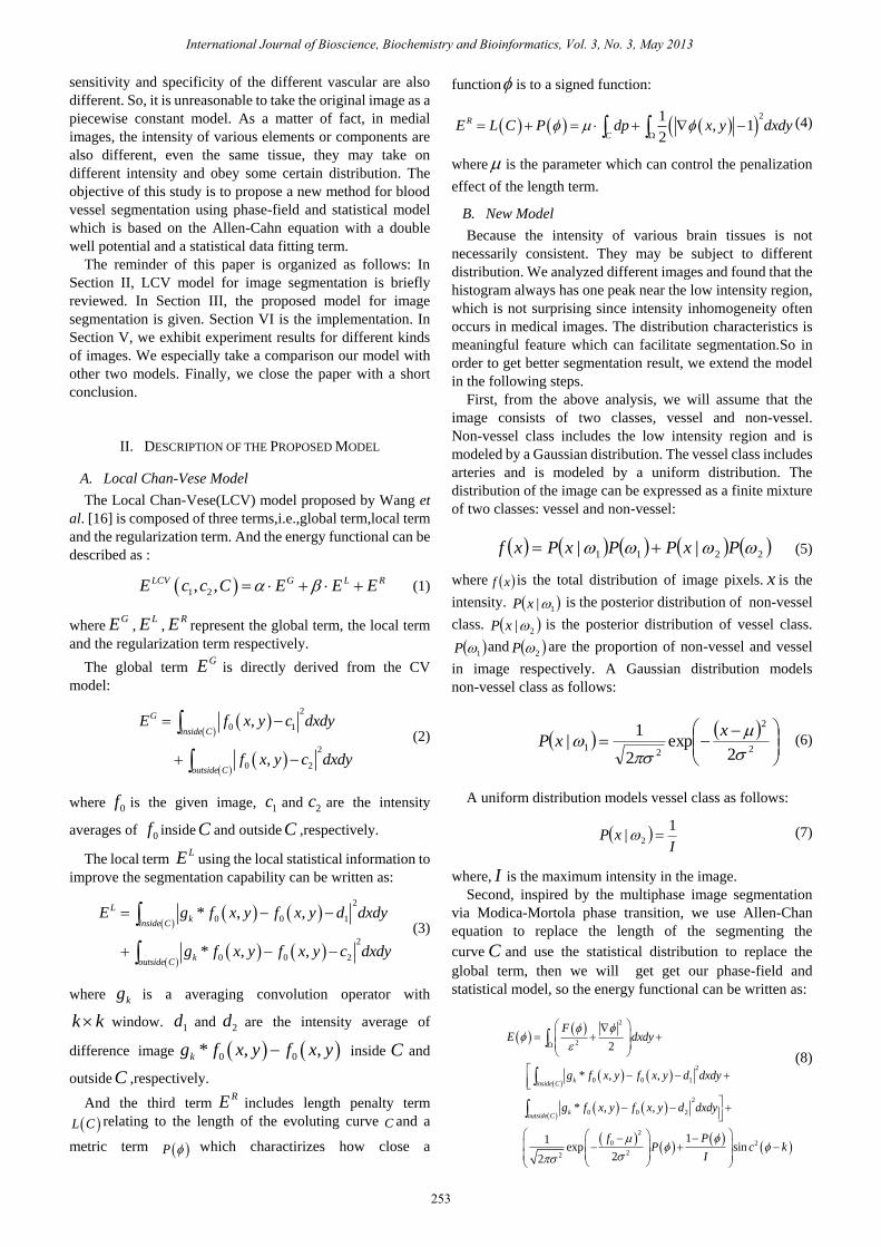
sensitivity and specificity of the different vascular are also
different. So, it is unreasonable to take the original image as a
piecewise constant model. As a matter of fact, in medial
images, the intensity of various elements or components are
also different, even the same tissue, they may take on
different intensity and obey some certain distribution. The
objective of this study is to propose a new method for blood
vessel segmentation using phase-field and statistical model
which is based on the Allen-Cahn equation with a double
well potential and a statistical data fitting term.
The reminder of this paper is organized as follows: In
Section II, LCV model for image segmentation is briefly
reviewed. In Section III, the proposed model for image
segmentation is given. Section VI is the implementation. In
Section V, we exhibit experiment results for different kinds
of images. We especially take a comparison our model with
other two models. Finally, we close the paper with a short
conclusion.
II. DESCRIPTION OF THE PROPOSED MODEL
A. Local Chan-Vese Model
The Local Chan-Vese(LCV) model proposed by Wang et
al. [16] is composed of three terms,i.e.,global term,local term
and the regularization term. And the energy functional can be
described as :
1 2, ,LCV G L RE c c C E E E (1)
whereGE ,
LE ,RE represent the global term, the local term
and the regularization term respectively.
The global term GE is directly derived from the CV
model:
2
0 1
2
0 2
,
,
G
inside C
outside C
E f x y c dxdy
f x y c dxdy
(2)
where 0f is the given image, 1c and 2c are the intensity
averages of 0f inside C and outside C ,respectively.
The local term LE using the local statistical information to
improve the segmentation capability can be written as:
2
0 0 1
2
0 0 2
* , ,
* , ,
L
kinside C
koutside C
E g f x y f x y d dxdy
g f x y f x y c dxdy
(3)
where kg is a averaging convolution operator with
k k window. 1d and 2d are the intensity average of
difference image 0 0* , ,kg f x y f x y inside C and
outside C ,respectively.
And the third termRE includes length penalty term
L C relating to the length of the evoluting curve C and a
metric term P which charactirizes how close a
function is to a signed function:
21
, 12
R
CE L C P dp x y dxdy
(4)
where is the parameter which can control the penalization
effect of the length term.
B. New Model
Because the intensity of various brain tissues is not
necessarily consistent. They may be subject to different
distribution. We analyzed different images and found that the
histogram always has one peak near the low intensity region,
which is not surprising since intensity inhomogeneity often
occurs in medical images. The distribution characteristics is
meaningful feature which can facilitate segmentation.So in
order to get better segmentation result, we extend the model
in the following steps.
First, from the above analysis, we will assume that the
image consists of two classes, vessel and non-vessel.
Non-vessel class includes the low intensity region and is
modeled by a Gaussian distribution. The vessel class includes
arteries and is modeled by a uniform distribution. The
distribution of the image can be expressed as a finite mixture
of two classes: vessel and non-vessel:
2211 || PxPPxPxf (5)
where xf is the total distribution of image pixels. x is the
intensity. 1|xP is the posterior distribution of non-vessel
class. 2|xP is the posterior distribution of vessel class.
1P and 2P are the proportion of non-vessel and vessel
in image respectively. A Gaussian distribution models
non-vessel class as follows:
2
2
21
2exp
2
1|
xxP (6)
A uniform distribution models vessel class as follows:
I
xP1
| 2 (7)
where, I is the maximum intensity in the image.
Second, inspired by the multiphase image segmentation
via Modica-Mortola phase transition, we use Allen-Chan
equation to replace the length of the segmenting the
curve C and use the statistical distribution to replace the
global term, then we will get get our phase-field and
statistical model, so the energy functional can be written as:
2
2
2
0 0 1
2
0 0 2
2
0 2
22
2
* , ,
* , ,
11exp sin
22
kinside C
koutside C
FE dxdy
g f x y f x y d dxdy
g f x y f x y d dxdy
f PP c k
I
(8)
International Journal of Bioscience, Biochemistry and Bioinformatics, Vol. 3, No. 3, May 2013
253

where 22 125.0 F is a double well potential. The
small constant is the gradient energy coefficient related to
the interfacial energy, is the image domain, P is the
proportion of non-vessel class in the image. 0f is the given
image, is the mean, 2 is the variance of the Gaussian
distribution, I is the maximum intensity in the image. And
the constant functions 1d , 2d are given as:
0 0
1
* , , ,
,
kg f x y f x y H x y dxdyd
H x y dxdy
(9)
0 0
2
* , , 1 ,
1 ,
kg f x y f x y H x y dxdyd
H x y dxdy
Once comes to a steady state, the evolving curve C will
separates the blood vessels form the background. From this
purpose, we seek a law of evolution in the form:
. gradEt (10)
The symbol „grad‟ here denotes the gradient in the space
2L .then we have:
'
2t
FgradE
2
0 0 1
2
0 0 2
* , ,
* , ,
k
k
g f x y f x y d
g f x y f x y d
2
0
22
11exp
22
f PP
I
2
2 32
sin 2 2sink k
k k
(11)
where N is the number of pixels of the image, ix is the
intensity of pixel i .
III. IMPLEMENTATION
In our numerical experiments, we normalize the given
image f as min0
max min
f ff
f f
,where
maxf and minf are the
maximum and the minimum values of the given image,
respectively. When the distribution is Gaussian, the
parameters of the distribution can be derived from the
following:
2
1
|( | )
|
n n
i k kn
k i n n
i j jj
P x PP x
P x P
(12)
1
1 1
1
1|
Nn n
i
i
P P xN
(13)
( 1) 1
1
|
|
Nn
i i in i
k Nn
i i
i
P x x
P x
(14)
21
1( 1) 2 1
1
1
|
( )
|
Nn n
i in i
Nn
i
i
P x x
P x
(15)
IV. EXPRIMENT RESULTS
In this section, the proposed algorithm has been tested on
real blood vessels. Fig. 1 shows the segmentation results for
three real blood vessel images with inhomogeneous intensity
via use of the the Local Binary Fitting mode[12], Local
Chan- Vese model[16] and the proposed method. In [16]
they had compared the CV model with the LCV model. So
we just compare LBF and LCV model with our proposed
model. It can be seen from the results that the proposed model
can achieve good segmenting results.
From the above experimental results, our method
illustrates the ability of segmenting image with the intensity
inhomogeneity and the images contain more perplexing
vascular. The three images all have the characteristics of
multi-branched. Because the real blood vessels in human
brain take on such features as complicated and arranged in a
crisscross pattern. As we all know,cerebral blood vessels are
composed of a large number of branched structures,which
form a highly entangled web. The methods used for blood
vessel segmentation discussed in introduction can work well
to deal with images of little size and the images just include
one or two banch vessels. But actually, to deal with images
containing more complicated branched blood vessels has
more practical significance.
In real vessel images, within blood vessels, the intensity
may be homogeneity, but surrongding the vicinity of vascular
edges, the intensity are usually different. Because the
background are normally composed of different brain tissues,
they generaly have different density. So using different
distribution functions to represent the different organizations
is meaningful.
(a) (b)
(c) (d)
(e) (f)
International Journal of Bioscience, Biochemistry and Bioinformatics, Vol. 3, No. 3, May 2013
254

International Journal of Bioscience, Biochemistry and Bioinformatics, Vol. 3, No. 3, May 2013
255
(g) (h)
(i) (j)
(k) (l)
Fig. 1. The comparisons of the three models on segmenting real blood image
with the intensity inhomogeneity . (a)(e)(f): original images. (b)(g)(h): final
segmentation result using LBF model. (c)(i)(j): final segmentation result
using LCV model. (d)(k)(l): final segmenting results using the proposed
model.
Their iteration number and processing time for segmenting
images in Fig. 1 are presented in Table I. It can be seen form
the table that the iteration numbers and the processing time
for the proposed model are both less than those of other two
models.
For the table above, we also come to that, our proposed
model not only has the ability of segmenting blood vessels
with intensity inhomogeneity, but also has more advantages
in handling larger images. When dealing with larger images,
our method can get better results with less iteration number
and less time.
TABLE I: THE PERFORMANCE COMPARISONS OF THREE MODELS
size =255*77(a) size =319*294(e) size =314*340(f)
Iteration
number
Total
time
Iteration
number
Total
time
Iteration
number
Total
time
LBF
model 20 16.7 50 52.2 100 88.9
LCV
model 20 14.4 20
22.5
6 80 87.6
Proposed
model 10 9.87 18 17.1 60 55.7
V. CONCLUSION
In this study, we propose a new model for blood vessel
image segmentation, which is based on the techniques of
curve evolution and global statistical distribution and level
set theory. The energy functional for the proposed model
consists of the double well potential and a statistical data
fitting term. By incorporating the global statistical
distribution information into the model, the images with
intensity inhomogeneity can be efficiently segmented. The
experiment results show that the proposed model is better
than other similar methods in medical image segmentation. In
our future work we will add more image properties and make
use of different geometry features to improve the
segmentation results and make our method more robust.
REFERENCES
[1] S. Kostopoulos et al., “A hybrid pixel-based classification method for
blood vessel segmentation and aneurysm detection on CTA,”
Computers & Graphics, vol. 31, issue 3, pp. 493–500, June 2007.
[2] K. Allen, C. Yau, and J. A. Noble, “A recursive stochastic vessel
segmentation framework that robustly handles bifurcations,” in Proc.
12th Annu. Conf. Medical Image Understanding and Analysis, Dundee,
Scotland, 2008.
[3] D. Mumford and J. Shah, “Optimal approximations of piecewise
smooth functions and associated variational problems (with J. Shah),”
Comm. in Pure and Appl. Math., vol. 42, pp. 577-685, 1989.
[4] L. M. Lorigo, O. D. Faugeras, W. E. L. Grimson, R. Keriven, R. Kikinis,
A. Nabavi, and C.-F. Westin, “CURVES: Curve evolution for vessel
segmentation, medical image analysis,” vol. 5, pp. 195-206, 2001.
[5] R. Malladi, J. A. Sethian, and B. Vemuri, “Shape Modeling with Front
Propagation: A Level Set Approach,” IEEE Trans. on Pattern Analysis
and Machine Intelligence, vol. 17, no. 2, February 1995.
[6] T. Chan and L. Vese, “Active contours without edges,” IEEE Trans.
Image Process , vol.10, issue 2, pp. 266–277, 2001.
[7] A. Tasi and A. Yezzi, “Curve evolution implementation of the
Mumford-Shah Functional for image segmentation, denoising,
interpolation and magnification,” IEEE transactions on image
processing, vol. 10. no. 8, pp. 1169-1186, August 2001.
[8] L. Vese and F .Tony, “A multiphase level set framework for image
segmentation using the mumford and shah model,” International
Journal of Computer Vision, vol.50, issue 3, pp. 271-293, 2002.
[9] X. Tai and F. Chan. “A survey on multiple level set methods with
applications for identifying piecewise constant functions,”
International Journal of Numerical Analysis and Modeling, vol. 1,
issue 1, pp. 25-47, 2004.
[10] G. Chung and L. Vese, “Energy minimization based segmentation and
denoising using multilayer level set approach,” LNCS, vol. 3757, pp.
439–455, 2005.
[11] L. Johan, L. Marius, and X. Tai, “A variant of the level set method and
applications to image segmentation,” Mathematics of Computation, vol.
75, pp. 1155-1174, 2006.
[12] C. M. Li, C. Kao, J. Gore, and Z. Ding, “Implicit active contours driven
by local binary fitting energy,” in Proc. Conf. Computer Vision and
Pattern, 2007.
[13] J. E. Solem, N. C. Overgaard, and A. Heyden, “Initialization techniques
for segmentation with the Chan–Vese model,” in Proc. 18th Conf.
Pattern Recognition, vol. 2, pp. 171–174, 2006.
[14] R. B. Xia, W. J. Liu, J. B. Zhao, and L. Li, “An optimal initialization
technique for improving the segmentation performance of Chan–Vese
model, ” in Proc. Confe. Automation and Logistics, pp. 411–415, 2007.
[15] Y. Pan, J. D. Birdwell, and S. M. Djouadi, “Efficient implementation of
the Chan–Vese models without solving PDEs,” in Proc. Workshop.
Multimedia Signal Processing, pp. 350–354, 2006.
[16] X. Wang, D. Huang, and H. Xu, “An efficient local chan-vese model
for image segmentation,” Pattern Recognition, vol. 43, issue 3, pp.
603-618, 2010.
[17] X. Feng and A. Prohl, “Numerical analysis of the Allen-Cahn equation
and approximation for mean curvature flows,” Numer. Math, vol. 94 pp.
33–65, 2003.
[18] S. Esedoglu and Y. H. R. Tsai, “Threshold dynamics for the piecewise
constant Mumford-Shah functional,” J. Comput. Phys., vol. 211, issue
1, pp. 367–384, 2006.
[19] J. Lie, M. Lysaker, and X. C. Tai, “A binary level set model and some
applications for Mumford-Shah image segmentation,” IEEE Trans.
Image Process., vol. 15, issue 4, pp. 1171–1181 ,2006.
[20] B. B. Thomas and C. Daniel, “On the statistical interpretation of the
piecewise smooth mumford-shah functiona, scale space and variational
methods in computer vision,” Lecture Notes in Computer Science, vol.
4485/2007, pp. 203-213, 2007.
[21] Y. Jung, S. Kang and J. Shen, “Multiphase Image Segmentation via
Modica-Mortola Phase transition,” SIAM applied Mathematics, vol. 67,
issue 5, pp. 1213-1232, 2007.
[22] F. Chen, Y. Chen and H. D. Tagare, “An extension of Sine-Sinc model
based on logarithm of likelihood,” in Proc. IPCV, vol. 1, pp. 222-227,
2008.
[23] Y. Li and J. Kim, “Multiphase image segmentation with a phase-field
model, Computers and Mathematics with Applications,” vol. 62, pp.
737-745, 2011.

International Journal of Bioscience, Biochemistry and Bioinformatics, Vol. 3, No. 3, May 2013
256
Shifeng Zhao received her B.Sc degree in Computer
Science and Technology (2004) from Qufu Normal
University, China. She obtained her M.Sc degree in
Computer Science and Technology (2007) from
Beijing Normal University, China. Since then, she has
been working with the College of Information Science
and Technology at BNU. Her research interests
include medical imaging and virtual reality.
Mingquan Zhou was born in Shanxi, China in 1954.
Mingquan Zhou received B. Sc in Mathematics and M.
Sc in Computer Software and Theory from Northwest
University, China, in 1976 and 1988 respectively.
Between 1992 and 1995, he was a visiting researcher
at the University of Bordeaux in France. Currently, he
is full Professor and Ph. D student supervisor in the
College of Information Science and Technology,
Beijing Normal University (BNU), China, where he is
also the dean of the College of Information Science and Technolgoy. Prof.
Zhou's current research interests include computer visualization, software
engineering and Chinese information processing, virtual reality and medical
imaging.
Zhongke Wu was born in Liaoning, China in 1965.
Zhongke Wu received B.Sc in Mathematics from
Peking University in China in 1988, and M. Eng and
PhD in CAD/CAM from Beijing University of
Aeronautics & Astronautics, China, in 1991 and 1995
respectively. Currently Zhongke WU, is Full Professor
and Ph. D student supervisor in College of Information
Science and Technology, Beijing Normal University
(BNU), China. Prior to joining in BNU, he worked in
Nanyang Technological University (NTU), Singapore, Institute National de
Recherche en Informatique et en Automatique (INRIA) in France, Institute
of High Performance Computing (IHPC) and Institute of Software, Chinese
Academy of Sciences in China from 1995 to 2006. Prof. WU has been
working in the field of computer graphics since 1988. He led and took part in
various research and development projects in computer graphics and related
areas. Prof. Wu's current research interests include computer graphics,
animation virtual reality, geometric modeling, volume graphics and medical
imaging.
Yun Tian received his B.Sc. degree in Computer
Science and Technology (2003) from Henan Normal
University, China, and his Ph. D. degree in Signal and
Information Processing (2007) from Northwestern
Polytechnic University, China. Currently, he is an
associate professor at the College of Information
Science and Technology, Beijing Normal University
(BNU), China. His research interests include medical
image processing and pattern recognition.
Lizhi Xie was born in 1984, doctoral level at Beijing
Normal University, Beijing (BNU), China. He works in
the field of medical image processing. In particular, his
research interests include volume rendering of medical
image, vascular segementation and vessel construction.