mosquito-borne viral pathogens detected in zambia: a
TRANSCRIPT
pathogens
Review
Mosquito-Borne Viral Pathogens Detected in Zambia:A Systematic Review
Rachel Milomba Velu 1,2,*, Geoffrey Kwenda 3,4, Liyali Libonda 5 , Caroline Cleopatra Chisenga 1,Bumbangi Nsoni Flavien 5 , Obvious Nchimunya Chilyabanyama 1 , Michelo Simunyandi 1,Samuel Bosomprah 1,6, Nicholus Chintu Sande 2, Katendi Changula 7 , Walter Muleya 8 ,Monicah Mirai Mburu 9, Benjamin Mubemba 10 , Simbarashe Chitanga 3,11,12 , John Tembo 13,Matthew Bates 13,14, Nathan Kapata 15, Yasuko Orba 16 , Masahiro Kajihara 17, Ayato Takada 2,4,17 ,Hirofumi Sawa 2,4,16,18,* , Roma Chilengi 1 and Edgar Simulundu 2,9
�����������������
Citation: Velu, R.M.; Kwenda, G.;
Libonda, L.; Chisenga, C.C.; Flavien,
B.N.; Chilyabanyama, O.N.;
Simunyandi, M.; Bosomprah, S.;
Sande, N.C.; Changula, K.; et al.
Mosquito-Borne Viral Pathogens
Detected in Zambia: A Systematic
Review. Pathogens 2021, 10, 1007.
https://doi.org/10.3390/
pathogens10081007
Academic Editor:
Andrew Taylor-Robinson
Received: 30 June 2021
Accepted: 5 August 2021
Published: 10 August 2021
Publisher’s Note: MDPI stays neutral
with regard to jurisdictional claims in
published maps and institutional affil-
iations.
Copyright: © 2021 by the authors.
Licensee MDPI, Basel, Switzerland.
This article is an open access article
distributed under the terms and
conditions of the Creative Commons
Attribution (CC BY) license (https://
creativecommons.org/licenses/by/
4.0/).
1 Centre for Infectious Disease Research in Zambia, Lusaka P.O. Box 34681, Zambia;[email protected] (C.C.C.); [email protected] (O.N.C.);[email protected] (M.S.); [email protected] (S.B.); [email protected] (R.C.)
2 Department of Disease Control, School of Veterinary Medicine, University of Zambia,Lusaka P.O. Box 32379, Zambia; [email protected] (N.C.S.); [email protected] (A.T.);[email protected] (E.S.)
3 Department of Biomedical Sciences, School of Health Sciences, University of Zambia,Lusaka P.O. Box 50110, Zambia; [email protected] (G.K.); [email protected] (S.C.)
4 Africa Center of Excellence for Infectious Diseases of Humans and Animals, University of Zambia,Lusaka P.O. Box 32379, Zambia
5 Department of Disease Control and Prevention, School of Medicine and Health Sciences, Eden University,Lusaka P.O. Box 37727, Zambia; [email protected] (L.L.); [email protected] (B.N.F.)
6 Department of Biostatistics, School of Public Health, University of Ghana, Accra P.O. Box LG13, Ghana7 Department of Paraclinical Studies, School of Veterinary Medicine, University of Zambia,
Lusaka P.O. Box 32379, Zambia; [email protected] Department of Biomedical Sciences, School of Veterinary Medicine, University of Zambia,
Lusaka P.O. Box 32379, Zambia; [email protected] Macha Research Trust, Choma P.O. Box 630166, Zambia; [email protected] Department of Zoology and Aquatic Sciences, School of Natural Resources, Copperbelt University,
Kitwe P.O. Box 21692, Zambia; [email protected] School of Veterinary Medicine, University of Namibia, Windhoek Private Bag 13301, Namibia12 School of Life Sciences, University of KwaZulu-Natal, Private Bag X54001, Durban 4000, South Africa13 HerpeZ Infection Research and Training, University Teaching Hospital, Lusaka Private Bag RW1X Ridgeway,
Lusaka P.O. Box 10101, Zambia; [email protected] (J.T.); [email protected] (M.B.)14 School of Life Sciences, University of Lincoln, Brayford Pool, Lincoln LN6 7TS, UK15 Zambia National Public Health Institute, Ministry of Health, Lusaka P.O. Box 30205, Zambia;
[email protected] Division of Molecular Pathobiology, International Institute for Zoonosis Control, Hokkaido University,
N 20 W10, Kita-ku, Sapporo 001-0020, Japan; [email protected] Division of Global Epidemiology, International Institute for Zoonosis Control, Hokkaido University,
N 20 W10, Kita-ku, Sapporo 001-0020, Japan; [email protected] Global Virus Network, 725 W Lombard St., Baltimore, MD 21201, USA* Correspondence: [email protected] (R.M.V.); [email protected] (H.S.)
Abstract: Emerging and re-emerging mosquito-borne viral diseases are a threat to global health. Thissystematic review aimed to investigate the available evidence of mosquito-borne viral pathogensreported in Zambia. A search of literature was conducted in PubMed and Google Scholar forarticles published from 1 January 1930 to 30 June 2020 using a combination of keywords. Eightmosquito-borne viruses belonging to three families, Togaviridae, Flaviviridae and Phenuiviridae werereported. Three viruses (Chikungunya virus, Mayaro virus, Mwinilunga virus) were reported amongthe togaviruses whilst four (dengue virus, West Nile virus, yellow fever virus, Zika virus) wereamong the flavivirus and only one virus, Rift Valley fever virus, was reported in the Phenuiviridaefamily. The majority of these mosquito-borne viruses were reported in Western and North-Westernprovinces. Aedes and Culex species were the main mosquito-borne viral vectors reported. Farming,fishing, movement of people and rain patterns were among factors associated with mosquito-borne
Pathogens 2021, 10, 1007. https://doi.org/10.3390/pathogens10081007 https://www.mdpi.com/journal/pathogens
Pathogens 2021, 10, 1007 2 of 15
viral infection in Zambia. Better diagnostic methods, such as the use of molecular tools, to detectthe viruses in potential vectors, humans, and animals, including the recognition of arboviral riskzones and how the viruses circulate, are important for improved surveillance and design of effectiveprevention and control measures.
Keywords: mosquito-borne; arboviruses; Togaviridae; Flaviviridae; Phenuiviridae; Zambia
1. Introduction
Arthropod-borne viruses (arboviruses) are transmitted to susceptible vertebrate hostsby hematophagous, or blood-sucking arthropods such as mosquitoes, sandflies, lice, ticksand fleas [1]. There are an estimated 700 known arboviruses among which about 100 areknown to cause infections in humans and animals [2].
Medically important arboviral infections mostly include flaviviral infections such asyellow fever, Zika virus disease, West Nile fever, and dengue fever. In addition, Chikun-gunya fever caused by Chikungunya virus, an alphavirus has caused many documentedoutbreaks in Africa, Asia, Europe, the South Pacific and recently the Caribbean region [3–5].The transmission dynamics of these viruses depend on several factors, which may varyfrom viral genetics to vector competence and ecological interactions between hosts andvectors [3,6].
Mosquitoes belonging to Aedes and Culex species play an important role in arboviraltransmission. For instance, Aedes aegypti and Aedes albopictus have been incriminated inthe transmission of prevalent arboviruses of medical importance. These include Chikun-gunya virus (CHIKV), dengue virus (DENV), and yellow Fever virus (YFV) [7]. On theother hand, Culex mosquitoes are principal vectors of many viruses including Japaneseencephalitis virus (JEV), West Nile virus (WNV), and St Louis encephalitis virus (SLEV)among others [8].
The clinical presentation of these viruses is non-specific and sometimes can lead tomisdiagnosis as most of them have a very similar clinical presentation. Symptoms caninclude fever, myalgia, polyarthralgia (migratory polyarthritis), rash, headache, photopho-bia, hyperemia and in some cases neurological complications such as meningitis, flaccidparalysis, and meningoencephalitis [9,10].
In sub-Saharan Africa, the presence of Aedes, Culex and Anopheles spp., transmittingmosquito-borne viral diseases and malaria are well established [11,12]. The co-circulationof these mosquito-borne viruses with other pathogens causing febrile illnesses such asmalaria poses a serious problem of diagnosis and management. This has led to an under-estimation of mosquito-borne viral infections as most of the febrile illnesses are treated asmalaria [4,13].
Due to her geographical location and tropical climate, Zambia hosts favourable breed-ing sites for mosquitoes that transmit malaria and mosquito-borne infections [14] and mostof these have been reported for instance, CHIKV was first reported in 1961 [15]. After along epidemiological silence of about three decades, serological studies have demonstratedevidence of exposure to other mosquito-borne viruses of public health importance such asdengue virus (DENV), yellow fever virus (YFV), Zika virus (ZIKV), Mayaro virus (MAYV)and West Nile virus (WNV) [16].
In light of recent global outbreaks, endemic conditions caused by these mosquito-borne viruses are likely to remain neglected and risk being forgotten. We undertook asystematic review to investigate the available evidence on the distribution of arbovirusesand their impact in Zambia, including pre-colonial period.
Pathogens 2021, 10, 1007 3 of 15
2. Results2.1. Electronic/Manual Searching
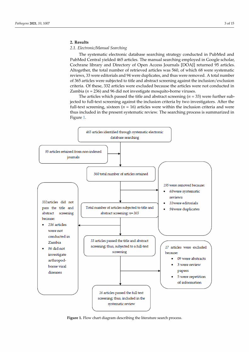
The systematic electronic database searching strategy conducted in PubMed andPubMed Central yielded 465 articles. The manual searching employed in Google scholar,Cochrane library and Directory of Open Access Journals [DOAJ] returned 95 articles.Altogether, the total number of retrieved articles was 560, of which 68 were systematicreviews, 33 were editorials and 94 were duplicates, and thus were removed. A total numberof 365 articles were subjected to title and abstract screening against the inclusion/exclusioncriteria. Of these, 332 articles were excluded because the articles were not conducted inZambia (n = 236) and 96 did not investigate mosquito-borne viruses.
The articles which passed the title and abstract screening (n = 33) were further sub-jected to full-text screening against the inclusion criteria by two investigators. After thefull-text screening, sixteen (n = 16) articles were within the inclusion criteria and werethus included in the present systematic review. The searching process is summarized inFigure 1.
Pathogens 2021, 10, x FOR PEER REVIEW 3 of 18
2.1. Electronic/Manual Searching The systematic electronic database searching strategy conducted in PubMed and
PubMed Central yielded 465 articles. The manual searching employed in Google scholar, Cochrane library and Directory of Open Access Journals [DOAJ] returned 95 articles. Altogether, the total number of retrieved articles was 560, of which 68 were systematic reviews, 33 were editorials and 94 were duplicates, and thus were removed. A total number of 365 articles were subjected to title and abstract screening against the inclu-sion/exclusion criteria. Of these, 332 articles were excluded because the articles were not conducted in Zambia (n = 236) and 96 did not investigate mosquito-borne viruses.
The articles which passed the title and abstract screening (n = 33) were further sub-jected to full-text screening against the inclusion criteria by two investigators. After the full-text screening, sixteen (n = 16) articles were within the inclusion criteria and were thus included in the present systematic review. The searching process is summarized in Figure 1.
Figure 1. Flow chart diagram describing the literature search process. Figure 1. Flow chart diagram describing the literature search process.
Pathogens 2021, 10, 1007 4 of 15
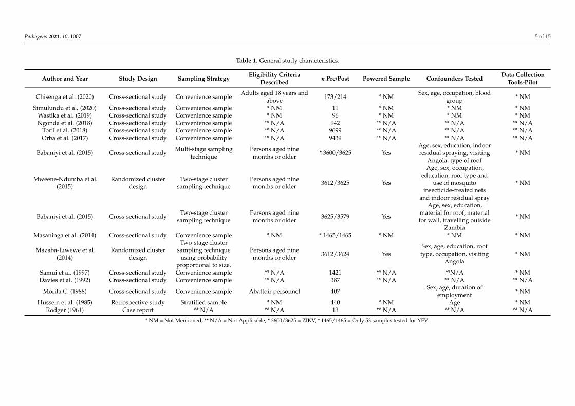
2.2. General Study Characteristics
A total of 16 studies were included in this review, and all the 16 studies described thesampling approaches used in selecting the sample. Most of the studies (12) were cross-sectionalstudies [16–27]. Seven research studies were conducted in humans [15,16,22,23,27–29] whilenine studies were conducted in animals and/or mosquitoes, and one study had bothhumans and animals [27].
The studies obtained in our search used different study designs as well as samplingstrategies to obtain data on the different mosquito-borne pathogens. The study designsemployed were cross sectional (12/16), randomized cluster (2/16), retrospective (1/16) andcase reports (1/16), with the sampling strategies used included convenience (10/16), two-stage cluster sampling (3/16), multi-stage (1/16), stratified (1/16) and a case report (1/16).
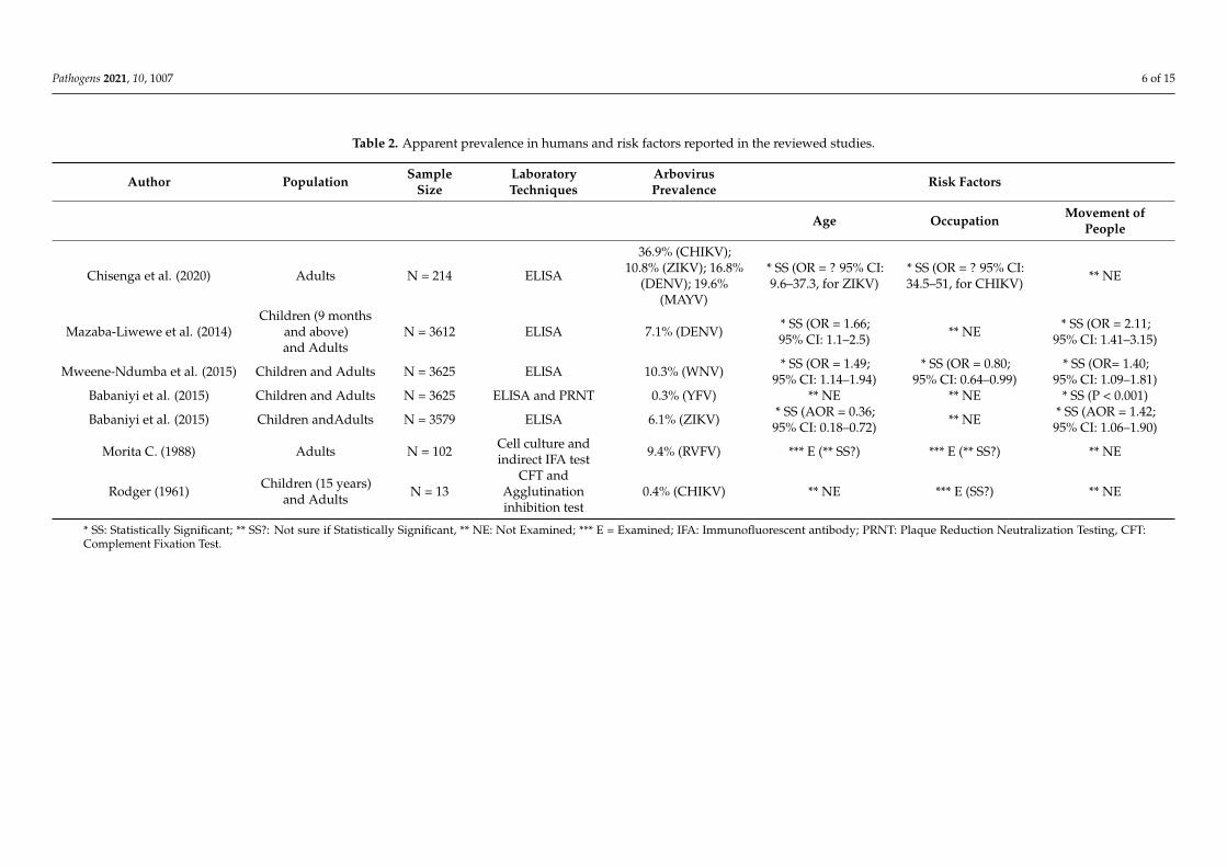
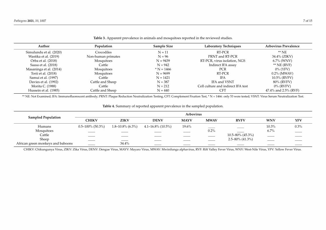
To determine association between prevalence of mosquito-borne viruses and somepotential risk factors, bivariate, multivariate and Pearson’s correlation tests were employedin the different studies. More details on study characteristics are provided in Table 1 whilstthe prevalence versus risk factors are summarised in Tables 2–4.
2.3. Geographical Location
Concerning geographical locations, seven of Zambia’s ten provinces reported mosquito-borne viruses. These include Copperbelt, Central, Eastern, Lusaka, North-Western, Southernand Western provinces. However, Western and North-Western provinces had the most com-mon sites for mosquito-borne virus studies as reported in eight and ten studies, respectively.
2.4. Mosquito-Borne Viruses
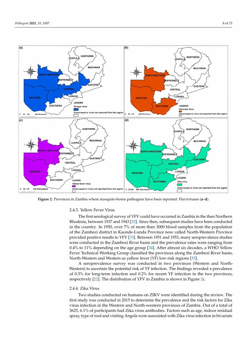
Eight mosquito-borne viruses belonging to three families, Togaviridae, Flaviviridaeand Phenuiviridae were recorded. Four viruses amid the flavivirus were reported DENV,WNV, YFV, and ZIKV (Figure 2). Among the togaviruses, CHIKV, MAYV and Mwinilungaalphavirus (MWAV) (Figure 3) were reported while among the phenuiviruses, only RVFVwas reported (Figure 3).
2.4.1. Dengue Virus
Evidence for the presence of dengue in Zambia was first shown serologically in1987 [30]. Since then, serological evidence of the virus has been reported in Western [28,31],North-Western [31] and Central Provinces [16] with reported prevalence ranging from 4.1%to 16.8%. Amongst the recognized risk factors for dengue virus infection in a population inWestern Province were the age, education and history of travel to Angola [28]. However,there is currently no knowledge of the serotypes circulating in the country [31]. Figure 2ashows the provinces of Zambia where DENV has been reported.
2.4.2. West Nile Virus
Serological evidence of WNV in Zambia was first reported in 2015, with a prevalenceof 10.3% [29]. This is the only study and report of WNV in humans in the country. Otherreports of WNV in Zambia were in Culex mosquitoes [21] at a prevalence of 6.7% and infarmed crocodiles [17]. Genetic characterization studies have revealed the presence oflineages 1a [17] and 2 [21] in the country. The identification of the virus in mosquitoesshows that the virus could be circulating within the communities, thus heightens the needfor proper understanding of disease epidemiology in those communities. Figure 2b showsthe provinces in Zambia where WNV has been reported.
Pathogens 2021, 10, 1007 5 of 15
Table 1. General study characteristics.
Author and Year Study Design Sampling Strategy Eligibility CriteriaDescribed n Pre/Post Powered Sample Confounders Tested Data Collection
Tools-Pilot
Chisenga et al. (2020) Cross-sectional study Convenience sample Adults aged 18 years andabove 173/214 * NM Sex, age, occupation, blood
group * NM
Simulundu et al. (2020) Cross-sectional study Convenience sample * NM 11 * NM * NM * NMWastika et al. (2019) Cross-sectional study Convenience sample * NM 96 * NM * NM * NMNgonda et al. (2018) Cross-sectional study Convenience sample ** N/A 942 ** N/A ** N/A ** N/A
Torii et al. (2018) Cross-sectional study Convenience sample ** N/A 9699 ** N/A ** N/A ** N/AOrba et al. (2017) Cross-sectional study Convenience sample ** N/A 9439 ** N/A ** N/A ** N/A
Babaniyi et al. (2015) Cross-sectional study Multi-stage samplingtechnique
Persons aged ninemonths or older * 3600/3625 Yes
Age, sex, education, indoorresidual spraying, visiting
Angola, type of roof* NM
Mweene-Ndumba et al.(2015)
Randomized clusterdesign
Two-stage clustersampling technique
Persons aged ninemonths or older 3612/3625 Yes
Age, sex, occupation,education, roof type and
use of mosquitoinsecticide-treated nets
and indoor residual spray
* NM
Babaniyi et al. (2015) Cross-sectional study Two-stage clustersampling technique
Persons aged ninemonths or older 3625/3579 Yes
Age, sex, education,material for roof, materialfor wall, travelling outside
Zambia
* NM
Masaninga et al. (2014) Cross-sectional study Convenience sample * NM * 1465/1465 * NM * NM * NM
Mazaba-Liwewe et al.(2014)
Randomized clusterdesign
Two-stage clustersampling technique
using probabilityproportional to size.
Persons aged ninemonths or older 3612/3624 Yes
Sex, age, education, rooftype, occupation, visiting
Angola* NM
Samui et al. (1997) Cross-sectional study Convenience sample ** N/A 1421 ** N/A **N/A * NMDavies et al. (1992) Cross-sectional study Convenience sample ** N/A 387 ** N/A ** N/A ** N/A
Morita C. (1988) Cross-sectional study Convenience sample Abattoir personnel 407 Sex, age, duration ofemployment * NM
Hussein et al. (1985) Retrospective study Stratified sample * NM 440 * NM Age * NMRodger (1961) Case report ** N/A ** N/A 13 ** N/A ** N/A ** N/A
* NM = Not Mentioned, ** N/A = Not Applicable, * 3600/3625 = ZIKV, * 1465/1465 = Only 53 samples tested for YFV.
Pathogens 2021, 10, 1007 6 of 15
Table 2. Apparent prevalence in humans and risk factors reported in the reviewed studies.
Author Population SampleSize
LaboratoryTechniques
ArbovirusPrevalence Risk Factors
Age Occupation Movement ofPeople
Chisenga et al. (2020) Adults N = 214 ELISA
36.9% (CHIKV);10.8% (ZIKV); 16.8%
(DENV); 19.6%(MAYV)
* SS (OR = ? 95% CI:9.6–37.3, for ZIKV)
* SS (OR = ? 95% CI:34.5–51, for CHIKV) ** NE
Mazaba-Liwewe et al. (2014)Children (9 months
and above)and Adults
N = 3612 ELISA 7.1% (DENV) * SS (OR = 1.66;95% CI: 1.1–2.5) ** NE * SS (OR = 2.11;
95% CI: 1.41–3.15)
Mweene-Ndumba et al. (2015) Children and Adults N = 3625 ELISA 10.3% (WNV) * SS (OR = 1.49;95% CI: 1.14–1.94)
* SS (OR = 0.80;95% CI: 0.64–0.99)
* SS (OR= 1.40;95% CI: 1.09–1.81)
Babaniyi et al. (2015) Children and Adults N = 3625 ELISA and PRNT 0.3% (YFV) ** NE ** NE * SS (P < 0.001)
Babaniyi et al. (2015) Children andAdults N = 3579 ELISA 6.1% (ZIKV) * SS (AOR = 0.36;95% CI: 0.18–0.72) ** NE * SS (AOR = 1.42;
95% CI: 1.06–1.90)
Morita C. (1988) Adults N = 102 Cell culture andindirect IFA test 9.4% (RVFV) *** E (** SS?) *** E (** SS?) ** NE
Rodger (1961) Children (15 years)and Adults N = 13
CFT andAgglutinationinhibition test
0.4% (CHIKV) ** NE *** E (SS?) ** NE
* SS: Statistically Significant; ** SS?: Not sure if Statistically Significant, ** NE: Not Examined; *** E = Examined; IFA: Immunofluorescent antibody; PRNT: Plaque Reduction Neutralization Testing, CFT:Complement Fixation Test.
Pathogens 2021, 10, 1007 7 of 15
Table 3. Apparent prevalence in animals and mosquitoes reported in the reviewed studies.
Author Population Sample Size Laboratory Techniques Arbovirus Prevalence
Simulundu et al. (2020) Crocodiles N = 11 RT-PCR ** NEWastika et al. (2019) Non-human primates N = 96 PRNT and RT-PCR 34.4% (ZIKV)
Orba et al. (2018) Mosquitoes N = 9439 RT-PCR, virus isolation, NGS 6.7% (WNV)Saasa et al. (2018) Cattle N = 942 Indirect IFA assay ** NE (RVF)
Masaninga et al. (2014) Mosquitoes * N = 1466 PCR 0% (YFV)Torii et al. (2018) Mosquitoes N = 9699 RT-PCR 0.2% (MWAV)
Samui et al. (1997) Cattle N = 1421 IFA 10.5% (RVFV)Davies et al. (1992) Cattle and Sheep N = 387 IFA and VSNT 80% (RVFV)
Morita C. (1988) Cattle N = 212 Cell culture and indirect IFA test 0% (RVFV)Hussein et al. (1985) Cattle and Sheep N = 440 CFT 47.4% and 2.5% (RVF)
** NE: Not Examined; IFA: Immunofluorescent antibody; PRNT: Plaque Reduction Neutralization Testing, CFT: Complement Fixation Test, * N = 1466: only 53 were tested; VSNT: Virus Serum Neutralization Test.
Table 4. Summary of reported apparent prevalence in the sampled population.
Sampled PopulationArbovirus
CHIKV ZIKV DENV MAYV MWAV RVFV WNV YFV
Humans 0.5–100% (50.3%) 1.8–10.8% (6.3%) 4.1–16.8% (10.5%) 19.6% ____ ____ 10.3% 0.3%Mosquitoes ____ ____ ____ ____ 0.2% ____ 6.7% ____
Cattle ____ ____ ____ ____ ____ 10.5–80% (45.3%) ____ ____Sheep ____ ____ ____ ____ ____ 2.5–80% (41.3%) ____ ____
African green monkeys and baboons ____ 34.4% ____ ____ ____ ____ ____ ____
CHIKV: Chikungunya Virus, ZIKV: Zika Virus, DENV: Dengue Virus, MAYV: Mayaro Virus, MWAV: Mwinilunga alphavirus, RVF: Rift Valley Fever Virus, WNV: West-Nile Virus, YFV: Yellow Fever Virus.
Pathogens 2021, 10, 1007 8 of 15
Pathogens 2021, 10, x FOR PEER REVIEW 10 of 18
2.3. Geographical Location Concerning geographical locations, seven of Zambia’s ten provinces reported
mosquito-borne viruses. These include Copperbelt, Central, Eastern, Lusaka, North-Western, Southern and Western provinces. However, Western and North-Western provinces had the most common sites for mosquito-borne virus studies as reported in eight and ten studies, respectively.
2.4. Mosquito-Borne Viruses Eight mosquito-borne viruses belonging to three families, Togaviridae, Flaviviridae
and Phenuiviridae were recorded. Four viruses amid the flavivirus were reported DENV, WNV, YFV, and ZIKV (Figure 2). Among the togaviruses, CHIKV, MAYV and Mwini-lunga alphavirus (MWAV) (Figure 3) were reported while among the phenuiviruses, only RVFV was reported (Figure 3).
Figure 2. Provinces in Zambia where mosquito-borne pathogens have been reported: Flaviviruses (a–d).
2.4.1. Dengue Virus Evidence for the presence of dengue in Zambia was first shown serologically in 1987
[30]. Since then, serological evidence of the virus has been reported in Western [28,31], North-Western [31] and Central Provinces [16] with reported prevalence ranging from 4.1% to 16.8%. Amongst the recognized risk factors for dengue virus infection in a pop-ulation in Western Province were the age, education and history of travel to Angola [28].
Figure 2. Provinces in Zambia where mosquito-borne pathogens have been reported: Flaviviruses (a–d).
2.4.3. Yellow Fever Virus
The first serological survey of YFV could have occurred in Zambia in the then NorthernRhodesia, between 1937 and 1943 [32]. Since then, subsequent studies have been conductedin the country. In 1950, over 7% of more than 3000 blood samples from the populationof the Zambezi district in Kaonde-Lunda Province now called North-Western Provinceprovided positive results to YFV [33]. Between 1951 and 1953, many seroprevalence studieswere conducted in the Zambezi River basin and the prevalence rates were ranging from0.4% to 11% depending on the age group [34]. After almost six decades, a WHO YellowFever Technical Working Group classified the provinces along the Zambezi River basin,North-Western and Western as yellow fever (YF) low-risk regions [35].
A seroprevalence survey was conducted in two provinces (Western and North-Western) to ascertain the potential risk of YF infection. The findings revealed a prevalenceof 0.3% for long-term infection and 0.2% for recent YF infection in the two provinces,respectively [22]. The distribution of YFV in Zambia is shown in Figure 2c.
2.4.4. Zika Virus
Two studies conducted on humans on ZIKV were identified during the review. Thefirst study was conducted in 2015 to determine the prevalence and the risk factors for Zikavirus infection in the Western and North-western provinces of Zambia. Out of a total of3625, 6.1% of participants had Zika virus antibodies. Factors such as age, indoor residualspray, type of roof and visiting Angola were associated with Zika virus infection in bivariate
Pathogens 2021, 10, 1007 9 of 15
analyses [36]. In the second study, Chisenga et al. reported a ZIKV seroprevalence of 10.8%(23/214) from serum samples collected in Central Province [16]. In 2019, 96 Non-humanprimates (African green monkeys and baboons) from Southern and Eastern provinceswere tested for ZIKV using plaque reduction neutralization test and RT-PCR. The findingsrevealed that 34.4% of their sera had neutralizing antibodies against ZIKV whereas theZIKV genomic RNA was not detected using RT-PCR [18]. The distribution of ZIKV acrossthe country is summarised in Figure 2d.
2.4.5. Chikungunya Virus
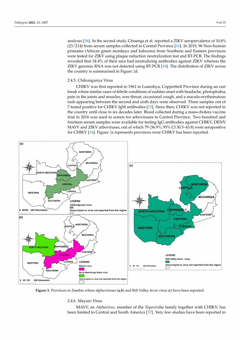
CHIKV was first reported in 1961 in Luanshya, Copperbelt Province during an out-break where similar cases of febrile conditions of sudden onset with headache, photophobia,pain in the joints and muscles, sore throat, occasional cough, and a maculo-erythematousrash appearing between the second and sixth days were observed. Three samples out of7 tested positive for CHIKV IgM antibodies [15]. Since then, CHIKV was not reported inthe country until close to six decades later. Blood collected during a mass-cholera vaccinetrial in 2016 was used to screen for arboviruses in Central Province. Two hundred andfourteen serum samples were available for testing IgG antibodies against CHIKV, DENV,MAYV and ZIKV arboviruses, out of which 79 (36.9%; 95% CI 30.5–43.8) were seropositivefor CHIKV [16]. Figure 3a represents provinces were CHIKV has been reported.
Pathogens 2021, 10, x FOR PEER REVIEW 12 of 18
30.5–43.8) were seropositive for CHIKV [16]. Figure 3a represents provinces were CHIKV has been reported.
2.4.6. Mayaro Virus MAYV, an Alphavirus, member of the Togaviridae family together with CHIKV, has
been limited to Central and South America [37]. Very few studies have been reported in Africa. In Zambia, one study, conducted in Central Province (Figure 3b) and published in 2020 is the only record of possible MAYV presence in the country. Its prevalence was es-timated to be 19.6% (42/214) [16].
2.4.7. Mwinilunga Alphavirus Mwinilunga alphavirus (MWAV) is a newly discovered virus. It was first reported
from a single Culex quinquefasciatus mosquito pool in Zambia in 2018, and its genome has since been sequenced. From the 9699 mosquitoes collected in North-Western Province (Figure 3b), a 0.2% prevalence of the virus was obtained [20]. However, studies in hu-mans have not been conducted, and its pathogenicity in human or other vertebrates is not well established.
Figure 3. Provinces in Zambia where alphaviruses (a,b) and Rift Valley fever virus (c) have been reported.
2.4.8. Rift Valley Fever Virus Rift Valley fever virus (RVFV) was first reported in 1985 after a seroprevalence study
conducted in 440 cattle and sheep in Central Province using a complement fixation test [38]. In 1992, serum samples from cattle and sheep, sampled from Lusaka, Copperbelt
Figure 3. Provinces in Zambia where alphaviruses (a,b) and Rift Valley fever virus (c) have been reported.
2.4.6. Mayaro Virus
MAYV, an Alphavirus, member of the Togaviridae family together with CHIKV, hasbeen limited to Central and South America [37]. Very few studies have been reported in
Pathogens 2021, 10, 1007 10 of 15
Africa. In Zambia, one study, conducted in Central Province (Figure 3b) and publishedin 2020 is the only record of possible MAYV presence in the country. Its prevalence wasestimated to be 19.6% (42/214) [16].
2.4.7. Mwinilunga Alphavirus
Mwinilunga alphavirus (MWAV) is a newly discovered virus. It was first reportedfrom a single Culex quinquefasciatus mosquito pool in Zambia in 2018, and its genome hassince been sequenced. From the 9699 mosquitoes collected in North-Western Province(Figure 3b), a 0.2% prevalence of the virus was obtained [20]. However, studies in humanshave not been conducted, and its pathogenicity in human or other vertebrates is notwell established.
2.4.8. Rift Valley Fever Virus
Rift Valley fever virus (RVFV) was first reported in 1985 after a seroprevalence studyconducted in 440 cattle and sheep in Central Province using a complement fixation test [38].In 1992, serum samples from cattle and sheep, sampled from Lusaka, Copperbelt andCentral provinces showed evidence of epizootic Rift Valley fever (RVF) in Zambia [26].Five years later, Samui et al. reported a positivity rate of 10.5% to RVFV in 27 herds ofcattle [25].
Using a recently developed diagnostic tool on a recombinant nucleocapsid protein(rNP)-based indirect immunofluorescent antibody assay (IFA), it was found that the sero-prevalence of RVF varied between 6.0% to 21.4% among cattle herds in Central, Southernand Western provinces [19].
A study conducted on humans in 1987 showed that 5 out of 53 (9.4%) workers at anabattoir dealing with cattle in Lusaka were seropositive for RVF, but none of the workers atthe abattoirs dealing with pigs was shown to be positive [27]. Figure 3c shows the locationswhere RVF has been reported in the country.
2.5. Factors Associated with Mosquito-Borne Viruses’ Distribution in Zambia
Several factors have been associated with mosquito-borne viral infections globallyand in Zambia. Anthropological activities, such as deforestation, land use patterns, demo-graphic density, global trade, and global warming (climate change) have all successfullycontributed to the emergence and re-emergence of these viruses [39]. Human activities in-cluding farming and fishing have been incriminated in CHIKV infection in the Copperbeltand Central Provinces [15,16].
The movement of people to neighbouring countries is most likely contributing toarboviral activity in Zambia. For instance, visiting Angola was associated with ZIKV andDENV infections [28,36], whereas travelling to the Democratic Republic of Congo (DRC)and South Africa was significantly related to YFV infection [22]
The transmission dynamic of mosquito-borne viruses has also been associated with cli-mate conditions. Most of RVF epidemics have been reported during the rainy seasons [25,26].
2.6. Mosquito Species from Which Potential Zoonotic Arboviruses Have Been Detected in Zambia
After the first record of mosquitoes belonging to Aedes species in 1950 [33], infor-mation has been scarce regarding the distribution and species composition of mosquitovectors in Zambia. An entomological investigation conducted in North-Western andNorthern provinces to identify yellow fever vectors showed that Aedes (Stegomyia) aegypti(Ae. aegypti) and Aedes (Stegomyia) africanus, the two main vectors of YFV, were presentin the study sites in low densities [24]. Other mosquito species identified included Aedes(Aedimorphus) mutilus; Aedes (Aedimorphus) minutus, Aedes (Finlaya) wellmani, Culex species(Cx. quinquefasciatus) and Mansonia species (Mansonia africanus).
Mosquito-borne viruses have been detected in mosquitoes on only two occasionsin Zambia. The first occasion was in 2017, where the WNV lineage 2 strain was iso-lated in a pool of Culex mosquitoes collected from Western Province [21]. Furthermore,
Pathogens 2021, 10, 1007 11 of 15
a novel alphavirus, tentatively named Mwinilunga alphavirus was identified from Culexquinquefasciatus mosquitoes in 2018 [20]. This discovery highlights the necessity of con-ducting robust entomological surveillance in the country for an improved understandingof mosquito-borne viral transmission dynamics in Zambia.
3. Discussion
The evidence gathered in this review indicated that a considerable array of mosquito-borne viruses have been detected in Zambia. Although no epidemics have been reportedin the country, there is evidence of an on-going mosquito-borne viral activity, mostly inNorth-Western, Western and Southern provinces [17,20–22,25,31]. It was interesting tonote that Muchinga Province did not report any form of mosquito-borne viral activity,seroprevalence evidence or virus isolation. Whichever the case, it is cardinal to do studiesin this part of the country to ensure an understanding of the prevailing situation.
Although most of the research studies did not elaborately highlight climatic conditionsto influence the distribution of mosquitoes, Masaninga et al. (2014) observed climatic factorssuch as precipitations and temperature to somewhat influence their distribution. Further, itwas noted that North-Western, Western, and Southern provinces host favourable breedingsites for mosquitoes due to their humid conditions, increasing the risk of mosquito-borneviruses [24]. This observation is in agreement with studies on DENV infection in Africawhich hypothesised climatological patterns to favour vector development and longevity ashumid warm tropical regions promote egg conservation and proliferation [40].
Following the analysis of the rainfall patterns, it was shown that provinces whichreceive on average 900 mm or higher than 1000 mm of rainfall are predisposed to high inci-dences of mosquito-borne diseases due to an increase in vector diversity and activities [41].RVF epidemics reported in 1992 and 1997 by Davies et al. (1992) and Samui et al. (1997),respectively, were related to high precipitation. Similar observations were noted in Kenyawhere RVF epizootics were associated with relatively high levels of rainfall [42,43].
Of note, human activities such as deforestation, trade and movement of people andanimals have been associated with the introduction of mosquito-borne viruses in previouslynon-endemic regions [44,45]. Human movement has also been linked to ZIKV, DENVand YFV seropositivity in Zambia [28,46]. This is also plausible because the countryshares borders with countries where major outbreaks of these arboviruses have beenpreviously reported [44,47]. It is also reasonable that livestock trade between Zambiaand her neighbouring countries likely promulgates the distribution of mosquito-borneviruses [41].
However, although different methods were used in the studies included with re-gards to detecting mosquito-borne viruses and thus introducing heterogeneity, the reviewlargely revealed information on circulating mosquito-borne viruses and their role in thetransmission dynamics of mosquito-borne viral infections; therefore, conducting moreentomological studies to investigate the vector population and associated infections iscrucial to control mosquito-borne viral diseases.
4. Materials and Methods4.1. Search Strategy
Using PRISMA guidelines for systematic review, we searched for information relatedto mosquito-borne viral pathogens found in Zambia on PubMed and PubMed Centralelectronic databases from 1 January 1930 to 30 June 2020. A manual search was done onGoogle scholar, Cochrane library and Directory of Open Access Journals [DOAJ].
The search strategy involved a combination of keywords (“Northern Rhodesia” OR“Republic of Zambia”) AND (“Mosquito-borne virus” OR “Arthropod-borne virus” OR“Arbovirus” OR “Dengue virus” OR “Yellow fever virus” OR “Chikungunya virus” OR“West-Nile virus” OR “Rift Valley fever virus” OR “Zika virus” OR “Mayaro virus” OR“flavivirus” OR “phlebovirus” OR “alphavirus” OR “bunyavirus”) AND (“Epidemiology”OR “Prevalence” OR “Distribution”).
Pathogens 2021, 10, 1007 12 of 15
The articles retained were populated in RefWorks (2020) database manager, andduplicates were automatically removed. Titles and abstracts of retained articles weresubjected to the inclusion and exclusion criteria.
4.2. Inclusion Criteria
All primary studies which focused on the occurrence and distribution of mosquito-borne viral diseases in Zambia from 1 January 1930 to 30 June December 2020 published inpeer-reviewed journals, in either English or French languages, were included. Demographichealth surveys indicating the burden of mosquito-borne viral diseases were also considered.
4.3. Exclusion Criteria
Studies published in languages other than English or French, or lacking extractabledata or not explicit in methodology, and systematic review papers were excluded. Abstractswithout full manuscript texts were also excluded.
4.4. Outcome Measures
The primary outcome was the distribution of mosquito-borne viral diseases. Thesecondary outcomes were the risk factors influencing their distribution.
4.5. Data Extraction
Potentially eligible articles were selected and screened using their title and abstract bytwo independent reviewers (RV and LL). The articles were divided into 2 subgroups “in-cluded” and “excluded” using the set inclusion and exclusion criteria. The final inclusionwas done by analysing the full texts of the included articles. When necessary, any disagree-ments were resolved by arbitration of the third reviewer (BF). Data was extracted usingan extraction tool from the Joana Briggs Institute Reviewers Manual (2018) for prevalencestudies. Information regarding the authors, location, study design, characteristics of thesampled population, diagnostic tests used, and specific results was extracted and enteredinto an Excel sheet.
4.6. Distribution Mapping
The data extracted was used to create a map of the distribution and/or occurrence ofmosquito-borne viruses across the country using ArcGIS version 10.3 (Figures 2 and 3).
4.7. Data Synthesis
A narrative summary of included studies was done by pooling the raw data with anemphasis on reporting their characteristics along with data extracted relevant to the reviewoutcomes. Analysis of quantitative studies was done based on the heterogeneity of theincluded studies.
5. Conclusions
The findings of this review demonstrate that mosquito-borne viruses constitute a pub-lic health threat to the country. Despite this threat, very few studies have been conductedto understand the virus, vector and reservoir host interactions and dynamics. Though noepidemic has been reported as yet, favourable ecological factors noted in the country maylead to a rise in cases of mosquito-borne viral infections. Accurate information regardingthe epidemiology and ecology of mosquito-borne viruses is of critical importance for imple-menting suitable surveillance strategies, prophylactic treatments, travel recommendationsand clinical therapies. Furthermore, better detection methods, such as molecular tools, todetect the viruses in potential vectors, humans and animals, including the recognition ofarboviral zones and how the viruses circulate, are important for improved surveillance andbetter appreciation of the impact of these viruses on animals and humans.
Pathogens 2021, 10, 1007 13 of 15
Author Contributions: Conceptualization, R.M.V., E.S. and L.L.; methodology, R.M.V., L.L. andB.N.F.; writing—original draft preparation, R.M.V. and C.C.C.; writing—review and editing, B.N.F.,C.C.C., O.N.C., M.S., S.B., N.C.S., K.C., W.M., M.M.M., B.M., S.C., J.T., M.B., N.K., Y.O., M.K., R.C.and E.S.; supervision, E.S., R.C., H.S. and G.K.; funding acquisition, H.S., A.T. and E.S. All authorshave read and agreed to the published version of the manuscript.
Funding: R.M.V. was supported by the Africa Center of Excellence for Infectious Disease of Hu-mans and Animals (ACEIDHA) project (grant number P151847) funded by the World Bank. Thework was also supported by the Japan Program for Infectious Diseases Research and Infrastructure(JP21wm0125008) from Japan Agency for Medical Research and Development (AMED).
Institutional Review Board Statement: Not applicable.
Informed Consent Statement: Not applicable.
Data Availability Statement: Data is contained within the article.
Acknowledgments: R.M.V. would like to thank the Centre for Infectious Disease Research in Zambia(CIDRZ) for their support.
Conflicts of Interest: The authors declare no conflict of interest.
References1. Conway, M.J.; Colpitts, T.M.; Fikrig, E. Role of the vector in arbovirus transmission. Ann. Rev. Virol. 2014, 1, 71–88. [CrossRef]2. Chandler, L.J. Arthropod-Borne Virus Information Exchange December 1997; Centers for Disease Control and Prevention (U.S.gov):
Atlanta, GA, USA, 1997.3. Weaver, S.C.; Reisen, W.K. Present and future arboviral threats. Antivir. Res. 2010, 85, 328–345. [CrossRef]4. Marchi, S.; Trombetta, C.M.; Montomoli, E. Emerging and Re-Emerging Arboviral Diseases as a Global Health Problem; Public Health;
Majumder, M.A.A., Ed.; IntechOpen: London, UK, 2018; pp. 25–46.5. Heinrich, N.; Saathoff, E.; Weller, N.; Clowes, P.; Kroidl, I.; Ntinginya, E.; Machibya, H.; Maboko, L.; Löscher, T.; Dobler, G. High
seroprevalence of Rift Valley fever and evidence for endemic circulation in Mbeya region, Tanzania, in a cross-sectional study.PLoS Negl. Trop. Dis. 2012, 6, e1557. [CrossRef] [PubMed]
6. Gan, V.; Leo, Y.S. Current epidemiology and clinical practice in arboviral infections-implications on blood supply in South-East Asia. ISBT Sci. Ser. 2014, 9, 262–267. [CrossRef] [PubMed]
7. Agarwal, A.; Parida, M.; Dash, P.K. Impact of transmission cycles and vector competence on global expansion and emergence ofarboviruses. Rev. Med.Virol. 2017, 27, e1941. [CrossRef]
8. Gould, E.; Pettersson, J.; Higgs, S.; Charrel, R.; De Lamballerie, X. Emerging arboviruses: Why today? One Health 2017, 4, 1–13.[CrossRef]
9. Petersen, L.R.; Marfin, A.A. West Nile virus: A primer for the clinician. Ann. Intern. Med. 2002, 137, 173–179. [CrossRef] [PubMed]10. Hayes, E.B.; Sejvar, J.J.; Zaki, S.R.; Lanciotti, R.S.; Bode, A.V.; Campbell, G.L. Virology, pathology, and clinical manifestations of
West Nile virus disease. Emerg. Infect. Dis. 2005, 11, 1174. [CrossRef] [PubMed]11. Weetman, D.; Kamgang, B.; Badolo, A.; Moyes, C.L.; Shearer, F.M.; Coulibaly, M.; Pinto, J.; Lambrechts, L.; McCall, P.J. Aedes
mosquitoes and Aedes-borne arboviruses in Africa: Current and future threats. Int. J. Environ. Res. Public Health 2018, 15, 220.[CrossRef]
12. Kamgang, B.; Vazeille, M.; Tedjou, A.; Yougang, A.P.; Wilson-Bahun, T.A.; Mousson, L.; Wondji, C.S.; Failloux, A.-B. Differentpopulations of Aedes aegypti and Aedes albopictus (Diptera: Culicidae) from Central Africa are susceptible to Zika virus infection.PLoS Negl. Trop. Dis. 2020, 14, e0008163. [CrossRef]
13. Ndhlovu, M.; Nkhama, E.; Miller, J.M.; Hamer, D.H. Antibiotic prescribing practices for patients with fever in the transitionfrom presumptive treatment of malaria to ‘confirm and treat’ in Zambia: A cross-sectional study. Trop. Med. Int. Health 2015, 20,1696–1706. [CrossRef]
14. Kamuliwo, M.; Babaniyi, O.A. Larval habitat distribution: Aedes mosquito vector for arboviruses and Culex spps in North-Western and Western provinces of Zambia. Int. Public Health J. 2016, 8, 51.
15. Rodger, L. An outbreak of suspected chikungunya fever in nothern Rhodesia. S. Afr. Med. J. 1961, 35, 126–128.16. Chisenga, C.C.; Bosomprah, S.; Musukuma, K.; Mubanga, C.; Chilyabanyama, O.N.; Velu, R.M.; Kim, Y.C.; Reyes-Sandoval, A.;
Chilengi, R. Sero-prevalence of arthropod-borne viral infections among Lukanga swamp residents in Zambia. PLoS ONE 2020,15, e0235322. [CrossRef]
17. Simulundu, E.; Ndashe, K.; Chambaro, H.M.; Squarre, D.; Reilly, P.M.; Chitanga, S.; Changula, K.; Mukubesa, A.N.; Ndebe, J.;Tembo, J. West Nile Virus in Farmed Crocodiles, Zambia, 2019. Emerg. Infect. Dis. 2020, 26, 811. [CrossRef] [PubMed]
18. Wastika, C.E.; Sasaki, M.; Yoshii, K.; Anindita, P.D.; Hang’ombe, B.M.; Mweene, A.S.; Kobayashi, S.; Kariwa, H.; Carr, M.J.; Hall,W.W. Serological evidence of Zika virus infection in non-human primates in Zambia. Arch.Virol. 2019, 164, 2165–2170. [CrossRef]
Pathogens 2021, 10, 1007 14 of 15
19. Saasa, N.; Kajihara, M.; Dautu, G.; Mori-Kajihara, A.; Fukushi, S.; Sinkala, Y.; Morikawa, S.; Mweene, A.; Takada, A.; Yoshimatsu,K. Expression of a recombinant nucleocapsid protein of Rift Valley fever virus in Vero cells as an immunofluorescence antigenand its use for serosurveillance in traditional cattle herds in Zambia. Vector-Borne Zoonotic Dis. 2018, 18, 273–277. [CrossRef][PubMed]
20. Torii, S.; Orba, Y.; Hang’ombe, B.M.; Mweene, A.S.; Wada, Y.; Anindita, P.D.; Phongphaew, W.; Qiu, Y.; Kajihara, M.; Mori-Kajihara, A. Discovery of Mwinilunga alphavirus: A novel alphavirus in Culex mosquitoes in Zambia. Virus Res. 2018, 250, 31–36.[CrossRef] [PubMed]
21. Orba, Y.; Hang’ombe, B.; Mweene, A.; Wada, Y.; Anindita, P.; Phongphaew, W.; Qiu, Y.; Kajihara, M.; Mori-Kajihara, A.; Eto, Y.First isolation of West Nile virus in Zambia from mosquitoes. Transbound. Emerg. Dis. 2018, 65, 933–938. [CrossRef]
22. Babaniyi, O.A.; Mwaba, P.; Mulenga, D.; Monze, M.; Songolo, P.; Mazaba-Liwewe, M.L.; Mweene-Ndumba, I.; Masaninga, F.;Chizema, E.; Eshetu-Shibeshi, M. Risk assessment for yellow fever in western and North-Western provinces of Zambia. J. Glob.Infect. Dis. 2015, 7, 11. [CrossRef] [PubMed]
23. Babaniyi, O.A.; Mwaba, P.; Songolo, P.; Mazaba-Liwewe, M.L.; Mweene-Ndumba, I.; Masaninga, F.; Rudatsikira, E.; Siziya, S.Seroprevalence of Zika virus infection specific IgG in Western and North-Western provinces of Zambia. Int. J. Public HealthEpidemiol. 2015, 4, 110–114.
24. Masaninga, F.; Muleba, M.; Masendu, H.; Songolo, P.; Mweene-Ndumba, I.; Mazaba-Liwewe, M.L.; Kamuliwo, M.; Ameneshewa,B.; Siziya, S.; Babaniyi, O.A. Distribution of yellow fever vectors in Northwestern and Western Provinces, Zambia. Asian Pac. J.Trop. Med. 2014, 7, S88–S92. [CrossRef]
25. Samui, K.L.; Inoue, S.; Mweene, A.S.; Nambota, A.M.; Mlangwa, J.E.; Chilonda, P.; Onuma, M.; Morita, C. Distribution of RiftValley fever among cattle in Zambia. Jpn. J. Med. Sci. Biol. 1997, 50, 73–77. [CrossRef] [PubMed]
26. Davies, F.; Kilelu, E.; Linthicum, K.; Pegram, R. Patterns of Rift Valley fever activity in Zambia. Epidemiol. Infect. 1992, 108,185–191. [CrossRef]
27. Morita, C. Prevalence of Rift Valley Fever in Lusaka and Mazabuka-Zambia. J. Vet. Med. Ser. B 1988, 35, 157–160. [CrossRef][PubMed]
28. Mazaba-Liwewe, M.L.; Babaniyi, O.; Monza, M.; Mweene-Ndumba, I.; Mulenga, D.; Masaninga, F.; Songolo, P.; Kasolo, F.; Siziya,S. Dengue fever and factors associated with it in Western provinces of Zambia. Int. Public Health J. 2016, 8, 65.
29. Mweene-Ndumba, I.; Siziya, S.; Monze, M.; Mazaba, M.L.; Masaninga, F.; Songolo, P.; Mwaba, P.; Babaniyi, O.A. Seroprevalenceof West Nile virus specific IgG and IgM antibodies in North-Western and Western provinces of Zambia. Afr. Health Sci. 2015, 15,803–809. [CrossRef]
30. Amarasinghe, A.; Kuritsk, J.; Letson, G.; Margolis, H. Dengue virus infection in Africa. Emerg. Infect. Dis. 2011, 17, 1349–1354.[CrossRef]
31. Mazaba-Liwewe, M.L.; Siziya, S.; Monze, M.; Mweene-Ndumba, I.; Masaninga, F.; Songolo, P.; Malama, C.; Chizema, E.; Mwaba,P.; Babaniyi, O.A. First sero-prevalence of dengue fever specific immunoglobulin G antibodies in Western and North-Westernprovinces of Zambia: A population based cross sectional study. Virol. J. 2014, 11, 1–8. [CrossRef]
32. Mahaffy, A.; Smithburn, K.; Hughes, T. The distribution of immunity to yellow fever in Central and East Africa. Trans. R. Soc.Trop. Med. Hyg. 1946, 40, 57–82. [CrossRef]
33. Robinson, G. A note on mosquitoes and yellow fever in Northern Rhodesia. East Afr. Med. J. 1950, 27, 284–288. [PubMed]34. Bonnel, P.; Deutschman, Z. La fièvre jaune en Afrique au cours des années récentes. Bull. World Health Organ. 1954, 11, 325.
[PubMed]35. Barnett, E.D. Yellow fever: Epidemiology and prevention. Clin. Infect. Dis. 2007, 44, 850–856. [CrossRef] [PubMed]36. Babaniyi, O.; Songolo, P.; Mazaba-Liwewe, M.L.; Mweene-Ndumba, I.; Masaninga, F.; Rudatsikira, E.; Siziya, S. Correlates of Zika
virus infection specific IgG in North-Western province of Zambia: Results from a population-based cross-sectional study. Int.Public Health J. 2016, 8, 39.
37. Mackay, I.M.; Arden, K.E. Mayaro virus: A forest virus primed for a trip to the city? Microbes Infect. 2016, 18, 724–734. [CrossRef]38. Hussein, N.A.; Snacken, M.; Moorhouse, P.; Moussa, M. A serological study of Rift Valley fever in Zambia. Rev. Sci. Tech. 1985, 4,
325–330. [CrossRef]39. Gould, E.A.; Higgs, S. Impact of climate change and other factors on emerging arbovirus diseases. Trans. R. Soc. Trop. Med. Hyg.
2009, 103, 109–121. [CrossRef]40. Simo, F.B.N.; Bigna, J.J.; Kenmoe, S.; Ndangang, M.S.; Temfack, E.; Moundipa, P.F.; Demanou, M. Dengue virus infection in people
residing in Africa: A systematic review and meta-analysis of prevalence studies. Sci. Rep. 2019, 9, 1–9. [CrossRef] [PubMed]41. Dautu, G.; Mweene, A.S.; Samui, K.L.; Sindato, C.; Roy, P.; Noad, R.; Paweska, J.; Majiwa, P.A.; Musoke, A.J. Rift Valley fever:
Real or perceived threat for Zambia? Onderstepoort J. Vet. Res. 2012, 79, 1–6. [CrossRef]42. Anyamba, A.; Linthicum, K.J.; Small, J.; Britch, S.C.; Pak, E.; de La Rocque, S.; Formenty, P.; Hightower, A.W.; Breiman, R.F.;
Chretien, J.-P. Prediction, assessment of the Rift Valley fever activity in East and Southern Africa 2006–2008 and possible vectorcontrol strategies. Am. J. Trop. Med. Hyg. 2010, 83, 43–51. [CrossRef] [PubMed]
43. Oyas, H.; Holmstrom, L.; Kemunto, N.P.; Muturi, M.; Mwatondo, A.; Osoro, E.; Bitek, A.; Bett, B.; Githinji, J.W.; Thumbi, S.M.Enhanced surveillance for Rift Valley Fever in livestock during El Niño rains and threat of RVF outbreak, Kenya, 2015–2016. PLoSNegl. Trop. Dis. 2018, 12, e0006353. [CrossRef]

Pathogens 2021, 10, 1007 15 of 15
44. Mbanzulu, K.M.; Mboera, L.E.; Luzolo, F.K.; Wumba, R.; Misinzo, G.; Kimera, S.I. Mosquito-borne viral diseases in the DemocraticRepublic of the Congo: A review. Parasites Vectors 2020, 13, 1–11. [CrossRef] [PubMed]
45. Zahouli, J.B.; Koudou, B.G.; Müller, P.; Malone, D.; Tano, Y.; Utzinger, J. Effect of land-use changes on the abundance, distribution,and host-seeking behavior of Aedes arbovirus vectors in oil palm-dominated landscapes, southeastern Côte d’Ivoire. PLoS ONE2017, 12, e0189082. [CrossRef] [PubMed]
46. Babaniyi, O.; Mazaba-Liwewe, M.L.; Masaninga, F.; Mwaba, P.; Mulenga, D.; Songolo, P.; Mweene-Ndumba, I.; Rudatsikira, E.;Siziya, S. Prevalence of yellow fever in North-Western Province of Zambia. Int. J. Public Health Epidemiol. 2016, 8, 29–32.
47. Makiala-Mandanda, S.; Ahuka-Mundeke, S.; Abbate, J.L.; Pukuta-Simbu, E.; Nsio-Mbeta, J.; Berthet, N.; Leroy, E.M.; Becquart, P.;Muyembe-Tamfum, J.-J. Identification of dengue and chikungunya cases among suspected cases of yellow fever in the DemocraticRepublic of the Congo. Vector-Borne Zoonotic Dis. 2018, 18, 364–370. [CrossRef] [PubMed]