morphologic changes due to area of smear thin area- spherocytes which are really...
TRANSCRIPT
MORPHOLOGIC CHANGES DUE TO AREA OF SMEAR
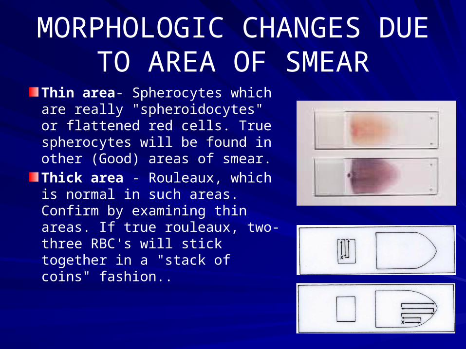
Thin area- Spherocytes which are really "spheroidocytes" or flattened red cells. True spherocytes will be found in other (Good) areas of smear.
Thick area - Rouleaux, which is normal in such areas. Confirm by examining thin areas. If true rouleaux, two-three RBC's will stick together in a "stack of coins" fashion..
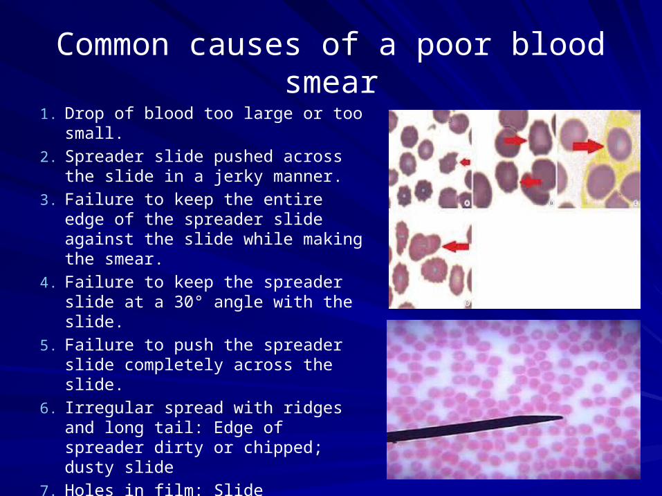
Common causes of a poor blood smear1. Drop of blood too large or too small.
2. Spreader slide pushed across the slide in a jerky manner.
3. Failure to keep the entire edge of the spreader slide against the slide while making the smear.
4. Failure to keep the spreader slide at a 30° angle with the slide.
5. Failure to push the spreader slide completely across the slide.
6. Irregular spread with ridges and long tail: Edge of spreader dirty or chipped; dusty slide
7. Holes in film: Slide contaminated with fat or grease
8. Cellular degenerative changes: delay in fixing, inadequate fixing time or methanol contaminated with water.
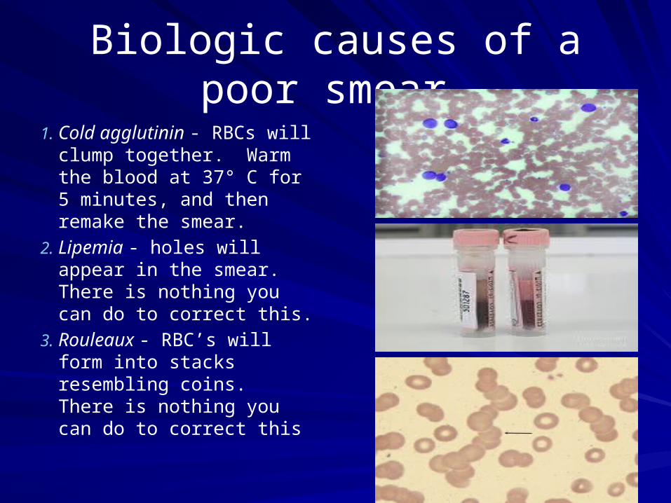
Biologic causes of a poor smear
1. Cold agglutinin - RBCs will clump together. Warm the blood at 37° C for 5 minutes, and then remake the smear.
2. Lipemia - holes will appear in the smear. There is nothing you can do to correct this.
3. Rouleaux - RBC’s will form into stacks resembling coins. There is nothing you can do to correct this
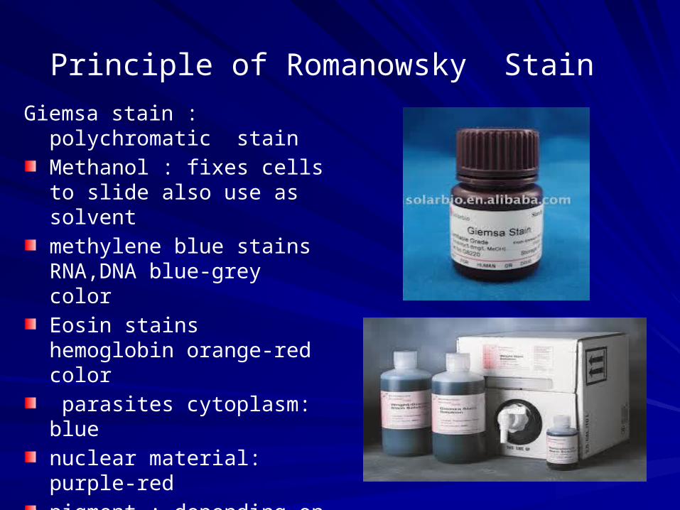
Principle of Romanowsky Stain
Giemsa stain : polychromatic stain
Methanol : fixes cells to slide also use as solvent
methylene blue stains RNA,DNA blue-grey color
Eosin stains hemoglobin orange-red color
parasites cytoplasm: blue
nuclear material: purple-red
pigment : depending on type& species
pH value of phosphate buffer is very important

Staining Procedure
Thin smear are air dried after fixation with absolute alcohol.
Dilute Giemsa 10%
Flood the smear with stain.
Leave the stain on the slide for 10 min.
Wash off by running water directly to the centre of the slide to prevent a residue of precipitated stain.
Stand slide on end, and let dry in air.

too acidic suitable too basicStaining result

Causes and correctionToo Acid Stain:
1. insufficient staining time
2. prolonged buffering or washing
3. old stain
Correction:
1) lengthen staining time
2) check stain and buffer pH
3) shorten buffering or wash time
Too Alkaline Stain:
1. thick blood smear
2. prolonged staining
3. insufficient washing
4. alkaline pH of stain components
Correction :
1) check pH
2) shorten stain time
3) prolong buffering time
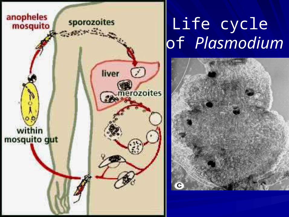
Life cycle of Plasmodium
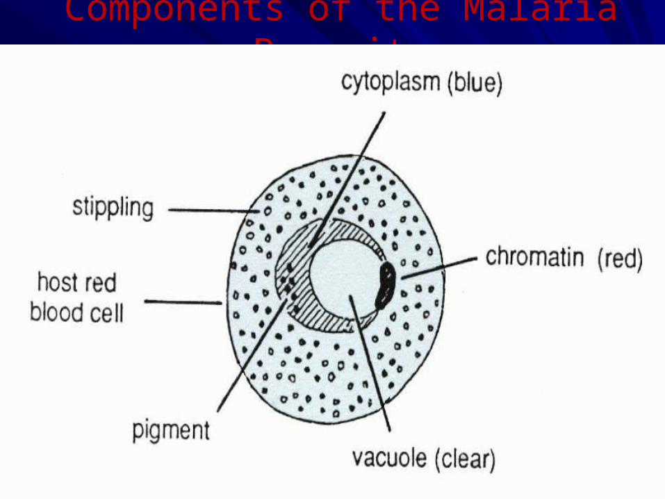
Components of the Malaria Parasite
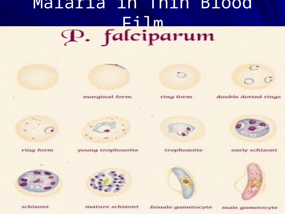
Malaria in Thin Blood Film

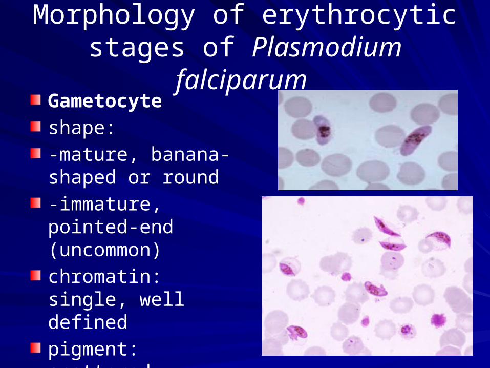
Morphology of erythrocytic stages of Plasmoduim falciparum
Trophozoitesize: small to mediumnumber: often numerousshape: ring and comma forms commonchromatin: often 2 dotscytoplasm: regular, fine to fleshy

Morphology of erythrocytic stages of Plasmodium falciparum
Schizont
size: small
number: few
shape: compact
uncommon, usually seen in severe malaria
mature forms: 12-30 or more merozoits
pigment: single dark mass
Morphology of erythrocytic stages of Plasmodium falciparum
Gametocyte
shape:
-mature, banana-shaped or round
-immature, pointed-end (uncommon)
chromatin: single, well defined
pigment: scattered, coarse, rice-grain-like

Malaria in Thin Blood Film

Plasmodium malariae
Morphology of erythrocytic stages of P. malariaeTrophozoitesize:smallnumber:usually fewshape:ring to rounded, compact formschromatin:single, largecytoplasm:regular, densepigment:scattered, abundant, with yellow tinge in older forms
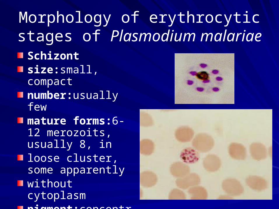
Morphology of erythrocytic stages of Plasmodium malariae
Schizontsize:small, compactnumber:usually fewmature forms:6-12 merozoits, usually 8, in loose cluster, some apparently without cytoplasmpigment:concentrated
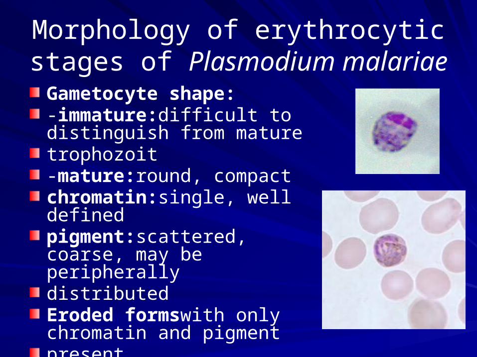
Morphology of erythrocytic stages of Plasmodium malariae
Gametocyte shape:-immature:difficult to distinguish from mature trophozoit-mature:round, compactchromatin:single, well definedpigment:scattered, coarse, may be peripherally distributedEroded formswith only chromatin and pigment present
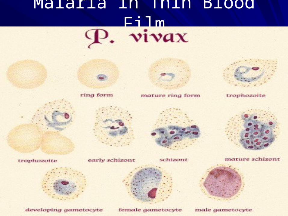
Malaria in Thin Blood Film

Morphology of erythrocytic stages of Plasmodium vivax
Trophozoite
size: small to large
number: few to moderate
shape: broken ring to irregular forms common
chromatin: single, occasionally 2
cytoplasm: irregular or fragmented (amoeboid)
pigment: scattered, fine
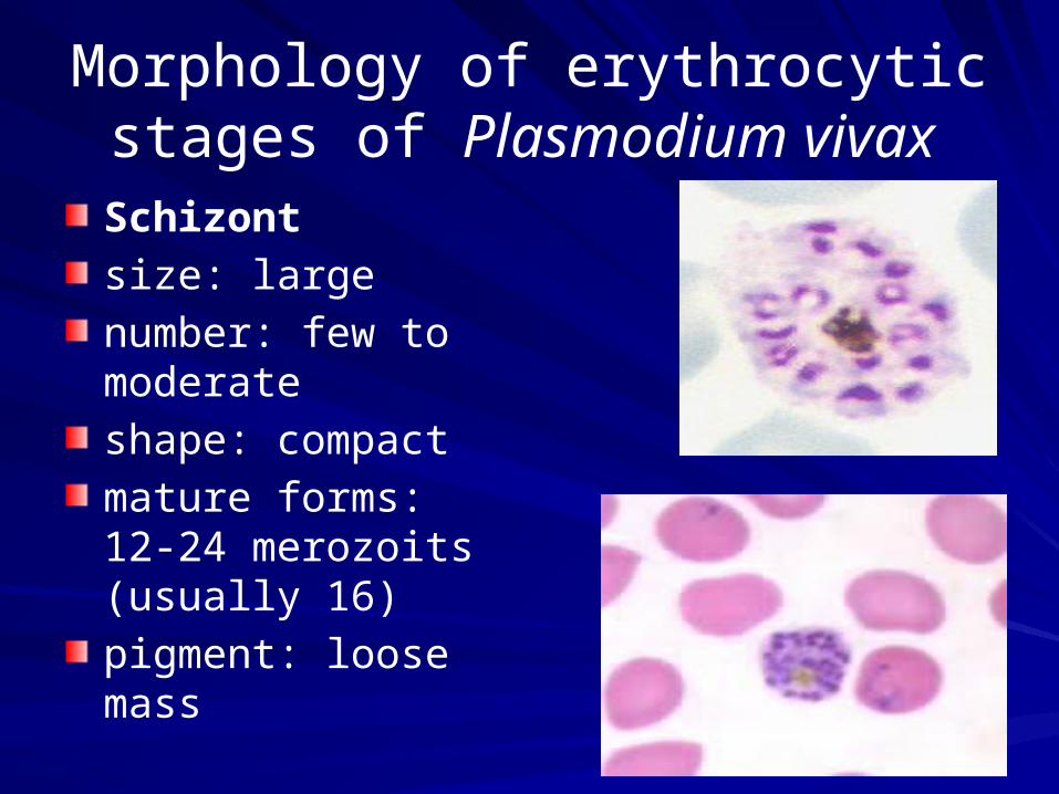
Morphology of erythrocytic stages of Plasmodium vivax
Schizont
size: large
number: few to moderate
shape: compact
mature forms: 12-24 merozoits (usually 16)
pigment: loose mass

Morphology of erythrocytic stages of Plasmodium vivax
Gametocyteshape: -immature: difficult to distinguish from mature trophozoit-mature: round, largechromatin: single, well definedpigment: scattered, fineEroded forms with scanty or no cytoplasm and only chromatin and pigment present
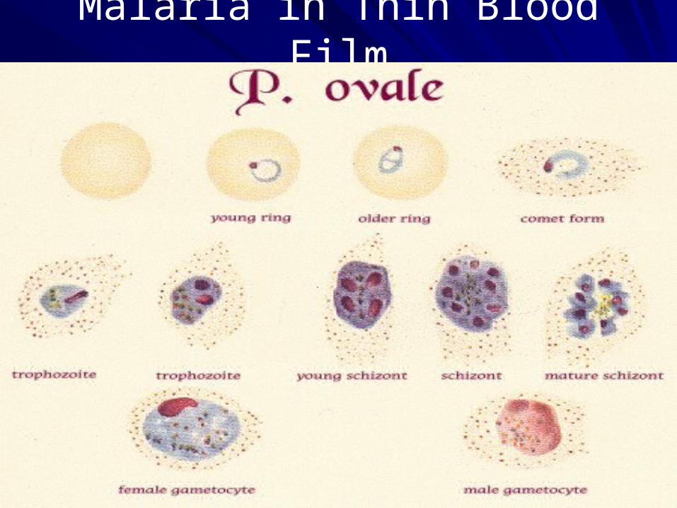
Malaria in Thin Blood Film

Morphology of erythrocytic stages of Plasmodium ovale
Trophozoite
size:may be smaller than P. vivax
number:usually few
shape:ring to rounded, compact forms
chromatin:single, prominent
cytoplasm:fairly regular, fleshy
pigment:scattered, coarse

Morphology of erythrocytic stages of Plasmodium ovale
Schizont
size:rather like P. malariae
number:few
mature forms:6-16 merozoits, usually 8, in loose cluster
pigment:concentrated mass
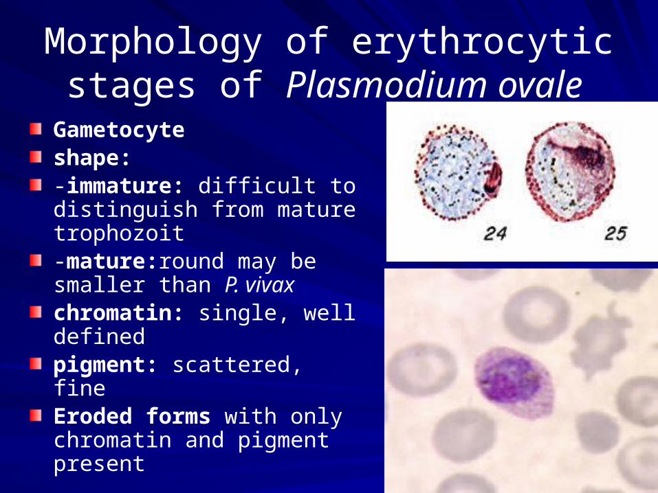
Morphology of erythrocytic stages of Plasmodium ovale
Gametocyteshape:-immature: difficult to distinguish from mature trophozoit-mature:round may be smaller than P. vivaxchromatin: single, well definedpigment: scattered, fineEroded forms with only chromatin and pigment present

Morphologic forms of haemoflagellate
There are 4 morphologic forms seen in hemoflagellates:
1) Amastigote2) Promastigote3) Epimastigote4) Trypomastigote - They can exist in two or more of the 4
morphologic forms depending on the species.
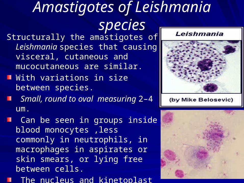
Amastigotes of Leishmania species
Structurally the amastigotes of Leishmania species that causing visceral, cutaneous and mucocutaneous are similar.
With variations in size between species.
Small, round to oval measuring 2–4 um.
Can be seen in groups inside blood monocytes ,less commonly in neutrophils, in macrophages in aspirates or skin smears, or lying free between cells.
The nucleus and kinetoplast stain dark reddish-mauve.
The cytoplasm stains palely and is difficult to see when the amastigotes are ingroups.
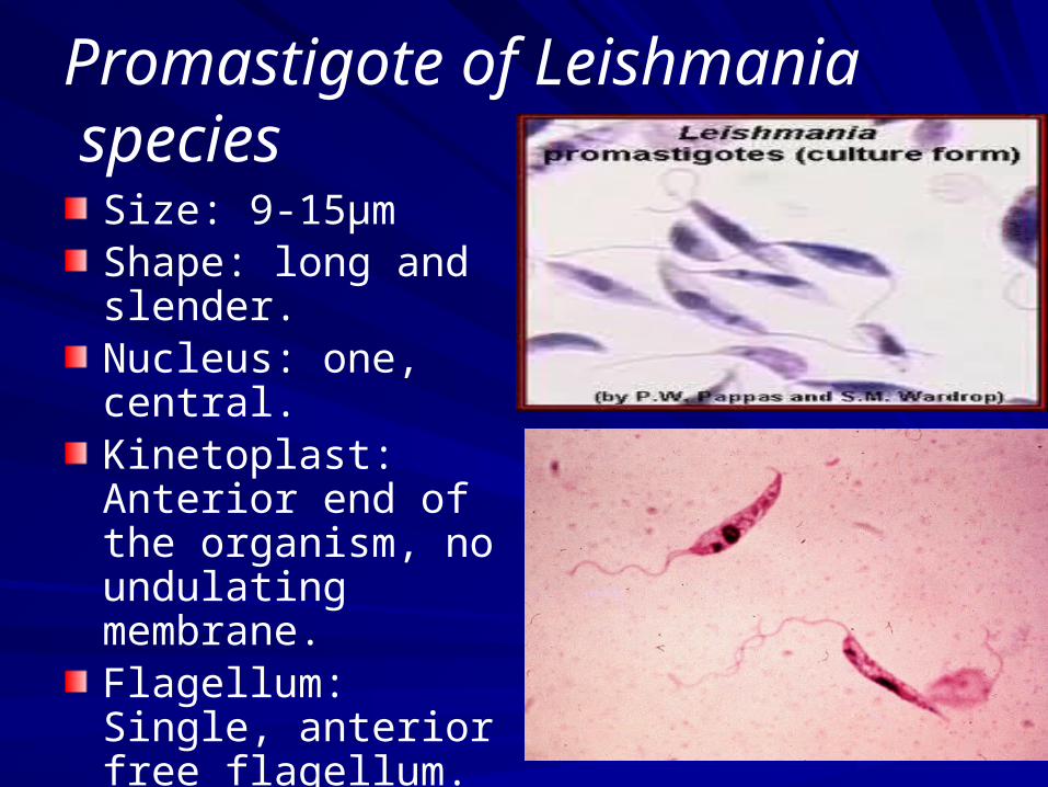
Promastigote of Leishmania species
Size: 9-15µmShape: long and slender.Nucleus: one, central.Kinetoplast: Anterior end of the organism, no undulating membrane.Flagellum: Single, anterior free flagellum.
Is the infective stage
Also result from culture (NNN)media.

Cutaneous leishmaniasis
Infection is often referred to as wet or dry oriental
sore. The early papule is often inflamed and resembles
a boil of 5–10 mm in diameter which rapidly develops
into a large uneven ulcer which is self-healing
in as little as 3–6 months. Multiple lesions may occur
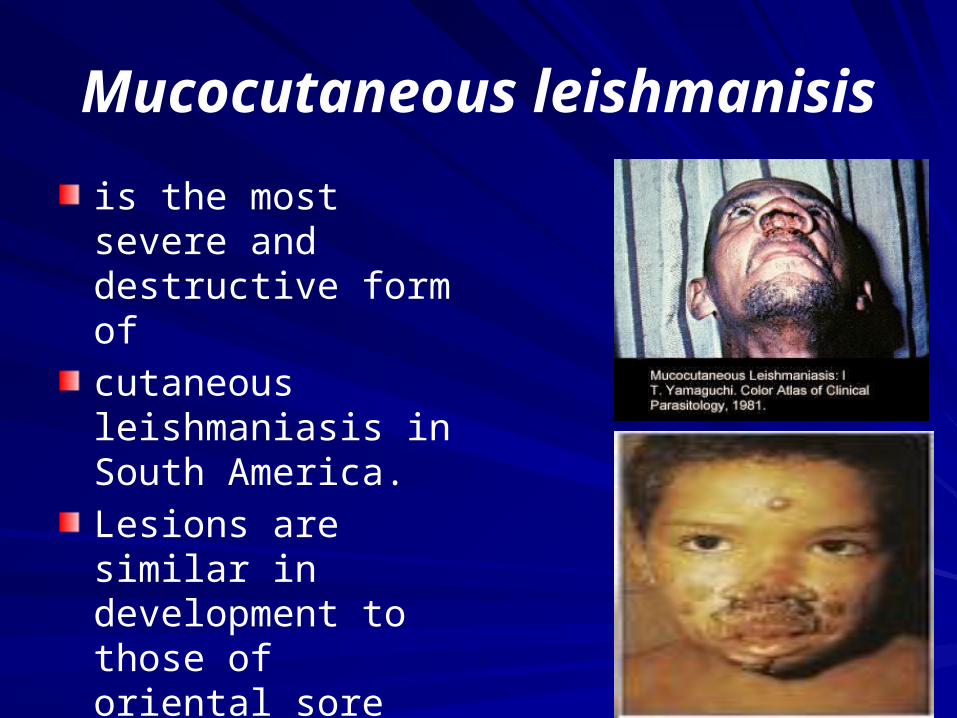
Mucocutaneous leishmanisis
is the most severe and destructive form of
cutaneous leishmaniasis in South America.
Lesions are similar in development to those of oriental sore and the resulting ulcers may become very large and
long-lasting.
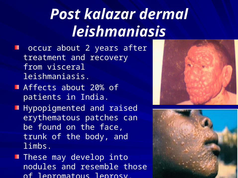
Post kalazar dermal leishmaniasis
occur about 2 years after treatment and recovery from visceral leishmaniasis.
Affects about 20% of patients in India.
Hypopigmented and raised erythematous patches can be found on the face, trunk of the body, and limbs.
These may develop into nodules and resemble those of lepromatous leprosy, fungal infections or other skin disorders.
Amastigotes are present in the papules and nodules.
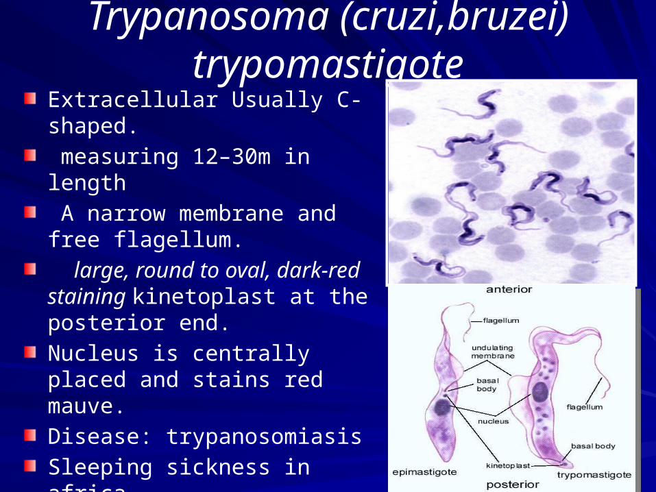
Trypanosoma (cruzi,bruzei) trypomastigote
Extracellular Usually C-shaped.
measuring 12–30m in length
A narrow membrane and free flagellum.
large, round to oval, dark-red staining kinetoplast at the posterior end.
Nucleus is centrally placed and stains red mauve.
Disease: trypanosomiasis
Sleeping sickness in africa
Chagas disease in america
Sample: Blood or Cerebrospinal fluid

Life Cycle African Trypanosomiasis
Life cycle of Trypanosoma brucei gambiense & T. b. rhodesiense

Cerebrospinal fluid Lymph node aspiration
CATT test trypanosoma

Blood CoccidiaTachyzoites of Toxoplasma gondii
Parasites are frequently seen in neutrophils and mononuclear cells.
They are crescent shaped and small,
measuring about 37m. One end is
rounded and the other end more pointed.
Nucleus is situated towards the roundedend and stains dark red.
Cytoplasm stains blue.

Diagnosis of Free-living Amoebae
They are amoebae that normally inhabit:- Water (lakes, swimming pools, air-conditioning units)- Moist soil.- Decaying vegetations.
Potentially Pathogenic Free-living amoebae
Amoeboid form
Trophozoite form
Naegleria fowleri Acanthamoeba species
In water
Flagellate form
Cyst form Cyst form
Trophozoite
In water or air
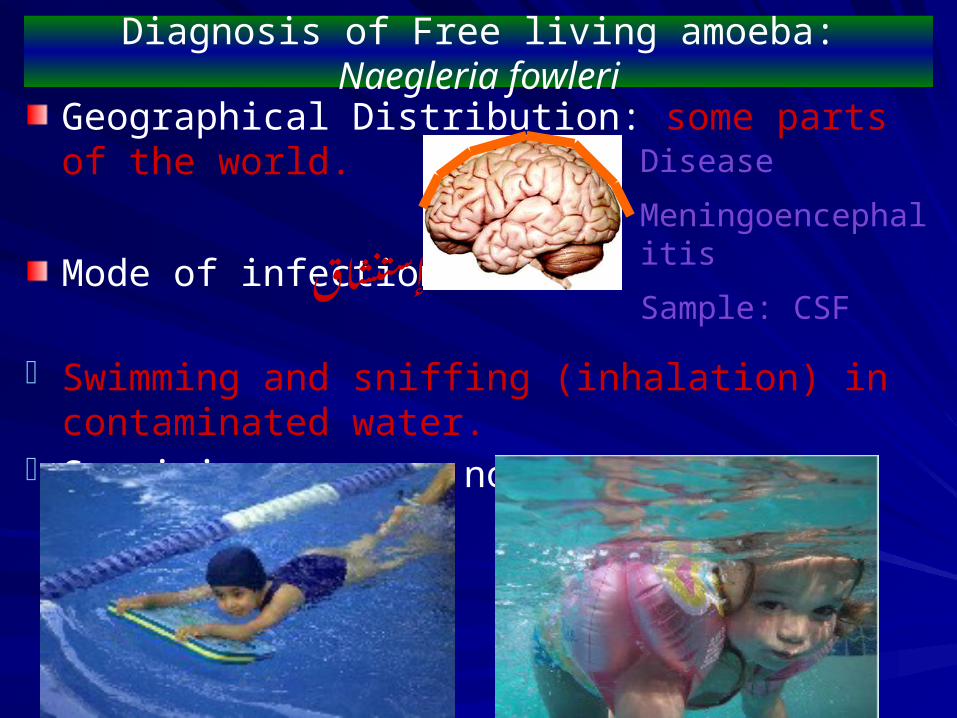
Diagnosis of Free living amoeba: Naegleria fowleri
Geographical Distribution: some parts of the world.
Mode of infection:
- Swimming and sniffing (inhalation) in contaminated water.
- Sappinia sp cause nonlethal amoebic encephalitis
إستنشاق
Disease
Meningoencephalitis
Sample: CSF
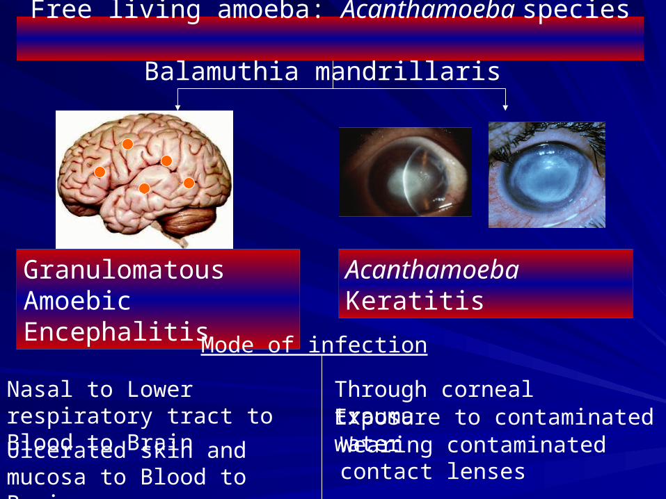
Free living amoeba: Acanthamoeba species Balamuthia mandrillaris
Granulomatous Amoebic Encephalitis
Acanthamoeba Keratitis
Mode of infection
Nasal to Lower respiratory tract to Blood to Brain
Ulcerated skin and mucosa to Blood to Brain
Through corneal traumaExposure to contaminated waterWearing contaminated contact lenses
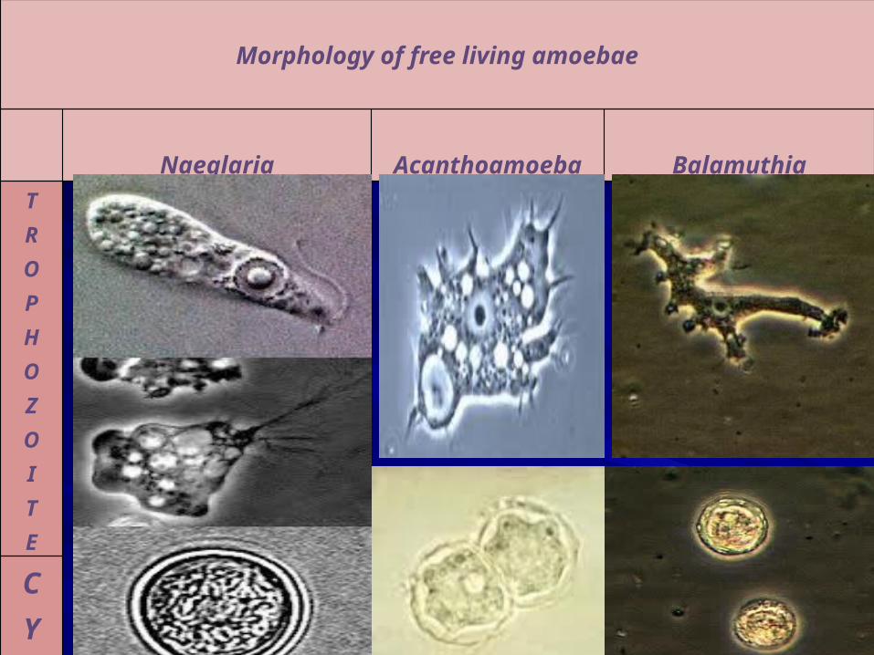
Morphology of free living amoebae
Naeglaria Acanthoamoeba Balamuthia
TROPHOZOITE
CYST

Urinogenital protozoa Trichomonas vaginalis
vaginal, urethral and prostatic tissue
only in humans; no animal
Structuer contain Fg=flagella
Bb=basal body
Nu=nucleus
Ax=axostyle
um=undulating membrane
Cy=cytostomal groove
Cs=costa
No cyst stage.
Multiplies by binary fission
Sample: urine sample,vaginal or urethral swab

Class insecta Anopheles
Morphology and medical importance
Female are blood feeder
Spotted wings
Maxillary palps as long as proboscis
Egg: floated, layed single
Proboscis and body in same straight line
Malaria transmition
In some areas it can also transmit filariasis

Class insectaCulex
Morphology and medical importance
Female are blood feeder
Uniform wings
Blunt tip abdomen
Maxillary palps shorter than proboscis
Egg: not floated, layed in group
Proboscis and body at an angle
vectors of filariasis and some viral
diseases

Class insectaAedes
Morphology and medical importance
Female are blood feeder
Uniform wings
Maxillary palps shorter than proboscis
Pointed tip abdomen
Egg: not floated, layed singly
Proboscis and body at an angle
vectors of dengue
yellow fever and other viral diseases
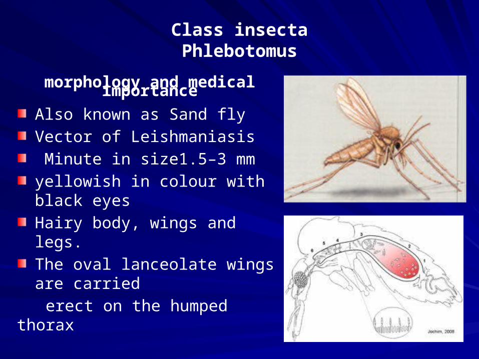
Class insectaPhlebotomus
morphology and medical importance
Also known as Sand fly
Vector of Leishmaniasis
Minute in size1.5–3 mm
yellowish in colour with black eyes
Hairy body, wings and legs.
The oval lanceolate wings are carried
erect on the humped thorax
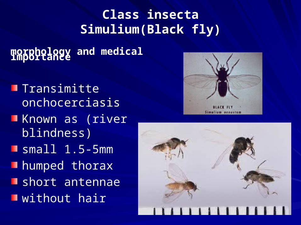
Class insectaSimulium(Black fly)
morphology and medical importance
Transimitte onchocerciasis
Known as (river blindness)
small 1.5-5mm
humped thorax
short antennae
without hair

Class insectaCulicoides
morphology and medical importance
AnnoyanceFilarial disease1.5-5 mmPair antennaesmall headcoered by black
spott
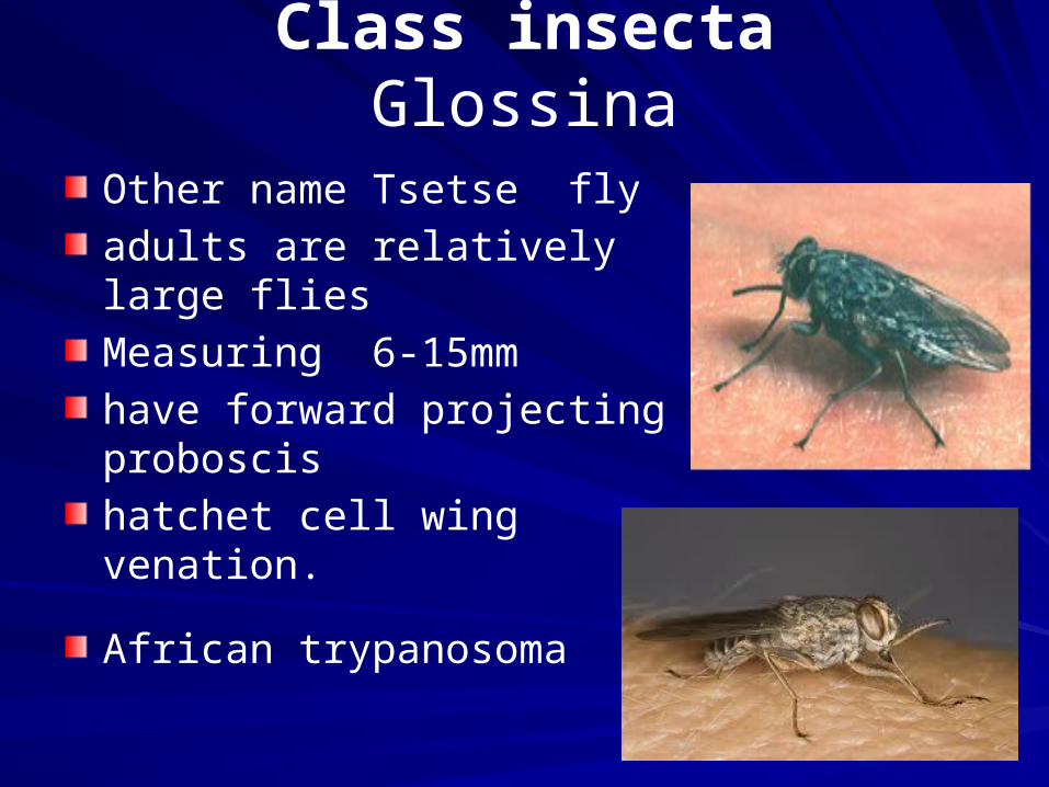
Class insectaGlossina
Other name Tsetse fly
adults are relatively large flies
Measuring 6-15mm
have forward projecting proboscis
hatchet cell wing venation.
African trypanosoma

Order hemipteraFamily:Reduviidae
Triatomine bug
Measure 1-4 cm
Elongate snout-like
head with two eye
4 segment antennae
3 pairs of legs
Vector of Chagas disease