morphogenesis and nature of the pigment granules in the adult human retinal pigment epithelium
TRANSCRIPT
Z. Zellforsch. 122, 378-388 (1971) �9 by Springer-Verlag 1971
Morphogenesis and Nature of the Pigment Granules in the Adult Human Retinal Pigment Epithelium*
MA~FRED SJPJ[TZNAS
Augenklinik des Klinikum Essen der Ruhr-Universit/it Bochum, Essen, Germany (Director: Prof. Dr. G. Meyer-Schwiekerath)
Received JuLy 19, 1971
Su~r~mary. The pigment epithelial cells of the retina are a layer of highly specialized melanocytes. Beginning in the early embryonic period they produce melanin throughout the entire life. The Golgi apparatus plays a key role in the biosynthesis of melanin. The following steps can be distinguished morphologically: (a) Golgi-vesicles, (b) intermediate vesicles, (c) melanosomes, (d) melanin granules. Structures with a ringlike appearance that are described as lipofuscin granules in the literature prove to be altered intermediate vesicles and melano- somes.
Key-Words: Pigment g r a n u l e s - Pigment ep i the l ium- Human retina --Melanin - - Morphogenesis.
Introduction
The ret inal p igment epi thel ium is a single layer of cells tha t is derived from the neuroectoderm. The cells themselves mus t be understood as highly specialized melanocytes (Fi tzpatr ick et al , 1966). The most characteristic feature of all melanocytes is tha t they contain pigment granules, most of which are melanin.
The ret inal melanocytes, i.e. the p igment epithelial cells, differ greatly from the epidermal, dermal, meningeal, uveal and other melanocytes in tha t they are arranged in a layer, are f irmly uni ted by intercellular junct ions and have highly differentiated functions.
Applying electron microscopic techniques the present work examines the morphology of the melanin granules in the pigment epithelium of the adul t huma n ret ina in order to elucidate the morphogenesis of these granules and to s tudy simi- larities and differences between the pigment epithelial cells and the unspecialized melanocytes in other areas of the human body.
Material and Methods
The study was carried out on three human eyes. The eyes came from a 35, a 47 and a 60 year-old patient. They were enucleated because of a malignant orbital or ocular tumor. The pigment epithelium areas under examination were far away from the primary disease and did not show any significant pathologic changes.
Immediately after enucleation (that is within two minutes after severing the optic nerve) the eyes were separated into anterior and posterior portions by making a circular cut in the pars plana region. The posterior portion was placed in a fixative solution of 2 % glutaraldehyde
* This investigation was carried out in part at the Francis I. Proctor Foundation for Research in Ophthalmology, San Francisco, California, U.S.A., and supported by United States Public Health Service Program Project Grant EY 00310, and Deutsche Forschungs- gemeinschaft, Training Grant Nr. Sp 102/1.
Granules in Human Retinal Pigment Epithelium 379
and 1% paraformaldehyde buffered with sodium cacodylate 0.2 M at pH 7.4 and containing 20 mg/100 ml of calcium chloride and 1% sucrose. The tissue then was cut in small strips, and the retina with the attached choroid was separated from the sclera with a thin spatula. This was done under a dissecting microscope in a drop of fixative. Fixation was continued for two hours in the same fixative at 37 ~ C, and then the tissue was postfixed in a 2 % osmium tetroxide- veronal-acetatc buffer solution for two hours at room temperature. After a two-hour en bloc staining in 2% uranyl acetate and dehydration in acetone and propyleneoxide, the specimens were embedded in epoxy resin (Araldite). Thin sections were cut with diamond knives on an ultramicrotome Porter-Blum, MT2 and examined with a Siemens Elmiskop IA.
Observations
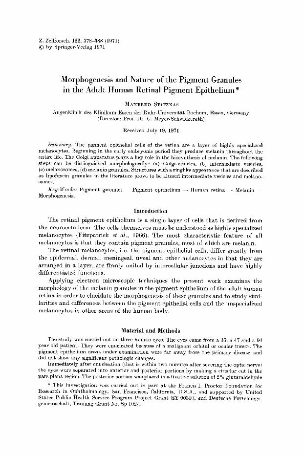
In addi t ion to other cell organelles two different types of p igment g r a n u l e s - - a lighter and a darker o n e - - c a n be dist inguished at low magnif icat ion (Fig. 1).
Fig. 1. a Survey picture of the retinal pigment epithelium: B Bruch's membrane, M mito- chondria, N nucleus, P pigment granules. Magnification: 3000 • b Pigment epithelial cell
with light and dark pigment granules. Magnification: 5300 •
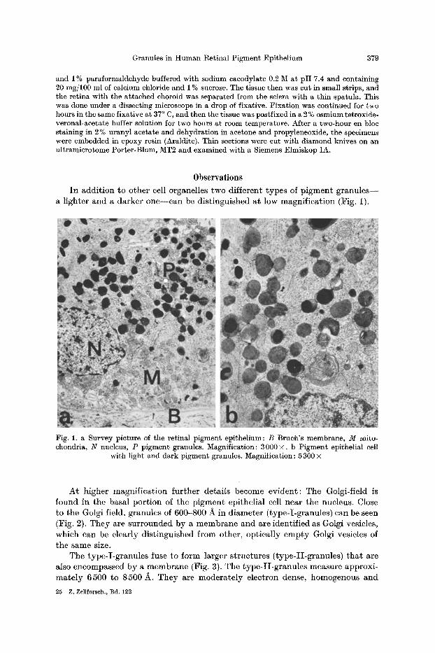
At higher magnif icat ion fur ther details become evident : The Golgi-field is found in the basal por t ion of the p igment epithelial cell near the nucleus. Close to the Golgi field, granules of 600-800 A in diameter (type-X-granules) can be seen (Fig. 2). They are surrounded by a membrane and are identif ied as Golgi vesicles, which can be clearly dist inguished from other, optically empty Golgi vesicles of the same size.
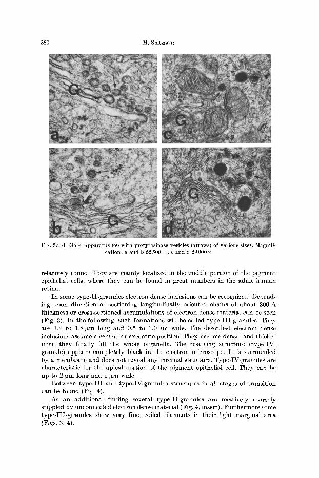
The type-X-granules fuse to form larger s t ructures ( type-H-granules) tha t are also encompassed by a membrane (Fig. 3). The type-H-granules measure approxi- mate ly 6500 to 8500/~. They are moderately electron dense, homogenous and
25 Z. Zellforsch., Bd. 122
380 M. Spitznas :
Fig. 2a-d. Golgi apparatus (G) with protyrosinase vesicles (arrows) of various sizes. Magnifi- cation: a and b 52 500 x ; c and d 29000 •
relatively round. They are mainly localized in the middle portion of the pigment epithelial cells, where they can be found in great numbers in the adult human retina.
I n some type-II -granules electron dense inclusions can be recognized. Depend- ing upon direction of sectioning longitudinally oriented chains of about 300 A thickness or cross-sectioned accumulations of electron dense material can be seen (Fig. 3). I n the following, such formations will be called type-III-granules . They are 1.4 to 1.8 ~zm long and 0.5 to 1.0 [zm wide. The described electron dense inclusions assume a central or excentric position. They become denser and thicker until they finally fill the whole organellc. The resulting structure (type-IV- granule) appears completely black in the electron microscope. I t is surrounded by a membrane and does not reveal any interna] structure. Type-IV-granules are characteristic for the apical portion of the pigment epithelial cell. They can be up to 2 ~zm long and 1 ~m wide.
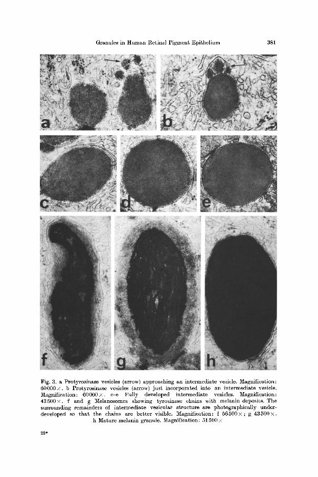
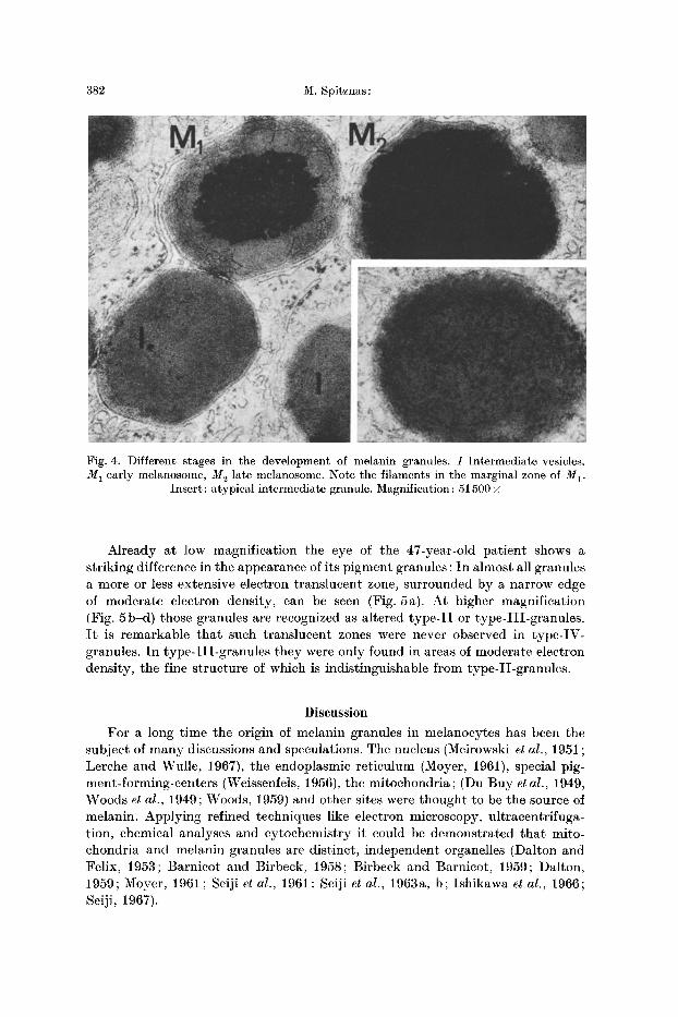
Between t y p e - I I I and type-IV-granules structures in all stages of transition can be found (Fig. 4).
As an additional finding several type-II-granules are relatively coarsely stippled by unconnected electron dense material (Fig. 4, insert). Fur thermore some type- I I I -granules show very fine, coiled filaments in their light marginal area (Figs. 3, 4).
Granules in Human Retinal Pigment Epi thel ium 381
Fig. 3. a Protyrosinase vesicles (arrow) approaching an intermediate vesicle. Magnification: 60000• b Protyrosinase vesicles (arrow) just incorporated into an intermediate vesicle. Magnification: 60000• c-e Fully developed intermediate vesicles. Magnification: 43500• f and g Melanosomes showing tyrosinase chains with melanin deposits. The surrounding remainders of intermediate vesicular s tructure are photographically under- developed so t h a t the chains are bet ter visible. Magnification: f 5 6 5 0 0 • g 43500•
h Mature melanin granule. Magnification : 51500 •
25*
382 M. Spitznas :
Fig. 4. Different stages in the development of melanin granules. I Intermediate vesicles, M 1 early melanosome, M~ late melanosome. Note the filaments in the marginal zone of My.
Insert: atypical intermediate granule. Magnification : 51500 •
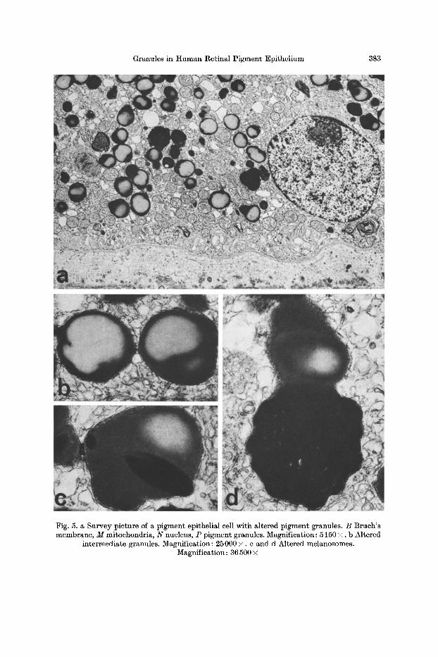
Already at low magnification the eye of the 47-year-old patient shows a striking difference in the appearance of its pigment granules: In almost all granules a more or less extensive electron translucent zone, surrounded by a narrow edge of moderate electron density, can be seen (Fig. 5a). At higher magnification (Fig. 5b-d) those granules are recognized as altered type- I I or type-III-granules. I t is remarkable tha t such translucent zones were never observed in type-IV- granules. In type-III-granules they were only found in areas of moderate electron density, the fine structure of which is indistinguishable from type-II-granulcs.
Discussion
For a long time the origin of melanin granules in melanocytes has been the subject of many discussions and speculations. The nucleus (Meirowski et al., 1951; Lerche and Wulle, 1967), the endoplasmie reticulum (Moyer, 1961), special pig- ment-forming-centers (Weissenfels, 1956), the mitochondria; (Du Buy et al., 1949, Woods et al., 1949; Woods, 1959) and other sites were thought to be the source of melanin. Applying refined techniques like electron microscopy, ultracentrifuga- tion, chemical analyses and cytochemistry it could be demonstrated that mito- chondria and melanin granules are distinct, independent organelles (Dalton and Felix, 1953; Barnicot and Birbeck, 1958; Birbeck and Barnicot, 1959; Dalton, 1959; Moyer, 1961; Seiji el al., 1961; Seiji et al., 1963a, b; Ishikawa et al., 1966; Seiji, 1967).
Granules in Human Retinal Pigment Epi thel ium 383
Fig. 5. a Survey picture of a pigment epithelial cell with altered pigment granules. B Bruch 's membrane, M mitochondria, N nucleus, P pigment granules. Magnification: 5150 X. b Altered
intermediate granules. Magnification: 25000 X. c and d Altered melanosomes. Magnification : 36 500 X
384 M. Spitznas :
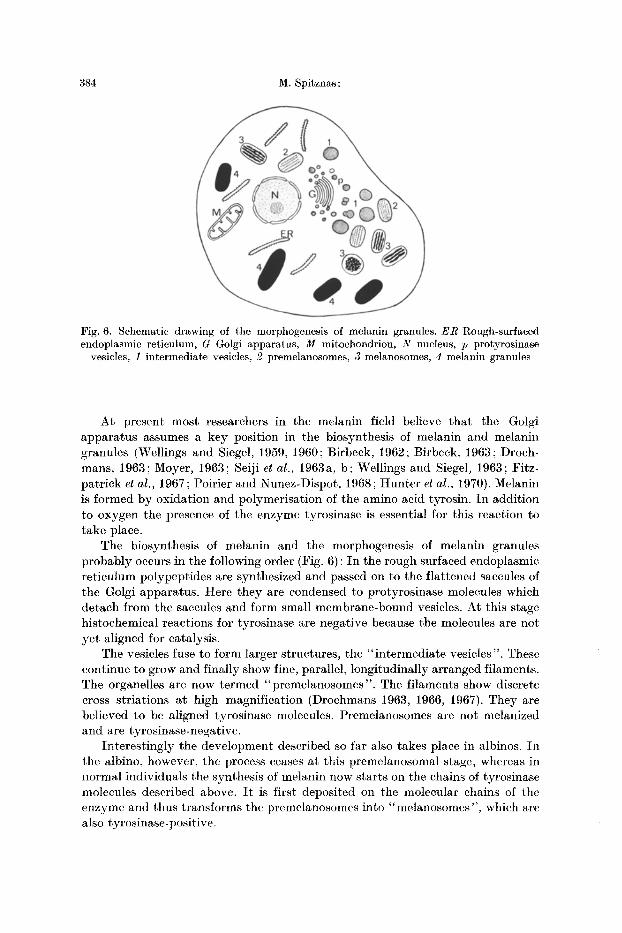
Fig. 6. Schematic drawing of the morphogenesis of melanin granules. E R Rough-surfaced endoplasmic reticulum, G Golgi apparatus, M mitochondrion, N nucleus, p protyrosinase
vesicles, 1 intermediate vesicles, 2 premelanosomes, 3 melanosomes, 4 melanin granules
At present most researchers in the melanin field believe that the Golgi apparatus assumes a key position in the biosynthesis of melanin and melanin granules (Wellings and Siegel, 1959, 1960; Birbeck, 1962; Birbeck, 1963; Droch- mans, 1963; Moyer, 1963; Seiji et al., 1963a, b; Wellings and Siegel~ 1963; Fitz- patrick et al., 1967; Poirier and Nunez-Dispot, 1968; Hunter et al., 1970). Melanin is formed by oxidation and polymerisation of the amino acid tyrosin. In addition to oxygen the presence of the enzyme tyrosinase is essential for this reaction to take place.
The biosynthesis of melanin and the morphogenesis of melanin granules probably occurs in the following order (Fig. 6): In the rough surfaced endoplasmic reticulum polypeptides are synthesized and passed on to the flattened saccules of the Golgi apparatus. Here they are condensed to protyrosinase molecules which detach from the saccules and form small membrane-bound vesicles. At this stage histoehemical reactions for tyrosinase are negative because the molecules are not yet aligned for catalysis.
The vesicles fuse to form larger structures, the "intermediate vesicles". These continue to grow and finally show fine, parallel, longitudinally arranged filaments. The organelles are now termed "premelanosomes". The filaments show discrete cross striations at high magnification (Drochmans 1963, 1966, 1967). They are believed to be aligned tyrosinase molecules. Premelanosomes are not melanized and are tyrosinase-negative.
Interestingly the development described so far also takes place in albinos. In the albino, however, the process ceases at this premelanosomal stage, whereas in normal individuals the synthesis of melanin now starts on the chains of tyrosinase molecules described above. I t is first deposited on the molecular chains of the enzyme and thus transforms the premelanosomes into "melanosomes", which are also tyrosinase-positive.
Granules in Human Retinal Pigment Epithelium: 385
The synthesis of melanin is continued until the whole stock of tyrosinase is used up and the melanosome is entirely filled with melanin. The thus formed organelle is tyrosinase-ncgative and is termed a "melanin granule".
Recently Stanka et al. (1969) and Stanka (1970) described premelanosomes without tyrosinase activity. He therefore believes tha t premelanosomes with their filamentous internal structure are derived from widened cisternae of the rough surfaced endoplasmie reticulnm (Stanka, 1971). This pre-formed structure is then assumed to receive its tyrosinase act ivi ty from the Golgi vesicles. In this case the nature of the filaments in the premelanosomes, the question why the Golgi vesicles are constantly tyrosinase negative, and the process by which tyrosinase is transfered from the vesicles to the premelanosomes are all obscure. In one case only (Maul, 1969) a fusion between prcmelanosomes and Golgi vesicles could be observed.
Comparing these par t ly complementary and part ly contradictory observations with the findings in the pigment epithelium of the adult human retina, the follow- ing conclusions can be drawn: The type-I-granules in the vicinity of the Golgi appa- ratus must be "protyrosinase vesicles". The type-II-granules tha t result from a fusion of those vesicles correspond to " intermediate vesicles". Premelanosomcs, i.e. granules with non-melanized tyrosinase molecules could not be observed.
The type-III-granules fulfill all the criteria for their identification as "melano- somes". The type-IV-granules are mature "melanin granules".
The eoarsly stippled granules tha t were observed in some instances (Fig. 4, insert) and which, without this stippling, would belong to the intermediate granules, have been observed by other authors in human malignant melanoma cells (Wellings and Siegel, 1963). They are probably atypical melanosomes in which tyrosinase molecules (condensation points for the forming melanin) are not arranged in long chains but in shorter units.
There does not seem to be an explanation for the fine fibrils tha t were seen in the marginal zone of some melanosomes (Fig. 4).
The few electron microscopic studies on the pigment epithelium of animal (Sebruyns, 1951; Francois et al., 1963; Moycr, 1969; Stanka, 1970, 1971) and human (Takeuehi, 1957; Lerchc, 1962; Feeney et al., 1965; Lerehe and Wulle, 1967) embryos and fetuses show that prior to birth the early stages of morpho- genesis of melanin granules prevail. Following birth, a shift toward more mature forms takes place. I t is interesting to see tha t there is a production of new melanin granules also in the adult and even in the aging human eye. This is demonstrated by the presence of melanosomes in all degrees of melanization. Apparently, however, with increasing age fewer intermediate vesicles are transformed into mature melanin granules so tha t - -compared to the number of melanosomes as a transitory s tage-- the number of intermediate vesicles increases.
In 1965 Feeney et al. described structures in the retinal pigment epithelium which they thought to be lipofusein granules. Looking at Feeney's electron micrographs it becomes apparent, however, tha t most of those granules are in fact intermediate granules, such as those described in this study. On some of Feeney's illustrations the supposed lipofuscin granules have an electron trans- lucent central zone surrounded by a more electron dense margin, which gives them the appearance of rings. Their structure is identical with tha t of the granules
386 M. Spitznas:
observed in the p igment epithelial cells of the 47-year-old pa t ient in the present study. I n spite of the fact tha t their fine s tructure differs from normal in termediate vesicles it was clearly demonst ra ted tha t we are dealing with altered intermediate vesicles or melanosomes and not with lipofuscin granules. The fine s tructure of lipofuscin granules is characterized by a marked heterogenity of their contents and a very irregular shape (Siebert et al., 1962; Bj6rkerud and Cummis, 1963; BjOrkerud, 1963; Malkoff and Strehler, 1963; Strehler, 1964; Hendley et al., 1965 ; Ishii et al., 1967 ; Miyagishi et al., 1967 ; Colcolough et al., 1970), whereas the granules under discussion are characterized by the homogeni ty of their contents and the regular i ty of their shape. They bear a slight resemblance to granules tha t are found in the wall cells of the atr ia of mammal hearts (Jamieson and Palade, 1964). This resemblance, however, is probably inc iden ta l
So far it cannot be explained what causes the al terat ion of the granules. Besides an uncer ta in in t rav i ta l change the possibility of an art ifact mus t be kept in mind, because such granules were observed exclusively in eyes tha t showed various signs of bad fixation.
These s ta tements should not imply tha t there is no lipofuscin in the ret inal p igment epithelium. Because of the close relationship between lipofuscin and lysosomes (Ishii et al., 1967), which seem to play an impor tan t role in the destruc- t ion of phagocytosed outer segments of the photoreceptors (Spitznas and Hogan, 1970), its presence in the p igment epi thel ium is more t han likely. I t must , however, be associated with s tructures other than the ones tha t were discussed last.
References Barnicot, N. A., Birbeck, M. S. C.: The electron microscopy of human melanocytes and
melanin granules. In: The biology of hair growth (W. Montagna and R. A. Ellis, eds.), p. 239-253. New York: Academic Press 1958.
Birbeck, M. S. C.: Electron microscopy of melanocytes. Brit. reed. Bull. 18, 220-222 (1962). - - Electron microscopy of melanocytes: The fine structure of hair-bulb premelanosomes.
Ann. N.Y. Acad. Sci. 100, 540-547 (1963). --Barnicot, N.A.: Electron microscopic studies on pigment formation in human hair
follicles. In: Pigment cell biology (M. Gordon, ed.), p. 549-561. New York: Academic Press 1959.
BjSrkerud, S.: The isolation of lipofuscin granules from bovine cardiac muscle, with obser- vations on the properties of the isolated granules on the light and electron microscopic level. J. Ultrastruct. Res., Suppl. ~, 5-42 (1963).
- - Cummis, J. T.: Selected enzymatic studies of lipofuscin granules isolated from bovine cardiac muscle. Exp. Cell Res. 32, 510--520 (1963).
Colcolough, H. L., Helmy, F.M., Hack, M.H.: Some histochemical observations on the lipofuscin of vertebrate liver, kidney and cardiac muscle. Acta. histochem. (Jena) 35, 345-356 (1970).
Dalton, A. J.: Organization in benign and malignant cells. Lab. Invest. 8~ 510-537 (1959). - - Felix, M. D.: Phase contrast and electron micrography of the Cloudman S-91 mouse
melanoma. In: Pigment cell growth (N. Gordon, ed.), p. 267 276. New York: Academic Press 1953.
Drochmans, P. : Melanin granules : Their fine structure, formation and degradation in normal and pathological tissues. Int. Rev. exp. Path. 2, 357-422 (1963).
- - The fine structure of melanin granules. In: Structure and control of the melanocyte (G. Della Porta and O. Miihlenbrock, eds.), Sixth International Pigment Cell Conference, p. 90-95. Berlin-Heidelberg-New York: Springer 1966.
- - Ultrastructure of melanin granules. In: Advances in biology of skin, vol. VIII: The pig- mcntary system (W. Montagna and F. Hu, eds.), p. 169 177. Oxford: Pergamon Press 1967.
Granules in Human Retinal Pigment Epithelium 387
Du Buy, H. G., Woods, M. W., Burk, D., Lackey, M. D.: Encymatic activities of isolated amelanotic and melanotic granules of mouse melanomas and a suggested relationship to mitochondria. J. nat. Cancer Inst. 9, 325-336 (1949).
Feeney, L., Grieshaber, A., Hogan, M. J . : Studies on human ocular pigment. In: Eye struc- ture, II. Symp. (J. W. Rohen, ed.), p. 535-548. Stuttgart : Schattauer-Verlag 1965.
Fitzpatrick, T. B., Miyamoto, M., Ishikawa, K. : The evolution of concepts of melanin biology. In: Advances in biology of skin, vol. VI I I : The pigmentary system (W. Montagna and F. Hu, eds.), p. 1-30. Oxford: Pergamon Press 1967.
- - Quevedo, W. C., Levene, A., McGovern, V. J., Mishima, Y., 0ettle, A. G.:Terminology of vertebrate melanin containing cells, their precursors and related cells. In: Structure and control of the melanocyte. Sixth Intern. Pigment Cell Conference (G. Della Porta and 0. Mfihlenbrock, eds.), p. 1-5. Berlin-Heidelberg-New York: Springer 1966.
Francois, J., Rabaey, M., Lagasse, A. : Electron microscopic observations on ehoroid, pigment epithelium and pecten of the developing chick in relation to melanin synthesis. Ophthal- mologica (Basel) 146, 415431 (1963).
Hendley, D. H., Strehler, B. L.: Enzymic activities of lipofuscin age pigments. Biochim. biophys. Acta (Amst.) 99, 406-417 (1065).
Hunter, J. A. A., Mottaz, J. H., Zelickson, A. S. : Melanogenesis : ultrastructural histochemical observations on ultraviolet irradiated human melanocytes. Invest. Derm. 51, 213-221 (1970).
Ishii, T., Murakami, Y., Kimura, R. S., Balogh, K., Jr . : Electron microscopic and histochemi- eal identification of lipofuscin in the inner ear. Acta oto-laryng. (Stockh.) 64, 17-29 (1967).
Ishikawa, K., Fitzpatrick, T. B., SzabS, G. : Some recent studies on the subcellular unit of melanin biosynthesis, the melanosome. In: Structure and control of the melanocyte (G. Della Porta and O. Miihlenbrock, eds.), p. 95-113. Sixth Internal Pigment Cell Conf. Berlin-Heidelberg-New York: Springer 1966.
Jamieson, J . D . , Palade, G. E. : Specific granules in atrial muscle cells. J. Cell Biol. 28, 151-172 (1964).
Lerehe, W. : Elektronenmikroskopische Beobachtungen fiber die Entwicklung der Pigment- granula in der Netzhaut menschlicher Embryonen. Albrecht v. Graefes Arch. Ophthal. 1 6 4 , 543-545 (1962).
- - Wulle, K. G. : t~bcr die Genese der Melaningranula in der embryonalen menschlichen Retina. Z. Zellforsch. 76, 452 457 (1967).
Malkoff, D., Strehler, B. : The ultrastructure of isolated and in situ human cardiac age pigment. J. Cell Biol. 26, 611-616 (1963).
Maul, G. G.: Golgi-melanosome relationship in human melanoma in vitro. J. Ultrastruct. Res. 26, 163 176 (1969).
Meirowski, E., Freeman, L. W.: Chromatin-melanin relationships in malignant melanoma. Invest. Derm. 16, 257-260 (1951).
Miyagishi, T., Naohiko, T., Iizuka, R. : Electron microscopic studies on the lipo-pigments in cerebral cortex nerve cells of senile and Vit. E. deficient rats. Acta neuropath. (Berl.) 9, 7-17 (1967).
Moyer, F. H. : Electron microscopic observations on the origin, development and genetic control of melanin granules in the mouse eye. In: Structure of the eye (G. K. Smelser, ed.), p. 469-486. New York: Academic Press 1961.
- - Genetic effects on melanosome fine structure and ontogeny in normal and malignant cells. Arm. N.Y. Acad. Sci. 100, 584-606 (1963).
- - Development, structure and function of the retinal pigment epithelium. In: The retina (Straatsma et al., eds.), p. 1-30. Los Angeles: Regents of UC 1969.
Poirier, J. , Nunez-Dispot, Ch. : Le m~lanocyte. I. Structure et ultrastructure. Presse m~d. 16, 1179-1181 (1968).
Sebruyns, M. : Study of the ultrastructure of the retinal epithelium by means of the electronic microscope. Amer. J. Ophthal. 34, 989-992 (1951).
Sciji, M.: Subcellular particles and melanin formation in melanocytes. In: Advances in biology of skin, vol. VIII. The pigmentary system (W. Montagna and F. Hu, eds.). Oxford: Pergamon Press 1967.
- - Fitzpatrick, T. B., Birbeck, M. S. C. : The melanosome: A distinctive subcellular particle of mammalian melanocytes and the site of melanogenesis. Invest. Derm. 36, 243-252 (1961).
388 M. Spitznas: Granules in Human Retinal Pigment Epithelium
Seiji, M., Fitzpatrick, T.B., Simpson, t~. T. :Chemical composition and terminology of speciali- zed organelles (melanosomes and melanin granules) in mammalian melanocytes. Nature (Lond.) 197, 1082 1084 (1963a).
- - Shimao, K., Birbeck, M. S.C., Fitzpatrick, T .B . : Subeellular localization of melanin biosynthesis. Ann. N.Y. Aead. Sci. 100, 497 533 (1963b).
Siebert, G., Diezel, P. B., Jahr, K., Krug, E., Sehmitt, A., Grfinberger, E., Bottke, I. : Isolie- rtmg and Eigenschaften von Lipofuscin aus Herzgewebe des Menschen. Histochemie 3, 17-45 (1962).
Spitznas, M., Hogan, M. J . : Outer segments of photoreceptors and the retinal pigment epithelium. Interrelationship in the human eye. Arch. Ophthal. 84, 810-819 (1970).
Stanka, P. : Die DOPA-Reaktion, eine brauchbare Methode in der Elektronenmikroskopie. Untersuchung am retinalen Pigmentepithel von Hiihnerembryonen. Mikroskopie 26, 169-174 (1970).
- -Elektronenmikroskopische Untersuchungen fiber die PrSomelanosomenentstehung im Pigmentepithel yon Hiihnerembryonen. Z. Zellforsch. 112, 120-128 (1971).
- - Kinzel, V., Mohr, U. : Elektronenmikroskopische Untersuchungen fiber Priimelanosomen- entstehung an Melanomzellen in vitro. Virchows Arch. Abt. B. Zellpath. 2, 91 102 (1969).
Strehler, B .L . : On the histochemistry and ultrastructure of age pigment. In: Advances in gerontological research, vol. I, (1964) (B. L. Strehler, ed.), p. 343 384. New York and London: Academic Press 1964.
Takeuchi, H.: Morphological studies of the pigment granules of the retina by the electron microscope. Jap. J. Ophthal. l , 100-106 (1957).
Weissenfels, N. : Licht-, Phasenkontrast and Elektronenmikroskopische Untersuchungen tiber die Entstehung der Pigmentgranula in Melanoblastenkulturen. Z. Zellforsch. 45, 60-73 (1956).
Wellings, S. R., Siegel, B. V. : Role of Golgi apparatus in the formation of melanin granules in human malignant melanoma. J. Ultrastruct. Res. 3, 147 154 (1959).
- - --- Electron microscopy of human malignant. J. nat. Cancer Inst. 24, 437-462 (1960). - - - - Electron microscopic studies on the subcellular origin and ultrastructure of melanin
granules in mammalian melanomas. Ann. N.Y. Acad. Sci. 100, 548-568 (1963). Woods, M. W.: Discussion. In: Pigment cell biology (M. Gordon, ed.), p. 560. NewYork:
Academic Press 1959. - - Du Buy, H. G., Burk, D., Hesselbach, M, L.: Cytological studies on the nature and the
cytoplasmic particulates in the Cloudman S-91 mouse melanoma, the derived Algire S-91 A partially amelanotic melanoma and the Harding-Passey mouse melanoma. J. nat. Cancer Inst. 9, 311 323 (1949).
Dr. Manfred Spitznas Universit~ts-Augenklinik D-4300 Essen, Hufelandstr. 55 Germany