molecular subclassification of gastrointestinal cancers
TRANSCRIPT

Li et al. Experimental Hematology & Oncology (2021) 10:53 https://doi.org/10.1186/s40164-021-00246-x
REVIEW
Molecular subclassification of gastrointestinal cancers based on cancer stem cell traitsMei‑Mei Li1,2†, Jun Yuan1,2†, Xin‑Yuan Guan3,4, Ning‑Fang Ma1,2 and Ming Liu1,2*
Abstract
Human gastrointestinal malignancies are highly heterogeneous cancers. Clinically, heterogeneity largely contributes to tumor progression and resistance to therapy. Heterogeneity within gastrointestinal cancers is defined by molecular subtypes in genomic and transcriptomic analyses. Cancer stem cells (CSCs) have been demonstrated to be a major source of tumor heterogeneity; therefore, assessing tumor heterogeneity by CSC trait‑guided classification of gastro‑intestinal cancers is essential for the development of effective therapies. CSCs share critical features with embryonic stem cells (ESCs). Molecular investigations have revealed that embryonic genes and developmental signaling path‑ways regulating the properties of ESCs or cell lineage differentiation are abnormally active and might be oncofetal drivers in certain tumor subtypes. Currently, multiple strategies allow comprehensive identification of tumor subtype‑specific oncofetal signatures and evaluation of subtype‑specific therapies. In this review, we summarize current knowledge concerning the molecular classification of gastrointestinal malignancies based on CSC features and elucidate their clinical relevance. We also outline strategies for molecular subtype identification and subtype‑based therapies. Finally, we explore how clinical implementation of tumor classification by CSC subtype might facilitate the development of more effective personalized therapies for gastrointestinal cancers.
Keywords: Gastrointestinal cancer, Heterogeneity, Cancer stem cell, Cancer subtype, Precision oncology
© The Author(s) 2021. Open Access This article is licensed under a Creative Commons Attribution 4.0 International License, which permits use, sharing, adaptation, distribution and reproduction in any medium or format, as long as you give appropriate credit to the original author(s) and the source, provide a link to the Creative Commons licence, and indicate if changes were made. The images or other third party material in this article are included in the article’s Creative Commons licence, unless indicated otherwise in a credit line to the material. If material is not included in the article’s Creative Commons licence and your intended use is not permitted by statutory regulation or exceeds the permitted use, you will need to obtain permission directly from the copyright holder. To view a copy of this licence, visit http:// creat iveco mmons. org/ licen ses/ by/4. 0/. The Creative Commons Public Domain Dedication waiver (http:// creat iveco mmons. org/ publi cdoma in/ zero/1. 0/) applies to the data made available in this article, unless otherwise stated in a credit line to the data.
IntroductionWorldwide, gastrointestinal cancers rank among the most frequent malignancies and are responsible for more than half of all cancer deaths. Common cancers of the gastrointestinal tract include liver, colorectal, pancreatic, gastric and esophageal malignancies. Current therapeu-tic treatments are ineffective, as most patients develop metastasis, resistance to radiation/chemotherapy, and recurrence. Thus, new strategies to improve treatment effects for patients with gastrointestinal cancers are urgently needed [1, 2].
Genomic and transcriptomic analyses reveal that human gastrointestinal malignancies are highly hetero-geneous cancers. Clinically, heterogeneity largely con-tributes to tumor progression, metastasis, resistance to therapy, and relapse. Bulk tumors contain diverse tumor cell subpopulations with distinct molecular signatures that display differential levels of sensitivity to treatments. Under therapeutic stress, the expansion of intrinsic sub-populations or the evolution of drug-tolerant cells can lead to resistance to treatment. In clinical pathology, these kinds of subpopulations, drug-tolerant cells or poorly differentiated tumors usually exhibit stem-like traits and lead to adverse clinical events. Cancer stem cells (CSCs) have been demonstrated to be a major source of tumor heterogeneity. CSCs are a very heteroge-neous subpopulation of “stem-like” cancer cells described
Open Access
Experimental Hematology & Oncology
*Correspondence: [email protected]†Mei‑Mei Li and Jun Yuan contributed equally to this work1 Affiliated Cancer Hospital and Institute of Guangzhou Medical University, Guangzhou 510095, ChinaFull list of author information is available at the end of the article

Page 2 of 23Li et al. Experimental Hematology & Oncology (2021) 10:53
as “tumor-initiating cells” or “sphere-forming cells” and share critical features with embryonic stem cells (ESCs), including multilineage differentiation, self-renewal and maintenance of the pluripotency state [3–5]. Specifically, the existence of molecular subtypes indicates the pres-ence of molecular heterogeneity. Elucidation of gastroin-testinal cancer classifications integrating CSC properties is critical, as such classifications may allow us to not only better understand the mechanisms of carcinogenesis from a CSC perspective but also improve diagnosis and prognostication and facilitate the development of preci-sion medicine through identification of subtypes that may respond to specific targeted therapies. In the pre-sent review, we describe recent advances in the molecu-lar classifications of five common gastrointestinal cancer types from the CSC perspective and elucidate their thera-peutic and clinical relevance, thereby providing an over-view of molecular subclassification by cancer stem cell traits for translation into clinical implementation and treatment selection.
Gastrointestinal tumor heterogeneity and therapeutic resistanceTumor heterogeneity, therapeutic resistance, and cancer stem cell propertiesTumor heterogeneity consists of intertumor (tumor by tumor) and intratumor (within each tumor) heterogene-ity. Tumor heterogeneity can arise from cells of origin. For example, PDAC (90% of all cases) and pancreatic neuroendocrine neoplasm (PanNEN, 3–5% of all cases) are two major histological subtypes of pancreatic cancer. PanNEN is further divided into well-differentiated pan-creatic neuroendocrine carcinoma and poorly differenti-ated pancreatic neuroendocrine carcinoma (PanNEC). The heterogeneity among PDAC, PanNEC and PanNET can be manifested by different driver genes. The critical driver gene mutations found in PDAC include those in KRAS, CDKN2A, TP53, and SMAD4. PanNEC harbors mutations in KRAS, TP53 and RB1, while the core driver gene mutations in PanNET include alterations in MEN1, DAXX/ATRX, and mTOR pathway genes (PTEN, TSC2 and PIK3CA), which completely differ from those in PDAC and PanNET. Furthermore, the origins of PDAC, PanNEN and PanNEC are complicated. Precursor cells of intralobular ducts or acinar cells with exocrine secre-tion can give rise to PDAC. PanNETs may originate from the α-cell lineage, β-cell lineage or islet cell precursors. PanNEC cells of origin may arise from undifferentiated progenitor cells and harbor stem cell-like properties [6]. Accumulating evidence suggests that CSCs origi-nate from nonmalignant stem or progenitor cells [7, 8]. CSC heterogeneity has been demonstrated to be a major source of intratumor heterogeneity within each tumor
population and contributes to inducing chemoresistance and subsequent tumor relapse [9–14]. Diverse subpopu-lations of CSCs show distinct functions, developmen-tal statuses or gene expression profiles [15, 16]. Cellular surface markers are a useful tool to isolate and identify CSC populations. Most of the markers are derived from hematopoietic and embryonic stem cells. Some markers have been proposed as preferential stemness markers, such as Nanog, Sox2, Oct4 and c-Myc. Some markers have been described to define CSC populations in dif-ferent cancer types (Table 1); for instance, the combina-tion of CD24 and CD44 markers delineates a common CSC population for colorectal cancer, liver cancer, pan-creatic cancer, and others. Interestingly, this population also characterizes the mesenchymal-like CSC popula-tion in breast cancer [17]. In addition, the expression of most CSC markers varies between tumor types and even between patients under the same subtype. For instance, CD24 showed significantly lower expression in oral squa-mous cell carcinoma, while CD24 had higher expression in pancreatic intraepithelial neoplasia [18]. However, marker functionality and CSC identification are still under debate because of the lack of consistency. A pos-sibility that heterogeneity remains in purified popula-tions remains, and the combination of multiple markers may promote optimal CSC enrichment. Indeed, EpCAM, CD166 and CD44 were more robust as markers of colo-rectal carcinoma (CRC) CSCs than CD133 alone [19].
CSCs display many features of ESCs because they tend to retain activation of one or several vital and highly con-served signaling pathways involved in the differentia-tion and pluripotency of stem cell phenotypes. CSCs can cause and sustain tumor growth, similar to ESCs, which develop into blastocysts and provide sustenance for fetal growth. They can both generate tumor cells from vari-ous stem cells and normal somatic cells. In addition, they have similar putative transcription factors (e.g., Nanog, Sox2, Oct4, Klf4, and c-Myc) and surface markers (e.g., CD133, CD90, CD24, and CD44). Furthermore, they are enriched in developmental signaling pathways regulating the features of embryonic cells or normal organogenesis or cell lineage differentiation, which may drive the initia-tion and progression of poorly differentiated malignan-cies. Five major signaling pathways have been identified as bestowing embryonic stemness traits upon tumor cells. These pathways included the Hedgehog, Hippo, Notch, TGF-β and Wnt/β-catenin pathways. All these pathways play important roles in conferring the ability of CSCs to turn into identical daughter cells by self-renewal, thereby maintaining immortality and differentiating into various types of cells. Moreover, these pathways are involved in gastrointestinal cancer initiation, migra-tion and resistance. As CSCs are highly heterogeneous,

Page 3 of 23Li et al. Experimental Hematology & Oncology (2021) 10:53
the expression of stemness pathways varies at different time points and in different gastrointestinal tumor types. Interestingly, activation of CSC pathways can also be identified in tumor cell expressing distinct CSC markers. For example, overexpression of Notch1 and Notch2 has been correlated with increased expression of CD44 and EpCAM in pancreatic cancer (PDAC) [20]. Wnt signal-ing has been shown to be activated to maintain the self-renewal and tumorigenicity of CD44+ gastric CSCs [21]. In HCC, Notch and Jagged have been shown to be highly expressed in CD133+ hepatocellular carcinoma (HCC) CSCs [22]. To date, an increasing number of biomarkers indicating activation of CSC pathways are being discov-ered continuously.
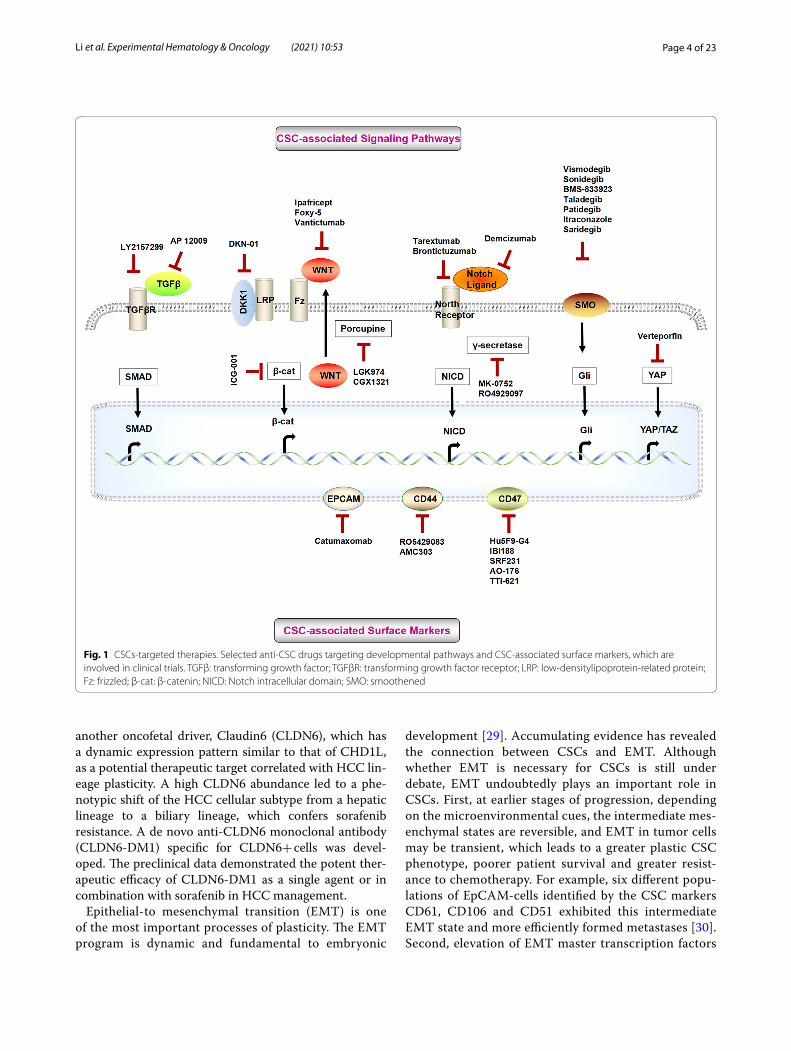
CSC biomarkers and signaling pathways are criti-cal factors distinguishing molecular classifications with stem-like traits. The expression levels plus the activation degrees of CSC biomarkers and signaling pathways differ in different subtypes, which has led to investigations into potential new avenues of targeted therapy. CSC-targeted therapies are currently in development, and many are already in clinical trials (Fig. 1; Table 2). To achieve better clinical outcomes, combination-based therapies should be implemented in CSC-targeting strategies. Moreover, CSC-directed therapy should be applied preferably early when CSC populations are still small and resistance path-ways have not yet been induced. CSC-directed therapy can also be applied in various stages of the patient treat-ment journey.
Tumor plasticityCancer cell plasticity has been proposed as one of the important mechanisms contributing to intratumor het-erogeneity. Plasticity enables cancer cells to shift between a nontransformed differentiated state and a tumo-rigenically transformed undifferentiated or CSC state in response to microenvironmental stimuli (e.g., onco-genic stresses, senescence, and inflammation). Plasticity usually includes stem cell multilineage interconversion, dedifferentiation and transdifferentiation [23, 24]. CSCs may arise from their normal stem cells, progenitors and/or differentiated somatic cells. CSCs have the potential to differentiate into cancer cells, dedifferentiate into original lineage cells, and/or transdifferentiate into other lineage cells [25, 26]. Aberrantly activated plasticity drives malig-nant transformation and confers tumors to accommodate the constraints of tumor growth and therapy resistance. In our previous study, we found that CHD1L (chromo-domain-helicase-DNA-binding protein 1-like gene) is a potential clinical developmental lineage oncogene in HCC. CHD1L expression is active in the embryonic stage but decreases progressively during terminal dif-ferentiation. However, CHD1L expression is abnormally amplified in HCC. This dynamic expression pattern is accompanied by elevated liver ancestral precursor mark-ers and reduced hepatic lineage differentiation markers. Further suppression of CHD1L may hinder poorly differ-entiated HCC and sensitize patients to chemotherapeu-tic drugs [27]. Our recently published study [28] found
Table 1 Representative markers of gastrointestinal CSCs
Liver cancer Colorectal cancer Pancreatic cancer Gastric cancer Esophageal cancer
(1) CD133+ [91] (1) ALDHhigh [101] (1) CD133+/ CXCR4+ [114] (1) CD44+ [119] (1) CD44+ [130]
(2) CD13+ [92] (2) Lgr5+ [102] (2) ALDH1A1+ [115] (2) CD44v8‑10+ [120] (2) Integrin α7+ [131]
(3) EpCAM+ [93] (3) ABCG2+/OCT4+ [103] (3) pAKT+/SOX9+ [116] (3) Snail+ [121] (3) ALDH1+ [132]
(4) SOX9 [94] (4) CD44v2+ [104] (4) FAM83A+ [117] (4) Lgr5+ [122] (4) ALDH1A1+ [133]
(5) Lin28B+ [95] (5) CD44v6+ [105] (5) CD133+/CD44+/CD24+/ESA+ [118]
(5) Frizzled7+ [123] (5) B7H4+ [134]
(6) β‑catenin+/GEP [96] (6) CD133+ [106] (6) CD44+/CD24+/EpCAM+ [118]
(6) CD47+ [124] (6) Gli1+ [135]
(7) CD133+/CD49f+ [97] (7) CD166+ [107] (7) CD133+ [125] (7) Musashi1+ [136]
(8) CD90+/CD45−, CD44+/CD90+ [98]
(8) EpCAM− [108] (8) ALDH+ [126] (8) Epiregulin+ [137]
(9) CD44+/CD133+ [99] (9) E‑cadherin− [109] (9) CD44+/CD24+ [127] (9) Numb+ [138]
(10) SALL4+/EpCAM+ [100] (10) CD133+/CD44+/ALDH1+ [110]
(10) CD44+/CD133+ [128] (10) WASH+ [139]
(11) EpCAM+/CD44+/CD166+ [19]
(11) CD44+/Snail1+/Vimentin+/E‑cadherin+ [129]
(11) CD47+/CD133+ [140]
(12) CD44+/CD24+ [111] (12) CD133+/CXCR4+ [141]
(13) CD133+/CXCR4+ [112](14) CD133+/CD24+ [113]
Page 4 of 23Li et al. Experimental Hematology & Oncology (2021) 10:53
another oncofetal driver, Claudin6 (CLDN6), which has a dynamic expression pattern similar to that of CHD1L, as a potential therapeutic target correlated with HCC lin-eage plasticity. A high CLDN6 abundance led to a phe-notypic shift of the HCC cellular subtype from a hepatic lineage to a biliary lineage, which confers sorafenib resistance. A de novo anti-CLDN6 monoclonal antibody (CLDN6-DM1) specific for CLDN6+ cells was devel-oped. The preclinical data demonstrated the potent ther-apeutic efficacy of CLDN6-DM1 as a single agent or in combination with sorafenib in HCC management.
Epithelial-to mesenchymal transition (EMT) is one of the most important processes of plasticity. The EMT program is dynamic and fundamental to embryonic
development [29]. Accumulating evidence has revealed the connection between CSCs and EMT. Although whether EMT is necessary for CSCs is still under debate, EMT undoubtedly plays an important role in CSCs. First, at earlier stages of progression, depending on the microenvironmental cues, the intermediate mes-enchymal states are reversible, and EMT in tumor cells may be transient, which leads to a greater plastic CSC phenotype, poorer patient survival and greater resist-ance to chemotherapy. For example, six different popu-lations of EpCAM-cells identified by the CSC markers CD61, CD106 and CD51 exhibited this intermediate EMT state and more efficiently formed metastases [30]. Second, elevation of EMT master transcription factors
Fig. 1 CSCs‑targeted therapies. Selected anti‑CSC drugs targeting developmental pathways and CSC‑associated surface markers, which are involved in clinical trials. TGFβ: transforming growth factor; TGFβR: transforming growth factor receptor; LRP: low‑densitylipoprotein‑related protein; Fz: frizzled; β‑cat: β‑catenin; NICD: Notch intracellular domain; SMO: smoothened

Page 5 of 23Li et al. Experimental Hematology & Oncology (2021) 10:53
Table 2 CSC‑targeting agents
Signaling pathways Cancer types Therapeutic agents Targets Phase Combination drugs Reference
TGF‑β signaling
Antisense oligonu‑cleotides
Pancreatic neo‑plasms, Colorectal Neoplasms
AP 12009 (Trabed‑ersen)
TGF‑β2 Phase 1 NCT00844064
TβR kinase inhibi‑tors/small‑mole‑cule inhibitors
(1) Hepatocellular carcinoma(2) Advanced or met‑astatic unresectable pancreatic cancer(3) Rectal adenocar‑cinoma(4) Advanced hepa‑tocellular carcinoma(5) Advanced hepa‑tocellular carcinoma(6) Metastatic cancer and advanced or metastatic unresect‑able pancreatic cancer(7) Metastatic pan‑creatic cancer(8) Advanced refrac‑tory solid tumors; Hepatocellular carcinoma(9) Metastatic pan‑creatic cancer
LY2157299(Galunisertib)
TβRI (1) Phase 1(2) Phase 1(3) Phase 2(4) Phase 2(5) Phase 2(6) Phase 1b/2(7) Phase 1(8) Phase 1b/2(9) Phase 1
(1) Sorafenib(2) Galunisertib(3) Capecitabine, Fluorouracil(4) Sorafenib(5) Sorafenib, Ramu‑cirumab(6) Gemcitabine(7) Durvalumab(8) Nivolumab(9) Durvalumab
(1) NCT02240433(2) NCT02154646(3) NCT02688712(4) NCT02178358(5) NCT01246986(6) NCT01373164(7) NCT02734160(8) NCT02423343(9) NCT02734160
Wnt signaling
β‑catenin inhibi‑tors
(1) Advanced pancreatic cancer; Metastatic pancreatic cancer; Pancreatic adenocarcinoma(2) Hepatitis C virus‑infected cirrhosis(3) Colorectal adeno‑carcinoma; Stage IVA colorectal cancer; Stage IVB colorectal cancer
ICG‑001(PRI‑724) CBP/β‑Catenin (1) Phase 1(2) Phase 1(3) Phase 2
(1) Gemcitabine(3) Bevacizumab,Leucovorin,Oxaliplatin,Fluorouracil
(1) NCT01764477(2) NCT02195440(3) NCT02413853
Wnt antibodies (1) Hepatocellular carcinoma(2) Pancreatic cancer; Stage IV pancreatic cancer
Ipafricept (OMP‑54F28)
Fzd8‑Fc fusion protein
(1) Phase 1(2) Phase 1
(1) Sorafenib(2) Nab‑PaclitaxelGemcitabine
(1) NCT02069145(2) NCT02050178
(1) Colorectal cancer(2) Metastatic colon cancer
Foxy‑5 WNT5a receptor (1) Phase 1(2) Phase 1
(1) NCT02020291(2) NCT02655952
Wnt antibodies Pancreatic cancer Vantictumab (OMP‑18R5)
Frizzled receptor Phase 1 Nab‑Paclitaxel and Gemcitabine
NCT02005315

Page 6 of 23Li et al. Experimental Hematology & Oncology (2021) 10:53
not only enforces metastatic potential but also exacer-bates the tumor-initiating capacity [9, 31, 32]. Indeed, most gastrointestinal cancer subtypes with stem cell
features display a strong association with the EMT phenotype.
Table 2 (continued)
Signaling pathways Cancer types Therapeutic agents Targets Phase Combination drugs Reference
DKK1 antibodies (1) Hepatocellular carcinoma(2) Esophageal neoplasms; Adeno‑carcinoma of the gastroesophageal junction; gastroe‑sophageal cancer; Gastric adenocarci‑noma(3) Carcinoma of intrahepatic and extra‑hepatic biliary system;Bile duct cancer;Cholangiocarcinoma
DKN‑01 DKK1 (1) Phase 1/2(2) Phase 1(3) Phase 1
(1) Sorafenib(2) Paclitaxel or pembrolizumab(3) Gemcitabine and cisplatin
(1) NCT03645980(2) NCT02013154(3) NCT02375880
Porcupine inhibi‑tors
(1) Metastatic colo‑rectal cancer(2) Pancreatic cancer; Esophageal squa‑mous cell cancer
LGK974 (WNT974) Porcupine (1) Phase 1(2) Phase 1
(1) LGX818, Cetuxi‑mab(2) PDR001
(1) NCT02278133(2) NCT01351103
Colorectal adenocar‑cinoma;Gastric adenocar‑cinoma; Pancreatic adenocarcinoma; Bile duct carcinoma; Hepatocellular car‑cinoma; Esophageal carcinoma;Gastrointestinal cancer
CGX1321 Porcupine Phase 1 Pembrolizumab NCT03507998
Notch signaling
DLL‑4 antibody (1) Metastatic pancreatic ductal adenocarcinoma(2) Locally advanced or metastatic pancre‑atic cancer(3) Colorectal cancer
Demcizumab (OMP‑21M18)
DLL4 (1) Phase 2(2) Phase 1(3) Phase 1
(1) Gemcitabine, Abraxane®
(2) Gemcitabine, Abraxane®
(1) NCT02289898(2) NCT01189929(3) NCT01189942
Notch receptor antibody
Untreated stage IV pancreatic cancer
Tarextumab(OMP‑59R5)
Notch2,Notch3
Phase 1/2 Nab‑Paclitaxel, Gem‑citabine
NCT01647828
Metastatic colorectal cancer
Brontictuzumab(OMP‑52M51)
Notch1 Phase 1 Trifluridine or tipiracil NCT03031691
γ‑secretase inhibi‑tor
Pancreatic cancer MK‑0752 γ‑secretase Phase 1 Gemcitabine hydro‑chloride
NCT01098344
γ‑secretase inhibi‑tor
(1) Metastatic pan‑creas cancer(2) Metastatic colo‑rectal cancer(3) Metastatic colo‑rectal cancer(4) Metastatic colo‑rectal cancer
RO4929097(R4733)
γ‑secretase (1) Phase 2(2) Phase 2(3) Phase 1(4) Phase 2
(2) FOLFOX regimen,Bevacizumab,Oxaliplatin,leucovorin calcium,fluorouracil(3) Cetuximab
(1) NCT01232829(2) NCT01270438(3) NCT01198535(4) NCT01116687

Page 7 of 23Li et al. Experimental Hematology & Oncology (2021) 10:53
Table 2 (continued)
Signaling pathways Cancer types Therapeutic agents Targets Phase Combination drugs Reference
Hedgehog signaling
SMO inhibitor (1) Metastatic pan‑creatic adenocarci‑noma(2) Metastatic colo‑rectal cancer(3) Pancreatic adeno‑carcinoma(4) Metastatic pancreatic cancer or solid tumors(5) Pancreatic ductal adenocarcinoma(6) Metastatic colo‑rectal cancer(7) Recurrent or metastatic pancreatic cancer
Vismodegib (GDC‑0449)
SMO (1) Phase 2(2) Phase 2(3) Phase 1(4) Phase 1(5) Phase II(6) Phase II(7) Phase II
(1) Gemcitabine, nab‑paclitaxel(2) Bevacizumab,Modified FOLFOX,FOLFIRI(3) Gemcitabine(4) Erlotinib, gemcit‑abine(6) Vismodegib, FOLFOX, FOLFIRI, Bevacizumab(7) Gemcitabine hydrochloride
(1) NCT01088815(2) NCT00636610(3) NCT01713218(4) NCT00878163(5) NCT01096732(6) NCT00959647(7) NCT01064622
Metastatic gastric, gastroesophageal, esophagealadenocarcinomas
BMS‑833923 (XL139) Phase 1 Cisplatin,capecitabine
NCT00909402
Esophageal cancer Taladegib (LY2940680)
Phase 1/2 Paclitaxel, carbopl‑atin, radiation
NCT02530437
(1) Metastatic pan‑creatic cancer(2) Advanced pancre‑atic adenocarcinoma
Patidegib (IPI‑926) (1) Phase 1(2) Phase 1
(1) Gemcitabine(2) FOLFIRINOX
(1) NCT01130142(2) NCT01383538
SMO inhibitor (1) Esophageal cancer(2) Esophageal Cancer(3) Locally Advanced Squamous Esopha‑geal Cancer
Itraconazole SMO (1) Phase 1(2) Phase 2(3) Phase 2
(1) NCT02749513(2) NCT04018872(3) NCT04481100
(1) Pancreatic cancer(2) Pancreatic cancer
Saridegib (IPI‑926) (1) Phase 1(2) Phase 1/2
(1) 5‑fluorouracil,Leucovorin,Irinotecan, Oxali‑platin(2) Gemcitabine
(1) NCT01383538(2) NCT01130142
(1) Resectable pan‑creatic adenocarci‑noma(2) Advanced or metastatic HCC(3) Esophageal cancer(4) Advanced pancre‑atic cancer(5) Pancreatic cancer(6) Pancreatic cancer(7) Pancreatic cancer(8) Resectable pan‑creatic cancer
Sonidegib (LDE225) (1) Phase 1/2(2) Phase 1(3) Phase 1(4) Phase 1(5) Phase 1/2(6) Phase 1(7) Phase 1(8) Phase 1
(1) Gemcitabine,nab‑paclitaxel(3) Everolimus(4) Fluorouracil, leu‑covorin, oxaliplatin, irinotecan(5) Gemcitabine,nab‑paclitaxel(6) Gemcitabine(7) Fluorouracil; Leu‑covorin; Oxaliplatin;Irinotecan
(1) NCT01431794(2) NCT02151864(3) NCT02138929(4) NCT01485744(5) NCT02358161(6) NCT01487785(7) NCT01485744(8) NCT01694589
Hippo signaling
YAP inhibitor Pancreatic cancer non‑resectable
Verteporfin YAP Phase 2 Photodynamic therapy
NCT03033225
CSC surface markers
Anti‑CD44 anti‑body
Malignant solid tumor
RO5429083 CD44 Phase 1 NCT01358903
CD44v6 inhibitor Malignant solid tumor
AMC303 CD44v6 Phase 1 NCT03009214

Page 8 of 23Li et al. Experimental Hematology & Oncology (2021) 10:53
Identification of molecular subtypes with CSC properties in gastrointestinal malignanciesGastrointestinal malignancies are highly heterogene-ous within tumors and have been defined by identifying so-called molecular subtypes. Transcriptomic, genomic, and/or epigenomic profiling of many tumors offers the basis for molecular classification. These distinct molec-ular subtypes reflect different biological backgrounds, including immunity, metabolism, and stemness. Spe-cifically, CSCs have been demonstrated to be a major source of intratumor heterogeneity. Integrative analyses of molecular subclassification from the CSC perspective may be encouraged with the aim of determining consen-sus molecular classification in patient prognostication and selection for therapies (Table 3).
Classification with CSC properties in liver cancerZhu et al. [33] used a 14-gene Notch score to stratify HCC into Notch-high HCC and Notch-low HCC sub-types. The Notch-high HCC subtype was associated with less differentiated tumors and poor survival, character-ized by increased expression of progenitor/cholangio-cyte markers (DCLK1 and KRT19), and highly enriched in genes related to developmental signaling and the fetal liver. In contrast, Notch-inactive HCC is a subtype of well-differentiated neoplasms with a better prognosis. In our recently published study [34], human ESCs were dif-ferentiated into human hepatocytes, and the whole differ-entiation process was defined by four stages: embryonic stem cells (ESs), endoderm (EN), liver progenitor cells
(LPs), and premature hepatocytes (PHs). We classified liver cancer into two major subtypes based on oncofe-tal gene expression patterns. We defined the genes from the ES and EN groups as the embryonic-like subtype (ES+ subtype) and genes from the LP and PH groups as the liver progenitor-like subtype (LP+ subtype). Inter-estingly, the ES+ subtype was mainly associated with genes in the pluripotency and stem cell self-renewal signaling pathway and the Gli signaling pathway, while the LP+ subtype was mainly associated with the TGF-β signaling pathway. Moreover, genes in the Notch and Wnt signaling pathways span all four stages. Lee et al. [35] uncovered two subgroups in their study: hepatocytes (HCs) and hepatoblasts (HBs). The specific HB subtype may arise from adult hepatic progenitor cells and fea-tures elevated expression of KRT7 and KRT19. Based on the oncofetal gene expression profiling, Yamashita et al. [36] distinguished two HCC subtypes: HpSC-HCC (referred to as EpCAM+ AFP+) and MH-HCC (referred to as EpCAM- AFP-). KRT19 and Wnt/β-catenin sign-aling are enriched in EpCAM+ AFP+ HCC cells. The EpCAM+ subgroup of HCC displayed a similar expres-sion pattern to the LP+ tumors in our study. In Hoshida et al.’s study [37], HCC patients were classified into S1, S2 and S3 subgroups based on the extent of tumor differ-entiation. The Wnt pathway was activated in S1 tumors by a mechanism of TGF-β signature activation. Class S2 was a progenitor cell group featuring Myc and AKT activation and EpCAM and AFP enrichment. S3 tumors were notable for differentiated hepatocyte function. Most of the patients’ tumors with oncofetal properties in our
Table 2 (continued)
Signaling pathways Cancer types Therapeutic agents Targets Phase Combination drugs Reference
Anti‑CD47 anti‑body
(1) Colorectal neo‑plasms/Solid tumors(2) Advanced malig‑nancies(3) Advanced solid cancers(4) Solid tumor
(1) Hu5F9‑G4(2) IBI188(3) SRF231(4) AO‑176
CD47 (1) Phase 1(2) Phase 1(3) Phase 1(4) Phase 1/2
(1) Cetuximab (1) NCT02953782(2) NCT03763149(3) NCT03512340(4) NCT03834948
Recombinant fusion protein binding CD47
Solid tumor TTI‑621 CD47 Phase 1 Rituximab or Nivolumab
NCT02663518
Anti‑EpCAM antibody
(1) Gastric cancer, Gastric adenocarci‑noma(2) Gastric Adeno‑carcinoma With Peritoneal Carcino‑matosis,Siewert Type II/III Adenocarcinoma of Esophagogastric Junction With Perito‑neal Carcinomatosis
Catumaxomab EpCAM (1) Phase 2(2) Phase 2
(1) NCT00464893(2) NCT01504256

Page 9 of 23Li et al. Experimental Hematology & Oncology (2021) 10:53
Tabl
e 3
Gas
troi
ntes
tinal
can
cer s
ubcl
assi
ficat
ions
with
CSC
trai
ts
Canc
er ty
pes
Refe
renc
eCl
assi
ficat
ions
Sign
atur
es &
Pat
hway
s (+
: A
ctiv
atio
n, −
: Sup
pres
sion
)Pr
ogno
sis
Inhi
bito
rs (+
: Sen
sitiv
e, −
: Re
sist
ance
)
Live
r can
cer
Zhu
et a
l. [3
0]N
otch
‑hig
hN
otch
and
TG
F‑β
sign
alin
g, K
RT19
, D
CLK
1, S
OX9
(+)
high
er tu
mor
sta
ge, w
orse
sur
viva
l ou
tcom
e
Liu
et a
l. [3
1]ES
+, L
P+ES
+: p
lurip
oten
cy a
nd s
tem
cel
l sel
f‑re
new
al s
igna
ling
path
way
s (+
), G
li,
Not
ch a
nd W
nt p
athw
ays
(+);
LP+
: TG
F‑β,
Not
ch a
nd W
nt p
ath‑
way
s (+
)
ES+
: HLM
6474
(+);
LP+
: SIS
3 (+
)
Lee
et a
l. [3
2]H
B, H
CH
B: K
RT7,
KRT
19, V
IM, A
P‑1
com
plex
(+
)H
B: tu
mor
‑inva
sive
, wor
se s
urvi
val,
poor
er p
rogn
osis
Yam
ashi
ta e
t al.
[33]
HpS
C‑H
CC (E
pCA
M+
AFP
+),
MH
‑H
CC (E
pCA
M−
AFP
−)
HpS
C‑H
CC: K
RT19
, c‑M
yc, W
nt/β
‑ca
teni
n (+
); M
H‑H
CC: H
epPa
r1 (+
)H
pSC
‑HCC
: tum
or‑in
vasi
ve, p
oor
prog
nosi
sM
H‑H
CC: g
ood
prog
nosi
s
HpS
C‑H
CC: β
‑cat
enin
inhi
bito
r (+
);Ep
CA
M+
HCC
cel
ls: G
SK‑3
β in
hibi
tor
BIO
, 5‑F
U (−
);Ep
CA
M−
HCC
cel
ls: 5
‑FU
(+)
Hos
hida
et a
l. [3
4]S1
, S2,
S3
S1: W
nt, T
GF‑
β, E
MT
(+);
S2: M
yc, A
KT, E
pCA
M, A
FP (+
);S3
: hep
atoc
yte
diffe
rent
iatio
n (+
)
S1: e
arlie
r rec
urre
nce,
tum
or‑in
va‑
sive
, poo
r sur
viva
l; S2
: poo
r sur
viva
l;S3
: goo
d su
rviv
al
Boya
ult e
t al.
[35]
G1,
G2,
G3,
G4,
G5,
G6
G1:
AXI
NI m
utat
ions
, fet
al li
ver
expr
essi
ng g
enes
, AFP
(+);
G2:
AKT
(+);
G3:
cel
l cyc
le g
enes
(+);
G5:
Wnt
(+);
G6:
Wnt
, LEF
1 (+
), C
DH
1 (−
)
G4‑
G6:
bet
ter s
urvi
val VS
G1‑
G3
Colo
rect
al c
ance
rM
aris
a et
al.
[36]
C1,
C2,
C3,
C4,
C5,
C6
C1:
EM
T (−
); C
2: W
nt (−
); C
3: E
MT
(−);
C4:
EM
T (+
); C
5: W
nt (+
); C
6:
EMT
(+)
C4
plus
C6:
wor
se p
rogn
osis
VS
all
othe
r sub
type
s

Page 10 of 23Li et al. Experimental Hematology & Oncology (2021) 10:53
Tabl
e 3
(con
tinue
d)
Canc
er ty
pes
Refe
renc
eCl
assi
ficat
ions
Sign
atur
es &
Pat
hway
s (+
: A
ctiv
atio
n, −
: Sup
pres
sion
)Pr
ogno
sis
Inhi
bito
rs (+
: Sen
sitiv
e, −
: Re
sist
ance
)
Colo
rect
al c
ance
rSa
dana
ndam
et a
l. [3
7]En
tero
cyte
, gob
let‑
like,
infla
m‑
mat
ory,
tran
sit‑
ampl
ifyin
g (C
S‑TA
, C
R‑TA
), st
em‑li
ke
Gob
let‑
like
and
ente
rocy
te: d
iffer
‑en
tiatio
n m
arke
rs (+
), st
em c
ell a
nd
Wnt
mar
kers
(−);
Tran
sit‑
ampl
ifyin
g: s
tem
and
pr
ogen
itor m
arke
rs (+
), W
nt‑t
arge
t ge
nes
(−);
Stem
‑like
: Wnt
, ste
m c
ell,
myo
epi‑
thel
ial a
nd m
esen
chym
al m
arke
rs
(+),
diffe
rent
iatio
n m
arke
rs (−
)
Gob
let‑
like
and
tran
sit‑
ampl
ifyin
g:
good
pro
gnos
is;
Ente
rocy
te a
nd in
flam
mat
ory:
in
term
edia
te D
FS; S
tem
‑like
tum
ors:
shor
test
DFS
Gob
let‑
like:
cet
uxim
ab (+
);In
flam
mat
ory:
FO
LFIR
I (+
);St
em‑li
ke: c
etux
imab
, FO
LFIR
I (+
);C
S‑TA
: cet
uxim
ab (+
);C
R‑TA
: cM
ET in
hibi
tors
(+),
cetu
xi‑
mab
(−)
De
Sous
a et
al.
[38]
CCS1
, CC
S2, C
CS3
CCS1
: Wnt
(+);
CCS3
: EM
T, m
atrix
re
mod
elin
g an
d TG
F‑β
(+)
CCS3
: poo
r pro
gnos
isCC
S3: c
etux
imab
(−)
Budi
nska
et a
l. [3
9]Su
rfac
e cr
ypt‑
like,
low
er c
rypt
‑like
, C
IMP‑
H‑li
ke, m
esen
chym
al, m
ixed
Surf
ace
cryp
t‑lik
e: E
MT
(−),W
nt (−
), β‑
cate
nin
(−);
Low
er c
rypt
‑like
: EM
T (−
), W
nt (+
);C
IMP‑
H‑li
ke: β
‑cat
enin
(−);
Mes
ench
ymal
: Eph
B2, E
MT
(+),
Wnt
(−
), β‑
cate
nin
(−);
Mix
ed: E
phB2
, EM
T, W
nt (+
)
Surf
ace
cryp
t‑lik
e an
d lo
wer
cry
pt‑
like:
bet
ter p
rogn
osis
VS
mes
en‑
chym
al;
CIM
P‑H
‑like
: poo
r OS
and
SAR;
M
esen
chym
al: r
ecur
renc
e ris
k, p
oor
OS;
Mix
ed: p
oore
rSA
R VS
low
er c
rypt
‑like
Roep
man
et a
l. [4
0]Ty
pe A
, Typ
e B,
Typ
e C
Type
A: E
MT
(−);
Type
B: E
MT
(−);
Type
C: E
MT
(+)
Type
A: g
ood
prog
nosi
s;Ty
pe B
: poo
r pro
gnos
is;
Type
C: p
oor p
rogn
osis
Type
A: 5
‑FU
(+);
Type
B: 5
‑FU
(+);
Type
C: 5
‑FU
(‑)
Gui
nney
et a
l. [4
1]C
MS1
, CM
S2, C
MS3
, CM
S4C
MS1
and
CM
S3: R
TK a
nd M
APK
pa
thw
ays
(+);
CM
S2: H
NF4
A, M
yc a
nd W
nt (+
); C
MS4
: EM
T, T
GF‑
β, a
ngio
gene
sis,
mat
rix re
mod
elin
g pa
thw
ays
(+)
CM
S1: w
orse
SA
R;C
MS4
: wor
se R
FS a
nd O
SC
MS1
: HSP
90 in
hibi
tors
(+);
CM
S2: H
SP90
inhi
bito
rs, E
GFR
inhi
bi‑
tors
, HER
2 in
hibi
tors
(+);
CM
S4: c
ombi
natio
n tr
eatm
ent o
f 5‑
FU a
nd lu
min
espi
b (+
)
Page 11 of 23Li et al. Experimental Hematology & Oncology (2021) 10:53
Tabl
e 3
(con
tinue
d)
Canc
er ty
pes
Refe
renc
eCl
assi
ficat
ions
Sign
atur
es &
Pat
hway
s (+
: A
ctiv
atio
n, −
: Sup
pres
sion
)Pr
ogno
sis
Inhi
bito
rs (+
: Sen
sitiv
e, −
: Re
sist
ance
)
Panc
reat
ic c
ance
rCo
lliss
on e
t al.
[43]
Cla
ssic
al, Q
M‑P
DA
, exo
crin
e‑lik
eC
lass
ical
: GAT
A6
(+);
QM
‑PD
A: m
esen
chym
e as
soci
ated
ge
nes
(+)
Cla
ssic
al: g
ood
prog
nosi
s;Ex
ocrin
e‑lik
e: in
term
edia
te p
rog‑
nosi
s;Q
M‑P
DA
: wor
st p
rogn
osis
Cla
ssic
al: e
rlotin
ib (+
), do
ceta
xel (−
);Q
M‑P
DA
: oxa
lipla
tine,
5‑F
U, B
ET
inhi
bito
r (+
);Ex
ocrin
e‑lik
e: S
N‑3
8 (−
)
Moffi
tt e
t al.
[44]
Nor
mal
str
omal
, act
ivat
ed s
trom
a,
basa
l‑lik
e, c
lass
ical
Act
ivat
ed s
trom
a: S
PARC
, WN
T2,
WN
T5A
, MM
P9, M
MP1
1 (+
);C
lass
ical
: GAT
A6
(+);
Act
ivat
ed s
trom
a: w
orse
sur
viva
l VS
norm
al s
trom
a;Ba
sal‑l
ike:
wor
se s
urvi
val VS
clas
sica
l
Nor
mal
str
oma:
Hed
geho
g pa
thw
ay
inhi
bito
r (+
);Ba
sal‑l
ike:
oxa
lipla
tine,
5‑F
U, B
ET
inhi
bito
r (+
);C
lass
ical
: doc
etax
el, S
N‑3
8 (−
)
Baile
y et
al.
[45,
81]
Panc
reat
ic p
roge
nito
r (PP
), sq
ua‑
mou
s, im
mun
ogen
ic a
nd a
berr
antly
di
ffere
ntia
ted
endo
crin
e ex
ocrin
e (A
DEX
)
Squa
mou
s: pa
ncre
atic
end
oder
mal
ce
ll‑fa
te d
eter
min
atio
n ge
nes,
Hed
geho
g/W
nt p
athw
ay (−
); TG
F‑β
and
Myc
pat
hway
(+);
PP: d
evel
opm
enta
l tra
nscr
iptio
n fa
c‑to
rs (+
), N
otch
pat
hway
(+)
AD
EX: p
ancr
eatic
dev
elop
men
tal
and
diffe
rent
iatio
nal g
enes
(+),
Not
ch p
athw
ay (+
)
PP: g
ood
surv
ival
out
com
es;
Imm
unog
enic
and
AD
EX: i
nter
me‑
diat
e su
rviv
al o
utco
mes
;Sq
uam
ous:
wor
st s
urvi
val o
utco
mes
Squa
mou
s: ox
alip
latin
e, 5
‑FU
, BET
in
hibi
tor,
GSK
3β in
hibi
tor (+
);Sq
uam
ous
and
PP s
ubty
pes:
doc‑
etax
el (−
);PP
: SN
‑38
(−)
Bied
erst
ädt e
t al.
[80]
Squa
mou
s/ba
sal‑
like
SUM
O p
athw
ay a
nd M
yc (+
)W
orse
pro
gnos
isSU
MO
ylat
ion
inhi
bito
r (+
)
Mue
ller e
t al.
[46]
Clu
ster
1, C
lust
er 2
, Clu
ster
3, C
lust
er
4, Clu
ster
5
Clu
ster
1: s
quam
ous
diffe
rent
iatio
n;C
lust
er 2
: epi
thel
ial d
iffer
entia
tion;
Clu
ster
3: e
mbr
yoni
c de
velo
pmen
t, EM
T, M
APK
pat
hway
;C
lust
er 5
: em
bryo
nic
deve
lopm
ent
Panc
reat
ic c
ance
rPu
leo
et a
l. [4
7]Pu
re c
lass
ical
, im
mun
e cl
assi
cal,
pure
bas
al‑li
ke, s
trom
a ac
tivat
ed,
desm
opla
stic
Stro
ma
activ
ated
and
pur
e ba
sal‑
like:
MET
, Hed
geho
g pa
thw
ay (+
)Pu
re c
lass
ical
and
imm
une
clas
sica
l: go
od p
rogn
osis
;St
rom
a ac
tivat
ed a
nd d
esm
opla
stic
: po
or p
rogn
osis
; Pur
e ba
sal‑l
ike:
w
orst
pro
gnos
is
Siva
kum
ar e
t al.
[48]
Not
ch, r
epre
ssed
Hed
geho
g/W
nt,
cell
cycl
eN
otch
: bes
t pro
gnos
is;
Repr
esse
d H
edge
hog/
Wnt
: wor
st
prog
nosi
s
Sein
o et
al.
[49]
W+
,W
−,
WRi
W+
: exo
geno
us W
nt (−
), R‑
spon
din
(+);
W−
: exo
geno
us W
nt (+
), R‑
spon
din
(+);
WRi
: Wnt
sig
nalin
g (−
)
W+
: poo
r sur
viva
l and
met
asta
tic
prog
ress
ion
Page 12 of 23Li et al. Experimental Hematology & Oncology (2021) 10:53
Tabl
e 3
(con
tinue
d)
Canc
er ty
pes
Refe
renc
eCl
assi
ficat
ions
Sign
atur
es &
Pat
hway
s (+
: A
ctiv
atio
n, −
: Sup
pres
sion
)Pr
ogno
sis
Inhi
bito
rs (+
: Sen
sitiv
e, −
: Re
sist
ance
)
Gas
tric
can
cer
Lei e
t al.
[53]
Prol
ifera
tive,
met
abol
ic, m
esen
‑ch
ymal
Prol
ifera
tive:
E2F
, Myc
, RA
S (+
);M
esen
chym
al: E
MT,
CSC
pat
hway
(+
)
No
sign
ifica
nt d
iffer
ence
s am
ong
the
3 su
btyp
esM
etab
olic
: 5‑F
U (+
);M
esen
chym
al: P
I3K‑
AKT
‑mTO
R in
hibi
‑to
rs (+
)
Cris
tesc
u et
al.
[54]
MSI
, MSS
/EM
T, M
SS/p
53+
and
MSS
/p5
3−M
SS/E
MT:
EM
T (+
)M
SS/E
MT:
wor
st p
rogn
osis
, rec
ur‑
renc
e, s
tage
III/I
V, e
arlie
r age
;M
SI: s
tage
I/II,
bes
t pro
gnos
is
Che
ul O
h et
al.
[55]
EP, M
PEP
: Wnt
(+),
EMT
(−);
MP:
EM
T, IG
F pa
thw
ay, H
edge
hog
path
way
(+)
EP: b
ette
r sur
viva
l;M
P: p
oor s
urvi
val
EP: a
djuv
ant c
hem
othe
rapy
(+);
MP:
adj
uvan
t che
mot
hera
py (−
), IG
F1/IG
F1R
path
way
inhi
bito
r (+
)
Che
ong
et a
l. [5
6]Ep
ithel
ial,
imm
une,
ste
m‑li
keEp
ithel
ial:
CD
X1 (+
);St
em‑li
ke: S
FRP4
(+)
Low
‑ris
k (im
mun
e‑hi
gh),
inte
rme‑
diat
e‑ris
k (im
mun
e‑lo
w a
nd s
tem
‑lik
e‑lo
w),
or h
igh
risk
(imm
une‑
low
an
d st
em‑li
ke‑h
igh)
No‑
bene
fit (i
mm
une‑
high
or
imm
une‑
low
and
epi
thel
ial‑l
ow) o
r ch
emot
hera
py‑b
enefi
t(im
mun
e‑lo
w a
nd e
pith
elia
l‑hig
h)
Oes
opha
geal
can
cer
Wal
ker e
t al.
[57]
ESCC
1, E
SCC
2, E
SCC
3ES
CC1:
SO
X2, T
P63
(+);
ESCC
2: Z
NF7
50 a
nd N
otch
1 m
utat
ion,
CD
K6 a
mpl
ifica
tion
(+),
KDM
6A a
nd K
DM
2D (−
), PI
K3R1
and
PT
EN (−
)ES
CC3:
mut
atio
ns a
ssoc
iate
d w
ith
RTK/
RAS/
PI3K
pat
hway
(+)
Wan
g et
al.
[58]
Subt
ype
I,Su
btyp
e II
Subt
ype
II: e
pith
eliu
m d
evel
opm
ent
gene
s (+
)N
o si
gnifi
cant
diff
eren
ces
betw
een
the
2 su
btyp
es
Jam
mul
a et
al.
[59]
Subt
ype
1,Su
btyp
e 2,
Subt
ype
3,Su
btyp
e 4
Subt
ype
1: D
NA
repa
ir an
d ce
ll cy
cle
driv
er g
enes
(+);
Subt
ype
2: d
iffer
entia
tiona
l and
de
velo
pmen
tal t
rans
crip
tion
fact
ors
(+);
Subt
ype
4: h
igh
leve
l of c
opy
num
ber a
ltera
tions
(+)
Subt
ype
1: C
DK4
/6 in
hibi
tors
, doc
‑et
axel
(+);
Subt
ype
2: C
DK4
/6 in
hibi
tors
(+);
Subt
ype
3: C
DK4
/6 in
hibi
tors
(+);
Subt
ype
4: C
DK4
/6 in
hibi
tors
(+),
CD
K2 in
hibi
tors
(+);
AD
EX: a
berr
antly
diff
eren
tiate
d en
docr
ine-
exoc
rine;
CCS
: col
on c
ance
r sub
type
; CIM
P: C
pG is
land
met
hyla
tor p
heno
type
; CM
S: c
onse
nsus
mol
ecul
ar s
ubty
pe; E
MT:
epi
thel
ial-t
omes
ench
ymal
tran
sitio
n; E
P: e
pith
elia
l ph
enot
ype;
EpC
AM
: epi
thel
ial c
ell a
dhes
ion
mol
ecul
e; E
S: e
mbr
yoni
c st
em c
ell;
ESCC
: oes
opha
geal
squ
amou
s ce
ll ca
rcin
omas
; FU
: 5-fl
uoro
urac
il; H
B: h
epat
obla
sts;
HC:
hep
atoc
ytes
; LP:
live
r pro
geni
tor c
ell;
MP:
m
esen
chym
al p
heno
type
; MSI
: mic
rosa
telli
te in
stab
ility
; MSS
: mic
rosa
telli
te-s
tabl
e; O
S: o
vera
ll su
rviv
al; P
H: p
rem
atur
e he
pato
cyte
s; P
P: p
ancr
eatic
pro
geni
tor;
QM
-PD
A: q
uasi
-mes
ench
ymal
-pan
crea
tic d
ucta
l ad
enoc
arci
nom
a; R
FS: r
elap
se-fr
ee s
urvi
val;
RTK:
rece
ptor
tyro
sine
kin
ase;
SA
R: s
urvi
val a
fter
rela
pse;
VS:
ver
sus

Page 13 of 23Li et al. Experimental Hematology & Oncology (2021) 10:53
study were consistent with the poorly differentiated S1 and S2 subgroups. Accordingly, the nononcofetal class of HCC matched the well-differentiated S3 subtype. Boyault et al.’s study [38] divided patients into G1 through G6 subgroups according to clinical and genetic character-istics. G1 and G2 tumors were characterized by AKT activation and fetal liver properties, G3 tumors were typi-fied by activation of cell cycle genes, heterogeneous G4 tumors were associated with rare TCF1 mutations, and G5 and G6 tumors were strongly related to Wnt pathway activation.
Classification of CSC properties in colorectal cancerSimilar to other gastrointestinal cancers, considerable effort has been dedicated to colorectal cancer stemness-based subtyping. Marisa et al. [39] revealed six subtypes: C1 (21%) is characterized by suppression of pathways associated with EMT, C2 (19%) is characterized by sup-pression of the Wnt pathway, C3 (13%) is characterized by suppression of EMT, C4 (10%) often shows upregulation of EMT and genes related to stem cell-like signatures, C5 (27%) exhibits overexpression of Wnt pathway genes, and C6 (10%) shows upregulation of the EMT pathway. Stud-ies performed by Sadanandam et al. [40] identified five subtypes and proposed that the five subtypes were asso-ciated with distinct cell subtypes found in normal colonic crypts. These subtypes are referred to as enterocyte, gob-let-like, inflammatory, transit-amplifying, and stem-like subtypes. The transit-amplifying subtype is a heterogene-ous subtype highly enriched for stem cell-relevant genes and the Wnt pathway and can be further divided into two groups based on the differential cetuximab response (CS-TA and CR-TA). Another stem-like subset is charac-terized by overexpression of Wnt signaling target genes and the presence of mesenchymal and myoepithelial stem-cell features, with downregulation of differentiation markers, whereas the goblet-like and enterocyte subsets are enriched in well-differentiated genes with few stem cell characteristics and low Wnt marker expression. In De Sousa et al. [41], they revealed three colon cancer subtypes: CCS1, CCS2 and CCS3. CCS1 (49%) refers to tumors with high activity of the Wnt signaling cascade, while CCS3 (27%) corresponds to heterogeneous and poorly differentiated tumors with upregulation of EMT, matrix remodeling and the TGF-β pathway. Unlike tradi-tional molecular classification according to gene expres-sion profiling, Budinska et al. [42] applied meta-gene profiles to identify five major subsets: surface crypt-like, lower crypt-like, CIMP-H-like, mesenchymal and mixed. Surface crypt-like and lower crypt-like subtypes are well differentiated with low expression of the EMT/stoma gene module when the mesenchymal subtype and the mixed subtypes are enriched for high expression of the
EMT/stroma gene module. In addition, the lower crypt-like and mixed subsets highly expressed Wnt signaling target signatures along with higher β-catenin nuclear immunoreactivity. In contrast, surface crypt-like and mesenchymal subgroups showed low expression of these signatures along with lower β-catenin nuclear immuno-reactivity. Moreover, the CIMP-H-like subtype exhibited almost no β-catenin nuclear immunoreactivity and low expression of gut development genes. Another classifica-tion based on whole-genome analysis of CRC patients in stages I-IV was discovered by Roepman et al. [43], who unveiled three molecular subtypes: Type A, Type B and Type C. Type A (22%) corresponds to a DNA mismatch repair (MMR)-deficient epithelial subtype, Type B (62%) represents an epithelial proliferative subtype, and Type C (16%) is characterized by the expression of EMT-related molecules. Intriguingly, these three subtypes overlapped with the three subtypes distinguished by De Sousa et al.
The above CRC subtyping systems considered three to six molecular subtypes with different characteristics that might lack compatibility and lead to some confusion. To standardize the different molecular subtypes, a large-scale study of 4000 CRC samples mainly in stages II-III was performed [44] to identify four distinct molecular classifications that correctly classified 78% of the sam-ples: CMS1 (14%, MSI immune), CMS2 (37%, canonical), CMS3 (13% metabolic) and CMS4 (23% mesenchymal). CMS2 is characterized by epithelial differentiation and strong activation of the Wnt and Myc signaling pathways. CMS4 is characterized by EMT upregulation, activa-tion of TGF-β signaling, enhanced matrix remodeling, complement-mediated inflammation and angiogenesis. In addition, NOTCH3 is a putative target for advanced CMS4 CRC patients [45]. CMS1-4 may reasonably be similar to any of the molecular subtypes mentioned above. CMS1 may fit the CCS2 class from De Sousa and the inflammatory class from Sadanandam. The class CMS2 consensus may be related to the CCS1 subtype from De Sousa and to the enterocyte and/or transit-amplifying subtypes from Sadanandam. The CMS4 sub-set can be associated with CCS3 tumors from De Sousa and with the stem-like module defined by Sadanandam.
Classification of CSC properties in pancreatic cancerCollisson et al.’s study [46] described three subtypes: clas-sical, quasi-mesenchymal (QM-PDA) and exocrine-like. The classical subtype is enriched in GATA6, while the QM-PDA subtype has comparatively low GATA6 expres-sion. GATA6 is essential for pancreatic development and differentiation. Moffitt et al. [47] extended the work from Collisson et al. by defining two subtypes for the tumor tissue (classical and basal-like) while adding stromal clas-sifications (normal and activated). The classical subtype
Page 14 of 23Li et al. Experimental Hematology & Oncology (2021) 10:53
overlaps with Collison’s classical subtype and is charac-terized by elevated GATA6 expression. In addition, “acti-vated” stroma is characterized by genes relevant to tumor promotion, such as the secreted protein SPARC, MMP family members MMP9 and MMP11, and WNT fam-ily members WNT2 and WNT5A. More recently, Bai-ley et al. [48] defined four molecular subtypes of PDAC: pancreatic progenitor (PP), squamous, immunogenic and aberrantly differentiated endocrine-exocrine (ADEX). The squamous subset entails downregulation of genes that control pancreatic endodermal cell fate determina-tion, repression of Hedgehog/Wnt signaling, and TGF-β signaling and MYC pathway activation. The PP subtype is characterized by developmental transcription factors and enrichment of Notch signaling. The ADEX class is enriched with genes that are important in lineage speci-fication and later stages of pancreatic development and differentiation. Intriguingly, a spectrum of differentiation that resembles embryonic lineages from early progenitors to fully differentiated cells exists in these subtypes. The Collison and Bailey classifications overlapped fairly well, with the exception of the immunogenic subtype. The Collison classical subtype is similar to the PP subtype, the QM-PDA subtype is similar to the squamous subtype, and the exocrine-like subtype is similar to the ADEX subtype. Following Bailey et al.’s study, Mueller et al. [49] defined five distinct clusters based on evolutionary tra-jectories and KRAS gene dosage. Cluster 1 resembles the squamous subtype. Clusters 2 and 5 are associated with epithelial cell differentiation and embryonic develop-ment. Cluster 3 is enriched for undifferentiated tumors and characterized by EMT and Ras downstream signal-ing. Cluster 4 is enriched for undifferentiated tumors and corresponds to the immunogenic subtype. Another sub-typing study from Puleo et al. [50] further distinguished five subtypes: pure classical, immune classical, pure basal-like, stroma activated and desmoplastic. The pure classical subtype is well differentiated (low-grade G1) and similar to the classical and PP subtypes. The pure basal-like subtype is poorly differentiated (high-grade G3) and associated with metastatic spread. The other subtypes correspond to an intermediate differentiation grade. The immune classical and desmoplastic subtypes fit Moffitt’s ‘normal stroma’ subtype, while the pure basal-like and stroma activated subtypes fit Moffitt’s ‘activated stroma’ subtype. Furthermore, the MET and Hedgehog signal-ing pathways are both activated in the stroma and pure basal-like subtypes.
Researchers have also classified PDAC based on CSC-related signal transduction pathways. In Sivakumar et al.’s study [51], three main biological processes gen-erated by the transcriptional signatures of oncogenic KRAS-specific master regulators were identified: Notch,
repressed Hedgehog/Wnt, and the cell cycle. All three subtypes represent three different transcriptional pro-grams during PDAC development and are linked to the Bailey subtypes. Suppression of Hedgehog/Wnt signal-ing is involved in the squamous subtype, Notch signaling is enriched in the ADEX and PP subtypes, and the cell cycle process is overrepresented by samples from the immunogenic subtype. Seino et al. [52] unveiled three subtypes based on the Wnt signaling pathway from a tumor organoid library: W+ (Wnt-secreting organoids), W- (Wnt-nonsecreting organoids) and WRi (Wnt and R-spondin-independent organoids). The W+ subtype is independent of exogenous Wnt ligands but requires R-spondin, the W- subtype depends on exogenous Wnt and R-spondin ligands, and the WRi subtype is Wnt signaling-independent.
Notably, potential overlap of defined subtypes may exist. Interestingly, plasticity occurs in these subtypes; that is, one subtype can switch to another, such as squa-mous to ADEX conversion [53, 54]. In mouse models, tumors shifted from squamous to classical after BET inhibitor treatment [55]. Another example is GATA6-mediated subgroup switching, as GATA6 downregula-tion contributes to the QM-like subtype in PDAC [48]. Conversely, GATA6-high PDACs exhibit higher levels of epithelial Wnt ligands, indicating GATA6-regulated Wnt niche dependency in patients with PDACs [52].
Classification of CSC properties in gastric cancerLei et al. [56] unveiled three subtypes in their study: proliferative, metabolic, and mesenchymal. The mesen-chymal subtype harbors CSC-like properties with the following four features. First, this subtype is strongly associated with CSC pathway activation. Second, it shows high CD44 and low CD24 levels compared with other types, which is similar to the QM-PDA subtype of PDAC. Third, it maintains an undifferentiated state, which is an essential feature of CSCs. Finally, the hypermethylated gene sets significantly overlap with genes expressed at low levels in HCC harboring hepatic stem cell proper-ties. In addition, the proliferative subtype shows elevated activities for several oncogenic pathways: E2F, MYC, and RAS. Cristescu et al. [57] used gene expression data to describe four patient subsets of gastric cancer: MSI, MSS/EMT, MSS/p53+ and MSS/p53− , where MSS refers to microsatellite stable tumors. The MSS/EMT mod-ule was significantly correlated with the EMT signature. Another Korean study led by Oh et al. [58] distinguished two distinct molecular subtypes: the epithelial phenotype (EP) and mesenchymal phenotype (MP). Higher recur-rence rates reflecting the clinical consequences of EMT were shown for the MP subtype, as the EMT-promot-ing pathway (TGF-β, Hedgehog pathway) and proteins

Page 15 of 23Li et al. Experimental Hematology & Oncology (2021) 10:53
(MYH11, RICTOR and CAV11) were highly activated in the MP module. The development and progression of EP-subtype gastric tumors are mainly due to activation of the Wnt pathway through repression of the SFRP fam-ily (SFRP1, SFRP2, SFRP3, and SFRP4). A retrospective study by Cheong et al. [59] identified four classifier genes to stratify patients into three subtypes: epithelial (CDX1), immune (GZMB and WARS), and stem-like (SFRP4). SFRP4 is a modulator of Wnt signaling-associated EMT, suggesting that EMT might contribute to the clinical consequences of the stem-like subtype.
Classification of CSC properties in esophageal cancerEsophageal cancer includes two main histological types: esophageal adenocarcinomas (EACs) and esophageal squamous cell carcinomas (ESCCs). In contrast to studies on other gastrointestinal tract tumors, molecular classi-fication studies of esophageal cancer are currently lack-ing. Walker et al. [60] unveiled three molecular subtypes: ESCC1, ESCC2, and ESCC3. ESCC1 tumors show high amplification of SOX2 and TP63. SOX2 is a pluripotent stem cell transcription factor that favors the development and maintenance of squamous epithelia. ESCC2 tumors contain more ZNF750 and NOTCH1 mutations, inacti-vation of the histone demethylases KDM6A and KDM2D, deactivation of the PIK3CA suppressors PIK3R1 and PTEN, and CDK6 amplification. The last group, ESCC3, contains mutations forecasting activation of the RTK/RAS/PI3K pathway. Another study performed by Wang et al. [61] revealed two distinct subtypes of ESCC. Sub-type I entails a highly activated pathway involved in the immune response, while subtype II is enriched in path-ways involved in ectoderm development. Epithelium development genes, including E2F4, JUN, KRT5 and KRT14, were enriched in Subtype II. PDPN and SIX1 have high expression levels in Subtype II ESCC, while SIX1 can maintain or increase PDPN-positive CSCs. Specifically, they discovered potential ESCC subset-spe-cific diagnostic markers: EYA2 and FOXA1 for subtype I and KRT14 and LAMC2 for subtype II, which may help guide ESCC clinical treatment. Most recently, Jammula et al. [62] identified four subtypes from Barrett’s esopha-gus and EAC. Subtype 1 showed elevation of driver gene alterations (CCND1, CCNE1, MYC, CDK6). Subtype 2 displayed significant overexpression of sets of key mas-ter transcription factors correlated with differentiation and development, including HNF4A/G, FOXA1/2/3, GATA6 and CDX2. Subtype 3 was enriched in all path-ways related to immune regulation, while subtype 4 contained a high quantity of copy number alterations. Considering the obvious parallels existing in these three classifications by Walker, Wang and Jammula, the extent
of connection among the three classifications remains to be fully addressed.
Common events of gastrointestinal subtypesCancer stem cell properties are included in molecular classification systems for gastrointestinal cancers. Most classifications are characterized by similar stem cell traits, poor differentiation, and poor clinical outcomes. Most gastrointestinal tumors appear to belong to sub-groups with EMT traits, for example, the C4, CCS3, mes-enchymal, type C and CMS4 subsets in CRC, the cluster 3 and pure basal-like subgroups in PDAC, and the MSS/EMT and MP subtypes in gastric cancer. Another con-sistently identified subtype is characterized by the activa-tion of signaling pathways involved in ESC differentiation and pluripotency, such as the Wnt pathway, TGF-β path-way, Hedgehog pathway and Myc pathway. For instance, the Wnt pathway is enriched in the C5, C6, transit-ampli-fying, stem-like, CCS1 and CMS2 subgroups in CRC, in the ‘activated’ stroma and W+ subgroups in PDAC, and in the EP subgroup in gastric cancer. Additionally, most classifications reflect the original functions of ESCs char-acterized by overexpression of key developmental and differentiation factors, for example, our ES and LP sub-types in liver cancer and the PP, squamous, ADEX, Clus-ter 2 and Cluster 5 subgroups in PDAC.
Notably, genetic mutations also contribute to the tumor stemness phenotype. For instance, ESCC2 esopha-geal cancers are enriched for NOTCH1 mutations, and mutations in ESCC3 drive activation of the RTK/RAS/PI3K pathway, indicating that genomic and transcrip-tomic subtypes interact with each other. Integrating both genomic and transcriptomic information may help iden-tify the related entities or entities with common origins. Furthermore, according to clinical observations of poorly differentiated gastrointestinal cancers with preserved lineage characteristics of their developmental precursor cells, such tumors may progress to acquire classifiable phenotypes, and the similarities between tumor subtypes from different organs may be defined from early embry-onic development events that are reflected in the devel-opmental signaling expression or mutational profiles of classified tumors. The inter- and intratumor heterogene-ity caused by these events can be used to foster patient welfare.
Evaluating strategies for subtype‑directed therapySubtyping identification strategiesThe tumor heterogeneity of each subtype is mainly explored by multiomics (transcriptomics, proteomics, metabolomics, lipidomics, glycomics) in many pub-licly available repositories (such as TCGA, ICGC and GEO) or institutional sources. For example, Liu et al.

Page 16 of 23Li et al. Experimental Hematology & Oncology (2021) 10:53
performed unsupervised clustering to define three immune subtypes with different features from mul-tiple HCC databases and developed a support vector machine (SVM) classifier based on multiomics sig-natures, and this multiomics SVM model provided potential predictors for prognosis and responses to immunotherapy in HCC [63]. Molecular subtyping typically requires tissue biopsy samples. However, sub-typing strategies may be hampered by the following aspects. First, in some hardly accessible tumors, such as PDAC, omics-based subtype classifications are difficult to obtain; in this case, small classifiers can be devised to circumvent this problem by working on small amounts of tumor tissues from routine diagnostic cytology. Sec-ond, intratumor heterogeneity may lead to sampling error and possibly tumor misclassification, and devel-oping marker panels or blood-based markers for tumor subtypes can help circumvent these problems [64]. Recently, liquid biopsy has become an appealing non-invasive clinical tool for the isolation and detection of blood-based markers. Jose et al. [65] provided an exam-ple to apply a microfluidic platform to identify CSC subtypes (CD133+CK+CD45−DAPI+EpCAM+ and CD133+CK+CD45-DAPI+EpCAM-) from patient blood samples in PDAC. Liquid biopsy can overcome the difficulties of obtaining tissue biopsies, capture spatial and/or temporal heterogeneity, and facilitate therapy response monitoring [66]. However, multiple technical issues, especially insufficient sensitivity and specificity, still need to be solved for future clinical application.
To date, markers for tumor subtypes can be measured using flow cytometry, real-time quantitative polymer-ase chain reaction (qPCR), and immunohistochemi-cal or immunofluorescent staining [67]. In addition, recent achievements of single-cell techniques such as scRNA-seq (single-cell RNA sequencing) have provided extraordinary insights into intratumor heterogeneity, which has already been highlighted in cancer classifica-tion, diagnosis, and treatment [68]. scRNA-seq can be used to characterize rare but important subtypes. For example, Daniel et al. [16] revealed a novel stemness-related cell subclone (CD24+/CD44+) within EPCAM+ HCC cells, and suppression of the signature gene CTSE in CD24+/CD44+cells abrogated the self-renewal ability of HCC. Lin et al. [69] applied scRNA-seq to identify the EMT+ PDAC subtype and epithelial tumor cell (ETC) population. The reported high mes-enchymal gene expression signals (i.e., QM subtype) were enriched in the EMT+ subtype, and the signature genes defining the classic, progenitor and squamous subtypes were enriched in the ETC population, whereas
the signature genes defining the basal subtype were enriched in both EMT and ETC tumor cells.
Preclinical models for subtype therapyVarious drug sensitivity studies have been performed using the most common models, such as tumor-derived cell lines and patient-derived xenografts (PDXs), which can retain the common molecular characteristics of primary tumors and generate valuable transcriptomic information for molecular subtypes and corresponding clinical and pharmacological data for association stud-ies. Several large-scale studies have been performed on a large set of tumor-derived cell lines for biomarker dis-covery and drug response prediction. Stefano et al. [70] screened the most commonly used liver cancer cell lines, including 34 models, and in combination with screening 31 anticancer agents, identified markers of therapeutic response. Another promising technique for large-scale functional screening using RNAi or CRISPR/Cas9 has also been applied to study cancer subtypes. For example, Robert et al. [71] performed a large-scale RNAi screen in 398 cancer cell lines to elucidate the vulnerabilities of specific cancer subtypes. Although tumor-derived cell lines are easily manipulated and acceptable for stem cell-based subtype identification and high-throughput screening, 2D culture cannot fully reproduce the native 3D microenvironment of tumor cells. Instead, the PDX model more reliably recapitulates patient subtypes than 2D culture by retaining patient histopathological and molecular features. Researchers have successfully trans-lated the CMS classification of CRC to preclinical PDX models for targeted treatment and distinguished patients with poor clinical consequences within the CMS groups [72–74]. However, the shortcomings of long engraftment periods and low engraftment efficiency hamper large-scale drug screening with PDX models. Alternatively, spheroids are used as important 3D preclinical models to test the effects of targeted drugs, especially to investigate the interaction between pharmacological and radiothera-peutic strategies. For example, Che et al. [75] established co-cultured pancreatic stellate cells/PDAC heterosphe-roids and found that this model exhibited higher resist-ance to gemcitabine than PDAC-only spheroids. The role of dCK in gemcitabine resistance was further stud-ied by using this model. Another useful 3D preclinical model is organoids. Cancer-derived organoids are good in vitro models that capture tumor subtype heterogene-ity, enable therapeutic screening and encompass unique subsets required for precision medicine development. Helen et al. [76] established a human gastric cancer orga-noid biobank that encompassing the most known molec-ular subtypes. Takashi et al. [52] developed a pancreatic tumor organoid library and identified three subtypes

Page 17 of 23Li et al. Experimental Hematology & Oncology (2021) 10:53
based on the stem cell niche factor associated with Wnt and R-spondin. Genetically engineered models appear to be another preclinical platform to evaluate molecular subtypes and therapeutic responses; however, they are unlikely to benefit patients whose tumors lack the target [77–80].
Clinical relevance and subtype‑driven therapiesLiver cancer subtypesZhu et al. [33] used a 14-gene Notch score to sort Notch-active signatures. Notch-active HCCs were found to resemble cholangiocarcinoma (CC)-like HCC and exhibit higher tumor stages and poorer prognoses than Notch-inactive HCCs. Notch signaling is best known for its role in cell fate determination. An overwhelming number of studies have shown that Notch signaling plays pro-moting roles in carcinogenesis and tumor progression; therefore, patients with cancer may benefit from Notch pathway blockade. Currently, multiple Notch inhibi-tors against γ-secretase, Notch receptors or ligands have been developed, including γ-secretase inhibitors, siRNA and monoclonal antibodies. The combination of Notch inhibitors with other chemotherapy or radiotherapy holds considerable promise for achieving better curative effects [81]. As a detailed subclassification of stem cell-like tumors is lacking, we established new classification models to mimic the whole differentiation process from human ESCs to human hepatocytes and classified HCC patients into two subtypes based on stem-like expression patterns. E2F1 and SMAD3 are two important oncofetal drivers of liver tumors with defined gene signatures. HCC patients with the ES-like subtype were more sensitive to the E2F1 inhibitor HLM6474, while HCC patients with the LP-like subtype were more sensitive to the SMAD3 inhibitor SIS3, indicating that targeting specific oncofe-tal drivers may promote drug selectivity and eliminate tumorigenicity effectively [34]. Lee et al. [35] uncovered a fetal HB subtype that might arise from hepatic progeni-tor cells with a poor prognosis. Another stemness-based HCC classification was proposed by Yamashita and col-leagues [36]. The EpCAM+ AFP+ HCC subgroup har-bored progenitor features with a poor prognosis, while the EpCAM-AFP-HCC subset had adult hepatocyte fea-tures with a good prognosis. Moreover, β-catenin inhibi-tors were more effective in EpCAM+ HCC cells than in EpCAM- HCC cells in vitro. In addition, a GSK-3β inhib-itor and 5-fluorouracil (FU) increased the EpCAM+ pop-ulation in HCC cells. Based on the extent of tumor differentiation, Hoshida et al. [37] classified HCC patients into S1, S2 and S3 subgroups. Subclass S1 is linked with a higher risk of early recurrence, with more satellite lesions and vascular invasion. As TGF-β boosts Wnt activ-ity by altering the subcellular localization of β-catenin,
cotargeting TGF-β and β-catenin may be an effective strategy for the treatment of the S1 subclass of HCC. S2 tumors demonstrate Myc and AKT activation, suggest-ing that AKT or PI3K inhibitors might be valuable in this particular subclass. In contrast, the S3 subclass contains the majority of well-differentiated tumors, which tend to have a lower grade and better survival outcomes.
Colorectal cancer subtypesIn Sadanandam et al.’s study [40], the goblet-like and transit-amplifying subtypes showed a good prognosis, the enterocyte and inflammatory subtypes were associ-ated with intermediate disease-free survival (DFS), while the stem-like tumors corresponded to the shortest DFS but were shown to benefit more from FOLFIRI than oth-ers, while CS-TA and CR-TA tumors were sensitive to cetuximab and cMET inhibitor treatment, respectively. De Sousa et al. [41] compared the clinical characteristics of CCS1 and CCS3 tumors in their study and found that patients with CCS1 tumors had a good prognosis. CCS3 tumors harbored malignant potential at an early stage of adenomas and were refractory to anti-EGFR therapy. Budinska et al. [42] assessed the associations of their clas-sifications with patient survival. Surface crypt-like and lower crypt-like subgroups showed a better prognosis. CIMP-H-like and mesenchymal subtypes were associated with poor overall survival (OS), while the former was also associated with short survival after relapse (SAR). The mixed subgroups showed a trend toward the worst OS. Molecular classification performed by Roepman et al. distinguished three subclasses [43]; Type A has the best prognosis, Type B has an intermediate prognosis but can benefit from adjuvant 5-FU chemotherapy, and Type C showed the worst survival and resistance to 5-FU-based chemotherapy. When assessing the existence of core subtype gene expression patterns among available CRC subtyping algorithms, four consensus molecular sub-types were observed to be related to clinical characteris-tics [44]. CMS1 patients are usually diagnosed at higher pathologic grades and show worse survival after relapse. Conversely, CMS2 patients had superior survival rates after relapse, whereas CMS4 patients had worse relapse-free and overall survival and were more likely to be in stage III and stage IV. Recently, Sveen et al. [72] trans-lated this CMS system to preclinical models contain-ing CRC-derived cell lines and PDX models to perform high-throughput in vitro drug screening. They found that CMS2 tumors were strongly responsive to EGFR and HER2 inhibitors and that CMS1 and CMS2 tumors were highly sensitive to HSP90 inhibitors. Furthermore, combination treatment with 5-FU and luminespib could relieve chemoresistance in CMS4 patients.
Page 18 of 23Li et al. Experimental Hematology & Oncology (2021) 10:53
Pancreatic cancer subtypesIn Collisson et al.’s study [46], among the three subtypes that they described, the classical subtype had a better prognosis than the other two types, while patients with QM-PDA subtype tumors had the worst prognosis. For subtype-specific drug responses, the classical subtype was more sensitive to erlotinib. Moffitt et al. [47] defined two major subtypes for the tumor (classical and basal-like) and stromal classifications (normal and activated). Patients with the activated stroma subtype had a worse median survival than those with the normal stroma subtype. Inhibition of the Hedgehog pathway could accelerate the development of PDAC and promote the delivery of chemotherapy in the normal stromal sub-type. In addition, patients with the basal-like subtype had a worse medium survival than those with the clas-sical subtype; however, the former type showed a better response to adjuvant therapy than the classical subtype. More recently, Bailey et al. [48] published four subtypes as described above. The squamous subtype is correlated with significantly worse clinical outcomes. Some patients in the PP subgroup had better survival outcomes than those in the immunogenic and ADEX subgroups. Mul-tivariate analysis found that this classification exhibited independent prognostic value [82]. A comprehensive analysis of drug sensitivity in the above three classifica-tions (Collisson, Moffitt and Bailey) showed that the QM-PDA, basal-like and squamous subtypes are sensitive to oxaliplatine and 5-FU. The activated stroma, QM-PDA and squamous subtypes show more resistance to gemcit-abine than the ADEX subtype and Collisson’s and Mof-fitt’s classical tumor subtypes. Collisson’s and Moffitt’s classical subtypes and Bailey’s squamous and PP subtypes are resistant to docetaxel. PP, Collisson’s exocrine-like and Moffitt’s classical subtypes are resistant to SN-38. When comparing gene expression in different sub-types, the QM-PDA, basal-like and squamous subtypes strongly expressed Myc, indicating that these subtypes may be more sensitive to BET inhibitors. EGFR signaling has been reported to enhance cancer cell stemness [83, 84]; although PDAC cells frequently present high EGFR expression, most are easily resistant to anti-EGFR treat-ment. Combined targeted therapy may help overcome this resistance. Moreover, Biederstädt et al. [85] detected coactivation of MYC and SUMO in the basal-like/squa-mous subtype of PDAC, which is known to be resistant to chemotherapies. SUMOylation inhibitor-based thera-pies might be a potential strategy to target this aggres-sive PDAC subtype. Specifically, Brunton et al. [86] found that loss of HNF4A and GATA6 could lead to a plastic-ity switch from the classical (progenitor) subtype to the squamous subtype and elevated expression of lyco-gen syn-thase kinase 3 beta (GSK3β). GSK3β inhibitors
showed selective sensitivity in the squamous subtype; however, a subgroup of squamous patient-derived cell lines (PDCLs) acquired drug tolerance and had access to the WNT gene program. In addition, another devel-opmental transcription factor, HNF1A, is a novel regu-lator of pancreatic cancer stem cell properties, and HNF1A + tumors (non-QM, overlap with the exocrine/ADEX subtype) benefit more from FOLFIRINOX than gemcitabine-based treatment [87]. Puleo et al. [50] rede-fined subtypes of PDAC into five groups. The pure clas-sical and immune classical subclasses had similar good prognoses. The patients in the stroma-activated and des-moplastic subgroups had a severe prognosis when pure basal-like tumors had the worst outcome. The Hedgehog pathway was highly enriched in stomal activated and pure basal tumors, suggesting that Hedgehog inhibitors may help prolong survival in PDAC patients with tumors in these two subgroups. In another study [51], three subtypes generated by the transcriptional signatures of oncogenic KRAS-specific master regulators were identi-fied: Notch, repressed Hedgehog/Wnt, and the cell cycle. Evidence of the potential clinical importance of the three groups revealed that the Hedgehog/Wnt group had the worst prognosis, while the Notch group showed the best prognosis. Seino and colleagues [52] established a library of PDAC-derived organoids and identified heterogene-ous subtypes dependent on Wnt ligands. They found that epithelial Wnt molecules (Wnt3, Wnt7a, Wnt7b, and Wnt10a) could serve as a surrogate marker for Wnt-producing PDACs. Notably, WRi and W + organoids displayed higher levels of epithelial Wnt gene expression than W-organoids, and high expression of epithelial Wnt molecules was closely linked to markedly poor survival and metastatic progression.
Gastric cancer subtypesLei et al. [56] developed a robust classification of pri-mary gastric adenocarcinomas: proliferative, metabolic, and mesenchymal. Analysis of survival information showed no significant differences in survival among the three subgroups. Patients with proliferative- and mes-enchymal-subtype tumors did not benefit from 5-FU treatment. In contrast, mesenchymal-subtype gastric cancer cells were preferentially sensitive to PI3K-AKT-mTOR inhibitors, possibly because this subtype of cells resembles CSCs. This finding is consistent with the observation that PI3K-AKT-mTOR inhibitors are also effective in prostate cancer and glioblastoma [88, 89]. High levels of CD44 are another distinctive feature of the mesenchymal subtype. CD44 is a well-known sur-face biomarker of CSCs and is aberrantly expressed in a variety of tumors in the forms of CD44s (standard iso-form) or CD44v (variant isoform). A high abundance of

Page 19 of 23Li et al. Experimental Hematology & Oncology (2021) 10:53
CD44 is closely associated with a malignant phenotype and poor clinical outcomes. CD44-positive cancer cells displayed lower sensitivity to sorafenib and 5-FU. Tar-geting CD44 may be a promising therapeutic strategy for cancer management. CD44 antibodies and block-ade of the HA-CD44 balance offer therapeutic inter-ventions to effectively impair the properties of CSCs among various cancers [90].
Cristescu et al. [57] investigated the clinical relevance of their four molecular subtypes and found that the age at occurrence of the MSS/EMT subtype was signifi-cantly lower than that of the other subtypes. Most sub-jects with this subtype were diagnosed at a late stage (III/IV) and showed the worst prognosis and the high-est recurrence frequency among the four subtypes. Oh et al. [58] described two subtypes: MP and EP. Clini-cally, the MP subtype is associated with significantly poor survival, a high recurrence rate, and resistance to standard adjuvant chemotherapy. The EP subtype is correlated with better survival and sensitivity to adju-vant chemotherapy. Importantly, MP-subtype cancer cells are significantly more sensitive to linsitinib treat-ment than EP-subtype cancer cells. Cheong et al. [59] uncovered three subtypes (immune, stem-like, and epithelial) for patients with resectable stage II-III gas-tric cancer and then developed a prognosis-based sin-gle-patient classifier to divide patients into low-risk (immune-high), intermediate-risk (immune-low and stem-like-low), or high risk (immune-low and stem-like-high) groups. They also developed a prediction-based single-patient classifier to divide patients into no-benefit (immune-high or immune-low and epithe-lial-low) or chemotherapy-benefit (immune-low and epithelial-high) groups. The association between the prognostic single-patient classifier groups and 5-year OS was significant. Furthermore, the association between the predictive single-patient classifier groups and adjuvant chemotherapy response in terms of OS was also notable. Collectively, the MSS/EMT, MP and stem-like subtypes have the worst prognosis in terms of clinical consequences for multiple cohorts, highlighting the significance of stemness-based subsets requiring clinical intervention.
Esophageal cancer subtypesClinically, molecular classification studies of esophageal cancer are still limited. Jammula et al. [62] unveiled four subtypes relevant to therapy. Subtype 1 was sensitized to CHFR, which is a cell cycle checkpoint inhibitor. In addition, CDK4/6 inhibitors were effective across all sub-types, whereas CDK2 inhibitors were preferentially effec-tive toward subtype 4 patients.
ConclusionsOur understanding of gastrointestinal cancer biology has drastically improved. The main genetic changes and tumor subtypes are gradually becoming well established, and their clinical relevance is being clarified. Evident dis-tinctions are present in the biological features and clinical properties of gastrointestinal cancers, which are probably a result of heterogeneity. Clinically, heterogeneity largely gives rise to tumor progression, metastasis, resistance to therapy, and relapse. Molecular heterogeneity arises from the existence of molecular subtypes. Due to the notable effect of CSCs on heterogeneity, CSC traits are undoubt-edly tightly associated with molecular classifications. Interestingly, CSCs usually resemble embryonic stem cells, which signifies the importance of developmental signals in cancer initiation and therapeutic resistance. Therefore, integrating the molecular subtypes associ-ated with stemness properties may offer new insights into treatment resistance.
Although molecular classifications based on CSC traits are substantially expanding our understanding of gastro-intestinal malignancies, the implementation of effective precision medicine is still hindered by some problems. First, sufficient studies describing the stemness-based molecular subtypes of each gastrointestinal cancer are lacking, and consensus subtypes may be identified and confirmed in future cancer expression data. Second, reli-able biomarkers corresponding to molecular subtypes to predict the response to current therapies are also lacking. Newer more effective approaches should be developed and applied in the detailed characterization of intra- and intertumoral heterogeneity, such as scRNA-seq and relevant preclinical models. Further precise targeting of tumor-initiating steps and driving events according to subtype-specific biomarkers might serve as a novel therapeutic strategy in gastrointestinal cancer treatment. Finally, systematic tumor and liquid biopsy techniques should be developed to define signature molecules allow-ing delineation of the complete molecular profile and patient classification.
In summary, we provide an overview of molecular clas-sifications from the CSC perspective that may facilitate improvement in the clinical management of patients with gastrointestinal malignancies and thus result in more favorable outcomes.
AbbreviationsADEX: Aberrantly differentiated endocrine‑exocrine; CCS: Colon cancer sub‑type; CHD1L: Chromodomain‑helicase‑DNA‑bindingprotein 1‑like gene; CIMP: CpG island methylator phenotype; CLDN6‑DM1: Anti‑CLDN6 monoclonal antibody conjugated with cytotoxic agent (Mertansine) DM1; CMS: Consensus molecular subtype; CRC : Colorectal carcinoma; CSC: Cancer stem cell; EAC: Oesophageal adenocarcinomas; EMT: Epithelial‑to‑mesenchymal transition; EN: Endoderm; EP: Epithelial phenotype; EpCAM: Epithelial cell adhesion

Page 20 of 23Li et al. Experimental Hematology & Oncology (2021) 10:53
molecule; ESC: Embryonic stem cell; ESCC: Oesophageal squamous cell carci‑nomas; FU: 5‑Fluorouracil; HB: Hepatoblasts; HC: Hepatocytes; HCC: Hepatocel‑lular carcinoma; LP: Liver progenitor cell; MP: Mesenchymal phenotype; MSI: Microsatellite‑instable; MSS: Microsattelite‑stable; OS: Overal survival; PDAC: Pancreatic cancer; PDXs: Patient‑derived xenografts; PH: Premature hepato‑cytes; PP: Pancreatic progenitor; QM‑PDA: Quasi‑mesenchymal‑pancreatic ductal adenocarcinoma.
AcknowledgementsNot applicable.
Authors’ contributionsMML wrote the manuscript with assistance from JY. XYG and NFM gave valuable comments. ML initiated the study and finalized the manuscript. All authors read and approved the final manuscript.
FundingThis work was supported by the National Natural Science Foundation of China (No. 82003773).
Availability of data and materialsNot applicable.
Declarations
Ethical approval and consent to participateNot applicable.
Consent for publicationNot applicable.
Competing interestsThe authors declare that they have no competing interests.
Author details1 Affiliated Cancer Hospital and Institute of Guangzhou Medical University, Guangzhou 510095, China. 2 Guangzhou Municipal and Guangdong Provincial Key Laboratory of Protein Modification and Degradation, School of Basic Medi‑cal Science, Guangzhou Medical University, Xinzao, Panyu District, Guang‑zhou 511436, China. 3 State Key Laboratory of Oncology in Southern China, Collaborative Innovation Center for Cancer Medicine, Sun Yat‑Sen University Cancer Center, Guangzhou 510060, China. 4 Department of Clinical Oncology, State Key Laboratory of Liver Research, University of Hong Kong, Hong Kong, China.
Received: 16 July 2021 Accepted: 5 November 2021
References 1. Rahib L, Smith BD, Aizenberg R, Rosenzweig AB, Fleshman JM, Matrisian
LM. Projecting cancer incidence and deaths to 2030: the unexpected burden of thyroid, liver, and pancreas cancers in the United States. Cancer Res. 2014;74(11):2913–21.
2. Siegel RL, Miller KD, Jemal A. Cancer statistics, 2019. CA Cancer J Clin. 2019;69(1):7–34.
3. Hadjimichael C, Chanoumidou K, Papadopoulou N, Arampatzi P, Papamatheakis J, Kretsovali A. Common stemness regulators of embry‑onic and cancer stem cells. World J Stem Cells. 2015;7(9):1150–84.
4. Taniguchi H, Moriya C, Igarashi H, Saitoh A, Yamamoto H, Adachi Y, et al. Cancer stem cells in human gastrointestinal cancer. Cancer Sci. 2016;107(11):1556–62.
5. Steinbichler TB, Dudas J, Skvortsov S, Ganswindt U, Riechelmann H, Skvortsova II. Therapy resistance mediated by cancer stem cells. Semin Cancer Biol. 2018;53:156–67.
6. Gao H‑L, Wang W‑Q, Xian‑Jun Yu, Liu L. Molecular drivers and cells of origin in pancreatic ductal adenocarcinoma and pancreatic neuroen‑docrine carcinoma. Exp Hematol Oncol. 2020;9:28.
7. Atashzar MR, Baharlou R, Karami J, Abdollahi H, Rezaei R, Pourramezan F, et al. Cancer stem cells: a review from origin to therapeutic implica‑tions. J Cell Physiol. 2020;235(2):790–803.
8. Clara JA, Monge C, Yang Y, Takebe N. Targeting signalling pathways and the immune microenvironment of cancer stem cells—a clinical update. Nat Rev Clin Oncol. 2020;17(4):204–32.
9. Batlle E, Clevers H. Cancer stem cells revisited. Nat Med. 2017;23(10):1124–34.
10. Craig AJ, von Felden J, Garcia‑Lezana T, Sarcognato S, Villanueva A. Tumour evolution in hepatocellular carcinoma. Nat Rev Gastroenterol Hepatol. 2020;17(3):139–52.
11. Bekaii‑Saab T, El‑Rayes B. Identifying and targeting cancer stem cells in the treatment of gastric cancer. Cancer‑Am Cancer Soc. 2017;123(8):1303–12.
12. Zhou P, Li B, Liu F, Zhang M, Wang Q, Liu Y, et al. The epithelial to mesenchymal transition (EMT) and cancer stem cells: implication for treatment resistance in pancreatic cancer. Mol Cancer. 2017;16(1):52.
13. Zeuner A, Todaro M, Stassi G, De Maria R. Colorectal cancer stem cells: from the crypt to the clinic. Cell Stem Cell. 2014;15(6):692–705.
14. Islam F, Gopalan V, Wahab R, Smith RA, Lam AK. Cancer stem cells in oesophageal squamous cell carcinoma: identification, prognostic and treatment perspectives. Crit Rev Oncol Hematol. 2015;96(1):9–19.
15. Fletcher RB, Das D, Ngai J. Creating Lineage Trajectory Maps Via Integra‑tion of Single‑Cell RNA‑Sequencing and Lineage Tracing: Integrating transgenic lineage tracing and single‑cell RNA‑sequencing is a robust approach for mapping developmental lineage trajectories and cell fate changes. Bioessays. 2018;40(8):e1800056.
16. Ho DW, Tsui YM, Sze KM, Chan LK, Cheung TT, Lee E, et al. Single‑cell transcriptomics reveals the landscape of intra‑tumoral heterogene‑ity and stemness‑related subpopulations in liver cancer. Cancer Lett. 2019;459:176–85.
17. Liu S, Cong Y, Wang D, Sun Y, Deng L, Liu Y, et al. Breast cancer stem cells transition between epithelial and mesenchymal states reflective of their normal counterparts. Stem Cell Rep. 2014;2(1):78–91.
18. Ghuwalewala S, Ghatak D, Das P, Dey S, Sarkar S, Alam N, et al. CD44(high)CD24(low) molecular signature determines the cancer stem cell and EMT phenotype in oral squamous cell carcinoma. Stem Cell Res. 2016;16(2):405–17.
19. Dalerba P, Dylla SJ, Park IK, Liu R, Wang X, Cho RW, et al. Phenotypic characterization of human colorectal cancer stem cells. Proc Natl Acad Sci USA. 2007;104(24):10158–63.
20. Wang Z, Zhang Y, Banerjee S, Li Y, Sarkar FH. Inhibition of nuclear factor kappab activity by genistein is mediated via Notch‑1 signaling pathway in pancreatic cancer cells. Int J Cancer. 2006;118(8):1930–6.
21. Wang B, Liu J, Ma LN, Xiao HL, Wang YZ, Li Y, et al. Chimeric 5/35 adenovirus‑mediated Dickkopf‑1 overexpression suppressed tumori‑genicity of CD44(+) gastric cancer cells via attenuating Wnt signaling. J Gastroenterol. 2013;48(7):798–808.
22. Wang XQ, Zhang W, Lui EL, Zhu Y, Lu P, Yu X, et al. Notch1‑Snail1‑E‑cadherin pathway in metastatic hepatocellular carcinoma. Int J Cancer. 2012;131(3):E163–72.
23. Da SV, Lorenzo‑Sanz L, Bernat‑Peguera A, Lopez‑Cerda M, Munoz P. Can‑cer cell plasticity: impact on tumor progression and therapy response. Semin Cancer Biol. 2018;53:48–58.
24. Huang Z, Wu T, Liu AY, Ouyang G. Differentiation and transdifferentia‑tion potentials of cancer stem cells. Oncotarget. 2015;6(37):39550–63.
25. Eun K, Ham SW, Kim H. Cancer stem cell heterogeneity: origin and new perspectives on CSC targeting. Bmb Rep. 2017;50(3):117–25.
26. Lathia JD, Mack SC, Mulkearns‑Hubert EE, Valentim CL, Rich JN. Cancer stem cells in glioblastoma. Genes Dev. 2015;29(12):1203–17.
27. Liu M, Chen L, Ma NF, Chow RK, Li Y, Song Y, et al. CHD1L promotes lineage reversion of hepatocellular carcinoma through opening chromatin for key developmental transcription factors. Hepatology. 2016;63(5):1544–59.
28. Kong FE, Li GM, Tang YQ, Xi SY, Loong J, Li MM, et al. Targeting tumor lineage plasticity in hepatocellular carcinoma using an anti‑CLDN6 antibody‑drug conjugate. Sci Transl Med. 2021;13(579):6282.
29. Polyak K, Weinberg RA. Transitions between epithelial and mesenchy‑mal states: acquisition of malignant and stem cell traits. Nat Rev Cancer. 2009;9:265–73.

Page 21 of 23Li et al. Experimental Hematology & Oncology (2021) 10:53
30. Thompson EW, Nagaraj SH. Transition states that allow cancer to spread. Nature. 2018;556:442–4.
31. Mani SA, Guo W, Liao MJ, Eaton EN, Ayyanan A, Zhou AY, et al. The epithelial‑mesenchymal transition generates cells with properties of stem cells. Cell. 2008;133(4):704–15.
32. Ye X, Tam WL, Shibue T, Kaygusuz Y, Reinhardt F, Ng EE, et al. Distinct EMT programs control normal mammary stem cells and tumour‑initiat‑ing cells. Nature. 2015;525(7568):256–60.
33. Zhu C, Ho YJ, Salomao MA, Dapito DH, Bartolome A, Schwabe RF, et al. Notch activity characterizes a common hepatocellular carcinoma sub‑type with unique molecular and clinicopathologic features. J Hepatol. 2021;74(3):613–26.
34. Liu M, Yan Q, Sun Y, Nam Y, Hu L, Loong JH, et al. A hepatocyte dif‑ferentiation model reveals two subtypes of liver cancer with different oncofetal properties and therapeutic targets. Proc Natl Acad Sci USA. 2020;117(11):6103–13.
35. Lee JS, Heo J, Libbrecht L, Chu IS, Kaposi‑Novak P, Calvisi DF, et al. A novel prognostic subtype of human hepatocellular carcinoma derived from hepatic progenitor cells. Nat Med. 2006;12(4):410–6.
36. Yamashita T, Ji J, Budhu A, Forgues M, Yang W, Wang HY, et al. EpCAM‑positive hepatocellular carcinoma cells are tumor‑initiating cells with stem/progenitor cell features. Gastroenterology. 2009;136(3):1012–24.
37. Hoshida Y, Nijman SM, Kobayashi M, Chan JA, Brunet JP, Chiang DY, et al. Integrative transcriptome analysis reveals common molecu‑lar subclasses of human hepatocellular carcinoma. Cancer Res. 2009;69(18):7385–92.
38. Boyault S, Rickman DS, de Reynies A, Balabaud C, Rebouissou S, Jeannot E, et al. Transcriptome classification of HCC is related to gene alterations and to new therapeutic targets. Hepatology. 2007;45(1):42–52.
39. Marisa L, de Reynies A, Duval A, Selves J, Gaub MP, Vescovo L, et al. Gene expression classification of colon cancer into molecular sub‑types: characterization, validation, and prognostic value. Plos Med. 2013;10(5):e1001453.
40. Sadanandam A, Lyssiotis CA, Homicsko K, Collisson EA, Gibb WJ, Wullschleger S, et al. A colorectal cancer classification system that associates cellular phenotype and responses to therapy. Nat Med. 2013;19(5):619–25.
41. De Sousa EMF, Wang X, Jansen M, Fessler E, Trinh A, de Rooij LP, et al. Poor‑prognosis colon cancer is defined by a molecularly distinct subtype and develops from serrated precursor lesions. Nat Med. 2013;19(5):614–8.
42. Budinska E, Popovici V, Tejpar S, D’Ario G, Lapique N, Sikora KO, et al. Gene expression patterns unveil a new level of molecular heterogene‑ity in colorectal cancer. J Pathol. 2013;231(1):63–76.
43. Roepman P, Schlicker A, Tabernero J, Majewski I, Tian S, Moreno V, et al. Colorectal cancer intrinsic subtypes predict chemotherapy benefit, deficient mismatch repair and epithelial‑to‑mesenchymal transition. Int J Cancer. 2014;134(3):552–62.
44. Guinney J, Dienstmann R, Wang X, de Reynies A, Schlicker A, Soneson C, et al. The consensus molecular subtypes of colorectal cancer. Nat Med. 2015;21(11):1350–6.
45. Varga J, Nicolas A, Petrocelli V, Pesic M, Mahmoud A, Michels BE, et al. AKT‑dependent NOTCH3 activation drives tumor progres‑sion in a model of mesenchymal colorectal cancer. J Exp Med. 2020;217(10):e20191515.
46. Collisson EA, Sadanandam A, Olson P, Gibb WJ, Truitt M, Gu S, et al. Subtypes of pancreatic ductal adenocarcinoma and their differing responses to therapy. Nat Med. 2011;17(4):500–3.
47. Moffitt RA, Marayati R, Flate EL, Volmar KE, Loeza SG, Hoadley KA, et al. Virtual microdissection identifies distinct tumor‑ and stroma‑specific subtypes of pancreatic ductal adenocarcinoma. Nat Genet. 2015;47(10):1168–78.
48. Bailey P, Chang DK, Nones K, Johns AL, Patch AM, Gingras MC, et al. Genomic analyses identify molecular subtypes of pancreatic cancer. Nature. 2016;531(7592):47–52.
49. Mueller S, Engleitner T, Maresch R, Zukowska M, Lange S, Kaltenbacher T, et al. Evolutionary routes and KRAS dosage define pancreatic cancer phenotypes. Nature. 2018;554(7690):62–8.
50. Puleo F, Nicolle R, Blum Y, Cros J, Marisa L, Demetter P, et al. Stratification of pancreatic ductal adenocarcinomas based on tumor and microenvi‑ronment features. Gastroenterology. 2018;155(6):1999‑2013.e3.
51. Sivakumar S, de Santiago I, Chlon L, Markowetz F. Master regulators of oncogenic KRAS response in pancreatic cancer: an integrative network biology analysis. PloS Med. 2017;14(1):e1002223.
52. Seino T, Kawasaki S, Shimokawa M, Tamagawa H, Toshimitsu K, Fujii M, et al. Human pancreatic tumor organoids reveal loss of stem cell niche factor dependence during disease progression. Cell Stem Cell. 2018;22(3):454‑467.e6.
53. Lee BH. Commentary on: "An integrated metabolic atlas of clear cell renal cell carcinoma." Hakimi AA, Reznik E, Lee CH, Creighton CJ, Bran‑non AR, Luna A, Aksoy BA, Liu EM, Shen R, Lee W, Chen Y, Stirdivant SM, Russo P, Chen YB, Tickoo SK, Reuter VE, Cheng EH, Sander C, Hsieh JJ.: Cancer Cell. 2016 Jan 11;29(1):104–16. Urol Oncol. 2017;35(9):579–580.
54. Candido JB, Morton JP, Bailey P, Campbell AD, Karim SA, Jamieson T, et al. CSF1R(+) Macrophages sustain pancreatic tumor growth through T cell suppression and maintenance of key gene programs that define the squamous subtype. Cell Rep. 2018;23(5):1448–60.
55. Andricovich J, Perkail S, Kai Y, Casasanta N, Peng W, Tzatsos A. Loss of KDM6A activates super‑enhancers to induce gender‑specific squamous‑like pancreatic cancer and confers sensitivity to BET inhibi‑tors. Cancer Cell. 2018;33(3):512‑526.e8.
56. Lei Z, Tan IB, Das K, Deng N, Zouridis H, Pattison S, et al. Identifica‑tion of molecular subtypes of gastric cancer with different responses to PI3‑kinase inhibitors and 5‑fluorouracil. Gastroenterology. 2013;145(3):554–65.
57. Cristescu R, Lee J, Nebozhyn M, Kim KM, Ting JC, Wong SS, et al. Molecu‑lar analysis of gastric cancer identifies subtypes associated with distinct clinical outcomes. Nat Med. 2015;21(5):449–56.
58. Oh SC, Sohn BH, Cheong JH, Kim SB, Lee JE, Park KC, et al. Clinical and genomic landscape of gastric cancer with a mesenchymal phenotype. Nat Commun. 2018;9(1):1777.
59. Cheong JH, Yang HK, Kim H, Kim WH, Kim YW, Kook MC, et al. Predictive test for chemotherapy response in resectable gastric cancer: a multi‑cohort, retrospective analysis. Lancet Oncol. 2018;19(5):629–38.
60. Walker RC, Underwood TJ. Molecular pathways in the development and treatment of oesophageal cancer. Best Pract Res Clin Gastroenterol. 2018;36–37:9–15.
61. Wang F, Yan Z, Lv J, Xin J, Dang Y, Sun X, et al. Gene Expression profiling reveals distinct molecular subtypes of esophageal squamous cell carcinoma in Asian populations. Neoplasia. 2019;21(6):571–81.
62. Jammula S, Katz‑Summercorn AC, Li X, Linossi C, Smyth E, Kill‑coyne S, et al. Identification of subtypes of Barrett’s esophagus and esophageal adenocarcinoma based on DNA methylation profiles and integration of transcriptome and genome data. Gastroenterology. 2020;158(6):1682–97.
63. Furong Liu Lu, Qin ZL, Song J, Yuan C, Liu Y, et al. Microenvironment characterization and multi‑omics signatures related to prognosis and immunotherapy response of hepatocellular carcinoma. Exp Hematol Oncol. 2020;9:10.
64. Bijlsma MF, Sadanandam A, Tan P, Vermeulen L. Molecular subtypes in cancers of the gastrointestinal tract. Nat Rev Gastroenterol Hepatol. 2017;14:333–42.
65. Varillas JI, Zhang J, Chen K, Barnes II, Liu C, George TJ, et al. Micro‑fluidic isolation of circulating tumor cells and cancer stem‑like cells from patients with pancreatic ductal adenocarcinoma. Theranostics. 2019;9(5):1417–25.
66. Parikh AR, Leshchiner I, Elagina L, Goyal L, Levovitz C, Siravegna G, et al. Liquid versus tissue biopsy for detecting acquired resistance and tumor heterogeneity in gastrointestinal cancers. Nat Med. 2019;25(9):1415–21.
67. Lathia J, Liu H, Matei D. The clinical impact of cancer stem cells. Oncolo‑gist. 2019;25(2):123.
68. Qian M, Wang DC, Chen H, Cheng Y. Detection of single cell heteroge‑neity in cancer. Semin Cell Dev Biol. 2017;64:143–9.
69. Lin W, Noel P, Borazanci EH, Lee J, Amini A, Han IW, et al. Single‑cell transcriptome analysis of tumor and stromal compartments of pan‑creatic ductal adenocarcinoma primary tumors and metastatic lesions. Genome Med. 2020;12(1):80.
70. Caruso S, Calatayud AL, Pilet J, La Bella T, Rekik S, Imbeaud S, et al. Analy‑sis of liver cancer cell lines identifies agents with likely efficacy against hepatocellular carcinoma and markers of response. Gastroenterology. 2019;157(3):760–76.

Page 22 of 23Li et al. Experimental Hematology & Oncology (2021) 10:53
71. McDonald ER, de Weck A, Schlabach MR, Billy E, Mavrakis KJ, Hoffman GR, et al. Project DRIVE: a compendium of cancer dependencies and synthetic lethal relationships uncovered by large‑scale. Deep RNAi Screening Cell. 2017;170(3):577‑592.e10.
72. Sveen A, Bruun J, Eide PW, Eilertsen IA, Ramirez L, Murumagi A, et al. Colorectal cancer consensus molecular subtypes translated to pre‑clinical models uncover potentially targetable cancer cell dependen‑cies. Clin Cancer Res. 2018;24(4):794–806.
73. Linnekamp JF, Hooff S, Prasetyanti PR, Kandimalla R, Buikhuisen JY, Fessler E, et al. Consensus molecular subtypes of colorectal cancer are recapitulated in in vitro and in vivo models. Cell Death Differ. 2018;25(3):616–33.
74. Prasetyanti PR, van Hooff SR, van Herwaarden T, de Vries N, Kalloe K, Rodermond H, et al. Capturing colorectal cancer inter‑tumor het‑erogeneity in patient‑derived xenograft (PDX) models. Int J Cancer. 2019;144(2):366–71.
75. Che PP, Gregori A, Firuzi O, Dahele M, Sminia P, Peters GJ, et al. Pan‑creatic cancer resistance conferred by stellate cells: looking for new preclinical models. Exp Hematol Oncol. 2020;9:18.
76. Yan H, Siu HC, Law S, Ho SL, Yue S, Tsui WY, et al. A Comprehensive human gastric cancer organoid biobank captures tumor subtype heterogeneity and enables therapeutic screening. Cell Stem Cell. 2018;23(6):882–97.
77. Russell R, Perkhofer L, Liebau S, Lin Q, Lechel A, Feld FM, et al. Loss of ATM accelerates pancreatic cancer formation and epithelial‑mesen‑chymal transition. Nat Commun. 2015;6:7677.
78. Cho K, Ro SW, Seo SH, Jeon Y, Moon H, Kim DY, et al. Genetically Engi‑neered Mouse Models for Liver Cancer. Cancers (Basel). 2019;12(1):14.
79. Till JE, Yoon C, Kim BJ, Roby K, Addai P, Jonokuchi E, et al. Onco‑genic KRAS and p53 loss drive gastric tumorigenesis in mice that can be attenuated by E‑Cadherin expression. Cancer Res. 2017;77(19):5349–59.
80. O’Rourke KP, Loizou E, Livshits G, Schatoff EM, Baslan T, Manchado E, et al. Transplantation of engineered organoids enables rapid generation of metastatic mouse models of colorectal cancer. Nat Biotechnol. 2017;35(6):577–82.
81. Yuan X, Hua Wu, Hanxiao Xu, Xiong H, Chu Q, Shiying Yu, et al. Notch signaling: an emerging therapeutic target for cancer treatment. Cancer Lett. 2015;369(1):20–7.
82. Bertucci F, Birnbaum DJ, Finetti P, Gilabert M, Poizat F, Raoul JL, et al. Prognostic Value of Molecular Subtypes in Pancreatic Cancer. Pan‑creas. 2017;46(4):e29–31.
83. Talukdar S, Emdad L, Das SK, Fisher PB. EGFR: an essential recep‑tor tyrosine kinase‑regulator of cancer stem cells. Adv Cancer Res. 2020;147:161–88.
84. Lv XX, Zheng XY, Yu JJ, Ma HR, Hua C, Gao RT. EGFR enhances the stemness and progression of oral cancer through inhibiting autophagic degradation of SOX2. Cancer Med. 2020;9(3):1131–40.
85. Biederstadt A, Hassan Z, Schneeweis C, Schick M, Schneider L, Muck‑enhuber A, et al. SUMO pathway inhibition targets an aggressive pancreatic cancer subtype. Gut. 2020;69(8):1472–82.
86. Brunton H, Caligiuri G, Cunningham R, Upstill‑Goddard R, Bai‑ley UM, Garner IM, et al. HNF4A and GATA6 loss reveals thera‑peutically actionable subtypes in pancreatic cancer. Cell Rep. 2020;31(6):107625.
87. Birnbaum DJ, Bertucci F, Finetti P, Birnbaum D, Mamessier E. Molecu‑lar classification as prognostic factor and guide for treatment decision of pancreatic cancer. Biochim Biophys Acta Rev Cancer. 2018;1869(2):248–55.
88. Dubrovska A, Elliott J, Salamone RJ, Kim S, Aimone LJ, Walker JR, et al. Combination therapy targeting both tumor‑initiating and dif‑ferentiated cell populations in prostate carcinoma. Clin Cancer Res. 2010;16(23):5692–702.
89. Eyler CE, Foo WC, LaFiura KM, McLendon RE, Hjelmeland AB, Rich JN. Brain cancer stem cells display preferential sensitivity to Akt inhibition. Stem Cells. 2008;26(12):3027–36.
90. Xu H, Niu M, Yuan X, Wu K, Liu A. CD44 as a tumor biomarker and thera‑peutic target. Exp Hematol Oncol. 2020;9(1):36.
91. Chen YL, Lin PY, Ming YZ, Huang WC, Chen RF, Chen PM, et al. The effects of the location of cancer stem cell marker CD133 on the prog‑nosis of hepatocellular carcinoma patients. BMC Cancer. 2017;17(1):474.
92. Haraguchi N, Ishii H, Mimori K, Tanaka F, Ohkuma M, Kim HM, et al. CD13 is a therapeutic target in human liver cancer stem cells. J Clin Invest. 2010;120(9):3326–39.
93. Kimura O, Takahashi T, Ishii N, Inoue Y, Ueno Y, Kogure T, et al. Characterization of the epithelial cell adhesion molecule (EpCAM)+ cell population in hepatocellular carcinoma cell lines. Cancer Sci. 2010;101(10):2145–55.
94. Richtig G, Aigelsreiter A, Schwarzenbacher D, Ress AL, Adiprasito JB, Stiegelbauer V, et al. SOX9 is a proliferation and stem cell factor in hepa‑tocellular carcinoma and possess widespread prognostic significance in different cancer types. PloS ONE. 2017;12(11):e0187814.
95. Cheng SW, Tsai HW, Lin YJ, Cheng PN, Chang YC, Yen CJ, et al. Lin28B is an oncofetal circulating cancer stem cell‑like marker associated with recurrence of hepatocellular carcinoma. PloS ONE. 2013;8(11):e80053.
96. Cheung PF, Cheung TT, Yip CW, Ng LW, Fung SW, Lo CM, et al. Hepatic cancer stem cell marker granulin‑epithelin precursor and beta‑catenin expression associate with recurrence in hepatocellular carcinoma. Oncotarget. 2016;7(16):21644–57.
97. Rountree CB, Senadheera S, Mato JM, Crooks GM, Lu SC. Expansion of liver cancer stem cells during aging in methionine adenosyltransferase 1A‑deficient mice. Hepatology. 2008;47(4):1288–97.
98. Yang ZF, Ho DW, Ng MN, Lau CK, Yu WC, Ngai P, et al. Significance of CD90+ cancer stem cells in human liver cancer. Cancer Cell. 2008;13(2):153–66.
99. Hou Y, Zou Q, Ge R, Shen F, Wang Y. The critical role of CD133(+)CD44(+/high) tumor cells in hematogenous metastasis of liver cancers. Cell Res. 2012;22(1):259–72.
100. Zeng SS, Yamashita T, Kondo M, Nio K, Hayashi T, Hara Y, et al. The transcription factor SALL4 regulates stemness of EpCAM‑positive hepa‑tocellular carcinoma. J Hepatol. 2014;60(1):127–34.
101. Fan F, Bellister S, Lu J, Ye X, Boulbes DR, Tozzi F, et al. The requirement for freshly isolated human colorectal cancer (CRC) cells in isolating CRC stem cells. Br J Cancer. 2015;112(3):539–46.
102. Kemper K, Prasetyanti PR, De Lau W, Rodermond H, Clevers H, Medema JP. Monoclonal antibodies against Lgr5 identify human colorectal cancer stem cells. Stem Cells. 2012;30(11):2378–86.
103. Hu J, Li J, Yue X, Wang J, Liu J, Sun L, et al. Expression of the cancer stem cell markers ABCG2 and OCT‑4 in right‑sided colon cancer predicts recurrence and poor outcomes. Oncotarget. 2017;8(17):28463–70.
104. Ozawa M, Ichikawa Y, Zheng YW, Oshima T, Miyata H, Nakazawa K, et al. Prognostic significance of CD44 variant 2 upregulation in colorectal cancer. Br J Cancer. 2014;111(2):365–74.
105. Saito S, Okabe H, Watanabe M, Ishimoto T, Iwatsuki M, Baba Y, et al. CD44v6 expression is related to mesenchymal phenotype and poor prognosis in patients with colorectal cancer. Oncol Rep. 2013;29(4):1570–8.
106. Coco C, Zannoni GF, Caredda E, Sioletic S, Boninsegna A, Migaldi M, et al. Increased expression of CD133 and reduced dystroglycan expres‑sion are strong predictors of poor outcome in colon cancer patients. J Exp Clin Cancer Res. 2012;31(1):71.
107. Han S, Yang W, Zong S, Li H, Liu S, Li W, et al. Clinicopathological, prog‑nostic and predictive value of CD166 expression in colorectal cancer: a meta‑analysis. Oncotarget. 2017;8(38):64373–84.
108. Han S, Zong S, Shi Q, Li H, Liu S, Yang W, et al. Is Ep‑CAM expression a diagnostic and prognostic biomarker for colorectal cancer? A system‑atic meta‑analysis. Ebiomedicine. 2017;20:61–9.
109. Choi JE, Bae JS, Kang MJ, Chung MJ, Jang KY, Park HS, et al. Expression of epithelial‑mesenchymal transition and cancer stem cell markers in colorectal adenocarcinoma: clinicopathological significance. Oncol Rep. 2017;38(3):1695–705.
110. Ricci‑Vitiani L, Lombardi DG, Pilozzi E, Biffoni M, Todaro M, Peschle C, et al. Identification and expansion of human colon‑cancer‑initiating cells. Nature. 2007;445(7123):111–5.
111. Yeung TM, Gandhi SC, Wilding JL, Muschel R, Bodmer WF. Cancer stem cells from colorectal cancer‑derived cell lines. Proc Natl Acad Sci U S A. 2010;107(8):3722–7.
112. Zhang SS, Han ZP, Jing YY, Tao SF, Li TJ, Wang H, et al. CD133(+)CXCR4(+) colon cancer cells exhibit metastatic potential and predict poor prognosis of patients. Bmc Med. 2012;10:85.
113. Choi D, Lee HW, Hur KY, Kim JJ, Park GS, Jang SH, et al. Cancer stem cell markers CD133 and CD24 correlate with invasiveness and

Page 23 of 23Li et al. Experimental Hematology & Oncology (2021) 10:53
• fast, convenient online submission
•
thorough peer review by experienced researchers in your field
• rapid publication on acceptance
• support for research data, including large and complex data types
•
gold Open Access which fosters wider collaboration and increased citations
maximum visibility for your research: over 100M website views per year •
At BMC, research is always in progress.
Learn more biomedcentral.com/submissions
Ready to submit your researchReady to submit your research ? Choose BMC and benefit from: ? Choose BMC and benefit from:
differentiation in colorectal adenocarcinoma. World J Gastroenterol. 2009;15(18):2258–64.
114. Hermann PC, Huber SL, Herrler T, Aicher A, Ellwart JW, Guba M, et al. Distinct populations of cancer stem cells determine tumor growth and metastatic activity in human pancreatic cancer. Cell Stem Cell. 2007;1(3):313–23.
115. Kahlert C, Bergmann F, Beck J, Welsch T, Mogler C, Herpel E, et al. Low expression of aldehyde dehydrogenase 1A1 (ALDH1A1) is a prognostic marker for poor survival in pancreatic cancer. BMC Cancer. 2011;11:275.
116. Xia S, Feng Z, Qi X, Yin Y, Jin J, Wu Y, et al. Clinical implication of Sox9 and activated Akt expression in pancreatic ductal adenocarcinoma. Med Oncol. 2015;32(1):358.
117. Chen S, Huang J, Liu Z, Liang Q, Zhang N, Jin Y. FAM83A is amplified and promotes cancer stem cell‑like traits and chemoresistance in pancreatic cancer. Oncogenesis. 2017;6(3):e300.
118. Li C, Heidt DG, Dalerba P, Burant CF, Zhang L, Adsay V, et al. Identifica‑tion of pancreatic cancer stem cells. Cancer Res. 2007;67(3):1030–7.
119. Takaishi S, Okumura T, Tu S, Wang SS, Shibata W, Vigneshwaran R, et al. Identification of gastric cancer stem cells using the cell surface marker CD44. Stem Cells. 2009;27(5):1006–20.
120. Lau WM, Teng E, Chong HS, Lopez KA, Tay AY, Salto‑Tellez M, et al. CD44v8‑10 is a cancer‑specific marker for gastric cancer stem cells. Cancer Res. 2014;74(9):2630–41.
121. He H, Chen W, Wang X, Wang C, Liu F, Shen Z, et al. Snail is an independ‑ent prognostic predictor for progression and patient survival of gastric cancer. Cancer Sci. 2012;103(7):1296–303.
122. Bu Z, Zheng Z, Zhang L, Li Z, Sun Y, Dong B, et al. LGR5 is a promising biomarker for patients with stage I and II gastric cancer. Chin J Cancer Res. 2013;25(1):79–89.
123. Li G, Su Q, Liu H, Wang D, Zhang W, Lu Z, et al. Frizzled7 promotes epithelial‑to‑mesenchymal transition and stemness via activating canonical Wnt/beta‑catenin pathway in gastric cancer. Int J Biol Sci. 2018;14(3):280–93.
124. Yoshida K, Tsujimoto H, Matsumura K, Kinoshita M, Takahata R, Matsu‑moto Y, et al. CD47 is an adverse prognostic factor and a therapeutic target in gastric cancer. Cancer Med. 2015;4(9):1322–33.
125. Ishigami S, Ueno S, Arigami T, Uchikado Y, Setoyama T, Arima H, et al. Prognostic impact of CD133 expression in gastric carcinoma. Antican‑cer Res. 2010;30(6):2453–7.
126. Nguyen PH, Giraud J, Chambonnier L, Dubus P, Wittkop L, Belleannee G, et al. Characterization of biomarkers of tumorigenic and chemore‑sistant cancer stem cells in human gastric carcinoma. Clin Cancer Res. 2017;23(6):1586–97.
127. Zhang C, Li C, He F, Cai Y, Yang H. Identification of CD44+CD24+ gastric cancer stem cells. J Cancer Res Clin Oncol. 2011;137(11):1679–86.
128. Lu L, Wu M, Sun L, Li W, Fu W, Zhang X, et al. Clinicopathological and prognostic significance of cancer stem cell markers CD44 and CD133 in patients with gastric cancer: a comprehensive meta‑analysis with 4729 patients involved. Medicine. 2016;95(42):e5163.
129. Ryu HS, Park DJ, Kim HH, Kim WH, Lee HS. Combination of epithelial‑mesenchymal transition and cancer stem cell‑like phenotypes has independent prognostic value in gastric cancer. Hum Pathol. 2012;43(4):520–8.
130. Zhao JS, Li WJ, Ge D, Zhang PJ, Li JJ, Lu CL, et al. Tumor initiating cells in esophageal squamous cell carcinomas express high levels of CD44. PloS ONE. 2011;6(6):e21419.
131. Ming XY, Fu L, Zhang LY, Qin YR, Cao TT, Chan KW, et al. Integrin alpha7 is a functional cancer stem cell surface marker in oesophageal squa‑mous cell carcinoma. Nat Commun. 2016;7:13568.
132. Wang Y, Zhe H, Gao P, Zhang N, Li G, Qin J. Cancer stem cell marker ALDH1 expression is associated with lymph node metastasis and poor survival in esophageal squamous cell carcinoma: a study from high incidence area of northern China. Dis Esophagus. 2012;25(6):560–5.
133. Sun L, Cao J, Guo C, Burnett J, Yang Z, Ran Y, et al. Associations of car‑boxypeptidase 4 with ALDH1A1 expression and their prognostic value in esophageal squamous cell carcinoma. Dis Esophagus. 2017;30(6):1–5.
134. Piao L, Yang Z, Jin J, Ni W, Qi W, Xuan Y. B7H4 is associated with stemness and cancer progression in esophageal squamous cell carci‑noma. Hum Pathol. 2018;80:152–62.
135. Yang Z, Cui Y, Ni W, Kim S, Xuan Y. Gli1, a potential regulator of esophageal cancer stem cell, is identified as an independent adverse prognostic factor in esophageal squamous cell carcinoma. J Cancer Res Clin Oncol. 2017;143(2):243–54.
136. Qin G, Lian J, Yue D, Chen X, Nan S, Qi Y, et al. Musashi1, a potential prognostic marker in esophageal squamous cell carcinoma. Oncol Rep. 2017;38(3):1724–32.
137. Sun L, Pan J, Yu L, Liu H, Shu X, Sun L, et al. Tumor endothelial cells pro‑mote metastasis and cancer stem cell‑like phenotype through elevated epiregulin in esophageal cancer. Am J Cancer Res. 2016;6(10):2277–88.
138. Liu Z, Luo C, Yang W, Wang K, Hu C, Zou J, et al. Increased Numb protein expression predicts poor clinical outcomes in esophageal squamous cell carcinoma patients. Cancer Biol Ther. 2018;19(1):34–41.
139. Huang L, Lian J, Chen X, Qin G, Zheng Y, Zhang Y. WASH overexpression enhances cancer stem cell properties and correlates with poor progno‑sis of esophageal carcinoma. Cancer Sci. 2017;108(12):2358–65.
140. Wang JH, Huang ST, Zhang L, Liu ZG, Liang RX, Jiang SW, et al. Combined prognostic value of the cancer stem cell markers CD47 and CD133 in esophageal squamous cell carcinoma. Cancer Med. 2019;8(3):1315–25.
141. Lu C, Xu F, Gu J, Yuan Y, Zhao G, Yu X, et al. Clinical and biological signifi‑cance of stem‑like CD133(+)CXCR4(+) cells in esophageal squamous cell carcinoma. J Thorac Cardiovasc Surg. 2015;150:386–95.
Publisher’s NoteSpringer Nature remains neutral with regard to jurisdictional claims in pub‑lished maps and institutional affiliations.