molecular selection, modification and development of
TRANSCRIPT

Hong Kong Baptist University
Molecular selection, modification and development of therapeuticoligonucleotide aptamersYU, Yuanyuan; LIANG, Chao; Lv, Quanxia; LI, Defang; Xu, Xuegong; Liu, Baoqin; LYU,Aiping; ZHANG, GePublished in:International Journal of Molecular Sciences
DOI:10.3390/ijms17030358
Published: 01/03/2016
Link to publication
Citation for published version (APA):YU, Y., LIANG, C., Lv, Q., LI, D., Xu, X., Liu, B., LYU, A., & ZHANG, G. (2016). Molecular selection, modificationand development of therapeutic oligonucleotide aptamers. International Journal of Molecular Sciences, 17(3), 1-19. https://doi.org/10.3390/ijms17030358
General rightsCopyright and intellectual property rights for the publications made accessible in HKBU Scholars are retained by the authors and/or othercopyright owners. In addition to the restrictions prescribed by the Copyright Ordinance of Hong Kong, all users and readers must alsoobserve the following terms of use:
• Users may download and print one copy of any publication from HKBU Scholars for the purpose of private study or research • Users cannot further distribute the material or use it for any profit-making activity or commercial gain • To share publications in HKBU Scholars with others, users are welcome to freely distribute the permanent publication URLs
Downloaded on: 14 Dec, 2021

Authors Authors Yuanyuan Yu, Chao Liang, Quanxia Lv, Defang Li, Xuegong Xu, Baoqin Liu, Aiping Lu, and Ge Zhang
This journal article is available at HKBU Institutional Repository: https://repository.hkbu.edu.hk/scmd_ja/63

International Journal of
Molecular Sciences
Review
Molecular Selection, Modification and Developmentof Therapeutic Oligonucleotide Aptamers
Yuanyuan Yu 1, Chao Liang 1, Quanxia Lv 1, Defang Li 1, Xuegong Xu 2, Baoqin Liu 2,*,Aiping Lu 1,* and Ge Zhang 1,*
1 Institute for Advancing Translational Medicine in Bone & Joint Diseases, School of Chinese Medicine,Hong Kong Baptist University, Hong Kong, China; [email protected] (Y.Y.);[email protected] (C.L.); [email protected] (Q.L.); [email protected] (D.L.)
2 Zhengzhou Hospital of Traditional Chinese Medicine, Zhengzhou 450007, China; [email protected]* Correspondence: [email protected] (B.L.); [email protected] (A.L.); [email protected] (G.Z.);
Tel.: +86-371-6744-7674 (B.L.); +852-3411-2958 (A.L.); +852-3411-2958 (G.Z.);Fax: +852-3411-2461 (A.L. & G.Z.)
Academic Editor: Ritva TikkanenReceived: 22 December 2015; Accepted: 29 February 2016; Published: 11 March 2016
Abstract: Monoclonal antibodies are the dominant agents used in inhibition of biological targetmolecules for disease therapeutics, but there are concerns of immunogenicity, production, costand stability. Oligonucleotide aptamers have comparable affinity and specificity to targets withmonoclonal antibodies whilst they have minimal immunogenicity, high production, low cost andhigh stability, thus are promising inhibitors to rival antibodies for disease therapy. In this review, wewill compare the detailed advantages and disadvantages of antibodies and aptamers in therapeuticapplications and summarize recent progress in aptamer selection and modification approaches.We will present therapeutic oligonucleotide aptamers in preclinical studies for skeletal diseases andfurther discuss oligonucleotide aptamers in different stages of clinical evaluation for various diseasetherapies including macular degeneration, cancer, inflammation and coagulation to highlight thebright commercial future and potential challenges of therapeutic oligonucleotide aptamers.
Keywords: oligonucleotide aptamers; monoclonal antibodies; diseases therapy; preclinical study;clinical evaluation
1. Introduction
Monoclonal antibodies have been the dominant agents in the biomedical field for detectionand inhibition of target molecules in biomedical research since they were introduced in 1975 [1].Highly sensitive antibody-based diagnostics and therapeutics have been aggressively applied inindustries without any intellectual property restriction [2]. However, the main issues of monoclonalantibodies are the high immunogenicity, low production, high cost and low stability. Recentlyoligonucleotide aptamers have become the most promising agents to compete with antibodies not onlyin diagnostics but also in therapeutics.
Aptamers are short (20–70 bases) single stranded oligonucleotides (ssRNA/ssDNA) which bind totheir targets through 3D conformational complementarities with high affinity and specificity. The termaptamer is derived from a Latin word “aptus” with the meaning of “to fix”, indicating the lock and keyrelationship of aptamers for their targets [3,4]. Aptamers can be tailored selectively against varioustargets including nucleotides, amino acids, proteins, small molecules, virus and live cells [5]; proteinsare the major targets in aptamer research.
Aptamers can be selected through an in vitro process called Systematic Evolution of Ligandsby EXponential enrichment (SELEX), which was first developed by three groups independently in
Int. J. Mol. Sci. 2016, 17, 358; doi:10.3390/ijms17030358 www.mdpi.com/journal/ijms

Int. J. Mol. Sci. 2016, 17, 358 2 of 19
1990 [3,4,6]. Compared to monoclonal antibodies, aptamers possess similar affinity and specificity, buthave minimal immunogenicity, high production, low cost and high stability, making them the mostadvanced reagents for detection and inhibition of target molecules beyond monoclonal antibodies.Until now, there have been over 900 aptamers developed against various targets for diagnostic andtherapeutic purposes [7]. For therapeutic applications, aptamers have been developed against a broadspectrum of diseases, including AIDS, cancer, diabetes, skeletal diseases. There are 11 aptamersunder different stages of clinical trials for treatment of macular degeneration, cancer, coagulation andinflammation. Pegaptanib, an aptamer against vascular endothelial growth factor (VEGF), the firsttherapeutic aptamer approved by the FDA for the treatment of wet age-related macular degeneration(wet AMD), has been successfully used in market [8–11]. It opens a wide window for the followingdevelopment of more therapeutic oligonucleotide aptamers.
In this review, we will first explain the advantages and limitations of oligonucleotide aptamersfrom the aspects of immunogenicity, production, cost and stability, and then talk about recent progressin optimization of aptamer selection process and downstream aptamer modifications. We willsummarize therapeutic oligonucleotide aptamers in preclinical studies for skeletal diseases andfurther discuss oligonucleotide aptamers in different stages of clinical evaluation for various diseasetherapies including macular degeneration, cancer, inflammation and coagulation, to highlight thebright commercial future and potential challenges of therapeutic oligonucleotide aptamers. At theend, we will discuss the potential targets for developing therapeutic oligonucleotide aptamers basedon the known targets of approved monoclonal antibodies, which will provide a clear direction fordevelopment of therapeutic oligonucleotide aptamers.
2. Monoclonal Antibodies versus Oligonucleotide Aptamers
2.1. Advantages of Oligonucleotide Aptamers
Aptamers possess similar affinity and specificity as monoclonal antibodies, but have some importantadvantages over antibodies. It is difficult to develop monoclonal antibodies with no immunogenicity, butaptamers are not recognized by the immune system as foreign and do not stimulate a negative immuneresponse because of the small size (around 30 kDa) [12]. On the other hand, special modifications such assubstitution of C or G with 21-O-methylribonucleotide could avoid stimulating immune response [13–15].There is no aptamer with high immunogenicity reported till now. Pegaptanib, the first aptamer approvedby FDA for treating wet AMD showed no immunogenicity in either preclinical evaluation in animals orclinical trials in patients. For production and cost, identification of antibodies starts in mice and requiresscreening a series of cells, which is rather laborious and expensive. Aptamers are identified in vitroso the selection conditions can be controlled and adjusted on demand, and nonphysiological buffersor nonphysiological temperatures could be used if necessary. Aptamers can be easily but accuratelysynthesized by chemical methods, so production of large quantities of aptamers is less expensive andless risky [16]. More importantly, there is no batch to batch variation in aptamer production. For stability,antibodies are proteins, which are very sensitive to temperature and would be denatured or degradedeasily under wrong storage or transport conditions. So antibodies have limited shelf life and require acontinuous cold chain during transportation to avoid denaturation [5]. Aptamers have an indefinite shelflife as they are temperature resistant and can tolerate transportation without any special requirements forcooling. This eliminates the need for a continuous cold chain in long-term storage or transportation [5].The function of aptamers could be regenerated easily even if they are denatured, as the denaturationcould be easily reversed. Thus, aptamers display distinct advantages over monoclonal antibodies in bothdiagnostic and therapeutic applications.
2.2. Limitations of Oligonucleotide Aptamers
There are also some barriers for aptamer identification and application. Aptamers can be degradedby nuclease in serum and have short half-lives and can be cleared rapidly in the circulation due to their

Int. J. Mol. Sci. 2016, 17, 358 3 of 19
small size. Therefore, downstream modifications are needed before use in vivo. Aptamer modifications arerather sequence dependent and have a high risk of failing as modifications may affect folding structures ofaptamers and lead to loss of function. Aptamers identified from SELEX that have high specificity in vitromay fail to inhibit their targets in vivo as expected. The successful rate of effective aptamer identification byconventional in vitro aptamer selection methods is lower than 30% [17]. Optimization of selection strategiesor conjugation of specific aptamers to an effective therapeutic payload such as microRNAs/siRNAs/smallmolecule compounds/monoclonal antibodies to form nanocomplex (aptamer guided target delivery) fordesired therapeutic aim can help to overcome this barrier [18].
2.3. Aptamer-Antibody Conjugation
Combination use of aptamers and antibodies is a novel therapeutic strategy that shows higherpotency than using aptamers or antibodies individually in some cases [18–20]. Antibody-aptamer pincers(AAPs) have been developed to increase binding affinity and inhibition potency of antibodies or aptamersto their targets (Figure 1). Anti-thrombin aptamer and antibody conjugation that binds to differentepitopes of thrombin have been designed. The AAP has significant lower dissociation constant value(Kd= 567 pM) than the antibody alone (Kd = 50 nM) or aptamer alone (Kd = 3.5 nM) to thrombin [21].In a breast cancer study, the AAP system was combined with human epidermal growth factor receptor 2(HER2) drug targeted delivery system. Anti-HER2 aptamer loaded with doxorubicin is conjugated withanti-HER2 antibody to form an AAP-HER2-Dox drug targeted delivery system. This system has muchhigher cytotoxicity (IC50 = 15.5 nM) to tumors than drug only (IC50 = 43.9 nM) or aptamer loaded withDox (IC50 = 38.6 nM). Therefore, this AAP system would be helpful to improve affinity and specificity ofantibody or aptamer to their targets in new drug development and existing highly toxic drug targeteddelivery systems, especially for therapeutic development for many malignancies [21].
Generally, aptamers are used in combination with antibodies without chemical conjugation.For example, anti-platelet-derived growth factor (PDGF) aptamer E10010 combined with anti-VEGFantibody ranibizumab shows higher therapeutic potency than antibody alone for wet AMD treatment,and have passed phase II clinical evaluation and are waiting for phase III clinical trial now (clinicaltrial IDs NCT01944839 and NCT01940900) [18,19]. On the other hand, combination therapeutics ofanti-PDGF antibody with anti-VEGF aptamer also has promising therapeutic effects.
Int. J. Mol. Sci. 2016, 17, 358 3 of 19
circulation due to their small size. Therefore, downstream modifications are needed before use in vivo. Aptamer modifications are rather sequence dependent and have a high risk of failing as modifications may affect folding structures of aptamers and lead to loss of function. Aptamers identified from SELEX that have high specificity in vitro may fail to inhibit their targets in vivo as expected. The successful rate of effective aptamer identification by conventional in vitro aptamer selection methods is lower than 30% [17]. Optimization of selection strategies or conjugation of specific aptamers to an effective therapeutic payload such as microRNAs/siRNAs/small molecule compounds/monoclonal antibodies to form nanocomplex (aptamer guided target delivery) for desired therapeutic aim can help to overcome this barrier [18].
2.3. Aptamer-Antibody Conjugation
Combination use of aptamers and antibodies is a novel therapeutic strategy that shows higher potency than using aptamers or antibodies individually in some cases [18–20]. Antibody-aptamer pincers (AAPs) have been developed to increase binding affinity and inhibition potency of antibodies or aptamers to their targets (Figure 1). Anti-thrombin aptamer and antibody conjugation that binds to different epitopes of thrombin have been designed. The AAP has significant lower dissociation constant value (Kd = 567 pM) than the antibody alone (Kd = 50 nM) or aptamer alone (Kd = 3.5 nM) to thrombin [21]. In a breast cancer study, the AAP system was combined with human epidermal growth factor receptor 2 (HER2) drug targeted delivery system. Anti-HER2 aptamer loaded with doxorubicin is conjugated with anti-HER2 antibody to form an AAP-HER2-Dox drug targeted delivery system. This system has much higher cytotoxicity (IC50 = 15.5 nM) to tumors than drug only (IC50 = 43.9 nM) or aptamer loaded with Dox (IC50 = 38.6 nM). Therefore, this AAP system would be helpful to improve affinity and specificity of antibody or aptamer to their targets in new drug development and existing highly toxic drug targeted delivery systems, especially for therapeutic development for many malignancies [21].
Generally, aptamers are used in combination with antibodies without chemical conjugation. For example, anti-platelet-derived growth factor (PDGF) aptamer E10010 combined with anti-VEGF antibody ranibizumab shows higher therapeutic potency than antibody alone for wet AMD treatment, and have passed phase II clinical evaluation and are waiting for phase III clinical trial now (clinical trial IDs NCT01944839 and NCT01940900) [18,19]. On the other hand, combination therapeutics of anti-PDGF antibody with anti-VEGF aptamer also has promising therapeutic effects.
Figure 1. Aptamer-antibody conjugation can be used directly against the same target or for drug targeted delivery (Reproduced with permission from Reference [21]). Upper: Anti-thrombin antibody and anti-thrombin aptamer bind to different sites to thrombin. Conjugation of antibody and aptamer (AAP) has 100 and 35 fold higher affinity to thrombin than antibody and aptamer alone, respectively. For conjugation, amine-functionalized aptamer was maleimide activated by sulfosuccinimidyl 4-(N-maleimidomethyl) cyclohexane-1-carboxylate (sulfo-SMCC), and thio-functionalized antibody was conjugated to N-succinimidyl-S-acetylthioacetate (SATA). Then aptamer and antibody were mixed and incubated for covalent conjugation. Lower: Anti-human epidermal growth factor receptor 2 (HER2) aptamer was conjugated with anti-HER2 antibody by same conjugation method (AAP) and then loaded with doxorubicin (AAP-Dox). Folding of the aptamer which loaded Dox changes when aptamer binds to HER2, then Dox will be released from AAP-Dox. AAP-Dox has approximately three- and six-fold higher cytotoxicity than Dox alone and antibody alone, respectively [21].
Figure 1. Aptamer-antibody conjugation can be used directly against the same target or for drugtargeted delivery (Reproduced with permission from Reference [21]). Upper: Anti-thrombin antibodyand anti-thrombin aptamer bind to different sites to thrombin. Conjugation of antibody and aptamer(AAP) has 100 and 35 fold higher affinity to thrombin than antibody and aptamer alone, respectively.For conjugation, amine-functionalized aptamer was maleimide activated by sulfosuccinimidyl4-(N-maleimidomethyl) cyclohexane-1-carboxylate (sulfo-SMCC), and thio-functionalized antibodywas conjugated to N-succinimidyl-S-acetylthioacetate (SATA). Then aptamer and antibody were mixedand incubated for covalent conjugation. Lower: Anti-human epidermal growth factor receptor 2(HER2) aptamer was conjugated with anti-HER2 antibody by same conjugation method (AAP) andthen loaded with doxorubicin (AAP-Dox). Folding of the aptamer which loaded Dox changes whenaptamer binds to HER2, then Dox will be released from AAP-Dox. AAP-Dox has approximately three-and six-fold higher cytotoxicity than Dox alone and antibody alone, respectively [21].

Int. J. Mol. Sci. 2016, 17, 358 4 of 19
3. Aptamer Selection and Modifications
3.1. Systematic Evolution of Ligands by EXponential Enrichment (SELEX)
3.1.1. Conventional SELEX
The method used to develop aptamers is a process called SELEX, which was originally performedand described by Gold and Ellington individually in the 1990s [3,4].
There are several steps in SELEX to find and develop specific aptamers (Figure 2). The first step issynthesis of a screening library, which contains a large number of randomly combinatorial ssDNAand/or ssRNA. All random ssDNA/RNAs have one conserved sequence at each end used for primerbinding and amplification and a central random region. The length of the random sequence is normally20–40 bases so the number of sequences in the whole library would be 1012–1015, which is enough forlibrary diversity. The second step is to incubate target proteins with the random library under properconditions. Then through a partition step, the sequences that bind to target proteins are separated fromthose that do not bind. In the third step, the binding sequences are eluted and amplified using the PCRmethod (for ssDNA) or RT-PCR (for ssRNA) based on the conserved primer sequences. After thesesteps, a single cycle of SELEX is completed, which would obtain only a small number of bindingsequences. Then in the last step, the selection process is repeated for about 7–20 rounds of incubation,partitioning and amplification, resulting in identification of a small number of binding sequences withhigh affinity and specificity for further processing and optimization. Generally, the binding sequencesare then transformed into bacteria (E. coli) for further sequencing as well as characterization. In thepost-SELEX process, the specific aptamers can be chemically modified to stabilize and protect themagainst nucleases in vivo. This is the general process of conventional SELEX.
Int. J. Mol. Sci. 2016, 17, 358 4 of 19
3. Aptamer Selection and Modifications
3.1. Systematic Evolution of Ligands by EXponential Enrichment (SELEX)
3.1.1. Conventional SELEX
The method used to develop aptamers is a process called SELEX, which was originally performed and described by Gold and Ellington individually in the 1990s [3,4].
There are several steps in SELEX to find and develop specific aptamers (Figure 2). The first step is synthesis of a screening library, which contains a large number of randomly combinatorial ssDNA and/or ssRNA. All random ssDNA/RNAs have one conserved sequence at each end used for primer binding and amplification and a central random region. The length of the random sequence is normally 20–40 bases so the number of sequences in the whole library would be 1012–1015, which is enough for library diversity. The second step is to incubate target proteins with the random library under proper conditions. Then through a partition step, the sequences that bind to target proteins are separated from those that do not bind. In the third step, the binding sequences are eluted and amplified using the PCR method (for ssDNA) or RT-PCR (for ssRNA) based on the conserved primer sequences. After these steps, a single cycle of SELEX is completed, which would obtain only a small number of binding sequences. Then in the last step, the selection process is repeated for about 7–20 rounds of incubation, partitioning and amplification, resulting in identification of a small number of binding sequences with high affinity and specificity for further processing and optimization. Generally, the binding sequences are then transformed into bacteria (E. coli) for further sequencing as well as characterization. In the post-SELEX process, the specific aptamers can be chemically modified to stabilize and protect them against nucleases in vivo. This is the general process of conventional SELEX.
Figure 2. Process of conventional Systematic Evolution of Ligands by EXponential enrichment (SELEX). Different sequences of ssDNA/RNA are shown in different color.
3.1.2. Modified SELEX
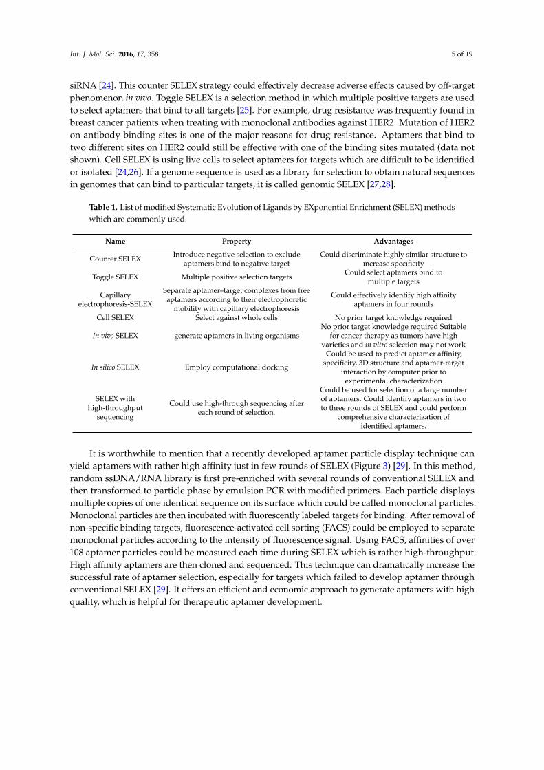
As mentioned above, aptamers identified from conventional SELEX process which have high specificity may fail to stimulate or inhibit their targets as expected. The successful rate of effective aptamer identification by conventional SELEX is lower than 30% [21]. Therefore, optimization or variations based on the conventional SELEX may be required in most cases. There are several modifications which are useful and used in research widely (Table 1) [22]. Counter (negative) SELEX is usually performed after positive SELEX to exclude aptamers which bind to negative targets to discriminate highly similar structures [23,24]. Using conventional SELEX in combination with counter SELEX strategy, aptamers that specifically recognized osteoblasts but did not enter hepatocytes and peripheral blood cells were successfully developed for targeted delivery of
Figure 2. Process of conventional Systematic Evolution of Ligands by EXponential enrichment (SELEX).Different sequences of ssDNA/RNA are shown in different color.
3.1.2. Modified SELEX
As mentioned above, aptamers identified from conventional SELEX process which have highspecificity may fail to stimulate or inhibit their targets as expected. The successful rate of effectiveaptamer identification by conventional SELEX is lower than 30% [21]. Therefore, optimization orvariations based on the conventional SELEX may be required in most cases. There are severalmodifications which are useful and used in research widely (Table 1) [22]. Counter (negative)SELEX is usually performed after positive SELEX to exclude aptamers which bind to negativetargets to discriminate highly similar structures [23,24]. Using conventional SELEX in combinationwith counter SELEX strategy, aptamers that specifically recognized osteoblasts but did not enterhepatocytes and peripheral blood cells were successfully developed for targeted delivery of therapeutic
Int. J. Mol. Sci. 2016, 17, 358 5 of 19
siRNA [24]. This counter SELEX strategy could effectively decrease adverse effects caused by off-targetphenomenon in vivo. Toggle SELEX is a selection method in which multiple positive targets are usedto select aptamers that bind to all targets [25]. For example, drug resistance was frequently found inbreast cancer patients when treating with monoclonal antibodies against HER2. Mutation of HER2on antibody binding sites is one of the major reasons for drug resistance. Aptamers that bind totwo different sites on HER2 could still be effective with one of the binding sites mutated (data notshown). Cell SELEX is using live cells to select aptamers for targets which are difficult to be identifiedor isolated [24,26]. If a genome sequence is used as a library for selection to obtain natural sequencesin genomes that can bind to particular targets, it is called genomic SELEX [27,28].
Table 1. List of modified Systematic Evolution of Ligands by EXponential Enrichment (SELEX) methodswhich are commonly used.
Name Property Advantages
Counter SELEX Introduce negative selection to excludeaptamers bind to negative target
Could discriminate highly similar structure toincrease specificity
Toggle SELEX Multiple positive selection targets Could select aptamers bind tomultiple targets
Capillaryelectrophoresis-SELEX
Separate aptamer–target complexes from freeaptamers according to their electrophoretic
mobility with capillary electrophoresis
Could effectively identify high affinityaptamers in four rounds
Cell SELEX Select against whole cells No prior target knowledge required
In vivo SELEX generate aptamers in living organismsNo prior target knowledge required Suitable
for cancer therapy as tumors have highvarieties and in vitro selection may not work
In silico SELEX Employ computational docking
Could be used to predict aptamer affinity,specificity, 3D structure and aptamer-target
interaction by computer prior toexperimental characterization
SELEX withhigh-throughput
sequencing
Could use high-through sequencing aftereach round of selection.
Could be used for selection of a large numberof aptamers. Could identify aptamers in twoto three rounds of SELEX and could perform
comprehensive characterization ofidentified aptamers.
It is worthwhile to mention that a recently developed aptamer particle display technique canyield aptamers with rather high affinity just in few rounds of SELEX (Figure 3) [29]. In this method,random ssDNA/RNA library is first pre-enriched with several rounds of conventional SELEX andthen transformed to particle phase by emulsion PCR with modified primers. Each particle displaysmultiple copies of one identical sequence on its surface which could be called monoclonal particles.Monoclonal particles are then incubated with fluorescently labeled targets for binding. After removal ofnon-specific binding targets, fluorescence-activated cell sorting (FACS) could be employed to separatemonoclonal particles according to the intensity of fluorescence signal. Using FACS, affinities of over108 aptamer particles could be measured each time during SELEX which is rather high-throughput.High affinity aptamers are then cloned and sequenced. This technique can dramatically increase thesuccessful rate of aptamer selection, especially for targets which failed to develop aptamer throughconventional SELEX [29]. It offers an efficient and economic approach to generate aptamers with highquality, which is helpful for therapeutic aptamer development.

Int. J. Mol. Sci. 2016, 17, 358 6 of 19Int. J. Mol. Sci. 2016, 17, 358 6 of 19
Figure 3. Mechanism of the aptamer particle display system (Reproduced with permission from Reference [29]). Different sequences of ssDNA/RNA are shown in different color.
3.2. Modifications of Aptamers for Preclinical Studies
For therapeutic purposes, as oligonucleotide aptamers could be degraded easily in serum, modifications after SELEX are required for stabilization. Various chemical modifications can significantly improve the stability of aptamers (Figure 4) [30].
3.2.1. Modifications on Linkage
The 15-mer guanine rich (G-rich) thrombin aptamer d(GGTTGGTGTGGTTGG) is a typical and popular model used for developing and illustrating of novel characterization or modification methods for aptamers [31]. It has a G-quadruplex structure formed by two stacking G-tetrads and a central loop TGT which is optimal for stability. It is interesting to find that adding an extra guanine at the 5′ end caused decreased stability while adding an extra guanine to the 3′ end caused increased stability, indicating that the themostability of an aptamer is sequence dependent [32]. It was found that the G-quadruplex is a rather common structure for aptamers, especially for DNA aptamers. DNA aptamers against various targets have a G-quadruplex structure with high sequence identity with high diversity. Another group tried to invert 5′–5′ of a polarity site to form a folded aptamer with a non-common structure d(GGTTGGTGTGGTTGG). This structure has higher stability and affinity to thrombin, although lower inhibiting activity compared to unmodified aptamers [33,34]. Terminal 3′–3′ and 3′–5′ internucleotide linkage was first tried in 1991. A sense deoxyoligonucleotide capping of both ends of the aptamer with inverted thymidine could not only increase stability significantly but also slowed nuclease degradation from 30 min (unmodified) down to 90 min (modified) in snake venom phosphodiesterase digestion [35]. 3′-capping with inverted thymidine modification is a commonly used approach to block 3′-exonuclease degradation by nucleases and prolong an aptamer’s half-life time in serum [23,36]. Most aptamers in clinical trials are modified with this method (Table 2).
Besides inverted thymidine modification, 3′-biotin-streptavidin conjugation is also designed to fight with 3′-exonuclease digestion in serum. It is found that 3′-biotin-streptavidin conjugating aptamers which have bigger size is not only protected from nucleases degradation, but also protected from rapid clearance by circulation system in vivo [37]. 3′-biotin modification is also used
Figure 3. Mechanism of the aptamer particle display system (Reproduced with permission fromReference [29]). Different sequences of ssDNA/RNA are shown in different color.
3.2. Modifications of Aptamers for Preclinical Studies
For therapeutic purposes, as oligonucleotide aptamers could be degraded easily in serum,modifications after SELEX are required for stabilization. Various chemical modifications cansignificantly improve the stability of aptamers (Figure 4) [30].
3.2.1. Modifications on Linkage
The 15-mer guanine rich (G-rich) thrombin aptamer d(GGTTGGTGTGGTTGG) is a typical andpopular model used for developing and illustrating of novel characterization or modification methodsfor aptamers [31]. It has a G-quadruplex structure formed by two stacking G-tetrads and a centralloop TGT which is optimal for stability. It is interesting to find that adding an extra guanine atthe 51 end caused decreased stability while adding an extra guanine to the 31 end caused increasedstability, indicating that the themostability of an aptamer is sequence dependent [32]. It was foundthat the G-quadruplex is a rather common structure for aptamers, especially for DNA aptamers.DNA aptamers against various targets have a G-quadruplex structure with high sequence identitywith high diversity. Another group tried to invert 51–51 of a polarity site to form a folded aptamer witha non-common structure d(GGTTGGTGTGGTTGG). This structure has higher stability and affinity tothrombin, although lower inhibiting activity compared to unmodified aptamers [33,34]. Terminal 31–31
and 31–51 internucleotide linkage was first tried in 1991. A sense deoxyoligonucleotide capping of bothends of the aptamer with inverted thymidine could not only increase stability significantly but alsoslowed nuclease degradation from 30 min (unmodified) down to 90 min (modified) in snake venomphosphodiesterase digestion [35]. 31-capping with inverted thymidine modification is a commonlyused approach to block 31-exonuclease degradation by nucleases and prolong an aptamer’s half-lifetime in serum [23,36]. Most aptamers in clinical trials are modified with this method (Table 2).
Besides inverted thymidine modification, 31-biotin-streptavidin conjugation is also designedto fight with 31-exonuclease digestion in serum. It is found that 31-biotin-streptavidin conjugatingaptamers which have bigger size is not only protected from nucleases degradation, but also protectedfrom rapid clearance by circulation system in vivo [37]. 31-biotin modification is also used for aptamers

Int. J. Mol. Sci. 2016, 17, 358 7 of 19
against other targets. Anti-SARS coronavirus helicase aptamer can remain intact for up to 16 h in 10%fetal bovine serum compared to 6 h for unmodified aptamers [23].
Int. J. Mol. Sci. 2016, 17, 358 7 of 19
for aptamers against other targets. Anti-SARS coronavirus helicase aptamer can remain intact for up to 16 h in 10% fetal bovine serum compared to 6 h for unmodified aptamers [23].
Figure 4. Various chemical modifications to stabilize aptamers (Adapted from Reference [30]). Modification sites are shown in red. The hydroxyl group of RNA is shown in blue to distinguish DNA and RNA.
Cholesterol can be added to the 5′-end of an aptamer to form a cholesteryl-oligonucleotide (cholODN) and further linked to low density lipoprotein (LDL) to form a compact cholODN-LDL complex. This complex has high stability and is highly resistant to nucleases degradation in serum which has a 10-fold longer half-life than unmodified aptamers [38]. Substitute phosphodiester linkage of DNA with methylphosphonate or phosphorothioate is also commonly used for aptamer stabilization [39–41].
Figure 4. Various chemical modifications to stabilize aptamers (Adapted from Reference [30]).Modification sites are shown in red. The hydroxyl group of RNA is shown in blue to distinguish DNAand RNA.
Cholesterol can be added to the 51-end of an aptamer to form a cholesteryl-oligonucleotide(cholODN) and further linked to low density lipoprotein (LDL) to form a compact cholODN-LDLcomplex. This complex has high stability and is highly resistant to nucleases degradation in serumwhich has a 10-fold longer half-life than unmodified aptamers [38]. Substitute phosphodiesterlinkage of DNA with methylphosphonate or phosphorothioate is also commonly used for aptamerstabilization [39–41].
Int. J. Mol. Sci. 2016, 17, 358 8 of 19
Table 2. Progress of aptamers for diseases therapy in on-going or completed clinical trials [42].
Therapeutic Purpose Name Target Form Modification Status Section
Macular degeneration
Pegaptanib Vascular endothelial growthfactor (VEGF) RNA
21-fluoro pyrimidines,21-O-methyl purines,
31-inverted dT, PEGylated
Approved for age-related maculardegeneration (wet AMD) 5.1.1
ARC1905 Complement component 5 RNA 31-inverted dT, PEGylated Phase I completed 5.1.2
E10030 Platelet-derived growthfactor (PDGF) DNA
21-fluoro pyrimidines,21-O-methyl purines
31-inverted dTPhase III await 5.1.3
CancerAS1411 Nucleolin RNA G-rich, PEGylated Phase II on-going 5.2.1
NOX-A12 The chemokine (C–X–Cmotif) ligand 12 (CXCL-12) L-RNA L-form, PEGylated Phase II on-going 5.2.2
Coagulation
REG1 Coagulation factor IXa RNA 31-inverted dT, PEGylated Phase III await 5.3.1
ARC1779 von Willebrand factor (vWF)A1 domain DNA 31-inverted dT, PEGylated Phase II on-going 5.3.2
NU172 Thrombin DNA Unmodified DNA Phase II on-going 5.3.3BAX499 Tissue factor pathway RNA 31-inverted dT, PEGylated Phase I on-going 5.3.4
InflammationNOX-H94 Hepcidlin L-RNA L-form, PEGylated Phase II on-going 5.4.1
NOX-E36 The chemokine (C–C motif)ligand 2 (CCL2) L-RNA L-form, PEGylated Phase II on-going 5.4.2

Int. J. Mol. Sci. 2016, 17, 358 9 of 19
3.2.2. Modifications on Sugar Ring or Bases
The natural oligonucleotides are all in D-form. L-form oligonucleotides (Spiegelmer) are chiralinversions of natural D-forms. In identification of L-form aptamers, D-form aptamers are firstly selectedagainst synthesized L-form protein targets from a general single strand random oligonucleotide library.After SELEX, L-form aptamers are synthesized according to the mirror image of corresponding D-formaptamers [43]. L-form aptamers are much more stable than D-form with high resistance to nucleasedegradation in vivo and do not hybridize to or affect the original nucleic acids in the cells [44–47].Clinical evaluated aptamers NOX-A12, NOX-H94 and NOX-E36 are all L-form aptamers.
Locked nucleic acid (LNA) is a modification on a sugar ring with a methylene linkage between21-O and 41-C, which can generate the most stable pairs to dramatically increase the themostabilityand nucleases resistance of aptamers [48,49]. The LNA/DNA chimera aptamer against HIV-1trans-activating response target could retain an intact structure without degradation for up to 20 h inserum [48].
Unlocked nucleic acid (UNA) is an opposite modification to LNA. There is a bond between C21
and C31 missing in UNA which makes aptamer more flexible. Different from LNA which can stabilizestructure, UNA has an uncertain affect to themostability of aptamers. It is found that UNA replacementon a loop region of an anti-thrombin aptamer increased its themostability while replacement onG-tetrads disrupted the structure formation [50]. It is uncertain whether UNA modification has anyeffect on protecting aptamers from nucleases degradation [50].
Other positions of the sugar ring could be amended for chemical modifications,such as 21-F [39,51,52], 41-C-(aminoethyl) thymidine [51], 5-N-(6-aminohexyl)carbamoyl-21-deoxyuridine [39,51] and so on. For more modifications in detail, please read reviews written byWang et al. [30]. More studies are required to characterize and discuss the stabilizing effects of thesemodifications and design more modification strategies in the future.
4. Aptamers for Skeletal Diseases Therapy in Preclinical Studies
The number of bone marrow mesenchymal stem cells (BMSCs) is decreased through aging whileincreased through adipocyte differentiation. It is found that miR-188 level is much higher in BMSCsin old than in young mice and human. Animals lacking miR-188 can be protected from age-relatedbone loss and fat accumulation in bone marrow. An aptamer that specifically recognizes BMSCs isdeveloped and conjugated with miR-188 to form a nanocomplex. This targeted delivery nanocomplexcould promote bone formation and reduce fat accumulation in bone marrow with high efficacy in agedmice, indicating a potential approach for age-related bone loss therapy [53].
Furthermore, BMSCs are important in bone marrow but there is no specific marker on their surface,making it difficult to be isolated from bone marrow directly. Aptamers with high binding affinityagainst porcine BMSCs are developed by SELEX. Using the high affinity and specificity aptamers,BMSCs could be fished out from cell solution and bone marrow, which is a novel method for BMSCsisolation and provide a foundation for aptamer applications in tissue engineering and regenerativemedicine for skeletal diseases therapy [54].
In another preliminary study, a specific aptamer against human jaw periosteal cells (JPCs) isdeveloped for tissue engineering in oral and maxillofacial surgery. This aptamer has high affinityto human osteogenically induced JPCs and BMSCs from bone marrow while it does not bind toany other cell lines or undifferentiated JPCs or JPCs induced from other sources. It can be usedto purify osteogenic progenitor cells from undifferentiated JPCs or stem cells of other sources.The mineralization capacity is higher in the aptamer positive fraction, which is a promising techniquefor tissue engineering [55].
There are other aptamers or other aptamer-ligand complexes developed and studied in preclinicalresearch at the moment. However, there is no aptamer evaluated in clinical trials. More efforts andstudies are needed for this therapeutic field in the future.
Adult mesenchymal stem cells (aMSCs) are stem cells.
Int. J. Mol. Sci. 2016, 17, 358 10 of 19
5. Aptamers in On-Going or Completed Clinical Trials for Therapeutics
At the moment, there are 11 aptamers evaluating in clinical trials for the treatment of maculardegeneration, cancer, inflammation and coagulation, and one of them has been approved by FDA forthe treatment of AMD. In this section, we will review the research and clinical evaluation progress ofthese aptamers.
5.1. Aptamers against Macular Degeneration
Wet (neovascular) and dry (atrophic) AMD are two major causes of vision loss in the elderly dueto retinal damage, and affect around eight million people in America. There are three aptamers beingevaluated in clinical trials now for wet or dry AMD therapy.
5.1.1. Pegaptanib
Pegaptanib (Macugen; Pfizer and Eyetech, New York, NY, USA), a 27-mer RNA aptamerspecifically binds to and inhibits VEGF against AMD, is the only aptamer approved by the FDAfor disease treatment on the market [56]. Pegaptanib is selected directly against VEGF165, the VEGFisoform primarily responsible for pathological ocular neovasculariztion and vascular permeability [8].After in vitro selection and characterization, the aptamer which inhibits VEGF165 with high affinityand efficacy is chosen and modified with 21-fluoro pyrimidines and 21-O-methyl purines and furthercapped with 31–31-linked deoxythymidine to avoid nuclease degradation and increase stability. In apreclinical animal study, a 40 kDa polyethylene glycol (PEG) is conjugated to the 51- end of the RNAaptamer to increase half life of the aptamer for better bioavailability. After around 10 years preclinicalstudies to optimize and evaluate its therapeutic potency, pegaptanib which shows high efficacy ininhibiting VEGF in different models was approved by the FDA for the treatment of AMD in 2004 withdosage of 0.3 mg per eye every 6 weeks administered intravitreally [56]. This is extraordinary progressas it is the first aptamer approved for use in human and it opens a wide window for therapeuticaptamers in disease treatment.
However, pegaptanib failed to compete with anti-VEGF monoclonal antibody ranibizumab(Lucentis; Genentech, South San Francisco, California, USA) as it only inhibits VEGF165 isoforms, whileantibodies inhibit all isoforms of VEGF [57], which has negative affects on the pharmacy investmentto therapeutic aptamers in these years. Fortunately, it was found recently that blocking all activityof VEGF may cause high risk of hypertension and other adverse effects [58]. Therefore, aptamerpegaptanib is better than antibody ranibizumab in long term maintenance therapy, especially inpatients with systemic comorbidities [18,59].
5.1.2. ARC1905
ARC1905 (Ophthotech Corp, New York, NY, USA) is a 39 bases RNA aptamer specifically againstcomplement component 5 (C5) for the treatment of both wet and dry AMDs [60]. C5 is a downstreampro-inflammatory protein in the complement system associated with AMD pathogenesis. Inhibition ofC5 can prevent the key terminal fragments formation which is critical for tissue pathology [61]. A PEGis also conjugated to the 31- end with an inverted thymidine. A phase I clinical trial using ARC1905 incombination with ranibizumab for the treatment of wet AMD (NCT00709527) was finished in 2011 and aphase I clinical for dry AMD treatment (NCT00950638) has recently been completed (data not shown).
5.1.3. E10030
It may not be sufficient to inhibit VEGF only for wet AMD treatment to prevent angiogenesisdue to the limit of new vessels regression associated with vision loss. PDGF plays an important rolein pericyte recruitment and maturation and new vessels may resist to anti-VEGF drugs due to therole of PDGF [19,62]. Combination treatment using pegaptanib and anti-PDGF antibody can not onlyprevent new vessel formation but also promote vessel regression [19]. On the other hand, combination

Int. J. Mol. Sci. 2016, 17, 358 11 of 19
treatment using anti-PDGF aptamer and anti-VEGF antibody also has promising therapeutic effect.A 29 bases RNA aptamer E10030 (Fovista; Ophthotech Corp) targeting PDGF is developed andmodified with 21-fluoro pyrimidine and 21-O-methyl purines. In preclinical studies, PEG-conjugatedE10030 was able to facilitate neovascular regression when combined with anti-VEGF agents [19].In clinical trials, patients treated with E10030 in combination with anti-VEGF antibody raninizumab(administered once a month) showed significant neovascular regression and 59% of them haveincreased visual acuity after three months of treatment without any side effects observed. In phase IIclinical trials of this combination therapy, patients treated with combination therapy gained 62% highervision than the patients treated with anti-VEGF antibody ranizumab only. The combination therapyis waiting for phase III clinical evaluation for wet AMD treatment at the moment (clinical trial IDsNCT01944839 and NCT01940900). The promising results suggest that combination therapy targetingtwo different antigens at the same time using aptamer-aptamer combination or aptamer-antibodycombination could be a new therapeutic direction in future study.
5.2. Aptamers against Cancer
Cancer treatment requires more effective and precise therapies, especially specific therapies,for discriminating normal cells and tumor cells to avoid toxicity. The highly disorganized vesselarchitecture inside the tumors and the surrounding extracellular matrixes as well as stromal cells areboth barriers for drug delivery [63]. Monoclonal antibodies can recognize targeting tumor cells withhigh specificity but are difficult to penetrate into the deep sites of tumor cells due to the large size(around 150 kDa). Aptamers with much smaller size (around 30 kDa) are able to cross the barriers andpenetrate into tumor cells and therefore are ideal therapeutic reagents for cancer. Generally, aptamerscould diffuse into tumors in 10 min after injection. For example, a fluorescent aptamer againstextracellular matrix protein tenascin-C was able to diffuse into tumors rapidly with perivascularfluorescence signal detected in tumor only 10 min after intravenous injection [64]. Furthermore,another group has compared the tumor penetration times between aptamer and antibody, and foundthat they were detected in tumors after 10 min and 3 h after intravenous injection, respectively [65].Immuno-therapy of cancer by aptamers is a recent novel research focus. There are two individualaptamers for cancer therapy now being evaluated in clinical trials.
5.2.1. AS1411
AS1411 (Antisoma, London, UK) is a guanine-rich aptamer with G-quadruplex structure identifiedfrom a guanine-rich ssRNA library by antiproliferation selection [66]. It has a G-quadruplex structurewhich is highly stable and resistant to nuclease degradation. It can penetrate into tumor cells easily.AS1411 binds to the external domain of nucleolin, which is a protein over-expressed on the surfaceof cancer cells and responsible for survival, growth, and proliferation of cells [66]. AS411 can inhibitover 80 types of cancer cells in in vitro studies and it is the first oligonucleotide aptamer approved forclinical trial for human cancer therapy. In preclinical studies, AS1411 has inhibition efficacy in multiplecancer models including non-small cell lung, renal cells and breast cancers. In phase I clinical trial(NCT00881244), AS1411 is well tolerated by patients with advanced cancers and has no side effects.AS1411 shows promising therapeutic efficacy especially for patients with renal cell carcinoma aftersix months of therapy. Phase II clinical trial shows therapeutic efficacy to acute myeloid leukemiapatients without toxicity and adverse effects. However, a following phase II evaluation for renal cellcarcinoma (clinical trial ID NCT00740441) found AS1411 only has therapeutic effect in 2.9% of patients,and shows minimal activity in unselected patients with metastatic renal cell carcinoma [67], indicatingmore research is required to optimize the therapeutic potency of AS1411 in the future.
5.2.2. NOX-A12
NOX-A12 (Olaptesed pegol; Noxxon, Berlin, Germany) is a 45-mer L-RNA aptamer developedfor use in autologous hematopoietic stem cell transplants [66]. NOX-A12 targets to stroma cell-derived

Int. J. Mol. Sci. 2016, 17, 358 12 of 19
factor-1 The chemokine (C–X–C motif) ligand 12 (CXCL-12), which plays important roles in stemcell migration towards the bone marrow and controls tumor growth, metastasis and vasculogenesis.Binding to CXCL-12 can block its receptor binding and prevent CXCL-12 tissue gradients and decreasethe possibility of tumor metastasis and drug resistance caused by cancer cell homing [68]. As L-formaptamers can not be recognized by nucleases, it does not require any chemical modifications for in vivostudies. It showed efficacy against non- Hodgkin’s lymphoma and myelomas in preclinical studies.Phase I clinical trial for safety and tolerability evaluation confirms NOX-A12 can be well toleratedwithout serious adverse effects. NOX-A12 has a 37 h long half-life and patients with NOX-A12treatment are more susceptible to chemotherapy due to its particular blockage of stromal cell-derivedfactor-1 (SDF-1), making it a promising and successful drug in cancer therapy [69]. Two phase IIclinical trials are in progress, one for treatment of Chromin Lymphocytic Leukemia (clinical trial IDNCT01486797) and the other one for evaluating and comparing the therapeutic efficacy of NOX-A12alone and combination therapy with chemotherapy for patients with multiple myeloma (clinical trialID NCT01521533).
5.3. Aptamers against Coagulation
More and more aptamers for coagulation therapy have been developed independently by differentresearch groups. Till now, there are four aptamers being evaluated in different stages of clinical trials.
5.3.1. REG1
REG1 (Regado Biosciences, Basking Ridge, NJ, USA) is an aptamer system consisting of a 37-merRNA aptamer RB006 (Pegnivacogin) and a 17-mer antidote RB007 with sequence complementary toRB006 [70]. RB006 is an antagonist of factor IXa for preventing the downstream conversion of factor Xand avoidance of clotting. RB007 is the antidote of RB006, which can specifically reverse the inhibitionfunction of RB006 to control sheath removal time after percutaneous coronary intervention. Phase Iclinical trials have shown REG1 is well tolerated in patients and no significant adverse effects or majorbleeding are found [71–73]. More importantly, therapeutic effects of REG1 can be controlled by dosageof RB007 and it is less toxic than heparin/protamine, which is also an anticoagulation/antidote paircurrently available [74]. Phase II clinical trials for using REG1 in percutaneous coronary interventionfor patients with coronary artery have just been completed (clinical trial ID NCT00715455). However,a later randomized clinical trial had to be terminated before it finished as patients treated with REG1showed severe allergic reactions and major bleeding with no significant efficacy after percutaneouscoronary intervention [75]. Therefore, more studies are required to evaluate the safety and efficacy ofthe REG1 system.
5.3.2. ARC1779
ARC1779 (Archemix Corp, Cambridge, MA, USA) is a 39-mer PEGylated DNA aptamer thatbinds to the A1 domain of von Willebrand factor (vWF) [76], which is a key factor in the coagulationcascade related to platelet recruitment, to block interaction between A1 domain and platelet receptorglycoprotein 1B [12,77]. Therefore, it is a potential therapeutic target for treatment of vWF-relatedplatelet disorders, von Willebrand disease as well as acute coronary syndromes [78]. A phase II pilotstudy showed that ARC1779 can inhibit platelet depletion induced by a vWF agonist desmopressin inpatients [79]. Phase II clinical trial for VWF 2D treatment is still on-going.
5.3.3. NU172
Aptamer NU172 (ARCA Biopharma, London, UK) is an unmodified DNA aptamer for short-termanticoagulation and is distinct from the long-term anticoagulation aptamer REG1 and ARC1779.In preclinical studies, NU172 showed efficacy to prolong clotting time and the anticoagulation effectwould be stopped rapidly due to nuclease degradation. A phase II clinical trial for evaluating thetherapeutic effects in coronary artery bypass graft surgery is in progress (clinical trial ID NCT00808964).

Int. J. Mol. Sci. 2016, 17, 358 13 of 19
5.3.4. BAX499
The above three anticoagulation aptamers all target to proteins in the intrinsic coagulationpathway, while BAX499 (Baxter, Deerfield, IL, USA) targets to the negative regulator of factor VIIa inthe extrinsic tissue factor pathway. In a preclinical hemophilia pathology mimicking monkey model,BAX499 is able to recover the clotting caused by anti-factor VIII antibody [80]. The phase I clinicaltrial was started from 2010 (clinical trial ID NCT01191372) and the evaluation results have not beenpublished yet.
5.4. Aptamers against Inflammation
Two anti-inflammation aptamers in clinical trials are both L-form aptamers from Noxxon.
5.4.1. NOX-H94
Hepcidin is over-expressed in patients with chronic inflammation induced by cancer or dialysis,which may lead to anemia due to the hepcidin-induced ferroportin degradation [81]. NOX-H94(Lexaptepid pegol; Noxxon) targets to hepcidin, a peptide hormone regulator for iron homeostasiswith the role to inhibit the interaction between hepcidin and ferroportin and reduce the anemiasymptom in patients [81,82]. In a preclinical study, cynomolgus monkey could be prevented frominterleukin-6 induced iron concentration decrease in serum when treated with NOX-H94. Phase IIaclinical trial for anemia therapeutic evaluation for patients with cancers have been completed. Clinicaltrials for treatment of erythropoiesis agent-induced anemia in patients with dialysis are still in progress(clinical trial ID NCT02079896).
5.4.2. NOX-E36
NOX-E36 is another L-form anti-inflammation aptamer from Noxxon [83]. The target ofNOX-E36 is chemokine ligand 2 (also called monocyte chemoattractant protein 1) which canmediate inflammation by recruiting leukocytes from intravascular to extravascular environments.Upon binding, NOX-E36 can inhibit chemokine ligands 2-induced inflammation to reduce therecruitment of leukocytes, which is quite effective for anti-inflammation treatment for lupus nephritisin mouse models. It is also useful for preventing type-2 diabetic glomerulosclerosis in mice [84].Phase II clinical trials for treatment of type-2 diabetes are in progress (clinical trial IDs NCT01085292and NCT01547897).
6. Conclusions
Oligonucleotide aptamers are more and more popular in recent years, especially from 2005 afterthe first aptamer Pegaptanib was approved for wet AMD therapy by FDA. There are over 900 aptamersdeveloped by SELEX for a broad spectrum of both diagnostic and therapeutic applications. You canfind over 5000 reports on aptamer research in PubMed, and this number currently generally increasesby two to five every day. There are 11 aptamers in clinical trials, which have advantages especially forsafety issues and have significant improvements in efficacy for therapeutic application. In addition tothese aptamers, there are nearly 100 aptamers waiting for approval for evaluation in clinical trials.
With promising advantages compared to monoclonal antibodies, oligonucleotide aptamers maybecome the predominant agent for therapeutic application. With monoclonal antibody technologycommonly developed, and used in therapeutic application, the targets of antibodies are wellcharacterized [85] (Table 3). Therefore in the future, based on the promising therapeutic effectsof inhibiting particular targets with monoclonal antibodies, oligonucleotide aptamers can be identifiedagainst the same targets to develop the second generation of therapy. This would save considerableresearch effort and allow more rapid progress in developing more economic and efficient therapeuticapproaches for various diseases with oligonucleotide aptamers.

Int. J. Mol. Sci. 2016, 17, 358 14 of 19
Table 3. Monoclonal antibodies approved by FDA for therapeutic use.
Antibody Trade Name Target Approved Indication
Muromomab Orthoclone CD3 Allograft rejection in allogeneicrenal transplantation
Abciximab ReoPro Glycoprotein IIb/IIIa Percutaneous coronary intervention
Rituximab Rituxan CD20 RA, Wegner granulomatosis,microscopic polyangiitis
Daclizumab Zenapax CD25 (II2r) Allograft rejectionBasiliximab Simulect CD25 (II2r) Allograft rejection
Palivizumab Synagis Protein F Respiratory syncytial virus (RSV inhibitor)in children
Infliximab Remicade TNFα Crohn’s disease and rheumatoid arthritisTrastuzumab Herceptin HER2/Neu Metastatic breast cancer
Etanercept Enbrel TNFα and βAutoimmune diseases such as
ankylosing spondylitisGemtuzumab Mylotarg CD33 CD33-positive acute myeloid leukemiaAlemtuzumab Mabcampath CD52 B-cell chronic lymphocytic leukemiaIbritomomab Zevalin 90Y CD20 B-cell non-Hodgkin’s lymphomaAdalimumab Trudexa TNFα Crohn’s disease and rheumatoid arthritis
Alefacept Amevive CD2 Chronic plaque psoriasisOmalizumab Xolair IgE asthema
Tositumomab Bexxar CD20 CD20-positive B-cell non-Hodgkin’slymphoma
Efalizumab Raptiva CD11a Moderate to severe plaque psoriasis
Cetuximab Erbitus EGFR Metastatic colorectal and head andneck carcinoma
Bevacizumab Avastin VEGF-A Metastatic colorectal and non-small celllung carcinoma
Natalizumab Tysabri Integrin-α4 Multiple sclerosisRanibizumab Lucentis VEGF-A Wet type age-related macular degenerationPanitumumab Vectibid EGFR Metastatic colorectal carcinoma
Eculizumab Soliris C5 Paroxysmal nocturnal haemoglobinuriaCertolizumab Cimzia TNFα Crohn’s diseaseDaratumumab Darzalex CD38 Multiple myeloma
Elotuzumab EMPLICITI CS1 In combination with lenalidomide anddexamethasone for Multiple myeloma
Mepolizumab Nucala IL-5 Asthma
Denosumab Prolia/Xgeva Nuclear factor kappa Bligand
Bone matastases, osteoporosis, giant celltumor of bone
Secukinumab Cosentyx IL-17 PsoriasisSirukumab (CNTO 136) IL-6 Rheumatoid arthritis (soon)
Acknowledgments: We thank academic staff members in Baoqin Liu’s group at Zhengzhou Hospital of TraditionalChinese Medicine, and Aiping Lu and Ge Zhang’s group at Hong Kong Baptist University. We also thank HongKong Baptist University for providing critical comments and technical support. This study was supported by theHong Kong General Research Fund (HKBU262913 to Ge Zhang).
Author Contributions: Yuanyuan Yu wrote the manuscript; Chao Liang, Quanxia Lv, Defang Li and Xuegong Xucontributed the manuscript for literature research; Baoqin Liu, Aiping Lu and Ge Zhang revised and approvedthe manuscript.
Conflicts of Interest: The authors declare no conflict of interest.
Abbreviations
The following abbreviations are used in this manuscript:
SELEX Systematic Evolution of Ligands by EXponential enrichmentAMD Age-related Macular DegenerationAAPs Antibody-aptamer pincersFACS Fluorescence-Activated Cell Sorting
Int. J. Mol. Sci. 2016, 17, 358 15 of 19
cholODN cholesteryl-oligonucleotideLDL low density lipoproteinVEGF vascular endothelial growth factorPDGF Platelet-Derived Growth FactorvWF von Willebrand factorCD Cluster of DifferentiationIL InterLeukinEGFR Epidermal Growth Factor Receptorsulfo-SMCC sulfosuccinimidyl 4-(N-maleimidomethyl) cyclohexane-1-carboxylateSATA N-succinimidyl-S-acetylthioacetateCXCL-12 The chemokine (C–X–C motif) ligand 12
References
1. Kohler, G.; Milstein, C. Continuous cultures of fused cells secreting antibody of predefined specificity. Nature1975, 256, 495–497. [CrossRef] [PubMed]
2. Bradbury, A.R.; Sidhu, S.; Dubel, S.; McCafferty, J. Beyond natural antibodies: The power of in vitro displaytechnologies. Nat. Biotechnol. 2011, 29, 245–254. [CrossRef] [PubMed]
3. Ellington, A.D.; Szostak, J.W. In vitro selection of RNA molecules that bind specific ligands. Nature 1990, 346,818–822. [CrossRef] [PubMed]
4. Tuerk, C.; Gold, L. Systematic evolution of ligands by exponential enrichment: RNA ligands to bacteriophaget4 DNA polymerase. Science 1990, 249, 505–510. [CrossRef] [PubMed]
5. Jayasena, S.D. Aptamers: An emerging class of molecules that rival antibodies in diagnostics. Clin. Chem.1999, 45, 1628–1650. [PubMed]
6. Robertson, D.L.; Joyce, G.F. Selection in vitro of an RNA enzyme that specifically cleaves single-strandedDNA. Nature 1990, 344, 467–468. [CrossRef] [PubMed]
7. Cruz-Toledo, J.; McKeague, M.; Zhang, X.; Giamberardino, A.; McConnell, E.; Francis, T.; DeRosa, M.C.;Dumontier, M. Aptamer base: A collaborative knowledge base to describe aptamers and SELEX experiments.Database 2012, 2012. [CrossRef] [PubMed]
8. Jellinek, D.; Green, L.S.; Bell, C.; Janjic, N. Inhibition of receptor binding by high-affinity RNA ligands tovascular endothelial growth factor. Biochemistry 1994, 33, 10450–10456. [CrossRef] [PubMed]
9. Ambati, J.; Ambati, B.K.; Yoo, S.H.; Ianchulev, S.; Adamis, A.P. Age-related macular degeneration: Etiology,pathogenesis, and therapeutic strategies. Surv. Ophthalmol. 2003, 48, 257–293. [CrossRef]
10. Que-Gewirth, N.S.; Sullenger, B.A. Gene therapy progress and prospects: RNA aptamers. Gene Ther. 2007,14, 283–291. [CrossRef] [PubMed]
11. Ruckman, J.; Green, L.S.; Beeson, J.; Waugh, S.; Gillette, W.L.; Henninger, D.D.; Claesson-Welsh, L.; Janjic, N.21-Fluoropyrimidine RNA-based aptamers to the 165-amino acid form of vascular endothelial growth factor(VEGF165). J. Biol. Chem. 1998, 273, 20556–20567. [CrossRef] [PubMed]
12. Keefe, A.D.; Pai, S.; Ellington, A. Aptamers as therapeutics. Nat. Rev. Drug Discov. 2010, 9, 537–550.[CrossRef] [PubMed]
13. Latz, E.; Verma, A.; Visintin, A.; Gong, M.; Sirois, C.M.; Klein, D.C.; Monks, B.G.; McKnight, C.J.;Lamphier, M.S.; Duprex, W.P.; et al. Ligand-induced conformational changes allosterically activate Toll-likereceptor 9. Nat. Immunol. 2007, 8, 772–779. [CrossRef] [PubMed]
14. Zhao, Q.; Temsamani, J.; Iadarola, P.L.; Jiang, Z.; Agrawal, S. Effect of different chemically modifiedoligodeoxynucleotides on immune stimulation. Biochem. Pharmacol. 1996, 51, 173–182. [CrossRef]
15. Yu, D.; Wang, D.; Zhu, F.G.; Bhagat, L.; Dai, M.; Kandimalla, E.R.; Agrawal, S. Modifications incorporated inCpG motifs of oligodeoxynucleotides lead to antagonist activity of Toll-like receptors 7 and 9. J. Med. Chem.2009, 52, 5108–5114. [CrossRef] [PubMed]
16. Banerjee, J. Antibodies are challenged. Indian J. Med. Sci. 2010, 64, 144–147. [PubMed]17. Gold, L.; Ayers, D.; Bertino, J.; Bock, C.; Bock, A.; Brody, E.N.; Carter, J.; Dalby, A.B.; Eaton, B.E.;
Fitzwater, T.; et al. Aptamer-based multiplexed proteomic technology for biomarker discovery. PLoS ONE2010, 5, e15004. [CrossRef] [PubMed]

Int. J. Mol. Sci. 2016, 17, 358 16 of 19
18. Lao, Y.H.; Phua, K.K.; Leong, K.W. Aptamer nanomedicine for cancer therapeutics: Barriers and potential fortranslation. ACS Nano 2015, 9, 2235–2254. [CrossRef] [PubMed]
19. Jo, N.; Mailhos, C.; Ju, M.; Cheung, E.; Bradley, J.; Nishijima, K.; Robinson, G.S.; Adamis, A.P.; Shima, D.T.Inhibition of platelet-derived growth factor B signaling enhances the efficacy of anti-vascular endothelialgrowth factor therapy in multiple models of ocular neovascularization. Am. J. Pathol. 2006, 168, 2036–2053.[CrossRef] [PubMed]
20. Zhang, P.; Zhao, N.; Zeng, Z.; Chang, C.C.; Zu, Y. Combination of an aptamer probe to CD4 and antibodiesfor multicolored cell phenotyping. Am. J. Clin. Pathol. 2010, 134, 586–593. [CrossRef] [PubMed]
21. Kang, S.; Hah, S.S. Improved ligand binding by antibody-aptamer pincers. Bioconjug. Chem. 2014, 25,1421–1427. [CrossRef] [PubMed]
22. Darmostuk, M.; Rimpelova, S.; Gbelcova, H.; Ruml, T. Current approaches in SELEX: An update to aptamerselection technology. Biotechnol. Adv. 2015, 33, 1141–1161. [CrossRef] [PubMed]
23. Shum, K.T.; Chan, C.; Leung, C.M.; Tanner, J.A. Identification of a DNA aptamer that inhibits sclerostin’santagonistic effect on Wnt signalling. Biochem. J. 2011, 434, 493–501. [CrossRef] [PubMed]
24. Liang, C.; Guo, B.; Wu, H.; Shao, N.; Li, D.; Liu, J.; Dang, L.; Wang, C.; Li, H.; Li, S.; et al.Aptamer-functionalized lipid nanoparticles targeting osteoblasts as a novel RNA interference-based boneanabolic strategy. Nat. Med. 2015, 21, 288–294. [CrossRef] [PubMed]
25. White, R.; Rusconi, C.; Scardino, E.; Wolberg, A.; Lawson, J.; Hoffman, M.; Sullenger, B. Generation of speciescross-reactive aptamers using “toggle“ SELEX. Mol. Ther. 2001, 4, 567–573. [CrossRef] [PubMed]
26. Thiel, T.; Graner, A.; Waugh, R.; Grosse, I.; Close, T.J.; Stein, N. Evidence and evolutionary analysis of ancientwhole-genome duplication in barley predating the divergence from rice. BMC Evol. Biol. 2009, 9. [CrossRef][PubMed]
27. Gold, L.; Brown, D.; He, Y.; Shtatland, T.; Singer, B.S.; Wu, Y. From oligonucleotide shapes to genomic SELEX:Novel biological regulatory loops. Proc. Natl. Acad. Sci. USA 1997, 94, 59–64. [CrossRef] [PubMed]
28. Shtatland, T.; Gill, S.C.; Javornik, B.E.; Johansson, H.E.; Singer, B.S.; Uhlenbeck, O.C.; Zichi, D.A.; Gold, L.Interactions of Escherichia coli RNA with bacteriophage MS2 coat protein: Genomic SELEX. Nucleic Acids Res.2000, 28. [CrossRef]
29. Wang, J.; Gong, Q.; Maheshwari, N.; Eisenstein, M.; Arcila, M.L.; Kosik, K.S.; Soh, H.T. Particle display:A quantitative screening method for generating high-affinity aptamers. Angew. Chem. Int. Ed. 2014, 53,4796–4801. [CrossRef] [PubMed]
30. Wang, R.E.; Wu, H.; Niu, Y.; Cai, J. Improving the stability of aptamers by chemical modification.Curr. Med. Chem. 2011, 18, 4126–4138. [CrossRef] [PubMed]
31. Bock, L.C.; Griffin, L.C.; Latham, J.A.; Vermaas, E.H.; Toole, J.J. Selection of single-stranded DNA moleculesthat bind and inhibit human thrombin. Nature 1992, 355, 564–566. [CrossRef] [PubMed]
32. Smirnov, I.; Shafer, R.H. Effect of loop sequence and size on DNA aptamer stability. Biochemistry 2000, 39,1462–1468. [CrossRef] [PubMed]
33. Martino, L.; Virno, A.; Randazzo, A.; Virgilio, A.; Esposito, V.; Giancola, C.; Bucci, M.; Cirino, G.; Mayol, L.A new modified thrombin binding aptamer containing a 51–51 inversion of polarity site. Nucleic Acids Res.2006, 34, 6653–6662. [CrossRef] [PubMed]
34. Pagano, B.; Martino, L.; Randazzo, A.; Giancola, C. Stability and binding properties of a modified thrombinbinding aptamer. Biophys. J. 2008, 94, 562–569. [CrossRef] [PubMed]
35. Seliger, H.; Frohlich, A.; Groger, G.; Krist, B.; Montenarh, M.; Rosch, H.; Rosch, R.; Ortigao, F.R. Syntheticoligonucleotides for biomedical applications. Nucleic Acids Symp. Ser. 1991, 193–196.
36. Shaw, J.P.; Kent, K.; Bird, J.; Fishback, J.; Froehler, B. Modified deoxyoligonucleotides stable to exonucleasedegradation in serum. Nucleic Acids Res. 1991, 19, 747–750. [CrossRef] [PubMed]
37. Dougan, H.; Lyster, D.M.; Vo, C.V.; Stafford, A.; Weitz, J.I.; Hobbs, J.B. Extending the lifetime of anticoagulantoligodeoxynucleotide aptamers in blood. Nucl. Med. Biol. 2000, 27, 289–297. [CrossRef]
38. De Smidt, P.C.; Le Doan, T.; de Falco, S.; van Berkel, T.J. Association of antisense oligonucleotides withlipoproteins prolongs the plasma half-life and modifies the tissue distribution. Nucleic Acids Res. 1991, 19,4695–4700. [CrossRef] [PubMed]
39. Sacca, B.; Lacroix, L.; Mergny, J.L. The effect of chemical modifications on the thermal stability of differentG-quadruplex-forming oligonucleotides. Nucleic Acids Res. 2005, 33, 1182–1192. [CrossRef] [PubMed]

Int. J. Mol. Sci. 2016, 17, 358 17 of 19
40. Zaitseva, M.; Kaluzhny, D.; Shchyolkina, A.; Borisova, O.; Smirnov, I.; Pozmogova, G. Conformation andthermostability of oligonucleotide d(GGTTGGTGTGGTTGG) containing thiophosphoryl internucleotidebonds at different positions. Biophys. Chem. 2010, 146, 1–6. [CrossRef] [PubMed]
41. Pozmogova, G.E.; Zaitseva, M.A.; Smirnov, I.P.; Shvachko, A.G.; Murina, M.A.; Sergeenko, V.I. Anticoagulanteffects of thioanalogs of thrombin-binding DNA-aptamer and their stability in the plasma. Bull. Exp.Biol. Med. 2010, 150, 180–184. [CrossRef] [PubMed]
42. Sundaram, P.; Kurniawan, H.; Byrne, M.E.; Wower, J. Therapeutic RNA aptamers in clinical trials. Eur. J.Pharm. Sci. 2013, 48, 259–271. [CrossRef] [PubMed]
43. Hoellenriegel, J.; Zboralski, D.; Maasch, C.; Rosin, N.Y.; Wierda, W.G.; Keating, M.J.; Kruschinski, A.;Burger, J.A. The spiegelmer NOX-A12, a novel CXCL12 inhibitor, interferes with chronic lymphocyticleukemia cell motility and causes chemosensitization. Blood 2014, 123, 1032–1039. [CrossRef] [PubMed]
44. Purschke, W.G.; Radtke, F.; Kleinjung, F.; Klussmann, S. A DNA spiegelmer to staphylococcal enterotoxin B.Nucleic Acids Res. 2003, 31, 3027–3032. [CrossRef] [PubMed]
45. Wlotzka, B.; Leva, S.; Eschgfaller, B.; Burmeister, J.; Kleinjung, F.; Kaduk, C.; Muhn, P.; Hess-Stumpp, H.;Klussmann, S. In vivo properties of an anti-GnRH spiegelmer: An example of an oligonucleotide-basedtherapeutic substance class. Proc. Natl. Acad. Sci. USA 2002, 99, 8898–8902. [CrossRef] [PubMed]
46. Leva, S.; Lichte, A.; Burmeister, J.; Muhn, P.; Jahnke, B.; Fesser, D.; Erfurth, J.; Burgstaller, P.; Klussmann, S.Gnrh binding RNA and DNA spiegelmers: A novel approach toward GnRH antagonism. Chem. Biol. 2002, 9,351–359. [CrossRef]
47. Eulberg, D.; Klussmann, S. Spiegelmers: Biostable aptamers. Chembiochem 2003, 4, 979–983. [CrossRef][PubMed]
48. Darfeuille, F.; Hansen, J.B.; Orum, H.; Di Primo, C.; Toulme, J.J. LNA/DNA chimeric oligomers mimic RNAaptamers targeted to the TAR RNA element of HIV-1. Nucleic Acids Res. 2004, 32, 3101–3107. [CrossRef][PubMed]
49. Schmidt, K.S.; Borkowski, S.; Kurreck, J.; Stephens, A.W.; Bald, R.; Hecht, M.; Friebe, M.; Dinkelborg, L.;Erdmann, V.A. Application of locked nucleic acids to improve aptamer in vivo stability and targeting function.Nucleic Acids Res. 2004, 32, 5757–5765. [CrossRef] [PubMed]
50. Pasternak, A.; Hernandez, F.J.; Rasmussen, L.M.; Vester, B.; Wengel, J. Improved thrombin binding aptamerby incorporation of a single unlocked nucleic acid monomer. Nucleic Acids Res. 2011, 39, 1155–1164.[CrossRef] [PubMed]
51. Matsuda, A. Development of highly nuclease-resistant chemically-modified oligonucleotides.Yakugaku Zasshi 2011, 131, 285–298. [CrossRef] [PubMed]
52. Floege, J.; Ostendorf, T.; Janssen, U.; Burg, M.; Radeke, H.H.; Vargeese, C.; Gill, S.C.; Green, L.S.; Janjic, N.Novel approach to specific growth factor inhibition in vivo: Antagonism of platelet-derived growth factor inglomerulonephritis by aptamers. Am. J. Pathol. 1999, 154, 169–179. [CrossRef]
53. Li, C.J.; Cheng, P.; Liang, M.K.; Chen, Y.S.; Lu, Q.; Wang, J.Y.; Xia, Z.Y.; Zhou, H.D.; Cao, X.; Xie, H.; et al.MicroRNA-188 regulates age-related switch between osteoblast and adipocyte differentiation. J. Clin. Invest.2015, 125, 1509–1522. [CrossRef] [PubMed]
54. Guo, K.T.; SchAfer, R.; Paul, A.; Gerber, A.; Ziemer, G.; Wendel, H.P. A new technique for the isolationand surface immobilization of mesenchymal stem cells from whole bone marrow using high-specific DNAaptamers. Stem Cells 2006, 24, 2220–2231. [CrossRef] [PubMed]
55. Ardjomandi, N.; Niederlaender, J.; Aicher, W.K.; Reinert, S.; Schweizer, E.; Wendel, H.P.; Alexander, D.Identification of an aptamer binding to human osteogenic-induced progenitor cells. Nucleic Acid Ther. 2013,23, 44–61. [CrossRef] [PubMed]
56. Ng, E.W.; Adamis, A.P. Anti-VEGF aptamer (pegaptanib) therapy for ocular vascular diseases. Ann. NYAcad. Sci. 2006, 1082, 151–171. [CrossRef] [PubMed]
57. Ferrara, N.; Damico, L.; Shams, N.; Lowman, H.; Kim, R. Development of ranibizumab, an anti-vascularendothelial growth factor antigen binding fragment, as therapy for neovascular age-related maculardegeneration. Retina 2006, 26, 859–870. [CrossRef] [PubMed]
58. Tunon, J.; Ruiz-Moreno, J.M.; Martin-Ventura, J.L.; Blanco-Colio, L.M.; Lorenzo, O.; Egido, J. Cardiovascularrisk and antiangiogenic therapy for age-related macular degeneration. Surv. Ophthalmol. 2009, 54, 339–348.[CrossRef] [PubMed]
Int. J. Mol. Sci. 2016, 17, 358 18 of 19
59. Friberg, T.R.; Tolentino, M.; Weber, P.; Patel, S.; Campbell, S.; Goldbaum, M. Pegaptanib sodium asmaintenance therapy in neovascular age-related macular degeneration: The level study. Br. J. Ophthalmol.2010, 94, 1611–1617. [CrossRef] [PubMed]
60. Biesecker, G.; Dihel, L.; Enney, K.; Bendele, R.A. Derivation of RNA aptamer inhibitors of human complementC5. Immunopharmacology 1999, 42, 219–230. [CrossRef]
61. Anderson, D.H.; Radeke, M.J.; Gallo, N.B.; Chapin, E.A.; Johnson, P.T.; Curletti, C.R.; Hancox, L.S.; Hu, J.;Ebright, J.N.; Malek, G.; et al. The pivotal role of the complement system in aging and age-related maculardegeneration: Hypothesis re-visited. Prog. Retin. Eye Res. 2010, 29, 95–112. [CrossRef] [PubMed]
62. Alvarez, R.H.; Kantarjian, H.M.; Cortes, J.E. Biology of platelet-derived growth factor and its involvement indisease. Mayo Clin. Proc. 2006, 81, 1241–1257. [CrossRef] [PubMed]
63. Jain, R.K.; Stylianopoulos, T. Delivering nanomedicine to solid tumors. Nat. Rev. Clin. Oncol. 2010, 7,653–664. [CrossRef] [PubMed]
64. Hicke, B.J.; Stephens, A.W.; Gould, T.; Chang, Y.F.; Lynott, C.K.; Heil, J.; Borkowski, S.; Hilger, C.S.; Cook, G.;Warren, S.; et al. Tumor targeting by an aptamer. J. Nucl. Med. 2006, 47, 668–678. [PubMed]
65. Xiang, D.; Zheng, C.; Zhou, S.F.; Qiao, S.; Tran, P.H.; Pu, C.; Li, Y.; Kong, L.; Kouzani, A.Z.; Lin, J.; et al.Superior performance of aptamer in tumor penetration over antibody: Implication of aptamer-basedtheranostics in solid tumors. Theranostics 2015, 5, 1083–1097. [CrossRef] [PubMed]
66. Bates, P.J.; Laber, D.A.; Miller, D.M.; Thomas, S.D.; Trent, J.O. Discovery and development of the G-richoligonucleotide AS1411 as a novel treatment for cancer. Exp. Mol. Pathol. 2009, 86, 151–164. [CrossRef][PubMed]
67. Rosenberg, M.J.; Vaske, D.; Killoran, C.E.; Ning, Y.; Wargowski, D.; Hudgins, L.; Tifft, C.J.; Meck, J.;Blancato, J.K.; Rosenbaum, K.; et al. Detection of chromosomal aberrations by a whole-genome microsatellitescreen. Am. J. Hum. Genet. 2000, 66, 419–427. [CrossRef] [PubMed]
68. Vater, A.; Klussmann, S. Toward third-generation aptamers: Spiegelmers and their therapeutic prospects.Curr. Opin. Drug Discov. Devel. 2003, 6, 253–261. [PubMed]
69. Marasca, R.; Maffei, R. NOX-A12: Mobilizing CLL away from home. Blood 2014, 123, 952–953. [CrossRef][PubMed]
70. Rusconi, C.P.; Scardino, E.; Layzer, J.; Pitoc, G.A.; Ortel, T.L.; Monroe, D.; Sullenger, B.A. RNA aptamers asreversible antagonists of coagulation factor IXa. Nature 2002, 419, 90–94. [CrossRef] [PubMed]
71. Dyke, C.K.; Steinhubl, S.R.; Kleiman, N.S.; Cannon, R.O.; Aberle, L.G.; Lin, M.; Myles, S.K.; Melloni, C.;Harrington, R.A.; Alexander, J.H.; et al. First-in-human experience of an antidote-controlled anticoagulantusing RNA aptamer technology: A phase 1a pharmacodynamic evaluation of a drug-antidote pair for thecontrolled regulation of factor IXa activity. Circulation 2006, 114, 2490–2497. [CrossRef] [PubMed]
72. Chan, M.Y.; Cohen, M.G.; Dyke, C.K.; Myles, S.K.; Aberle, L.G.; Lin, M.; Walder, J.; Steinhubl, S.R.;Gilchrist, I.C.; Kleiman, N.S.; et al. Phase 1b randomized study of antidote-controlled modulation offactor IXa activity in patients with stable coronary artery disease. Circulation 2008, 117, 2865–2874. [CrossRef][PubMed]
73. Chan, M.Y.; Rusconi, C.P.; Alexander, J.H.; Tonkens, R.M.; Harrington, R.A.; Becker, R.C. A randomized,repeat-dose, pharmacodynamic and safety study of an antidote-controlled factor IXa inhibitor.J. Thromb. Haemost. 2008, 6, 789–796. [CrossRef] [PubMed]
74. Cohen, M.G.; Purdy, D.A.; Rossi, J.S.; Grinfeld, L.R.; Myles, S.K.; Aberle, L.H.; Greenbaum, A.B.; Fry, E.;Chan, M.Y.; Tonkens, R.M.; et al. First clinical application of an actively reversible direct factor IXa inhibitoras an anticoagulation strategy in patients undergoing percutaneous coronary intervention. Circulation 2010,122, 614–622. [CrossRef] [PubMed]
75. Lincoff, A.M.; Mehran, R.; Povsic, T.J.; Zelenkofske, S.L.; Huang, Z.; Armstrong, P.W.; Steg, P.G.; Bode, C.;Cohen, M.G.; Buller, C.; et al. Effect of the REG1 anticoagulation system versus bivalirudin on outcomesafter percutaneous coronary intervention (REGULATE-PCI): A randomised clinical trial. Lancet 2015, 387,349–356. [CrossRef]
76. Diener, J.L.; Daniel Lagasse, H.A.; Duerschmied, D.; Merhi, Y.; Tanguay, J.F.; Hutabarat, R.; Gilbert, J.;Wagner, D.D.; Schaub, R. Inhibition of von willebrand factor—mediated platelet activation and thrombosisby the anti-von willebrand factor A1—domain aptamer ARC1779. J. Thromb. Haemost. 2009, 7, 1155–1162.[CrossRef] [PubMed]

Int. J. Mol. Sci. 2016, 17, 358 19 of 19
77. Lillicrap, D. Genotype/phenotype association in von willebrand disease: Is the glass half full or empty?J. Thromb. Haemost. 2009, 7, 65–70. [CrossRef] [PubMed]
78. Sadler, J.E.; Budde, U.; Eikenboom, J.C.; Favaloro, E.J.; Hill, F.G.; Holmberg, L.; Ingerslev, J.; Lee, C.A.;Lillicrap, D.; Mannucci, P.M.; et al. Update on the pathophysiology and classification of von willebranddisease: A report of the subcommittee on von willebrand factor. J. Thromb. Haemost. 2006, 4, 2103–2114.[CrossRef] [PubMed]
79. Jilma, B.; Paulinska, P.; Jilma-Stohlawetz, P.; Gilbert, J.C.; Hutabarat, R.; Knobl, P. A randomised pilottrial of the anti-von willebrand factor aptamer ARC1779 in patients with type 2b von willebrand disease.Thromb. Haemost. 2010, 104, 563–570. [CrossRef] [PubMed]
80. Waters, E.K.; Genga, R.M.; Schwartz, M.C.; Nelson, J.A.; Schaub, R.G.; Olson, K.A.; Kurz, J.C.; McGinness, K.E.Aptamer ARC19499 mediates a procoagulant hemostatic effect by inhibiting tissue factor pathway inhibitor.Blood 2011, 117, 5514–5522. [CrossRef] [PubMed]
81. Schwoebel, F.; van Eijk, L.T.; Zboralski, D.; Sell, S.; Buchner, K.; Maasch, C.; Purschke, W.G.; Humphrey, M.;Zollner, S.; Eulberg, D.; et al. The effects of the anti-hepcidin spiegelmer NOX-H94 on inflammation-inducedanemia in cynomolgus monkeys. Blood 2013, 121, 2311–2315. [CrossRef] [PubMed]
82. Ganz, T. Hepcidin, a key regulator of iron metabolism and mediator of anemia of inflammation. Blood 2003,102, 783–788. [CrossRef] [PubMed]
83. Kulkarni, O.; Pawar, R.D.; Purschke, W.; Eulberg, D.; Selve, N.; Buchner, K.; Ninichuk, V.; Segerer, S.;Vielhauer, V.; Klussmann, S.; et al. Spiegelmer inhibition of CCL2/MCP-1 ameliorates lupus nephritis inMRL-(Fas)lpr mice. J. Am. Soc. Nephrol. 2007, 18, 2350–2358. [CrossRef] [PubMed]
84. Ninichuk, V.; Clauss, S.; Kulkarni, O.; Schmid, H.; Segerer, S.; Radomska, E.; Eulberg, D.; Buchner, K.;Selve, N.; Klussmann, S.; et al. Late onset of Ccl2 blockade with the spiegelmer mNOX-E36-31PEG preventsglomerulosclerosis and improves glomerular filtration rate in db/db mice. Am. J. Pathol. 2008, 172, 628–637.[CrossRef] [PubMed]
85. Chames, P.; van Regenmortel, M.; Weiss, E.; Baty, D. Therapeutic antibodies: Successes, limitations andhopes for the future. Br. J. Pharmacol. 2009, 157, 220–233. [CrossRef] [PubMed]
© 2016 by the authors; licensee MDPI, Basel, Switzerland. This article is an open accessarticle distributed under the terms and conditions of the Creative Commons by Attribution(CC-BY) license (http://creativecommons.org/licenses/by/4.0/).