molecular phylogenetics of multiple genes on …aspergillus sp. jv3 unknown (cocoa beans ) tiii...
TRANSCRIPT

Introduction
Aspergillosis is a clinically important mycosis that comprises a wide variety of bronchopulmonary infections, such as invasive pulmonary aspergillosis, fungus ball in the lung cavity and allergic bronchopulmonary aspergillosis. The disease also manifests as corneal ulcer, nasal sinusitis with/without fungus balls and infections in the other organs and tissues. Among opportunistic fungal infections, invasive pulmonary aspergillosis is the most serious due to its high frequency and mortality rates. The causative agents are Aspergillus fumigatus, A. flavus, A. niger and other
species in this genus. The most significant causative agent is A. fumigatus and it has been reported that cases of invasive infections are caused by species of a related teleomorphic genus, Neosartorya 1-3). However, clinical isolates of the species are not necessarily morphologically uniform, and mistaken identifications of them by morphological characteristics have often happened. In order to develop diagnostic techniques, including DNA identification and assessment of sensitivity to antifungal agents, it is essential to clarify intra- and interspecies diversity in A. fumigatus and closely related species. Several biochemical and molecular techniques have recently been applied to A. fumigatus and related species. Profiles of secondary metabolites produced by A. fumigatus and described varieties
Original Article
Molecular Phylogenetics of Multiple Genes on AspergillusSection Fumigati Isolated from Clinical Specimens in Japan
Takashi Yaguchi 1*, Yoshikazu Horie 2, Reiko Tanaka 1,Tetsuhiro Matsuzawa 1, Junko Ito 1, Kazuko Nishimura 1
1 Research Center for Pathogenic Fungi and Microbial Toxicoses, Chiba University,
1-8-1 Inohana, Chuo-ku, Chiba 260-8673, Japan2 Natural History Museum and Institute Chiba,
123 Yoshio, Katsura, Chiba 299-5242, Japan
〔Received: 1, August 2006. Accepted: 13, October 2006〕
Abstract A phylogenetic study based on sequence analysis of the β-tubulin, hydrophobin and calmodulin genes was performed in 19 strains of Aspergillus fumigatus and related species isolated from clinical specimens in Japan. Correlations between detailed morphology and phylogeny were examined. Species in the section Fumigati were divided into five clades: clade I, typical strains of A. fumigatus; clade II, species including A. lentulus and A. fumisynnematus; clade III, species including A. fumigatiaffinis and A. novofumigatus, clade IV, atypical strains of A. fumigatus including A. viridinutans; and clade V, species including A. brevipes, A. duricaulis and A. unilateralis. Most of the examined strains from clinical specimens in Japan clustered together in clade I and exhibit globose conidia with lobate-reticulate ornamentation. Other strains from clinical specimens were divided into two clades (clades II and IV). The strains in clades II and the six strains in clade IV exhibit conidia with microtuberculate ornamentation, while A. viridinutans-complex in clades IV and the strains in clade V have conidia with lobate-reticulate ornamentation. The six strains are clearly distinguished from A. viridinutans-complex and are considered to be related to Neosartorya udagawae. The maximal growth temperatures of clades I, II, IV and V were above 50°C, 45°C, 42°C and 42°C, respectively. These data are useful for classification of species within the Aspergillus section Fumigati. Key words: Aspergillus Section Fumigati, clinical isolates in Japan, molecular phylogenetics
Corresponding author: Takashi YaguchiResearch Center for Pathogenic Fungi and Microbial Toxicoses, Chiba University, 1-8-1 Inohana, Chuo-ku, Chiba 260-8673, Japan.
Jpn. J. Med. Mycol.Vol. 48, 37-46, 2007ISSN 0916-4804

真菌誌 第48巻 第 1 号 平成19年38
Table 1. List of taxa sequenced in this study and additional taxa included in the analysis
Group Taxon Strain number Origin (substrate)
I A. fumigatus IAM 13869 NT USA (chicken) A. fumigatus IFM 47447 Japan (human) A. fumigatus IFM 51745 Japan (human) A. fumigatus IFM 51925 Japan (horse) A. fumigatus IFM 51941 Japan (human) A. fumigatus IFM 51942 Japan (human) A. fumigatus IFM 51977 Japan (human) A. fumigatus IFM 51978 Japan (human) A. fumigatus IFM 53869 Japan (human) A. fumigatus IFM 53870 Japan (human) A. fumigatus IFM 53872 Japan (human) A. fumigatus IFM 54304 Japan (human) A. fumigatus CBS 112389 Germany (indoor) A. fumigatus CBS 110.46 Unknown A. fumigatus CBS 148.89 France (corn oil) A. fumigatus CBS 386.75 India (soil) A. fumigatus var. ellipticus CBS 487.65 T USA (human) A. arvii CBM FD-0144 T Finland (cow)
II A. lentulus FH5 T USA (human) A. lentulus FH7 USA (human) A. lentulus IFM 41090 Venezuela (soil) A. lentulus IFM 47063 Japan (human) A. lentulus IFM 47457 Japan (human) A. lentulus CBS 153.89 USA (soil) A. lentulus CBS 175.97 Netherlands (dolphin) A. lentulus CBS 116883 Korea (soil) A. lentulus KACC 41642 Korea (soil) A. lentulus MK245 Australia (human) A. fumisynnematus IFM 42277 T Venezuela (soil) A. fumisynnematus 90-BP-70 Brazil (soil) A. fumisynnematus 90-BP-177 Brazil (soil) Aspergillus sp. JV3 Unknown (cocoa beans)
III A. fumigatiaffinis CBS 117194 T USA (fungus) A. novofumigatus CBS 117520 T Ecuador (soil)
IV Aspergillus sp. IFM 5058 Japan (human) Aspergillus sp. IFM 51744 Japan (human) Aspergillus sp. IFM 53867 Japan (human) Aspergillus sp. IFM 53868 Japan (human) Aspergillus sp. IFM 54302 Japan (human) Aspergillus sp. CBM FD-0143 Japan (food) Aspergillus sp. MK285 Australia (human) A. viridinutans IMI 062875 Australia (dung) A. viridinutans IMI 133982 Russia (soil) A. viridinutans IMI 182127 Sri Lanka (plant) A. viridinutans IMI 280490 Zambia (soil) A. viridinutans NRRL 6106 Unknown A. viridinutans CBS 127.56 T Australia (dung) A. viridinutans IFM 54303 Japan (human) A. viridinutans FRR 1266 Australia (soil) Aspergillus sp. MK246 Australia (human) Aspergillus sp. MK284 Australia (human)
V A. brevipes NRRL 2439 T Australia (soil) A. duricaulis NRRL 4021 T Argentina (soil) A. unilateralis NRRL 577 Unknown A. unilateralis CBS 126.56 T Australia (soil) Others Aspergillus sp. NWS3 Australia (human) N. aureola NRRL 2244 T Ghana (soil) N. fennelliae (type A) NRRL 5534 T USA (rabbit) N. fennelliae (type a) NRRL 5535 T USA (rabbit) N. fischeri NRRL 181 T Unknown N. glabra NRRL 2163 T USA (rubber) N. hiratsukae NRRL 20819 Unknown N. hiratsukae IFM 47035 T Japan (aloe juice) N. pseudofischeri NRRL 20748 T USA (human) N. spinosa NRRL 5034 T Nicaragua (soil) N. udagawae (type A) CBM FA-0702 T Brazil (soil) N. udagawae (type a) CBM FA-0703 T Brazil (soil)
Out group A. clavatus H522 A. oryzae RIB40
T: type strain, NT: neotype strain
are homogeneous 4). Isozyme electrophoretic patterns have been examined in the A. fumigatus complex by several groups 5-8). The phylogenetic relationships among A. fumigatus and related species have also been analyzed by sequencing parts of the β-tubulin 9, 10) and cytochrome b genes 11). In 2005, species of poorly sporulating A. fumigatus found among clinical isolates in Australia were determined to be atypical A. fumigatus based on DNA sequence analysis of 18S rDNA, and the alkaline protease and β-tubulin genes. Based on morphology, however, it was concluded that all isolates could be classified as A. viridinutans 12). Aspergillus lentulus isolated from clinical specimens in USA was described as a new species 13). It is not able to survive at 48°C, and is potentially drug resistant 14). The clade comprising this species was distinct from the A. fumigatus-complex, which includes the varieties of A. fumigatus. Variability within A. fumigatus and related species in Korea was recently examined using morphology, growth temperature regimens, extrolite patterns and DNA analyses of the partial β-tubulin, calmodulin and actin genes, and two new species, A. fumigatiaffinis and A. novofumigatus, were proposed 15). We re-evaluated the identification of the A. fumigatus strains preserved at the Research Center for Pathogenic Fungi and Microbial Toxicoses, Chiba University (IFM), as causative agents of mycosis in human and animals. Because we identified morphologically atypical species of A. fumigatus, we then determined the phylogenetic relationships among A. fumigatus and related species, including Neosartorya species, by analyzing the DNA sequences of the partial β-tubulin, hydrophobin and calmodulin genes. Furthermore, correlations among detailed morphology, maximal growth temperatures, minimal inhibitory concentrations (MICs) of antifungal agents and phylogeny were analyzed.
Materials and Methods
Fungal isolates Isolates were preserved at IFM and the Natural History Museum and Institute, Chiba, Japan (CBM), or were purchased from the Centraalbureau voor Schimmelcultures (CBS). Isolates are listed in Table 1.
Incubation and observation Cultures were grown in incubators at 25°C or 37°C. Fungal structure of isolates grown for 14
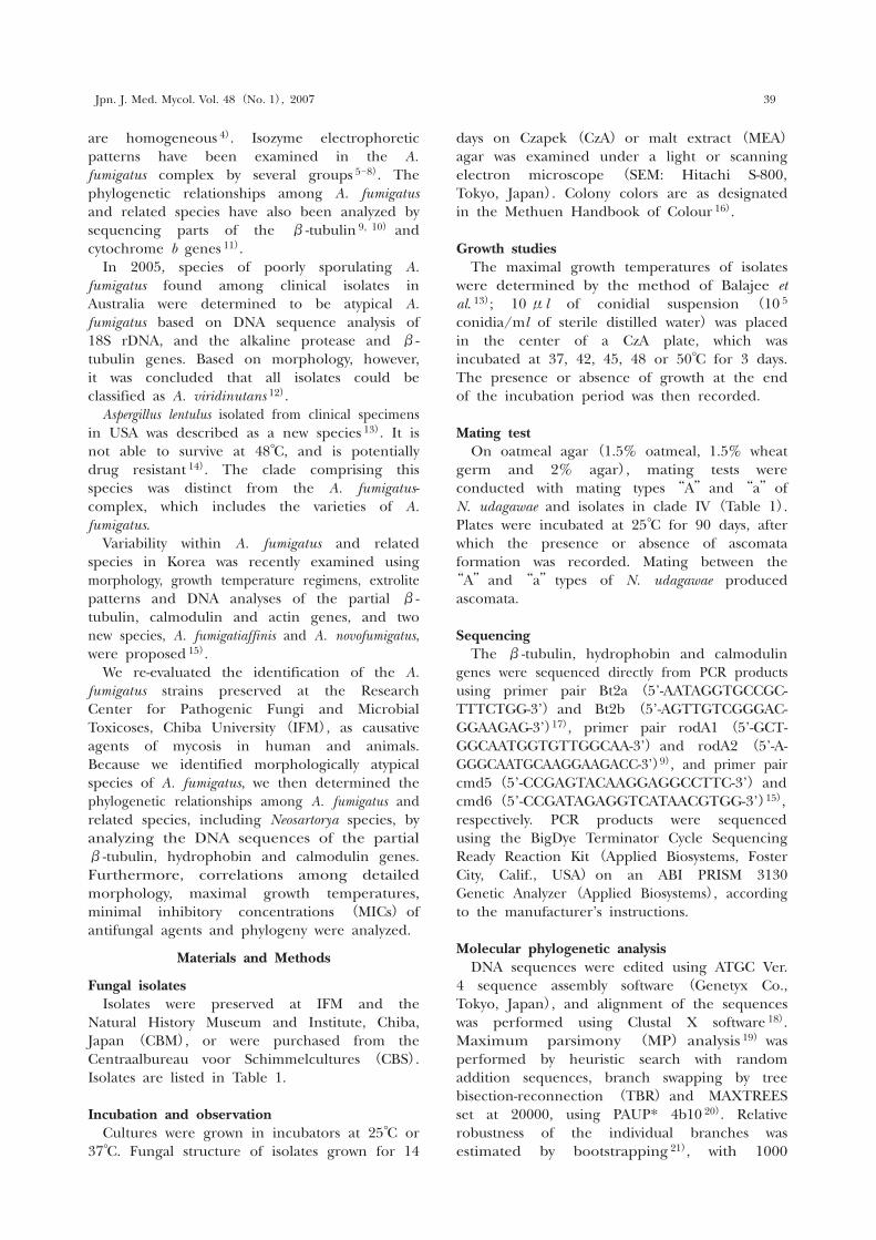
days on Czapek (CzA) or malt extract (MEA) agar was examined under a light or scanning electron microscope (SEM: Hitachi S-800, Tokyo, Japan). Colony colors are as designated in the Methuen Handbook of Colour 16).
Growth studies The maximal growth temperatures of isolates were determined by the method of Balajee et al. 13); 10 μl of conidial suspension (10 5 conidia/ml of sterile distilled water) was placed in the center of a CzA plate, which was incubated at 37, 42, 45, 48 or 50°C for 3 days. The presence or absence of growth at the end of the incubation period was then recorded.
Mating test On oatmeal agar (1.5% oatmeal, 1.5% wheat germ and 2% agar), mating tests were conducted with mating types “A” and “a” of N. udagawae and isolates in clade IV (Table 1). Plates were incubated at 25°C for 90 days, after which the presence or absence of ascomata formation was recorded. Mating between the “A” and “a” types of N. udagawae produced ascomata.
Sequencing The β-tubulin, hydrophobin and calmodulin genes were sequenced directly from PCR products using primer pair Bt2a (5’-AATAGGTGCCGC-TTTCTGG-3’) and Bt2b (5’-AGTTGTCGGGAC-GGAAGAG-3’)17), primer pair rodA1 (5’-GCT-GGCAATGGTGTTGGCAA-3’) and rodA2 (5’-A-GGGCAATGCAAGGAAGACC-3’)9), and primer pair cmd5 (5’-CCGAGTACAAGGAGGCCTTC-3’) and cmd6 (5’-CCGATAGAGGTCATAACGTGG-3’)15), respectively. PCR products were sequenced using the BigDye Terminator Cycle Sequencing Ready Reaction Kit (Applied Biosystems, Foster City, Calif., USA) on an ABI PRISM 3130 Genetic Analyzer (Applied Biosystems), according to the manufacturer’s instructions.
Molecular phylogenetic analysis DNA sequences were edited using ATGC Ver. 4 sequence assembly software (Genetyx Co., Tokyo, Japan), and alignment of the sequences was performed using Clustal X software 18). Maximum parsimony (MP) analysis 19) was performed by heuristic search with random addition sequences, branch swapping by tree bisection-reconnection (TBR) and MAXTREES set at 20000, using PAUP* 4b10 20). Relative robustness of the individual branches was estimated by bootstrapping 21), with 1000
Jpn. J. Med. Mycol. Vol. 48 (No. 1), 2007 39

真菌誌 第48巻 第 1 号 平成19年40
5
A. clavatus H522 (AF057312)
N. pseudofischeri NRRL 20748T (AF057325)
A. brevipes NRRL 2439T (AF057311)A. duricaulis NRRL 4021T (AF057313)
N. hiratsukae NRRL 20819 (AF057324)A. unilateralis NRRL 577 (AF057316)
N. glabra NRRL 2163T (AF057323)N. fennelliae NRRL 5534T (AF057320)N. fennelliae NRRL 5535T (AF057321)
Aspergillus sp. JV3 (AF132222)A. lentulus CBS 153.89 (AB250093*)
A. fumisynnematus IFM 42277T (AB248076*)A. fumisynnematus 90-BP-70 (AB248077*)A. fumisynnematus 90-BP-177 (AB248078*)
A. lentulus IFM 41090 (AB248073*)Aspergillus sp. MK245 (AY590128)
A. lentulus IFM 47063 (AB248074*)A.lentulus IFM 47457 (AB248075*)
A. lentulus FH5T (AY738513)A. lentulus FH7 (AY738520)A. lentulus CBS 175.97 (AB250094*)
N. fischeri NRRL 181T (AF057322)A. fumigatus IFM 47447 (AB248063*)A. fumigatus IFM 51745 (AB248064*)A. fumigatus IFM 51925 (AB248065*)A. fumigatus IFM 51941 (AB248066*)A. fumigatus IFM 51942 (AB248067*)A. fumigatus IFM 51977 (AB248068*)A. fumigatus IFM 53870 (AB248060*)A. fumigatus IFM 54304 (AB248062*)A. arvii CBM FD-0144T (AB248072*)A. fumigatus var. ellipticus
CBS 487.65T (AB248071*)A. fumigatus IAM 13869NT (AB248059*)A. fumigatus IFM 51978 (AB248069*)A. fumigatus IFM 53872 (AB248061*)A. fumigatus IFM 53869 (AB248070*)
N. spinosa NRRL 5034T (AF057329)Aspergillus sp. NSW3 (AY590132)
A. fumigatiaffinis CBS 117194T (DQ094884)A. novofumigatus CBS 117520T (DQ094886)
A. viridinutans FRR 1266 (AF132223)A. viridinutans CBS 127.56T (AB248298*)
A. viridinutans IMI 062875 (AF134779)A. viridinutans IFM 54303 (AB248299*)
Aspergillus sp. MK246 (AY590129)Aspergillus sp. MK284 (AY590130)
A. viridinutans IMI 280490 (AF134780)A. viridinutans IMI 182127 (AF134777)
A. viridinutans NRRL 6106 (AF134778)A. viridinutans IMI 133982 (AF134775)N. aureola NRRL 2244T (AF057319)Aspergillus sp. MK285 (AY590133)Aspergillus sp. IFM 53867 (AB248294*)Aspergillus sp. IFM 53868 (AB248295*)Aspergillus sp. IFM 54302 (AB248296)Aspergillus sp. CBM FD-0143 (AB248297*)
Aspergillus sp. IFM 51744 (AB248293*)Aspergillus sp. IFM 5058 (AB248292*)
N. udagawae CBM FA-0702T (AB248302*)N. udagawae CBM FA-0703T (AB248303*)
57
58
74
91
95
100
100
93
97 100
75
55100
63
79
83
6299
83
65
100
99
84
IV
V
I
II
III
84
94
62
Fig. 1. One of 95 equally parsimonious trees obtained from analysis of the β-tubulin gene using PAUP. Trees were 459 steps in length with a CI of 0.721 and an RI of 0.879. Numbers above or below the nodes represent bootstrap values of >50% (out of 1000 bootstrap replications). A. Aspergillus; N. Neosartorya; *, this study. SEM photographs; conidia (scale bars=3 μm).

replicates, using heuristic search and branch swapping by TBR and MAXTREES set at 100. For neighbor-joining (NJ) analysis 22), the distances between sequences were calculated using Kimura’s two-parameter model 23). MICs of antifungal agents MICs of antifungal agents against the isolated
fungi were measured by the microdilution method, which was proposed for filamentous fungi by the National Committee for Clinical Laboratory Standards (NCCLS M38-A)24). Microtiter plates (Dry Plate; Eiken Chemicals, Tokyo, Japan) containing lyophilized antifungals that were serially diluted two-fold were used according to the manufacturer’s instructions, with slight
Jpn. J. Med. Mycol. Vol. 48 (No. 1), 2007 41
Fig. 2. One of six equally parsimonious trees obtained from analysis of the hydrophobin gene using PAUP. Trees were 368 steps in length with a CI of 0.690 and an RI of 0.819. Numbers above or below the nodes represent bootstrap values of >50% (out of 1000 bootstrap replications). A. Aspergillus; N. Neosartorya; *, this study.

modification. The inoculum suspension was adjusted to 10 4 CFU/ml in RPMI 1640 broth, added to the microtiter wells at 0.1 ml, and then incubated at 35°C for 48 hours. The growth control included the same inoculum in the absence of antifungals, while the negative control was sterile RPMI 1640 broth. Visual examination
of growth inhibition was performed at 48 hours, as further incubation at 72 hours revealed the same reading.
Results
Phylogenetic analysis DNA sequences of the partial β-tubulin,
真菌誌 第48巻 第 1 号 平成19年42
Fig. 3. One of 95 equally parsimonious trees obtained from analysis of the calmodulin gene using PAUP. Trees were 465 steps in length with a CI of 0.733 and an RI of 0.845. Numbers above or below the nodes represent bootstrap values of >50% (out of 1000 bootstrap replications). A. Aspergillus; N. Neosartorya; *, this study.

hydrophobin and calmodulin genes in the strains listed in Table 1 were determined. New sequences were deposited in the DNA Data Bank of Japan (DDBJ) and the accession numbers are listed in Figs. 1-3. MP analysis of the β-tubulin gene sequences (Fig. 1) yielded 95 equally parsimonious trees based on 137 parsimony informative characters, 459 steps in length with a consistency index (CI) of 0.721 and a retention index (RI) of 0.879. That of the hydrophobin gene sequences (Fig. 2) yielded six equally parsimonious trees based on 110 parsimony informative characters, 368 steps in length with a CI of 0.690 and an RI of 0.819, and that of the calmodulin gene sequences (Fig. 3) yielded three parsimonious trees based on 145 parsimony informative characters, 465 steps in length with a CI of 0.733 and an RI of 0.845. No differences were seen between tree topologies from MP and NJ analyses (NJ trees not shown) of the β-tubulin, hydrophobin and calmodulin genes. The three trees based on the three loci were found to be similar. The species of the section Fumigati were divided into five clades: clade I, typical strains of A. fumigatus; clade II, species including A. lentulus and A. fumisynnematus; clade III, species including A. fumigatiaffinis and A. novofumigatus; IV, atypical strains of A. fumigatus including A. viridinutans; and clade V, species including A. brevipes, A. duricaulis and A. unilateralis. Most of the examined strains, including ex-neotype strain IAM 13869, clustered together in the same clade (clade I), while A. fumigatus var. ellipticus and A. arvii were also placed here. Other strains from clinical specimens in Japan were divided into two
clades (clades II and IV). Clade II formed a sister group with clade I and included A. lentulus, A. fumisynnematus and three strains, IFM 47063, 47475 and 41090. Clade III was related to clades I and II and no strains from Japanese clinical specimens belonged to this clade. Clade IV was separated from clades I, II and III. The six variant isolates, IFM 5058, 51744, 53867, 53868, 54302 and CBM FD-0143, belonged to this clade and were closely related to two species of Neosartorya, N. udagawae and N. aureola. This clade also included all strains of A. viridinutans and strain IFM 54303 from a clinical specimen in Japan. A. brevipes, A. duricaulis and A. unilateralis clustered together in clade V, which included no strains from clinical specimens.
Morphology Conidium ornamentation on SEM in strains belonging to clade I was found to vary from almost smooth to echinulate; however, most strains were echinulate. This ornamentation was classified into the lobate-reticulate category 25). A. fumigatus var. ellipticus exhibited ellipsoidal conidia with almost smooth ornamentation, while that of A. arvii was lobatereticulate. Strains IFM 47063, 47475 and 41090, which exhibited almost the same alignment as A. lentulus FH 5, were found to have subglobose conidia with microtuberculate ornamentation 25). Strains IFM 42277, 90-BP-70 and 177, which were in clade II and were identified as A. fumisynnematus, had ellipsoidal conidia with microtuberculate ornamentation and their colonies were floccose and grayish green. The six strains in clade IV (IFM 5058, 51744,
Jpn. J. Med. Mycol. Vol. 48 (No. 1), 2007 43
Table 2. Maximum growth temperatures and MICs on species of Aspergillus section Fumigati
Maximum growth MIC(mg/ml )* Group Species Strain temperature( C) AMPH 5-FC FCZ ITZ MCZ MCFG
A. fumigatus IAM 13869 >50 1 >64 >64 0.5 2 >16 A. fumigatus CBS 110.46 >50 0.5 >64 >64 0.5 4 >16 I A. fumigatus IFM 59125 >50 1 >64 >64 0.5 4 >16 A. arvii CBM FD-0144 >50 1 >64 >64 0.5 2 >16 A. fumigatus var. ellipticus CBS 478.65 >50 1 32 >64 0.5 2 >16
A. lentulus IFM 47457 45 2 >64 >64 0.5 4 >16 II A. lentulus IFM 47063 45 2 >64 >64 1 32 >16 A. lentulus CBS 175.97 45 2 >64 >64 1 4 >16 A. fumisynnematus IFM 42277 45 1 >64 >64 1 2 >16
Aspergillus sp. IFM 51744 42 1 64 >64 0.5 2 >16 IV Aspergillus sp. IFM 53868 42 1 64 >64 1 32 >16 Aspergillus sp. IFM 54302 42 1 >64 >64 1 2 >16
V A. viridinutans CBS127.56 42 1 64 >64 0.5 1 >16 A. viridinutans IFM 54303 42 1 >64 >64 2 >32 >16
* MICs shown were determined by the NCCLS methods. AMPH=amphotericin B, 5-FC=flucytosine, FCZ=fluconazole, ITZ= itraconazole, MCZ=miconazole, MCFG=micafungin.

53867, 53868, 54302 and CBM FD-0143) had globose conidia with microtuberculate ornamenta-tion, while the ex-type strain of A. viridinutans CBS 127.56 had finely echinulate conidia classified as lobate-reticulate. Therefore, these strains were distinguished from A. viridinutans by conidia ornamentation. Furthermore, most strains of A. viridinutans possessed nodding vesicles. The strains in clade VI had globose conidia with lobate-reticulate ornamentation, which were identical to those of typical A. fumigatus. Maximal growth temperature Strains belonging to clade I, including A. fumigatus var. ellipticus and A. arvii, grew at more than at 50°C. Strains belonging to clade II grew poorly at 45°C and did not grow at 48°C, while those belonging to clades IV and V grew well at 42°C but did not grow at 45°C (Table 2).
Mating test Mating between the six strains in clade IV (IFM 5058, 51744, 53867, 53868, 54302 and CBM FD-0143) and mating types “A” and “a” of N. udagawae did not occur.
MIC of antifungal agents MICs of antifungal agents against the isolates are listed in Table 2. The MIC of amphotericin B (AMPH) in A. lentulus was high when compared with other species.
Discussion
Our results concurred with those of Katz et al. 12) and Hong et al. 15) regarding the outline of phylogenetic trees based on DNA sequences of the β-tubulin gene. Namely, typical strains of A. fumigatus clustered in clade I (2a by Katz et al., A. fumigatus s. str. by Hong et al.) and all strains of A. viridinutans clustered in clade IV (1a and 1b by Katz et al., A. viridinutans-complex by Hong et al.). Varga et al. 26) showed that all strains of A. viridinutans belonged to a cluster based on the β-tubulin gene. However, we found six unknown strains in clade IV. The sequences of the hydrophobin and calmodulin genes gave trees with identical topology as that based on the β-tubulin gene. These strains are very closely related to N. udagawae, a heterothallic species, isolated from soil in Brazil 27). However, mating between the strains and N. udagawae did not occur. It is often difficult to do successful mating experiments on clinical isolates and fungi that have been routinely sub-cultured. Therefore these strains need to be further
investigated before they are identified as the anamorphic state of N. udagawae. The strains have conidia with microtuberculate ornamentation, while A. viridinutans has conidia with lobate-reticulate ornamentation. The maximal growth temperature of this species is 42°C, which differs from those of A. fumigatus and A. lentulus. This phenotype is important and helpful to rapidly classify the species in the section Fumigati. Strain CBM FD-0143 belonging to clade IV was isolated from food in Japan and was found to produce the neurotropic mycotoxins fumitremorgin A and B 28). Strain IFM 5058 29) was isolated from a corneal ulcer and showed invasiveness in a mouse brain infection model, and it was also found to belong to this clade. Aspergillus sp. MK 285 12) isolated from the respiratory tract of a cat in Australia was included in this clade based on β-tubulin gene sequence, and is considered to be closely related to those strains. Additional study is necessary to confirm the identity of the Australian isolate. Strains IFM 47063 and 47457 from clinical specimens in Japan were identified as A. lentulus, which was proposed as a new species demonstrating low in vitro susceptibility to antifungal drugs, including amphotericin B and itaconazole 13, 14). Our MP tree based on the β-tubulin gene had the same topology of the ML tree by Balajee et al. 13) among A. fumigatus, A. lentulus and N. fischeri. These strains had smaller conidial heads than A. fumigatus, did not survive at 48°C and had the same susceptibilities as A. lentulus (Table 2). Therefore, they are considered to belong to A. lentulus based on phylogeny, and morphological and physiological characteristics. According to de Hoog et al. 30), the MICs of AMPH for A. fumigatus ranged from 0.125 μm/ml to 2 μm/ml. It was confirmed that Japanese isolates of A. lentulus had lower susceptibility to AMPH than typical isolates of A. fumigatus. Strain IFM 41090 from soil in Venezuela was also identified as A. lentulus and this species was reported to be isolated from clinical specimens in USA and Australia, soils in Korea and a dolphin in the Netherlands 13, 15). Therefore, A. lentulus is widely distributed in the world and the number of mycoses caused by this species is expected to increase from now. Aspergillus fumigatus var. ellipticus is known to be a human pathogen and is distinguished from A. fumigatus by light yellow/green, short conidial heads and ellipsoidal, smooth or nearly so conidia, while A. fumigatus has dark green,
真菌誌 第48巻 第 1 号 平成19年44

long conidial heads and globose echinulate conidia 31). In 1989, Kozakiewicz 25) promoted this variety to an identical species, A. neoellipticus, which resulted in much debate regarding species independence. Our results showed that for three different genes, the ex-type strain of A. fumigatus var. ellipticus (CBS 487.65) could not be distinguished from the strains identified as A. fumigatus, including the ex-neotype strain IAM 13869. Similar data were seen for β-tubulin and hydrophobin gene analysis 9, 13) and mitochondrial cytochrome b gene analysis 11). However, there has been no discussion regarding molecular phylogenetic analysis and morphology examined under SEM. The ornamentation of conidia on A. fumigatus var. ellipticus is almost smooth, while that of A. fumigatus is lobate-reticulate. Therefore, this taxon should remain as a variety of A. fumigatus, and A. fumigatus var. ellipticus is a suitable name. In 1994, A. arvii was isolated from liver lesions in a dairy cow in Finland and was reported as a new species classified in section Fumigati based on its buff color on all standard media 32). This characteristic is not found in the original descriptions of A. fumigatus. However, this taxon has the same alignment with typical species of A. fumigatus in three different genes. Moreover, the shape and ornamentation of the conidia are the same as those of A. fumigatus. Therefore, it was concluded that this taxon is a variety of A. fumigatus. Aspergillus fumigatiaffinis and A. novofumigatus were recently proposed as two new species 15) clustered together with N. spinosa on the β-tubulin gene. We have not found any strains related to the two species. These taxa were isolated from soils in Korea and Ecuador. Therefore, additional isolates identified as these species will be identified with further research. Aspergillus brevipes, A. duricaulis and A. unilateralis were very distant from other members of the section Fumigati. All three species are known from their type strains or very few isolates 25), and so are not considered clinically important. In conclusion, we found that species of Aspergillus section Fumigati were divided into five clades, and there were correlations regard-ing phylogeny, morphology and physiological characteristics. The present polyphasic analysis demonstrates its ability to classify pathogenic fungi and its great potential as a tool in developing diagnostic techniques and in clarifying pathogenesis and epidemiology.
Acknowledgements
This work was supported in part by the National Bioresource Project-Pathogenic microbes in Japan (http://www.nbrp.jp/) and a Grant-in-Aid for Scientific Research (C-18510201, 18927002) from the Japan Society for the Promotion of Science.
References
1) Balajee SA, Gribskov J, Brandt M, Ito J, Fothergill A, Marr KA: Mistaken identity: Neosartorya pseudofischeri and its anamorph masquerading as Aspergillus fumigatus. J Clin Microbiol 43: 5996-5999, 2005.
2) Guarro J, Kallas EG, Godoy P, Karenina A, Gene J, Stchigel A, Colombo AL: Cerebral aspergillosis caused by Neosartorya hiratsukae, Brazil Emerg Infec Dis 8: 989-991, 2002.
3) Jarv H, Lehtmaa J, Summerbell RC, Hoekstra ES, Samson RA, Naaber P: Isolation of Neosartorya pseudofischeri from blood: First hint of pulmonary aspergillosis. J Clin Miclobiol 42: 925-928, 2004.
4) Frisvad JC, Samson RA: Chemotaxnomy and morphology of Aspergillus and related taxa, In Modern concepts in Penicillium and Aspergillus classification. R.A. Samson and J.I. Pitt (ed.), pp.201-208. Plenum Press, New York, 1990.
5) Lin DM, Lehmann PF, Hamory BH, Padhye AA, Durry E, Pinner RW, Lasker BA: Comparison of three typing methods for clinical and environmental isolates of Aspergillus fumigatus. J Clin Microbiol 33: 1596-1601, 1995.
6) Matsuda H, Kohno S, Maesaki S, Yamada H, Koga H, Tamura M, Kuraishi H, Sugiyama J: Application of ubiquinone systems and electro-phoretic comparison of enzymes to identifica-tion of clinical isolates of Aspergillus fumigatus and several other species Aspergillus. J Clin Microbiol 30: 1999-2005, 1992.
7) Rinyu E, Varga J, Ferenczy L: Phenotypic and genotypic analysis of variability in Aspergillus fumigatus. J Clin Microbiol 33: 2567-2575, 1995.
8) Rodriguez E, de Meeus T, Mallie M, Symoens F, Mondon P, Piens MA, Lebeau B, Viviani MA, Geillot R, Nolard N, Chapuis F, Tortorano AM, Bastide JM: Multicentric epidemiology study of Aspergillus fumigatus isolated by multilocus emzyme electrophoresis. J Clin Microbiol 34: 2559-2568, 1996.
9) Geiser DM, Frisvad JC, Taylor JW: Evolutionary relationships in Aspergillus section Fumigati inferred from partial β-tubulin and hydrophobin DNA sequences. Mycologia 90: 831-845, 1998.
10) Varga J, Vide Z, Toth B, Debets F, Horie Y: Phylogenetic analysis of newly described Neosartorya species. Antonie Leeuwenhoek 77: 235-239, 2000.
Jpn. J. Med. Mycol. Vol. 48 (No. 1), 2007 45

11) Wang L, Yohoyama K, Miyaji M, Nishimura K: Mitochondrial cytochrome b gene analysis of Aspergillus fumigatus and related species. J Clin Miclobiol 38: 1352-1358, 2000.
12) Katz ME, Dougall AM, Weeks K, Cheetham BF: Multiple genetically distinct group revealed among clinical isolates identified as atypical Aspergillus fumigatus. J Clin Miclobiol 43: 551-555, 2005.
13) Balajee SA, Gribskov JL, Hanley E, Nickle D, Marr K: Aspergillus lentulus sp. nov., a new sibling species of A. fumigatus. Eukaryotic Cell 4: 625-632, 2005.
14) Balajee SA, Weaver M, Imhof A, Gribskov J, Marr KA: Aspergillus fumigatus variant with decreased susceptibility to multiple antifungals. Antimicrob Agents Chmother 48: 1197-1203, 2004.
15) Hong S-B, Go S-J, Frisvad JC, Samson R: Polyphasic taxonomy of Aspergillus fumigatus and related species. Mycologia 97: 1316-1329, 2005.
16) Kornerup A, Wanscher JH: Methuen Hand-book of Colour, 3rd ed. Eyre Methuen, London, 1978.
17) Glass NL, Donaldson GC: Development of primer sets designed for use with the PCR to amplify genes from filamentous ascomycetes. Appl Environ Microbiol 61: 1323-1330, 1995.
18) Thompson JD, Gibson TJ, Plewniak F, Jeanmougin F, Higgins DG: The clustal X windows interface: flexible strategies for multiple sequence alignment aided by quality analysis tools. Nucleic Acids Res 24: 4876-4882, 1997.
19) Fitch WM: On the problem of discovering the most parsimonious tree. Am Nat 111: 223-257, 1977.
20) Swofford DL: PAUP*: phylogenetic analysis using parsimony (*and other methods). Version 4b10. Sunderland, Massachusetts: Sinauer Associate, 2002.
21) Felsenstein J: Confidence limits on phylogenies:
An approach using the bootstrap. Evolution 39: 783-791, 1985.
22) Saitou N, Nei M: The neighbor-joining method: A new method for reconstructing phylogenetic trees. Mol Biol Evol 4: 406-425, 1987.
23) Kimura M: A simple method for estimating evolutionary rate of base substitutions through comparative studies of nucleotide sequences. J Mol Evol 16: 111-120, 1980.
24) National Committee for Clinical Laboratory Standards. Reference method for broth dilution antifungal susceptibility testing of filamentous fungi. Approved standard M38-A. National Committee for Clinical Laboratory Standards. Wayne, Pa, 2002.
25) Kozakiewicz Z: Aspergillus species on the stored products. Mycol Pap 161: 1-181, 1989.
26) Varga J, Toth B, Rigo K, Debets F, Kozakiewicz Z: Genetic variety within the Aspergillus viridinutans species. Folia Microbiol 45: 423-428, 2000.
27) Horie Y, Miyaji M, Nishimura K, Franco MF, Coelho KIR: New and interesting species of Neosartorya from Brazilian soil. Mycoscience 36: 199-204, 1995.
28) Yamazaki M, Suzuki S, Miyaki K: Tremorgenic toxins from Aspergillus fumigatus Fres. Chem Pharm Bull 19: 1739-1740, 1971.
29) Nishimura K, Miyaji M: Studies on the growth of Aspergillus fumigatus in the brain of mouse. Jpn J Med Mycol 12: 24-29, 1971.
30) De Hoog G.S, Guarro J, Gene J, Figueras MJ: Aspergillus fumigatus. In Atlas of Clinical Fungi, 2nd ed. pp.473-475, Centraalbureau voor Schimmelcultures, Utrecht, 2000.
31) Raper KB, Fennell DI: The Aspergillus fumigatus group. In The genus of Aspergillus (Raper KB, Fennell DI ed), pp.238-268, The Williams & Wilkins Co., Baltitimore, 1965.
32) Aho R, Horie Y, Nishimura K, Miyaji M: Aspergillus arvii spec. nov., a new animal pathogen? Mycoses 37: 389-392, 1994.
真菌誌 第48巻 第 1 号 平成19年46