molecular l-jak, janus kinase
TRANSCRIPT

Proc. Nati. Acad. Sci. USAVol. 91, pp. 6374-6378, July 1994Immunology
Molecular cloning of L-JAK, a Janus family protein-tyrosine kinaseexpressed in natural killer cells and activated leukocytesMASARU KAWAMURA*, DANIEL W. MCVICAR*, JAMES A. JOHNSTONt, TREVOR B. BLAKEO, YI-QING CHENX,BRAJESH K. LAL*, ANDREW R. LLOYDt, DAVID J. KELVINt, J. ERIN STAPLESt, JOHN R. ORTALDO*,AND JOHN J. O'SHEA*§*Leukocyte Cell Biology Section, Laboratory of Experimental Immunology, the tLaboratory of Molecular Immunoregulation, Biological Response ModifiersProgram, and *Biological Carcinogenesis and Development Program, Program Resources, Inc./DynCorp, National Cancer Institute, Frederick, MD 21702-1201
Communicated by Richard D. Klausner, March 10, 1994
ABSTRACT Protein-tyrosine kinases (PTKs) are criticalenzymes for receptor-mediated signaling In lymphocytes. Be-cause natural killer (NK) cells are large granular lymphocyteswith s effector function, we set out to Identify PTKsprferentially exessed In these cells. One such PFK wasIdentified and molecularly cloned. The predicted amino acidsequence shows that this kinase lacks SH2 or SH3 dotypical of src family kinses but has tandem nonidenticalcatalytic domains, idcing that It is a member of the Janusfamily of PTKs. Immunoprecipitation using antiserum gener-ated against a peptide corresponding to the deduced amino acidsequence of this gene revealed a kinase with a molecular weightof "125,000. The pattern of expression of this kinase con-trast sharply with that of other Janus kinases, which areubiquitously expressed. The kinase described in the presentstudy was found to be more limited in its expression; expressionwas found inNK cells and an NK-like cell line but not in restingT cells or in other tissues. In contrast, stimulated and trans-formed T cells expressed the gene, suggesting a role in lym-phold activation. Because of its homology and tissue expres-sion, we have tentatively termed this PTK gene L-JAK forleukocyte Janus kinase.
Protein-tyrosine phosphorylation is an early and requisiteevent in lymphocyte receptor-mediated signaling (reviewedin ref. 1) for both multichain immune recognition receptorssuch as the T-cell antigen receptor (TCR) (2-4) and cytokinereceptors (5-7). Unlike growth factor receptors, neither ofthese types of receptors has intrinsic protein-tyrosine kinase(PTK) activity. Rather, they are coupled to nonreceptortyrosine kinases. For example, there is considerable evi-dence indicating a role for the src family PTKs Lck and Fynin TCR-mediated signaling (2, 3, 8-17) and the non-src familyPTK Zap-70 has been shown to associate with the TCR uponactivation (18-21). Additionally, the src family PTKs, Lck,Fyn, and Lyn have been implicated in interleukin 2 receptor-mediated signaling (5, 6, 22-25). Recently, an additionalfamily ofPTKs, the Janus family ofkinases (JAKs), has beendescribed. These kinases, JAK1, JAK2, and Tyk2, are struc-turally quite distinct in that they possess tandem nonidenticalcatalytic domains (26-29). These PTKs have also been shownto be involved in signaling by a number of cytokine andhormone receptors (30-36). These family members are alsoof interest in that they appear to exert their effect throughtyrosine-phosphorylated transcription factors (37, 38). Thusthere is now abundant evidence implicating a variety ofPTKsin lymphocyte activation.
Natural killer (NK) cells are a distinct lymphocyte subsetthat do not undergo rearrangement of antigen receptor chaingenes but otherwise shares a number of similarities with T
lymphocytes (reviewed in refs. 39-41). NK cells and T cellsare developmentally related and express similar surfacemolecules (42, 43). Nonetheless, they are functionally quitedifferent. NK cells can be thought of as primed effector cellscapable of killing susceptible targets without additional ac-tivation. In contrast, T cells generally do not lyse targets inthe absence of prior stimulation. To begin to understand themolecular underpinning of this difference and because of thecritical role of PTKs in NK cell function (44-47), we com-pared the status of tyrosine-phosphorylated substrates inthese two lymphocyte subsets (unpublished results). Wedemonstrated that NK cells have high levels of tyrosinephosphorylation of a variety of substrates relative to T cells.This was not due to differential expression or activity of Lckor differences in protein-tyrosine phosphatase activity. Wehypothesized, therefore, that NK cells might express PTKsnot expressed in resting T cells, and to this end used aPCR-based strategy to identify such kinases. In the presentstudy we report the molecular cloning of one such PTKI thatis a member of the Janus family of PTKs. However, unlikeother JAKs, which are widely expressed, the expression ofthe NK-derived kinase is much more limited; it is expressedonly in NK cells and other activated leukocytes. We have,therefore, termed this kinase gene L-JAK for leukocyte JAK.
MATERIALS AND METHODST Cells and NK Cell PTK. A modification of the strategy
described by Wilks et al. (28) was used to clone PTKexpressed in NK cells (48). The forward primer used was5'-CCAGCGGCCGCGT(G/A/T/C)CA(C/T)CG(G/A/T/C)GA(C/T)CT(G/A/T/C)GC-3' and the reverse primer was5'-CCAGCGGCCGCCC(G/A)AA(G/A/T/C/)(G/C)(A/T)CCA(G/A/T/C)AC(G/A)TC-3'. The resulting productswere digested with Not I, subcloned, and sequenced. ThePCR fiagment corresponding to one novel kinase was iso-lated, labeled, and used to screen several libraries includingAgtll, oligo(dT)-primed cDNA libraries derived from phyto-hemagglutinin (PHA)-stimulated peripheral blood T cells andthe HUT-78 T-cell line (Clontech), a AZAP YT library(provided by Warren Leonard, National Heart, Lung, andBlood Institute), and a A ZAP library from PHA-activated Tcells (provided by K. Kelly, National Cancer Institute).Purified phage DNA was digested and subcloned into pBlue-script for sequencing. Sequence data were manipulated andanalyzed using the programs of the Genetics ComputerGroup ofthe University ofWisconsin and the BLAST programof the National Center for Biotechnology Information.
Abbreviations: JAK, Janus kinase; JH, JAK homology; L-JAK, leuko-cyte JAK; NK, natural killer; PHA, phytohemagglutinin; PIK, pro-tein-tyrosine kinase; TCR, T-cell antigen receptor.1To whom reprint requests should be addressed.1The sequence reported in this paper has been deposited in theGenBank data base (accession no. U09607).
6374
The publication costs of this article were defrayed in part by page chargepayment. This article must therefore be hereby marked "advertisement"in accordance with 18 U.S.C. §1734 solely to indicate this fact.

Proc. Natl. Acad. Sci. USA 91 (1994) 6375
For Northern analysis, total RNA from various humantissues was purchased (Clontech) or prepared from NK cellsand T cells.Tmmunopenptatond Imnunoblotng. A peptide cor-
responding to the deduced C terminus ofthe L-JAK gene (seeFig. 1; amino acids 1104-1124) was synthesized (MultiplePeptide Systems, San Diego) coupled to keyhole limpethemocyanin with m-maleimidobenzoyl-N-hydroxysuccin-imide ester (Pierce) and used as an immunogen in rabbits.Cells (107 cells per point) were labeled with [35S]methionine(0.5 mCi/ml; 1 Ci = 37 GBq) for 2 hr. washed with phosphate-buffered saline, and lysed in buffer containing 1% TritonX-100 (lysis buffer). Postnuclear supernatants were immu-noprecipitated with 10 pI of antiserum prebound to proteinA-Sepharose, washed in buffer containing 0.1% Triton X-100(wash buffer), eluted, and electrophoresed in 8% polyacryl-amide gels that were subsequently fixed, rinsed in Fluoro-Hance (Research Products International), and dried for au-toradiography.
Kinase assays were performed as described (33, 34) bysolubilizing cells in lysis buffer supplemented with 1 mMNa3VO4 and 1 mM EDTA and immunoprecipitating with theantipeptide antiserum. The washed immunoprecipitates wereincubated in 50 1d of buffer containing 20 mM Tris, 5 mMMgCl2, 5 mM MnCl2, 1 juM ATP, and [y-32P]ATP (Amer-sham) at 200 ,uCi/ml. The reaction was carried out for 15 minat 25°C and was terminated by the addition of ice-cold washbuffer. After washing the beads again, the reaction productswere eluted and electrophoresed.For immunoblot analysis, cells were solubilized in lysis
buffer, and postnuclear supernatants (=100 jug of protein)from the indicated cells were electrophoresed, transferred tonitrocellulose, and immunoblotted. Filters were blocked,incubated with antiserum (1:1000), washed, and incubatedwith peroxidase-conjugated goat anti-rabbit IgG. Antibodybinding was detected by enhanced chemiluminescence(ECL; Amersham).
RESULTS
Cloning of an NK Cell-Derived Janus Family PTK. Todetect previously unknown PTKs expressed in NK cells, weemployed PCR, an approach used successfully by others (28,48). We prepared cDNA from NK cell mRNA using reversetranscriptase and then performed PCR with degenerate oli-gonucleotide primers corresponding to conserved motifs inthe catalytic domains of PTKs. The forward primers weredesigned to correspond to residues in subdomain VI and weredesigned to exclude src family PTKs (48). The reverse primercorresponded to the reverse complement of the DVWSFGmotif (subdomain IX) conserved in a large number of PTKs.Out of =200 clones, seven previously unknown putativekinases were identified. One of these genes was found byNorthern analysis to be expressed preferentially in NK andactivated T cells (see below). This PCR-generated fragmentwas, therefore, used to screen libraries derived from theNK-like cell line, YT, PHA-activated T cells, and HUT-78cells. Approximately 5 x 10⁵ plaques from each library werescreened to obtain multiple overlapping clones that generatedsequence corresponding to a single large open reading frame.Fig. 1 depicts the deduced amino acid sequence of the NKcell-derived gene compared to closely related PTKs.The deduced polypeptide encoded by this gene exhibits
features typical of a PTK (49). Like other PTKs, a catalyticdomain is present in the C-terminal portion of the molecule(subdomains I-XI). The domain begins with a typical ATP-binding motif at residues 829-834 (subdomain I) in which thecanonical GXGXXG motif is evident (Fig. 1, shaded) that isfollowed by a critical lysine residue in subdomain II (residue855). Just C-terminal to subdomain VII is a pair of tyrosine
residues following an acidic residue that likely represents theautophosphorylation site. In subdomain VIII, phenylalanineand tyrosine residues surround the invariant tryptophanresidue. This atypical motif contrasts with the motifs seen insrc- and abl-related proteins and growth factor receptors.Notably though, this motif (FWYAPE; Fig. 1, shaded) ispresent in the Janus family of kinases. The entire catalyticdomain, termed the JH1 domain, is composed of 273 aminoacids (residues 822-1095) and is followed by a unique Cterminus.
In addition to a kinase catalytic domain, the NK-derivedgene has a region N-terminal to the PTK catalytic domain thatalso has elements typical ofa protein kinase catalytic domain(Fig. 1, Ia-XIa). This tandem kinase-like (JH2) domain is acharacteristic feature of the JAKs (28-31). However, likeother family members, this domain in the NK cell-derivedgene lacks some standard features ofa PTK catalytic domainsuch as an autophosphorylation site in subdomain VII.The known JAK family PTKs have large extracatalytic
segments (JH3-7 domains) N-terminal to the kinase (H11)and kinase-like (JH2) domains. While motifs correspondingto SH2 or SH3 domains are lacking, there is a motif that hasbeen suggested to be SH2-like (30), which (Fig. 1, shaded) isconserved in three of the four family members, including the,NK cell-derived gene. Immediately N-terminal to this motifis a highly conserved motif that is a potential tyrosinephosphorylation site (VDGXFRL). Other areas of strikinghomology between the NK-derived gene and other JAKs areevident in the remaining domains (JH5-7). Structurally then,it appears that the NK-derived gene has all of the character-istics of a JAK family PTK. The overall homology ofthe NKPTK to the most closely related Janus family member, JAK2,is =68% identity. A hydrophilicity plot did not show thepresence of a hydrophobic domain, suggesting that the NK-derived gene encodes a nonreceptor type of PTK like otherJanus proteins.Using the initiation site indicated, the open reading frame
of this gene encompasses 3372 nucleotides and is predicted toencode a polypeptide of 1124 residues. This predicts amolecular weight of 125,014, roughly equivalent to, butslightly smaller than, other JAKs. This predicted molecularweight is consistent with that of the polypeptide identifiedusing the antiserum prepared against this kinase (see below),supporting the contention that this is the correct initiationsite.
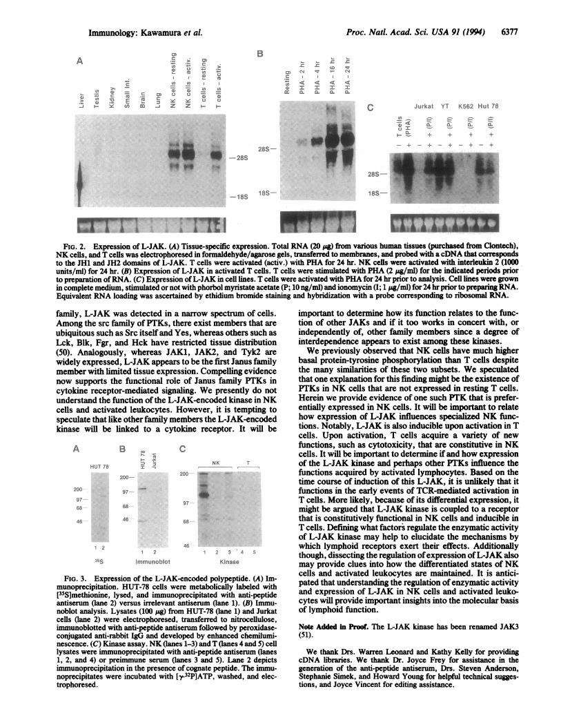
Expression of the NK-Derived Janus Family PTK. Othermembers of the Janus family of PTKs (JAK1, JAK2, andTyk2) are present in a variety of tissues (27-29). We sought,therefore, to determine if the NK-derived Janus PTK alsowas widely expressed or if its expression was more limited.As shown in Fig. 2A, unlike other family members, this PTKgene was found to have restricted tissue distribution. In theabsence of stimulation, the gene was found to be expressedsolely in NK cells. This filter was also probed with a JAKicDNA and, as previously reported, ubiquitous tissue expres-sion was noted with the exception of small intestine (data notshown). Equivalent loading ofRNA was confirmed by ethid-ium bromide staining and by hybridization with a probeagainst ribosomal RNA (not shown). It should be noted thatthe signal observed to the left of the liver lane is artifactualand represents binding of the probe to the edge of the filterand not to RNA. Interestingly, while the NK PTK gene wasexpressed at very low levels in resting T cells, we observedthat following activation this gene was induced whereasactivation of NK cells did not alter the level of expression.Because the time course ofinduction of this gene might beginto provide clues to its function, this was more carefullyanalyzed in the experiment shown in Fig. 2B. The expressionof this gene in T cells was found to peak 16-24 hr afterstimulation with PHA. We next analyzed the expression of
Immunology: Kawamura et A

6376 Immunology: Kawamura et al. Proc. Natl. Acad. Sci. USA 91 (1994)
L-JAK " A P PSCP T ? L: P ; R5: S LL S- nA u EAV AJ -2KG# A. LT XT ItEA TS S FVSI lP P AS FvY..k - -- AFPKm
S S P r VE V F YA P _ RT,*2 - - - - - - M P ~~~R V S M A R A IV
S ,iMAAYaAL1VL.MWAr
JUrLrtSm P VsriSAAH A A K
V
ICjILISa.j:F-TE-ANELE- -ARA- A..FHN- ;
JL 1'TPlNTAS L A HPY RSF Yr T'I rski ' DISTL T IR: F S HAH F E A .N F A ^Y -
C -i -- FEAELAA1 -AYP* F FI5- A. .
L-JAK A ST P V A RKXI RG'5* r .. . t
*U K IA| A AS H.
X VA A
JU-J P3 S KP| HLNLFS L LFIH AIE AALY z ADATU-i FAASAI..LAASL1T YPL IRFGA HNH APAS TISH. FI1 9SA
L-JAK A I T A A
JAAA AS S(LjN. IINN0RP~nN.LuTVV i C P A AADS T IxLjLLLIA.S TIAIXLKMALPLY X LSIIALLILAP R
r AV EPA A I R
IJ1A~SNAALHAArnmAVIA
~ I'..LL LLQN1F1 - ---A5. 1NTNUIS1LHSLI ALIIILILICAAPTIIIIFPA ISJ A2 ll AP¢ 0 P -V sA- LI A R
J*1lPUE;|R £|r~ll N I r Z C S S Y S t' H D~l|K V~l Y L|A T|L ZIT|LIT It HIYIGXICILP I: XMJO2 11 9r ;mlLAPO-4 S.R S ItTL t|IRI KJ P ItG R V ',iwR
C4_~~~~~~~~~ ~ ~~~~~~VItr I Y -LE
pUJNS U-,
Ty A2 - LA- L --I E I SAL F| IANAA ItASHA., VV....v .
L-JAK I AA- N~ A -D I E; _~A F|*,JH m i
mu 1 I APA-R SI[F-ILLEH IANS NTp2VNAIt .LA S S- iA S PAN VKVAA,. A A A RJ 1ArFR J
CO-90.6-~~~~~ ~PRAFFK Y F.
L-jAK .Y m n P PSO£3~H 11L T T I > Pmo_ D ;7 T
JU-2 ISISSIPV1SIAIIN0A 11 -e sIfESS I:lT|^YlP S..SA~vL1|| IS xE A:SHFAA{; lAU lI l.I. .k. I AI A it PA D IN I.Ar SI H A A
ISENEI AIPAFSSPA NlI
TLAA T L 5I5 5 ELVAL7 SAP CPAI VT1G|flITC-. A S YAlPH Al T . VIICSL JTI AlG I'I-
AS ....u' H A H H A A F: F
JLa I v sv IK- -l- fA2U- IvR I. S11 PT Ns R K P ASAP F IF I V ANT-
Hl SQA
PS StV A _jfA JN n mFflfV S m fl ,, oU-2 SICITSIILIPK1SjLISIAIS A^ 1E S1SIAIN roDP1I F1ASF1 L PNINIGS DILISIPIA1S4JAIAS ISI":TLYj15r||IUJI ISI I! l IOICiI:I 1N11 IIVIA-S^THAN LWtNIJSUJAWHILIA;GA C L[ W
A' W1 HL
ASST V REU S
I [A F.NU-A TA SAN5 KPLNITALIjLHI.jHIA I P Ak FV.l TA AN
c_ .r-K V K L;:F--:.-r ; eA
L-JAK A L IV OPAs1QrX1PI1TLrA...NPIARJAIEA CAIL JA1HI T F, 7 C
DA T
JlST A A LL, D VA SP |IV N P 3 ]g iyRali }FF |CS| 4l~iX9II^VF B,, K - - - - - - - z }~~~~~~~~~~~~~~~~~~L;PfV Y 4l I F |rEl |'x |jV, |1R4-|||K:YSII
C - . ,FA I L V AFSP Pl s H~l| F A1: wH~JLILN!BkF
C_~~~~~~~~~~~~~~~J7 RS -Q -A ^F H:-E: .. O
A.kll A
I E A A S'NS tLF uNfE9.W__9S.
EA
5 -1ADJ571 rm A|:KV^._.L-A lKI, I3Y::Y;..;P^VEY
A A K 'i
.-JAX4.2
Trk2
- jAKJak2
Tyh
.,AK S ii r : 4:--FLE r.-wF I;v ; e;
Tvk2 ? f i 0 i ~|l t .NA It:~!:sFi-
AV AL . A A A SN- W A; .
VAlI VLAl.F s SP A
li. F. Fi ;rr .IAHiIA
Jdk2 11 O-F I E 1:F V ' S E t ir: si,>;rVrTrI ISr-_~
C- i9K.. . <, _::TvAN N
X. Xh,-JAK rfcVI _ N ItiS ,k Y K| i,7 < A RW An F, X[AT pi VU; rG
IlAl.^-I LSJ M
2JL;It. A]CoIu HASAE.r~rrm:NflN~Y~fl'l~ AAU-N2' j-AAAA.A E.A
.--AKJak2Jk
Tak2jai
L-JAK
Ty2C-,.,-
[7Sff11 Gf' -Ill.. -. A.. .AY[F-* A 1. _R K r r tRDP11.g E E ;P>
P. - JUT NH
A. T NQIH A -
LzllS I :~ q 5
rAA Hill.iI 1.
S L ! I A ATn F IT l l.P IS VCPS FL 1R:RI D pi C Il~ I L OSL|wl DIl| ;'V ' r & VYIT S A|I HK |N TP J L1 Y:| 3
:115f : LLTIEI~l.H.HA~~~~~~~~~~~~lS~~~~~~~
L-AK k 'a R A P. k :4 R I :R '>fAg
it s|=1w HVITJLAN 2 ; rx_:1 N RIV1S A5
A VTvk2 . H ASL= A 4 PUPtAIR K
All VillL A AlA R P Sr a IX
n uA :: 7 . C' : . S . --Ak 1A A E RI 4'y
SL I C on.F...... JAL:.: :: L IIIL .._ALlp
515I5III~~~~~~~IPmA EI
-JAK P AI PA I clil LII I ,A. R P ~ ;.11DSv
Sl || M - EI |N S N { | 5 S.^ ||JFk, X l VY 1- IR xl W E 9X11lI{ l 5 |1T,.l2 r L L]L. l WF-Ftl^IF L L,~~~~~~~
FiG. 1. Predicted amino acid sequence of the L-JAK gene and alignment to the known members of the Janus family of PTKs. The deducedprimary structure of the L-JAK gene is depicted in the single-letter amino acid code, and residues that are identical among JAKs are boxed.The subdomains of the PTK domain are indicated with Roman numerals. The subdomains of the kinase-like domain are indicated by Romannumerals followed by the letter a (e.g., Ia). The boundaries ofJAK homology (JH) domains are denoted by arrows. Motifs of particular interestare shaded.
this gene in cell lines (Fig. 2C). The YT cell line hascharacteristics of NK cells and, as expected, this gene wasfound to be present constitutively in these cells. Also aspredicted by studies with peripheral blood T cells, the genewas found not to be expressed in-the Jurkat T-cell line but wasinducible upon activation. HUT-78 cells are a transformedT-cell line, and, interestingly, expression of the gene wasfound to be constitutive in this cell line. No expression ofthisgene was detected in a variety ofother cell lines, including theerythroleukemia cell line K562. Moreover, it was not induc-ible in this cell line. Because of the homology to other JAKsand the pattern of tissue expression of the NK-derived PTKgene, we have termed the gene L-JAK for leukocyte JAK.The deduced C terminus ofL-JAK was found to be unique,
so a synthetic peptide was generated corresponding to thisportion of this gene product (Fig. 1, amino acids 1104-1124)and used as an immunogen. In good agreement with themolecular weight predicted by the deduced primary struc-ture, analysis of metabolically labeled HUT-78 cells showedspecific immunoprecipitation of a polypeptide with a Mr of%125,000 (Fig. 3A). Immunoblot analysis of these cells alsoshowed reactivity of the antibody with a protein of approx-imately the same mobility in HUT-78 cells. In contrast,Jurkat T cells expressed minimal levels of this protein. In
additional experiments (data not shown), the expression ofthe L-JAK-encoded polypeptide was found to parallel theexpression seen by analysis of mRNA. Expression of theprotein was detected in NK cells, activated T cells, and insome transformed leukocyte cell lines. Immunoblotting withpreimmune serum or antiserum competed with cognate pep-tide versus irrelevant peptide confirmed the specificity ofthisreactivity (data not shown).To ascertain that the L-JAK-encoded kinase had enzymatic
activity, in vitro kinase assays were performed. A phosphor-ylated polypeptide with the expected Mr was evident inimmunoprecipitates from NK cells (Fig. 3C, lane 1) but not inresting T cells (lane 4) or in control immunoprecipitates Qanes2, 3, and 5). The phosphorylated residues were resistant toKOH, consistent with tyrosine phosphorylation. This likelyrepresents autophosphorylation of L-JAK.
DISCUSSIONThe Janus family comprises PTKs that have tandem non-identical catalytic domains and a large extracatalytic segment(27-29). The structural features of the NK-derived PTK,L-JAK, clearly argues for membership in the Janus family ofPTKs. However, unlike the three other members of this
Proc. Nati. Acad. Sci. USA 91 (1994) 6377
A
.8 o =
> T2 EH N 3u
LI)-T
t)r_ CD 0
c
0n Xj z
> 0)t
C1) m
~c 0 0.Z H H
B
CT) CN -
c) I I IM a_ XL X-
*,"-... C
ICL
Jurkat YT K562 Hut 78
U,-o oU Ir .
+ &- 4t +
.s
'III_A.
-128S_*
-loo
_ + +28S-
- + I
28S-
18S- 18S-
mI
FIG. 2. Expression of L-JAK. (A) Tissue-specific expression. Total RNA (20 yg) from various human tissues (purchased from Clontech),NK cells, and T cells was electrophoresed in formaldehyde/agarose gels, transferred to membranes, and probed with acDNA that correspondsto the JH1 and JH2 domains of L-JAK. T cells were activated (activ.) with PHA for 24 hr. NK cells were activated with interleukin 2 (1000units/ml) for 24 hr. (B) Expression of L-JAK in activated T cells. T cells were stimulated with PHA (2 pg/ml) for the indicated periods priorto preparation ofRNA. (C) Expression of L-JAK in cell lines. T cells were activated with PHA for 24 hr prior to analysis. Cell lines were grownin complete medium, stimulated or not with phorbol myristate acetate (P; 10 ng/ml) and ionomycin (1; 1 mg/ml) for 24 hr prior to preparing RNA.Equivalent RNA loading was ascertained by ethidium bromide staining and hybridization with a probe corresponding to ribosomal RNA.
family, L-JAK was detected in a narrow spectrum of cells.Among the src family of PTKs, there exist members that areubiquitous such as Src itself and Yes, whereas others such asLck, Blk, Fgr, and Hck have restricted tissue distribution(50). Analogously, whereas JAK1, JAK2, and Tyk2 arewidely expressed, L-JAK appears to be the first Janus familymember with limited tissue expression. Compelling evidencenow supports the functional role of Janus family PTKs incytokine receptor-mediated signaling. We presently do notunderstand the function of the L-JAK-encoded kinase in NKcells and activated leukocytes. However, it is tempting tospeculate that like other family members the L-JAK-encodedkinase will be linked to a cytokine receptor. It will be
B
HUT 78
200
97
68
46
2
J5s
CNK T
200 _
97
68
i 2
Immunoblot
46
1 2 3 4
Kinase
FIG. 3. Expression of the L-JAK-encoded polypeptide. (A) Im-munoprecipitation. HUT-78 cells were metabolically labeled with[35S]methionine, lysed, and immunoprecipitated with anti-peptideantiserum (lane 2) versus irrelevant antiserum (lane 1). (B) Immu-noblot analysis. Lysates (100 pg) from HUT-78 (lane 1) and Jurkatcells (lane 2) were electrophoresed, transferred to nitrocellulose,immunoblotted with anti-peptide antiserum followed by peroxidase-conjugated anti-rabbit IgG and developed by enhanced chemilumi-nescence. (C) Kinase assay. NK (lanes 1-3) and T (lanes 4 and 5) celllysates were immunoprecipitated with anti-peptide antiserum (lanes1, 2, and 4) or preimmune serum (lanes 3 and 5). Lane 2 depictsimmunoprecipitation in the presence of cognate peptide. The immu-noprecipitates were incubated with [y.32P]ATP, washed, and elec-trophoresed.
important to determine how its function relates to the func-tion of other JAKs and if it too works in concert with, orindependently of, other family members since a degree ofinterdependence appears to exist among these kinases.We previously observed that NK cells have much higher
basal protein-tyrosine phosphorylation than T cells despitethe many similarities of these two subsets. We speculatedthat one explanation for this finding might be the existence ofPTKs in NK cells that are not expressed in resting T cells.Herein we provide evidence of one such PTK that is prefer-entially expressed in NK cells. It will be important to relatehow expression of L-JAK influences specialized NK func-tions. Notably, L-JAK is also inducible upon activation in Tcells. Upon activation, T cells acquire a variety of newfunctions, such as cytotoxicity, that are constitutive in NKcells. It will be important to determine ifand how expressionof the L-JAk kinase and perhaps other PTKs influence thefunctions acquired by activated lymphocytes. Based on thetime course of induction of this L-JAK, it is unlikely that itfunctions in the early events of TCR-mediated activation inT cells. More likely, because of its differential expression, itmight be argued that L-JAK kinase is coupled to a receptorthat is constitutively functional in NK cells and inducible inT cells. Defining what factors regulate the enzymatic activityof L-JAK kinase may help to elucidate the mechanisms bywhich lymphoid receptors exert their effects. Additionallythough, dissecting the regulation ofexpression ofL-JAK alsomay provide clues into how the differentiated states of NKcells and activated leukocytes are maintained. It is antici-pated that understanding the regulation of enzymatic activityand expression of L-JAK in NK cells and activated leuko-cytes will provide important insights into the molecular basisof lymphoid function.
Note Added in Proof. The L-JAK kinase has been renamed JAK3(51).
We thank Drs. Warren Leonard and Kathy Kelly for providingcDNA libraries. We thank Dr. Joyce Frey for assistance in thegeneration of the anti-peptide antiserum, Drs. Steven Anderson,Stephanie Simek, and Howard Young for helpful technical sugges-tions, and Joyce Vincent for editing assistance.
A
200
97
68
46
Immunology: Kawamura et al.

6378 Immunology: Kawamura et al.
1. Perlmutter, R. M., Levin, S. D., Appleby, M. W., Anderson,S. J. & Alberola-ila, J. (1993) Annu. Rev. Immunol. 11, 451-499.
2. Klausner, R. D. & Samelson, L. E. (1991) Cell 64, 875-878.3. Weiss, A. (1993) Cell 73, 209-212.4. Keegan, R. D. & Paul, W. C. (1992) Immunol. Today 13,
63-68.5. Taniguchi, T. & Minami, Y. (1993) Cell 73, 5-8.6. Minami, Y., Kono, T., Miyazaki, T. & Taniguchi, T. (1993)
Annu. Rev. Immunol. 11, 245-267.7. Stahl, N. & Yancopoulos, G. D. (1993) Cell 74, 587-590.8. Samelson, L. E., Phillips, A. F., Luong, E. T. & Klausner,
R. D. (1992) Proc. Nall. Acad. Sci. USA 87, 4358-4362.9. TimsonGauen, L. K., Kong, A. N., Samelson, L. E. & Shaw,
A. S. (1992) Mol. Cell. Biol. 12, 5438-5446.10. Appleby, M. W., Gross, J. A., Cooke, M. P., Levin, S. D.,
Qian, X. & Perlmutter, R. M. (1992) Cell 70, 751-763.11. Stein, P. L., Lee, H. M., Rich, S. & Soriano, P. (1992) Cell 70,
741-750.12. Cooke, M. P., Abraham, K. M., Korbush, K. A. & Perlmutter,
R. M. (1991) Cell 65, 281-291.13. Veillette, A., Bookman, M. A., Horak, E. M. & Bolen, J. B.
(1988) Cell 55, 301-308.14. Veillette, A., Abraham, N., Caron, L. & Davidson, D. (1991)
Semin. Immunol. 3, 143-152.15. Rudd, C. E., Trevillyan, J. M., Dasgupta, J. D., Wong, L. L.
& Schlossman, S. F. (1988) Proc. Nail. Acad. Sci. USA 85,5190-5194.
16. Abraham, N., Miceli, M. C., Parnes, J. R. & Veillette, A.(1991) Nature (London) 350, 62-66.
17. Molina, T. J., Kisihara, K., Sidervoski, D. P., van Ewijk, W.,Narendren, A., Timers, E., Wakenman, A., Paige, C. J.,Hartmann, K. U., Veillette, A., Davidson, D. & Mak, T. W.(1992) Nature (London) 357, 161-164.
18. Strauss, D. B. & Weiss, A. (1992) Cell 70, 585-593.19. Chan, A. C., Irving, B. A., Fraser, J. D. & Weiss, A. (1991)
Proc. Natl. Acad. Sci. USA 88, 9166-9170.20. Wange, R. L., Kong, A. T. & Samelson, L. (1992) J. Biol.
Chem. 267, 11685-11688.21. Chan, A. C., Iwashima, M., Turck, C. W. & Weiss, A. (1992)
Cell 71, 649-662.22. Horak, I. D., Gress, R. E., Lucas, P. J., Horak, E. M., Wald-
man, T. A. & Bolen, J. B. (1991) Proc. Nail. Acad. Sci. USA88, 1196-2001.
23. Hatakeyama, M., Kono, T., Kobayashi, N., Kawahara, A.,Levin, S. D., Perlmutter, R. M. & Taniguchi, T. (1991) Science252, 1523-1528.
24. Kobayashi, N., Kono, T., Hataklyama, M., Minami, Y.,Miyazaki, T., Perlmutter, R. M. & Taniguchi, T. (1993) Proc.Nail. Acad. Sci. USA 90, 4201-4205.
25. Torigoe, T., Saragovi, H. U. & Reed, J. C. (1992) Proc. Nail.Acad. Sci. USA 89, 2674-2678.
26. Wilks, A. F. (1989) Proc. Nail. Acad. Sci. USA 86,1603-1607.27. Wilks, A. F., Harpur, A. G., Kurban, R. R., Ralph, S. J.,
Zurcher, G. & Ziemiecki, A. (1991) Mol. Cell. Biol. 11, 2057-2065.
28. Harpur, A. G., Andres, A. C., Ziemiecki, A., Aston, R. R. &Wilks, A. F. (1992) Oncogene 7, 1347-1353.
29. Firmbach-Kraft, I., Byers, M., Shows, T., Dalla-Favera, R. &Krolewski, J. J. (1990) Oncogene S. 1329-1336.
30. Velazquez, L., Fellous, M., Stark, G. R. & Pellegrini, S. (1992)Cell 70, 313-322.
31. Argetsinger, L. S., Campbell, G. S., Zang, X., Witthuhn,B. A., Silvennoinen, 0., IhIe, J. N. & Carter-Su, C. (1993) Cell74, 237-244.
32. Witthuhn, B. A., Quelle, F. W., Silvennoinen, O., Yi, T.,Miura, 0. & IhIe, J. N. (1993) Cell 74, 227-236.
33. Muller, M., Briscoe, J., Laxton, C., Gushin, D., Ziemiecki, A.,Silvennoinen, O., Harpur, A. G., Barbieri, G., Witthuhn,B. A., Schindler, C., Pellegrini, S., Wilks, A. F., Ihle, J. N.,Stark, G. R. & Kerr, I. M. (1993) Nature (London) 36, 129-135.
34. Watling, D., Guschin, D., Muller, M., Silvennionen, Witthuhn,B. A., QueUe, F. W., Rogers, N. C., Schindler, C., Stark,G. R., Ihle, J. N. & Ken, I. M. (1993) Nature (London) 366,166-170.
35. Shuai, K., Ziemiecki, A., Wilks, A. F., Harpur, A. G., Sad-owski, H. B., Gilman, M. Z. & Darnell, J. E. (1993) Nature(London) 366, 580-582.
36. Silvennoinen, O., Ihle, J. N., Schlessinger, J. & Levy, D. E.(1993) Nature (London) 366, 583-585.
37. Fu, X.-Y. (1992) Cell 70, 323-325.38. Pellegrini, S. & Schindler, C. (1993) Trends Biochem. Sci. 18,
338-342.39. Trinchieri, G. (1989) Adv. Immunol. 47, 187-303.40. Robertson, M. J. & Ritz, J. (1990) Blood 76, 2421-2438.41. Kennedy, I. C. S., Ortaldo, J. R. & O'Shea, J. J. (1992) inNK
Cell Mediated Cytotoxicity: Receptors, Signaling and Mecha-nism, eds. Lotzova, E. & Herberman, R. B. (CRC, BocaRaton, FL), pp. 147-167.
42. Rodewald, H. R., Moingeon, P., Lucich, J. L., Dosiou, C.,Lopez, P. & Reinherz, E. L. (1992) Cell 69, 139-150.
43. Lanier, L. L., Spits, H. & Phillips, J. H. (1992) Immunol.Today 13, 392-395.
44. Einspahr, K. J., Abraham, R. T., Binstadt, B. A., Uehara, Y.& Liebson, P. J. (1991) Proc. Nail. Acad. Sci. USA 88,6279-6283.
45. O'Shea, J. J., Weissman, A. M., Kennedy, I. C. S. & Ortaldo,J. R. (1991) Proc. Nail. Acad. Sci. USA 88, 350-354.
46. Vivier, E., Morin, P., O'Brien, C., Druker, B., Schlossman,S. F. & Anderson, P. (1991) J. Immunol. 146, 206-210.
47. O'Shea, J. J., McVicar, D. W., Kuhns, D. B. & Ortaldo, J. R.(1992) J. Immunol. 148, 2497-2502.
48. Siliciano, J. D., Morrow, T. A. & Desiderio, S. V. (1992) Proc.Natl. Acad. Sci. USA 89, 11194-11198.
49. Hanks, S. K., Q*uinn, A. M. & Hunter, T. (1988) Science 24,42-52.
50. Hunter, T. & Cooper, J. A. (1986) The Enzymes 17 Part A(Academic, San Diego), pp. 192-246.
51. Johnston, J. A., Kawamura, M., Kirken, R., Chen, Y.-Q.,Blake, T. B., Shibuya, K., Ortaldo, J. R., McVicar, D. W. &O'Shea, J. J. (1994) Nature (London), in press.
Proc. Nad. Acad Sci. USA 91 (1994)