synthesis of small molecule inhibitors of janus kinase 2
TRANSCRIPT

University of South Florida University of South Florida
Scholar Commons Scholar Commons
Graduate Theses and Dissertations Graduate School
2011
Synthesis of Small Molecule Inhibitors of Janus Kinase 2, Synthesis of Small Molecule Inhibitors of Janus Kinase 2,
Phosphodiesterase IV, GABAA and NMDA receptors: Investigation Phosphodiesterase IV, GABAA and NMDA receptors: Investigation
of Mcmurry, Mannich and Chemoenzymatic Strategies of Mcmurry, Mannich and Chemoenzymatic Strategies
Meghanath Gali University of South Florida, [email protected]
Follow this and additional works at: https://scholarcommons.usf.edu/etd
Part of the American Studies Commons, and the Organic Chemistry Commons
Scholar Commons Citation Scholar Commons Citation Gali, Meghanath, "Synthesis of Small Molecule Inhibitors of Janus Kinase 2, Phosphodiesterase IV, GABAA and NMDA receptors: Investigation of Mcmurry, Mannich and Chemoenzymatic Strategies" (2011). Graduate Theses and Dissertations. https://scholarcommons.usf.edu/etd/3110
This Dissertation is brought to you for free and open access by the Graduate School at Scholar Commons. It has been accepted for inclusion in Graduate Theses and Dissertations by an authorized administrator of Scholar Commons. For more information, please contact [email protected].

Synthesis of Small Molecule Inhibitors of Janus Kinase 2,
Phosphodiesterase IV, GABAA and NMDA receptors: Investigation of
Mcmurry, Mannich and Chemoenzymatic Strategies
by
Meghanath Gali
A dissertation is submitted in partial fulfillment
of the requirements for the degree of
Doctor of Philosophy
Department of Chemistry
College of Arts and Sciences
University of South Florida
Major Professor: Kirpal S. Bisht, Ph.D.
Mark L McLaughlin, Ph.D.
Abdul Malik, Ph.D.
Jianfeng Cai, Ph.D.
Date of Approval:
July 7, 2011
Keywords: Stilbenes, Stilbenoids, Mcmurry reaction, Stilbene bisoxazines, Ketamine
analogues, Rolipram, Lactone, Regioselective acylation
©Copyright 2011, Meghanath Gali
Dedication
I dedicate this dissertation to my parents, who supported me to achieve greater
goals in life and always there with me to uplift my strength when I am at troughs of life. I
could not have done this without them. I would also like to thank my brother for his
support. My friends are my strength and thank you all for valuable help provided to me
throughout the Ph.D.
Acknowledgements
I would like to express my gratitude to Dr. Kirpal S. Bisht, who driven me to
successful career and providing constant support for my pursuit of dissertation. I could
not have it without him. Thanks Dr. Bisht. I would like to thank my committee members
Dr. Mark McLaughlin, Dr. Abdul Malik, Dr. Jianfeng Cai for helpful discussions and
suggestions during the dissertation. I would also like to thank Dr. Roman Manetsch and
Dr. Wayne Guida for their assistane during the dissertation. I would like to thank Dr.
Peter P. Sayeski, Dr. Jahanshah Amin, Dr. Alberto Van Olphen and Dr. Lindsey Shaw for
the enormous assistance provided through collaboration for projects. I would also like to
thank Dr. Edwin Rivera, Dr. Mohan Kumar and Philip Murray for assistance provided in
instrumental use.
I like to thank Ruizhi Wu, Pasha Khan, Kiran Kirthi Muppalla, Sumedh
Parulekar, Ali Husain, Demetrious Pantages, Solomon Okbazghi, Cyrus Vahdatpour,
Arthur Maknenko, Isabella Garriga, Carolina Lopez, Michelle Cortess-salva, Praveen
Ramaraju, Sameer Kulkarni, Arun Babu Kumar, Srinivas Reddy Vennapusa, Jayakumar,
Srinivas Ragam, Sashidhar Panta, Satya Vooturi, Sailakshmana Vankayala, Ranjani
Muralidharan Vankayala for the wonderful moments we have shared and helpful advises.
Last but not least, I would also like to thank Department of Chemistry and University of
South Florida for giving me an opportunity to carry out my research successfully.
i
Table of Contents
List of Tables iii
List of Figures iv
List of Schemes xii
List of Symbols and Abbreviations xv
Abstract xviii
Chapter-1: A short review of small molecule inhibitors of Janus Kinase 2,
Phosphodiesterase IV, and GABAA and NMDA receptors 1
1.1. Significance of small molecule inhibitors 1
1.2. Small molecules as Jak2 inhibitors 5
1.3. Phosphodiesterase 4 inhibitors as anti-inflammatory drugs 10
1.4. Small molecules as agonists of GABAA receptors 13
1.5. NMDA antagonists 17
1.6. References 19
Chaper-2: Synthesis of Novel Stilbenoids as Inhibitors of Jak2 enzyme:
Application of Mcmurry Reaction and Mannich Condensation 24
2.1. Janus Kinase and its role in cell signaling 24
2.2. Jak2 inhibition by small molecular inhibitors 27
2.3. Significance of stilbenes in biological applications 28
2.4. Synthetic approaches towards stilbenes 32
2.5. Synthesis of novel stilbenes via Mcmurry reaction 37
2.6. Synthesis of novel stilbenoids via Mannich condensation 42
2.7. Synthesis of novel N-substituted stilbenebisoxazine analogues via
double condensation 48
2.8. Evaluation of the novel stilbenes and stilbenoids against JAK2-
V617F Mutated JAK2 Enzyme 50
2.9. Inhibition of JAK2-V617F kinase activity by G6 (Stilbenoid 11) 57
2.10. Antimicrobial activity of stilbenoids 58
2.11. Experimental Procedures 61
2.12. References 72

ii
Chapter-3: Synthesis of Novel Ketamine Analogues and Their Activity at GABAA
and NMDA Receptors. 84
3.1. Introduction 84
3.2. Synthetic approaches towards ketamine analogues 88
3.3. Synthesis of Novel ketamine analogues: 91
3.4. Agonist activities of ketamine analogues on 62 and 22
receptors: 98
3.5. Antagonist activities of ketamine analogues on NMDA Receptors: 101
3.6. Conclusions 105
3.7. Experimental Section 106
3.8. References 109
Chapter -4: Selective Acylation of Diols: Formal Synthesis of (±)-Rolipram and
synthesis of (±)-Lactone 115
4.1. General Introduction 115
4.2. Synthetic approaches towards Rolipram and -substituted lactone 116
4.3. Formal synthesis of (±)-Rolipram 122
4.3.1. Synthesis of arylbromide (5) 123
4.3.2. Synthesis of the prochiral 1, 3-diol (7) 124
4.3.3. Synthesis of the monoacetate (9) via selective protection 126
4.3.4. Synthesis of methoxyalkene (13) via homologation 127
4.3.5. Synthesis of ester alcohol (17) 130
4.3.6. Synthesis of azide (19) for rolipram 132
4.4. Synthesis of -substituted--(±)-Lactone 133
4.4.1. Synthesis of 1, 4-diol (24) via heck reaction 134
4.4.2. Synthesis of the monoacetate (26) via regioselective
deacylation 137
4.4.3. Synthesis of ester (29) 139
4.4.4. Synthesis of lactone (30) via lactonization 140
4.5. Conclusions 141
4.6. Experimental Section 142
4.7. References 159
Appendices 165
Appendix-A: Spectroscopic data for compounds of Chapter 2 166
Appendix-B: Spectroscopic Data for Compounds of Chapter 3 204
Appendix-C: Spectroscopic Data for Compounds of Chapter 4 209
iii
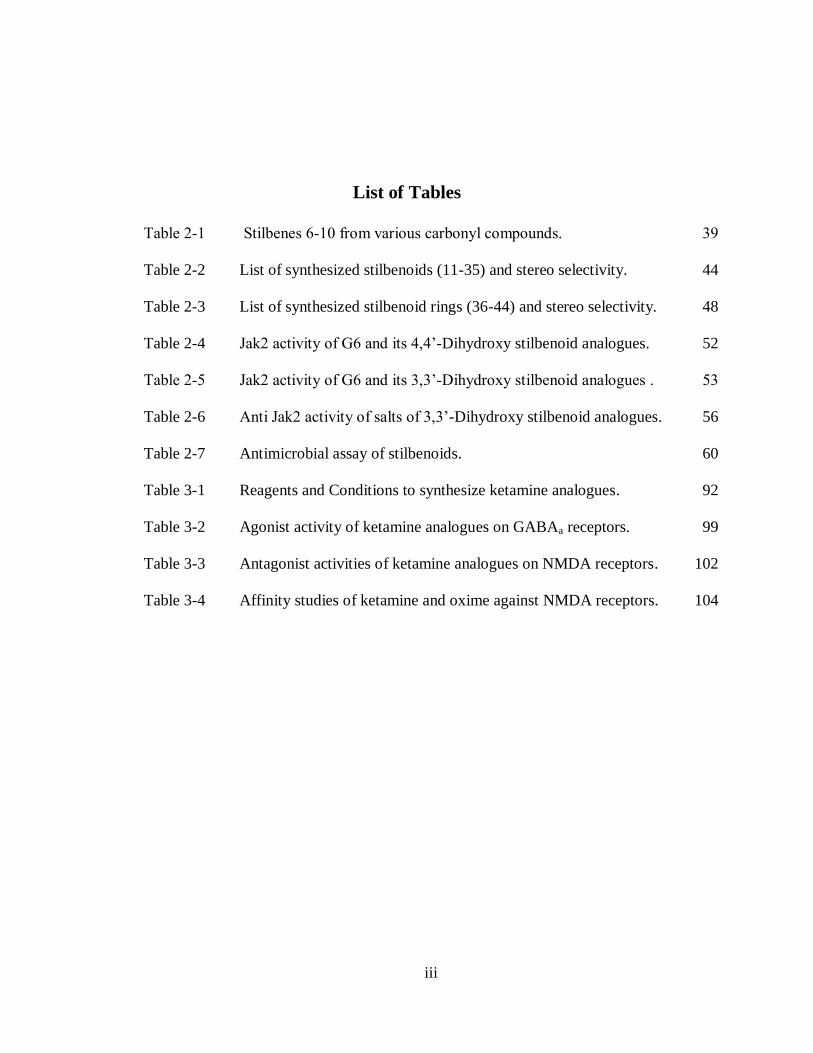
List of Tables
Table 2-1 Stilbenes 6-10 from various carbonyl compounds. 39
Table 2-2 List of synthesized stilbenoids (11-35) and stereo selectivity. 44
Table 2-3 List of synthesized stilbenoid rings (36-44) and stereo selectivity. 48
Table 2-4 Jak2 activity of G6 and its 4,4’-Dihydroxy stilbenoid analogues. 52
Table 2-5 Jak2 activity of G6 and its 3,3’-Dihydroxy stilbenoid analogues . 53
Table 2-6 Anti Jak2 activity of salts of 3,3’-Dihydroxy stilbenoid analogues. 56
Table 2-7 Antimicrobial assay of stilbenoids. 60
Table 3-1 Reagents and Conditions to synthesize ketamine analogues. 92
Table 3-2 Agonist activity of ketamine analogues on GABAa receptors. 99
Table 3-3 Antagonist activities of ketamine analogues on NMDA receptors. 102
Table 3-4 Affinity studies of ketamine and oxime against NMDA receptors. 104
iv
List of Figures
Figure 1-1 Structures of kinase inhibitors 1
Figure 1-2 Structures of Phosphodiesterase-4 inhibitors. 2
Figure 1-3 Structures of Antagonists of NMDA receptors. 3
Figure 1-4 Structures of Agonists of GABAA receptors. 4
Figure 1-5 Structures of Jak2 inhibitors in clinical studies. 5
Figure 1-6 Aminopyrimidine compounds as potent Jak2 inhibitors. 7
Figure 1-7 Benzoxazole compounds as Jak2 inhibitors. 8
Figure 1-8 Recently reported potent Jak2 inhibitors. 9
Figure 1-9 Oxazoles as Phosphodiesterase-4 inhibitors. 11
Figure 1-10 Triazolodiazines as inhibitors of phosphodiesterase 4 inhibitors. 12
Figure 1-11 Diagramme representing GABAA receptor with binding sites for
allosteric modulators 13
Figure 1-12 Benzodiazepine class of compounds as agonist and antagonists of
GABAA Receptors. 14
Figure 1-13 Neurosteroids as agonists of GABAA receptors for anesthesia. 15
Figure 1-14 Barbiturates and other small molecules as agonists for GABAA
receptors. 16
Figure 1-15 Schematic representation of potential sites for drug action within
the NMDAR protein complex. 17
Figure 1-16 Small cycloalkane rings with phenyl substituents as NMDA
antagonists. 18
Figure 2-1 The JAK–STAT pathway 26
v
Figure 2-2 Structures of some Jak2 inhibitors. 27
Figure 2-3 Resveratrol and its analogues as inhibitors of platelet aggregation
induced by Collagen. 28
Figure 2-4 Structures and Antimicrobial activities of Piperidino stilbenes and
Resveratrol analogues against Gram-positive and Gram-
negative bacteria. 29
Figure 2-5 Stilbenes as inhibitors of tubulin assembly and with activity in
prostate Cancer cell lines. 30
Figure 2-6 Tamoxifen and its analogues as antiestrogen compounds for the
treatment of advanced breast cancer. 31
Figure 2-7 Structure of G6 (compound 11). 38
Figure 2-8 1H NMR spectra of Diethyl stilbestrol 6. 40
Figure 2-9 Comparisons of 1H NMR spectrums of 4, 4’-dihydroxy
dimethylstilbene 7 as a mixture and trans isolated. 41
Figure 2-10 Observed stereo selectivity in the synthesized stilbene in Mcmurry
reaction. 42
Figure 2-11 Compound 11 spectrums. (a). 1H NMR spectrum. (b).
13C NMR
spectrum. 43
Figure 2-12 1H NMR spectrums. a. Compound 9. b. Signals for stilbenoid 29. 45
Figure 2-13 1H NMR spectrums. (a). Compound 17. (b). Compound 22. (c).
Compound 34. 46
Figure 2-14 1H NMR spectrum of Compound 39. 47
Figure 2-15 1H NMR spectrums. (a). N-Benzyl stilbenebisoxazine 38. (b). N-
Cyclopentyl stilbenebisoxazine 41. (c). N-(p-methoxybenzyl)
stilbenebisoxazine 43. 49
Figure 2-16 Activity Comparison of 3,3’ and 4,4’-Dihydroxy Stilbenoids. 54
Figure 2-17 Stilbenoids which shows potent activity against JAK2 enzyme. 55
Figure 2-18 Structures of salts of stilbenoids. 55
Figure 2-19 Binding mode of G6 (grey) at the Jak2 (green) vanderwall surface. 57

vi
Figure 2-20 Anti Jak2 enzyme activity of G6 (stilbenoid 11) 58
Figure 2-21 Structures of active stilbenoids and N-Substituted stilbene
bisoxazines. 59
Figure 3-1 General representation of GABAa (Left) and NMDA (Right)
receptors. 85
Figure 3-2 Structures of Ketamine and Phencyclidine (PCP). 87
Figure 3-3 NMR Spectra of Oxime 2. 94
Figure 3-4 NMR Spectra of Methoxime 2. 95
Figure 3-5 NMR Spectra of Benzoxime 4. 96
Figure 3-6 NMR Spectra of Hydroxy 6 (Diastereomeric ratio 1:0.5). 97
Figure 3-7 Agonist activities of ketamine 1 and oxime 2 on GABAa receptors. 100
Figure 3-8 Activity of ketamine analogues against NMDA receptors at 50 M
concentration inhibiting NMDA response. 102
Figure 3-9 % Recovery of response from NMDA receptors post drug (50 M
conc.) treatment. 103
Figure 3-10 % Recovery of response from NMDA receptors after post drug
treatment. 104
Figure 4-1 Structure of Rolipram and its analogue -Lactone. 116
Figure 4-2 NMR Spectra of 1, 3-diester 6. 125
Figure 4-3 NMR Spectra. (a). 1, 3-diol 7. (b). 1, 3-diacetate 8. (c).
monoacetate 9. 127
Figure 4-4 1H NMR spectra of Aldehyde 10. 128
Figure 4-5 1H NMR spectra of (E/Z )-homologated alkene (11). 129
Figure 4-6 1H NMR spectra of Aldehyde 14. 130
Figure 4-7 1H NMR spectra of Ester 17. 131
Figure 4-8 1H NMR spectra of Azide 19. 133

vii
Figure 4-9 13
C NMR spectra. (a). Aryl bromide 5. (b). Aryl iodide 21. 136
Figure 4-10 1H NMR spectra. (a). 1, 4-Diacetate 25. (b). Mixtures of
monoacetates 26 and 26a. 138
Figure 4-11 1H NMR spectra. (a). Carboxylic acid 28. (b). Ester 29. 140
Figure A-1
1H and
13C NMR spectra of E-4, 4’-[1, 2-diethyl-1, 2-ethenediyl]
bis phenol (6). 165
Figure A-2 1H and
13C NMR spectra of E/Z-4, 4’-[1, 2-dimethyl-1, 2-
ethenediyl] bis phenol (7). 166
Figure A-3 1H and
13C NMR spectra of E- 4, 4’-(1, 2-ethenediyl) bis phenol
(8). 167
Figure A-4 1H and
13C NMR spectra of E/Z-3, 3’-[1, 2-dimethyl-1, 2-
ethenediyl] bis phenol (9). 168
Figure A-5 1H and
13C NMR spectra of E-4, 4’-(1, 2-diethyl-1, 2-ethenediyl)
bis [2-[(diethylamino) methyl]-Phenol (11). 169
Figure A-6 1H and
13C NMR spectra of E-4, 4’-(1, 2-diethyl-1, 2-ethenediyl)
bis [2-[(dimethylamino) methyl]-Phenol (12). 170
Figure A-7 1H and
13C NMR spectra of E-4, 4’-(1, 2-diethyl-1, 2-ethenediyl)
bis [2-[(pyrrolidine) methyl]-Phenol (13). 171
Figure A-8 1H and
13C NMR spectra of E-4, 4’-(1, 2-diethyl-1, 2-ethenediyl)
bis [2-[(piperidine) methyl]-Phenol (14). 172
Figure A-9 1H and
13C NMR spectra of E-4, 4’-(1, 2-diethyl-1, 2-ethenediyl)
bis [2-[(morpholine) methyl]-Phenol (15). 173
Figure A-10 1H and
13C NMR spectra of 4, 4’-(1, 2-dimethyl-1, 2-ethenediyl)
bis [2-[(diethylamino) methyl]-Phenol (16). 174
Figure A-11 1H and
13C NMR spectra of 4, 4’-(1, 2-dimethyl-1, 2-ethenediyl)
bis [2-[(dimethylamino) methyl]-Phenol (17). 175
Figure A-12 1H and
13C NMR spectra of 4, 4’-(1, 2-dimethyl-1, 2-ethenediyl)
bis [2-[(pyrrolidine) methyl]-Phenol (18). 176
Figure A-13 1H and
13C NMR spectra of 4, 4’-(1, 2-dimethyl-1, 2-ethenediyl)
bis [2-[(Piperidine) methyl]-Phenol (19). 177
viii
Figure A-14 1H and
13C NMR spectra of E-4, 4’-(1, 2-ethenediyl) bis [2-
[(morpholine) methyl]-Phenol (20). 178
Figure A-15 1H and
13C NMR spectra of E-4, 4’-(1, 2-ethenediyl) bis [2-
[(diethylamino) methyl]-Phenol (21). 179
Figure A-16 1H and
13C NMR spectra of E-4, 4’-(1, 2-ethenediyl) bis [2-
[(dimethylamino) methyl]-Phenol (22). 180
Figure A-17 1H and
13C NMR spectra of E-4, 4’-(1, 2-ethenediyl) bis [2-
[(pyrrolidine) methyl]-Phenol (23). 181
Figure A-18 1H and
13C NMR spectra of E-4, 4’-(1, 2-ethenediyl) bis [2-
[(piperidine) methyl]-Phenol (24). 182
Figure A-19 1H and
13C NMR spectra of E-4,4'-(1,2-ethenediyl)bis[2-
[(morpholine)methyl]-Phenol (25). 183
Figure A-20 1H and
13C NMR spectra of 3,3'-(1,2-dimethyl-1,2-
ethenediyl)bis[2-[(diethylamino)methyl]-Phenol (26). 184
Figure A-21 1H and
13C NMR spectra of 3,3'-(1,2-dimethyl-1,2-
ethenediyl)bis[2-[(dimethylamino)methyl]-Phenol (27). 185
Figure A-22 1H and
13C NMR spectra of 3,3'-(1,2-dimethyl-1,2-
ethenediyl)bis[2-[(pyrrolidine)methyl]-Phenol (28). 186
Figure A-23 1H and
13C NMR spectra of 3,3'-(1,2-dimethyl-1,2-
ethenediyl)bis[2-[(piperidine)methyl]-Phenol (29). 187
Figure A-24 1H and
13C NMR spectra of 3,3'-(1,2-dimethyl-1,2-
ethenediyl)bis[2-[(morpholine)methyl]-Phenol (30). 188
Figure A-25 1H and
13C NMR spectra of E-3,3'-(1,2-ethenediyl)bis[2-
[(diethylamino)methyl]-Phenol (31). 189
Figure A-26 1H and
13C NMR spectra of E-3, 3’-(1, 2-ethenediyl) bis [2-
[(dimethylamino) methyl]-Phenol (32). 190
Figure A-27 1H and
13C NMR spectra of E-3, 3’-(1, 2-ethenediyl) bis [2-
[(pyrrolidine) methyl]-Phenol (33). 191
Figure A-28 1H and
13C NMR spectra of E-3, 3’-(1, 2-ethenediyl) bis [2-
[(piperidine) methyl]-Phenol (34). 192

ix
Figure A-29 1H and
13C NMR spectra of E-3, 3’-(1, 2-ethenediyl) bis [2-
[(morpholine) methyl]-Phenol (35). 193
Figure A-30 1H and
13C NMR spectra of E-6, 6’-(vinylene) bis [3-cyclohexyl-3,
4-dihydro- 2H-1, 3-Benzoxazine] (36). 194
Figure A-31 1H and
13C NMR spectra of E-6, 6’-(vinylene) bis [3-cyclopentyl-
3, 4-dihydro- 2H-1, 3-Benzoxazine] (37). 195
Figure A-32 1H and
13C NMR spectra of E-6, 6’-(vinylene) bis [3-benzyl-3,4-
dihydro- 2H-1,3-Benzoxazine] (38). 196
Figure A-33 1H and
13C NMR spectra of E-6,6'-(vinylene)bis[3-(4-methoxy
benzyl)-3,4-dihydro- 2H-1,3-Benzoxazine] (39). 197
Figure A-34 1H and
13C NMR spectra of E-7,7'-(vinylene)bis[3-cyclohexyl-3,4-
dihydro- 2H-1,3-Benzoxazine] (40). 198
Figure A-35 1H and
13C NMR spectra of E-7,7'-(vinylene)bis[3-cyclopentyl-
3,4-dihydro- 2H-1,3-Benzoxazine] (41). 199
Figure A-36 1H and
13C NMR spectra of E-7,7'-(vinylene)bis[3-benzyl-3,4-
dihydro- 2H-1,3-Benzoxazine] (42). 200
Figure A-37 1H and
13C NMR spectra of E-7,7'-(vinylene)bis[3-(4-methoxy
benzyl)-3,4-dihydro- 2H-1,3-Benzoxazine] (43). 201
Figure A-38 1H and
13C NMR spectra of E-7,7'-(vinylene)bis[3-furfuryl-3,4-
dihydro- 2H-1,3-Benzoxazine] (44). 202
Figure B-1 1H and
13C-NMR spectra of 2-(2-Chloro-phenyl)-2-methylamino-
cyclohexanone oxime (2). 203
Figure B-2 1H and
13C-NMR spectra of 2-(2-Chloro-phenyl)-2-methylamino-
cyclohexanone O-methyl-oxime (3). 204
Figure B-3 1H and
13C-NMR spectra of 2-(2-Chloro-phenyl)-2-methylamino-
cyclohexanone O-benzyl-oxime (4). 205
Figure B-4 1H and
13C-NMR spectra of 2-(2-Chloro-phenyl)-2-methylamino-
cyclohexanone O-semicarbazone (5). 206
Figure B-5 1H and
13C-NMR spectra of 2-(2-Chloro-phenyl)-2-methylamino-
cyclohexanol (6). 207
x
Figure C-1 1H and
13C-NMR spectra of Acetic acid 2-methoxy-phenyl ester
(2). 208
Figure C-2 1H and
13C-NMR spectra of Acetic acid 5-bromo-2-methoxy-
phenyl ester (3). 209
Figure C-3 1H and
13C-NMR spectra of 5-Bromo-2-methoxy-phenol (4). 210
Figure C-4 1H and
13C-NMR spectra of 4-bromo-2-(cyclopentyloxy)-1-
methoxybenzene (5). 211
Figure C-5 1H and
13C-NMR spectra of 2-(3-Cyclopentyloxy-4-methoxy-
phenyl)-malonic acid diethyl ester (6). 212
Figure C-6 1H and DEPT-NMR spectra of 2-(3-Cyclopentyloxy-4-methoxy-
phenyl)-propane-1,3-diol (7). 213
Figure C-7 1H NMR spectra of Acetic acid 3-acetoxy-2-(3-cyclopentyloxy-4-
methoxy-phenyl)propyl ester (8) and 1H -NMR spectra of
Acetic acid-2-(3-cyclopentyloxy-4-methoxy-phenyl)-3-
hydroxy-propyl ester (9). 214
Figure C-8: 1H and 13C-NMR spectra of Acetic acid 2-(3-cyclopentyloxy-4-
methoxy-phenyl)-3-oxo-propyl ester (10). 215
Figure C-9 1H NMR spectra of (E/Z)-Acetic acid 2-(3-cyclopentyloxy-4-
methoxy-phenyl)-4-methoxy-but-3-enyl ester (11) and 1H -
NMR spectra of (E/Z)-2-(3-Cyclopentyloxy-4-methoxy-
phenyl)-4-methoxy-but-3-en-1-ol (12). 216
Figure C-10 1H NMR spectra of 4-(1-Benzyloxymethyl-3-methoxy-allyl)-2-
cyclopentyloxy-1-methoxy-benzene (13) and 1H -NMR spectra
of 4-Benzyloxy-3-(3-cyclopentyloxy-4-methoxy-phenyl)-
butyraldehyde (14). 217
Figure C-11 1H NMR spectra of 4-Benzyloxy-3-(3-cyclopentyloxy-4-methoxy-
phenyl)-butyric acid (15) and 1H -NMR spectra of 4-
Benzyloxy-3-(3-cyclopentyloxy-4-methoxy-phenyl)-butyric
acid ethyl ester (16). 218
Figure C-12 1H NMR spectra of 3-(3-Cyclopentyloxy-4-methoxy-phenyl)-4-
hydroxy-butyric acid ethyl ester (17) and 1H -NMR spectra of
4-Bromo-3-(3-cyclopentyloxy-4-methoxy-phenyl)-butyric acid
ethyl ester (18). 219

xi
Figure C-13 DEPT- NMR spectra of 4-Bromo-3-(3-cyclopentyloxy-4-methoxy-
phenyl)-butyric acid ethyl ester (18) and 1H -NMR spectra of
4-Azido-3-(3-cyclopentyloxy-4-methoxy-phenyl)-butyric acid
ethyl ester (19). 220
Figure C-14 DEPT- NMR spectra of 4-Azido-3-(3-cyclopentyloxy-4-methoxy-
phenyl)-butyric acid ethyl ester (19). 221
Figure C-15 1H and
13C-NMR spectra of 2-Cyclopentyloxy-4-iodo-1-methoxy-
benzene (21). 222
Figure C-16 1H and
13C-NMR spectra of 2-(3-Cyclopentyloxy-4-methoxy-
phenyl)-but-2-enedioic acid diethyl ester (22). 223
Figure C-17 1H NMR spectra of 2-(3-Cyclopentyloxy-4-methoxy-phenyl)-
succinic acid diethyl ester (23) and 1H -NMR spectra of 2-(3-
Cyclopentyloxy-4-methoxy-phenyl)-butane-1,4-diol (24). 224
Figure C-18 13
C -NMR spectra of 2-(3-Cyclopentyloxy-4-methoxy-phenyl)-
butane-1,4-diol (24) and 1H -NMR spectra of Acetic acid 4-
acetoxy-2-(3-cyclopentyloxy-4-methoxy-phenyl)-butyl ester
(25). 225
Figure C-19 13
C -NMR spectra of Acetic acid 4-acetoxy-2-(3-cyclopentyloxy-4-
methoxy-phenyl)-butyl ester (25). 226
Figure C-20 1H and
13C-NMR spectra of Acetic acid 2-(3-cyclopentyloxy-4-
methoxy-phenyl)-4-hydroxy-butyl ester (26). 227
Figure C-21 1H and
13C-NMR spectra of Acetic acid 2-(3-cyclopentyloxy-4-
methoxy-phenyl)-4-oxo-butyl ester (27). 228
Figure C-22 1H and
13C-NMR spectra of 4-Acetoxy-3-(3-cyclopentyloxy-4-
methoxy-phenyl)-butyric acid (28). 229
Figure C-23 1H and
13C-NMR spectra of 4-Acetoxy-3-(3-cyclopentyloxy-4-
methoxy-phenyl)-butyric acid methyl ester (29). 230
Figure C-24 1H NMR spectra of 4-(3-Cyclopentyloxy-4-methoxy-phenyl)-
dihydro-furan-2-one (30). 231

xii
List of Schemes
Scheme 1-1. Synthesis of various pyrimidine analogues by Burns et. al. 7
Scheme 1-2. Synthesis of various benzoxazole analogues by Gespacher et. al. 8
Scheme 1-3. Synthesis of Oxazole compounds as potent Phosphodiesterase-4
inhibitors. 11
Scheme 1-4. Synthesis of Triazolodiazines in two steps by skoumbourdis et. al. 12
Scheme 1-5. Chlordiazepoxide 48 synthesis by sternback rearrangement. 15
Scheme 2-1. Synthesis of E-Resveratrol by Guiso et. al.via heck reaction. 32
Scheme 2-2. Synthesis of Stilbenes by sengupta et. al.via heck reaction. 32
Scheme 2-3. Synthesis of Stilbenes by Andrus et. al.via suzuki reaction. 33
Scheme 2-4. Synthesis of Stilbenes by Nishibayashi et. al.via Stille coupling. 33
Scheme 2-5. Synthesis of Stilbenes by Velder et. al. via cross-metathesis. 34
Scheme 2-6. Synthesis of (Z)-Stilbenes by Pettit et. al. via wittig reaction. 35
Scheme 2-7. Synthesis of (Z)-Stilbenes by Heynekamp72
et. al.via Horner-
Emmons-Wadsworth reaction. 35
Scheme 2-8. Observed stereoselectivity in the synthesis of (Z)-4-
Hydroxytamoxifen. 36
Scheme 2-9. Synthesis of (Z)-4-Hydroxytamoxifen by Gautheir et. al. 52
via
Mcmurry reaction with modified reaction conditions. 36
Scheme 2-10. Synthesis of stilbenoids from ketones and aldehydes. 38
Scheme 2-11. Reagents and Conditions. i. TiCl4, Zn, Dry. THF, reflux. 39
Scheme 2-12. Reagents and Conditions. (a). amine, paraformaldehyde, MeOH,
reflux. 43

xiii
Scheme 2-13. Reagents and Conditions. (a). amine, paraformaldehyde, MeOH,
reflux. 48
Scheme 3-1. Synthesis of tetralone analogues of ketamine by Yang et. al. 88
Scheme 3-2. Synthesis of lactam with methylamine analogues of ketamine by
arantonello et. al. 89
Scheme 3-3. Synthesis of lactam with piperidine analogues of ketamine by
Zarantonello et. al. 90
Scheme 3-4. Synthesis of ketamine analogue with phenyl group substitution. 91
Scheme 3-5. Synthesis of ketamine analogues. 91
Scheme 4-1. Garcia et. al synthesis of (±)-Rolipram via Heck-Matsuda arylation
of 3-pyrroline. 117
Scheme 4-2. Demnitz et. al synthesis of (R) & (S)-Rolipram via
chromatographic separation of two diastereomers. 117
Scheme 4-3. Paraskar et. al synthesis of (R)-Rolipram via chiral bis-oxozoline
ligand, NaBH4 and CoCl2. 118
Scheme 4-4. Hynes et. al synthesis of (R)-Rolipram via Michael addition. 119
Scheme 4-5. Mulzer et. al synthesis of (R)-Rolipram via Michael addition. 120
Scheme 4-6. Yoon et. al synthesis of (±)-Rolipram via Rhodium catalyzed C-H
insertion. 120
Scheme 4-7. Honda et. al synthesis of (R)-Rolipram via -Aryl--
butyrolactone. 121
Scheme 4-8. Mandai et. al synthesis of -Aryl--butyrolactone. 121
Scheme 4-9. Retrosynthetic analysis for the synthesis of -Lactam or (±)-
Rolipram. 123
Scheme 4-10. Synthesis of Aryl Bromide from Guaiacol 123
Scheme 4-11. Synthesis of 1,3-diol 7 via 1,3-diester 6. 125
Scheme 4-12. Synthesis of monoacetate via selective acylation of 1,3-Diol. 126
Scheme 4-13. Synthesis of Alkene 13 from monoacetate 9 via homologation. 128

xiv
Scheme 4-14. Synthesis of Ester alcohol 17. 130
Scheme 4-15. Synthesis of azide for the Rolipram from Esteralcohol 17. 132
Scheme 4-16. Retrosynthetic analysis for the synthesis of Lactone (30) 134
Scheme 4-17. Synthesis of 1,4-Diol 24 via Heck arylation of diethylmaleate. 135
Scheme 4-18. Synthesis of Monoacetate 26 via Regioselective deacylation of 1,4-
Diacetate by Lipase Ps-30 (Lipase Pseudomonas Sepacia). 137
Scheme 4-19. Synthesis of Ester acetate 29 from monoacetate 26. 139
Scheme 4-20. Synthesis of -Lactone 30 via Ester acetate 29. 141

xv
List of Symbols and Abbreviations
δ Chemical shift in parts per million units
1H Isotope of hydrogen with mass of 1 amu
13
C Isotope of carbon with mass of 13 amu
Ac acetyl
Ac2O acetic anhydride
AcOH acetic acid
Bn benzyl
BuLi butyl lithium
CDCl3 Deuterated chloroform
CH3OD Deuterated methanol
CNS central nervous system
COSY COrrelation SpectroscopY
DCM dichloromethane
DEPT Distortionless Enhancement by Polarization Transfer
DEPT-135 Distortionless Enhancement by Polarization Transfer at a flip angle
of 135 degrees
DMAP N, N-dimethyl aminopyridine
DME 1,2-dimethoxyethane (glyme)
DMF dimethylformamide
DMSO dimethylsulfoxide
DMSO-d6 Deuterated dimethylsulfoxide
xvi
e. e enantiomeric excess
Et ethyl
EtOAc ethyl acetate
EA ethyl acetate
EtOH ethanol
FDA Food and Drug Administration
GABA -Amino butyric acid
Hz a unit of frequency defined as the number of cycles per second
MHz Megahertz
hrs hour(s)
HPLC high pressure (performance) liquid chromatography
HTS high throughput screening
iPr isopropyl
iPrOH isopropanol
LAH lithium aluminum hydride
LC-MS liquid chromatography-mass spectrometry
Me methyl
MeOH methanol
NaH Sodium hydride
NMDA N-Methyl-D-Aspartate
NMR nuclear magnetic resonance
Novozyme 435 Lipase from Candida Antarctica
Pd Palladium

xvii
Ph phenyl
PPh3 triphenylphosphine
Ppm parts per million
PPTS Pyridinium p-toluene sulfonate
Pr propyl
Ps-30 Pseudomonas Sepacia Lipase
PTSA p-Toluene sulfonic acid
Pyr pyridine
R R enantiomer
rt room temperature
S S enantiomer
SAR structure activity relationship
SPR structure property relationship
tBu tert-butyl
TEA triethylamine
TFA trifluoroacetic acid
THF tetrahydrofuran
TLC thin layer chromatography
TMS Tetramethylsilane
USF University of South Florida
WHO World Health Organization
xviii
Abstract
Stilbenoids possess a wide range of biological properties such as, anticancer,
antiplatelet aggregation, antiestrogenic, antibacterial, antifungal and antiatherogenic, etc.
Owing to these therapeutic values, a great deal of attention attracted in the synthesis of
derivatives of stilbenes. During the course of the study, G6 a novel stilbenoid was
discovered, through high throughput screening, to be a potent inhibitor of mutated JAK2-
V617F. The mutated JAK2 variant has been implicated in various myeloproliferative
disorders (MPDs) including polycythemia vera (PV), essential thrombocythemia (ET)
and primary myelofibrosis (PMF) has been targeted by therapeutics. Chapter 2 describes
the synthesis of analogs of the stilbenoid G6 and N-substituted stilbenes bisoxazines by
utilizing Mcmurry reaction and Mannich condensation methods. The main emphasis of
this work is to develop novel stilbenoids as inhibitors of JAK2-V617F mutated Jak2
enzyme in Human erythroleukemia cells (HEL) since this mutation is discovered in the
majority of patients with myeloproliferative disorders (MPDs). Using Mcmurry reaction,
five novel trans-hydroxystilbenes have been synthesized from carbonyl compounds.
Subsequently using Mannich coupling with five secondary amines and five primary
amines, 25 novel stilbenoids and 9 novel N-substituted stilbene bisoxazines have been
synthesized. In HEL cell assay, 8 stilbenoid analogues have been identified as potent
inhibitors of Jak2 enzyme.

xix
Chapter 3 describes the modification of ketamine structurally for the synthesis of
novel analogues to study for their agonist activity at GABAA receptors and antagonist
activity at NMDA receptors. Ligand gated ion channels like GABAA and NMDA
receptors are membrane-embedded proteins at synaptic cleft which controls
intercommunication among neurons and plays an important role in motor control activity,
learning. GABAA receptors are responsible for inhibitory action potentials while NMDA
receptors are responsible for excitory action potentials. Ketamine, known as dissociative
anesthetic, produces profound analgesia at low doses to a unique cardiovascular
stimulation and a cataleptic state at higher doses with dose dependent side effects like
vivid dreams, disruptions of cognitive functions. The main emphasis of this work is the
synthesis of novel analogues of ketamine by transforming carbonyl group in ketamine to
imine functionality with small to bulkier groups and to identify an analogue of ketamine
which is highly potent in its activity at the both GABAA and NMDA receptors and
improved clinical actions. Studies of analogues activity against GABAA subtypes 62,
122 receptors and NMDA subtypes NR1/2A, NR1/2B, NR1/2D receptors have been
described.
Chapter 4 describes the formal synthesis of (±)-Rolipram and the
chemoenzymatic synthesis of -aryl--lactone, a Rolipram analogue. The key steps, Pd
catalyzed arylation of diethylmalonate and the efficient use of selective acylation of 1, 3-
diol entails the formal synthesis of (±)-Rolipram. The regioselective deacylation of -
aryl-1, 4-diacetate by lipase Pseudomonas Sepacia entails the formation of -aryl--
lactone. The efficient use of various methods including halogen exchange, Heck arylation

xx
of diethylmaleate and lactonization for the synthesis of -aryl--lactone have been
discussed. The present work provides an efficient and general route to -lactones.

1
Chapter 1
A short review of small molecule inhibitors of Janus Kinase 2, Phosphodiesterase
IV, and GABAA and NMDA receptors
1.1 Significance of small molecule inhibitors.
A number of small molecule inhibitors1 have been discovered and are currently
used in therapy of many diseases including cancer2 and inflammation.
3 The successful
treatment of chronic myeloid leukemia by imitinib4 (1, Fig. 1-1) among other different
types of cancers and treatment of non small lung cancer (NSCLC) by gefitinib (2)5 and
erlotinib (3)6 serves as examples that small molecule inhibitors can be effective drugs.
Currently, Most of the drug discovery efforts are focused on developing small molecule
inhibitors with exquisite selectivity, specificity, affinity for binding with proteins and to
identify off-target interactions. Small molecule inhibitors have a greater advantage for the
elucidation of the mechanism of cellular processes and so are very useful tools. Since
they are more stable than peptide inhibitors and are often easily cell permeable, use of
them as inhibitors of protein-protein interactions7 is particularly helpful.
Fig. 1-1. Structures of kinase inhibitors

2
Imitinib 1 (Fig. 1-1) is a potent inhibitor of three tyrosine kinases: ABL
(Ableson), platelet-derived growth factor receptor (PDGFR) and KIT. The main feature
that prompted the use of imitinib in clinical trials8 is its inhibitory action against ABL
tyrosine kinase. Constitutively activated Janus Kinase 2 (Jak2) has been implicated in
myeloproliferative neoplasms10
(MPNs), i.e. polycythemia
vera (PV), essential
thrombocythemia (ET) and primary myelofibrosis (PMF). A large number of patients
with myeloproliferative neoplasms (MPNs) have mutated JAK2-V617F,11
which
prompted the development of Jak2 inhibitors12
for the treatment of MPNs. Several groups
are working on Jak2 inhibitors with favourable pharmacokinetics and toxicity profiles
which are good for clinical evaluation.
Fig. 1-2. Structures of Phosphodiesterase-4 inhibitors.
Phosphodiesterase-4 (PDE4), an enzyme belonging to the family of eleven
phosphodiesterase isoenzymes identified so far, has absolute specificity for cyclic
adenosine-3’,5’-monophosphate (cAMP) and is considered potential therapeutic targets13
for the treatment of chronic inflammatory disorders,14
such as chronic obstructive
pulmonary disease (COPD).15
PDE4 is abundant and is the major regulator of cAMP
metabolism16
in almost every proinflammatory and immune cell. PDE4 inhibitors, of
varied structural classes, suppress a myriad of in vitro responses, such as proliferation
and the generation and/or release of histamine.

3
For example small molecule Cilomilast17
(4) (Fig. 1-2), a selective
phosphodiesterase-4 inhibitor, has demonstrated encouraging therapeutic efficacy in
clinical trials of COPD. Now, several other selective PDE4 inhibitors are in clinical trials
such as tetomilast18
(5), roflumilast19
(6) (Fig. 1-2).
Antagonists of NMDA receptors20
are currently under constant development to
treat neurogenerative disorders such as Parkinson’s, Alzheimer’s, Huntington’s diseases,
cerebral ischemia, epilepsy, neuropathic pain, brain stroke and multiple sclerosis21
. The
rational for design of inhibitors is strongest for the acute treatment of ischemia22
. A
majority of NMDA antagonists developed so far have adverse effects such as
hallucinations, increase in blood pressure, catatonia and anesthesia. Fortunately, all of the
side effects so far identified are dose-limiting. A number of potent small molecule
NMDA antagonists have been developed. For example, Ifenprodil23
(7) proved to be
neuroprotective in animal models of stroke without the severe side effects of earlier
drugs.
Fig. 1-3. Structures of Antagonists of NMDA receptors.
4
Various animal models also revealed that Ifenprodil (7) is beneficial and
significantly superior in neuroprotection of Anoxia (Hypoxia), Seizures, Ischemia and
brain injury. Traxoprodil24
(8) (Fig. 1-3) is another small molecule NMDA antagonist
which also has neuroprotective, analgesic and anti-Parkinson’s effects in animal studies
although human studies proved less beneficial for the treatment of brain injury after
stroke. The clinical trial, however, revealed another important therapeutic benefit of
Traxoprodil (8), as a rapid acting antidepressant. NMDA subtype selectivity has been the
driving force for development of small molecule antagonist; similar to that of
benzodiazepines (such as Valium 9) which enhance activation of GABAA receptors.
Several small molecules have also been developed which are agonists for GABAA
receptors with subtype selectivity such as sedatives, hypnotics, anxiolytics, anxiogenic,
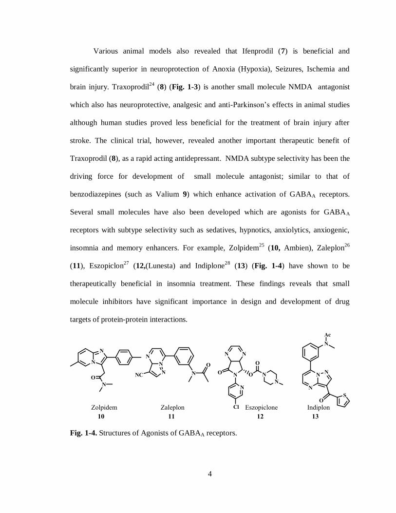
insomnia and memory enhancers. For example, Zolpidem25
(10, Ambien), Zaleplon26
(11), Eszopiclon27
(12,(Lunesta) and Indiplone28
(13) (Fig. 1-4) have shown to be
therapeutically beneficial in insomnia treatment. These findings reveals that small
molecule inhibitors have significant importance in design and development of drug
targets of protein-protein interactions.
Fig. 1-4. Structures of Agonists of GABAA receptors.

5
1.2. Small molecules as Jak2 inhibitors.
Small-molecule kinase inhibitors most commonly target the highly conserved
adenosine triphosphate (ATP)-binding domain in enzyme. Since the discovery of the
mutation JAK2-V617F29
in Jak2 and its implication in myeloproliferative neoplasms30
(MPNs), many number of Jak2 inhibitors have been developed and some are under
clinical trials. Majority of them are small molecules.
Pyrrolopyrimidine
INCB018424 (14 in Fig. 1-5) has shown potent inhibition activity against Jak2
with IC50 of 2.8 nM. However, the selectivity has been an issue and it has also shown
potent to moderate activity against Jak1, Tyk2 kinase proteins.
Fig. 1-5. Structures of Jak2 inhibitors in clinical studies.
6
INCB01842431
(14) treatment of murine model of JAK2-V617F driven
malignancy resulted in significant attenuation
of spleen growth and significantly
increased mice survival compared with mice treated with vehicle alone. These inhibitors
belongs to pyrrolopyrimidine class compounds.
Another clinical candidate CP-690,55033
(16 in Fig. 1-5) for rheumatoid arithritis,
also been shown to have potent inhibition activity against Jak2 enzyme with IC50 of 20
nM. It has also shown higher degree of selectivity for Jak2 over Jak1/3 enzymes. This
inhibitor has been identified as a lead candidate at Pfizer for rheumatoid arthritis
treatment from a screening of over 400,000 small molecules library. Interestingly this
compound, like INCB018424 (14 in Fig. 1-5) also belongs to the pyrrolopyrimidines
class of molecules. Based on this pyrrolopyrimidine ring system, several structure-
activity studies have revealed other potent Jak family inhibitors.
Aminopyrimidines
Extensive in vitro and in vivo studies have revealed an interesting
aminopyrimidine class compound TG10134834
(17 in Fig. 1-5) as a potent Jak2 inhibitor
with IC50 of 3 nM. This inhibitor also turned out to be highly selective for Jak2 when
profiled against over 230 kinases.
The potency and promising clinical efficacy reported for TG101348 (17) has led
to structure-activity relationship investigations and new compounds have been developed.
For example CYT-38735
18 (Fig. 1-6) has been recently found to be another potent Jak2
inhibitor which belongs to the aminopyrimidine class of compounds. Series of other
aminopyrimidines (19-25) developed in structure-active relationship studies35
with CYT-
387 (19) have shown high potency than 19 (Fig. 1-6).

7
Fig. 1-6. Aminopyrimidine compounds as potent Jak2 inhibitors.
Synthesis of these aminopyrimidines (19-25) involves two steps, starting from
various phenyl boronicacids. Regioselective cross-coupling of phenylboronic acids with
pyrimidine dichloride and condensation with anilines or palladium catalyzed Buchwald
reaction yields various aminopyrimidines (19-25) as shown in Scheme 1-1.
Scheme 1-1. Synthesis of various pyrimidine analogues by Burns et. al.

8
Benzoxazoles
Benzoxazole compounds also have shown attractive Jak2 inhibitory profiles in
cellular assays. For example novel benzoxazole 26-31 (Fig. 1-7) synthesized by
Gerspacher et. al.36
showed high inhibitory potency against Jak2 with IC50 values in 3.0
to 8.0 nM range. Synthesis of the benzoxazoles 26-31 was reported to be relatively
straightforward and convergent (Scheme 1-2).
Scheme 1-2. Synthesis of various benzoxazole analogues by Gespacher et. al.
Fig. 1-7. Benzoxazole compounds as Jak2 inhibitors.

9
Miscellaneous
Apart from aminopyrimidines and benzoxazoles, small molecules belonging to
various other scaffolds such as Tricyclic pyridones, pyrrolotriazines, purines,
aminopyrazolopyrimidines, monocyclic pyridines and pyrimidines (Fig. 1-8) have also
shown promising therapeutic efficiency in anti Jak2 activity. For example, INCB1656232
15 (Fig. 1-5) is a potent inhibitor of Jak2 enzyme with IC50 of 0.25 nM. In a combination
studies, the growth of myeloma xenografts in mice was suppressed and antitumor activity
was enhanced by oral administration of INCB16562 (15) which belongs to the pyridine
class of compounds.
Fig. 1-8. Recently reported potent Jak2 inhibitors.
10
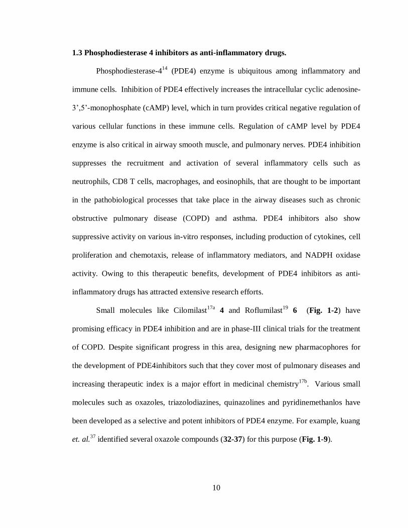
1.3 Phosphodiesterase 4 inhibitors as anti-inflammatory drugs.
Phosphodiesterase-414
(PDE4) enzyme is ubiquitous among inflammatory and
immune cells. Inhibition of PDE4 effectively increases the intracellular cyclic adenosine-
3’,5’-monophosphate (cAMP) level, which in turn provides critical negative regulation of
various cellular functions in these immune cells. Regulation of cAMP level by PDE4
enzyme is also critical in airway smooth muscle, and pulmonary nerves. PDE4 inhibition
suppresses the recruitment and activation of several inflammatory cells such as
neutrophils, CD8 T cells, macrophages, and eosinophils, that are thought to be important
in the pathobiological processes that take place in the airway diseases such as chronic
obstructive pulmonary disease (COPD) and asthma. PDE4 inhibitors also show
suppressive activity on various in-vitro responses, including production of cytokines, cell
proliferation and chemotaxis, release of inflammatory mediators, and NADPH oxidase
activity. Owing to this therapeutic benefits, development of PDE4 inhibitors as anti-
inflammatory drugs has attracted extensive research efforts.
Small molecules like Cilomilast17a
4 and Roflumilast19
6 (Fig. 1-2) have
promising efficacy in PDE4 inhibition and are in phase-III clinical trials for the treatment
of COPD. Despite significant progress in this area, designing new pharmacophores for
the development of PDE4inhibitors such that they cover most of pulmonary diseases and
increasing therapeutic index is a major effort in medicinal chemistry17b
. Various small
molecules such as oxazoles, triazolodiazines, quinazolines and pyridinemethanlos have
been developed as a selective and potent inhibitors of PDE4 enzyme. For example, kuang
et. al.37
identified several oxazole compounds (32-37) for this purpose (Fig. 1-9).
11
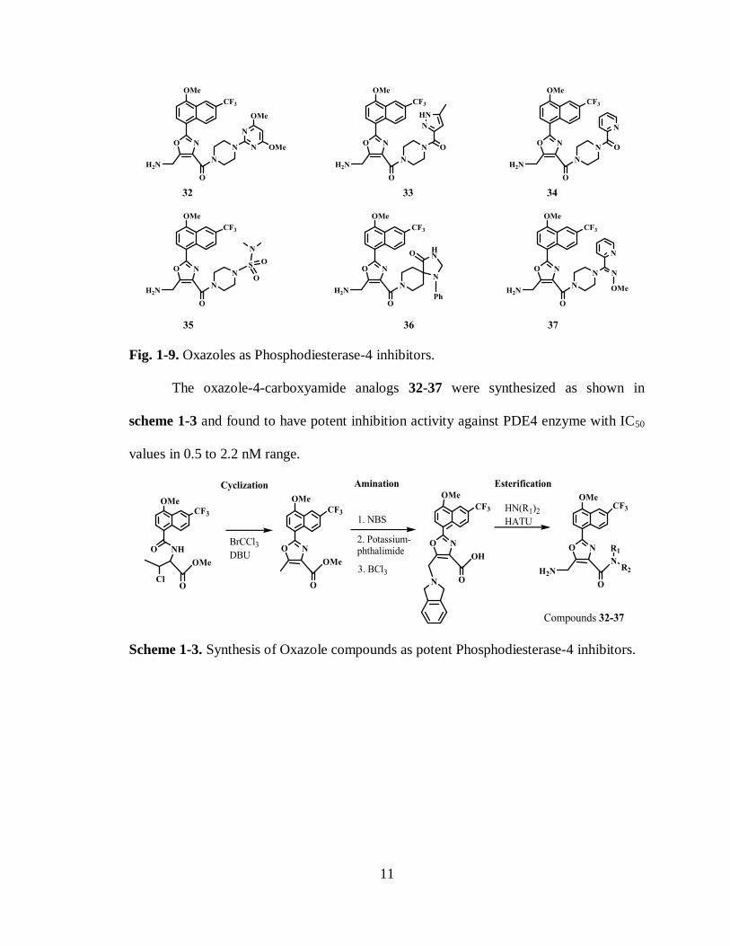
Fig. 1-9. Oxazoles as Phosphodiesterase-4 inhibitors.
The oxazole-4-carboxyamide analogs 32-37 were synthesized as shown in
scheme 1-3 and found to have potent inhibition activity against PDE4 enzyme with IC50
values in 0.5 to 2.2 nM range.
Scheme 1-3. Synthesis of Oxazole compounds as potent Phosphodiesterase-4 inhibitors.
12
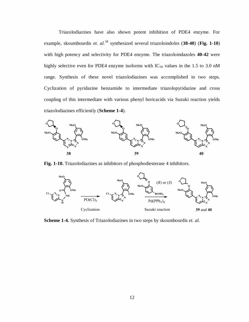
Triazolodiazines have also shown potent inhibition of PDE4 enzyme. For
example, skoumbourdis et. al.38
synthesized several triazoloindoles (38-40) (Fig. 1-10)
with high potency and selectivity for PDE4 enzyme. The triazoloindazoles 40-42 were
highly selective even for PDE4 enzyme isoforms with IC50 values in the 1.5 to 3.0 nM
range. Synthesis of these novel triazolodiazines was accomplished in two steps.
Cyclization of pyridazine benzamide to intermediate triazolopyridazine and cross
coupling of this intermediate with various phenyl boricacids via Suzuki reaction yields
triazolodiazines efficiently (Scheme 1-4).
Fig. 1-10. Triazolodiazines as inhibitors of phosphodiesterase 4 inhibitors.
Scheme 1-4. Synthesis of Triazolodiazines in two steps by skoumbourdis et. al.

13
1.4 Small molecules as agonists of GABAA receptors
GABAA receptors have been therapeutic target39
of many drugs. Majority of them
are small molecules which interact with GABAA receptors as allosteric modulators. For
example, benzodiazepines, barbiturates and neurosteroids interact at these receptors.
GABAA receptors are functionally characterized as ligand gated Cl- ion channels with a
GABA (-amino butyric acid) recognition site. Normally, GABA binds to GABAA
receptor fecilitating the Cl- ion flux into the cells leading to inhibitory neurotransmission.
These receptors also have binding sites for external modulators called allosteric
modulators.
Fig. 1-11. Diagramme representing GABAA receptor with binding sites for allosteric
modulators (Reprinted from Richards. G, Schoch. P, Haefely. W; Seminars in
Neurosciences. 1991, 3, 191. with permission from Elsevier).

14
Allosteric modulators typically modulates Cl- ion flux along with GABA at these
receptors resulting many beneficial effects for the treatment of anxiety, insomnia,
agitation, seizures, muscle spasms, alcohol withdrawal and premedication for medical,
dental procedures. Thus barbiturates, benzodiazepines, neurosteroids and their analogues
have been developed as small molecule agonists and inverse agonists for therapeutic
benefits. Benzodiazepines40
such as diazepam 41, midazolam 42, lorazepam 43,
oxazepam 44 and quazepam 45 (Fig. 1-12) are extensively used as sedatives, hypnotics
and anticonvulsants which enhance the effect of GABA potentiation at GABAA receptors
and thus characterized as agonists or positive modulators. Flumazenil 46 and Imidazenil
47 (Fig. 1-12) are other benzodiazepines which acts as antagonists mainly and reverse the
effects of benzodiazepines by competitive inhibition at their binding site on GABAA
receptors and thus used as antidote for benzodiazepine overdose. Benzodiazepines are
heterocycles derived by fusion of benzene and 1, 4-diazepines.
Fig. 1-12. Benzodiazepine class of compounds as agonist and antagonists of GABAA
Receptors.
15
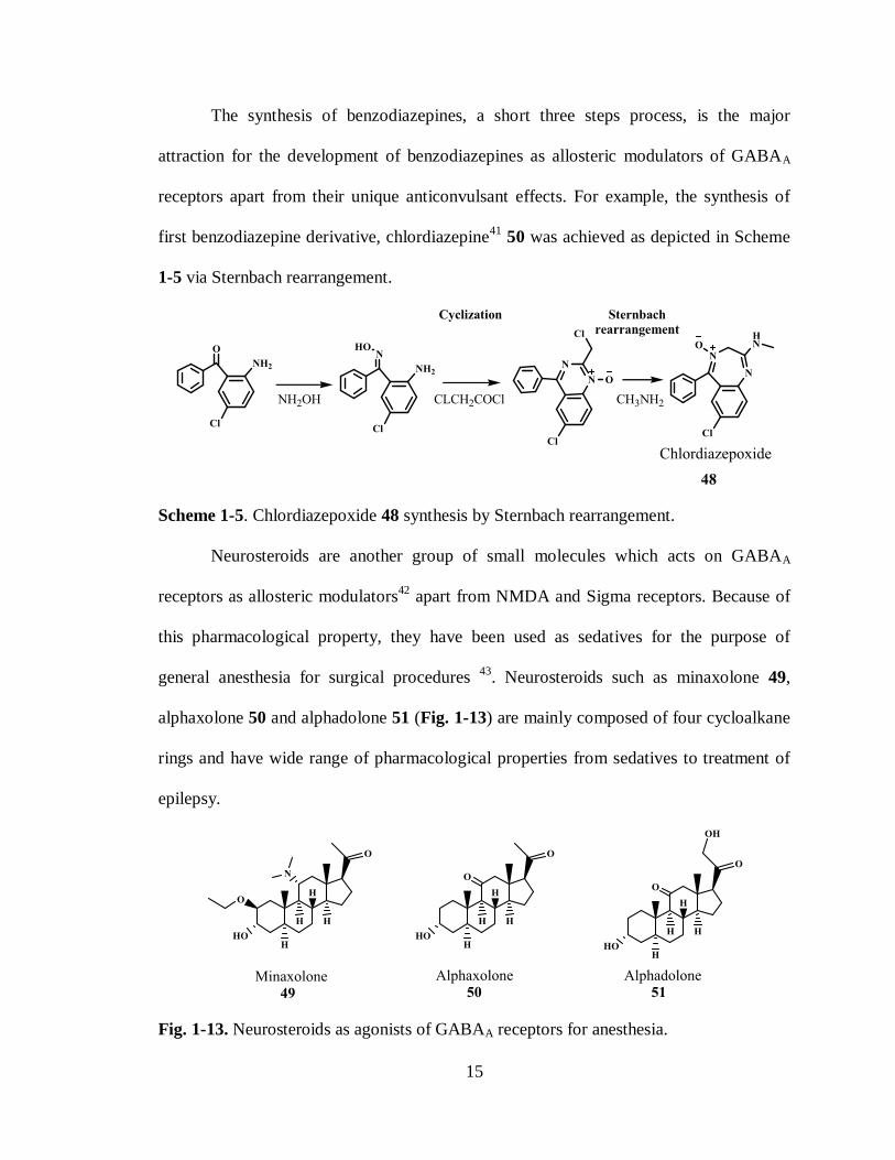
The synthesis of benzodiazepines, a short three steps process, is the major
attraction for the development of benzodiazepines as allosteric modulators of GABAA
receptors apart from their unique anticonvulsant effects. For example, the synthesis of
first benzodiazepine derivative, chlordiazepine41
50 was achieved as depicted in Scheme
1-5 via Sternbach rearrangement.
Scheme 1-5. Chlordiazepoxide 48 synthesis by Sternbach rearrangement.
Neurosteroids are another group of small molecules which acts on GABAA
receptors as allosteric modulators42
apart from NMDA and Sigma receptors. Because of
this pharmacological property, they have been used as sedatives for the purpose of
general anesthesia for surgical procedures 43
. Neurosteroids such as minaxolone 49,
alphaxolone 50 and alphadolone 51 (Fig. 1-13) are mainly composed of four cycloalkane
rings and have wide range of pharmacological properties from sedatives to treatment of
epilepsy.
Fig. 1-13. Neurosteroids as agonists of GABAA receptors for anesthesia.

16
Phenobarbitol 52 and Pentobarbitol 53 (Fig. 1-14) are also small molecules which
at submicromolar concentration act on GABAA receptors as agonists producing
anesthesia. Etomidate 54 and propofol 55 (Fig. 1-14) are also well known small
molecules which primarily acts as agonists at GABAA receptors and thus used
extensively as intravenous anesthetic agents.
Fig. 1-14. Barbiturates and other small molecules as agonists for GABAA receptors.
Many number of small molecules are being constantly developed for the allosteric
modulation of GABAA receptors since beneficial effects of targeting GABAA receptors
therapeutically encompasses over a wide range from hypnotics to neuropathic pain
management.

17
1.5 NMDA antagonists
NMDA receptors as therapeutic target by antagonists plays critical role in pain
management and anesthesia. Vast number of small molecule antagonists exerts this
pharmacological purpose. This is feasible because protein-protein interactions have been
better understood mechanistically over the last several years. Due to the enhanced
mechanistic understanding, significant progress in the development of small, drug like
molecules that are capable of blocking NMDA receptor has been made. Functionally,
NMDA receptors also have several binding sites that can bind to other antagonists.
Fig. 1-15. Schematic representation of potential sites for drug action within the NMDAR
protein complex. (Reprinted from Kalia, L. V.; Kalia, S. K.; Salter, M. W. The Lancet
Neur. 2008, 7, 742. with permission from Elsevier)
.

18
So modulation of NMDA receptors can be achieved through actions at different
recognition sites such as the primary transmitter site (competitive), the phencyclidine site
located inside the cation channel(uncompetitive), the polyamine modulatory site and the
strychnine-insensitive, co agonistic glycine site (glycine) (Fig. 1-15).
Following few small molecules exemplify the purpose of negative modulation of
NMDA receptors. Memantine 56 (Fig. 1-16) is fused cycloalkane ring system which
function at NMDA receptors as uncompetitive antagonist. Memantine has been widely
accepted as a very well tolerated medicine towards the treatment of Parkinson’s disease
and Alzheimer’s dementia. Memantine functions as antagonist at NMDA receptor with
low affinity. This unique feature results in its fast binding and dissociating from the
receptor and thus less psychotomimetic effects. Memantine, an aminoadamantane, has
been used in more than 200000 patients for the treatment of dementia.
Fig. 1-16. Small cycloalkane rings with phenyl substituents as NMDA antagonists.
Ketamine 57 is an another NMDA receptor blocker which has been used in
clinical practice for many years. Being used as dissociative anesthetic, ketamine has also
been in clinical trials for neuropathic pain treatment. Phencyclidine 58 is another
dissociative anesthetic which was used as surgical anesthetic but discontinued due to
adverse side effects. Dizocilpine (MK-801) 59 is also another antagonist which is in
clinical trials as an antidepressant.

19
Various other small molecule drugs have been in clinical practice and many more
are undergoing clinical trials as antagonists of NMDA receptors. The significance of
using small molecules as drugs comes with many advantages from wide range of libraries
being screened for the lead compound to ready access to preparation.
In summary, the development of various small molecules as drugs has advantage
over large molecules such as peptides. Small molecules can cross cell membranes easily
than large molecules, provided enough lipophilicity. Large molecules such as peptides
can be degraded easily if orally administered and cannot cross cell membranes easily.
Since fewer interactions like hydrogen bonds are enough to inhibit or activate enzymes
and receptors activity of the molecules can be modulated by small changes in its
structures. This advantage combined with the ease of preparation of the library of small
molecules for screening makes small molecules as attractive drugs.
1.7 References
(1). (a). Gadek, T. Biochem. Pharmacol. 2003, 65, 1.(b). Arkin, M. R.; Wells, J. A.
Nat. Rev. Drug Discov. 2004, 3, 301.
(2). (a). Zhang, J.; Yang, P. L.; Gray, N. S. Nat. Rev. Canc. 2009, 9, 28.(b). Crews, C.
Curr. Opin. Chem. Biol. 2000, 4, 47.
(3). Gaestel, M.; Mengel, A.; Bothe, U.; Asadullah, K. Curr. Med. Chem. 2007, 14,
2214.
(4). (a). Gambacorti-Passerini, C. B.; Gunby, R. H.; Piazza, R.; Galietta, A.;
Rostagno, R.; Scapozza, L. The Lancet Oncol. 2003, 4, 75.(b). Gambacorti-
Passerini, C. The Lancet Oncol. 2008, 9, 600.(c). Hochhaus, A.; Druker, B.;
Sawyers, C.; Guilhot, F.; Schiffer, C. A.; Cortes, J.; Niederwieser, D. W.;

20
Gambacorti-Passerini, C.; Stone, R. M.; Goldman, J.; Fischer, T.; O'Brien, S. G.;
Reiffers, J. J.; Mone, M.; Krahnke, T.; Talpaz, M.; Kantarjian, H. M. Blood. 2007,
111, 1039.
(5). (a). Paez, J. G. Science. 2004, 304, 1497.(b). Kris, M. G. J. Amer. Med. Associa.
2003, 290, 2149.
(6). (a). Herbst, R. S. J. Clinic. Oncol. 2005, 23, 5892.(b). Haas-Kogan, D. A.; Prados,
M. D.; Tihan, T.; Eberhard, D. A.; Jelluma, N.; Arvold, N. D.; Baumber, R.;
Lamborn, K. R.; Kapadia, A.; Malec, M.; Berger, M. S.; Stokoe, D. J. Nation.
Cancer Instit. 2005, 97, 880.
(7). (a). Jones, S. Prog. Biophy. Mol. Biol. 1995, 63, 31(b). Arkin, M. Curr. Opin.
Chem. Biol. 2005, 9, 317(c). Yang, C.; Wang, S. Ann. Rep. Comp. Chem. 2006, 2,
197.
(8). Ohare, T.; Walters, D.; Deininger, M.; Druker, B. Cancer Cell. 2005, 7, 117.
(9). Noble, M. E. M. Science. 2004, 303, 1800.
(10). (a). Verstovsek, S. Hematology. 2009, 636.(b). Atallah, E.; Verstovsek, S. Expert
Rev. Anticanc. 2009, 9, 663.(c). Apostolidou, E.; Kantarjian, H. M.; Verstovsek,
S. Clin. Lymphoma Myelom. 2009, 9, S340.
(11). (a). Aringer, M.; Cheng, A.; Nelson, J. W.; Chen, M.; Sudarshan, C.; Zhou, Y. J.;
O'Shea, J. J. Life Sci. 1999, 64, 2173.(b). Baxter, E. J.; Scott, L. M.; Campbell, P.
J.; East, C.; Fourouclas, N.; Swanton, S.; Vassiliou, G. S.; Bench, A. J.; Boyd, E.
M.; Curtin, N.; Scott, M. A.; Erber, W. N.; Green, A. R. Lancet. 2005, 365, 1054.
(12). Verma, A.; Kambhampati, S.; Parmar, S.; Platanias, L. C. Cancer Metastasis Rev.
2003, 22, 423.

21
(13). Houslap, M.; Sullivan, M.; Bolgerz, G. Adv. Pharmacol. 1998, 44, 225.
(14). Giembycz, M. A. Proceed. Amer. Thor. Soc. 2005, 2, 326.
(15). Kodimuthali, A.; Jabaris, S. S. L.; Pal, M. J. Med. Chem. 2008, 51, 5471.
(16). Bourne, H. R.; Weinstein, Y.; Melmon, K. L.; Lichtenstein, L. M.; Henney, C. S.;
Shearer, G. M. Science. 1974, 184, 19.
(17). (a). Giembycz, M. A. Exp. Opin. Invest. Drugs 2001, 10, 1361.(b). Dyke, H. J.;
Montana, J. G. Exp. Opin. Invest. Drugs 1999, 8, 1301.
(18). Chihiro, M.; Nagamoto, H.; Takemura, I.; Kitano, K.; Komatsu, H.; Sekiguchi,
K.; Tabusa, F.; Mori, T.; Tominaga, M.; Yabuuchi, Y. J. Med. Chem. 1995, 38,
353.
(19). Hatzelmann, A.; Schudt, C. J. Pharmacol. Exp. Ther. 2001, 297, 267.
(20). Kemp, J. A.; McKernan, R. M. Nat. Neurosci. 2002, 5, 1039.
(21). Kalia, L. V.; Kalia, S. K.; Salter, M. W. The Lancet Neur. 2008, 7, 742.
(22). Chen, M.; Lu, T. J.; Chen, X. J.; Zhou, Y.; Chen, Q.; Feng, X. Y.; Xu, L.; Duan,
W. H.; Xiong, Z. Q. Stroke. 2008, 39, 3042.
(23). Reynolds, I. J.; Miller, R. J. Mol. Pharmacol. 1989, 36, 758.
(24). Chenard, B. L.; Bordner, J.; Butler, T. W.; Chambers, L. K.; Collins, M. A.; De
Costa, D. L.; Ducat, M. F.; Dumont, M. L.; Fox, C. B. J. Med. Chem. 1995, 38,
3138.
(25). (a). Depoortere, H.; Zivkovic, B.; Lloyd, K. G.; Sanger, D. J.; Perrault, G.;
Langer, S. Z.; Bartholini, G. J. Pharmacol. Exp. Ther. 1986, 237, 649. (b). Salvà,
P.; Costa, J. Clin. Pharmacokinetics. 1995, 29, 142.
(26). Patat, A.; Paty, I.; Hindmarch, I. Human Psychopharma. Clin. Exp. 2001, 16, 369.

22
(27). Halas, C. J. Amer. J. Health Sys. Pharm. 2006, 63, 41.
(28). Petroski, R. E. J. Pharmacol. Exp. Ther. 2005, 317, 369.
(29). Kralovics, R.; Passamonti, F.; Buser, A. S.; Teo, S. S.; Tiedt, R.; Passweg, J. R.;
Tichelli, A.; Cazzola, M.; Skoda, R. C. N. Eng. J. Med. 2005, 352, 1779.
(30). Levine, R. L.; Wadleigh, M.; Cools, J.; Ebert, B. L.; Wernig, G.; Huntly, B. J.;
Boggon, T. J.; Wlodarska, I.; Clark, J. J.; Moore, S.; Adelsperger, J.; Koo, S.;
Lee, J. C.; Gabriel, S.; Mercher, T.; D'Andrea, A.; Frohling, S.; Dohner, K.;
Marynen, P.; Vandenberghe, P.; Mesa, R. A.; Tefferi, A.; Griffin, J. D.; Eck, M.
J.; Sellers, W. R.; Meyerson, M.; Golub, T. R.; Lee, S. J.; Gilliland, D. G. Cancer.
Cell. 2005, 7, 387.
(31). Wrobleski, S. T.; Pitts, W. J. 2009, 44, 247.
(32). Li, J.; Favata, M.; Kelley, J. A.; Caulder, E.; Thomas, B.; Wen, X.; Sparks, R. B.;
Arvanitis, A.; Rogers, J. D.; Combs, A. P.; Vaddi, K.; Solomon, K. A.; Scherle, P.
A.; Newton, R.; Fridman, J. S. Neoplasia. 2010, 12, 28.
(33). Flanagan, M. E.; Blumenkopf, T. A.; Brissette, W. H.; Brown, M. F.; Casavant, J.
M.; Shang-Poa, C.; Doty, J. L.; Elliott, E. A.; Fisher, M. B.; Hines, M.; Kent, C.;
Kudlacz, E. M.; Lillie, B. M.; Magnuson, K. S.; McCurdy, S. P.; Munchhof, M.
J.; Perry, B. D.; Sawyer, P. S.; Strelevitz, T. J.; Subramanyam, C.; Sun, J.;
Whipple, D. A.; Changelian, P. S. J. Med. Chem. 2010, 53, 8468.
(34). Wernig, G.; Kharas, M. G.; Okabe, R.; Moore, S. A.; Leeman, D. S.; Cullen, D.
E.; Gozo, M.; McDowell, E. P.; Levine, R. L.; Doukas, J.; Mak, C. C.; Noronha,
G.; Martin, M.; Ko, Y. D.; Lee, B. H.; Soll, R. M.; Tefferi, A.; Hood, J. D.;
Gilliland, D. G. Cancer Cell. 2008, 13, 311.
23
(35). Burns, C. J.; Bourke, D. G.; Andrau, L.; Bu, X.; Charman, S. A.; Donohue, A. C.;
Fantino, E.; Farrugia, M.; Feutrill, J. T.; Joffe, M. Bioorg. Med. Chem. Lett. 2009,
19, 5887.
(36). Gerspacher, M.; Furet, P.; Pissot-Soldermann, C.; Gaul, C.; Holzer, P.;
Vangrevelinghe, E.; Lang, M.; Erdmann, D.; Radimerski, T.; Regnier, C. H.
Bioorg. Med. Chem. Lett. 2010, 20, 1724.
(37). Kuang, R.; Shue, H.-J.; Blythin, D. J.; Shih, N.-Y.; Gu, D.; Chen, X.; Schwerdt,
J.; Lin, L.; Ting, P. C.; Zhu, X. Bioorg. Med. Chem. Lett. 2007, 17, 5150.
(38). Skoumbourdis, A. P.; LeClair, C. A.; Stefan, E.; Turjanski, A. G.; Maguire, W.;
Titus, S. A.; Huang, R.; Auld, D. S.; Inglese, J.; Austin, C. P. Bioorg. Med. Chem.
Lett. 2009, 19, 3686.
(39). Rudolph, U.; Mohler, H. Curr. Opin. Pharmacol. 2006, 6, 18.
(40). (a) Olkkola, K. T.; Ahonen, J. Handb. Exp. Pharmacol. 2008, 182, 335(b) Lader,
M. Exp. Rev. Neurother. 2008, 8, 1189.
(41). Sternbach, L. H.; Reeder, E. J. Org. Chem. 1961, 26, 1111.
(42). Lan, N. Hormones Behav. 1994, 28, 537.
(43). Morrow, A. Pharma. Therap. 2007, 116, 1.
24
Chaper-2
Synthesis of Novel Stilbenoids as Inhibitors of Jak2 enzyme: Application of
Mcmurry Reaction and Mannich Condensation
2.1 Janus Kinase and its role in cell signaling
Janus kinases (Jaks) are cytoplasmic protein kinases bound to the cytoplasmic
tails of cytokine receptors. Upon binding of the ligand in the extracellular portion of the
cell, JAK undergo conformational changes causing autophopshorylation of tyrosine
residues on the C-terminal end of the cytokine receptors and further activation of Signal
Transducers and Activators of Transcription (STAT) proteins (Fig. 2-1), which function
as a gene transcription factors.1 The mammalian JAK kinase family comprises four
members: JAK1, JAK2, JAK3 and TYK2.2 JAKs are expressed ubiquitously, except
JAK3 which is primarily expressed in hematopoietic cells.3 JAKs comprises seven
regions (JH1-JH7) with significant sequence homology which are referred to as the Jak
homology domains (Fig. 2-1).4 JH1 domain, also known as tyrosine kinase domain, binds
ATP and harbors phosphor-transferase activity of the protein. JH2 domain, which lacks
tyrosine kinase activity and also known as pseudo kinase domain, negatively regulates
the kinase activity of JH1 domain.4b,5
JH4-JH7 domains, collectively referred to FERM
domain, directly mediate the interaction of the Janus kinases with other cellular proteins
such as cytokine receptors.6
Genetic knockout studies of Jaks to gain insight into the function of each Jaks
have shown importance of Jak2 implied in the death of Jak2 deficient mice

25
embryonically due to impaired erythropoiesis and profound anemia.7 Apart from playing
a critical role in embryonic development,7 the Jaks have been implicated in several
cancers.8 The discovery of a somatic mutation in Jak2 resulting in valine to phenylalanine
substitution at position 617 (Jak2-V617F)9 in majority of patients with myeloproliferative
disorders (MPDs) including polycythemia vera (PV), essential thrombocythemia (ET)
and primary myelofibrosis (PMF) has been targeted by therapeutics.10
Its enhanced activity is usually associated with abnormal cell proliferation in a
series of hematologic malignancies including lymphoid and myeloid leukemias,
Hodgkin’s lymphoma and various B-cell non-Hodgkin’s lymphomas.11
Several
molecular inhibitors of constitutively activated Jak2 have been under study in high
throughput screening and significant structural improvement in these inhibitors could be
the clinical benefit in treating this MPDs at molecular pathogenesis.
26
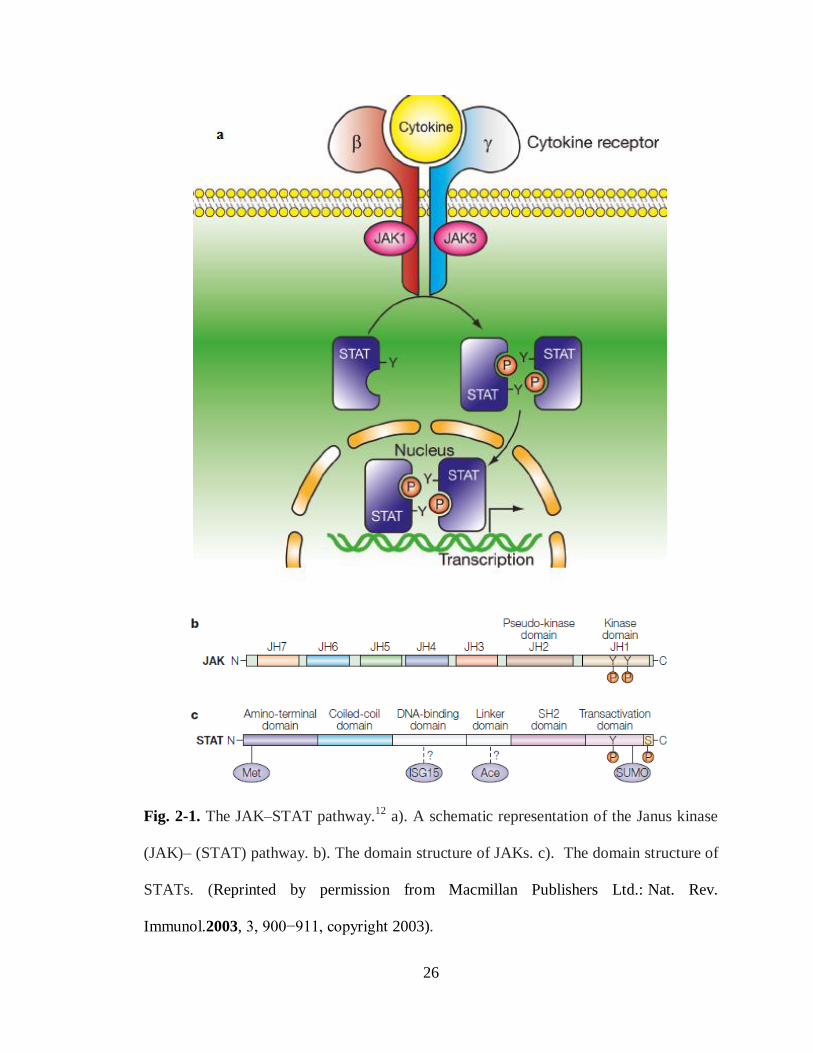
Fig. 2-1. The JAK–STAT pathway.12
a). A schematic representation of the Janus kinase
(JAK)– (STAT) pathway. b). The domain structure of JAKs. c). The domain structure of
STATs. (Reprinted by permission from Macmillan Publishers Ltd.: Nat. Rev.
Immunol.2003, 3, 900−911, copyright 2003).
27
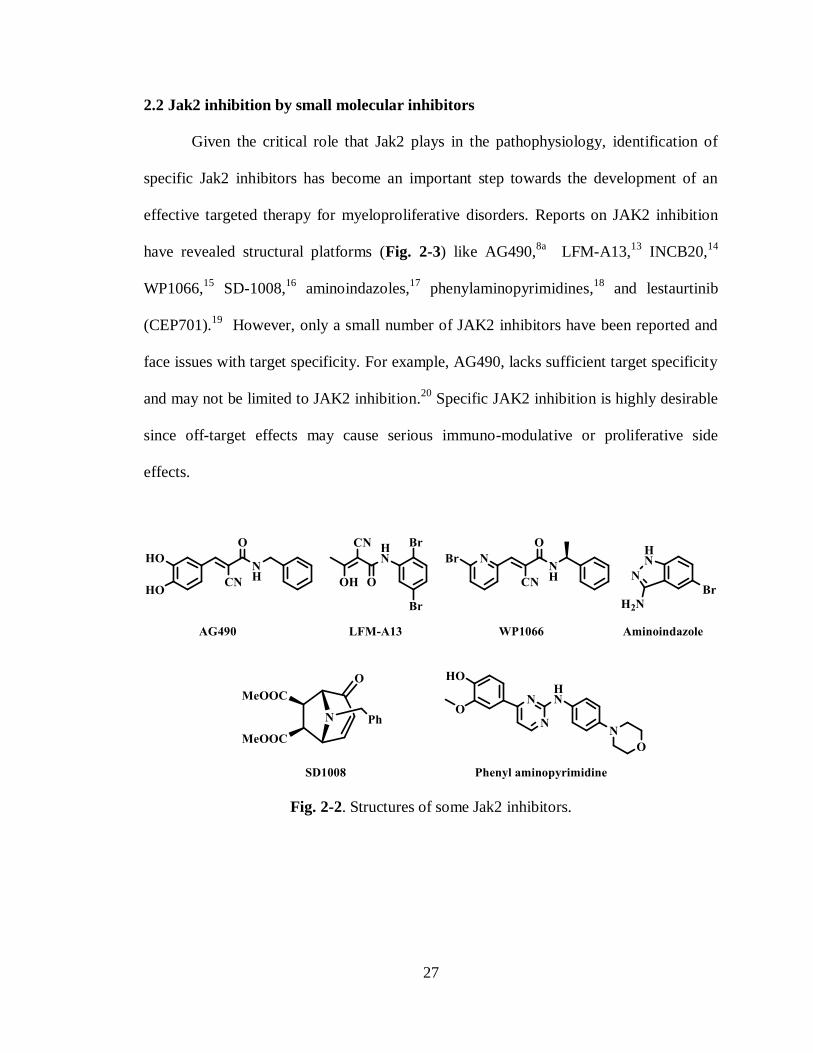
2.2 Jak2 inhibition by small molecular inhibitors
Given the critical role that Jak2 plays in the pathophysiology, identification of
specific Jak2 inhibitors has become an important step towards the development of an
effective targeted therapy for myeloproliferative disorders. Reports on JAK2 inhibition
have revealed structural platforms (Fig. 2-3) like AG490,8a
LFM-A13,13
INCB20,14
WP1066,15
SD-1008,16
aminoindazoles,17
phenylaminopyrimidines,18
and lestaurtinib
(CEP701).19
However, only a small number of JAK2 inhibitors have been reported and
face issues with target specificity. For example, AG490, lacks sufficient target specificity
and may not be limited to JAK2 inhibition.20
Specific JAK2 inhibition is highly desirable
since off-target effects may cause serious immuno-modulative or proliferative side
effects.
Fig. 2-2. Structures of some Jak2 inhibitors.

28
2.3 Significance of stilbenes in biological applications
The activity of stilbenes has been widely studied for their biological role against
pathogens and for pharmacological properties.21
The biological activity encompasses a
wide range that includes anticancer therapy. Resveratrol (Fig. 1-4), present in grapes and
other medicinal plants22
, is a well known stilbene as chemopreventive agent against
major stages of carcinogenesis23
. It’s other biological properties24
include antifungal,
antibacterial, anticancer,25
antiviral,26
estrogenic,27
platelet antiaggregating28
(Fig. 2-4)
and heart protecting activities.29
Resveratrol was found to be highly beneficial to the
cardiovascular system and has shown potent anti tumorigenic effect.30
Piceatannol, a
naturally occurring phenolic stilbenoid (Fig. 2-6), is the only stilbene that is a known
protein tyrosine kinase inhibitor. It inhibits LMP2A, a viral tyrosine kinase implicated in
diseases associated with the Epstein- Barr virus.31
Fig. 2-3. Resveratrol and its analogues as inhibitors of platelet aggregation induced by
collagen.
Antimicrobial compounds32
in grape wines belong to family of stilbenes. For
example, 3,5-dimethoxy-4′-hydroxy stilbene (Dimethoxy Resveratrol in Fig. 2-5),
present in grapevine leaves has relatively high activity as antifungal compound.32a
It has been shown that the total phenolic compounds extracted from red wine
inhibit the oxidation of human low-density lipoproteins (LDL) which otherwise may
promote atherogenesis.33
Analogues of resveratrol,34
like piceatannol,35
are also known to

29
possess antimicrobial activities. The presence of phenolic hydroxyl/methoxyl groups in
this stilbenes, which have high affinity for proteins, is thought to cause scavenging of the
the reactive oxygen species (ROS) and partly explains their associated biological roles.36
Previous studies also showed that hydroxystilbenes are capable of DNA binding and
cleavage can also occurs in combination with metals.37
Owing to these therapeutic values,
the synthesis of derivatives of resveratrol has attracted a great deal of attention recently
since they cannot be obtained in large quantities by extractive procedures. Thus several
stilbene derivatives have been synthesized and found to be showing good antibacterial
activity against Staphylococcus aureus, Streptococcus faecalis, Bacillus subtilis and
Bacillus anthracis.32d,38
For example, Dimethoxy Resveratrol derivatives were shown to
be active against several gram-positive and negative bacteria. Piperidino stilbenes was
also found to be active against Stephylococccus Aureas with MIC of 5 mG (Fig. 1-5).
Fig. 2-4. Structures and Antimicrobial activities of Piperidino stilbenes and Resveratrol
analogues against Gram-positive and Gram-negative bacteria.
Since Resveratrol has been studied extensively for its antiproliferative activity,39
there have been many number of other stilbene analogues studied. Combretastatin A4 is a
naturally found stilbene (Fig. 2-6) in the bush willow tree Comretum caffrum and shown
to be a potent inhibitor of tubilin assembly (IC50=1.2 M)40
and strongly cytotoxic
against selected human cancer cell lines. It’s evaluation against DU-145 prostate cancer
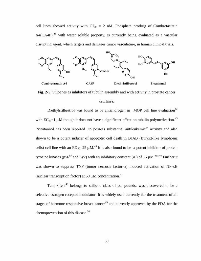
30
cell lines showed activity with GI50 = 2 nM. Phosphate prodrug of Combretastatin
A4(CA4P),41
with water soluble property, is currently being evaluated as a vascular
disrupting agent, which targets and damages tumor vasculature, in human clinical trials.
Fig. 2-5. Stilbenes as inhibitors of tubulin assembly and with activity in prostate cancer
cell lines.
Diethylstilbestrol was found to be antiandrogen in MOP cell line evaluation42
with EC50=1 M though it does not have a significant effect on tubulin polymerization.43
Piceatannol has been reported to possess substantial antileukemic44
activity and also
shown to be a potent inducer of apoptotic cell death in BJAB (Burkitt-like lymphoma
cells) cell line with an ED50=25 M.45
It is also found to be a potent inhibitor of protein
tyrosine kinases (p56lck
and Syk) with an inhibitory constant (Ki) of 15 µM.31a,46
Further it
was shown to suppress TNF (tumor necrosis factor-) induced activation of NF-B
(nuclear transcription factor) at 50 M concentration.47
Tamoxifen,48
belongs to stilbene class of compounds, was discovered to be a
selective estrogen receptor modulator. It is widely used currently for the treatment of all
stages of hormone-responsive breast cancer49
and currently approved by the FDA for the
chemoprevention of this disease.50

31
Fig. 2-6. Tamoxifen and its analogues as antiestrogen compounds for the treatment of
advanced breast cancer.
Z-Hydroxytamoxifen,51
an active metabolite of Tamoxifen,52
was found to be an
active antiestrogenic stilbene.53
It also possess a high in-vitro potency for the estrogenic
receptor54
and is a weaker agent than the tamoxifen in-vivo due to rapid clearance55
from
cell line. Z-Toremifen was also found to be effective for the treatment of estrogen
receptor positive advanced breast cancer56
and has been marketed for the treatment of
advanced breast cancer.57
E-Droloxifen is another stilbene found to be a new
antiestrogen58
and is under clinical trials for the treatment of breast cancer in
postmenopausal women. Potential uses of E-Droloxifen59
could be in endometriosis,
Alzheimer’s disease, Uterine fibroid disease among others. The pronounced biological
activities of tamoxifen analogues have encouraged the synthesis of many novel structural
congeners.60

32
2.4 Synthetic approaches towards stilbenes
Stilbenes have been synthesized via various methods.61
These methods include
reactions catalyzed by Pd, Ru, Ti, Zn, Rh, Ni, Mo and ylide formation in wittig reaction.
Following sections will describe a few methods that are widely used.
Guiso et. al. 62
have synthesized E-Resveratrol (Scheme 2-1) via a Heck reaction
between 3,5-diacetoxy styrene and 4-acetoxy phenyl iodide catalyzed by Pd(OAc)2 and
PPh3 in 70% yield. Since aryl iodide is more reactive than ArBr and ArCl in Heck
reaction, it was chosen in this reaction. This reaction also yielded small amounts of
partially deacylated by products. The resulted triacetoxy stilbene was hydrolyzed to E-
Resveratrol with NaOMe.
Scheme 2-1. Synthesis of E-Resveratrol by Guiso et. al.via heck reaction. (a). Pd(OAc)2,
PPh3, Et3N, CH3CN, 850C. (b). NaOMe, MeOH, THF.
Sengupta et. al. 63
used aryldiazonium salts in double heck reaction (Scheme 2-2)
with vinyltriethoxysliane to synthesize symmetrical trans-stilbenes with 40-70% yields
for various substituents.
Scheme 2-2. Synthesis of Stilbenes by sengupta et. al.via heck reaction. (a). Pd(OAc)2,
MeOH, 600C, 1hr.

33
Suzuki-Miyaura reaction has also been used for the synthesis of stilbenes since
the method is highly selective, tolerant of functional groups on either coupling partners
and reagents is fairly insensitive towards water. Andrus et. al. 64
(Scheme 2-3) used
imodozolium chloride catalyst in combination with Pd(OAc)2 to couple aryldiazonium
salts and aryl vinylboronic acids in suzuki reaction for the synthesis of trans-stilbenes.
Scheme 2-3. Synthesis of Stilbenes by Andrus et. al.via suzuki reaction. (a). Pd(OAc)2,
THF, 00C to room temperature, imidazolium chloride, 3hrs, 68-87% yields.
Stille coupling65
with modified reagents is also an useful method for the synthesis
of stilbenes. For example, Nishibayashi et. al. 66
(Scheme 2-4) used organotellurides
instead of organostannes in Stille type coupling for the synthesis of trans-stilbenes.
Various organotellurides reacted with alkenes in the presence of palladium salts and
oxidant like AgOAc for the efficient synthesis of stilbenes.
Scheme 2-4. Synthesis of Stilbenes by Nishibayashi et. al.via Stille coupling. (a).
Pd(Cl)2, AgOAc, Et3N, MeOH, 250C, 20 hrs.

34
Cross metathesis reaction presents another possible method for the synthesis of
both symmetrical and unsymmetrical stilbenes.67
Since schrock et. al. 68
demonstrated that
trans-stilbenes can be synthesized by using Mo-based catalyst, this method has garnered
attention recently. Chang et. al. 69
synthesized symmetrical stilbenes from the
corresponding para-substituted styrene by using Ru-based Grubbs 2nd
generation catalyst.
Velder et. al. 70
synthesized a broad spectrum of unsymmetrically substituted stilbenes
(Scheme 2-5) using Grubbs 2nd
gen. catalyst.
Scheme 2-5. Synthesis of Stilbenes by Velder et. al. 70
via cross-metathesis. (a). Grubb’s
2nd
generation cat.(2 mol%), CH2Cl2, 400C, 1.5 hrs.
Wittig or Horner-Wadsworth-Emmons reactions have attracted greater attention
for the synthesis of stilbenes from two aromatic building blocks although it affords both
(E)- and (Z)-alkenes. This reaction of benzyl ylides with substituted benzyl aldehydes
proved to be unsatisfactory since it afforded poor control over the configuration of the
newly formed C=C double bond yielding mixtures. This is mainly attributed to semi
stability or instability of the ylides. Pettit et. al. 71
synthesized cis-stilbenes (Scheme 2-6)
using wittig reaction with a Z to E ratio of 1.4:1. Both isomers were isolated by column
chromatography. Photochemical isomerization of trans isomer proceeded to yield cis-
stilbenes with 62% overall yield.
35
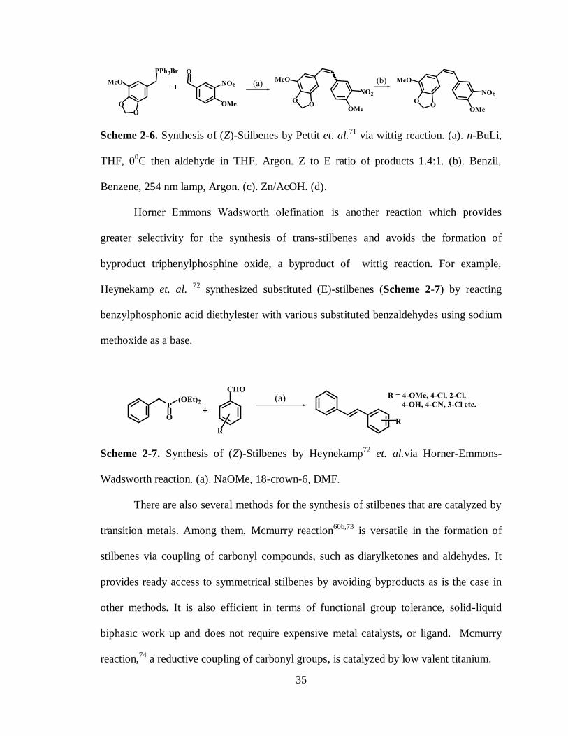
Scheme 2-6. Synthesis of (Z)-Stilbenes by Pettit et. al.71
via wittig reaction. (a). n-BuLi,
THF, 00C then aldehyde in THF, Argon. Z to E ratio of products 1.4:1. (b). Benzil,
Benzene, 254 nm lamp, Argon. (c). Zn/AcOH. (d).
Horner−Emmons−Wadsworth olefination is another reaction which provides
greater selectivity for the synthesis of trans-stilbenes and avoids the formation of
byproduct triphenylphosphine oxide, a byproduct of wittig reaction. For example,
Heynekamp et. al. 72
synthesized substituted (E)-stilbenes (Scheme 2-7) by reacting
benzylphosphonic acid diethylester with various substituted benzaldehydes using sodium
methoxide as a base.
Scheme 2-7. Synthesis of (Z)-Stilbenes by Heynekamp72
et. al.via Horner-Emmons-
Wadsworth reaction. (a). NaOMe, 18-crown-6, DMF.
There are also several methods for the synthesis of stilbenes that are catalyzed by
transition metals. Among them, Mcmurry reaction60b,73
is versatile in the formation of
stilbenes via coupling of carbonyl compounds, such as diarylketones and aldehydes. It
provides ready access to symmetrical stilbenes by avoiding byproducts as is the case in
other methods. It is also efficient in terms of functional group tolerance, solid-liquid
biphasic work up and does not require expensive metal catalysts, or ligand. Mcmurry
reaction,74
a reductive coupling of carbonyl groups, is catalyzed by low valent titanium.

36
The formation of the stilbene is the result of a induced reductive dimerization to
form carbon-carbon bond which is followed by the deoxygenation of the pinacolate
intermediate. The Mcmurry reaction has been exploited in organic synthesis and usually
provides (E)-stilbenes. (Z)-stilbenes75
can be preferentially obtained if geometric
constraints control the orientation of the two carbonyl moieties. Thus this reaction is
versatile in terms of controlling stereochemistry of the double bond and functional group
tolerance. Some of the stilbenes synthesized by this method have shown potent
nematocidal activity. 76
Mcmurry reaction has been used in the synthesis of highly
functionalized stilbenes such as Tamoxifen, 73a
Droloxifen59
and related compounds. 52
Scheme 2-8. Observed stereoselectivity in the synthesis of (Z)-4-Hydroxytamoxifen.
Scheme 2-9. Synthesis of (Z)-4-Hydroxytamoxifen by Gautheir et. al. 52
via Mcmurry
reaction with modified reaction conditions. (a). TiCl4, Zn, THF, 00C to reflux. (b).
Cl(CH2)2N(CH3)2, K2CO3, acetone, H2O. (c). MeLi, THF, -780C.
37
Mcmurry reaction yields mixed results for synthesis of unsymmetrical stilbenes
especially when the carbonyl compound used is diaryl ketone and is reacted with
equimolar amounts of other ketone or aldehyde. However, by using diaryl ketone in less
than equimolar amount, the desired unsymmetrical stilbene can be obtained in good
stereoselectivity and yield. For example, Gauthier et. al. 52
demonstrated that using diaryl
ketone and propiophenone in 1:3 ratio, the cross coupled product was obtained with E/Z
ratio of 100:1 in 82% yield (Scheme 2-9). The mechanistic investigation52,59
of the
reaction revealed a steric preference in the transition state in which the sterically smaller
alkyl chain aligned with the bulkier substituted phenol group (Scheme 2-8) thus
minimizing the steric interaction. This steric preference was elegantly exploited by
Gauthier et. al. in the design of the synthetic route to (Z)-4-Hydroxytamoxifen and other
analogues like (E)-Droloxifen.
2.5 Synthesis of novel stilbenes via Mcmurry reaction
Recently, we have identified a novel small-molecule inhibitor of Jak2, named G6,
(Fig. 2-8) using structure-based virtual screening. 77
We showed that G6 has a specific
inhibitory effect on Jak2 kinase activity as measured by in vitro enzyme assays. In an in
vivo and ex vivo study, G6 greatly reduced growth of Jak2-Val617phe mutated human
pathological cells isolated from the bone marrow of a polycythemia vera patient who was
Jak2-V617F positive in a dose dependent manner with IC50 value of 50 nM. Our
laboratories have also reported that the core stilbenoid ring system78
present in G6 is
essential for maintaining its ability to inhibit Jak2 kinase activity. In the course of our
studies towards the synthesis of G6 and its analogs, our efforts were led on to the
investigation of the Mcmurry reaction.

38
Fig. 2-7 Structure of G6 (compound 11).
As illustrated in Scheme 2-10, synthesis of the various G6 analogs was
envisioned using Mcmurry coupling of the hydroxyl benzaldehyde or ketones to
synthesize the corresponding hydroxystilbene followed by the Mannich coupling with the
appropriate amine. Five stilbenes, 6-10 (see Table. 2-1, Scheme 2-11) were synthesized.
Mcmurry coupling of the ketone and aldehyde in the presence of Zinc and titanium
tetrachloride in dry THF led to a mixture of E- and Z- stilbenoids to a varying degree.
Scheme 2-10. Synthesis of stilbenoids from ketones and aldehydes.
Separation of these isomers involving column chromatography proved to be a
difficult task due to the dynamic isomerization on the column. Next, we tried various
solvents for recrystallization to isolate the isomers selectively. However, several
attempts to separate and isolate the Z- isomer were unsuccessful. The E-stilbenoid, being
more stable, was the only isomer isolated in these recrystallization. When 4’-hydroxy
propiophenone 1 was treated with the low valent titanium reagent, prepared from
TiCl4/Zn, at reflux, diethylstilbestrol was obtained as an inseparable E/Z mixture. This
E/Z mixture upon recrystallization from 15% ethylacetate/hexane resulted 6 (>100:1 E/Z
ratio) with pure E isomer in 73% yield. 79

39
Scheme 2-11. Reagents and Conditions. i. TiCl4, Zn, Dry. THF, reflux.
Table 2-1. Stilbenes 6-10 from various carbonyl compounds.
Aldehyde or ketone R1 R2 R3 Stilbene E/Z
ratio
%
Yield
4’-Hydroxy propiophenone (1) -C2H5 -H -OH 6 100 : 1 73
4’-Hydroxy acetophenone (2) -CH3 -H -OH 7 100 : 1 65
4’-Hydroxy benzaldehyde (3) -H -H -OH 8 100 : 1 71
3’-Hydroxy acetophenone (4) -CH3 -OH -H 9 85 : 15 76
3’-Hydroxy benzaldehyde (5) -H -OH -H 10 100 : 1 84
trans- 4,4’-Diethyl stilbestrol(6): The 1H NMR of trans- diethylstilbestrol 6 was in
complete agreement with the one in literature (Fig. 2-9b).79a
A NMR spectral comparison
of the cis- and trans- mixture before and after crystallization is shown in figure 1-8. The
methylene (Ha’) and methyl (Hb’) protons in the trans isomer were upfield than of those
(Ha and Hb) in the cis-stilbestrol (Fig. 1-8a). Methyl (Hb’) protons in trans- 6 were at
0.6 ppm while those (Hb) were at 0.7 ppm in the cis-isomer. This upfield shift in the
resonance of the methyl and methylene in the trans isomer can be attributed to the
diamagnetic shielding due to their proximity to the aromatic ring. The diamagnetic
shielding was also observed in the upfield shift in the resonance position of the aromatic
protons in the cis- isomer (Fig. 2-9a). 79a

40
Fig. 2-8. 1H NMR spectra of Diethyl stilbestrol 6 (a). trans and cis mixture . (b). isolated
trans isomer after recrystallization.
trans- 4,4’-Dihydroxy dimethylstilbene (7): Using reaction conditions similar to as
previously described the 4’-Hydroxy acetophenone (2) in Mcmurry coupling also gave
E/Z mixture which upon recrystallization from (9:1) methanol and water furnished 7 (>99
: 1 ratio) with 65% yield. In the 1H-NMR spectrum, the methyl groups in the trans-
isomer of 7 also showed signal upfield than that of in the cis-isomer (Fig. 2-10); as seen
in the case of Diethylstilbestrol 6. Similar trend was also observed for aromatic protons
of 7 in which were upfield in the cis-isomer that in the trans- isomer. This could also be
attributed to diamagnetic shielding effect as explained in the case of Diethylstilbestrol 6.

41
Fig. 2-9. Comparisons of 1H NMR spectrums of 4,4’-dihydroxy dimethylstilbene 7 as
mixture and trans isolated. (a). Mixture of isomers trans and cis. (b). Pure trans isomer
after recrystallization.
Next, 4’ and 3’-Hydroxy benzaldehydes (3 and 5) also gave E/Z mixtures which
upon recrystallization from methanol furnished pure isomers with 71% and 84% yields.
Crude product from 3’-Hydroxy acetophenone in recrystallization from methanol resulted
in 85:15 (E/Z ratio) which used in next step directly. Unambiguous structural
characterization of the stilbene was carried out using the 1H and
13C NMR spectroscopic
analysis . 76,79
In all of the stilbenes synthesized, trans-isomer was obtained predominantly. This
predominant formation of trans-product suggests that the orientation of phenyl rings
plays an important role in the transition state as shown in Fig. 2-11. The generally
accepted mechanism, proposed by Mcmurry73b,80
and several other, 59
is that the carbonyl
compounds are reduced by metallic titanium to give a radical anion species leading to
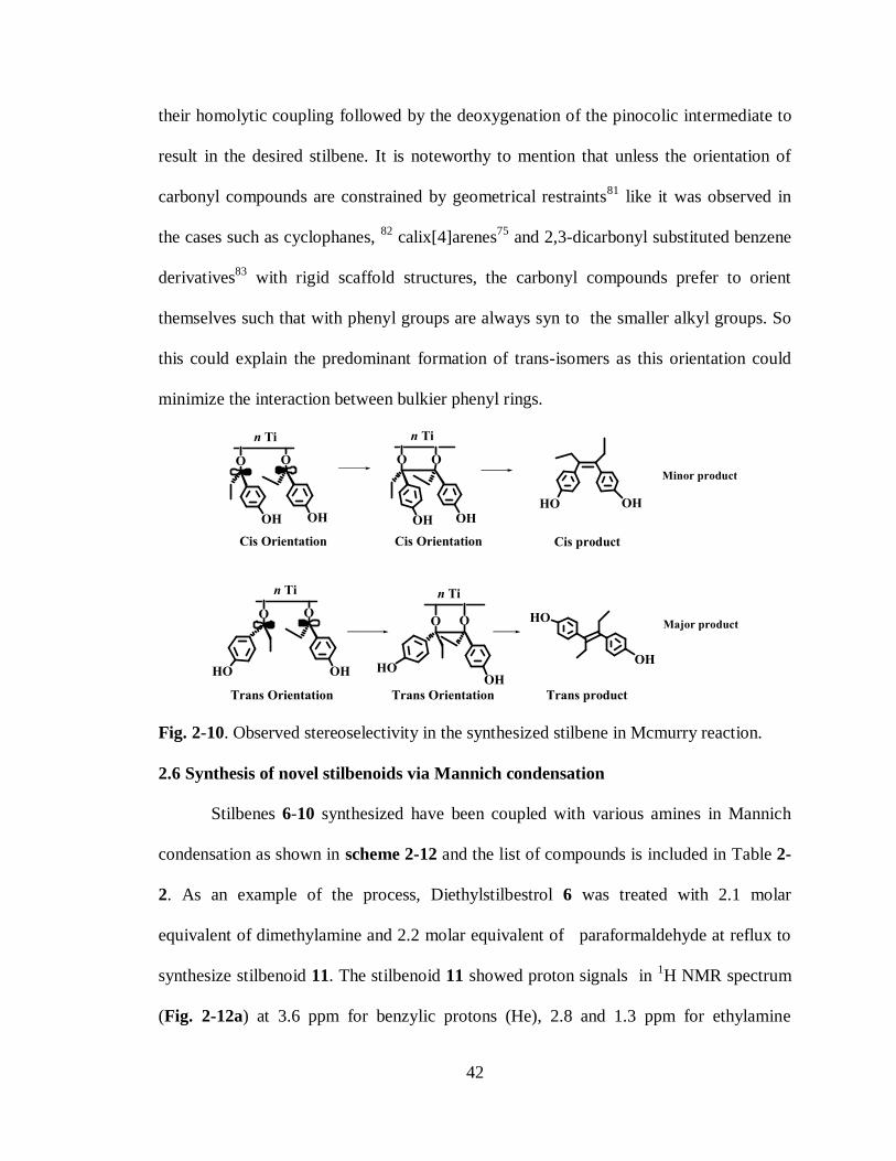
42
their homolytic coupling followed by the deoxygenation of the pinocolic intermediate to
result in the desired stilbene. It is noteworthy to mention that unless the orientation of
carbonyl compounds are constrained by geometrical restraints81
like it was observed in
the cases such as cyclophanes, 82
calix[4]arenes75
and 2,3-dicarbonyl substituted benzene
derivatives83
with rigid scaffold structures, the carbonyl compounds prefer to orient
themselves such that with phenyl groups are always syn to the smaller alkyl groups. So
this could explain the predominant formation of trans-isomers as this orientation could
minimize the interaction between bulkier phenyl rings.
Fig. 2-10. Observed stereoselectivity in the synthesized stilbene in Mcmurry reaction.
2.6 Synthesis of novel stilbenoids via Mannich condensation
Stilbenes 6-10 synthesized have been coupled with various amines in Mannich
condensation as shown in scheme 2-12 and the list of compounds is included in Table 2-
2. As an example of the process, Diethylstilbestrol 6 was treated with 2.1 molar
equivalent of dimethylamine and 2.2 molar equivalent of paraformaldehyde at reflux to
synthesize stilbenoid 11. The stilbenoid 11 showed proton signals in 1H NMR spectrum
(Fig. 2-12a) at 3.6 ppm for benzylic protons (He), 2.8 and 1.3 ppm for ethylamine

43
protons (Hc and Hd), aromatic protons at 6.8-7.0 ppm, the Ha at 2.1 ppm and the Hb
protons at 0.7ppm. 13
C NMR spectrum also confirmed the structure of the symmetrical
stilbenoid 11. Importantly, the NMR data confirmed that the reflux condition used in this
reaction did not alter the trans-configuration of this product.
Fig. 2-11. Compound 11 spectrums. (a). 1H NMR spectrum. (b).
13C NMR spectrum.
Following the general synthetic protocol established for the synthesis of 11 above,
Diethyl stilbestrol 6 was treated with four other amines dimethyl amine, piperidine,
pyrrolidine and morpholine to synthesize four other stilbenoids 12-15 (Table 2-2). All of
them obtained in excellent yield with preserved trans- configuration.
Scheme 2-12. Reagents and Conditions. (a). amine, paraformaldehyde, MeOH, reflux.
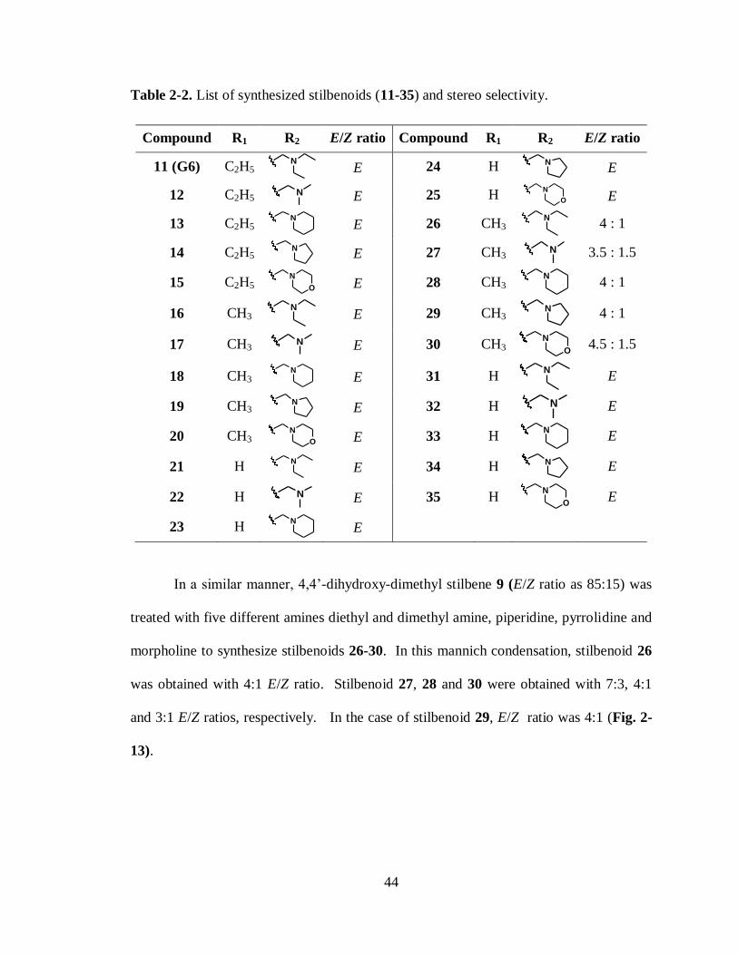
44
Table 2-2. List of synthesized stilbenoids (11-35) and stereo selectivity.
Compound R1 R2 E/Z ratio Compound R1 R2 E/Z ratio
11 (G6) C2H5 N
E 24 H N
E
12 C2H5 N E 25 H
N
O E
13 C2H5 N
E 26 CH3
N
4 : 1
14 C2H5 N
E 27 CH3 N
3.5 : 1.5
15 C2H5 N
O E 28 CH3
N
4 : 1
16 CH3 N
E 29 CH3
N
4 : 1
17 CH3 N E 30 CH3
N
O 4.5 : 1.5
18 CH3 N
E 31 H
N
E
19 CH3 N
E 32 H N
E
20 CH3 N
O E 33 H
N
E
21 H N
E 34 H N
E
22 H N E 35 H
N
O E
23 H N
E
In a similar manner, 4,4’-dihydroxy-dimethyl stilbene 9 (E/Z ratio as 85:15) was
treated with five different amines diethyl and dimethyl amine, piperidine, pyrrolidine and
morpholine to synthesize stilbenoids 26-30. In this mannich condensation, stilbenoid 26
was obtained with 4:1 E/Z ratio. Stilbenoid 27, 28 and 30 were obtained with 7:3, 4:1
and 3:1 E/Z ratios, respectively. In the case of stilbenoid 29, E/Z ratio was 4:1 (Fig. 2-
13).

45
Fig. 2-12. 1H NMR spectrums. a. Compound 9. b. Signals for stilbenoid 29.
Stilbenes 7, 8, and 10 were also treated with 2.2 mmols of appropriate amines
(dimethyl and diethylamine, pyrrolidine, piperidine and morpholine) and
paraformaldehyde in methanol at 800 C to synthesize the 11 analogues in good to
excellent yield (Scheme 2-12, Table 2-2).
The chemical structures of the stilbenoids obtained were confirmed by 1H- and
13C- NMR analysis. For example,
1H NMR spectra of stilbenoids 17, 22 and 34 shown
below (Fig. 2-14) indicates excellent purity and the absence of any side products.
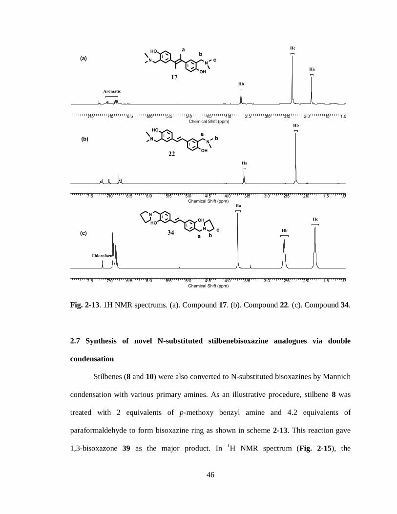
46
Fig. 2-13. 1H NMR spectrums. (a). Compound 17. (b). Compound 22. (c). Compound 34.
2.7 Synthesis of novel N-substituted stilbenebisoxazine analogues via double
condensation
Stilbenes (8 and 10) were also converted to N-substituted bisoxazines by Mannich
condensation with various primary amines. As an illustrative procedure, stilbene 8 was
treated with 2 equivalents of p-methoxy benzyl amine and 4.2 equivalents of
paraformaldehyde to form bisoxazine ring as shown in scheme 2-13. This reaction gave
1,3-bisoxazone 39 as the major product. In 1H NMR spectrum (Fig. 2-15), the
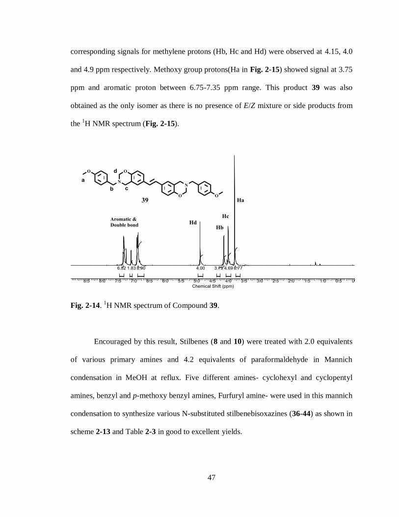
47
corresponding signals for methylene protons (Hb, Hc and Hd) were observed at 4.15, 4.0
and 4.9 ppm respectively. Methoxy group protons(Ha in Fig. 2-15) showed signal at 3.75
ppm and aromatic proton between 6.75-7.35 ppm range. This product 39 was also
obtained as the only isomer as there is no presence of E/Z mixture or side products from
the 1H NMR spectrum (Fig. 2-15).
Fig. 2-14. 1H NMR spectrum of Compound 39.
Encouraged by this result, Stilbenes (8 and 10) were treated with 2.0 equivalents
of various primary amines and 4.2 equivalents of paraformaldehyde in Mannich
condensation in MeOH at reflux. Five different amines- cyclohexyl and cyclopentyl
amines, benzyl and p-methoxy benzyl amines, Furfuryl amine- were used in this mannich
condensation to synthesize various N-substituted stilbenebisoxazines (36-44) as shown in
scheme 2-13 and Table 2-3 in good to excellent yields.

48
Scheme 2-13. Reagents and Conditions. (a). amine, paraformaldehyde, MeOH, reflux.
Table 2-3. List of synthesized stilbenoid rings (36-44) and stereo selectivity.
Stilbene R1 R2 Product R3
8 OH H 36
8 OH H 37
8 OH H 38
8 OH H 39 OMe
9 H OH 40
9 H OH 41
9 H OH 42
9 H OH 43 OMe
9 H OH 44 O
The chemical structures of all the N-substituted stilbenebisoxazines were
confirmed by 1H- and
13C- NMR analysis.
1H NMR spectra of N-substituted
stilbenebisoxazines 38, 41 and 43 shown below (Fig. 2-16) indicates excellent purity and
the absence of any side products.
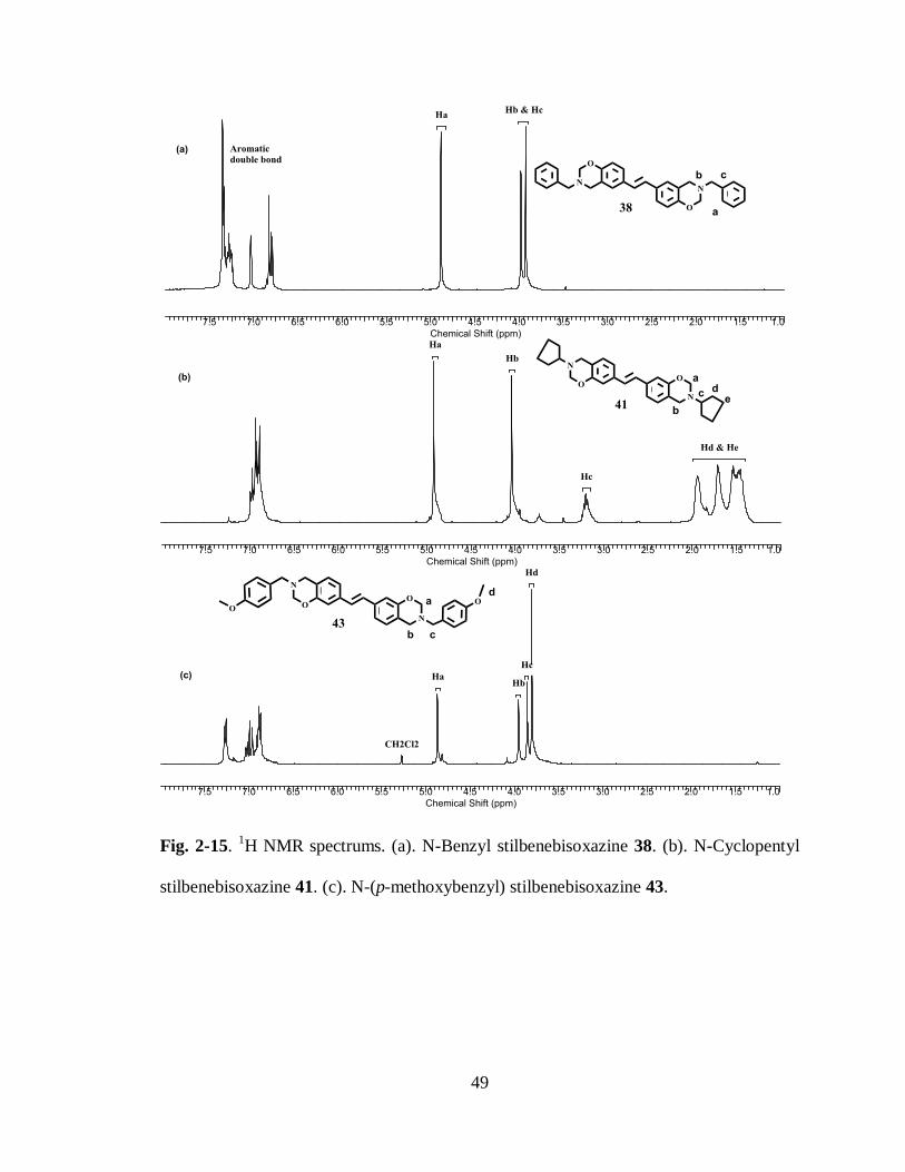
49
Fig. 2-15. 1H NMR spectrums. (a). N-Benzyl stilbenebisoxazine 38. (b). N-Cyclopentyl
stilbenebisoxazine 41. (c). N-(p-methoxybenzyl) stilbenebisoxazine 43.

50
2.8 Evaluation of the novel stilbenes and stilbenoids against JAK2-V617F Mutated
JAK2 Enzyme
Stilbenes (6 to 10) and stilbenoids (11-35) synthesized were evaluated for JAK2
enzyme activity. Proliferation of HEL cells is mediated by the constitutively active Jak2-
V617F signaling which promotes a G1/S phase transition, thereby leading to increased
cellular proliferation.10a-e
G6 (Stilbenoid 11) has been identified as a potent inhibitor of
Jak2 enzyme in structure based virtual screening. In an in vivo and ex vivo study, G6
greatly reduced growth of JAK2-Val617phe mutated human pathological cells.
Elaborated details of this work have been discussed at the end of this chapter. We
observed that the phenol OH in G6 forms strong hydrogen bonds with the backbone NH
group of Leu932 and a salt bridge is formed between N of 30 amine in G6 and Asp994
(see Fig. 2-20 and Fig. 2-21). These interactions might be the important to consider
during further modification of the G6. To study these interactions and tolerance of
various groups on the G6, we here describe structural modification of G6 in an effort to
derive a structure activity relationship (Fig. 2-8). G6 and its structurally related
derivatives were therefore first analyzed for their ability to inhibit the Jak2-V617F
dependent proliferation of HEL cells. Viable cell numbers were determined by trypan
blue exclusion and hemocytometer after 72 hrs. Each sample was measured in triplicate.
Inhibition by G6 (Compound 11) was arbitrarily set at 100% and the percent inhibition
for all the other compounds relative to G6 was defined as 1.00 - (∆ drug / ∆ vehicle
control). To determine the ability of each of these compounds to inhibit Jak2-V617F
mediated HEL cell proliferation, cells were treated with G6 or its derivatives. Viable cell
numbers for each treatment were determined.
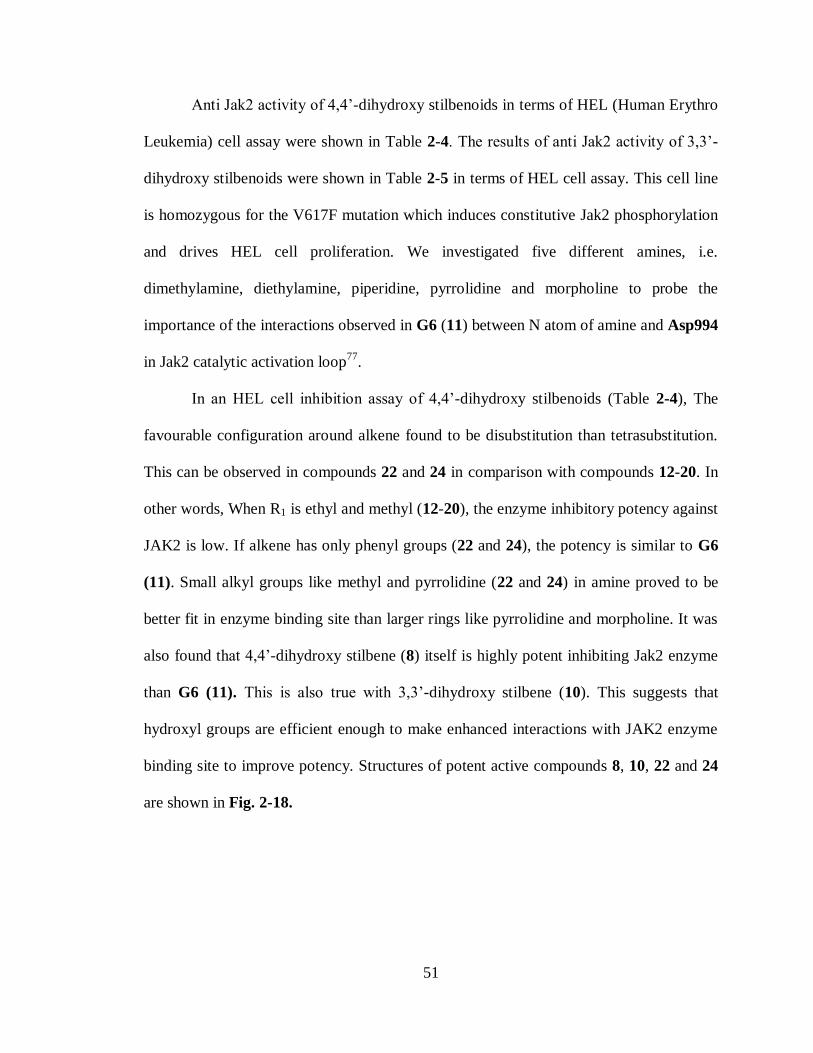
51
Anti Jak2 activity of 4,4’-dihydroxy stilbenoids in terms of HEL (Human Erythro
Leukemia) cell assay were shown in Table 2-4. The results of anti Jak2 activity of 3,3’-
dihydroxy stilbenoids were shown in Table 2-5 in terms of HEL cell assay. This cell line
is homozygous for the V617F mutation which induces constitutive Jak2 phosphorylation
and drives HEL cell proliferation. We investigated five different amines, i.e.
dimethylamine, diethylamine, piperidine, pyrrolidine and morpholine to probe the
importance of the interactions observed in G6 (11) between N atom of amine and Asp994
in Jak2 catalytic activation loop77
.
In an HEL cell inhibition assay of 4,4’-dihydroxy stilbenoids (Table 2-4), The
favourable configuration around alkene found to be disubstitution than tetrasubstitution.
This can be observed in compounds 22 and 24 in comparison with compounds 12-20. In
other words, When R1 is ethyl and methyl (12-20), the enzyme inhibitory potency against
JAK2 is low. If alkene has only phenyl groups (22 and 24), the potency is similar to G6
(11). Small alkyl groups like methyl and pyrrolidine (22 and 24) in amine proved to be
better fit in enzyme binding site than larger rings like pyrrolidine and morpholine. It was
also found that 4,4’-dihydroxy stilbene (8) itself is highly potent inhibiting Jak2 enzyme
than G6 (11). This is also true with 3,3’-dihydroxy stilbene (10). This suggests that
hydroxyl groups are efficient enough to make enhanced interactions with JAK2 enzyme
binding site to improve potency. Structures of potent active compounds 8, 10, 22 and 24
are shown in Fig. 2-18.

52
Table 2-4. Jak2 activity of G6 and its 4,4’-Dihydroxy stilbenoid analogues.
Comp. R1 R2
% HEL
Inhibition
(10 µM)
at 72 hrs
Comp. R1 R2
% HEL
Inhibition
(10 µM)
at 72 hrs
6 C2H5 H 65 17 CH3 N
60
7 CH3 H 63 18 CH3 N
72
8 H H 106 19 CH3 N
74
11 (G6) C2H5 N
100 20 CH3
N
O nd
12 C2H5 N
74 21 H N
85
13 C2H5 N
80 22 H N
99
14 C2H5 N
89 23 H
N
nd
15 C2H5 N
O 48 24 H N
96
16 CH3 N
26 25 H
N
O nd

53
Table 2-5. Jak2 activity of G6 and its 3,3’-Dihydroxy stilbenoid analogues .
Compound R1 R2 E/Z ratio
% HEL
Inhibition
(10 µM) at 72 hrs
11 (G6) C2H5 N
100 : 1 100
9 CH3 H 85 : 15 35
10 H H 100 : 1 106
26 CH3 N
4 : 1 61
27 CH3 N 3.5 : 1.5 55
28 CH3 N
4 : 1 84
29 CH3 N
4 : 1 68
30 CH3 N
O 4.5 : 1.5 39
31 H N
E 74
32 H N
E 36
33 H N
E 50
34 H N
E 35
35 H N
O E 25
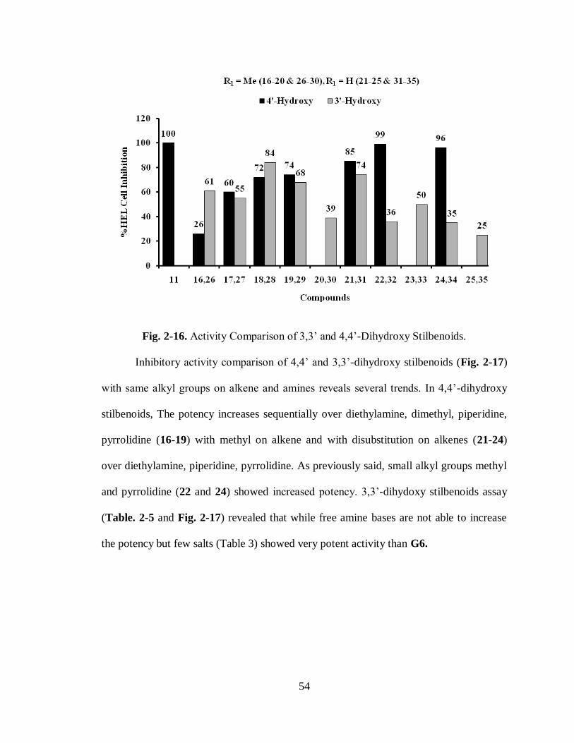
54
Fig. 2-16. Activity Comparison of 3,3’ and 4,4’-Dihydroxy Stilbenoids.
Inhibitory activity comparison of 4,4’ and 3,3’-dihydroxy stilbenoids (Fig. 2-17)
with same alkyl groups on alkene and amines reveals several trends. In 4,4’-dihydroxy
stilbenoids, The potency increases sequentially over diethylamine, dimethyl, piperidine,
pyrrolidine (16-19) with methyl on alkene and with disubstitution on alkenes (21-24)
over diethylamine, piperidine, pyrrolidine. As previously said, small alkyl groups methyl
and pyrrolidine (22 and 24) showed increased potency. 3,3’-dihydoxy stilbenoids assay
(Table. 2-5 and Fig. 2-17) revealed that while free amine bases are not able to increase
the potency but few salts (Table 3) showed very potent activity than G6.

55
Fig. 2-17. Stilbenoids which shows potent activity against JAK2 enzyme.
Fig. 2-18. Structures of salts of stilbenoids.
The assay of few salts (Fig. 2-19) was done to follow the inhibition activity
(Table 2-6) dependence on solubility. Even in salts, large alkyl ring on N did not
improve the inhibitory activity as evident in 33a than G6 (Compound 11). But dimethyl
and diethyl amine, showed good inhibitory activity and greatly increased the potency
than G6. This may be due to fact that salts have the advantage of having charges on N
already making them more soluble in carrier polar media and making better interactions
in the binding pocket.
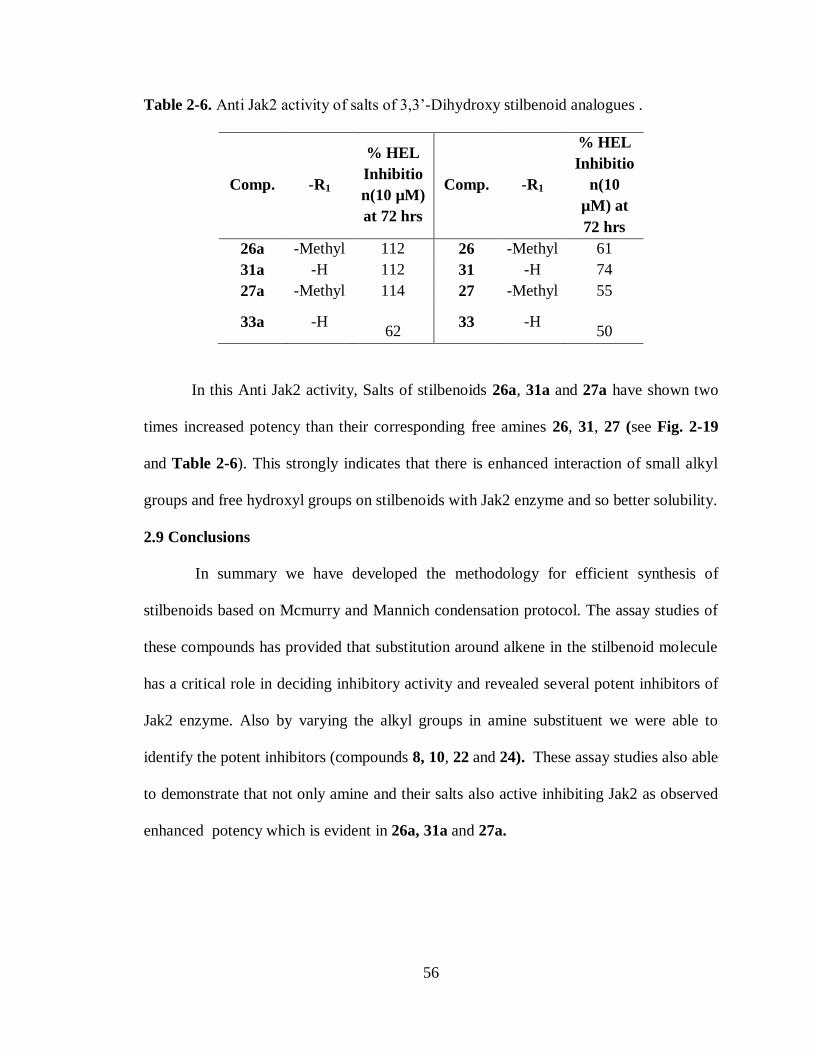
56
Table 2-6. Anti Jak2 activity of salts of 3,3’-Dihydroxy stilbenoid analogues .
Comp. -R1
% HEL
Inhibitio
n(10 µM)
at 72 hrs
Comp. -R1
% HEL
Inhibitio
n(10
µM) at
72 hrs
26a -Methyl 112 26 -Methyl 61
31a -H 112 31 -H 74
27a -Methyl 114 27 -Methyl 55
33a -H 62
33 -H 50
In this Anti Jak2 activity, Salts of stilbenoids 26a, 31a and 27a have shown two
times increased potency than their corresponding free amines 26, 31, 27 (see Fig. 2-19
and Table 2-6). This strongly indicates that there is enhanced interaction of small alkyl
groups and free hydroxyl groups on stilbenoids with Jak2 enzyme and so better solubility.
2.9 Conclusions
In summary we have developed the methodology for efficient synthesis of
stilbenoids based on Mcmurry and Mannich condensation protocol. The assay studies of
these compounds has provided that substitution around alkene in the stilbenoid molecule
has a critical role in deciding inhibitory activity and revealed several potent inhibitors of
Jak2 enzyme. Also by varying the alkyl groups in amine substituent we were able to
identify the potent inhibitors (compounds 8, 10, 22 and 24). These assay studies also able
to demonstrate that not only amine and their salts also active inhibiting Jak2 as observed
enhanced potency which is evident in 26a, 31a and 27a.
57
2.9 Inhibition of JAK2-V617F kinase activity by G6 (Stilbenoid 11)
Recently, In collaboration with Dr. Peter, P. Sayeski, we identified G6 (Stilbenoid
11) as a inhibitor of JAK2 enzyme using structure-based virtual screening.29
It was shown
that G6 has a specific inhibitory effect on Jak2 kinase activity as measured by in vitro
enzyme assays.29
In an in vivo and ex vivo study, G6 greatly reduced growth of JAK2-
Val617phe mutated human pathological cells isolated from the bone marrow of a
polycythemia vera patient who was Jak2-V617F positive in a dose dependent manner
with IC50 value of 50 nM (Fig. 1-20). We have also reported that the core stilbenoid ring
system present in G6 is essential for maintaining its ability to inhibit Jak2 kinase activity.
From analysis of the G6 binding mode in Jak2 active site77
, The human erythroleukemia
(HEL 92.1.7) cell line is homozygous for the Jak2-V617F mutation and this gain-of-
function mutation is responsible for its transformed phenotype.10d
Fig. 2-19. Binding mode of G6(grey) at the Jak2(green) vanderwall surface.77

58
Fig. 2-20. Anti Jak2 enzyme activity of G6 (stilbenoid 11)77
. Left. Inhibition of JAK2
Val617Phe kinase activity by G6 (IC50 = 60 ± 4.5 nM). Right. Growth inhibition of G6
on a human pathological cells isolated from polycythemia vera patient .
2.10 Antimicrobial activity of stilbenoids
Previous studies revealed that hydroxy stilbenes and analogues of Resveratrol
possess antimicrobial activities. The presence of phenolic hydroxyl/methoxyl groups in
these stilbenes, which have high affinity for proteins, generally attributed to scavenge the
reactive oxygen species(ROS) and partly explain their associated biological roles.36
Studies also shown that hydroxystilbenes are capable of DNA binding and cleavage can
also occurs in combination with metals.37
Thus pronounced biological role of stilbenes
encouraged us to screen several stilbenoids synthesized in our lab. Each of the stilbenoid
synthesized, as illustrated in Scheme 2-12 and Table 2-2, and N-substituted
stilbenebisoxazine synthesized, as illustrated in Scheme 2-13 and Table 2-3, was assayed
for the ability to inhibit the growth of select strain of clinically significant bacteria.

59
The invitro MIC determination for each compound were performed against a
multi drug resistant hospital acquired methicillin resistant S. aureus (MRSA) and
methicillin resistant S. epidermidis (MRSE). From the results of these MIC (minimum
inhibitory concentration) assays, most active compounds are presented in Table 2-7. To
explore the broad spectrum activity of these compounds (Fig. 2-22), we also employed
the modified Kirby-Bauer assay using two gram-positive pathogens, E. Faecalis and B.
Anthracis, Gram-negative pathogen, E. Coli, and the eukaryote Saccharomyces
cerevisiae (yeast) (see Table 2-7).
Fig. 2-21. Structures of active stilbenoids and N-Substituted stilbenebisoxazines.
From the analysis of data, Most observable feature is that not only stilbenes with
free hydroxyl groups are able to show potent antibacterial activity but also stilbenes with
bisoxazine rings on phenyl groups were able to show moderate antimicrobial activity.
This fact is quite interesting than previously predicted observation that free hydroxyl
moiety in stilbenes is vital to show enhanced activity. Alkene with methyl group and
dimethyl groups on tertiary amine in stilbenoid (Compound 27) was able to show
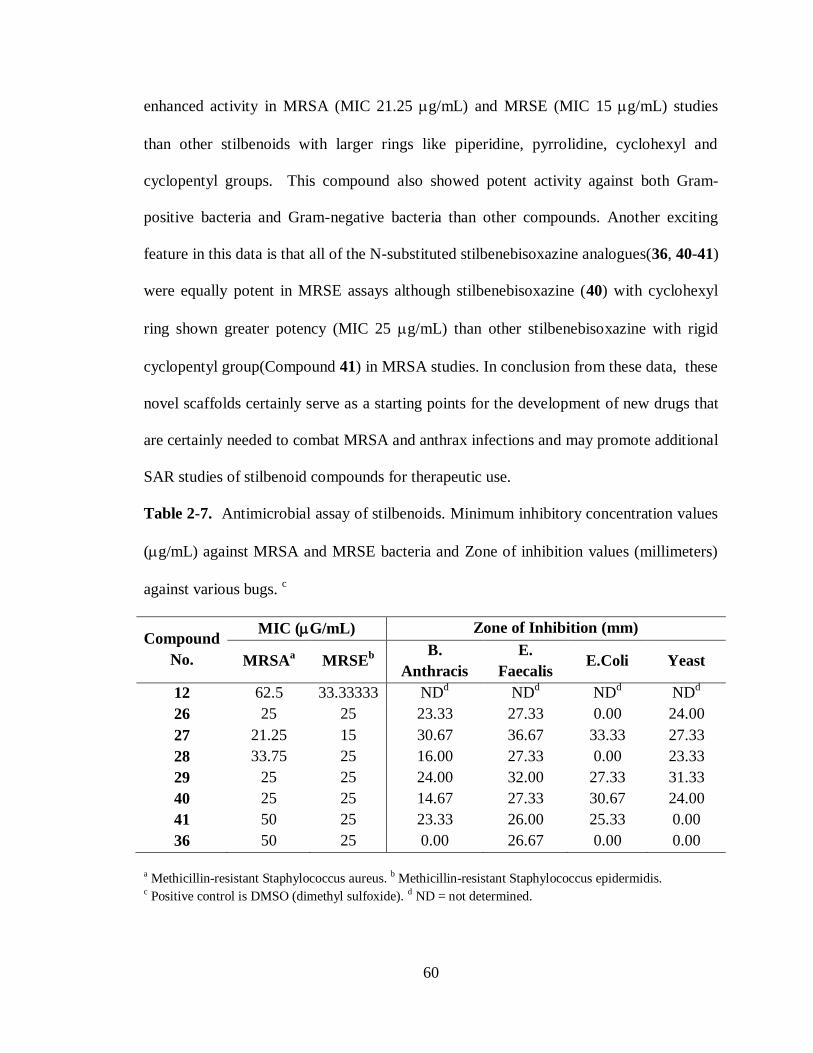
60
enhanced activity in MRSA (MIC 21.25 g/mL) and MRSE (MIC 15 g/mL) studies
than other stilbenoids with larger rings like piperidine, pyrrolidine, cyclohexyl and
cyclopentyl groups. This compound also showed potent activity against both Gram-
positive bacteria and Gram-negative bacteria than other compounds. Another exciting
feature in this data is that all of the N-substituted stilbenebisoxazine analogues(36, 40-41)
were equally potent in MRSE assays although stilbenebisoxazine (40) with cyclohexyl
ring shown greater potency (MIC 25 g/mL) than other stilbenebisoxazine with rigid
cyclopentyl group(Compound 41) in MRSA studies. In conclusion from these data, these
novel scaffolds certainly serve as a starting points for the development of new drugs that
are certainly needed to combat MRSA and anthrax infections and may promote additional
SAR studies of stilbenoid compounds for therapeutic use.
Table 2-7. Antimicrobial assay of stilbenoids. Minimum inhibitory concentration values
(g/mL) against MRSA and MRSE bacteria and Zone of inhibition values (millimeters)
against various bugs. c
Compound
No.
MIC (G/mL) Zone of Inhibition (mm)
MRSAa MRSE
b
B.
Anthracis
E.
Faecalis E.Coli Yeast
12 62.5 33.33333 NDd ND
d ND
d ND
d
26 25 25 23.33 27.33 0.00 24.00
27 21.25 15 30.67 36.67 33.33 27.33
28 33.75 25 16.00 27.33 0.00 23.33
29 25 25 24.00 32.00 27.33 31.33
40 25 25 14.67 27.33 30.67 24.00
41 50 25 23.33 26.00 25.33 0.00
36 50 25 0.00 26.67 0.00 0.00
a Methicillin-resistant Staphylococcus aureus. b Methicillin-resistant Staphylococcus epidermidis. c Positive control is DMSO (dimethyl sulfoxide). d ND = not determined.
61
2.11 Experimental Procedures
All solvents were dried and distilled prior to use and organic solvent extracts
dried over Na2SO4. 1H NMR and
13C NMR spectra were recorded on a Bruker 250 MHz
spectrometer in CDCl3 and DMSO-d6 with TMS as the standard. Chemical shifts are
reported in parts per million, multiplicities are indicated by s (singlet), d (doublet), t
(triplet), q (quartet), m (multiplet) and bs (broad singlet). Thin-layer chromatography
(TLC) was performed on glass plates coated with 0.25 mm thickness of silica gel.
2.12.1 General Procedure for the synthesis of Stilbenes:
In a flame dried 2 neck round bottom flask fitted with magnetic stirrer bar and
reflux condensor, dry THF (180 mL or 1.5 Molar) and Zinc (8 equivalents) was taken
under N2 atmosphere. Then reaction mixture was cooled to 0Oc in an ice bath. To this
mixture TiCl4 (4 equivalents) was added dropwise while maintaining the reaction
temperature at 0OC. After addition was complete, reaction mixture was refluxed for 2
hours. Then this brown color reaction mixture was cooled again to 0Oc. Aldehyde or
Ketone(1 equivalent) was taken in 20 mL of dry THF was added to this ice cooled
reaction mixture slowly portion wise. Then this reaction mixture was allowed to reflux
until TLC (2:3 mixture of ethylacete/hexane) shows that all the aldehyde or ketone is
consumed in the reaction. After reaction is complete, reaction mixture was concentrated
and ethylacetate was added. To this mixture saturated K2CO3 solution was added and
allowed to stir for another 7 hours. Reaction mixture was filtered and residue was washed
with ethylacetate. Filtrate was extracted with ethylacetate. Organic layer was washed
with saturated NaCl solution, water, dried over anhydrous Na2SO4 and concentrated. This

62
crude mixture was chromatographed on silica column eluting with 1:9 mixture of
ethylacetate:hexane mixture to receive E and Z stilbenes as a mixture.
Representative procedure for E-4,4'-[1,2-diethyl-1,2-ethenediyl]bis Phenol (6):79a
To a solution of Zinc (20.9 g, 0.319 mol) in dry THF (150 mL) was added TiCl4
(22.73 g, 0.119 mol) dropwise at 00C under N2 atmosphere. After addition was complete,
reaction mixture was refluxed for 2 hours. Then this brown color reaction mixture was
cooled again to 0Oc. A solution of 4’-hydroxy propiophenone (6 g, 0.039 mol) in dry
THF (50 mL) was added to this ice cooled reaction mixture slowly portion wise. Then
this reaction mixture was allowed to reflux for 6 hrs. Then the reaction mixture was
concentrated and ethylacetate was added. To this mixture sat. K2CO3 solution was added
and allowed to stir for another 7 hours. Reaction mixture was filtered and residue was
washed with ethylacetate. Filtrate was extracted with ethylacetate. Organic layer was
washed with saturated NaCl solution, water, dried over anhydrous Na2SO4 and
concentrated. This crude mixture was chromatographed on silica column eluting with 1:9
mixture of ethylacetate:hexane mixture to receive cis/trans mixture. This cis/trans
mixture received in column has been recrystallized from 15% ethylacetate/hexane
mixture to receive pure E isomer with >99% purity, which is also confirmed from
literature values, with 73% yield. 1H NMR (d6-DMSO, MHz): δ 9.1 (bs, 2H), 6.7-6.6 (d,
4H, j=8.5 Hz), 6.5-6.4 (d, 4H, j=8.5 Hz), 1.8 (q, 4H, j=7.5 Hz), 0.5 (t, 6H, j=7.5 Hz). ; 13
C
NMR (62.5 MHz, d6-DMSO): δ 155.7, 137.9, 132.5, 129.3, 114.8, 28.0, 13.3.
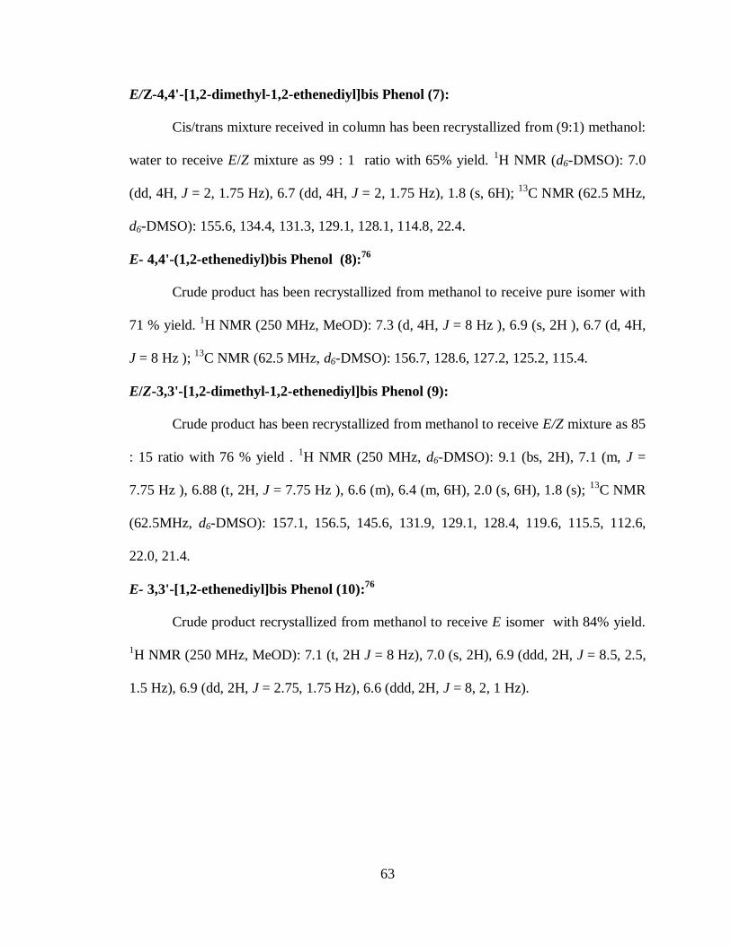
63
E/Z-4,4'-[1,2-dimethyl-1,2-ethenediyl]bis Phenol (7):
Cis/trans mixture received in column has been recrystallized from (9:1) methanol:
water to receive E/Z mixture as 99 : 1 ratio with 65% yield. 1H NMR (d6-DMSO): 7.0
(dd, 4H, J = 2, 1.75 Hz), 6.7 (dd, 4H, J = 2, 1.75 Hz), 1.8 (s, 6H); 13
C NMR (62.5 MHz,
d6-DMSO): 155.6, 134.4, 131.3, 129.1, 128.1, 114.8, 22.4.
E- 4,4'-(1,2-ethenediyl)bis Phenol (8):76
Crude product has been recrystallized from methanol to receive pure isomer with
71 % yield. 1H NMR (250 MHz, MeOD): 7.3 (d, 4H, J = 8 Hz ), 6.9 (s, 2H ), 6.7 (d, 4H,
J = 8 Hz ); 13
C NMR (62.5 MHz, d6-DMSO): 156.7, 128.6, 127.2, 125.2, 115.4.
E/Z-3,3'-[1,2-dimethyl-1,2-ethenediyl]bis Phenol (9):
Crude product has been recrystallized from methanol to receive E/Z mixture as 85
: 15 ratio with 76 % yield . 1H NMR (250 MHz, d6-DMSO): 9.1 (bs, 2H), 7.1 (m, J =
7.75 Hz ), 6.88 (t, 2H, J = 7.75 Hz ), 6.6 (m), 6.4 (m, 6H), 2.0 (s, 6H), 1.8 (s); 13
C NMR
(62.5MHz, d6-DMSO): 157.1, 156.5, 145.6, 131.9, 129.1, 128.4, 119.6, 115.5, 112.6,
22.0, 21.4.
E- 3,3'-[1,2-ethenediyl]bis Phenol (10):76
Crude product recrystallized from methanol to receive E isomer with 84% yield.
1H NMR (250 MHz, MeOD): 7.1 (t, 2H J = 8 Hz), 7.0 (s, 2H), 6.9 (ddd, 2H, J = 8.5, 2.5,
1.5 Hz), 6.9 (dd, 2H, J = 2.75, 1.75 Hz), 6.6 (ddd, 2H, J = 8, 2, 1 Hz).

64
2.12.2 General Procedure for the synthesis of Stilbenoids:
Stilbene(1 equivalent) was taken in one neck round bottom flask and 15 mL of
methanol was added. Paraformaldehyde(2.1 equivalents) and appropriate amine(2.2
equivalents) was added. Then this reaction mixture was allowed to reflux until TLC (2:3
mixture of ethylacetate:hexane) shows that reaction is complete. Then mixture is cooled
to room temperature and concentrated. To this mixture ethylacetate and 1M HCl solution
is added. Aqueous phase is separated and treated with 1M NaOH solution until pH is 7.
Then this aqueous solution is extracted with ethylacetate. Organic layer is washed with
saturated NaCl solution, water and dried over anhydrous Na2SO4. This organic layer is
concentrated and dried in vacuuo. This mixture containing mannich products has been
purified by flash chromatography over silicagel characterized by eluting with
dichloromethane : methanol : triethylamine mixture(99:0.5:0.5) to receive pure product
yields ranging from 80 to 95 %.
Representative procedure:
E-4,4'-(1,2-diethyl-1,2-ethenediyl)bis[2-[(diethylamino)methyl]-Phenol (Pure
isomer) (11) :
To a solution of 6(0.388g, 1.447 mmol) in MeOH was added
paraformaldehyde(0.095 g, 3.183 mmol) and diethyl amine(0.232g, 3.183 mmol). The
reaction mixture was refluxed for 8 hrs. Then reaction mixture was cooled to room
temperature and concentrated under reduced pressure. To this mixture ethylacetate and
1M HCl solution is added. Aqueous phase is separated and treated with 1M NaOH
solution until pH is 7. Then this aqueous solution is extracted with ethylacetate. Organic
layer is washed with saturated NaCl solution, water and dried over anhydrous Na2SO4.

65
This organic layer is concentrated and dried in vacuuo. Crude reaction mixture
chromatographed over silica gel by eluting with dichloromethane : methanol :
triehtylamine mixture( 99:0.5:0.5) to receive pure product 11 as white color solid with 92
% yield. 1
H NMR (CDCl3): δ 6.9 (d, 2H, J = 1.5 Hz ), 6.8 (d, 4H, J = 8 Hz ), 3.79 (S,
4H), 2.6-2.6 (q, 8H, J = 7.25 Hz ), 2.1-2.08 (q, 4H, J = 7.5 Hz ), 1.1-1.0 (t, 12H, J = 7.25
Hz), 0.8-0.7 (t, 6H, J = 7.5 Hz ) ; 13
C NMR (CDCl3): δ 156.4, 138.7, 133.3, 129.8, 128.7,
128.5, 121.5, 115.4, 57.1, 54.0, 46.5, 46.3, 28.6, 13.5, 11.4, 11.3.
E-4,4'-(1,2-diethyl-1,2-ethenediyl)bis[2-[(dimethylamino)methyl]-Phenol (Pure
isomer) (12) :
1H NMR (CDCl3): δ 6.7 (d, 2H, J = 8.25 Hz), 6.7 (d, 4H, J = 3Hz), 3.5 (s, 4H),
2.2 (s, 12H), 2.0 (q, 4H, J = 7.25 Hz), 0.6 (t, 6H, J = 7.5 Hz); 13
C NMR (CDCl3): δ 156.2,
138.7, 133.4, 129.0, 128.4, 121.4, 115.4, 63.0, 44.5, 28.5, 13.4;
E-4,4'-(1,2-diethyl-1,2-ethenediyl)bis[2-[(pyrrolidine)methyl]-Phenol (Pure isomer)
(13) :
1H NMR (CDCl3): δ 6.9 (d, 1H, J = 2 Hz ), 6.8 (d, 1H, J = 2 Hz ), 6.7 (d, 2H, J =
2.5 Hz ), 6.7 (d, 2H, J = 3.5 Hz), 3.7 (s, 4H), 2.5 (bs, 8H), 2.0 (q, 4H, J = 7.5 Hz), 1.7 (m,
8H, J = 3.25 Hz), 0.6 (t, 6H, J = 7.5 Hz); 13
C NMR (CDCl3): δ 156.2, 138.7, 133.3,
128.8, 128.0, 121.9, 115.2, 59.0, 53.5, 28.5, 23.6, 13.5.
E-4,4'-(1,2-diethyl-1,2-ethenediyl)bis[2-[(piperidine)methyl]-Phenol (Pure isomer)
(14) :
1H NMR (CDCl3): δ 6.90 (d, 1H, J = 2 Hz ), 6.8 (d, 1H, J = 2 Hz ), 6.7 (d, 2H, J =
2 Hz), 6.7 (d, 2H, J = 2 Hz), 3.5 (s, 4H), 2.4 (bs, 8H), 2.03 (q, 4H, J = 7.5 Hz), 1.5 (m,

66
8H, J = 5 Hz ), 1.4 (bs, 4H), 0.6 (t, 6H, J = 7.5 Hz ); 13
C NMR (CDCl3): δ 156.3, 138.7,
133.4, 128.8, 128.6, 121.0, 115.3, 62.3, 54.1, 53.9, 28.5, 25.9, 24.0, 13.5.
E-4,4'-(1,2-diethyl-1,2-ethenediyl)bis[2-[(morpholine)methyl]-Phenol (Pure isomer)
(15) :
1H NMR (CDCl3): δ 6.9 (d, 1H, J = 2 Hz), 6.9 (d, 1H, J = 2 Hz ), 6.7 (m, 4H), 3.7
(t, 8H, J = 4.5 Hz), 3.6 (s, 4H), 2.5 (bs, 8H), 2.0 (q, 4H, J = 7.5 Hz), 0.6 (t, 6H, J = 7.5
Hz); 13
C NMR (CDCl3): δ 155.7, 138.7, 133.7, 129.3, 128.9, 120.1, 115.5, 66.8, 62.0,
52.9, 28.5, 13.4.
4,4'-(1,2-dimethyl-1,2-ethenediyl)bis[2-[(diethylamino)methyl]-Phenol (16) :
(E/Z: 5:1) 1H NMR (CDCl3): δ 6.9-6.9 (m, 3H), 6.7-6.6 (m, 6H), 6.5-6.3(m, 1H),
3.7 (s, 4H), 3.4 (s), 2.6-2.5 (q, 8H, J = 7.5 Hz ), 2.3 (q, 2H, J = 7.5 Hz), 2.0 (s), 1.7 (s,
6H) 1.0 (t, 6H, J = 7.5 Hz), 1.0-0.9 (m) ; 13
C NMR (CDCl3): δ 156.4, 156.1, 155.8, 154.8,
135.6,135.2, 132.2, 131.6, 131.5, 129.9, 129.6, 121.5, 121.1, 115.0, 114.4, 57.1, 56.7,
46.3, 46.1, 22.7, 21.4, 11.3, 11.0 .
4,4'-(1,2-dimethyl-1,2-ethenediyl)bis[2-[(dimethylamino)methyl]-Phenol (17) :
(E/Z 100 : 1) 1H NMR (CDCl3): δ 7.1 (dd, 2H, J = 2 Hz ), 6.8 (d, 2H, J = 2 Hz ),
6.8 (s, 1H), 6.8 (s, 1H), 3.6 (s, 4H), 2.3 (s, 12H), 1.8 (s, 6H); 13
C NMR (CDCl3): δ 156.2,
135.3, 132.1, 128.6, 128.1, 121.4, 115.5, 63.0, 44.5, 22.6.
4,4'-(1,2-dimethyl-1,2-ethenediyl)bis[2-[(pyrrolidine)methyl]-Phenol (18) :
(E/Z 100 : 1) 1H NMR (CDCl3): δ 6.9 (dd, 2H, J = 2 Hz ), 6.7 (d, 2H, J = 2 Hz ),
6.7 (s, 1H), 6.7 (s, 1H), 3.7 (s, 4H), 2.5 (bs, 8H), 1.8-1.7 (bs, 8H); 13
C NMR (CDCl3): δ
156.2, 135.2, 132.1, 128.4, 127.7, 121.9, 115.3, 58.9, 53.5, 23.6, 22.6.

67
4,4'-(1,2-dimethyl-1,2-ethenediyl)bis[2-[(Piperidine)methyl]-Phenol (19) :
(E/Z 100 : 1) 1H NMR (CDCl3): δ 6.9 (d, 1H, J = 2 Hz ), 6.9 (d, 1H, J = 2 Hz ),
6.8 (d, 2H, J = 2 Hz ), 6.7 (s, 1H), 6.6 (s, 1H), 3.6 (s, 4H), 2.4 (bs, 8H), 1.7 (s, 6H), 1.5
(m, 8H, J = 5.25 Hz ), 1.4 (bs, 4H); 13
C NMR (CDCl3): δ 156.3, 135.3, 132.1, 128.4,
128.3, 121.1, 115.4, 62.3, 53.9, 25.9, 24.0, 22.7.
E-4,4'-(1,2-ethenediyl)bis[2-[(diethylamino)methyl]-Phenol (21) :
1H NMR (CDCl3): δ 7.3 (d, 1H, J = 2 Hz ), 7.2 (d, 1H, J = 2 Hz), 7.1 (s, 2H), 6.8
(s, 2H), 6.8 (d, 2H, J = 8.25 Hz ), 3.8 (s, 4H), 2.6 (q, 8H, J = 7 Hz ), 1.1 (t, 12H, J = 7.5
Hz ); 13
C NMR (CDCl3): δ 157.8, 128.9, 126.5, 126.1, 125.6, 122.2, 116.3, 57.0, 46.3,
11.2.
E-4,4'-(1,2-ethenediyl)bis[2-[(dimethylamino)methyl]-Phenol (22) :
1H NMR (CDCl3): δ 7.2 (d, 1H, J = 2 Hz ), 7.1 (d, 1H, J = 2 Hz ), 7.0 (s, 2H), 6.7
(s, 2H), 6.7 (d, 2H, J = 8.25 Hz ), 3.5 (s, 4H), 2.2 (s, 12H); 13
C NMR (CDCl3): δ 157.6,
128.9, 126.7, 126.1, 125.6, 122.0, 116.3, 62.9, 44.5.
E-4,4'-(1,2-ethenediyl)bis[2-[(pyrrolidine)methyl]-Phenol (23) :
1H NMR (CDCl3): δ 7.2 (d, 1H, J = 2 Hz ), 7.1 (d, 1H, J = 2 Hz ), 7.0 (s, 2H), 6.7
(s, 2H), 6.7 (d, 2H, J = 8.25 Hz ), 3.7 (s, 4H), 2.5 (bs, 8H), 1.7 (bs, 8H); 13
C NMR
(CDCl3): δ 157.5, 128.9, 126.5, 125.6, 122.5, 116.1, 58.9, 53.5, 23.6.
E-4,4'-(1,2-ethenediyl)bis[2-[(piperidine)methyl]-Phenol (24) :
1H NMR (CDCl3): δ 7.2 (d, 1H, J = 2 Hz ), 7.0 (s, 2H), 6.7 (s, 2H), 6.7 (d, 2H, J =
8.25 Hz ), 3.6 (s, 4H), 2.4 (bs, 8H), 1.5 (m, 8H, J = 5 Hz ), 1.4 (bs, 4H); 13
C NMR
(CDCl3): δ 157.6, 128.9, 126.5, 126.2, 125.6, 121.7, 116.2, 62.2, 53.9, 25.8, 24.0.
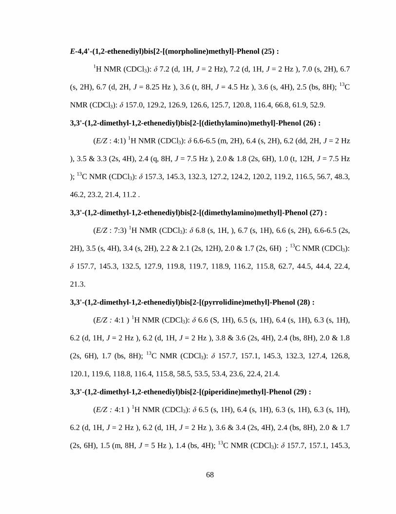
68
E-4,4'-(1,2-ethenediyl)bis[2-[(morpholine)methyl]-Phenol (25) :
1H NMR (CDCl3): δ 7.2 (d, 1H, J = 2 Hz), 7.2 (d, 1H, J = 2 Hz ), 7.0 (s, 2H), 6.7
(s, 2H), 6.7 (d, 2H, J = 8.25 Hz ), 3.6 (t, 8H, J = 4.5 Hz ), 3.6 (s, 4H), 2.5 (bs, 8H); 13
C
NMR (CDCl3): δ 157.0, 129.2, 126.9, 126.6, 125.7, 120.8, 116.4, 66.8, 61.9, 52.9.
3,3'-(1,2-dimethyl-1,2-ethenediyl)bis[2-[(diethylamino)methyl]-Phenol (26) :
(E/Z : 4:1) 1H NMR (CDCl3): δ 6.6-6.5 (m, 2H), 6.4 (s, 2H), 6.2 (dd, 2H, J = 2 Hz
), 3.5 & 3.3 (2s, 4H), 2.4 (q, 8H, J = 7.5 Hz ), 2.0 & 1.8 (2s, 6H), 1.0 (t, 12H, J = 7.5 Hz
); 13
C NMR (CDCl3): δ 157.3, 145.3, 132.3, 127.2, 124.2, 120.2, 119.2, 116.5, 56.7, 48.3,
46.2, 23.2, 21.4, 11.2 .
3,3'-(1,2-dimethyl-1,2-ethenediyl)bis[2-[(dimethylamino)methyl]-Phenol (27) :
(E/Z : 7:3) 1H NMR (CDCl3): δ 6.8 (s, 1H, ), 6.7 (s, 1H), 6.6 (s, 2H), 6.6-6.5 (2s,
2H), 3.5 (s, 4H), 3.4 (s, 2H), 2.2 & 2.1 (2s, 12H), 2.0 & 1.7 (2s, 6H) ; 13
C NMR (CDCl3):
δ 157.7, 145.3, 132.5, 127.9, 119.8, 119.7, 118.9, 116.2, 115.8, 62.7, 44.5, 44.4, 22.4,
21.3.
3,3'-(1,2-dimethyl-1,2-ethenediyl)bis[2-[(pyrrolidine)methyl]-Phenol (28) :
(E/Z : 4:1 ) 1H NMR (CDCl3): δ 6.6 (S, 1H), 6.5 (s, 1H), 6.4 (s, 1H), 6.3 (s, 1H),
6.2 (d, 1H, J = 2 Hz ), 6.2 (d, 1H, J = 2 Hz ), 3.8 & 3.6 (2s, 4H), 2.4 (bs, 8H), 2.0 & 1.8
(2s, 6H), 1.7 (bs, 8H); 13
C NMR (CDCl3): δ 157.7, 157.1, 145.3, 132.3, 127.4, 126.8,
120.1, 119.6, 118.8, 116.4, 115.8, 58.5, 53.5, 53.4, 23.6, 22.4, 21.4.
3,3'-(1,2-dimethyl-1,2-ethenediyl)bis[2-[(piperidine)methyl]-Phenol (29) :
(E/Z : 4:1 ) 1H NMR (CDCl3): δ 6.5 (s, 1H), 6.4 (s, 1H), 6.3 (s, 1H), 6.3 (s, 1H),
6.2 (d, 1H, J = 2 Hz ), 6.2 (d, 1H, J = 2 Hz ), 3.6 & 3.4 (2s, 4H), 2.4 (bs, 8H), 2.0 & 1.7
(2s, 6H), 1.5 (m, 8H, J = 5 Hz ), 1.4 (bs, 4H); 13
C NMR (CDCl3): δ 157.7, 157.1, 145.3,

69
145.1, 132.5, 132.3, 128.0, 127.3, 120.2, 119.4, 118.9, 118.7, 116.4, 115.8, 61.9, 61.7,
53.9, 53.8, 25.9, 24.0, 22.4, 21.4.
3,3'-(1,2-dimethyl-1,2-ethenediyl)bis[2-[(morpholine)methyl]-Phenol (30) :
(E/Z : 4.5:1.5 ) 1H NMR (CDCl3): δ 6.6 (s, 1H), 6.5 (s, 1H), 6.4 (s, 1H), 6.3 (s,
1H), 6.2 (d, 1H, J = 2 Hz ), 6.1 (d, 1H, J = 2 Hz ), 3.6 (t, 8H, J = 4.75 Hz ), 3.5 (s, 4H),
2.4 & 2.3 (bs, 8H), 2.0 & 1.7 (2s, 6H); 13
C NMR (CDCl3): δ 157.2, 156.6, 145.7, 145.3,
132.3, 128.5, 127.8, 120.5, 119.3, 118.5, 117.8, 116.6, 115.9, 66.8, 61.7, 61.6, 53.2, 52.8,
22.4, 21.4.
E-3,3'-(1,2-ethenediyl)bis[2-[(diethylamino)methyl]-Phenol (31) :
1H NMR (CDCl3): δ 6.9 (m, 6H), 6.8 (d, 2H, J = 2.75 Hz), 3.7 (s, 4H), 2.5 (q, 8H,
J = 7.25 Hz), 1.0 (t, 12H, J = 7.25 Hz) ; 13
C NMR (CDCl3): δ 158.4, 138.0, 128.6, 128.3,
121.6, 117.6, 113.7, 56.6, 46.3, 11.1.
E-3,3'-(1,2-ethenediyl)bis[2-[(dimethylamino)methyl]-Phenol (32) :
1H NMR (CDCl3): δ 7.0-6.9 (m, 8H), 3.6 (s, 4H), 2.2 (s, 12H);
13C NMR (CDCl3):
δ 158.2, 138.1, 128.5, 128.3, 121.5, 117.7, 113.5, 62.6, 44.5.
E-3,3'-(1,2-ethenediyl)bis[2-[(pyrrolidine)methyl]-Phenol (33) :
1H NMR (CDCl3): δ 6.9-6.9 (m, 4H), 6.8-6.8 (m, 4H), 3.7 (s, 4H), 2.5 (bs, 8H),
1.7 (bs, 8H); 13
C NMR (CDCl3): δ 158.2, 138.0, 128.3, 128.0, 122.1, 117.5, 113.4, 58.6,
53.5, 23.6.
E-3,3'-(1,2-ethenediyl)bis[2-[(piperidine)methyl]-Phenol (34) :
1H NMR (CDCl3): δ 7.0-6.8 (m, 8H), 3.7 (s, 4H), 2.5 (bs, 8H), 1.3-1.2 (m, 12H);
13C NMR (CDCl3): δ 158.2, 157.9, 157.8, 138.3, 138.0, 129.8, 128.6, 128.3, 128.2, 128.1,
121.2, 119.6, 117.6, 117.3, 166.3, 114.3, 113.5, 61.9, 54.4, 53.9, 25.8, 24.0.

70
E-3,3'-(1,2-ethenediyl)bis[2-[(morpholine)methyl]-Phenol (35) :
1H NMR (CDCl3): δ 7.2-6.9 (m, 8H), 3.7 (s, 8H), 3.6 (s, 4H), 2.6 (bs, 8H);
13C
NMR (CDCl3): δ 157.6, 138.3, 129.0, 128.4, 120.2, 118.0, 113.6, 66.8, 61.6, 52.9.
2.12.3 General procedure for the synthesis of stilbenebisoxazine
analogues:
Stilbene (1 equivalent) was taken in one neck round bottom flask and 15 mL of
methanol was added. Paraformaldehyde(4.2 equivalents) and appropriate amine(2
equivalents) was added. Then this reaction mixture was allowed to reflux until TLC (2:3
mixture of ethylacetate:hexane) shows that reaction is complete. Then mixture is cooled
to room temperature. Then the precipitation observed was collected by filtration and the
residue was washed with cold. Methanol several times. This solid received, pure product
in all cases, was dried in vacuuo and characterized by 1H and
13C NMR.
Representative procedure :
E-6,6'-(vinylene)bis[3-cyclohexyl-3,4-dihydro- 2H-1,3-Benzoxazine] (36) :
To a solution of 4,4’-dihydroxystilbene 8 (0.2 g, 0.952 mmol) in MeOH (15 mL)
was added paraformaldehyde (0.12 g, 4.0 mmol) and cyclohexylamine (0.22 mL, 1.904
mmol). The reaction mixture was refluxed for 8 hrs. Then reaction mixture was cooled to
room temperature. The resulting white color precipitate was collected by filtration and
washed with cold MeOH (5 mL). This precipitate was pure enough in NMR spectra and
obtained yield was 93% as white color precipitate. 1H NMR (400 MHz, CDCl3) 7.1 -
7.2 (m, 2H), 7.0 - 7.1 (m, 2H), 6.7 - 6.8 (m, 3H), 6.6 - 6.7 (m, 2H), 4.9 - 5.0 (m, 4H), 4.0 -
4.1 (m, 4H), 2.6 - 2.7 (m, 2H), 1.8 - 2.0 (m, 5H), 1.6 - 1.8 (m, 7H), 1.5 - 1.6 (m, 3H), 0.9 -

71
1.3 (m, 12H). 13
C NMR (101 MHz, CHLOROFORM-d) 154.8, 130.3, 126.4, 125.7,
124.9, 122.0, 116.9, 80.7, 59.0, 47.6, 31.8, 26.3, 25.7.
E-6,6'-(vinylene)bis[3-cyclopentyl-3,4-dihydro- 2H-1,3-Benzoxazine] (37):
1H NMR (400 MHz, CDCl3) 7.1 - 7.3 (m, 2H), 6.9 - 7.1 (m, 2H), 6.6 - 6.9 (m,
4H), 4.9 (br. s., 4H), 4.0 (br. s., 4H), 3.1 - 3.2 (m, 2H), 1.8 - 2.0 (m, 4H), 1.6 - 1.8 (m,
5H), 1.3 - 1.6 (m, 10H). 13
C NMR (101 MHz, CHLOROFORM-d) 154.2, 130.2, 126.1,
125.6, 125.1, 120.5, 116.5, 81.7, 59.5, 49.5, 31.4, 23.8.
E-6,6'-(vinylene)bis[3-benzyl-3,4-dihydro- 2H-1,3-Benzoxazine] (38):
1H NMR (400 MHz, CDCl3) 7.2 - 7.4 (m, 13H), 7.0 (m, 2H), 6.7 - 6.8 (m, 4H),
4.8 - 4.9 (m, 4H), 3.8 - 4.0 (m, 8H). 13
C NMR (101 MHz, CDCl3) 153.5, 130.5, 129.0,
128.5, 127.5, 126.2, 125.8, 125.4, 116.7, 82.2, 55.5, 49.6.
E-6,6'-(vinylene)bis[3-(4-methoxy benzyl)-3,4-dihydro- 2H-1,3-Benzoxazine] (39):
1H NMR (400 MHz, CDCl3) 7.2 - 7.3 (m, 6H), 7.0 - 7.1 (m, 2H), 6.8 - 6.9 (m,
12H), 4.8 - 4.9 (m, 4H), 4.1 - 4.1 (m, 4H), 3.9 - 4.0 (m, 4H), 3.7 - 3.8 (m, 7H). 13
C NMR
(101 MHz, CDCl3) 159.0, 153.7, 130.2, 127.6, 126.2, 125.7, 125.4, 120.0, 116.1, 113.8,
82.2, 55.3, 54.9, 49.5.
E-7,7'-(vinylene)bis[3-cyclohexyl-3,4-dihydro- 2H-1,3-Benzoxazine] (40):
1H NMR (400 MHz, CDCl3) 6.8 - 7.0 (m, 10H), 4.9 - 5.0 (m, 4H), 4.0 (s, 4H),
2.6 - 2.7 (m, 2H), 1.8 - 2.0 (m, 7H), 1.6 - 1.8 (m, 8H), 1.5 (dd, J = 3.71, 15.81 Hz, 4H),
0.9 - 1.3 (m, 17H). 13
C NMR (101 MHz, CDCl3) 155.2, 136.8, 127.9, 121.3, 118.7,
114.0, 80.3, 58.7, 47.3, 31.6, 25.9, 25.5.

72
E-7,7'-(vinylene)bis[3-cyclopentyl-3,4-dihydro- 2H-1,3-Benzoxazine] (41):
1H NMR (400 MHz, CDCl3) 6.7 - 7.0 (m, 8H), 4.8 - 4.9 (m, 4H), 3.9 - 4.0 (m,
4H), 3.0 - 3.2 (m, 2H), 1.8 - 2.0 (m, 5H), 1.3 - 1.7 (m, 16H). 13
C NMR (101 MHz,
CDCl3) 155.1, 137.2, 128.7, 127.8, 120.3, 119.1, 114.1, 81.8, 59.8, 49.7, 31.6, 24.0.
E-7,7'-(vinylene)bis[3-benzyl-3,4-dihydro- 2H-1,3-Benzoxazine] (42):
1H NMR (400 MHz, CDCl3) 7.1 - 7.4 (m, 21H), 6.8 - 7.0 (m, 11H), 4.8 - 4.9
(m, 4H), 3.8 - 4.0 (m, 9H), 3.6 - 3.7 (m, 3H). 13
C NMR (101 MHz, CDCl3) 154.3,
138.2, 137.2, 129.0, 128.5, 128.3, 128.2, 127.9, 127.4, 119.1, 114.0, 82.3, 73.7, 57.0,
55.7.
E-7,7'-(vinylene)bis[3-(4-methoxy benzyl)-3,4-dihydro- 2H-1,3-Benzoxazine] (43):
1H NMR (400 MHz, CDCl3) 7.2 - 7.3 (m, 6H), 6.8 - 7.0 (m, 10H), 4.8 - 4.9 (m,
4H), 3.9 (s, 4H), 3.8 (m, 5H), 3.7 - 3.8 (m, 9H). 13
C NMR (101 MHz, CDCl3) 159.2,
154.6, 137.4, 130.4, 119.7, 119.3, 114.2, 114.1, 113.8, 82.3, 55.5, 55.1, 49.1.
E-7,7'-(vinylene)bis[3-furfuryl-3,4-dihydro- 2H-1,3-Benzoxazine] (44):
1H NMR (400 MHz, CDCl3) 7.3 - 7.4 (m, 2H), 6.8 - 7.0 (m, 10H), 6.2 - 6.3 (m,
2H), 4.8 - 4.9 (m, 4H), 3.9 - 4.0 (m, 4H), 3.8 - 3.9 (m, 4H). 13
C NMR (101 MHz, CDCl3)
154.1, 151.5, 142.6, 137.2, 128.1, 119.2, 114.1, 110.2, 109.0, 81.9, 49.5, 48.2.
2.12 References
(1). (a). Bromberg, J.; Darnell, J. E. Oncogene 2000, 19, 2468. (b). Levy, D. E.;
Darnell, J. E. Nat Rev Mol Cell Bio 2002, 3, 651. (c). Fu, X. Y.; Schindler, C.;
Improta, T.; Aebersold, R.; Darnell, J. E., Jr. Proc. Natl. Acad. Sci. U. S. A. 1992,
89, 7840. (d). Schindler, C.; Fu, X. Y.; Improta, T.; Aebersold, R.; Darnell, J. E.,

73
Jr. Proc. Natl. Acad. Sci. U. S. A. 1992, 89, 7836. (e). Schindler, C.; Shuai, K.;
Prezioso, V. R.; Darnell, J. E., Jr. Science. 1992, 257, 809. (f). Greenlund, A. C.;
Morales, M. O.; Viviano, B. L.; Yan, H.; Krolewski, J.; Schreiber, R. D.
Immunity. 1995, 2, 677.
(2). Leonard, W. J.; O'Shea, J. J. Annu. Rev. Immunol. 1998, 16, 293.
(3). Rane, S. G.; Reddy, E. P. Oncogene. 2000, 19, 5662.
(4). (a) Saharinen, P.; Silvennoinen, O. J. Biol. Chem. 2002, 277, 47954. (b).
Saharinen, P.; Vihinen, M.; Silvennoinen, I. Mol Biol Cell 2003, 14, 1448.
(5). Luo, H.; Rose, P.; Barber, D.; Hanratty, W. P.; Lee, S.; Roberts, T. M.; D'Andrea,
A. D.; Dearolf, C. R. Mol. Cell. Biol. 1997, 17, 1562.
(6). (a). Girault, J. A.; Labesse, G.; Mornon, J. P.; Callebaut, I. Mol. Med. 1998, 4,
751. (b). Kampa, D.; Burnside, J. Biochem. Biophys. Res. Commun. 2000, 278,
175.(c). Tanner, J. W.; Chen, W.; Young, R. L.; Longmore, G. D.; Shaw, A. S. J.
Biol. Chem. 1995, 270, 6523. (d). Zhao, Y.; Wagner, F.; Frank, S. J.; Kraft, A. S.
J. Biol. Chem. 1995, 270, 13814.
(7). (a). Neubauer, H.; Cumano, A.; Muller, M.; Wu, H.; Huffstadt, U.; Pfeffer, K.
Cell. 1998, 93, 397. (b). Parganas, E.; Wang, D.; Stravopodis, D.; Topham, D. J.;
Marine, J. C.; Teglund, S.; Vanin, E. F.; Bodner, S.; Colamonici, O. R.; van
Deursen, J. M.; Grosveld, G.; Ihle, J. N. Cell. 1998, 93, 385.
(8). (a). Meydan, N.; Grunberger, T.; Dadi, H.; Shahar, M.; Arpaia, E.; Lapidot, Z.;
Leeder, J. S.; Freedman, M.; Cohen, A.; Gazit, A.; Levitzki, A.; Roifman, C. M.
Nature. 1996, 379, 645. (b). Podewski, E. K.; Hilfiker-Kleiner, D.; Hilfiker, A.;
Morawietz, H.; Lichtenberg, A.; Wollert, K. C.; Drexler, H. Circulation. 2003,

74
107, 798. (c). Sayeski, P. P.; Ali, M. S.; Safavi, A.; Lyles, M.; Kim, S. O.; Frank,
S. J.; Bernstein, K. E. J. Biol. Chem. 1999, 274, 33131. (d). Aringer, M.; Cheng,
A.; Nelson, J. W.; Chen, M.; Sudarshan, C.; Zhou, Y. J.; O'Shea, J. J. Life Sci.
1999, 64, 2173. (e). Schindler, C. W. J. Clin. Inv. 2002, 109, 1133.
(9). Levine, R. L.; Wadleigh, M.; Cools, J.; Ebert, B. L.; Wernig, G.; Huntly, B. J.;
Boggon, T. J.; Wlodarska, I.; Clark, J. J.; Moore, S.; Adelsperger, J.; Koo, S.;
Lee, J. C.; Gabriel, S.; Mercher, T.; D'Andrea, A.; Frohling, S.; Dohner, K.;
Marynen, P.; Vandenberghe, P.; Mesa, R. A.; Tefferi, A.; Griffin, J. D.; Eck, M.
J.; Sellers, W. R.; Meyerson, M.; Golub, T. R.; Lee, S. J.; Gilliland, D. G. Cancer.
Cell. 2005, 7, 387.
(10). (a). Baxter, E. J.; Scott, L. M.; Campbell, P. J.; East, C.; Fourouclas, N.; Swanton,
S.; Vassiliou, G. S.; Bench, A. J.; Boyd, E. M.; Curtin, N.; Scott, M. A.; Erber, W.
N.; Green, A. R. Lancet. 2005, 365, 1054. (b). Levine, R. L.; Loriaux, M.; Huntly,
B. J.; Loh, M. L.; Beran, M.; Stoffregen, E.; Berger, R.; Clark, J. J.; Willis, S. G.;
Nguyen, K. T.; Flores, N. J.; Estey, E.; Gattermann, N.; Armstrong, S.; Look, A.
T.; Griffin, J. D.; Bernard, O. A.; Heinrich, M. C.; Gilliland, D. G.; Druker, B.;
Deininger, M. W. Blood. 2005, 106, 3377. (c). James, C.; Ugo, V.; Le Couedic, J.
P.; Staerk, J.; Delhommeau, F.; Lacout, C.; Garcon, L.; Raslova, H.; Berger, R.;
Bennaceur-Griscelli, A.; Villeval, J. L.; Constantinescu, S. N.; Casadevall, N.;
Vainchenker, W. Nature. 2005, 434, 1144. (d). Kralovics, R.; Passamonti, F.;
Buser, A. S.; Teo, S. S.; Tiedt, R.; Passweg, J. R.; Tichelli, A.; Cazzola, M.;
Skoda, R. C. N. Eng. J. Med. 2005, 352, 1779. (e). Zhao, R.; Xing, S.; Li, Z.; Fu,
X.; Li, Q.; Krantz, S. B.; Zhao, Z. J. J. Biol. Chem. 2005, 280, 22788. (f).

75
Pardanani, A. Leukemia. 2008, 22, 23. (g). Levine, R. L.; Pardanani, A.; Tefferi,
A.; Gilliland, D. G. Nat. Rev. Cancer. 2007, 7, 673.
(11). (a) Ward, A. C.; Touw, I.; Yoshimura, A. Blood. 2000, 95, 19. (b). Verma, A.;
Kambhampati, S.; Parmar, S.; Platanias, L. C. Cancer Metastasis Rev. 2003, 22,
423.
(12). Shuai, K.; Liu, B. Nature Reviews Immunology 2003, 3, 900.
(13). van den Akker, E.; van Dijk, T. B.; Schmidt, U.; Felida, L.; Beug, H.; Lowenberg,
B.; von Lindern, M. Biol. Chem. 2004, 385, 409.
(14). Burger, R.; Le Gouill, S.; Tai, Y. T.; Shringarpure, R.; Tassone, P.; Neri, P.;
Podar, K.; Catley, L.; Hideshima, T.; Chauhan, D.; Caulder, E.; Neilan, C. L.;
Vaddi, K.; Li, J.; Gramatzki, M.; Fridman, J. S.; Anderson, K. C. Mol. Cancer
Ther. 2009, 8, 26.
(15). Verstovsek, S.; Manshouri, T.; Quintas-Cardama, A.; Harris, D.; Cortes, J.; Giles,
F. J.; Kantarjian, H.; Priebe, W.; Estrov, Z. Clin. Cancer. Res. 2008, 14, 788.
(16). Duan, Z.; Bradner, J.; Greenberg, E.; Mazitschek, R.; Foster, R.; Mahoney, J.;
Seiden, M. V. Mol. Pharmacol. 2007, 72, 1137.
(17). Antonysamy, S.; Hirst, G.; Park, F.; Sprengeler, P.; Stappenbeck, F.; Steensma,
R.; Wilson, M.; Wong, M. Bioorg. Med. Chem. Lett. 2009, 19, 279.
(18). (a). Burns, C. J.; Bourke, D. G.; Andrau, L.; Bu, X.; Charman, S. A.; Donohue, A.
C.; Fantino, E.; Farrugia, M.; Feutrill, J. T.; Joffe, M.; Kling, M. R.; Kurek, M.;
Nero, T. L.; Nguyen, T.; Palmer, J. T.; Phillips, I.; Shackleford, D. M.; Sikanyika,
H.; Styles, M.; Su, S.; Treutlein, H.; Zeng, J.; Wilks, A. F. Bioorg. Med. Chem.
76
Lett. 2009, 19, 5887. (b). Pardanani, A.; Lasho, T.; Smith, G.; Burns, C. J.;
Fantino, E.; Tefferi, A. Leukemia. 2009, 23, 1441.
(19). Hexner, E. O.; Serdikoff, C.; Jan, M.; Swider, C. R.; Robinson, C.; Yang, S.;
Angeles, T.; Emerson, S. G.; Carroll, M.; Ruggeri, B.; Dobrzanski, P. Blood.
2008, 111, 5663.
(20). (a). Osherov, N.; Gazit, A.; Gilon, C.; Levitzki, A. J. Biol. Chem. 1993, 268,
11134. (b). Gu, Y.; Zou, Y.; Aikawa, R.; Hayashi, D.; Kudoh, S.; Yamauchi, T.;
Uozumi, H.; Zhu, W.; Kadowaki, T.; Yazaki, Y.; Komuro, I. Mol. Cell. Biochem.
2001, 223, 35. (c). Oda, Y.; Renaux, B.; Bjorge, J.; Saifeddine, M.; Fujita, D. J.;
Hollenberg, M. D. Can. J. Physiol. Pharmacol. 1999, 77, 606.
(21). Inayat-Hussain, S. H.; Thomas, N. F. Expert Opin Ther Pat 2004, 14, 819.
(22). (a). Eddarir, S.; Abdelhadi, Z.; Rolando, C. Tet. Lett. 2001, 42, 9127. (b).
Adesanya, S. A.; Nia, R.; Martin, M.-T.; Boukamcha, N.; Montagnac, A.; Païs,
M. J. Nat. Prod. 1999, 62, 1694. (c). Soleas, G.; Diamandis, E.; Goldberg, D.
Clin. Biochem. 1997, 30, 91.
(23). Jang, M. S.; Cai, E. N.; Udeani, G. O.; Slowing, K. V.; Thomas, C. F.; Beecher,
C. W. W.; Fong, H. H. S.; Farnsworth, N. R.; Kinghorn, A. D.; Mehta, R. G.;
Moon, R. C.; Pezzuto, J. M. Science 1997, 275, 218.
(24). (a). Aziz, M. H.; Kumar, R.; Ahmad, N. Int. J. Oncol. 2003, 23, 17. (b). Wolter,
F.; Stein, J. Drug Future 2002, 27, 949. (c). Wu, J. M.; Wang, Z. R.; Hsieh, T. C.;
Bruder, J. L.; Zou, J. G.; Huang, Y. Z. Int. J. Mol. Med. 2001, 8, 3. (d). Fremont,
L. Life Sci. 2000, 66, 663.

77
(25). (a). Jang, M.; Cai, L.; Udeani, G. O.; Slowing, K. V.; Thomas, C. F.; Beecher, C.
W.; Fong, H. H.; Farnsworth, N. R.; Kinghorn, A. D.; Mehta, R. G.; Moon, R. C.;
Pezzuto, J. M. Science. 1997, 275, 218. (b). Schneider, Y.; Vincent, F.; Duranton,
B.; Badolo, L.; Gosse, F.; Bergmann, C.; Seiler, N.; Raul, F. Cancer Lett. 2000,
158, 85.
(26). Docherty, J. J.; Fu, M. M. H.; Stiffler, B. S.; Limperos, R. J.; Pokabla, C. M.;
DeLucia, A. L. Antivir. Res. 1999, 43, 145.
(27). (a). Bowers, J. L.; Tyulmenkov, V. V.; Jernigan, S. C.; Klinge, C. M.
Endocrinology. 2000, 141, 3657. (b). Gehm, B. D.; McAndrews, J. M.; Chien, P.
Y.; Jameson, J. L. P Natl Acad Sci USA 1997, 94, 14138.
(28). (a). Wang, Z. R.; Huang, Y. Z.; Zou, J. G.; Cao, K. J.; Xu, Y. N.; Wu, J. M. Int. J.
Mol. Med. 2002, 9, 77. (b). Wang, Z. R.; Zou, J. G.; Huang, Y. Z.; Cao, K. J.; Xu,
Y. N.; Wu, J. M. Chinese Med. J-Peking. 2002, 115, 378.
(29). (a). Lee, H. J.; Seo, J. W.; Lee, B. H.; Chung, K. H.; Chi, D. Y. Bioorg. Med.
Chem. Lett. 2004, 14, 463. (b). Babich, H.; Reisbaum, A. G.; Zuckerbraun, H. L.
Toxicol. Lett. 2000, 114, 143.
(30). Pace-Asciak, C. R.; Hahn, S.; Diamandis, E. P.; Soleas, G.; Goldberg, D. M.
Clinica. Chimica. Acta. 1995, 235, 207.
(31). (a). Geahlen, R. L.; Mclaughlin, J. L. Biochem. Bioph. Res. Co. 1989, 165, 241.
(b). Swanson-Mungerson, M.; Ikeda, M.; Lev, L.; Longnecker, R.; Portis, T. J.
Antimicrob. Chemoth. 2003, 52, 152.
(32). (a). Langcake, P.; Cornford, C. A.; Pryce, R. J. Phytochemistry 1979, 18, 1025.
(b). Langcake, P.; Pryce, R. J. Physiol Plant Pathol 1976, 9, 77. (c). Breuil, A. C.;

78
Adrian, M.; Pirio, N.; Meunier, P.; Bessis, R.; Jeandet, P. Tetrahedron Lett 1998,
39, 537. (d). Wyrzykiewicz, E.; Wendzonka, M.; Kedzia, B. Eur J Med Chem
2006, 41, 519.
(33). (a). Renaud, S.; Delorgeril, M. Lancet. 1992, 339, 1523. (b). Frankel, E. N.;
Waterhouse, A. L.; Kinsella, J. E. Lancet. 1993, 341, 1103. (c). Frankel, E. N.;
Waterhouse, A. L.; Teissedre, P. L. J. Agr. Food Chem. 1995, 43, 890. (d).
Fauconneau, B.; WaffoTeguo, P.; Huguet, F.; Barrier, L.; Decendit, A.; Merillon,
J. M. Life Sci. 1997, 61, 2103. (e). Vitrac, X.; Desmouliere, A.; Brouillaud, B.;
Krisa, S.; Deffieux, G.; Barthe, N.; Rosenbaum, J.; Merillon, J. M. Life Sci. 2003,
72, 2219.
(34). (a). Pettit, G. R.; Grealish, M. P.; Jung, M. K.; Hamel, E.; Pettit, R. K.; Chapuis,
J. C.; Schmidt, J. M. J. Med. Chem. 2002, 45, 2534. (b). Orsini, F.; Pelizzoni, F.;
Verotta, L.; Aburjai, T.; Rogers, C. B. J. Nat. Prod. 1997, 60, 1082. (c). Bolton,
R. J. Labelled Compd. Radiopharm. 2002, 45, 485. (d). Lasne, M. C.; Perrio, C.;
Rouden, J.; Barre, L.; Roeda, D.; Dolle, F.; Crouzel, C. Top Curr Chem 2002,
222, 201.
(35). Yim, N.; Do, T. H.; Trung, T. N.; Kim, J. P.; Lee, S.; Na, M.; Jung, H.; Kim, H.
S.; Kim, Y. H.; Bae, K. Bioorg Med Chem Lett. 2010, 20, 1165.
(36). Subramanian, M.; Shadakshari, U.; Chattopadhyay, S. Bioorg. Med. Chem. 2004,
12, 1231.
(37). Fukuhara, K.; Miyata, N. Bioorg. Med. Chem. Lett.. 1998, 8, 3187.
(38). (a). Wyrzykiewicz, E.; Blaszczyk, A.; Kedzia, B. Farmaco 2000, 55, 151. (b).
Aslam, S. N.; Stevenson, P. C.; Kokubun, T.; Hall, D. R. Microbiol Res 2009,

79
164, 191. (c). Lin, L. G.; Yang, X. Z.; Tang, C. P.; Ke, C. Q.; Zhang, J. B.; Ye, Y.
Phytochemistry 2008, 69, 457.
(39). (a). Kimura, Y. In Vivo. 2005, 19, 37. (b). Larrosa, M.; Tomas-Barberan, F. A.;
Espin, J. C. J. Agr. Food Chem. 2003, 51, 4576. (c). Aggarwal, B. B.; Bhardwaj,
A.; Aggarwal, R. S.; Seeram, N. P.; Shishodia, S.; Takada, Y. Anticancer Res.
2004, 24, 2783. (d). Le Corre, L.; Chalabi, N.; Delort, L.; Bignon, Y. J.; Bernard-
Gallon, D. J. Molecular Nutrition & Food Research 2005, 49, 462.
(40). (a). Lin, C. M.; Ho, H. H.; Pettit, G. R.; Hamel, E. Biochemistry. 1989, 28, 6984.
(b). Lin, C. M.; Singh, S. B.; Chu, P. S.; Dempcy, R. O.; Schmidt, J. M.; Pettit, G.
R.; Hamel, E. Mol. Pharmacol. 1988, 34, 200. (c). Pettit, G. R.; Singh, S. B.;
Schmidt, J. M.; Niven, M. L.; Hamel, E.; Lin, C. M. J. Nat. Prod. 1988, 51, 517.
(d). Pettit, G. R.; Rhodes, M. R.; Herald, D. L.; Chaplin, D. J.; Stratford, M. R. L.;
Hamel, E.; Pettit, R. K.; Chapuis, J. C.; Oliva, D. Anti-Cancer Drug Des. 1998,
13, 981.
(41). (a). Chaplin, D. J.; Hill, S. A.; Dougherty, G. J.; Prise, V. E.; Tozer, G. M.; Pettit,
R. Hum. Gene Ther. 1999, 10, 840. (b). Chaplin, D. J.; Dougherty, G. J. Brit. J.
Cancer 1999, 80, 57. (c). Chaplin, D. J.; Pettit, G. R.; Hill, S. A. Anticancer Res.
1999, 19, 189. (d). Tozer, G. M.; Prise, V. E.; Wilson, I.; Vojnovic, B.; Chaplin,
D. J. J. Vasc. Res. 1999, 36, 328. (e). Siemann, D. W.; Chaplin, D. J.; Walicke, P.
A. Expert. Opin. Investig. Drugs 2009, 18, 189. (f). Mooney, C. J.; Nagaiah, G.;
Fu, P. F.; Wasman, J. K.; Cooney, M. M.; Savvides, P. S.; Bokar, J. A.; Dowlati,
A.; Wang, D.; Agarwala, S. S.; Flick, S. M.; Hartman, P. H.; Ortiz, J. D.; Lavertu,
P. N.; Remick, S. C. Thyroid 2009, 19, 233.

80
(42). Kalach, J.; Jolypharaboz, M.; Chantepie, J.; Nicolas, B.; Descotes, F.; Mauduit,
C.; Benahmed, M.; Andre, J. The Journal of Steroid Biochemistry and Molecular
Biology. 2005, 96, 119.
(43). D'Amato, R. J.; Lin, C. M.; Flynn, E.; Folkman, J.; Hamel, E. Proc. Natl. Acad.
Sci. U. S. A. 1994, 91, 3964.
(44). Ferrigni, N. R.; Mclaughlin, J. L.; Powell, R. G.; Smith, C. R. J. Nat. Prod. 1984,
47, 347.
(45). Wieder, T.; Prokop, A.; Bagci, B.; Essmann, F.; Bernicke, D.; Schulze-Osthoff,
K.; Dorken, B.; Schmalz, H. G.; Daniel, P. T.; Henze, G. Leukemia. 2001, 15,
1735.
(46). Thakkar, K.; Geahlen, R. L.; Cushman, M. J. Med. Chem. 1993, 36, 2950.
(47). Ashikawa, K.; Majumdar, S.; Banerjee, S.; Bharti, A. C.; Shishodia, S.;
Aggarwal, B. B. J. Immunol. 2002, 169, 6490.
(48). (a). Jordan, V. C. Lancet Oncol, 2000, 1, 43. (b). Jordan, V. C. J. Natl. Cancer
Inst. 2000, 92, 92. (c). Schafer, J. M.; Lee, E. S.; O'Regan, R. M.; Yao, K.;
Jordan, V. C. Clin. Cancer. Res. 2000, 6, 4373.
(49). Jordan, V. C. J. Steroid Biochem. Mol. Biol. 2000, 74, 269.
(50). Jordan, V. C. Br. J. Pharmacol. 2006, 147 Suppl 1, S269.
(51). (a). Daniel, C. P.; Gaskell, S. J.; Bishop, H.; Nicholson, R. I. J. Endocrinol. 1979,
83, 401. (b). Jordan, V. C. Breast Cancer Res. Treat. 1982, 2, 123.
(52). Gauthier, S.; Mailhot, J.; Labrie, F. J. Org. Chem. 1996, 61, 3890.
(53). Schafer, J. M.; Liu, H.; Bentrem, D. J.; Zapf, J. W.; Jordan, V. C. Cancer Res.
2000, 60, 5097.

81
(54). (a). Jordan, V. C.; Collins, M. M.; Rowsby, L.; Prestwich, G. J. Endocrinol. 1977,
75, 305. (b). Binart, N.; Catelli, M. G.; Geynet, C.; Puri, V.; Hahnel, R.; Mester,
J.; Baulieu, E. E. Biochem. Biophys. Res. Commun. 1979, 91, 812.
(55). Robertson, D. W.; Katzenellenbogen, J. A.; Long, D. J.; Rorke, E. A.;
Katzenellenbogen, B. S. J. Steroid Biochem. 1982, 16, 1.
(56). Valavaara, R.; Pyrhonen, S.; Heikkinen, M.; Rissanen, P.; Blanco, G.; Tholix, E.;
Nordman, E.; Taskinen, P.; Holsti, L.; Hajba, A. Eur. J. Cancer Clin. Oncol.
1988, 24, 785.
(57). Valavaara, R.; Kangas, L. Ann. Clin. Res. 1988, 20, 380.
(58). (a). Rauschning, W.; Pritchard, K. I. Breast Cancer Res. Treat. 1994, 31, 83. (b).
Bruning, P. F. Eur. J. Cancer 1992, 28A, 1404.
(59). Gauthier, S.; Sanceau, J. Y.; Mailhot, J.; Caron, B.; Cloutier, J. Tetrahedron.
2000, 56, 703.
(60). (a). Detsi, A.; Koufaki, M.; Calogeropoulou, T. J. Org. Chem. 2002, 67, 4608.
(b). Tanpure, R. P.; Harkrider, A. R.; Strecker, T. E.; Hamel, E.; Trawick, M. L.;
Pinney, K. G. Bioorg. Med. Chem. 2009, 17, 6993.
(61). Ferrefilmon, K.; Delaude, L.; Demonceau, A.; Noels, A. coord. chem. rev. 2004,
248, 2323.
(62). (a) Guiso, M.; Marra, C.; Farina, A. Tet. Lett. 2002, 43, 597. (b) Ziegler, C. B.;
Heck, R. F. J. Org. Chem. 1978, 43, 2941.
(63). Sengupta, S.; Bhattacharyya, S.; Kumar Sadhukhan, S. J. Chem. Soc., Perkin
Trans. 1. 1998, 275.
(64). Andrus, M. B.; Song, C. Org. Lett. 2001, 3, 3761.

82
(65). Kikukawa, K. J. Organomet. Chem. 1986, 311, C44.
(66). Nishibayashi, Y. J. Organomet. Chem. 1996, 507, 197.
(67). Ferre-Filmon, K.; Delaude, L.; Demonceau, A.; Noels, A. F. Eur. J. Org. Chem.
2005, 3319.
(68). Fox, H. H.; Schrock, R. R.; Odell, R. Organometallics. 1994, 13, 635.
(69). Chang, S.; Na, Y.; Shin, H. J.; Choi, E.; Jeong, L. S. Tet. Lett. 2002, 43, 7445.
(70). Schmalz, H.-G.; Velder, J.; Ritter, S.; Lex, J. Synthesis. 2006, 273.
(71). Pettit, G. R.; Anderson, C. R.; Herald, D. L.; Jung, M. K.; Lee, D. J.; Hamel, E.;
Pettit, R. K. J. Med. Chem. 2003, 46, 525.
(72). Heynekamp, J. J.; Weber, W. M.; Hunsaker, L. A.; Gonzales, A. M.; Orlando, R.
A.; Deck, L. M.; Vander Jagt, D. L. J. Med. Chem. 2006, 49, 7182.
(73). (a). Coe, P. L.; Scriven, C. E. J. Chem. Soc., Perkin Trans. 1. 1986, 475. (b).
McMurry, J. E. Chem. Rev. 1989, 89, 1513.
(74). McMurry, J. E. Acc. Chem. Res. 1983, 16, 405.
(75). Sukwattanasinitt, M.; Rojanathanes, R.; Tuntulani, T.; Sritana-Anant, Y.;
Ruangpornvisuti, V. Tet. Lett. 2001, 42, 5291.
(76). Ali, M. A.; Kondo, K.; Tsuda, Y. Chem. Pharm. Bull. 1992, 40, 1130.
(77). Kiss, R.; Polgar, T.; Kirabo, A.; Sayyah, J.; Figueroa, N. C.; List, A. F.; Sokol, L.;
Zuckerman, K. S.; Gali, M.; Bisht, K. S.; Sayeski, P. P.; Keseru, G. M. Bioorg.
Med. Chem. Lett. 2009, 19, 3598.
(78). Majumder, A.; Govindasamy, L.; Magis, A.; Kiss, R.; Polgar, T.; Baskin, R.;
Allan, R. W.; Agbandje-McKenna, M.; Reuther, G. W.; Keseru, G. M.; Bisht, K.
S.; Sayeski, P. P. J. Biol. Chem. 2010, 285, 31399.
83
(79). (a). White, W. A.; Ludwig, N. H. J. Agr. Food Chem. 1971, 19, 388(b) Leimner,
J.; Weyerstahl, P. Chem. Ber. 1982, 115, 3697.
(80). (a). Mcmurry, J. E. Acc. Chem. Res. 1974, 7, 281. (b). Mcmurry, J. E.; Fleming,
M. P. J. Am. Chem. Soc. 1974, 96, 4708.
(81). Mayekar, N. V.; Chattopadhyay, S.; Nayak, S. K. Letters in Organic Chemistry
2004, 1, 203.
(82). Chen, H.-B.; Yin, J.; Wang, Y.; Pei, J. Org. Lett. 2008, 10, 3113.
(83). Some, S.; Dutta, B.; Ray, J. K. Tet. Lett. 2006, 47, 1221.

84
Chapter 3
Synthesis of Novel Ketamine Analogues and Their Activity at GABAA and NMDA
Receptors.
3.1 Introduction
Ligand gated ion channels are membrane-embedded proteins at synaptic cleft
which controls intercommunication among neurons. Each receptor has a function of
allowing the flow of ions like Cl-, Na
+, K
+ resulting in excitation or inhibition. When a
neurotransmitter like -aminobutyric acid (GABA) binds to its respective receptor-
channel (GABA receptors), chloride ions flow through the receptor 1 channel reducing
the likelihood of initiating action potentials. GABAa receptors2 are most ubiquitously
expressed ligand gated Cl- ion channels in the mammalian central nervous system.
3 These
receptors are assembled from a diverse family of 19 homologous subunits4 ((1-6), (1-3),
(1-3), , and (1-3)), which are differentially expressed throughout the brain.5 The
GABAa receptor is a pentameric complex composed of closely related subunits in 2:2:1
stoichiometry in which , and subunits expressed as most abundant subunits.6 From
previous studies with the transgenic mice and with subtype selective subunits, it has been
established that while 1-containing receptors are responsible for mediating sedative and
muscle relaxant properties, 3 and/or 2-containing receptors are responsible for anxiety.
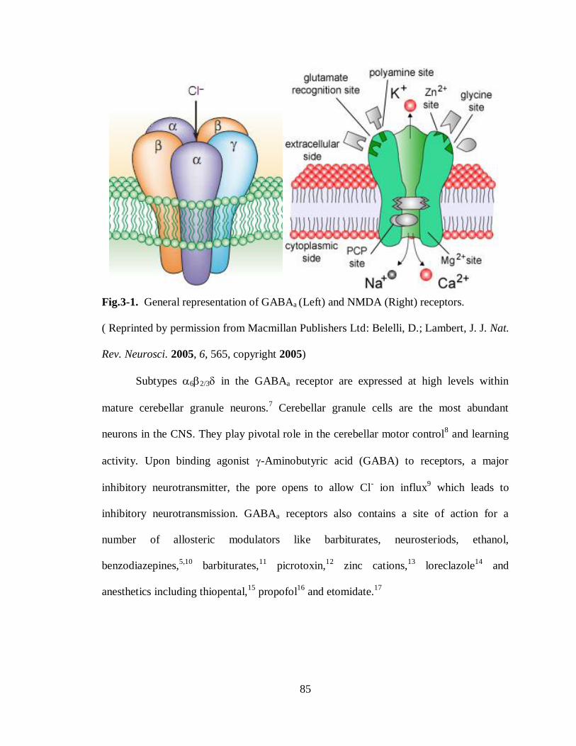
85
Fig.3-1. General representation of GABAa (Left) and NMDA (Right) receptors.
( Reprinted by permission from Macmillan Publishers Ltd: Belelli, D.; Lambert, J. J. Nat.
Rev. Neurosci. 2005, 6, 565, copyright 2005)
Subtypes 62/3 in the GABAa receptor are expressed at high levels within
mature cerebellar granule neurons.7 Cerebellar granule cells are the most abundant
neurons in the CNS. They play pivotal role in the cerebellar motor control8 and learning
activity. Upon binding agonist -Aminobutyric acid (GABA) to receptors, a major
inhibitory neurotransmitter, the pore opens to allow Cl- ion influx
9 which leads to
inhibitory neurotransmission. GABAa receptors also contains a site of action for a
number of allosteric modulators like barbiturates, neurosteriods, ethanol,
benzodiazepines,5,10
barbiturates,11
picrotoxin,12
zinc cations,13
loreclazole14
and
anesthetics including thiopental,15
propofol16
and etomidate.17

86
In contrast N-methyl-D-aspartate (NMDA) receptors18
are glycine and glutamate
gated receptors. When a neurotransmitter binds to NMDA receptors, the pore opens to
allow influx of cations19
like a Ca+ ions which are implicated in synaptic plasticity and
learning.20
The influx of Ca+
ions into neuronal cell results membrane depolarization
causing the propagation of excitory signal and also triggering intracellular signaling
pathways.21
Overactivation of these receptors has been implicated in conditions such as
ischemic stroke and traumatic brain injury,22
where the glutamate levels are elevated. In a
central nervous system (CNS), subunits like NR1, NR2 and NR3 forms the
heterotetrameric NMDA receptor complex. 23
Two NR1 and two NR2A or NR2B
subunits assembles in a hetereotetrameric complex to form most common NMDA
receptors in adult CNS. In general, NMDA receptors contributes to differing biophysical
and pharmacological properties.23
Ketamine and Phencyclidine (PCP) (Fig. 3-2), known
as dissociative anesthetics, produce profound analgesia at low doses to a unique
cardiovascular stimulation and a cataleptic state at higher doses.24
Both ketamine and
PCP, primarily acting at NMDA receptors as uncompetitive antagonists25
via reducing
excitory input, show differing CNS depression levels. PCP shows markedly higher
affinity26
for this target than ketamine while the ketamine possesses a higher CNS
depressant activity and produce a better quality of anesthesia than PCP.27
Neurotransmitter systems like dopamine D2 receptors,28
5-HT2 receptors,29
mu and
kappa opioid receptors,30
sigma receptors and muscarinic cholinergic receptors,31
voltage
gated K+, Na
+ and Ca
2+ channels
32 are also shown to get affected by ketamine and PCP
besides NMDA receptors though at significantly higher concentrations.

87
Fig.3-2. Structures of Ketamine and Phencyclidine (PCP).
Studies have yet to find about the mechanism by which ketamine produces a
higher CNS depression than PCP while at anesthetic doses.33
Dose-dependent side
effects like vivid dreams, illusions, disruptions of cognitive function, mood changes have
been reported with ketamine administration which are also observed in schizophrenia.34
The psychotomimetic effects of the ketamine have been shown or proposed to mediated
by its inhibitory effect on GABAergic interneurons leading to increase in the glutamate
release.35
Administration of subanesthetic doses of the ketamine shown to have
therapeutic efficacy for the treatment resistant major depression.36
Other symptoms
associated with ketamine like saccade, nystagmus, hyperlocomotion and prepulse
inhibition deficit found to be a model system37
to study schizophrenia and to identify
even hit-to-lead compounds for the inhibition of a ketamine induced hyperlocomotion for
the schizophrenic studies in animals. However, the spectrum of ketamine analogues38
is
narrow so far in terms of broadening ketamine’s desired effects to improve clinical
actions, decreasing its incidence of side effects and therapeutic efficacy on the selective
affinity towards receptors.
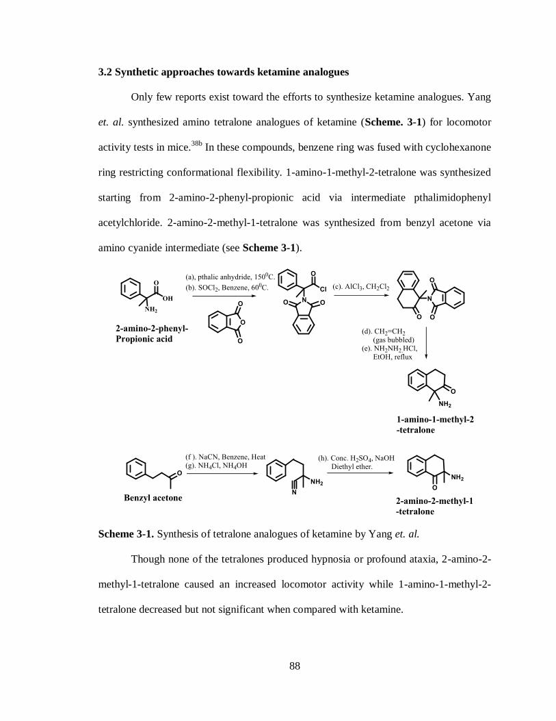
88
3.2 Synthetic approaches towards ketamine analogues
Only few reports exist toward the efforts to synthesize ketamine analogues. Yang
et. al. synthesized amino tetralone analogues of ketamine (Scheme. 3-1) for locomotor
activity tests in mice.38b
In these compounds, benzene ring was fused with cyclohexanone
ring restricting conformational flexibility. 1-amino-1-methyl-2-tetralone was synthesized
starting from 2-amino-2-phenyl-propionic acid via intermediate pthalimidophenyl
acetylchloride. 2-amino-2-methyl-1-tetralone was synthesized from benzyl acetone via
amino cyanide intermediate (see Scheme 3-1).
Scheme 3-1. Synthesis of tetralone analogues of ketamine by Yang et. al.
Though none of the tetralones produced hypnosia or profound ataxia, 2-amino-2-
methyl-1-tetralone caused an increased locomotor activity while 1-amino-1-methyl-2-
tetralone decreased but not significant when compared with ketamine.

89
Recently, Zarantonello et.al. reported novel ketamine analogues. In their strategy,
ketamine was modified by introducing amide in cyclohexanone ring and with various
tertiary and secondary amines attached in the place of methylamine for evaluation as
NMDA receptor antagonists.39
Ketamine analogues A and B, which have amide group in
cyclohexanone ring (Scheme. 3-2) were synthesized from chlorophenyl amine acetic
acid. Intermediate amino nitrile was synthesized from chlorophenyl amineacetic acid by
alkylation. Upon reductive cyclization of this intermediate in the presence of CoCl2 and
NaBH4, the resulting cyclic amide was further methylated with methyl iodide to isolate
novel analogues A and B as a mixture which were separated by HPLC purification.
Scheme 3-2. Synthesis of lactam with methylamine analogues of ketamine by
Zarantonello et. al.

90
In a similar strategy, analogues of phencyclidine (C and D) were synthesized
from bromophenyl methylacetate via piperidine nitrile intermediate (see Scheme 3-3 ).
Nitrile group in this intermediate was then reduced and cyclized in the case of synthesis
of six membered ring as in C and cyclized directly by reductive hydrogenation in the case
of synthesis of 5 membered ring as in D. Methylated amides ( E and F ) were synthesized
from the amides (C and D ) by methylation.
The evaluation of A and B against NMDA receptor with NR2-A and NR2-B
subunits showed that amide functionality in cyclohexanone ring was not tolerated. The
same loss of functional activity was observed even in the case of C-F in comparison to
ketamine.
Scheme 3-3. Synthesis of lactam with piperidine analogues of ketamine by Zarantonello
et. al.
From this structure and activity relationship studies, any appreciable activity was
observed only in the analogue (G) in which cyclohexyl ring was preserved and phenyl
group has methoxy substituent (Scheme 3-4).

91
Scheme 3-4. Synthesis of ketamine analogue with phenyl group substitution.
3.3 Synthesis of Novel ketamine analogues:
Ketamine analogues with modification of cyclohexyl ring to amide39
(Scheme 3-
2 and 3-3) or fused cyclohexane with benzene38b
(Scheme 3-1) resulted in a loss of
functional activity. The functional activity was sustained only when cyclohexyl ring is
preserved (Scheme. 3-4) in ketamine or phencyclidine. When the phenyl group was
introduced with various substitutions, the activity was modestly retained in comparison
with ketamine. So to synthesize novel analogues of ketamine with cyclohexyl ring intact,
we transformed the carbonyl group in cyclohexane to few other analogues with imine
substitutions as depicted in Scheme 3-5 and Table 3-1.
Scheme 3-5. Synthesis of ketamine analogues. See Table 3-1 for Appropriate reagents
and conditions.
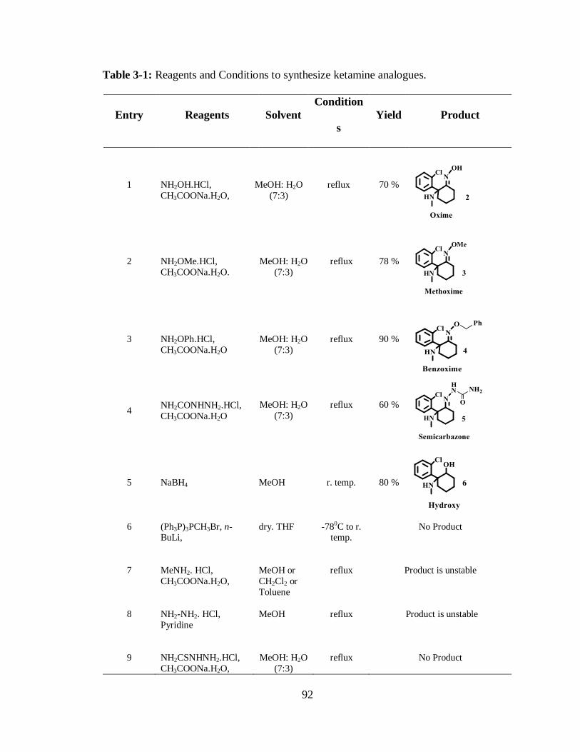
92
Table 3-1: Reagents and Conditions to synthesize ketamine analogues.
Entry Reagents Solvent
Condition
s
Yield Product
1
NH2OH.HCl,
CH3COONa.H2O,
MeOH: H2O
(7:3)
reflux 70 %
2
NH2OMe.HCl,
CH3COONa.H2O.
MeOH: H2O
(7:3)
reflux 78 %
3
NH2OPh.HCl,
CH3COONa.H2O
MeOH: H2O
(7:3)
reflux 90 %
4 NH2CONHNH2.HCl,
CH3COONa.H2O
MeOH: H2O
(7:3)
reflux
60 %
5 NaBH4
MeOH r. temp. 80 %
6 (Ph3P)3PCH3Br, n-BuLi,
dry. THF -780C to r. temp.
No Product
7 MeNH2. HCl,
CH3COONa.H2O,
MeOH or
CH2Cl2 or
Toluene
reflux Product is unstable
8 NH2-NH2. HCl,
Pyridine
MeOH
reflux Product is unstable
9 NH2CSNHNH2.HCl,
CH3COONa.H2O,
MeOH: H2O
(7:3)
reflux No Product

93
Ketamine carbonyl group was converted to the oxime by treating with
hydroxylamine in the presence of sodium acetate at reflux to synthesize oxime 2 in 70%
yield as a white color solid. The structure of the oxime 2 was confirmed by its NMR
spectra (Fig. 3-3). In a 1H NMR spectrum, methylene hydrogens (Ha and Ha’) next to
C=N group showed resonance at 2.5 and 2.8 ppm. Carbon in C=N-OH groups showed
resonance at 161 ppm in 13
C NMR. The absence of any signal between 200-220 confirms
that ketamine has been transformed to oxime 2. Absence of any isomers of hydroxyl
group on C=N could be observed in both 1H and
13C NMR spectra (Fig. 3-3).

94
Fig.3-3. NMR Spectra of Oxime 2. TOP. 1H NMR. BOTTOM.
13C NMR.
In a similar fashion, ketamine 1 was treated with methoxylamine to synthesize
methoxime 3. In this reaction, methoxime 3 has obtained in 78% yield as a light yellow
color solid. In the structural determination of methoxime 3 by NMR spectra, methylene
hydrogens (Ha and Ha’) have showed resonance at 3.3 ppm and methoxy hydrogens
showed resonance at 3.9 ppm (Fig. 3-4). In both 1H and
13C NMR spectra, isomers of
methoxime 3 were also not present (Fig. 3-4).

95
Fig.3-4. NMR Spectra of Methoxime 2. TOP. 1H NMR. BOTTOM.
13C NMR.
When the ketamine 1 was treated with benzoxylamine in the presence of sodium
acetate, benzoxime 4 was isolated as white color solid. Although benzoxylamine group is
bulky, the reaction proceeded smoothly yielding the product in 90% yield. Presence of
resonance for benzyl methylene hydrogens (Hf) at 5.4 ppm in 1H NMR spectrum and
C=N carbon at 158 ppm in 13
C NMR spectrum confirmed the product. It could be worthy
to assume that bulkier benzoxyl group allows the formation of only one isomer which
could be observed in 1H and
13C NMR spectra (Fig. 3-5).

96
Fig.3-5. NMR Spectra of Benzoxime 4. TOP. 1H NMR. BOTTOM.
13C NMR.
Semicarbazone 5 was also synthesized by treating the ketamine 1 with
semicarbazide as light yellow color solid. In a wittig reaction with (PPh3)3CH2PBr
reagent, the ketamine carbonyl was attempted to convert into a terminal alkene by
treating wittig reagent and n-BuLi at low temperature (-780C). But the product could not
be observed as only the reactant ketamine 1 was recovered. This reaction was repeated
with varying equivalent ratios of reagents and temperatures. But in all cases, only
ketamine 1 was recovered. So to test the reagent purity, wittig reagent and n-BuLi was
treated with benzophenone. In this case, the reaction mixture with 1 hr stirring at room
temperature, after addition of benzophenone to wittig reagent and n-BuLi, showed the
product on TLC (1:4 EtOAc:Hexanes) which was confirmed in both 1H NMR and
13C
NMR spectra.
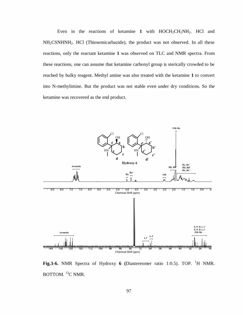
97
Even in the reactions of ketamine 1 with HOCH2CH2NH2. HCl and
NH2CSNHNH2. HCl (Thiosemicarbazide), the product was not observed. In all these
reactions, only the reactant ketamine 1 was observed on TLC and NMR spectra. From
these reactions, one can assume that ketamine carbonyl group is sterically crowded to be
reached by bulky reagent. Methyl amine was also treated with the ketamine 1 to convert
into N-methylimine. But the product was not stable even under dry conditions. So the
ketamine was recovered as the end product.
Fig.3-6. NMR Spectra of Hydroxy 6 (Diastereomer ratio 1:0.5). TOP. 1H NMR.
BOTTOM. 13
C NMR.

98
The hydroxy analogue 6 was synthesized by treating ketamine 1 with sodium
borohydride. This reaction yielded both diastereomers. The presence of the diastereomers
was observed on both 1H and
13C NMR spectra (Fig. 3-6). The CH-OH hydrogen (Ha
and Ha’ in Fig. 3-6) for both diastereomers showed resonances at 4.5 and 4.25 ppm in
the 1H NMR spectrum and the respective CH-OH carbon at 64 and 63 ppm in the
13C
NMR spectrum. The ratio of diastereomers has been calculated as 7:4 from 1H NMR
spectrum. All of the ketamine analogues synthesized was used for the GABAA and
NMDA receptors assay to evaluate their agonist activity at GABAA receptors and
antagonist activity at NMDA receptors. Hydroxy 6 analogue, which received as
diastereomeric mixture was used for receptor assay as such without separation.
3.4 Agonist activities of ketamine analogues on 62 and 22 receptors:
The GABAa subtypes 62 and 122 receptors are expressed at high levels
exclusively within mature cerebellar granule neurons. In the previous study,40
ketamine
has potentiated the GABA current arising from 6- and -containing GABAa receptors in
oocytes, dissociated granule neurons and cerebellar slices isolated from rodents within
anesthecally relevant concentration. This study also shown that PCP (Phencyclidine) and
Mk-801 (a potent non-competitive inhibitor of NMDA receptor) was less active
compared to ketamine in potentiating tonic chloride current arising from GABAA
receptors containing 6- and -subtypes. These subtypes were chosen to test against new
analogues within the oocyte expression system since their association with cerebellum
has a pivotal role in motor control activity.
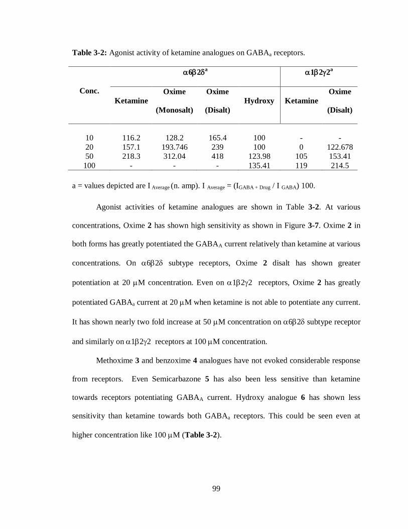
99
Table 3-2: Agonist activity of ketamine analogues on GABAa receptors.
Conc.
62a 122
a
Ketamine
Oxime
(Monosalt)
Oxime
(Disalt)
Hydroxy Ketamine
Oxime
(Disalt)
10
116.2
128.2
165.4
100
-
-
20 157.1 193.746 239 100 0 122.678
50 218.3 312.04 418 123.98 105 153.41
100 - - - 135.41 119 214.5
a = values depicted are I Average (n. amp). I Average = (IGABA + Drug / I GABA) 100.
Agonist activities of ketamine analogues are shown in Table 3-2. At various
concentrations, Oxime 2 has shown high sensitivity as shown in Figure 3-7. Oxime 2 in
both forms has greatly potentiated the GABAA current relatively than ketamine at various
concentrations. On 62 subtype receptors, Oxime 2 disalt has shown greater
potentiation at 20 M concentration. Even on 122 receptors, Oxime 2 has greatly
potentiated GABAa current at 20 M when ketamine is not able to potentiate any current.
It has shown nearly two fold increase at 50 M concentration on 62 subtype receptor
and similarly on 122 receptors at 100 M concentration.
Methoxime 3 and benzoxime 4 analogues have not evoked considerable response
from receptors. Even Semicarbazone 5 has also been less sensitive than ketamine
towards receptors potentiating GABAA current. Hydroxy analogue 6 has shown less
sensitivity than ketamine towards both GABAa receptors. This could be seen even at
higher concentration like 100 M (Table 3-2).

100
Fig 3-7. Agonist activities of ketamine 1 and oxime 2 on GABAa receptors. TOP.
62receptors. BOTTOM. 122 receptors.

101
From this analysis, Bulkier groups like methoxime and benzoxime are less
effective in potentiation. Among polar groups, Bulkier group like one in semicarbazone 5
and smaller group like hydroxyl were also insensitive towards GABAa receptors. It
indicates that double bond and the presence of small polar groups as in oxime 2 are
crucial to potentiate. The increased potentiation can be attributed to the reduced
lipophilicity in oxime 2 than ketamine.
3.5 Antagonist activities of ketamine analogues on NMDA Receptors:
Initially, new analogues synthesized were tested at 50 M concentration against
three different receptors (Table 3-3 and Fig. 3-8) for their antagonist activity.
Modification of the carbonyl group in ketamine to oxime has increased inhibition at
NMDA-A and –B receptors while the comparable activity can be observed at NMDA-D
receptors (Table 3-3 and Fig. 3-8). Particularly it had a greater inhibition at NMDA-A
receptors than ketamine and similar to PCP. It even had a similar inhibition at NMDA-B
and –D receptors. This could establish the fact that oxime 2 is target specific towards
NMDA-A receptors at 50 M concentration. Replacement of the carbonyl group in
ketamine with other analogues like methoxime, benzoxime and semicarbazone has
resulted decreased effect on all receptors (Table 3-3, Fig. 3-8). % Recovery of current
measured post drug treatment to reflect anesthetically effectiveness of drug has also
shown that oxime 2 retains similar effect on A and B receptors (Fig. 3-9) while showing
ineffectiveness at D receptors compared to ketamine. Oxime 2 also shown comparable
activity with PCP (Phencyclidine) at NMDA-A and –D receptors in inhibition. At
NMDA-B and –D receptors, it shown greater affinity than PCP as obvious from %
recovery of current measured at these receptors (Table 3-3 and Fig. 3-9).
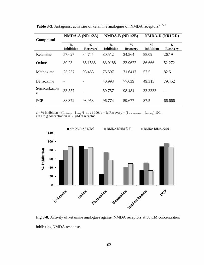
102
Table 3-3: Antagonist activities of ketamine analogues on NMDA receptors.a, b, c
Compound NMDA-A (NR1/2A) NMDA-B (NR1/2B) NMDA-D (NR1/2D)
%
Inhibition
%
Recovery
%
Inhibition
%
Recovery %
Inhibition
%
Recovery
Ketamine 57.627 84.745 80.512 34.564 88.09 26.19
Oxime 89.23 86.1538 83.0188 33.9622 86.666 52.272
Methoxime 25.257 98.453 75.597 71.6417 57.5 82.5
Benzoxime - - 40.993 77.639 49.315 79.452
Semicarbazon
e 33.557 - 50.757 98.484 33.3333 -
PCP 88.372 93.953 96.774 59.677 87.5 66.666
a = % Inhibition = (I Glu/Gly – I Drug /I Glu/Gly) 100. b = % Recovery = (I Post treatment – I Glu/Gly) 100. c = Drug concentration is 50 M at receptor.
Fig 3-8. Activity of ketamine analogues against NMDA receptors at 50 M concentration
inhibiting NMDA response.

103
Fig. 3-9. % Recovery of response from NMDA receptors post drug(50 mM conc.)
treatment .
Thus encouraged by this result, oxime 2 affinity studies have been conducted at
various concentrations to determine IC50 values (Table 3-4 and Fig. 3-10). The %
recovery reflects long lasting effect of drug as anesthetic to determine anesthetically
relevant concentration. Though oxime 2 showed greater or similar inhibition at 50 M
concentration with ketamine at all NMDA receptors, it’s affinity was attenuated over a
range of concentrations from 0.1 to 200 M in terms of % recovery of response (Table
3-4 and Fig. 3-10) by observing IC50 values.

104
Table 3-4 : Affinity studies of ketamine and oxime against NMDA receptors with IC50
values.
NMDA Receptor
% Recovery
(Ketamine)
% Recovery
(Oxime)
IC50
(Ketamine)
IC50
(Oxime)
NMDA-NR1/2A 28 74 2.32 5.911
NMDA-NR1/2B 19 31 0.82 1.068
NMDA-NR1/2C - 44 - 0.747
NMDA-NR1/2D 24 68 0.76 1.068
Fig 3-10. % Recovery of response from NMDA receptors after post drug treatment (over
various conc.).

105
Recovery is very less for ketamine than oxime means ketamine has long lasting
effect and so more affinity. Oxime shown similar inhibition at NMDA-B, C, D receptors
while it has two fold increase in terms of IC50 values than ketamine at NMDA-A
receptors.
3.6 Conclusions
Modification of carbonyl group in ketamine to oxime, methoxime, benzoxime,
semicarbazone and hydroxyl groups has provided an insight into the effect of size of the
groups towards GABAa receptors. Oxime has shown to reduce lipophilicity while
methoxime, benzoxime shown increased lipophilicity. This study has also shown that
polar groups like semicarbazone and hydroxyl were not able to increase the potency
towards GABAa receptors. Thus oxime 2 has been better agonist at GABAa receptors
exhibiting two times more excitory post synaptic potential than ketamine. From this study
, though anesthetic efficacy of ketamine and PCP (Phencyclidine) as non-competitive
inhibitors of NMDA (N-Methyl-D-Aspartate) led to classifying them as dissociate
anesthetics, Oxime 2 has shown improved or similar potency towards various NMDA
receptors. This could be beneficial to study further anesthetic efficacy of oxime 2 in
epileptic seizures, other convulsant side effects which are associated with ketamine.
Ketamine is known to be more effective anesthetic across different animal species but in
humans it is associated with various adverse effects like inducing catalepsy at higher
doses and hallucinations at lower doses. Since oxime has shown greater or comparable
potency at lower doses as antagonist exciting postsynaptic potential at various NMDA
receptors and less duration of affinity, it could be interesting to study its efficacy across
various animals.
106
3.7 Experimental Section
General
1H-NMR and
13C-NMR spectra were recorded on a Bruker 250 MHz and Varian
400 MHz spectrometer in CDCl3 and DMSO-d6 with TMS as the standard. Chemical
shifts are reported in ppm, multiplicities are indicated by s (singlet), d (doublet), t
(triplet), q (quartet), p (quintet), h (sextet), m (multiplet) and bs (broad singlet) coupling
constants reported in hertz (Hz). All 13C NMR spectra were proton decoupled. Thin-
Layer chromatography (TLC) was performed on glass plates coated with 0.25 mm
thickness of silica-gel. All solvents were dried and distilled prior to use and organic
solvent extracts were dried over Anhydrous. Na2SO4. Toluene and tetrahydrofuran were
distilled from sodium and benzophenone and were stored in a dry box. Chromatographic
purifications were performed by flash chromatography using silica gel (63-200μ) from
Dynamic Adsorbents Inc. Pd(dba)2 and P(t-Bu)3 were purchased from Aldrich Inc. All
reagents and bases were purchased from Aldrich, Fisher Science, VWR and used without
further purification. Aryl bromide 5 was prepared using literature procedure. Ketamine
was purchased from Aldrich chemical company and used as received.
2-(2-Chloro-phenyl)-2-methylamino-cyclohexanone oxime (2): To as solution of
NH2OH. HCl and CH3COONa. H2O in MeOH:H2O (7:3) stirred at room temperature for
15 minutes, was added ketamine and the resulting solution was stirred at reflux for 16 hrs
before cooling to room temperature. The reaction mixture was filtered and the filtrate was
partitioned between CH2Cl2 and sat. NaHCO3 solution. The filtrate was extracted with
CH2Cl2 (2×30 mL) and the combined organic layer was washed with brine solution. The
107
organic layer was dried over anhydrous. Na2SO4 and concentrated under reduced
pressure. The crude reaction mixture received was purified over silica gel column
chromatography by eluting with initially CH2Cl2 and later with (1:9) MeOH:CH2Cl2 to
receive oxime 2 as pale yellow solid.
1H NMR (250 MHz, CDCl3) 7.4 - 7.5 (m, 1H), 7.0
- 7.3 (m, 3H), 2.6 - 2.8 (m, 1H), 2.4 - 2.5 (q, J = 5.8 Hz, 1H), 2.1 - 2.3 (m, 1H), 2.0 - 2.1
(m, 3H), 1.5 - 1.7 (m, 5H). 13
C NMR (63 MHz, CDCl3) 160.1, 140.1, 133.8, 131.6,
129.1, 128.1, 126.5, 64.0, 37.1, 29.4, 24.5, 22.2, 21.7.
2-(2-Chloro-phenyl)-2-methylamino-cyclohexanone O-methyl-oxime (3): To as
solution of NH2OMe. HCl and CH3COONa. H2O in MeOH:H2O (7:3) stirred at room
temperature for 15 minutes, was added ketamine and the resulting solution was stirred at
reflux for 16 hrs before cooling to room temperature. The reaction mixture was filtered
and the filtrate was partitioned between CH2Cl2 and sat. NaHCO3 solution. The filtrate
was extracted with CH2Cl2 (2×30 mL) and the combined organic layer was washed with
brine solution. The organic layer was dried over anhydrous. Na2SO4 and concentrated
under reduced pressure. The crude reaction mixture received was purified over silica gel
column chromatography by eluting with initially CH2Cl2 and later with (1:9)
MeOH:CH2Cl2 to receive methoxime 3 as pale yellow solid.
1H NMR (400 MHz, CDCl3)
7.9 - 8.0 (m, 1H), 7.2 - 7.4 (m, 3H), 3.91 - 4.0 (m, 3H), 3.3 (td, J = 4.10, 13.96 Hz, 1H),
3.2 (td, J = 4.49, 13.73 Hz, 1H), 2.4 - 2.5 (m, 3H), 2.1 - 2.3 (m, 1H), 1.6 - 1.8 (m, 3H),
1.4 - 1.5 (m, 1H), 1.2 - 1.4 (m, 1H). 13
C NMR (101 MHz, CDCl3) 156.3, 135.5, 132.4,
132.2, 131.9, 131.0, 128.3, 67.4, 62.7, 39.4, 28.8, 26.1, 24.7, 22.0.
108
2-(2-Chloro-phenyl)-2-methylamino-cyclohexanone O-benzyl-oxime (4): To as
solution of NH2OCH2Ph. HCl and CH3COONa. H2O in MeOH:H2O (7:3) stirred at room
temperature for 15 minutes, was added ketamine and the resulting solution was stirred at
reflux for 16 hrs before cooling to room temperature. The reaction mixture was filtered
and the filtrate was partitioned between CH2Cl2 and sat. NaHCO3 solution. The filtrate
was extracted with CH2Cl2 (2×30 mL) and the combined organic layer was washed with
brine solution. The organic layer was dried over anhydrous. Na2SO4 and concentrated
under reduced pressure. The crude reaction mixture received was purified over silica gel
column chromatography by eluting with initially CH2Cl2 and later with (1:9)
MeOH:CH2Cl2 to receive benzoxime 4 as pale yellow solid.
1H NMR (400 MHz, CDCl3)
7.7 (d, J = 8.06 Hz, 1H), 7.1 - 7.4 (m, 8H), 5.0 - 5.2 (q, J = 8.2 Hz, 2H), 2.9 - 3.1 (m,
2H), 2.2 - 2.3 (m, 3H), 1.9 - 2.0 (m, 2H), 1.3 - 1.8 (m, 4H). 13
C NMR (101 MHz, CDCl3)
158.3, 138.7, 135.1, 132.1, 131.1, 129.9, 128.7, 128.4, 127.8, 127.6, 66.4, 38.8, 29.1,
26.0, 24.5, 22.1.
2-(2-Chloro-phenyl)-2-methylamino-cyclohexanone O-semicarbazone (5): To as
solution of NH2CONHNH2. HCl and CH3COONa. H2O in MeOH:H2O (7:3) stirred at
room temperature for 15 minutes, was added ketamine and the resulting solution was
stirred at reflux for 16 hrs before cooling to room temperature. The reaction mixture was
filtered and the filtrate was partitioned between CH2Cl2 and sat. NaHCO3 solution. The
filtrate was extracted with CH2Cl2 (2×30 mL) and the combined organic layer was
washed with brine solution. The organic layer was dried over anhydrous. Na2SO4 and
concentrated under reduced pressure. The crude reaction mixture received was purified

109
over silica gel column chromatography by eluting with initially CH2Cl2 and later with
(1:9) MeOH:CH2Cl2 to receive semicarbazone 5 as pale yellow solid.
1H NMR (400
MHz, DMSO-d6) 9.6 (br. s., 1H), 7.7 - 7.8 (m, 1H), 7.4 - 7.5 (m, 3H), 4.0 - 4.1 (m, 2H),
3.0 - 3.1 (d, J = 4.9 Hz, 2H), 2.1 (s, 3H), 1.5 - 1.8 (m, 3H), 1.2 - 1.4 (m, 3H). 13
C NMR
(101 MHz, DMSO-d6) 158.3, 149.1, 134.8, 133.2, 133.0, 132.3, 128.7, 67.8, 36.8, 28.2,
26.7, 25.9, 22.3.
2-(2-Chloro-phenyl)-2-methylamino-cyclohexanol (6): To a solution of ketamine in
MeOH at 0OC was added NaBH4 portion wise and allowed to stir at room temperature for
12 hrs. The reaction mixture was cooled to 0OC
and quenched with sat. NH4Cl solution
by adding dropwise. The resulting reaction mixture was partitioned between CH2Cl2 and
sat. NH4Cl solution and extracted with CH2Cl2 (2×30 mL), The organic layer was dried
over anhydrous. Na2SO4 and concentrated at reduced pressure. The crude reaction
mixture received was purified over silica gel column chromatography by eluting with
(2:8) EtOAc:Hexanes to receive hydroxyl analogue 6 as amorphous white solid.
1H
NMR (250 MHz, CDCl3) 7.2 - 7.4 (m, 4H), 7.0 - 7.2 (m, 4H), 4.4 - 4.5 (m, 1H), 4.2 -
4.3 (m, 1H), 2.4 - 2.5 (m, 1H), 1.9 - 2.1 (m, 2H), 1.8 - 1.9 (m, 3H), 1.2 - 1.7 (m, 5H), 0.8 -
1.0 (m, 1H). 13
C NMR (63 MHz, CDCl3) 140.5, 137.9, 133.2, 132.3, 132.0, 131.9,
131.2, 128.3, 126.7, 69.8, 64.0, 62.9, 29.9, 28.7, 28.3, 28.0, 26.5, 22.5, 20.8, 20.3, 19.3.
3.8 References
(1). Bormann, J. Trends in Neurosciences 1988, 11, 112.
(2). Bormann, J. Trends in Pharmacological Sciences 2000, 21, 16.
(3). Sivilotti, L.; Nistri, A. Progress in Neurobiology 1991, 36, 35.

110
(4). Simon, J.; Wakimoto, H.; Fujita, N.; Lalande, M.; Barnard, E. A. j biol. chem.
2004, 279, 41422.
(5). Sieghart, W. Pharmacol. Rev. 1995, 47, 181.
(6). (a). Stephenson, F. A. Biochemical Journal 1995, 310 ( Pt 1), 1. (b). McKernan,
R. M.; Whiting, P. J. Trends in Neurosciences 1996, 19, 139. (c). Farrar, S. J.;
Whiting, P. J.; Bonnert, T. P.; McKernan, R. M. j biol. chem. 1999, 274, 10100.
(7). (a). Wisden, W.; Korpi, E. R.; Bahn, S. Neuropharmacology 1996, 35, 1139. (b).
Laurie, D. J.; Wisden, W.; Seeburg, P. H. Journal of Neuroscience 1992, 12,
4151.
(8). Tyrrell, T.; Willshaw, D. Philos Trans R Soc Lond B Biol Sci 1992, 336, 239.
(9). Feigenspan, A.; Bormann, J. Prog Retin Eye Res 1998, 17, 99.
(10). Gupta, S. P. Prog Drug Res 1995, 45, 67.
(11). Olsen, R. W. Journal of Neurochemistry 1981, 37, 1.
(12). Newland, C. F.; Cull-Candy, S. G. J Physiol 1992, 447, 191.
(13). Barberis, A.; Cherubini, E.; Mozrzymas, J. W. Journal of Neuroscience 2000, 20,
8618.
(14). Wingrove, P. B.; Wafford, K. A.; Bain, C.; Whiting, P. J. Proc Natl Acad Sci U S
A 1994, 91, 4569.
(15). Yang, C. X.; Xu, H.; Zhou, K. Q.; Wang, M. Y.; Xu, T. L. Anesthesia and
Analgesia 2006, 102, 1114.
(16). (a) Orser, B. A.; Wang, L. Y.; Pennefather, P. S.; MacDonald, J. F. Journal of
Neuroscience 1994, 14, 7747(b) Hara, M.; Kai, Y.; Ikemoto, Y. Anesthesiology
1993, 79, 781.

111
(17). Tomlin, S. L.; Jenkins, A.; Lieb, W. R.; Franks, N. P. Anesthesiology 1998, 88,
708.
(18). Kohr, G. Cell and Tissue Research 2006, 326, 439.
(19). Mayer, M. L.; Westbrook, G. L.; Guthrie, P. B. Nature 1984, 309, 261.
(20). Li, F.; Tsien, J. Z. N Engl J Med 2009, 361, 302.
(21). Mayer, M. L. Nature 2006, 440, 456.
(22). (a). Park, C. K.; Nehls, D. G.; Graham, D. I.; Teasdale, G. M.; Mcculloch, J.
Annals of Neurology 1988, 24, 543. (b). Dirnagl, U.; Iadecola, C.; Moskowitz, M.
A. Trends in Neurosciences 1999, 22, 391. (c). Whetsell, W. O. Journal of
Neuropathology and Experimental Neurology 1996, 55, 1. (d). Lipton, S. A.
Current Opinion in Neurology and Neurosurgery 1993, 6, 588. (e). Wang, C. X.;
Shuaib, A. Curr Drug Targets CNS Neurol Disord 2005, 4, 143.
(23). (a). Vicini, S.; Wang, J. F.; Li, J. H.; Zhu, W. J.; Wang, Y. H.; Luo, J. A. H.;
Wolfe, B. B.; Grayson, D. R. Journal of Neurophysiology 1998, 79, 555. (b).
Yuan, H. J.; Hansen, K. B.; Vance, K. M.; Ogden, K. K.; Traynelis, S. F. Journal
of Neuroscience. 2009, 29, 12045. (c). Neyton, J.; Paoletti, P. Journal of
Neuroscience 2006, 26, 1331.
(24). (a). Lodge, D.; Anis, N. A. European Journal of Pharmacology 1982, 77, 203.
(b). MacDonald, J. F.; Nowak, L. M. Trends in Pharmacological Sciences 1990,
11, 167. (c). MacDonald, J. F.; Miljkovic, Z.; Pennefather, P. Journal of
Neurophysiology. 1987, 58, 251. (d). Anis, N. A.; Berry, S. C.; Burton, N. R.;
Lodge, D. British Journal of Pharmacology 1983, 79, 565.

112
(25). Parsons, C. G.; Quack, G.; Bresink, I.; Baran, L.; Przegalinski, E.; Kostowski, W.;
Krzascik, P.; Hartmann, S.; Danysz, W. Neuropharmacology 1995, 34, 1239.
(26). (a). Johnstone, M.; Evans, V.; Baigel, S. British Journal of Anaesthesia 1959, 31,
433. (b). Macdonald, J. F.; Bartlett, M. C.; Mody, I.; Pahapill, P.; Reynolds, J. N.;
Salter, M. W.; Schneiderman, J. H.; Pennefather, P. S. Journal of Physiology-
London 1991, 432, 483. (c). Rogawski, M. A.; Wenk, G. L. Cns Drug Reviews
2003, 9, 275. (d). Chen, G. Archives Internationales De Pharmacodynamie Et De
Therapie 1965, 157, 193. (e). Chen, G.; Ensor, C. R.; Russell, D.; Bohner, B.
Journal of Pharmacology and Experimental Therapeutics 1959, 127, 241.
(27). Mccarthy, D. A.; Chen, G.; Kaump, D. H.; Ensor, C. Journal of New Drugs 1965,
5, 21.
(28). Seeman, P.; Guan, H. C.; Hirbec, H. Synapse 2009, 63, 698.
(29). Kapur, S.; Seeman, P. Molecular Psychiatry 2002, 7, 837.
(30). Hirota, K.; Okawa, H.; Appadu, B. L.; Grandy, D. K.; Devi, L. A.; Lambert, D. G.
Anesthesiology 1999, 90, 174.
(31). Hustveit, O.; Maurset, A.; Oye, I. Pharmacology & Toxicology 1995, 77, 355.
(32). (a). Kawano, T.; Oshita, S.; Takahashi, A.; Tsutsumi, Y.; Tanaka, K.; Tomiyama,
Y.; Kitahata, H.; Nakaya, Y. Anesthesiology 2005, 102, 93. (b). Wagner, L. E.;
Gingrich, K. J.; Kulli, J. C.; Yang, J. Anesthesiology 2001, 95, 1406.
(33). (a). Kress, H. G. Anaesthesist 1997, 46, S8. (b). Hevers, W.; Luddens, H.
Neuropharmacology 2002, 42, 34. (c). Hevers, W.; Luddens, H. Molecular
Neurobiology 1998, 18, 35.

113
(34). (a). Adler, C. M.; Malhotra, A. K.; Elman, I.; Goldberg, T.; Egan, M.; Pickar, D.;
Breier, A. American Journal of Psychiatry 1999, 156, 1646. (b). Morgan, C. J.;
Mofeez, A.; Brandner, B.; Bromley, L.; Curran, H. V. Neuropsychopharmacology
2004, 29, 208. (c). Morgan, C. J.; Mofeez, A.; Brandner, B.; Bromley, L.; Curran,
H. V. Psychopharmacology (Berl) 2004, 172, 298. (d). Krystal, J. H.; Karper, L.
P.; Seibyl, J. P.; Freeman, G. K.; Delaney, R.; Bremner, J. D.; Heninger, G. R.;
Bowers, M. B., Jr.; Charney, D. S. Arch Gen Psychiatry 1994, 51, 199.
(35). (a). Olney, J. W.; Labruyere, J.; Wang, G.; Wozniak, D. F.; Price, M. T.; Sesma,
M. A. Science 1991, 254, 1515. (b). Seamans, J. Nature Chemical Biology 2008,
4, 91.
(36). (a). Hota, D.; Bansal, V.; Pattanaik, S. Methods Find Exp Clin Pharmacol 2007,
29, 443. (b). Mathew, S. J.; Manji, H. K.; Charney, D. S.
Neuropsychopharmacology. 2008, 33, 2080. (c). Krystal, J. H. Swiss Med Wkly
2007, 137, 215.
(37). (a). Avila, M. T.; Weiler, M. A.; Lahti, A. C.; Tamminga, C. A.; Thaker, G. K.
Am J Psychiatry 2002, 159, 1490. (b). Radant, A. D.; Bowdle, T. A.; Cowley, D.
S.; Kharasch, E. D.; Roy-Byrne, P. P. Neuropsychopharmacology 1998, 19, 434.
(c). Bowdle, T. A.; Radant, A. D.; Cowley, D. S.; Kharasch, E. D.; Strassman, R.
J.; Roy-Byrne, P. P. Anesthesiology 1998, 88, 82.(d). Petit, L.; Clark, V. P.;
Ingeholm, J.; Haxby, J. V. Journal of Neurophysiology 1997, 77, 3386. (e). Braff,
D. L.; Geyer, M. A.; Light, G. A.; Sprock, J.; Perry, W.; Cadenhead, K. S.;
Swerdlow, N. R. Schizophrenia Research 2001, 49, 171.

114
(38). (a). Burak, K.; Lipnicka, U.; Orszanska, H.; Rykowski, Z.; Witkiewicz, K.;
Wrzesien, J.; Bogdal, M.; Krzywasinski, L.; Borkowska, B. Farmaco Sci. 1985,
40, 285. (b). Yang, D. J.; Davisson, J. N. J. MED. CHEM. 1985, 28, 1361. (c).
Cone, E. J.; McQuinn, R. L.; Shannon, H. E. J. Pharm. and Exp. Ther. 1984, 228,
147.
(39). Zarantonello, P.; Bettini, E.; Paio, A.; Simoncelli, C.; Terreni, S.; Cardullo, F.
Bioorg. & Med. Chem. Lett. 2011, 21, 2059.
(40). Hevers, W.; Hadley, S. H.; Luddens, H.; Amin, J. J. Neurosci. 2008, 28, 5383.
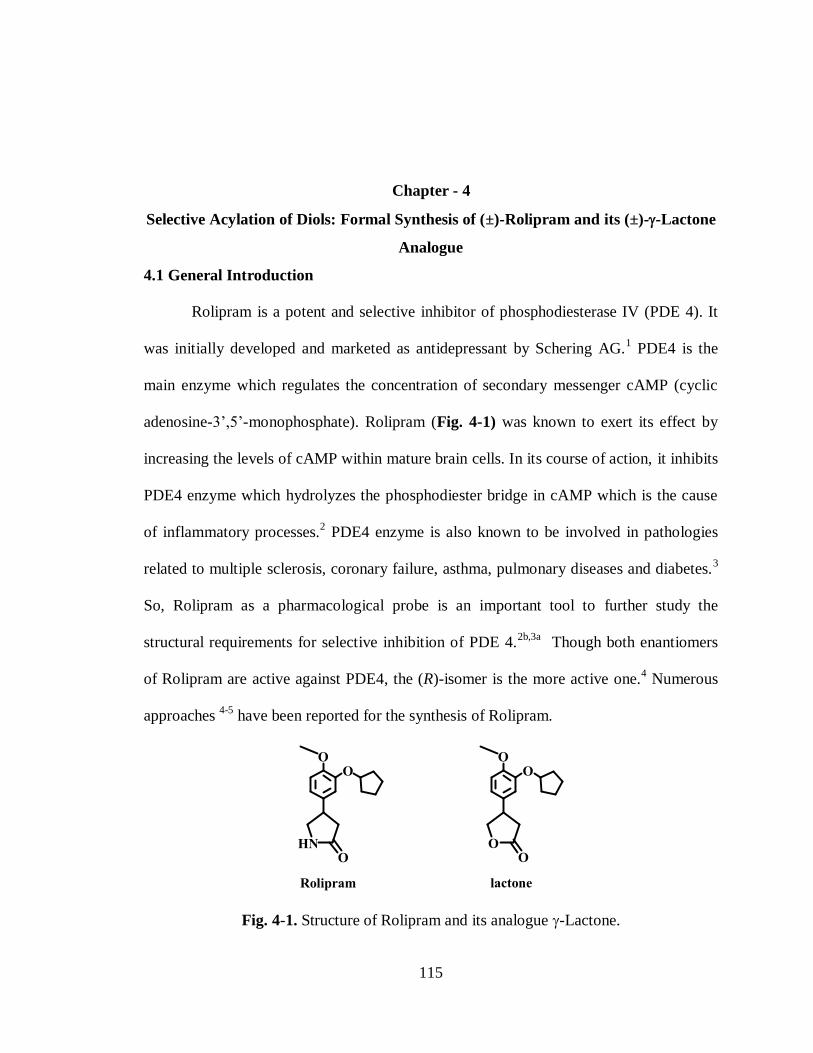
115
Chapter - 4
Selective Acylation of Diols: Formal Synthesis of (±)-Rolipram and its (±)--Lactone
Analogue
4.1 General Introduction
Rolipram is a potent and selective inhibitor of phosphodiesterase IV (PDE 4). It
was initially developed and marketed as antidepressant by Schering AG.1 PDE4 is the
main enzyme which regulates the concentration of secondary messenger cAMP (cyclic
adenosine-3’,5’-monophosphate). Rolipram (Fig. 4-1) was known to exert its effect by
increasing the levels of cAMP within mature brain cells. In its course of action, it inhibits
PDE4 enzyme which hydrolyzes the phosphodiester bridge in cAMP which is the cause
of inflammatory processes.2 PDE4 enzyme is also known to be involved in pathologies
related to multiple sclerosis, coronary failure, asthma, pulmonary diseases and diabetes.3
So, Rolipram as a pharmacological probe is an important tool to further study the
structural requirements for selective inhibition of PDE 4.2b,3a
Though both enantiomers
of Rolipram are active against PDE4, the (R)-isomer is the more active one.4 Numerous
approaches 4-5
have been reported for the synthesis of Rolipram.
Fig. 4-1. Structure of Rolipram and its analogue -Lactone.
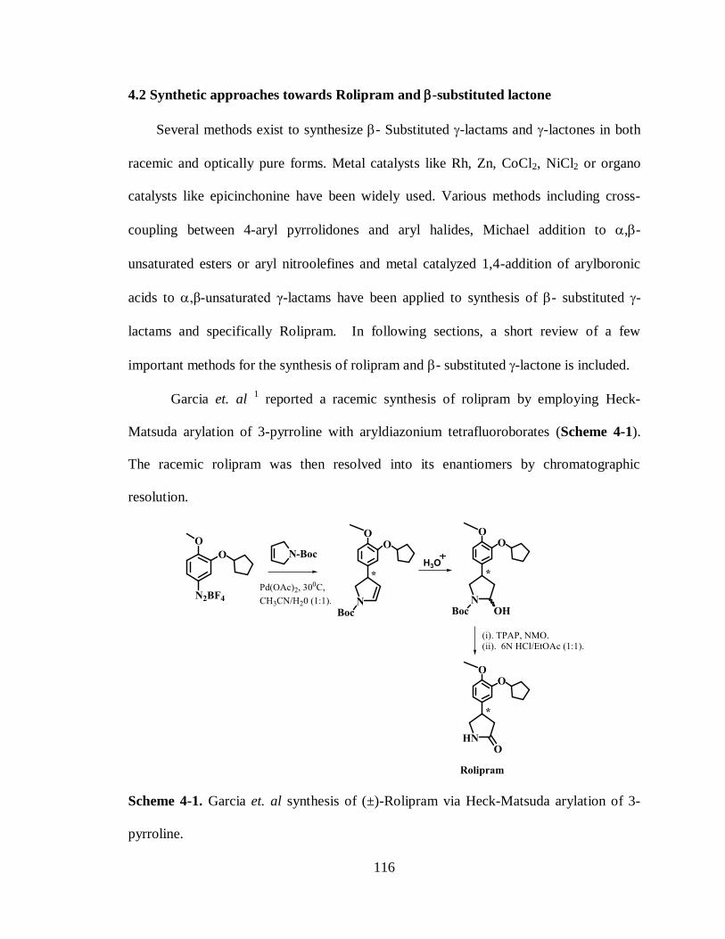
116
4.2 Synthetic approaches towards Rolipram and -substituted lactone
Several methods exist to synthesize - Substituted -lactams and -lactones in both
racemic and optically pure forms. Metal catalysts like Rh, Zn, CoCl2, NiCl2 or organo
catalysts like epicinchonine have been widely used. Various methods including cross-
coupling between 4-aryl pyrrolidones and aryl halides, Michael addition to ,-
unsaturated esters or aryl nitroolefines and metal catalyzed 1,4-addition of arylboronic
acids to ,β-unsaturated γ-lactams have been applied to synthesis of - substituted -
lactams and specifically Rolipram. In following sections, a short review of a few
important methods for the synthesis of rolipram and - substituted -lactone is included.
Garcia et. al 1 reported a racemic synthesis of rolipram by employing Heck-
Matsuda arylation of 3-pyrroline with aryldiazonium tetrafluoroborates (Scheme 4-1).
The racemic rolipram was then resolved into its enantiomers by chromatographic
resolution.
Scheme 4-1. Garcia et. al synthesis of (±)-Rolipram via Heck-Matsuda arylation of 3-
pyrroline.

117
Scheme 4-2. Demnitz et. al synthesis of (R) & (S)-Rolipram via chromatographic
separation of two diastereomers.
Demnitz et. al. 5g
(Scheme 4-2) added nitromethane to aryl ,-unsaturated
esters via Michael addition. The resulting racemic nitrobutyric acid was resolved via
chromatographic separation of two readily separable diastereomeric amides obtained with
(S)-(-)-phenyl ethylamine. Reduction followed by a reflux resulted in formation of
Rolipram.
Paraskar et. al. 5l
(Scheme 4-3) used NaBH4 in combination with CoCl2 catalyst
and chiral ligand for reductive cyclizations of a suitably substituted azido ,-unsaturated
ester in the key step to synthesize (R)-Rolipram. The intermediate, azido ,-unsaturated
ester, was synthesized via suzuki reaction from Guaiacol.

118
Scheme 4-3. Paraskar et. al synthesis of (R)-Rolipram via chiral bis-oxozoline ligand,
NaBH4 and CoCl2.
Michael addition is another alternative method used in the synthesis of rolipram.
Hynes et. al 5m
(Scheme 4-4) utilized organocatalyst epicinchonine derivative for the
Michael addition of malonate nucleophile to aryl nitro olefin enantioselectively in the key
step. The resulting nitro ester was further reduced and cyclized using NiCl2 and NaBH4
followed by decarboxylation to (R)-rolipram.

119
Scheme 4-4. Hynes et. al synthesis of (R)-Rolipram via Michael addition.
Mulzer et. al 5a
chose enolate of N-acetyloxozolidone to add to the nitroolefine in
a Michael addition (Scheme 4-5). Resulting nitro oxozolidone was recrystallized from
methanol to receive single diastereomer which upon catalytic hydrogenation yielded (R)-
rolipram.
Scheme 4-5. Mulzer et. al synthesis of (R)-Rolipram via Michael addition.
Yoon et. al 5j
utilized Rhodium catalyzed regioselective intramolecular C-H
insertion of a -diazo--(phenylsulfonyl)-acetamide in the key step for the synthesis of
rolipram (Scheme 4-6).

120
Scheme 4-6. Yoon et. al synthesis of (±)-Rolipram via Rhodium catalyzed C-H
insertion.
Honda et. al. 5d
used the enantioselective silyl ether formation from phenyl
cyclobutanone in a key step to synthesize -substituted -butyrolactones. This lactone is
further used to synthesize (R)-Rolipram (Scheme 4-7).
Scheme 4-7. Honda et. al 5d
synthesis of (R)-Rolipram via -Aryl--butyrolactone.

121
Mandai et. al.pused Pd(OAc)2 catalyzed coupling of (Z)-2-butene-1,4-diols with
aryl iodide to synthesize tetrahydro-2-furanols (Scheme 4-8) which upon oxidation with
PCC led to -substituted -butyrolactones.
Scheme 4-8. Mandai et. al synthesis of -Aryl--butyrolactone.
4.3 Formal synthesis of (±)-Rolipram
Selective protection of diols6 is an important concept in which one hydroxyl
group is protected while the other hydroxyl could be converted to various functional
groups. This selective protection of hydroxyl groups has been reported in various
symmetric and unsymmetric diols. Selective protection of alcohols can be achieved with
groups like acetates, benzoates and carbonates, e.g., the boc group. Adsorbents like silica
gel (SiO2),7 Alumina (Al2O3),
8 Phase transfer catalysts,
8a metal catalysts
6c and lipases
9
have been widely used as catalysts to achieve monoprotection in diols. Infact, lipases
have been used in our lab previously for selective protection of diols. Lipase catalyzed
disymmetrization of meso 1,3-diols is an efficient strategy which has been used in our lab
to synthesize (S)-Imperanene10
and Carbafuranomycin11
analogues. In our effort to
synthesize rolipram, we applied the monoacylation of 1,3-diols by silicagel (SiO2)
catalyst to achieve maximum yield. The strategy for the formal synthesis of (±)-Rolipram
is shown in Scheme (4-9). The synthesis of the azido ester (19) from the monoacetate (9)
represents the formal synthesis of Rolipram. The synthesis of the diester (6) was

122
envisioned through cross coupling of the aryl bromide (5) to diethylmalonate via Hartwig
protocol. The precursor aryl bromide (5) was synthesized from guaiacol. The diester (6)
would then be reduced to the prochiral 1,3-diol (7) which would be monoacylated with
SiO2 catalyst. The monoacetate(9) would then be converted to the azidoester (19) through
homologation of free hydroxyl group and azide substitution. The synthesis of the
Rolipram from azidoester (19) can then be accomplished through the reported protocol
described by Honda et. al.5d
The following sections document our effort to accomplish
the synthesis of Rolipram by selective protection of diols.
Scheme 4-9. Retrosynthetic analysis for the synthesis of -Lactam or (±)-Rolipram.

123
4.3.1 Synthesis of arylbromide (5)
Aryl bromide (5) was synthesized from guaiacol (1) in 4 steps by following the
reported literature procedure as shown in scheme (4-10). 5l
Scheme 4-10. Synthesis of Aryl Bromide from Guaiacol
Phenolic group in Guaiacol 1 was acylated to fecilitate bromine substitution at
para to the methoxy group. The acylated guaiacol (2) was then treated with NBS (N-
Bromosuccinimide) in acetonitrile at 600C to receive compound 3 in 85% yield as a light
yellow color solid. The deacylation in compound 3 was performed by treating with
NaHCO3 in methanol at reflux to receive bromophenol 4 as a light yellow color solid in
95% yield. The cyclopentyl group was introduced in bromophenol 4 by treating it with
cyclopentyl bromide and K2CO3 in DMF at 1000C to receive aryl bromide 5 as light
brown color liquid in 96% yield (Scheme 4-10).
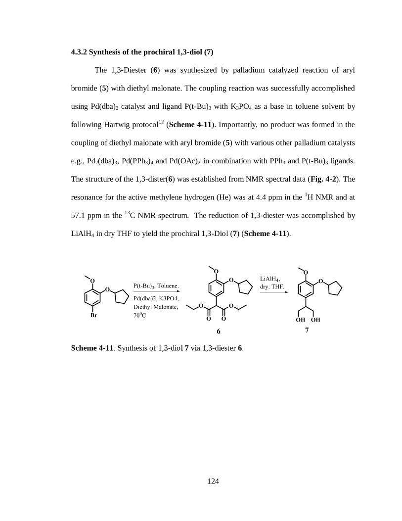
124
4.3.2 Synthesis of the prochiral 1,3-diol (7)
The 1,3-Diester (6) was synthesized by palladium catalyzed reaction of aryl
bromide (5) with diethyl malonate. The coupling reaction was successfully accomplished
using Pd(dba)2 catalyst and ligand P(t-Bu)3 with K3PO4 as a base in toluene solvent by
following Hartwig protocol12
(Scheme 4-11). Importantly, no product was formed in the
coupling of diethyl malonate with aryl bromide (5) with various other palladium catalysts
e.g., Pd2(dba)3, Pd(PPh3)4 and Pd(OAc)2 in combination with PPh3 and P(t-Bu)3 ligands.
The structure of the 1,3-dister(6) was established from NMR spectral data (Fig. 4-2). The
resonance for the active methylene hydrogen (He) was at 4.4 ppm in the 1H NMR and at
57.1 ppm in the 13
C NMR spectrum. The reduction of 1,3-diester was accomplished by
LiAlH4 in dry THF to yield the prochiral 1,3-Diol (7) (Scheme 4-11).
Scheme 4-11. Synthesis of 1,3-diol 7 via 1,3-diester 6.

125
Fig. 4-2. NMR Spectra of 1,3-diester 6. (a). 1H NMR. (b).
13C NMR
This 1,3-diol is prochiral9b,c,10
and can be used to introduce enantiomeric center by
enantioselective acylation or it can also be diacylated and the resulting 1,3-diacetate (8)
can be disymmetrized to yield enantiomeric center. The importance of this
disymmetrization is that the quantitative conversion (~ 100%) can be achieved as against
resolution where the highest yield of enantiopure compound is limited to 50%.
4.3.3 Synthesis of the monoacetate (9) via selective protection
Absorbents such as silicagel (SiO2) 7 and alumina (Al2O3)
8 are reported to be
efficient catalysts for the selective monoacetylation of various symmetric diols, hindered
alcohols in unsymmetric diols and even per-O-acylation of hydroxyl groups in
carbohydrates.6d
Following this approach, 1,3-Diol (7) was preferentially acylated to

126
monoacetate (9) using SiO2 (0.6 equival.) and acetyl chloride (0.94 equival.) in CH2Cl2
solvent. The monoacetate (9) was isolated in 78% yield along with 1,3-diacetate (8) and
1,3-Diol (7). The reaction procedure was optimized after screening with various SiO2 and
acetyl chloride ratios to synthesize monoacetate (9) in high yield.
Scheme 4-12. Synthesis of monoacetate via selective acylation of 1,3-Diol.
The structure of the monoacetate (9) was established from its NMR spectrum and
its comparison with 1,3-diol (7) and 1,3-diacetate (8) (Fig. 4-3). The methylene
hydrogen (He) in 1,3-diol (7) and in monoacetate (9) was observed at 2.9 and 3.0 ppm
respectively, while it was at 3.3 ppm in the 1,3-diacetate (8) (Fig. 4-3). The resonance for
CH2-OH protons (Hi) in monoacetate (9) appears at 3.6 ppm while it is at 3.8 ppm (Hf) in
1,3-diol (7). Having the acetoxy methylene (Hf) hydrogens at 4.5 ppm and the CH2-OH
hydrogens at 3.6 ppm (Hi) in monoacetate 9 confirms its structure (Fig. 4-3).

127
Fig. 4-3. NMR Spectra. (a). 1,3-diol 7 .(b). 1,3-diacetate 8 .(c). monoacetate 9.
4.3.4 Synthesis of methoxyalkene (13) via homologation
The monoacetate (9) was oxidized to the aldehyde (10) with dess-martin
periodinane13
in dry dichloromethane in 96% yield (Scheme 4-13). The structure of the
aldehyde was confirmed by its 1H NMR spectrum in which aldehyde hydrogen (Hj)
resonance was present at 9.6 ppm and acetoxy methylene hydrogens (Hf) resonance was
present at 4.2 ppm (Fig. 4-4).

128
Scheme 4-13 Synthesis of Alkene 13 from monoacetate 9 via homologation.
Fig. 4-4. 1H NMR spectra of Aldehyde 10.

129
In a homologation reaction, the aldehyde 10 was treated with wittig reagent
(methoxymethyl)triphenylphosphonium chloride,14
at -780C with n-BuLi to yield the
vinyl ether 12 as a E/Z mixture.15
Presence of both isomers was confirmed by its 1H
NMR spectrum (Fig. 4-5). For example, the alkene hydrogens showed resonance at 6.4
(Hj) and 4.8 (Hi) in the E isomer and at 6.0 (Hj) and 4.5 (Hi) ppm in the Z- isomer (Fig.
4-5). Since this methoxy alkene moiety will be hydrolyzed further to aldehyde, (E/Z )-
homologated alkene (11) was used in next step without separation of the isomers.
Fig. 4-5. 1H NMR spectra of (E/Z )-homologated alkene (11).
Since acetate group is labile in acidic condition needed in subsequent steps, (E/Z
)-homologated alkene (11) was hydrolyzed to the (E/Z ) -homologated alcohol (12) by
K2CO3 and methanol. The hydroxyl group in 12 was then protected with benzyl group16
by treating with benzyl bromide to yield the benzylated vinylether (13) (Scheme 4-13).

130
4.3.5 Synthesis of esteralcohol (17)
Hydrolysis17
of vinyl ether in 13 with 1N HCl in dry THF at 600c led to the
aldehyde (14) in 97% yield (Scheme 4-14). Temperature higher than 600c led to cleavage
of benzyl protection in the hydrolysis reaction. The presence of resonance for aldehyde
proton (Hi) at 9.6 ppm and benzylic protons (Hg) at 4.4 ppm in 1H NMR (Fig. 4-6)
confirmed the structure of aldehyde (14).
Scheme 4-14 Synthesis of Ester alcohol 17.
Fig. 4-6. 1H NMR spectra of Aldehyde 14.

131
Aldehyde (14) was then oxidized18
with Oxone® (Potassium
peroxymonosulfate)19
to receive carboxylic acid (15) in 96% yield. The carboxylic acid
was further esterified with ethanol in the presence of conc. H2SO4 at 450c (Scheme 4-14).
In this reaction maintaining temperature below 500c and excess solvent was crucial to
retain benzyl group protection on the alcohol since reflux led to removal of benzyl group
on the alcohol.
Once the desired ester (16) was received, benzyl protection on the alcohol was
removed by hydrogenolysis over palladium and charcoal (Pd/C 10 wt%) (Scheme 4-14).
The debenzylation was monitored using TLC, after shaking reaction mixture for 12 hrs,
all of the starting material was debenzylated. The structure of ester alcohol (17) was
confirmed by its 1H NMR spectrum (Fig. 4-7). The CH2-OH hydrogens (Hf) showed
resonance at 3.6 ppm and ethyl hydrogens (Hi and Hj) showed resonances at 4.0 ppm and
1.2 ppm respectively.
Fig. 4-7. 1H NMR spectra of Ester 17.

132
4.3.6 Synthesis of azide (19) for rolipram
Scheme 4-15 Synthesis of azide for the Rolipram from Esteralcohol 17.
Having established a synthetic route to the ester alcohol (17), the next task was to
introduce the azide group (Scheme 4-15). The bromide serves as facile leaving group to
substitute with azide nucleophile,20
so the alcohol (17) was treated with PBr3 to
synthesize bromide (18). The bromide was then treated with NaN3 in
dimethylformamide to synthesize the azide (19). The structure of the azide(19) was
established from its 1H NMR spectrum (Fig. 4-8). The resonance for CH2-N3 hydrogens
(Hf) was present at 3.4 ppm while the methylene (Hg) hydrogens showed resonance at
2.6 ppm. Since azide (19) was already transformed into Rolipram by Honda et. al.,5d
the
synthesis of azide (19) constitutes the formal synthesis to Rolipram.

133
Fig. 4-8. 1H NMR spectra of Azide 19.
4.4 Synthesis of -substituted--(±)-Lactone
Previous sections described the utilization of selective protection of hydroxyl
group in 1,3-diol for the formal synthesis of Rolipram. In a chemoenzymatic study for the
synthesis of -lactone, unsymmetric 1,4-diol was also regioselectively deacylated with
lipases. The following sections demonstrate the regioselective deacylation of 1,4-diols by
Ps-30 lipase for the synthesis of -lactone. The strategy for the synthesis of -aryl--
lactone was shown in scheme (4-16). The synthesis of arylmaleate (21) from aryl
bromide (5) was envisioned via Heck cross coupling protocol. The synthesis of the
precursor aryl bromide (5) from guaiacol (1) was shown in scheme 4-10. The arylmaleate
(21) would be converted to the monoacetate (26) via lipase catalyzed regioselective
deacylation. The monoacetate (26) would then be converted to the methyl ester (29) via
oxidation and esterification. The methyl ester (29) would finally be lactonized to the
lactone (30).
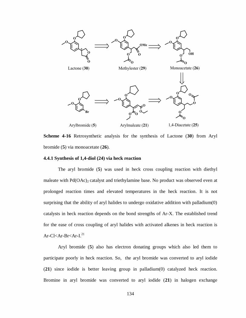
134
Scheme 4-16 Retrosynthetic analysis for the synthesis of Lactone (30) from Aryl
bromide (5) via monoacetate (26).
4.4.1 Synthesis of 1,4-diol (24) via heck reaction
The aryl bromide (5) was used in heck cross coupling reaction with diethyl
maleate with Pd(OAc)2 catalyst and triethylamine base. No product was observed even at
prolonged reaction times and elevated temperatures in the heck reaction. It is not
surprising that the ability of aryl halides to undergo oxidative addition with palladium(0)
catalysts in heck reaction depends on the bond strengths of Ar-X. The established trend
for the ease of cross coupling of aryl halides with activated alkenes in heck reaction is
Ar-Cl<Ar-Br<Ar-I.21
Aryl bromide (5) also has electron donating groups which also led them to
participate poorly in heck reaction. So, the aryl bromide was converted to aryl iodide
(21) since iodide is better leaving group in palladium(0) catalyzed heck reaction.
Bromine in aryl bromide was converted to aryl iodide (21) in halogen exchange

135
reaction22
with KI in the presence of CuI catalyst. The aryl iodide (21) was received as
pale yellow oil. The structure of the aryl iodide (21) was confirmed by its 13
C NMR
spectrum (Fig. 4-9). The presence of resonance for the carbon in aromatic ring with
iodide at 82.5 ppm confirmed the structure of aryl iodide (21). Same carbon in aromatic
ring attached to bromide was showed resonance at 112 ppm in 13
C NMR spectrum (Fig.
4-9).
Scheme 4-17 Synthesis of 1,4-Diol 24 via Heck arylation of diethylmaleate.

136
Fig. 4-9. 13
C NMR spectra. (a). Aryl bromide 5. (b). Aryl iodide 21.
The heck reaction23
between aryl iodide (21) and diethyl maleate was successfully
accomplished with Pd(OAc)2 and triethylamine base in CH3CN yielding aryl maleate
(22) with 90% yield24
(Scheme 4-17). In this reaction, the aryl maleate (22) was received
as E/Z mixture which was used directly in next step without separation of the isomers
since alkene will be hydrogenated in the following reaction. The double bond in the aryl
maleate (22) was hydrogenated to 1,4-diester (23) in the presence of Pd/C and H2 gas at 1
atm pressure.25
Further reduction of ester groups in 1,4-diester (23) was accomplished by
using lithium aluminiumhydride (LAH)26
to 1,4-diol (24) ( Scheme 4-17).
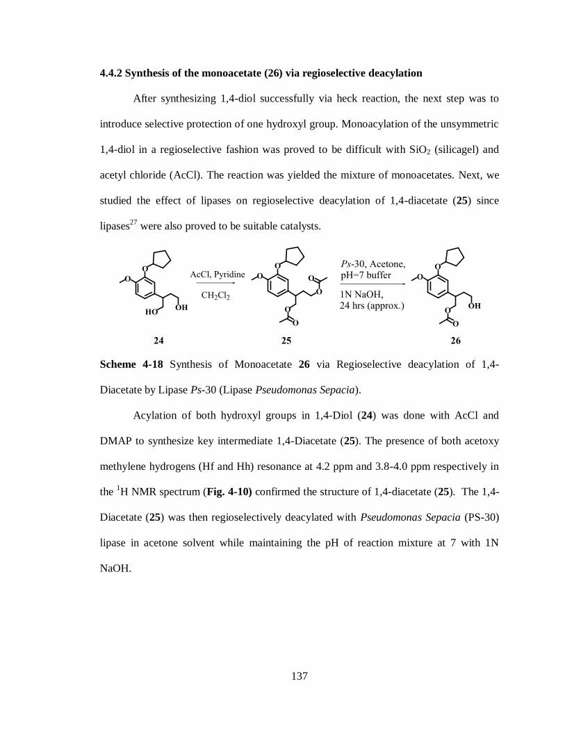
137
4.4.2 Synthesis of the monoacetate (26) via regioselective deacylation
After synthesizing 1,4-diol successfully via heck reaction, the next step was to
introduce selective protection of one hydroxyl group. Monoacylation of the unsymmetric
1,4-diol in a regioselective fashion was proved to be difficult with SiO2 (silicagel) and
acetyl chloride (AcCl). The reaction was yielded the mixture of monoacetates. Next, we
studied the effect of lipases on regioselective deacylation of 1,4-diacetate (25) since
lipases27
were also proved to be suitable catalysts.
Scheme 4-18 Synthesis of Monoacetate 26 via Regioselective deacylation of 1,4-
Diacetate by Lipase Ps-30 (Lipase Pseudomonas Sepacia).
Acylation of both hydroxyl groups in 1,4-Diol (24) was done with AcCl and
DMAP to synthesize key intermediate 1,4-Diacetate (25). The presence of both acetoxy
methylene hydrogens (Hf and Hh) resonance at 4.2 ppm and 3.8-4.0 ppm respectively in
the 1H NMR spectrum (Fig. 4-10) confirmed the structure of 1,4-diacetate (25). The 1,4-
Diacetate (25) was then regioselectively deacylated with Pseudomonas Sepacia (PS-30)
lipase in acetone solvent while maintaining the pH of reaction mixture at 7 with 1N
NaOH.
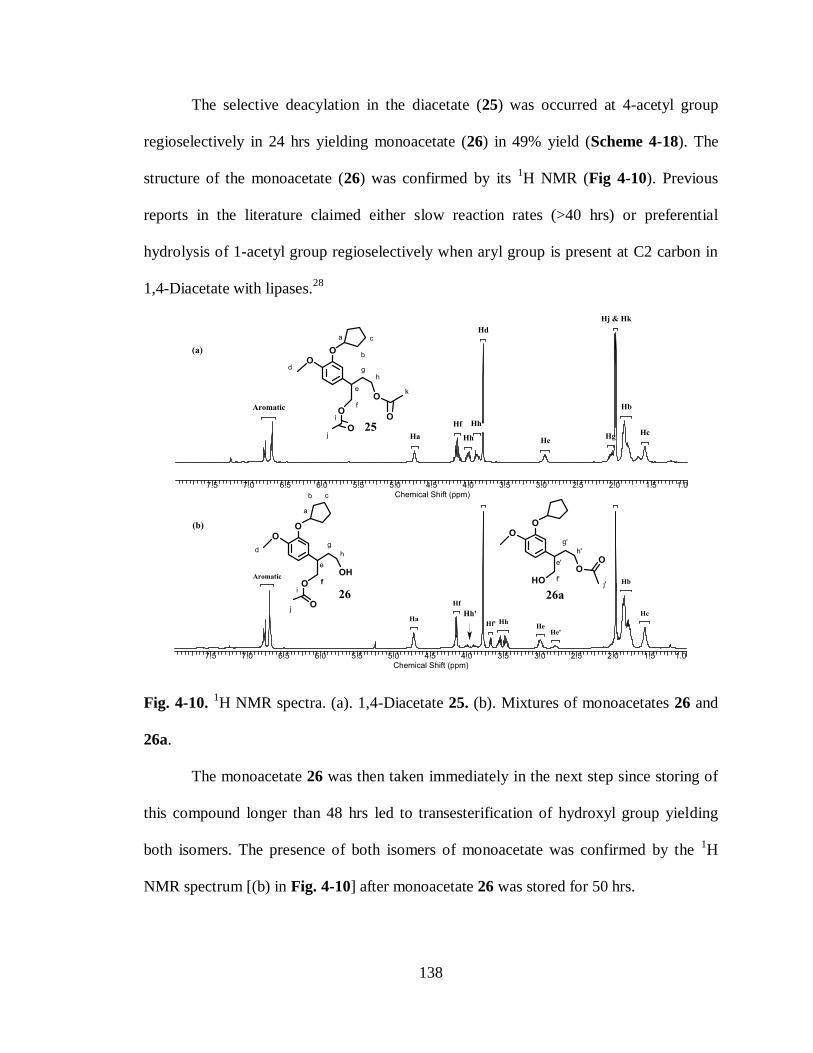
138
The selective deacylation in the diacetate (25) was occurred at 4-acetyl group
regioselectively in 24 hrs yielding monoacetate (26) in 49% yield (Scheme 4-18). The
structure of the monoacetate (26) was confirmed by its 1H NMR (Fig 4-10). Previous
reports in the literature claimed either slow reaction rates (>40 hrs) or preferential
hydrolysis of 1-acetyl group regioselectively when aryl group is present at C2 carbon in
1,4-Diacetate with lipases.28
Fig. 4-10. 1H NMR spectra. (a). 1,4-Diacetate 25. (b). Mixtures of monoacetates 26 and
26a.
The monoacetate 26 was then taken immediately in the next step since storing of
this compound longer than 48 hrs led to transesterification of hydroxyl group yielding
both isomers. The presence of both isomers of monoacetate was confirmed by the 1H
NMR spectrum [(b) in Fig. 4-10] after monoacetate 26 was stored for 50 hrs.

139
4.4.3 Synthesis of ester (29)
Scheme 4-19. Synthesis of Ester acetate 29 from monoacetate 26.
The free hydroxyl group in the monoacetate (26) was oxidized by treating with
dess-martin periodinane29
to the aldehyde (27) which was then further oxidized with
Oxone to the carboxylic acid (28) (Scheme 4-19). Direct esterification19,30
of the
aldehyde (27) to the methyl ester (29) was not proceded with Oxone rather it yielded the
acid (28). So the esterification of the acid (28) to the methyl ester (29) was accomplished
with DCC (dicyclohexylcarbodiimide), DMAP (dimethylamino pyridine) and MeOH in
CH2Cl2 solvent (Scheme 4-19). The presence of the resonance for methoxy group (Hk) in
1H NMR spectrum at 3.6 ppm was confirmed the structure of ester (29) (Fig. 4-11).

140
Fig. 4-11. 1H NMR spectra. (a). Carboxylic acid 28. (b). Ester 29.
4.4.4 Synthesis of lactone (30) via lactonization
The deacylation of 1-acetoxy group in the ester (29) was performed to fecilitate
the final assembly of the lactone (30). No reaction was observed when the lipase PPL
(Pigliver pancreatic lipase)31
was used at pH=7 to hydrolyse 1-acetoxy group.32
The
deacylation of 1-acetoxy group was also unsuccessful even with the Novozyme 435
lipase33
in the presence of n-butanol and dry. tetrahydrofuran at prolonged reaction times
(>60 hrs). Mild hydrolysis of this acetate group with sodium acetate (CH3COONa)34
or
LiCl35
was also not proceeded to the product.
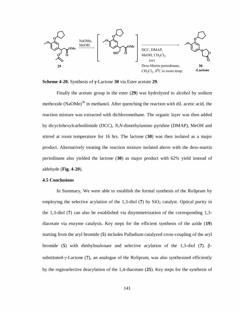
141
Scheme 4-20. Synthesis of -Lactone 30 via Ester acetate 29.
Finally the acetate group in the ester (29) was hydrolyzed to alcohol by sodium
methoxide (NaOMe)36
in methanol. After quenching the reaction with dil. acetic acid, the
reaction mixture was extracted with dichloromethane. The organic layer was then added
by dicyclohexylcarbodiimide (DCC), N,N-dimethylamino pyridine (DMAP), MeOH and
stirred at room temperature for 16 hrs. The lactone (30) was then isolated as a major
product. Alternatively treating the reaction mixture isolated above with the dess-martin
periodinane also yielded the lactone (30) as major product with 62% yield instead of
aldehyde (Fig. 4-20).
4.5 Conclusions
In Summary, We were able to establish the formal synthesis of the Rolipram by
employing the selective acylation of the 1,3-diol (7) by SiO2 catalyst. Optical purity in
the 1,3-diol (7) can also be established via disymmetrization of the corresponding 1,3-
diacetate via enzyme catalysis. Key steps for the efficient synthesis of the azide (19)
starting from the aryl bromide (5) includes Palladium catalyzed cross-coupling of the aryl
bromide (5) with diethylmalonate and selective acylation of the 1,3-diol (7). -
substituted--Lactone (7), an analogue of the Rolipram, was also synthesized efficiently
by the regioselective deacylation of the 1,4-diacetate (25). Key steps for the synthesis of

142
-substituted--Lactone (30) includes the heck arylation of diethyl maleate, regioselective
deacylation of -substituted-1,4-diacetate (25) via lipase catalysis and lactonization.
4.6 Experimental Section
4.6.1 General
Lipase PS-30 was generous gifts from Amano Enzymes. 1H-NMR and
13C-NMR spectra
were recorded on a Bruker 250 MHz and Varian 400 MHz spectrometer in CDCl3 and
DMSO-d6 with TMS as the standard. Chemical shifts are reported in ppm, multiplicities
are indicated by s (singlet), d (doublet), t (triplet), q (quartet), quin (quintet), h (sextet), m
(multiplet), dd (doublet of doublet), td (triplet of doublet), qd (quartet of doublet) and bs
(broad singlet). Coupling constants are reported in hertz (Hz). All 13
C NMR spectra were
proton decoupled. Thin-Layer chromatography (TLC) was performed on glass plates
coated with 0.25 mm thickness of silica-gel. All solvents were dried and distilled prior to
use and organic solvent extracts were dried over anhydrous Na2SO4. Toluene and
tetrahydrofuran were distilled from sodium and benzophenone and were stored in a dry
box. Chromatographic purifications were performed by flash chromatography using silica
gel (63-200μ) from Dynamic Adsorbents Inc. Pd(dba)2 and P(t-Bu)3 were purchased from
Aldrich Inc. All reagents and bases were purchased from Aldrich, Fisher Scientific, VWR
and used without further purification.
Acetic acid 2-methoxy-phenyl ester (2): To a solution of a guaiacol 1 (15 g, 0.12 mol)
and pyridine (22.75 mL, 0.279 mol) in CH2Cl2 (300 mL) at 00C was added AcCl (13 mL,
0.181 mol) portion wise and the resulting mixture was allowed to stir for overnight at
room temperature. The reaction mixture was poured into 1M H3PO4 solution (100 mL)
143
and the aqueous layer was extracted with CH2Cl2 (4×50 mL). The Combined organic
phases were then washed with brine solution (80 mL), water (2×30 mL), dried over
anhydrous Na2SO4 and filtered. The resulting light filtrate was concentrated under
reduced pressure and chromatographed over silica gel column by eluting with
EtOAc:Hexanes (1:9) to receive compound 2 as light brown color oil in 95% yield (18.9
g, 0.114 mol). 1H NMR (250 MHz, CDCl3) 7.0 - 7.1 (m, 1H), 6.7 - 6.9 (m, 3H), 3.6 -
3.7 (bs, 3H), 2.1 - 2.2 (bs, 3H). 13
C NMR (62.5 MHz, CDCl3) 169.1, 151.1, 139.8,
126.9, 122.8, 120.8, 112.4, 55.8, 20.7.
Acetic acid 5-bromo-2-methoxy-phenyl ester (3): To a solution of 2 (13.639 g, 0.082
mol) in CH3CN (180 mL) was added N-Bromosuccinimide (17.533 g, 0.098 mol) under
N2 atmosphere and the reaction mixture was allowed to stir overnight at 600C and
filtered. The resulting filtrate was added by EtOAc in a separatory funnel and (200 mL),
washed with brine solution (2×100 mL). The combined organic layers were dried over
anhydrous Na2SO4 and filtered. The filtrate was then concentrated under reduced pressure
and chromatographed over silicagel column to receive 3 as a light yellow color solid
(16.17 g, 85% yield). 1H NMR (250 MHz, CDCl3) 7.3 (dd, J = 2.4, 8.7 Hz, 1H), 7.1 (d,
J = 2.3 Hz, 1H), 6.8 (d, J = 8.6 Hz, 1H), 3.8 (s, 3H), 2.30 (s, 3H). 13
C NMR (62.5 MHz,
CDCl3) 168.6, 150.6, 140.3, 129.6, 126.1, 113.7, 112.0, 56.1, 20.6.
5-Bromo-2-methoxy-phenol (4): To a solution of 3 (1.05 g, 4.3 mmol) in MeOH (6 mL)
was added NaHCO3 (0.6 g, 8.0 mmol) and the reaction mixture was allowed to stir
overnight at reflux and filtered. The filtrate was then poured into 1N HCl solution (10

144
mL) and the aqueous layer was extracted with diethyl ether (3×10 mL). The combined
organic phases were then washed with brine solution (2×10 mL), dried over anhydrous
Na2SO4 and filtered. The resulting filtrate was then concentrated under reduced pressure
and chromatographed over silicagel column by eluting with EtOAc:Hexanes (1:9) to
receive compound 4 as light yellow color solid (0.829 g, 95% yield). 1H NMR (250 MHz,
CDCl3) 7.0 (m, 1H), 6.9 (m, 1H), 6.8 (m, 1H), 6.7 (d, J = 8.5 Hz, 1H), 3.8 (s, 3H). 13
C
NMR (62.5 MHz, CDCl3) 146.5, 145.9, 122.8, 117.8, 113.2, 111.9, 56.1.
4-bromo-2-(cyclopentyloxy)-1-methoxybenzene (5): To a mixture of bromo phenol 4
(23.5 g, 0.115 mol) and K2CO3 (31.79 g, 0.225 mol) in DMF (100 ml) was added
cyclopentyl bromide (16.5 mL, 0.149 mol) using a syringe at 500C with vigorous stirring.
After completion of the reaction (monitored by TLC (1:4 EtOAc:Hexanes), the reaction
mixture was allowed to cool to room temperature. The reaction mixture was then poured
into 1M NaOH solution (100 mL) and EtOAc (200 mL) added to it. The organic layer
was separated and the aqueous layer was extracted with EtOAc (2×50 ml). The combined
organic extracts were washed with water (30 ml) followed by the brine solution (50mL),
dried over anhydrous MgSO4 and filtered. The organic layer was then concentrated under
reduced pressure to give crude product (30 g), which was further purified by column
chromatography on silica gel using hexanes/EtOAc (9:1) as eluent to afford the aryl
bromide 5 (26.5 g, 96% yield, yellowish liquid). 1H NMR (250MHz, CDCl3) = 6.9 - 6.8
(m, 2 H), 6.6 (d, J = 8.5 Hz, 1 H), 4.6 (bs, 1 H), 3.7 (s, 3 H), 1.9 - 1.6 (m, 6 H), 1.5 (bs., 2
H). 13
C NMR (62.5 MHz , CDCl3) = 148.7, 148.0, 122.6, 117.3, 112.6, 112.1, 80.1,
55.6, 32.2, 23.5.

145
2-(3-Cyclopentyloxy-4-methoxy-phenyl)-malonic acid diethyl ester (6): To a schlenk
tube containing diethyl malonate (0.176 g, 1.1 mmol) and aryl bromide 5 (0.271 g, 1.0
mmol) were added P(t-Bu)3 (0.009 g, 0.040 mmol), Pd(dba)2 (0.0115 g, 0.020 mmol),
and K3PO4 (0.636 g, 3.0mmol) followed by toluene (3.0 mL) under N2 atmosphere. The
tube was sealed with the stopper and the heterogeneous reaction mixture was stirred at 70
°C for 16 hrs and monitored by TLC (1:4 EtOAc:Hexanes). The crude reaction mixture
was filtered through a plug of celite and concentrated in vacuuo. The residue was purified
by column chromatography on silica gel (10:90 EtOAc/hexanes) to give the desired
product 6 (yellowish liquid, 60% yield). 1H NMR (250 MHz, CDCl3) 6.8 - 6.95(m, 1H),
6.7 (d, J = 9.6 Hz, 2H), 4.6 - 4.7 (m, 1H), 4.4 - 4.4 (m, 1H), 4.1 (d, J = 7.1 Hz, 4H), 3.7
(m, 3H), 1.6 - 1.9 (m, 6H), 1.4 - 1.5 (m, 2H), 1.1 (d, J = 2.2 Hz, 3H) 13
C NMR (62.5 MHz
, CDCl3) = 168.3, 150.0, 147.5, 125.1, 121.7, 115.8, 111.6, 80.3, 61.6, 57.3, 55.9, 32.7,
24.0, 14.0.
2-(3-Cyclopentyloxy-4-methoxy-phenyl)-propane-1,3-diol (7): To a solution of 6
(0.573 g, 1.63 mmol) in diethyl ether, cooled at 00C was added lithium aluminiumhydride
(0.745 g, 19.64 mmol). The reaction warmed to room temperature and allowed to stir for
a further 8 hours. The reaction was quenched at 00C with water (3 mL) and 2N NaOH
solution (6 mL) until white color suspension received and filtered. The filtrate was
extracted with ethyl acetate (3×25 ml). The organic layer was dried over sodium sulfate
and filtered. The organic layer was concentrated under reduced pressure and purified over
column chromatography on silica gel (3:7 EtOAc/ Hexanes) to receive 1,3-Diol as a

146
colorless viscous liquid (68% yield). 1H NMR (250 MHz, CDCl3) 6.5 - 6.7 (m, 3H), 4.6
- 4.7 (m, 1H), 3.6 - 3.9 (m, 7H), 2.8 - 3.0 (m, 1H), 1.6 - 1.8 (m, 6H), 1.4 - 1.5 (m, 2H).
Acetic acid 3-acetoxy-2-(3-cyclopentyloxy-4-methoxy-phenyl)propyl ester (8): To a
solution of 1,3-Diol (7) (0.827 g, 3.109 mmol) in CH2Cl2 was added acetylchloride (0.8
mL, 12.43 mmol) and pyridine (1.01 mL, 12.43 mmol). The reaction was stirred for 6 hrs
at room temperature and quenched with NaHCO3 (20 mL). The reaction mixture was
extracted with CH2Cl2 (2×25 mL). Combined organic layer was washed with 1N HCl,
brine solution and dried over Anhydrous Na2SO4. The resulting crude reaction mixture
from evaporation under rotavap was purified by column chromatography on silica gel by
eluting with EtOAc/Hexanes (1:9) to receive 8 as yellowish liquid (85% yield). 1H NMR
(250 MHz, CDCl3) 6.6 - 6.8 (m, 3H), 4.62 - 4.7 (m, 1H), 4.2 (d, J = 6.79 Hz, 4H), 3.7 -
3.8 (m, 3H), 3.1 (t, J = 6.56 Hz, 1H), 1.97 (s, 6H), 1.70 - 1.89 (m, 6H), 1.46 - 1.60 (m,
2H)
Acetic acid 2-(3-cyclopentyloxy-4-methoxy-phenyl)-3-hydroxy-propyl ester (9):
Acetyl chloride (0.183 ml, 2.57 mmol) was added to a stirred solution of diol (0.720 g,
2.7 mmol), SiO2 (0.100 g, 1.664 mmol) in CH2Cl2 (5 ml) under a nitrogen atmosphere
and the resulting reaction mixture was stirred at room temperature. When judged
complete by TLC (approx. 3 hrs 1:1 EtOAc:Hexanes), the reaction was diluted with Et2O
and washed twice with a saturated NaHCO3 solution and once with brine. The organic
layer was dried over anhydrous. MgSO4 and filtered. The organic layer was concentrated
under reduced pressure and purified via flash column chromatography (1:4 EtOAc–

147
hexane mixtures) to receive monoacetate 9 (64% yield, 78% yield based on recovered
1,3-Diol 7). 1H NMR (250MHz , CDCl3) = 6.8 - 6.6 (m, 3 H), 4.7 - 4.6 (m, 1 H), 4.2 (d,
J = 6.8 Hz, 2 H), 3.8 - 3.6 (m, 5 H), 2.9 (t, J = 6.5 Hz, 1 H), 2.0 - 1.9 (m, 3 H), 1.9 - 1.6
(m, 6 H), 1.6 - 1.4 (m, 2 H).
Acetic acid 2-(3-cyclopentyloxy-4-methoxy-phenyl)-3-oxo-propyl ester (10): To a
solution of 9 (0.156 g, 1.75 mmol) in dry dichloromethane (5 ml) at 00C was added Dess-
Martin periodinane (5ml, 15%w/v). The reaction was allowed to stir at 00C for a further
90 minutes then allowed to warm to room temperature and stirred for an additional 2
hours. A solution of sodium thiosulfate (15ml) was added followed by a solution of sat.
NaHCO3 and the solution stirred for an additional 15 minutes, then vacuum-filtered over
celite. An additional 30 ml of dichloromethane was added and extracted with aqueous
sodium chloride (20 ml). The organic layer was dried over sodium sulfate, concentrated,
and the product (amber liquid, 0.508 g, 1.73 mmol, 96% yield) used immediately in the
next reaction without further purification. 1H NMR (250 MHz, CDCl3) 9.5 - 9.6 (m,
1H), 6.7 - 6.8 (m, 1H), 6.5 - 6.6 (m, 2H), 4.6 - 4.7 (m, 1H), 4.4 - 4.6 (m, 1H), 4.2 (dd, J =
4.4, 5.8 Hz, 1H), 3.6 - 3.8 (m, 5H), 1.8 - 1.9 (m, 3H), 1.6 - 1.8 (m, 6H), 1.4 - 1.5 (m, 2H).
13C NMR (62.5 MHz , CDCl3) = 190.8, 170.6, 154.9, 147.8, 128.0, 121.9, 113.1, 110.5,
80.6, 65.8, 56.1, 56.0, 32.7, 24.1, 20.6.
148
(E/Z)-Acetic acid 2-(3-cyclopentyloxy-4-methoxy-phenyl)-4-methoxy-but-3-enyl
ester (11): To a solution of the Wittig reagent [(Methoxymethyl)
triphenylphosphoniumchloride] (2.97 g, 8.68 mmol) in dry THF under a nitrogen
atmosphere at -780C was added n-butyl lithium (5.64 mL, 9.03 mmol). The reaction was
allowed to stir for an additional 30 minutes and a solution of the aldehyde 10 (0.508 g,
1.736 mmol) in dry THF (10 ml) was added to the sanguineous solution at -780C. The
reaction was allowed to warm to room temperature and stirred for an additional 19 hours.
The solution was then concentrated and added by ethyl acetate (50 ml),brine solution (20
ml). The organic layer was concentrated under reduced pressure and purified over wet
silica chromatography using ethyl acetate: hexane (2:8) to receive compound 11 as a
viscous, amber liquid (88% yield) as E/Z mixture. 1H NMR (250 MHz, CDCl3) 6.7 - 6.8
(m, 3H), 6.3 (d, J = 12.6 Hz, 1H), 6.0 (d, J = 8.3 Hz, 1H), 4.8 (d, J = 8.3 Hz), 4.7 - 4.8
(bs, 1H), 4.2 (d, J = 7.4 Hz, 1H), 3.7 - 3.8 (m, 3H), 3.6 (s, 2H), 3.5 - 3.6 (m, 3H), 1.9 - 2.0
(m, 3H), 1.7 - 1.9 (m, 6H), 1.5 - 1.7 (m, 2H)
(E/Z)-2-(3-Cyclopentyloxy-4-methoxy-phenyl)-4-methoxy-but-3-en-1-ol (12): To a
solution of the alkene 11 (0.35 g, 1.04 mmol) in dry methanol (20 ml) was added
anhydrous potassium carbonate (0.3 g). The reaction was allowed to stir at room
temperature for 3 hours upon which it was quenched with sat. NaHCO3 solution (10 mL)
and extracted with dichloromethane. The organic layer was dried over anhydrous.
Na2SO4, concentrated and purified by flash column chromatography using ethyl acetate:
hexane (4:6) to receive viscous colorless liquid 12 (0.22 g, 0.75 mmol, 72% yield) 1
H
NMR (250 MHz, CDCl3) 6.6 - 6.8 (m, 3H), 6.3 (d, J = 12.00 Hz, 1H), 5.9 (d, J = 7.27
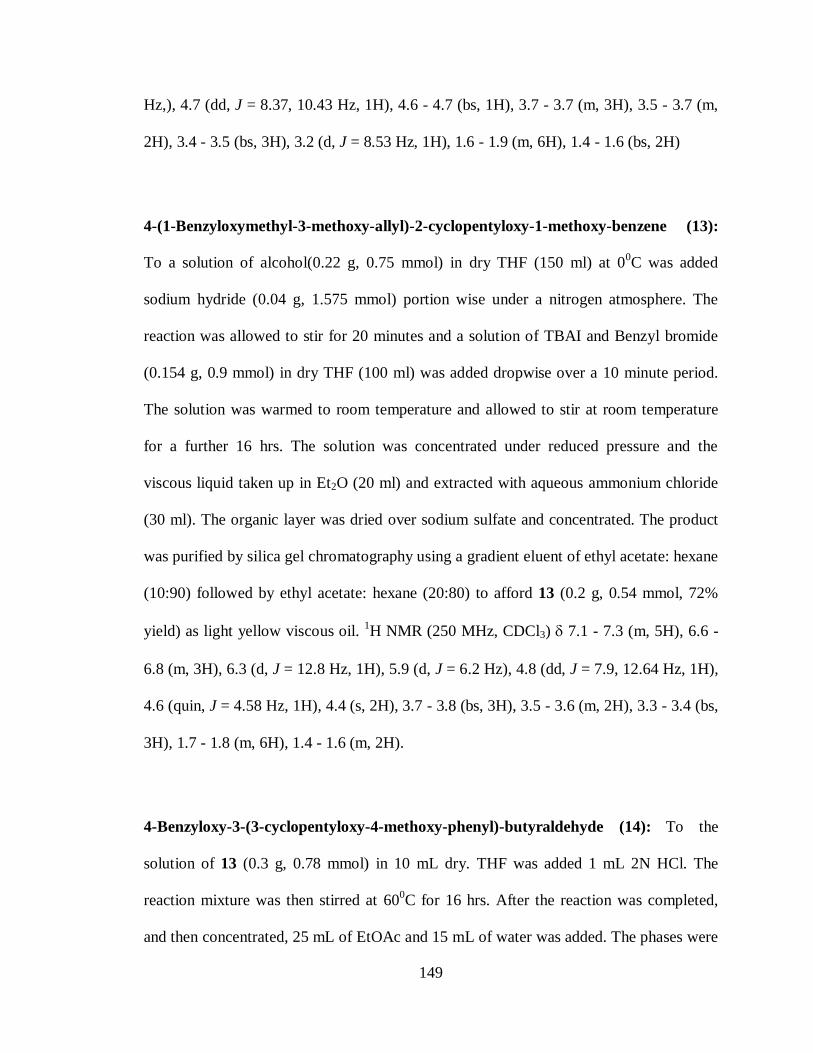
149
Hz,), 4.7 (dd, J = 8.37, 10.43 Hz, 1H), 4.6 - 4.7 (bs, 1H), 3.7 - 3.7 (m, 3H), 3.5 - 3.7 (m,
2H), 3.4 - 3.5 (bs, 3H), 3.2 (d, J = 8.53 Hz, 1H), 1.6 - 1.9 (m, 6H), 1.4 - 1.6 (bs, 2H)
4-(1-Benzyloxymethyl-3-methoxy-allyl)-2-cyclopentyloxy-1-methoxy-benzene (13):
To a solution of alcohol(0.22 g, 0.75 mmol) in dry THF (150 ml) at 00C was added
sodium hydride (0.04 g, 1.575 mmol) portion wise under a nitrogen atmosphere. The
reaction was allowed to stir for 20 minutes and a solution of TBAI and Benzyl bromide
(0.154 g, 0.9 mmol) in dry THF (100 ml) was added dropwise over a 10 minute period.
The solution was warmed to room temperature and allowed to stir at room temperature
for a further 16 hrs. The solution was concentrated under reduced pressure and the
viscous liquid taken up in Et2O (20 ml) and extracted with aqueous ammonium chloride
(30 ml). The organic layer was dried over sodium sulfate and concentrated. The product
was purified by silica gel chromatography using a gradient eluent of ethyl acetate: hexane
(10:90) followed by ethyl acetate: hexane (20:80) to afford 13 (0.2 g, 0.54 mmol, 72%
yield) as light yellow viscous oil. 1H NMR (250 MHz, CDCl3) 7.1 - 7.3 (m, 5H), 6.6 -
6.8 (m, 3H), 6.3 (d, J = 12.8 Hz, 1H), 5.9 (d, J = 6.2 Hz), 4.8 (dd, J = 7.9, 12.64 Hz, 1H),
4.6 (quin, J = 4.58 Hz, 1H), 4.4 (s, 2H), 3.7 - 3.8 (bs, 3H), 3.5 - 3.6 (m, 2H), 3.3 - 3.4 (bs,
3H), 1.7 - 1.8 (m, 6H), 1.4 - 1.6 (m, 2H).
4-Benzyloxy-3-(3-cyclopentyloxy-4-methoxy-phenyl)-butyraldehyde (14):
To the
solution of 13 (0.3 g, 0.78 mmol) in 10 mL dry. THF was added 1 mL 2N HCl. The
reaction mixture was then stirred at 600C for 16 hrs. After the reaction was completed,
and then concentrated, 25 mL of EtOAc and 15 mL of water was added. The phases were
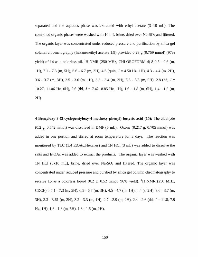
150
separated and the aqueous phase was extracted with ethyl acetate (3×10 mL). The
combined organic phases were washed with 10 mL brine, dried over Na2SO4 and filtered.
The organic layer was concentrated under reduced pressure and purification by silica gel
column chromatography (hexanes/ethyl acetate 1:9) provided 0.28 g (0.759 mmol) (97%
yield) of 14 as a colorless oil. 1H NMR (250 MHz, CHLOROFORM-d) 9.5 - 9.6 (m,
1H), 7.1 - 7.3 (m, 5H), 6.6 - 6.7 (m, 3H), 4.6 (quin, J = 4.50 Hz, 1H), 4.3 - 4.4 (m, 2H),
3.6 - 3.7 (m, 3H), 3.5 - 3.6 (m, 1H), 3.3 - 3.4 (m, 2H), 3.3 - 3.3 (m, 0H), 2.8 (dd, J =
10.27, 11.06 Hz, 0H), 2.6 (dd, J = 7.42, 8.85 Hz, 1H), 1.6 - 1.8 (m, 6H), 1.4 - 1.5 (m,
2H).
4-Benzyloxy-3-(3-cyclopentyloxy-4-methoxy-phenyl)-butyric acid (15): The aldehyde
(0.2 g, 0.542 mmol) was dissolved in DMF (6 mL). Oxone (0.217 g, 0.705 mmol) was
added in one portion and stirred at room temperature for 3 days. The reaction was
monitored by TLC (1:4 EtOAc:Hexanes) and 1N HCl (3 mL) was added to dissolve the
salts and EtOAc was added to extract the products. The organic layer was washed with
1N HCl (3x10 mL), brine, dried over Na2SO4 and filtered. The organic layer was
concentrated under reduced pressure and purified by silica gel column chromatography to
receive 15 as a colorless liquid (0.2 g, 0.52 mmol, 96% yield). 1H NMR (250 MHz,
CDCl3) 7.1 - 7.3 (m, 5H), 6.5 - 6.7 (m, 3H), 4.5 - 4.7 (m, 1H), 4.4 (s, 2H), 3.6 - 3.7 (m,
3H), 3.3 - 3.61 (m, 2H), 3.2 - 3.3 (m, 1H), 2.7 - 2.9 (m, 2H), 2.4 - 2.6 (dd, J = 11.8, 7.9
Hz, 1H), 1.6 - 1.8 (m, 6H), 1.3 - 1.6 (m, 2H).

151
4-Benzyloxy-3-(3-cyclopentyloxy-4-methoxy-phenyl)-butyric acid ethyl ester (16):
The acid 15 (0.115 g, 0.3 mmol) was dissolved in ethanol and stirred at 500C for 10 min.
5 drops of Conc. H2SO4 was added to above solution at 450C and allowed to stir for 16
hrs. The reaction was monitored by TLC (1:4 EtOAc:Hexanes). After starting material
was consumed completely, reaction was concentrated and partitioned between diethyl
ether and sat. NaHCO3. The organic layer was dried over anhydrous. Na2SO4 and
concentrated. Product was purified over column chromatography on silicagel by eluting
with EtOAc/hexanes (15:85) to receive 16 as colorless liquid (0.108 g, 0.261 mmol, 87%
yield). 1H NMR (250 MHz, CDCl3) 7.1 - 7.3 (m, 5H), 6.5 - 6.8 (m, 3H), 4.5 - 4.7 (m,
1H), 4.3 - 4.5 (m, 2H), 3.8 - 4.0 (m, 2H), 3.6 - 3.8 (m, 3H), 3.2 - 3.6 (m, 3H), 2.7 - 2.8
(dd, J = 8.1, 8.1 Hz,, 1H), 2.4 - 2.6 (dd, J = 8.1, 8.1 Hz,,,1H), 1.6 - 1.9 (m, 6H), 1.4 - 1.6
(m, 2H), 0.9 - 1.1 (m, 3H).
3-(3-Cyclopentyloxy-4-methoxy-phenyl)-4-hydroxy-butyric acid ethyl ester (17): To
a solution of 16 (0.108 g, 0.262 mmol) in absolute ethanol (20 mL) was added
palladium/charcoal(0.05 g, 10wt%), and the suspension was shaken for 12 hrs under
H2 atmosphere (45 psi) in a Parr apparatus. The suspension was filtered over a short
pad of silica gel and the filtrate was evaporated to an oily residue. Product was purified
over column chromatography on silica gel to receive alcohol 17 (0.6 g, 0.186 mmol, 72%
yield) as yellowish color liquid. 1H NMR (250 MHz, CDCl3) 6.5 - 6.8 (m, 3H), 4.6 - 4.7
(m, 1H), 3.9 - 4.0 (q, J = 6.8 Hz, 2H), 3.5 - 3.7 (m, 5H), 3.1 (t, J = 7.11 Hz, 1H), 2.4 - 2.7
(dd, J = 10.4, 8.2 Hz,2H), 1.6 - 1.9 (m, 6H), 1.4 - 1.6 (m, 2H), 1.0 - 1.1 (m, 3H).
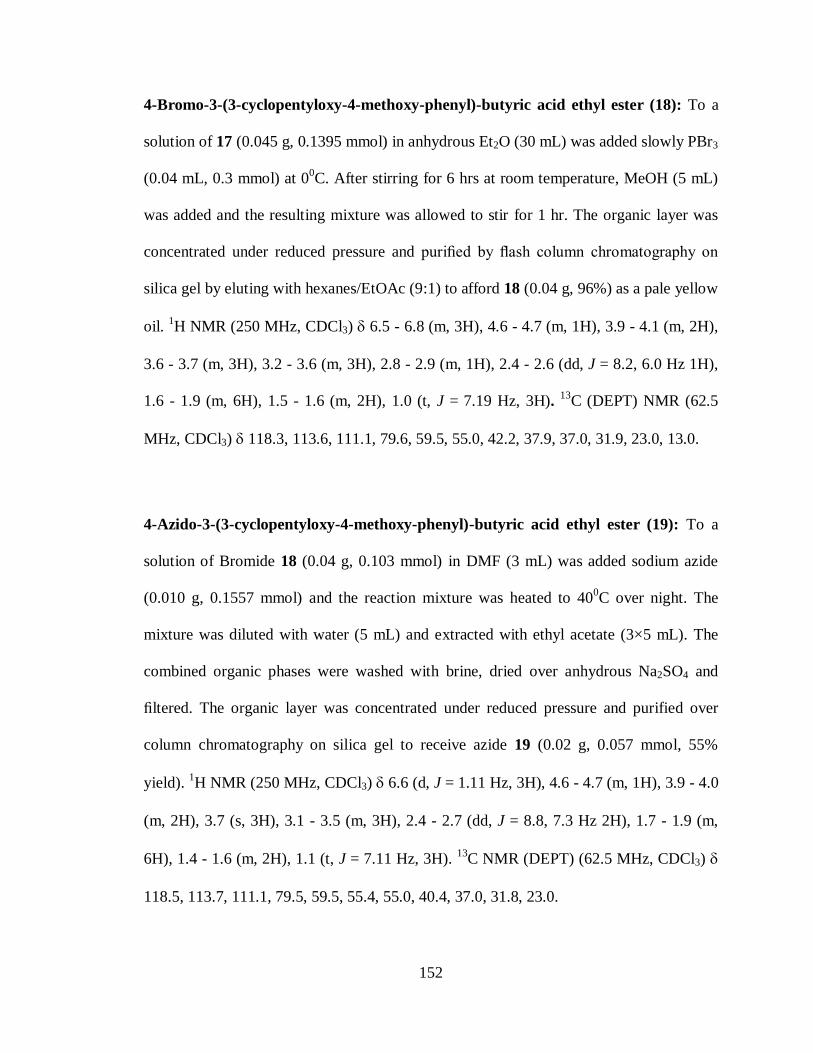
152
4-Bromo-3-(3-cyclopentyloxy-4-methoxy-phenyl)-butyric acid ethyl ester (18): To a
solution of 17 (0.045 g, 0.1395 mmol) in anhydrous Et2O (30 mL) was added slowly PBr3
(0.04 mL, 0.3 mmol) at 00C. After stirring for 6 hrs at room temperature, MeOH (5 mL)
was added and the resulting mixture was allowed to stir for 1 hr. The organic layer was
concentrated under reduced pressure and purified by flash column chromatography on
silica gel by eluting with hexanes/EtOAc (9:1) to afford 18 (0.04 g, 96%) as a pale yellow
oil. 1H NMR (250 MHz, CDCl3) 6.5 - 6.8 (m, 3H), 4.6 - 4.7 (m, 1H), 3.9 - 4.1 (m, 2H),
3.6 - 3.7 (m, 3H), 3.2 - 3.6 (m, 3H), 2.8 - 2.9 (m, 1H), 2.4 - 2.6 (dd, J = 8.2, 6.0 Hz 1H),
1.6 - 1.9 (m, 6H), 1.5 - 1.6 (m, 2H), 1.0 (t, J = 7.19 Hz, 3H). 13
C (DEPT) NMR (62.5
MHz, CDCl3) 118.3, 113.6, 111.1, 79.6, 59.5, 55.0, 42.2, 37.9, 37.0, 31.9, 23.0, 13.0.
4-Azido-3-(3-cyclopentyloxy-4-methoxy-phenyl)-butyric acid ethyl ester (19): To a
solution of Bromide 18 (0.04 g, 0.103 mmol) in DMF (3 mL) was added sodium azide
(0.010 g, 0.1557 mmol) and the reaction mixture was heated to 400C over night. The
mixture was diluted with water (5 mL) and extracted with ethyl acetate (3×5 mL). The
combined organic phases were washed with brine, dried over anhydrous Na2SO4 and
filtered. The organic layer was concentrated under reduced pressure and purified over
column chromatography on silica gel to receive azide 19 (0.02 g, 0.057 mmol, 55%
yield). 1H NMR (250 MHz, CDCl3) 6.6 (d, J = 1.11 Hz, 3H), 4.6 - 4.7 (m, 1H), 3.9 - 4.0
(m, 2H), 3.7 (s, 3H), 3.1 - 3.5 (m, 3H), 2.4 - 2.7 (dd, J = 8.8, 7.3 Hz 2H), 1.7 - 1.9 (m,
6H), 1.4 - 1.6 (m, 2H), 1.1 (t, J = 7.11 Hz, 3H). 13
C NMR (DEPT) (62.5 MHz, CDCl3)
118.5, 113.7, 111.1, 79.5, 59.5, 55.4, 55.0, 40.4, 37.0, 31.8, 23.0.

153
2-Cyclopentyloxy-4-iodo-1-methoxy-benzene (21): To a solution of aryl bromide 5 (1
g, 3.7 mmol) in DMF (10 mL) was added potassium iodide (9.213 g, 55.5 mmol) and
copper(I) iodide (3.52 g, 18.5 mmol). The heterogeneous reaction mixture was stirred at
1600C for 24 hrs. After the reaction was quenched with 1N HCl, EtOAc (25 mL) was
added and insoluble copper(I) was filtered over celite. The filtrate was extracted with
EtOAc (50 mL). The combined organic phase was washed with 1N HCl, brine, dried over
anhydrous. Na2SO4 and filtered. The organic layer was concentrated under reduced
pressure and purified over column chromatography on silica gel to receive aryl iodide 21
as a pale yellow oil (0.9 g, 2.83 mmol, 76% yield). 1H NMR (250 MHz, CDCl3) 6.9 -
7.2 (m, 2H), 6.5 (d, J = 8.37 Hz, 1H), 4.6 (dt, J = 3.00, 5.92 Hz, 1H), 3.6 - 3.7 (m, 3H),
1.6 - 1.9 (m, 6H), 1.4 - 1.6 (m, 2H). 13
C NMR (62.5 MHz, CDCl3) 150.1, 148.7, 129.6,
123.6, 113.7, 82.4, 80.7, 56.1, 32.7, 24.1.
2-(3-Cyclopentyloxy-4-methoxy-phenyl)-but-2-enedioic acid diethyl ester (22): To a
solution of aryl iodide 21 (0.9 g, 2.83 mmol) in CH3CN (5 mL) was added diethyl
maleate (1.3 mL, 7.86 mmol), Triethylamine (1.11 ml, 7.86 mmol), Pd(OAc)2 (0.007 g,
0.03 mmol) and stirred at 700C for 16 hrs. The reaction mixture was cooled and filtered
over celite plug. The filtrate was concentrated in vacuuo and purified over column
chromatography on silica gel by eluting with EtOAc/hexanes (1:4) to receive aryl maleate
22 as yellowish liquid (0.92 g, 2.547 mol, 90% yield). 1H NMR (250 MHz, CDCl3) 6.8
- 7.0 (m, 1H), 6.8 (s, 1H), 6.6 - 6.8 (m, 2H), 6.1 (s, 1H), 4.5 - 4.7 (m, 1H), 4.3 (q, J = 7.11
Hz, 1H), 4.1 (qd, J = 7.16, 10.58 Hz, 2H), 3.9 - 4.0(q, J = 9.2 Hz, 1H) , 3.6 - 3.7 (m, 3H),
1.6 - 1.9 (m, 6H), 1.4 - 1.6 (m, 2H), 0.9 - 1.3 (t, J = 8.5 Hz, 6H).
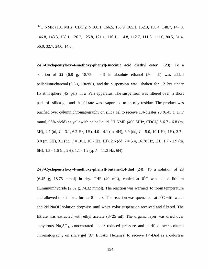
154
13
C NMR (101 MHz, CDCl3) 168.1, 166.5, 165.9, 165.1, 152.3, 150.4, 148.7, 147.8,
146.8, 143.3, 128.1, 126.2, 125.8, 121.1, 116.1, 114.8, 112.7, 111.6, 111.0, 80.5, 61.4,
56.0, 32.7, 24.0, 14.0.
2-(3-Cyclopentyloxy-4-methoxy-phenyl)-succinic acid diethyl ester (23): To a
solution of 22 (6.8 g, 18.75 mmol) in absolute ethanol (50 mL) was added
palladium/charcoal (0.8 g, 10wt%), and the suspension was shaken for 12 hrs under
H2 atmosphere (45 psi) in a Parr apparatus. The suspension was filtered over a short
pad of silica gel and the filtrate was evaporated to an oily residue. The product was
purified over column chromatography on silica gel to receive 1,4-diester 23 (6.45 g, 17.7
mmol, 95% yield) as yellowish color liquid. 1H NMR (400 MHz, CDCl3) 6.7 - 6.8 (m,
3H), 4.7 (td, J = 3.1, 6.2 Hz, 1H), 4.0 - 4.1 (m, 4H), 3.9 (dd, J = 5.0, 10.1 Hz, 1H), 3.7 -
3.8 (m, 3H), 3.1 (dd, J = 10.1, 16.7 Hz, 1H), 2.6 (dd, J = 5.4, 16.78 Hz, 1H), 1.7 - 1.9 (m,
6H), 1.5 - 1.6 (m, 2H), 1.1 - 1.2 (q, J = 11.3 Hz, 6H).
2-(3-Cyclopentyloxy-4-methoxy-phenyl)-butane-1,4-diol (24): To a solution of 23
(6.45 g, 18.75 mmol) in dry. THF (40 mL), cooled at 00C was added lithium
aluminiumhydride (2.82 g, 74.32 mmol). The reaction was warmed to room temperature
and allowed to stir for a further 8 hours. The reaction was quenched at 00C with water
and 2N NaOH solution dropwise until white color suspension received and filtered. The
filtrate was extracted with ethyl acetate (3×25 ml). The organic layer was dried over
anhydrous Na2SO4, concentrated under reduced pressure and purified over column
chromatography on silica gel (3:7 EtOAc/ Hexanes) to receive 1,4-Diol as a colorless

155
viscous liquid (4 g, 14.466 mmol, 77% yield). 1H NMR (400 MHz, CDCl3) 6.6 - 6.8
(m, 3H), 4.6 - 4.7 (m, 1H), 3.7 - 3.8 (d, J = 5.9 Hz, 3H), 3.4 - 3.7 (m, 4H), 2.8 (quin, J =
6.9 Hz, 1H), 2.5 - 2.7 (m, 1H), 1.7 - 1.9 (m, 8H), 1.4 - 1.6 (m, 2H). 13
C NMR (101 MHz,
CDCl3) 148.9, 147.7, 134.7, 119.8, 114.9, 112.1, 80.5, 67.5, 61.0, 56.1, 45.4, 35.9, 32.7,
24.0.
Acetic acid 4-acetoxy-2-(3-cyclopentyloxy-4-methoxy-phenyl)-butyl ester (25): To a
solution of 1,4-Diol (24) (4 g, 14.466 mmol) in CH2Cl2 was added acetylchloride (2.05
mL, 28.93 mmol) and pyridine (2.35 mL, 28.93 mmol). The reaction was stirred for 6 hrs
at room temperature and quenched with NaHCO3 (20 mL). The reaction mixture was
extracted with CH2Cl2 (2×25 mL). Combined organic layer was washed with 1N HCl,
brine solution and dried over Anhydrous. Na2SO4. Resulting crude reaction mixture from
evaporation under rotavap was purified by column chromatography on silica gel by
eluting with EtOAc/Hexanes (10:90) to receive 25 as yellowish liquid (4.4 g, 12.06
mmol, 84% yield).1H NMR (400 MHz, CDCl3) 6.7 - 6.8 (m, 1H), 6.6 - 6.7 (m, 2H), 4.6
- 4.7 (m, 1H), 4.1 - 4.2 (m, J = 7.0 Hz, 2H), 3.9 - 4.0 (m, 1H), 3.8 - 3.9 (m, 1H), 3.7 - 3.8
(m, 3H), 2.8 - 3.0 (m, 1H), 1.95 - 2.11 (m, 7H), 1.7 - 1.9 (m, 7H), 1.5 - 1.6 (m, 2H). 13
C
NMR (101 MHz, CDCl3) 170.9, 149.1, 147.6, 132.8, 119.8, 114.8, 112.1, 80.5, 68.3,
62.4, 56.0, 41.3, 32.7, 31.4, 24.0, 20.9.

156
Acetic acid 2-(3-cyclopentyloxy-4-methoxy-phenyl)-4-hydroxy-butyl ester (26):
10.23g (0.0284 mol) of 1,4-diacetate 25, was stirred in a mixture of phosphate buffer
(pH 7.0; 60ml) and acetone (3ml) in a round bottom flask at room temperature. Lipase
PS-30 (1.0 g) was added while maintaining the pH of the reaction mixture at 7.0 using 1N
NaOH solution. The reaction was stopped when no change in the pH of the reaction
medium occurred (approx. 24 hrs). The reaction was monitored by TLC (1:4
EtOAc:Hexanes). The reaction mixture was extracted with ethyl acetate (3 x 200 mL).
The organic layer was dried over Na2SO4. The organic layer was concentrated under
reduced pressure and purified by column chromatography over silica gel using
ethylacetate: hexane (1:3) to isolate the monoacetate 26 (49% yield). 1H NMR (400 MHz,
CDCl3) 6.6 - 6.8 (m, 3H), 4.6 - 4.7 (m, 1H), 4.1 - 4.2 (m, 2H), 3.7 - 3.8 (m, 3H), 3.4 -
3.6 (m, 2H), 2.9 - 3.0 (m, 1H), 1.9 - 2.0 (m, 3H), 1.7 - 1.9 (m, 6H), 1.5 - 1.6 (m, 2H). 13
C
NMR (101 MHz, CDCl3) 171.1, 149.0, 147.7, 133.5, 119.9, 115.0, 112.2, 80.4, 68.4,
60.5, 56.0, 41.0, 35.3, 32.8, 24.0, 20.9.
Acetic acid 2-(3-cyclopentyloxy-4-methoxy-phenyl)-4-oxo-butyl ester (27): To a
solution of monoacetate 25 (4.3 g, 13.33 mmol) in dry dichloromethane (20 ml) at 00C
was added Dess-Martin periodinane (40ml, 15%w/v). The reaction was allowed to stir at
00C for a further 90 minutes then allowed to warm to room temperature and stirred for an
additional 3 hours. A solution of sodium thiosulfate (30ml) was added followed by a
solution of sat. NaHCO3 (30 mL) and the solution stirred for an additional 60 minutes,
then vacuum-filtered over celite. An additional 30 ml of dichloromethane was added and
extracted with aqueous sodium chloride (20 ml). The organic layer was dried over
157
sodium sulfate, concentrated, and the product (yellow liquid, 4.2 g, 13.02 mmol, 98%
yield) is pure enough by 1H NMR and used immediately in the next reaction without
further purification.1H NMR (400 MHz, CDCl3) 9.6 - 9.7 (m, 1H), 6.6 - 6.8 (m, 3H),
4.6 - 4.8 (m, 1H), 4.0 - 4.3 (m, 2H), 3.7 - 3.8 (m, 3H), 3.4 - 3.5 (m, 1H), 2.7 - 2.8 (m, 2H),
1.7 - 2.0 (m, 9H), 1.5 - 1.6 (m, 2H). 13
C NMR (101 MHz, CDCl3) 200.7, 170.8, 149.3,
147.7, 132.2, 119.6, 114.8, 112.1, 80.4, 67.8, 56.0, 46.8, 38.5, 32.8, 24.02, 20.9.
4-Acetoxy-3-(3-cyclopentyloxy-4-methoxy-phenyl)-butyric acid (28): The aldehyde 27
(5.62 g, 17.541 mmol) was dissolved in DMF (20 mL). The Oxone (5.059 g, 26.312
mmol) was added to above solution in one portion and stirred at room temperature for 24
hrs. The reaction was monitored by TLC (1:4 EtOAc:Hexanes) and 1N HCl (3 mL) was
added to dissolve the salts and EtOAc was added to extract the products. The organic
layer was washed with 1N HCl (3x10 mL), brine and dried over anhydrous Na2SO4. The
organic layer was concentrated under reduced pressure and purified by silica gel column
chromatography to receive 28 as a colorless liquid (5.510 g, 16.38 mmol, 94% yield). 1H
NMR (400 MHz, CDCl3) 6.6 - 6.8 (m, 3H), 4.7 (tt, J = 3.17, 6.00 Hz, 1H), 4.1 - 4.2 (dd,
J = 7.8, 10.9 Hz, 1H), 4.0 (dd, J = 7.8, 10.9 Hz, 1H), 3.7 - 3.8 (m, 3H), 3.3 - 3.4 (m, 1H),
2.7 - 2.8 (m, 1H), 2.5 - 2.6 (m, 1H), 1.7 - 2.0 (m, 9H), 1.5 - 1.6 (m, 2H). 13
C NMR (101
MHz, CDCl3) 177.3, 170.8, 149.2, 147.6, 132.3, 119.5, 114.8, 112.1, 80.5, 67.7, 56.1,
40.5, 37.5, 32.7, 24.0, 20.7.

158
4-Acetoxy-3-(3-cyclopentyloxy-4-methoxy-phenyl)-butyric acid methyl ester (29): To
a solution of the acid 28 (5.51 g, 16.38 mmol) in dichloromethane (30 mL) was added
dicyclohexyldicarbodiimide (DCC) (4.4 g, 21.3 mmol), dimethylaminopyrimidine
(DMAP) (0.2 g, 1.638 mmol) MeOH (13.2 mL) and stirred at room temperature for 24
hrs. The resulting reaction mixture was quenched by adding 1N HCl (20 mL) and
extracted with CH2Cl2 (2×50 mL). Combined organic phase was washed with brine, dried
over anhydrous Na2SO4. The organic layer was concentrated under reduced pressure and
purified by silica gel column chromatography to receive 29 as a colorless liquid (4.87 g,
13.923 mmol, 94% yield). 1H NMR (400 MHz, CDCl3) 6.6 - 6.8 (m, 3H), 4.6 - 4.7 (m,
1H), 4.1 - 4.3 (m, 1H), 4.0 (dd, J = 7.61, 11.13 Hz, 1H), 3.7 - 3.8 (m, 3H), 3.5 - 3.6 (m,
3H), 3.3 - 3.4 (m, 1H), 2.6 - 2.7 (dd, J = 7.8, 7.8 Hz, 1H), 2.5 - 2.6 (m, 1H), 1.9 - 2.0 (m,
3H), 1.6 - 1.9 (m, 6H), 1.4 - 1.6 (m, 2H). 13
C NMR (101 MHz, CDCl3) 172.2, 170.7,
149.2, 147.5, 132.6, 119.5, 115.0, 112.1, 80.6, 67.5, 56.0, 51.5, 40.6, 37.6, 32.6, 24.0,
20.8.
4-(3-Cyclopentyloxy-4-methoxy-phenyl)-dihydro-furan-2-one (30): To a solution of
ester acetate 29 (0.160 g, 0.456 mmol) in dry. MeOH (4 mL) was added a solution
prepared from sodium (0.1 g) and dry. MeOH (4 mL) and the resulting mixture was
stirred for 3 hrs at room temperature. The reaction was quenched with glacial acetic acid
until pH=7 and water (5 mL) was added. The resulting reaction mixture was extracted
with CH2Cl2. To this reaction mixture was added DCC (0.147 g), DMAP (0.025 g) and
stirred at 400C for 6 hrs. The reaction mixture was then concentrated without heat under
reduced pressure and the crude product was purified by column chromatography on silica
159
gel by eluting with EtOAc/Hexanes (10:90) to receive 30 as yellowish liquid (0.103 g,
3.74 mmol, 82% yield). 1H NMR (400 MHz, CDCl3) 6.6 - 6.8 (m, 3H), 4.6 - 4.7 (m,
1H), 4.6- 4.5 (m, 1H), 4.0-4.2 (m,1H), 3.7 - 3.8 (m, 3H), 3.5 - 3.6 (m, 1H), 2.7 - 2.9 (m,
1H), 2.5 - 2.6 (m, 1H), 1.6 - 1.9 (m, 6H), 1.4 - 1.6 (m, 2H).
4.7. References
(1). Garcia, A. L. L.; Carpes, M. J. S.; de Oca, A. C. B. M.; dos Santos, M. A. G.;
Santana, C. C.; Correia, C. R. D. J. Org. Chem. 2005, 70, 1050.
(2). (a). Beavo, J. A.; Conti, M.; Heaslip, R. J. Mol. Pharmacol. 1994, 46, 399. (b).
Brackeen, M. F.; Cowan, D. J.; Stafford, J. A.; Schoenen, F. J.; Veal, J. M.;
Domanico, P. L.; Rose, D.; Strickland, A. B.; Verghese, M.; Feldman, P. L. J.
Med. Chem. 1995, 38, 4848. (c). Sommer, N.; Loschmann, P. A.; Northoff, G. H.;
Weller, M.; Steinbrecher, A.; Steinbach, J. P.; Lichtenfels, R.; Meyermann, R.;
Riethmuller, A.; Fontana, A.; Dichgans, J.; Martin, R. Nat. Med. 1995, 1, 244(d)
Schwabe, U.; Miyake, M.; Ohga, Y.; Daly, J. W. Mol. Pharmacol. 1976, 12, 900.
(3). (a). Marivet, M. C.; Bourguignon, J. J.; Lugnier, C.; Mann, A.; Stoclet, J. C.;
Wermuth, C. G. J. Med. Chem. 1989, 32, 1450. (b). Doherty, A. M. Curr. Opin.
Chem. Biol. 1999, 3, 466. (c). Pearse, D. D.; Pereira, F. C.; Marcillo, A. E.; Bates,
M. L.; Berrocal, Y. A.; Filbin, M. T.; Bunge, M. B. Nat.. Med. 2004, 10, 610. (d).
Weishaar, R. E.; Cain, M. H.; Bristol, J. A. J. Med. Chem. 1985, 28, 537.
(4). (a). Baures, P. W.; Eggleston, D. S.; Erhard, K. F.; Cieslinski, L. B.; Torphy, T.
J.; Christensen, S. B. J. Med. Chem. 1993, 36, 3274. (b). Barluenga, J.;
Fernandez-Rodriguez, M. A.; Aguilar, E.; Fernandez-Mari, F.; Salinas, A.; Olano,
B. Chem-Eur. J. 2001, 7, 3533.

160
(5). (a). Mulzer, J.; Zuhse, R.; Schmiechen, R. Angew. Chem. Int. Edit. 1992, 31, 870.
(b). Meyers, A. I.; Snyder, L. J Org Chem 1993, 58, 36. (c). Mulzer, J. J. Prak.
Chem. Chem. Ztg. 1994, 336, 287. (d). Honda, T.; Ishikawa, F.; Kanai, K.; Sato,
S.; Kato, D.; Tominaga, H. Heterocycles. 1996, 42, 109. (e). Langlois, N.; Wang,
H. S. Syn. Comm. 1997, 27, 3133. (f). Diaz, A.; Siro, J. G.; GarciaNavio, J. L.;
Vaquero, J. J.; AlvarezBuilla, J. Synthesis. 1997, 559. (g). Demnitz, J.;
LaVecchia, L.; Bacher, E.; Keller, T. H.; Muller, T.; Schurch, F.; Weber, H. P.;
Pombo-Villar, E. Molecules 1998, 3, 107. (h). Anada, M.; Mita, O.; Watanabe,
H.; Kitagaki, S.; Hashimoto, S. Synlett. 1999, 1775. (i). Chang, M. Y.; Sun, P. P.;
Chen, S. T.; Chang, N. C. Heterocycles. 2003, 60, 1865. (j). Yoon, C. H.; Nagle,
A.; Chen, C.; Gandhi, D.; Jung, K. W. Org. Lett. 2003, 5, 2259. (k). Barnes, D.
M.; Ji, J. G.; Fickes, M. G.; Fitzgerald, M. A.; King, S. A.; Morton, H. E.; Plagge,
F. A.; Preskill, M.; Wagaw, S. H.; Wittenberger, S. J.; Zhang, J. J. Am. Chem.
Soc. 2002, 124, 13097. (l). Paraskar, A. S.; Sudalai, A. Tetrahedron. 2006, 62,
4907(m) Hynes, P. S.; Stupple, P. A.; Dixon, D. J. Org. Lett. 2008, 10, 1389. (n).
Helmchen, G.; Becht, J.-M.; Meyer, O. Synthesis. 2003, 2805. (o). Feng, C. G.;
Shao, C.; Yu, H. J.; Wu, N. Y.; Tian, P.; Wang, R.; Lin, G. Q. Org. Lett. 2011, 13,
788. (p). Mandai, T.; Hasegawa, S.; Fujimoto, T.; Kawada, M.; Nokami, J.; Tsuji,
J. Synlett. 1990, 85.
(6). (a). Ogawa, H.; Amano, M.; Chihara, T. Chem. Commun. 1998, 495. (b). Ogawa,
H.; Ide, Y.; Honda, R.; Chihara, T. J. Phys. Org. Chem. 2003, 16, 355. (c).
Clarke, P. A. Tet. Lett. 2002, 43, 4761. (d). Misra, A. K.; Tiwari, P.; Madhusudan,
S. K. Carbohyd. Res. 2005, 340, 325.
161
(7). (a). Chihara, T.; Teratani, S.; Ogawa, H. J Chem Soc Chem Comm 1981, 1120.
(b). Chihara, T.; Takagi, Y.; Teratani, S.; Ogawa, H. Chem Lett 1982, 1451. (c).
Nishiguchi, T.; Taya, H. J Chem Soc Perk T 1 1990, 172. (d). Nishiguchi, T.;
Kawamine, K.; Ohtsuka, T. J Org Chem 1992, 57, 312. (e). Nishiguchi, T.; Taya,
H. J. Am. Chem. Soc. 1989, 111, 9102.
(8). (a). Delazerda, J.; Barak, G.; Sasson, Y. Tetrahedron 1989, 45, 1533. (b). Ogawa,
H.; Ichimura, Y.; Chihara, T.; Teratani, S.; Taya, K. B Chem Soc Jpn 1986, 59,
2481.
(9). (a). Oger, C.; Marton, Z.; Brinkmann, Y.; Bultel-Ponce, V.; Durand, T.; Graber,
M.; Galano, J. M. J Org Chem 2010, 75, 1892. (b). Egri, G.; Balint, J.; Peredi, R.;
Fogassy, E.; Novak, L.; Poppe, L. J Mol Catal B-Enzym 2000, 10, 583. (c).
Shishido, K.; Bando, T. J Mol Catal B-Enzym 1998, 5, 183. (d). Hisano, T.;
Onodera, K.; Toyabe, Y.; Mase, N.; Yoda, H.; Takabe, K. Tetrahedron Lett 2005,
46, 6293. (e). Takabe, K.; Mase, N.; Hisano, T.; Yoda, H. Tetrahedron Lett 2003,
44, 3267. (f). Ciuffreda, P.; Casati, S.; Santaniello, E. Tetrahedron Lett 2003, 44,
3663. (g). Ciuffreda, P.; Alessandrini, L.; Terraneo, G.; Santaniello, E.
Tetrahedron-Asymmetr 2003, 14, 3197. (h). Wang, Y. F.; Lalonde, J. J.;
Momongan, M.; Bergbreiter, D. E.; Wong, C. H. J Am Chem Soc 1988, 110,
7200(i) Guanti, G.; Narisano, E.; Podgorski, T.; Thea, S.; Williams, A.
Tetrahedron. 1990, 46, 7081.
(10). Carr, J. A.; Bisht, K. S. Org. Lett. 2004, 6, 3297.
(11). (a). Khan, P. M.; Wu, R. Z.; Bisht, K. S. Tetrahedron.. 2007, 63, 1116. (b). Khan,
P. M.; Bisht, K. S. Tet. Lett. 2010, 51, 1407.

162
(12). Beare, N. A.; Hartwig, J. F. J Org Chem. 2002, 67, 541.
(13). Ishii, Y.; Nagumo, S.; Arai, T.; Akuzawa, M.; Kawahara, N.; Akita, H.
Tetrahedron. 2006, 62, 716.
(14). Maehr, H.; Smallheer, J. M. J Org Chem. 1981, 46, 1752.
(15). Dussault, P. H.; Zope, U. R. J. Org. Chem. 1995, 60, 8218.
(16). Liang, Q.; Sun, Y.; Yu, B.; She, X.; Pan, X. J. Org. Chem. 2007, 72, 9846.
(17). Doyle, M. P.; Griffin, J. H.; Chinn, M. S.; Vanleusen, D. J Org Chem 1984, 49,
1917.
(18). He, W. Synlett 2006, 2006, 3548.
(19). Travis, B. R.; Sivakumar, M.; Hollist, G. O.; Borhan, B. Org. Lett. 2003, 5, 1031.
(20). Wijdeven, M. A.; Botman, P. N. M.; Wijtmans, R.; Schoemaker, H. E.; Rutjes, F.
P. J. T.; Blaauw, R. H. Org Lett 2005, 7, 4005.
(21). Whitcombe, N. Tetrahedron 2001, 57, 7449.
(22). Suzuki, H.; Kondo, A.; Ogawa, T. Chem Lett 1985, 411.
(23). Cortese, N. A.; Heck, R. F. J Org Chem 1978, 43, 3985.
(24). Cortese, N. A.; Ziegler, C. B.; Hrnjez, B. J.; Heck, R. F. J Org Chem 1978, 43,
2952.
(25). Ferland, J. M.; Demerson, C. A.; Humber, L. G. Can J Chem 1985, 63, 361.
(26). Lin, C.; Hiraga, Y.; Masaki, K.; Iefuji, H.; Ohkata, K. Journal of Molecular
Catalysis B: Enzymatic 2006, 38, 1.
(27). Ferraboschi, P.; Casati, S.; Verza, E.; Santaniello, E. Tet.Asymm. 1995, 6, 1027.
(28). (a). Fuganti, C.; Pedrocchi-Fantoni, G.; Servi, S. Chem Lett 1990, 1137. (b).
Pedrocchi-Fantoni, G.; Servi, S. Journal of the Chemical Society, Perkin

163
Transactions 1 1992, 1029. (c). Degueil-Castaing, M. Tetrahedron Lett 1987, 28,
953. (d). Wang, Y. F.; Lalonde, J. J.; Momongan, M.; Bergbreiter, D. E.; Wong,
C. H. J Am Chem Soc 1988, 110, 7200. (e). Fang, J.-M.; Wong, C.-H. Synlett
1994, 1994, 393. (f). Faber, K.; Riva, S. Synthesis 1992, 1992, 895. (g).
Ferraboschi, P. Tetrahedron: Asymmetry 1994, 5, 691. (h). Ferraboschi, P.;
Casati, S.; Grandi, S. D.; Grisenti, P.; Santaniello, E. Biocatalysis and
Biotransformation 1994, 10, 279. (i). Lutz, D. Tetrahedron: Asymmetry 1990, 1,
783. (j). Foelsche, E.; Hickel, A.; Hoenig, H.; Seufer-Wasserthal, P. The Journal
of Organic Chemistry 1990, 55, 1749. (k). Kazlauskas, R. J.; Weissfloch, A. N.
E.; Rappaport, A. T.; Cuccia, L. A. The Journal of Organic Chemistry 1991, 56,
2656. (l). Xie, Z. Tetrahedron: Asymmetry 1993, 4, 973(m) Carrea, G. Tet.:
Asymm. 1993, 4, 1063.
(29). Mendgen, T.; Scholz, T.; Klein, C. D. Bioorganic & Medicinal Chemistry Letters
2010, 20, 5757.
(30). Knowles, J. P.; O′Connor, V. E.; Whiting, A. Org. & Bio. Chem. 2011, 9, 1876.
(31). Mulzer, J.; Greifenberg, S.; Beckstett, A.; Gottwald, M. Liebigs Annalen Der
Chemie 1992, 1992, 1131.
(32). (a) Fadnavis, N. Tetrahedron: Asymmetry 2001, 12, 691(b) Shimotori, Y.; Sekine,
K.; Miyakoshi, T. Flavour and Fragrance Journal 2007, 22, 531.
(33). Prasad, A.; Mukherjee, C.; Sharma, D.; Rastogi, S.; Mangalam, A.; Jha, A.;
Olsen, C.; Patkar, S.; Parmar, V. Journal of Molecular Catalysis B: Enzymatic
2006, 40, 101.
(34). Lee, D. Tett. Lett. 1998, 39, 2163.

164
(35). Mattsson, S.; Dahlström, M.; Karlsson, S. Tet. Lett. 2007, 48, 2497.
(36). Gamalevich, G. D.; Serebryakov, E. P.; Vlasyuk, A. L. Mend. Comm. 1998, 8, 8.

165
Appendices
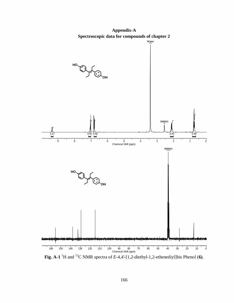
166
Appendix-A
Spectroscopic data for compounds of chapter 2
OH
OH
9 8 7 6 5 4 3 2 1 0
Chemical Shift (ppm)
5.984.004.064.001.67
DMSO
Water
OH
OH
160 150 140 130 120 110 100 90 80 70 60 50 40 30 20 10 0
Chemical Shift (ppm)
DMSO
Fig. A-1
1H and
13C NMR spectra of E-4,4'-[1,2-diethyl-1,2-ethenediyl]bis Phenol (6).

167
10 9 8 7 6 5 4 3 2 1 0
Chemical Shift (ppm)
5.694.004.041.48
DMSO
Water
OH
OH
160 150 140 130 120 110 100 90 80 70 60 50 40 30 20 10 0
Chemical Shift (ppm)
DMSO
OH
OH
Fig. A-2
1H and
13C NMR spectra of E/Z-4,4'-[1,2-dimethyl-1,2-ethenediyl]bis Phenol
(7).
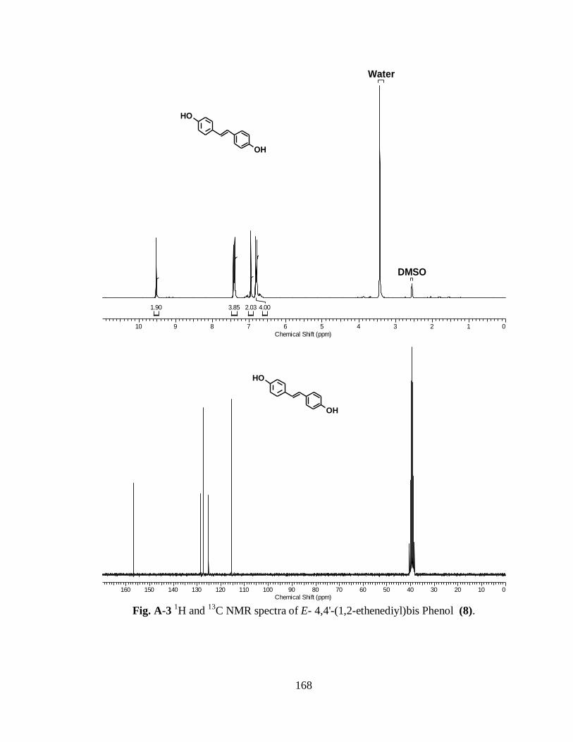
168
10 9 8 7 6 5 4 3 2 1 0
Chemical Shift (ppm)
4.002.033.851.90
DMSO
Water
OH
OH
160 150 140 130 120 110 100 90 80 70 60 50 40 30 20 10 0
Chemical Shift (ppm)
OH
OH
Fig. A-3
1H and
13C NMR spectra of E- 4,4'-(1,2-ethenediyl)bis Phenol (8).

169
10 9 8 7 6 5 4 3 2 1 0
Chemical Shift (ppm)
0.446.106.080.582.000.141.91
OHOH
160 150 140 130 120 110 100 90 80 70 60 50 40 30 20 10 0
Chemical Shift (ppm)
DMSO
OHOH
Fig. A-4 1H and
13C NMR spectra of E/Z-3,3'-[1,2-dimethyl-1,2-ethenediyl]bis Phenol
(9).

170
8.5 8.0 7.5 7.0 6.5 6.0 5.5 5.0 4.5 4.0 3.5 3.0 2.5 2.0 1.5 1.0 0.5 0
Chemical Shift (ppm)
6.6513.554.318.854.004.192.05
OH
N
OH
N
11 (G6)
170 160 150 140 130 120 110 100 90 80 70 60 50 40 30 20 10 0
Chemical Shift (ppm)
OH
N
OH
N
11 (G6)
Fig. A-5
1H and
13C NMR spectra of E-4,4'-(1,2-diethyl-1,2-ethenediyl)bis[2-
[(diethylamino)methyl]-Phenol (11).
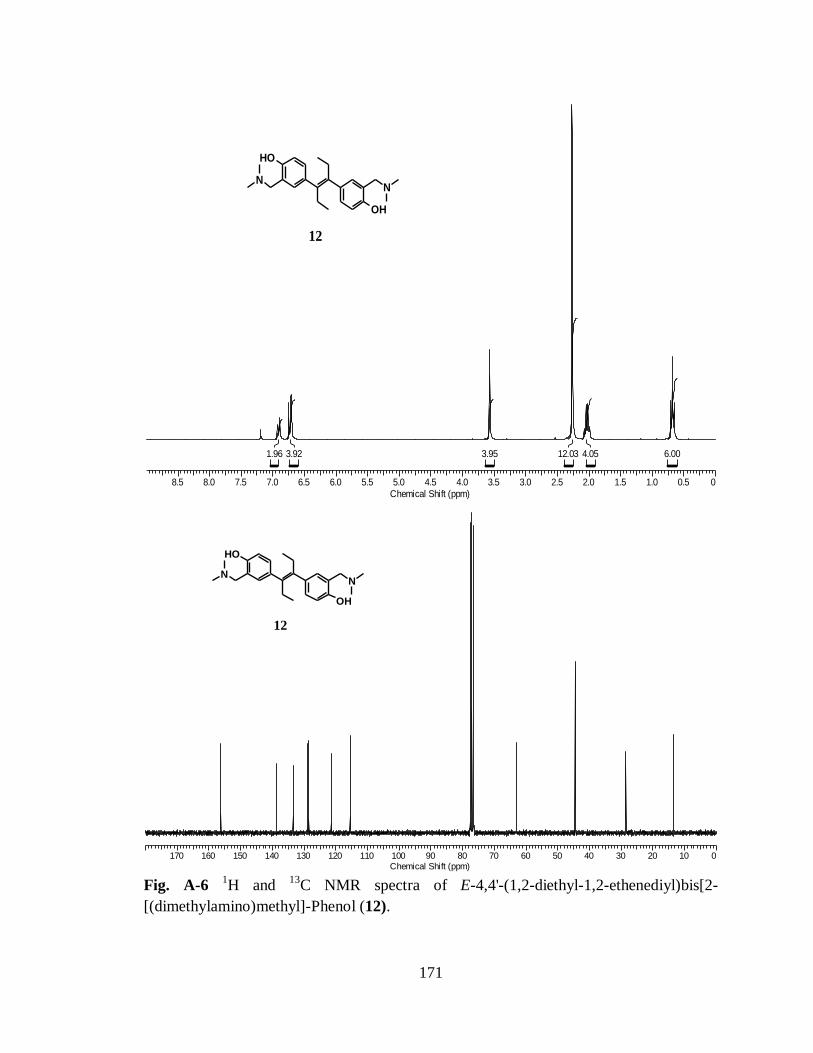
171
8.5 8.0 7.5 7.0 6.5 6.0 5.5 5.0 4.5 4.0 3.5 3.0 2.5 2.0 1.5 1.0 0.5 0
Chemical Shift (ppm)
6.004.0512.033.953.921.96
12
OH
N
OH
N
170 160 150 140 130 120 110 100 90 80 70 60 50 40 30 20 10 0
Chemical Shift (ppm)
12
OH
N
OH
N
Fig. A-6
1H and
13C NMR spectra of E-4,4'-(1,2-diethyl-1,2-ethenediyl)bis[2-
[(dimethylamino)methyl]-Phenol (12).

172
8.5 8.0 7.5 7.0 6.5 6.0 5.5 5.0 4.5 4.0 3.5 3.0 2.5 2.0 1.5 1.0 0.5 0
Chemical Shift (ppm)
5.9712.624.207.774.003.741.87
OH
N
OH
N
13
170 160 150 140 130 120 110 100 90 80 70 60 50 40 30 20 10 0
Chemical Shift (ppm)
OH
N
OH
N
13
Fig. A-7
1H and
13C NMR spectra of E-4,4'-(1,2-diethyl-1,2-ethenediyl)bis[2-
[(pyrrolidine)methyl]-Phenol (13).

173
8.5 8.0 7.5 7.0 6.5 6.0 5.5 5.0 4.5 4.0 3.5 3.0 2.5 2.0 1.5 1.0 0.5 0
Chemical Shift (ppm)
6.108.104.117.884.003.981.97
OH
N
OH
N
14
170 160 150 140 130 120 110 100 90 80 70 60 50 40 30 20 10 0
Chemical Shift (ppm)
OH
N
OH
N
14
Fig. A-8
1H and
13C NMR spectra of E-4,4'-(1,2-diethyl-1,2-ethenediyl)bis[2-
[(piperidine)methyl]-Phenol (14).
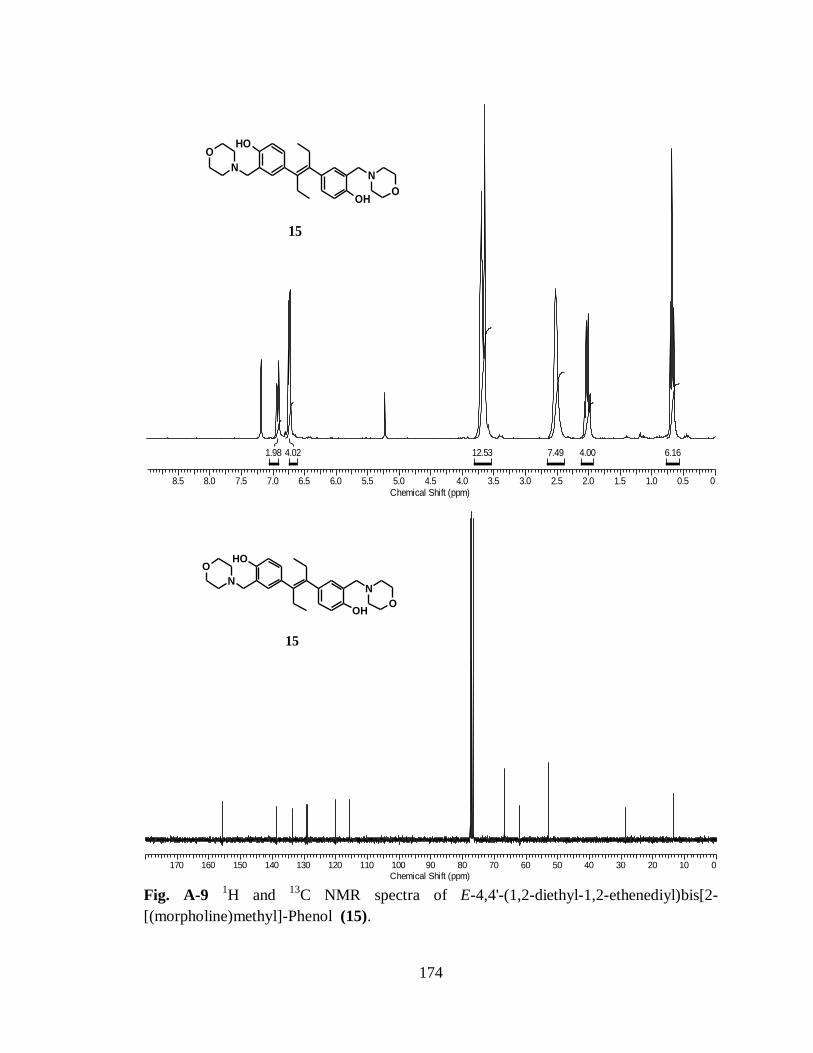
174
8.5 8.0 7.5 7.0 6.5 6.0 5.5 5.0 4.5 4.0 3.5 3.0 2.5 2.0 1.5 1.0 0.5 0
Chemical Shift (ppm)
6.164.007.4912.534.021.98
OH
N
OH
N
O
O
15
170 160 150 140 130 120 110 100 90 80 70 60 50 40 30 20 10 0
Chemical Shift (ppm)
OH
N
OH
N
O
O
15
Fig. A-9
1H and
13C NMR spectra of E-4,4'-(1,2-diethyl-1,2-ethenediyl)bis[2-
[(morpholine)methyl]-Phenol (15).

175
8.5 8.0 7.5 7.0 6.5 6.0 5.5 5.0 4.5 4.0 3.5 3.0 2.5 2.0 1.5 1.0 0.5 0
Chemical Shift (ppm)
14.176.599.524.005.372.86
16
OH
N
OH
N
180 170 160 150 140 130 120 110 100 90 80 70 60 50 40 30 20 10 0
Chemical Shift (ppm)
16
OH
N
OH
N
Fig. A-10
1H and
13C NMR spectra of 4,4'-(1,2-dimethyl-1,2-ethenediyl)bis[2-
[(diethylamino)methyl]-Phenol (16).
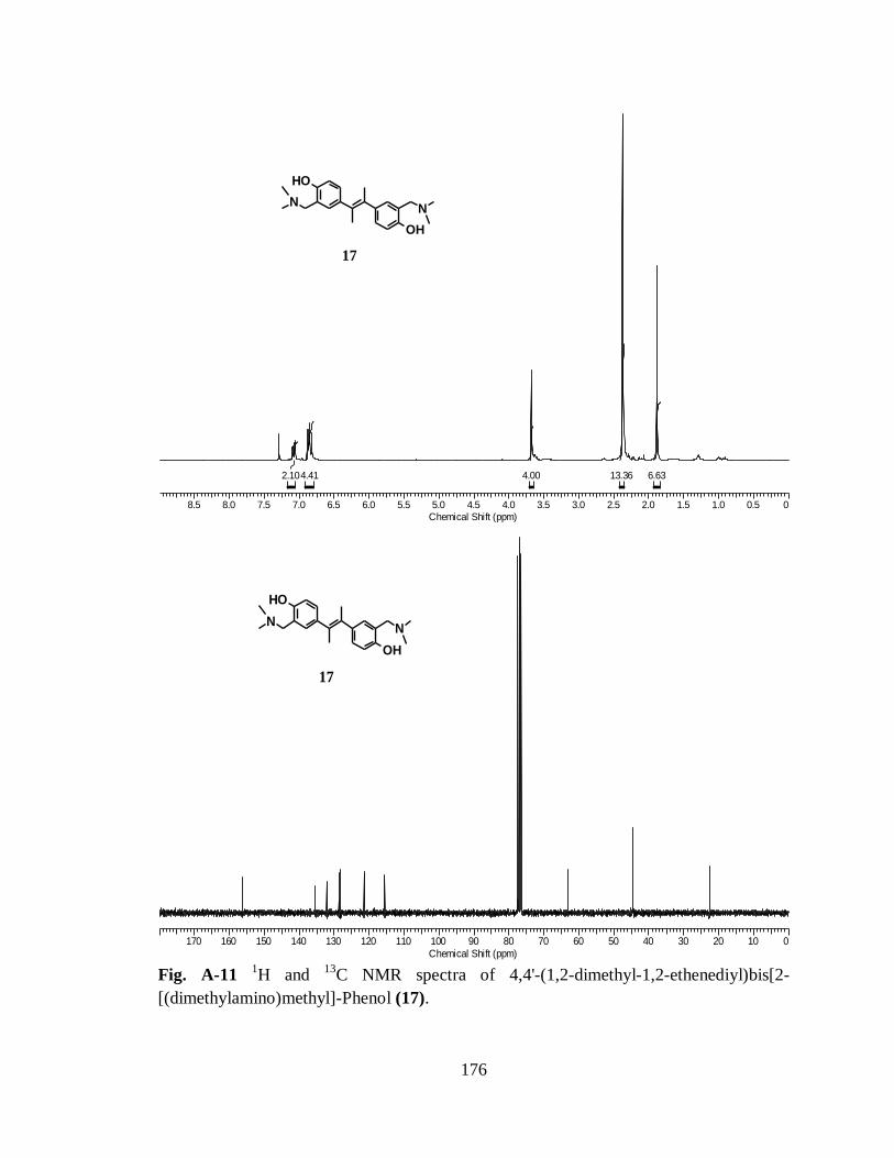
176
8.5 8.0 7.5 7.0 6.5 6.0 5.5 5.0 4.5 4.0 3.5 3.0 2.5 2.0 1.5 1.0 0.5 0
Chemical Shift (ppm)
6.6313.364.004.412.10
OH
N
OH
N
17
170 160 150 140 130 120 110 100 90 80 70 60 50 40 30 20 10 0
Chemical Shift (ppm)
OH
N
OH
N
17
Fig. A-11
1H and
13C NMR spectra of 4,4'-(1,2-dimethyl-1,2-ethenediyl)bis[2-
[(dimethylamino)methyl]-Phenol (17).

177
8.5 8.0 7.5 7.0 6.5 6.0 5.5 5.0 4.5 4.0 3.5 3.0 2.5 2.0 1.5 1.0 0.5 0
Chemical Shift (ppm)
14.896.787.904.004.332.08
OH
N
OH
N
18
170 160 150 140 130 120 110 100 90 80 70 60 50 40 30 20 10 0
Chemical Shift (ppm)
OH
N
OH
N
18
Fig. A-12
1H and
13C NMR spectra of 4,4'-(1,2-dimethyl-1,2-ethenediyl)bis[2-
[(pyrrolidine)methyl]-Phenol (18).

178
8.5 8.0 7.5 7.0 6.5 6.0 5.5 5.0 4.5 4.0 3.5 3.0 2.5 2.0 1.5 1.0 0.5 0
Chemical Shift (ppm)
14.437.984.004.001.99
OH
N
OH
N
19
170 160 150 140 130 120 110 100 90 80 70 60 50 40 30 20 10 0
Chemical Shift (ppm)
OH
N
OH
N
19
Fig. A-13
1H and
13C NMR spectra of 4,4'-(1,2-dimethyl-1,2-ethenediyl)bis[2-
[(Piperidine)methyl]-Phenol (19).

179
8.5 8.0 7.5 7.0 6.5 6.0 5.5 5.0 4.5 4.0 3.5 3.0 2.5 2.0 1.5
Chemical Shift (ppm)
6.009.6713.323.711.78
OH
N
OH
N
O
O
20
Fig. A-14
1H and
13C NMR spectra of E-4,4'-(1,2-ethenediyl)bis[2-[(morpholine)methyl]-
Phenol (20).

180
8.5 8.0 7.5 7.0 6.5 6.0 5.5 5.0 4.5 4.0 3.5 3.0 2.5 2.0 1.5 1.0 0.5 0
Chemical Shift (ppm)
14.579.534.004.582.032.79
21
OH
N
OH
N
170 160 150 140 130 120 110 100 90 80 70 60 50 40 30 20 10 0
Chemical Shift (ppm)
21
OH
N
OH
N
Fig. A-15
1H and
13C NMR spectra of E-4,4'-(1,2-ethenediyl)bis[2-
[(diethylamino)methyl]-Phenol (21).
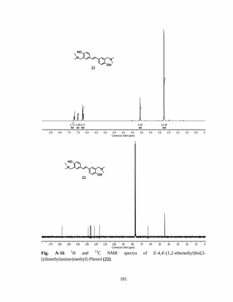
181
8.5 8.0 7.5 7.0 6.5 6.0 5.5 5.0 4.5 4.0 3.5 3.0 2.5 2.0 1.5 1.0 0.5 0
Chemical Shift (ppm)
12.484.004.171.982.73
OH
N
OH
N
22
170 160 150 140 130 120 110 100 90 80 70 60 50 40 30 20 10 0
Chemical Shift (ppm)
OH
N
OH
N
22
Fig. A-16
1H and
13C NMR spectra of E-4,4'-(1,2-ethenediyl)bis[2-
[(dimethylamino)methyl]-Phenol (22).

182
8.5 8.0 7.5 7.0 6.5 6.0 5.5 5.0 4.5 4.0 3.5 3.0 2.5 2.0 1.5 1.0 0.5 0
Chemical Shift (ppm)
11.343.744.004.032.032.37
OH
N
OH
N
23
170 160 150 140 130 120 110 100 90 80 70 60 50 40 30 20 10 0
Chemical Shift (ppm)
OH
N
OH
N
23
Fig. A-17
1H and
13C NMR spectra of E-4,4'-(1,2-ethenediyl)bis[2-[(pyrrolidine)methyl]-
Phenol (23).

183
8.5 8.0 7.5 7.0 6.5 6.0 5.5 5.0 4.5 4.0 3.5 3.0 2.5 2.0 1.5 1.0 0.5 0
Chemical Shift (ppm)
8.007.773.993.992.002.24
OH
N
OH
N
24
170 160 150 140 130 120 110 100 90 80 70 60 50 40 30 20 10 0
Chemical Shift (ppm)
OH
N
OH
N
24
Fig. A-18
1H and
13C NMR spectra of E-4,4'-(1,2-ethenediyl)bis[2-[(piperidine)methyl]-
Phenol (24).
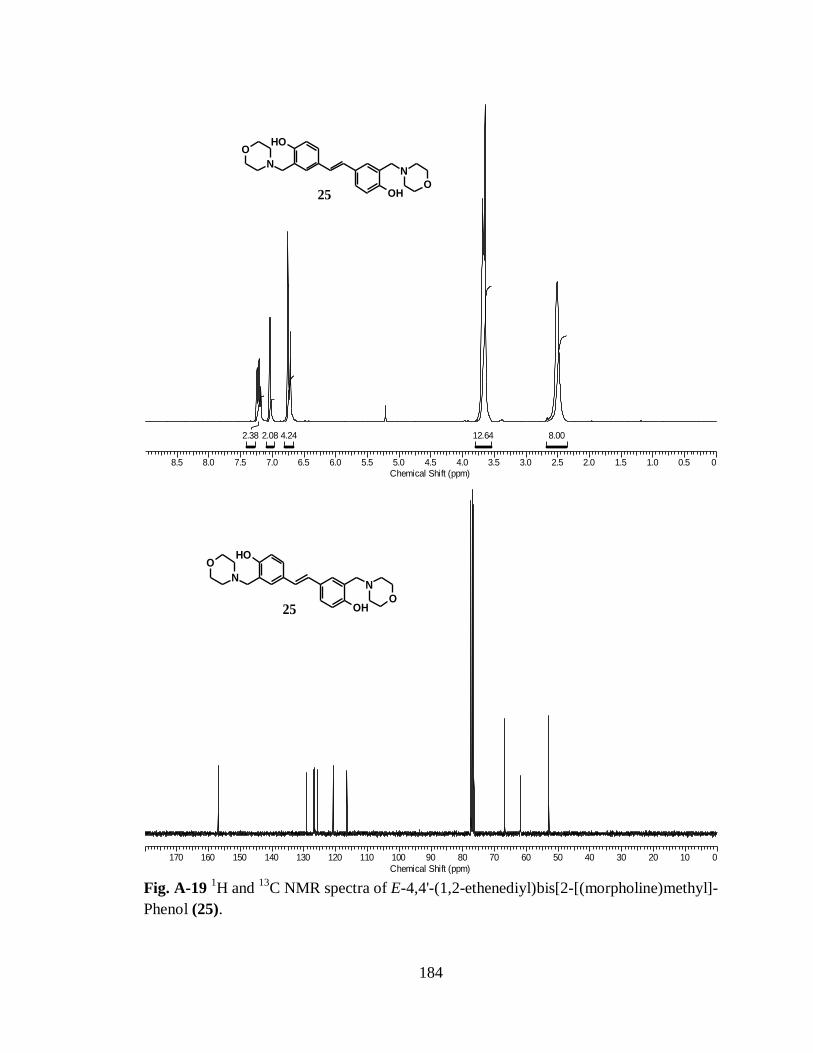
184
8.5 8.0 7.5 7.0 6.5 6.0 5.5 5.0 4.5 4.0 3.5 3.0 2.5 2.0 1.5 1.0 0.5 0
Chemical Shift (ppm)
8.0012.644.242.082.38
OH
N
OH
N
O
O25
170 160 150 140 130 120 110 100 90 80 70 60 50 40 30 20 10 0
Chemical Shift (ppm)
OH
N
OH
N
O
O25
Fig. A-19
1H and
13C NMR spectra of E-4,4'-(1,2-ethenediyl)bis[2-[(morpholine)methyl]-
Phenol (25).

185
8.5 8.0 7.5 7.0 6.5 6.0 5.5 5.0 4.5 4.0 3.5 3.0 2.5 2.0 1.5 1.0 0.5 0
Chemical Shift (ppm)
11.450.866.007.560.903.177.26
OHOH
N
N
26
170 160 150 140 130 120 110 100 90 80 70 60 50 40 30 20 10 0
Chemical Shift (ppm)
26
OHOH
N
N
Fig. A-20
1H and
13C NMR spectra of 3,3'-(1,2-dimethyl-1,2-ethenediyl)bis[2-
[(diethylamino)methyl]-Phenol (26).

186
8.5 8.0 7.5 7.0 6.5 6.0 5.5 5.0 4.5 4.0 3.5 3.0 2.5 2.0 1.5 1.0 0.5 0
Chemical Shift (ppm)
6.222.075.5612.601.484.005.682.02
27
OHOH
N
N
170 160 150 140 130 120 110 100 90 80 70 60 50 40 30 20 10 0
Chemical Shift (ppm)
27
OHOH
N
N
Fig. A-21
1H and
13C NMR spectra of 3,3'-(1,2-dimethyl-1,2-ethenediyl)bis[2-
[(dimethylamino)methyl]-Phenol (27).
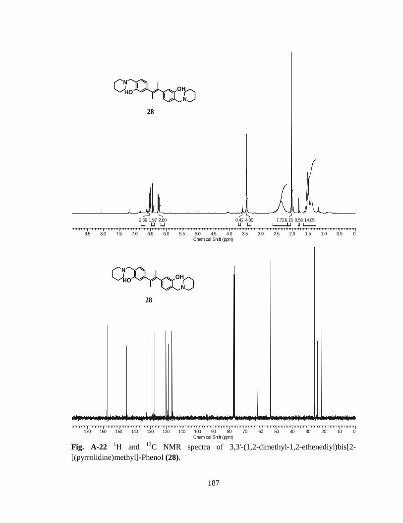
187
8.5 8.0 7.5 7.0 6.5 6.0 5.5 5.0 4.5 4.0 3.5 3.0 2.5 2.0 1.5 1.0 0.5 0
Chemical Shift (ppm)
14.050.586.157.724.000.422.001.972.36
OHOH
N
N
28
170 160 150 140 130 120 110 100 90 80 70 60 50 40 30 20 10 0
Chemical Shift (ppm)
OHOH
N
N
28
Fig. A-22
1H and
13C NMR spectra of 3,3'-(1,2-dimethyl-1,2-ethenediyl)bis[2-
[(pyrrolidine)methyl]-Phenol (28).

188
8.5 8.0 7.5 7.0 6.5 6.0 5.5 5.0 4.5 4.0 3.5 3.0 2.5 2.0 1.5 1.0 0.5 0
Chemical Shift (ppm)
0.260.160.240.100.010.050.050.06
OHOH
N
N
29
170 160 150 140 130 120 110 100 90 80 70 60 50 40 30 20 10 0
Chemical Shift (ppm)
OHOH
N
N
29
Fig. A-23
1H and
13C NMR spectra of 3,3'-(1,2-dimethyl-1,2-ethenediyl)bis[2-
[(piperidine)methyl]-Phenol (29).
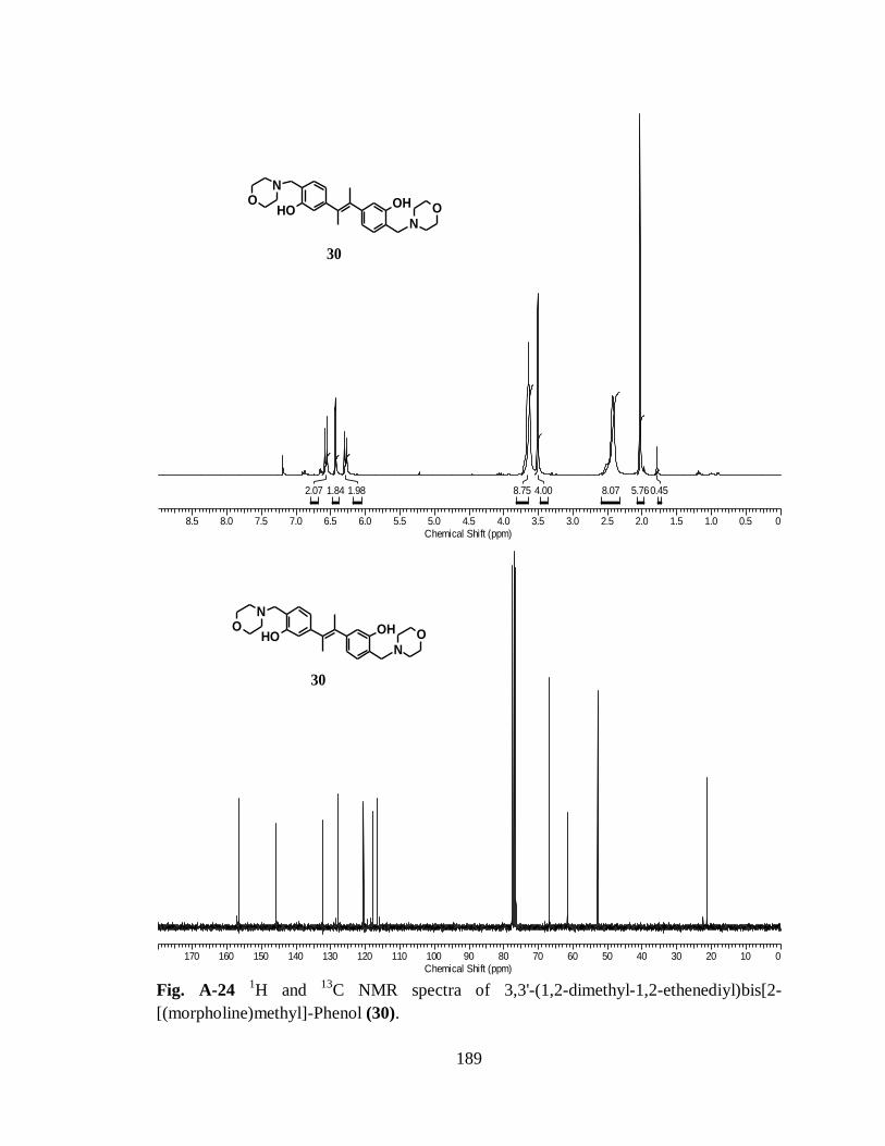
189
8.5 8.0 7.5 7.0 6.5 6.0 5.5 5.0 4.5 4.0 3.5 3.0 2.5 2.0 1.5 1.0 0.5 0
Chemical Shift (ppm)
0.455.768.074.008.751.981.842.07
OHOH
N
NO
O
30
170 160 150 140 130 120 110 100 90 80 70 60 50 40 30 20 10 0
Chemical Shift (ppm)
OHOH
N
NO
O
30
Fig. A-24
1H and
13C NMR spectra of 3,3'-(1,2-dimethyl-1,2-ethenediyl)bis[2-
[(morpholine)methyl]-Phenol (30).

190
8.5 8.0 7.5 7.0 6.5 6.0 5.5 5.0 4.5 4.0 3.5 3.0 2.5 2.0 1.5 1.0 0.5 0
Chemical Shift (ppm)
14.539.154.008.21
31
OHOH
N
N
170 160 150 140 130 120 110 100 90 80 70 60 50 40 30 20 10 0
Chemical Shift (ppm)
31
OHOH
N
N
Fig. A-25
1H and
13C NMR spectra of E-3,3'-(1,2-ethenediyl)bis[2-
[(diethylamino)methyl]-Phenol (31).

191
8.5 8.0 7.5 7.0 6.5 6.0 5.5 5.0 4.5 4.0 3.5 3.0 2.5 2.0 1.5 1.0 0.5 0
Chemical Shift (ppm)
1.2212.594.0010.26
32
OHOH
N
N
170 160 150 140 130 120 110 100 90 80 70 60 50 40 30 20 10 0
Chemical Shift (ppm)
32
OHOH
N
N
Fig. A-26
1H and
13C NMR spectra of E-3,3'-(1,2-ethenediyl)bis[2-
[(dimethylamino)methyl]-Phenol (32).
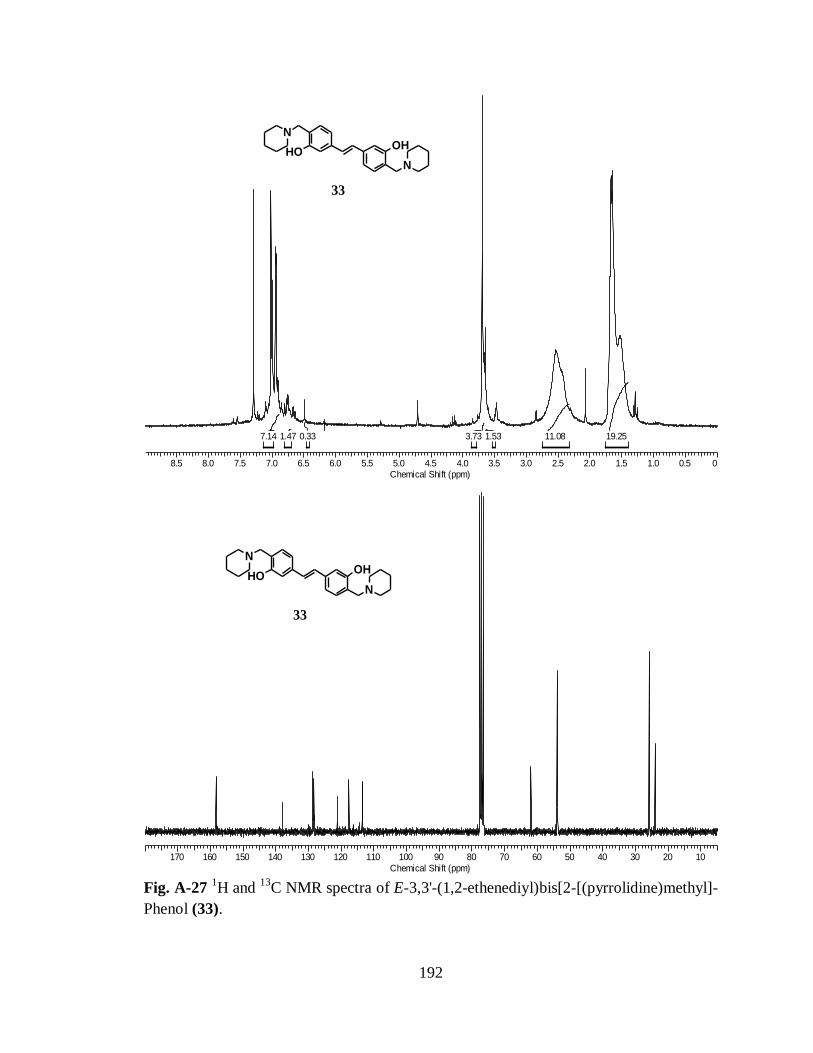
192
8.5 8.0 7.5 7.0 6.5 6.0 5.5 5.0 4.5 4.0 3.5 3.0 2.5 2.0 1.5 1.0 0.5 0
Chemical Shift (ppm)
19.2511.081.533.730.331.477.14
OHOH
N
N
33
170 160 150 140 130 120 110 100 90 80 70 60 50 40 30 20 10
Chemical Shift (ppm)
OHOH
N
N
33
Fig. A-27
1H and
13C NMR spectra of E-3,3'-(1,2-ethenediyl)bis[2-[(pyrrolidine)methyl]-
Phenol (33).

193
8.5 8.0 7.5 7.0 6.5 6.0 5.5 5.0 4.5 4.0 3.5 3.0 2.5 2.0 1.5 1.0 0.5 0
Chemical Shift (ppm)
8.147.834.008.05
OHOH
N
N
34
170 160 150 140 130 120 110 100 90 80 70 60 50 40 30 20 10 0
Chemical Shift (ppm)
OHOH
N
N
34
Fig. A-28
1H and
13C NMR spectra of E-3,3'-(1,2-ethenediyl)bis[2-[(piperidine)methyl]-
Phenol (34).
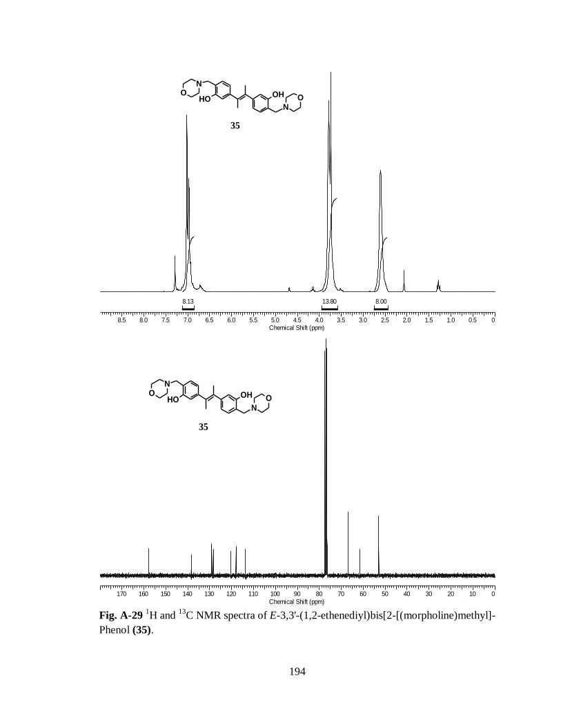
194
8.5 8.0 7.5 7.0 6.5 6.0 5.5 5.0 4.5 4.0 3.5 3.0 2.5 2.0 1.5 1.0 0.5 0
Chemical Shift (ppm)
8.0013.808.13
OHOH
N
NO
O
35
170 160 150 140 130 120 110 100 90 80 70 60 50 40 30 20 10 0
Chemical Shift (ppm)
OHOH
N
NO
O
35
Fig. A-29
1H and
13C NMR spectra of E-3,3'-(1,2-ethenediyl)bis[2-[(morpholine)methyl]-
Phenol (35).
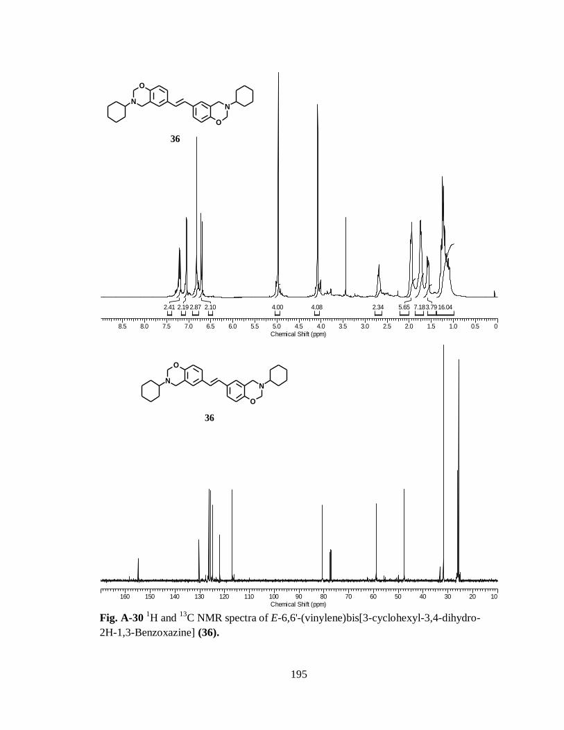
195
8.5 8.0 7.5 7.0 6.5 6.0 5.5 5.0 4.5 4.0 3.5 3.0 2.5 2.0 1.5 1.0 0.5 0
Chemical Shift (ppm)
16.043.797.185.652.344.084.002.102.872.192.41
O
N
O
N
36
160 150 140 130 120 110 100 90 80 70 60 50 40 30 20 10
Chemical Shift (ppm)
O
N
O
N
36
Fig. A-30
1H and
13C NMR spectra of E-6,6'-(vinylene)bis[3-cyclohexyl-3,4-dihydro-
2H-1,3-Benzoxazine] (36).

196
8.5 8.0 7.5 7.0 6.5 6.0 5.5 5.0 4.5 4.0 3.5 3.0 2.5 2.0 1.5 1.0 0.5 0
Chemical Shift (ppm)
10.385.204.642.004.144.244.402.132.37
O
N
O
N
37
170 160 150 140 130 120 110 100 90 80 70 60 50 40 30 20 10 0
Chemical Shift (ppm)
O
N
O
N
37
Fig. A-31
1H and
13C NMR spectra of E-6,6'-(vinylene)bis[3-cyclopentyl-3,4-dihydro-
2H-1,3-Benzoxazine] (37).

197
8.5 8.0 7.5 7.0 6.5 6.0 5.5 5.0 4.5 4.0 3.5 3.0 2.5 2.0 1.5 1.0 0.5 0
Chemical Shift (ppm)
8.484.004.462.0113.74
O
N
O
N
38
170 160 150 140 130 120 110 100 90 80 70 60 50 40 30 20 10 0
Chemical Shift (ppm)
O
N
O
N
38
Fig. A-32
1H and
13C NMR spectra of E-6,6'-(vinylene)bis[3-benzyl-3,4-dihydro- 2H-
1,3-Benzoxazine] (38).

198
8.5 8.0 7.5 7.0 6.5 6.0 5.5 5.0 4.5 4.0 3.5 3.0 2.5 2.0 1.5 1.0 0.5 0
Chemical Shift (ppm)
6.774.693.794.008.901.836.32
39
O
N
O
N
O
O
170 160 150 140 130 120 110 100 90 80 70 60 50 40 30 20 10 0
Chemical Shift (ppm)
39
O
N
O
N
O
O
Fig. A-33
1H and
13C NMR spectra of E-6,6'-(vinylene)bis[3-(4-methoxy benzyl)-3,4-
dihydro- 2H-1,3-Benzoxazine] (39).

199
8.5 8.0 7.5 7.0 6.5 6.0 5.5 5.0 4.5 4.0 3.5 3.0 2.5 2.0 1.5 1.0 0.5 0
Chemical Shift (ppm)
17.254.648.057.002.523.954.0010.22
N
O
N
O
40
170 160 150 140 130 120 110 100 90 80 70 60 50 40 30 20 10 0
Chemical Shift (ppm)
N
O
N
O
40
Fig. A-34
1H and
13C NMR spectra of E-7,7'-(vinylene)bis[3-cyclohexyl-3,4-dihydro-
2H-1,3-Benzoxazine] (40).
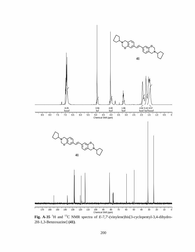
200
8.5 8.0 7.5 7.0 6.5 6.0 5.5 5.0 4.5 4.0 3.5 3.0 2.5 2.0 1.5 1.0 0.5 0
Chemical Shift (ppm)
9.575.104.941.964.003.568.29
N
O
N
O
41
170 160 150 140 130 120 110 100 90 80 70 60 50 40 30 20 10 0
Chemical Shift (ppm)
N
O
N
O
41
Fig. A-35
1H and
13C NMR spectra of E-7,7'-(vinylene)bis[3-cyclopentyl-3,4-dihydro-
2H-1,3-Benzoxazine] (41).

201
8.5 8.0 7.5 7.0 6.5 6.0 5.5 5.0 4.5 4.0 3.5 3.0 2.5 2.0 1.5 1.0 0.5 0
Chemical Shift (ppm)
3.219.894.0011.2723.15
N
O
N
O
42
170 160 150 140 130 120 110 100 90 80 70 60 50 40 30 20 10 0
Chemical Shift (ppm)
N
O
N
O
42
Fig. A-36
1H and
13C NMR spectra of E-7,7'-(vinylene)bis[3-benzyl-3,4-dihydro- 2H-
1,3-Benzoxazine] (42).

202
8.5 8.0 7.5 7.0 6.5 6.0 5.5 5.0 4.5 4.0 3.5 3.0 2.5 2.0 1.5 1.0 0.5 0
Chemical Shift (ppm)
15.314.244.0016.186.08
43
N
O
N
O
O
O
170 160 150 140 130 120 110 100 90 80 70 60 50 40 30 20 10 0
Chemical Shift (ppm)
43
N
O
N
O
O
O
Fig. A-37
1H and
13C NMR spectra of E-7,7'-(vinylene)bis[3-(4-methoxy benzyl)-3,4-
dihydro- 2H-1,3-Benzoxazine] (43).

203
9.5 9.0 8.5 8.0 7.5 7.0 6.5 6.0 5.5 5.0 4.5 4.0 3.5 3.0 2.5
Chemical Shift (ppm)
5.364.053.995.9012.082.92
N
O
N
OO O
44
170 160 150 140 130 120 110 100 90 80 70 60 50 40 30 20 10 0
Chemical Shift (ppm)
N
O
N
OO O
44
Fig. A-38
1H and
13C NMR spectra of E-7,7'-(vinylene)bis[3-furfuryl-3,4-dihydro- 2H-
1,3-Benzoxazine] (44).

204
APPENDIX B
Spectroscopic Data for Compounds of Chapter 3
7.5 7.0 6.5 6.0 5.5 5.0 4.5 4.0 3.5 3.0 2.5 2.0 1.5 1.0
Chemical Shift (ppm)
5.253.001.051.000.973.280.98
NH-Mea a'
d
d', b,cAromatic
Oxime 2
a & a'
b
c
d
N
NH
OHCl
170 160 150 140 130 120 110 100 90 80 70 60 50 40 30 20 10
Chemical Shift (ppm)
CDCl3
e
NH-Me
C=N-OH
a, b, c, dAromatic
Oxime 2
a
b
c
d
e
N
NH
OHCl
Fig. B-1 1H and
13C-NMR spectra of 2-(2-Chloro-phenyl)-2-methylamino-cyclohexanone
oxime (2).

205
8.5 8.0 7.5 7.0 6.5 6.0 5.5 5.0 4.5 4.0 3.5 3.0 2.5 2.0 1.5 1.0 0.5 0
Chemical Shift (ppm)
-OMe
-NH-Me
HdHa, Ha'
Hb, Hc, Hd'
Aromatic
a
bc
d
e
N
NH
Cl O
170 160 150 140 130 120 110 100 90 80 70 60 50 40 30 20 10 0
Chemical Shift (ppm)
-NH-Me
e
-OMe
C=N-OMe
CDCl3
a, b, c, d
a
bc
d
e
N
NH
Cl O
Aromatic
Fig. B-2 1H and
13C-NMR spectra of 2-(2-Chloro-phenyl)-2-methylamino-cyclohexanone
O-methyl-oxime (3).

206
8.5 8.0 7.5 7.0 6.5 6.0 5.5 5.0 4.5 4.0 3.5 3.0 2.5 2.0 1.5 1.0 0.5 0
Chemical Shift (ppm)
-NH-Me
Hf
Ha
Aromatic
Hb, Hc, Hd
a
bc
d
ef
N
NH
Cl O
a
bc
d
ef
N
NH
Cl O
160 150 140 130 120 110 100 90 80 70 60 50 40 30 20 10 0
Chemical Shift (ppm)
f
C=N-OBne
-NH-Me
a, b, c, d
Fig. B-3 1H and
13C-NMR spectra of 2-(2-Chloro-phenyl)-2-methylamino-cyclohexanone
O-benzyl-oxime (4).

207
9 8 7 6 5 4 3 2 1 0
Chemical Shift (ppm)
2.772.863.058.662.383.141.000.80
solvent
WaterDMSO
a
bc
d
e
Semicarbazone 5
N
NH
ClNH
NH2
O
170 160 150 140 130 120 110 100 90 80 70 60 50 40 30 20 10 0
Chemical Shift (ppm)
a
bc
d
e
Semicarbazone 5
N
NH
ClNH
NH2
O
Fig. B-4 1H and
13C-NMR spectra of 2-(2-Chloro-phenyl)-2-methylamino-cyclohexanone
O-semicarbazone (5)

208
7.5 7.0 6.5 6.0 5.5 5.0 4.5 4.0 3.5 3.0 2.5 2.0 1.5 1.0 0.5 0
Chemical Shift (ppm)
0.968.535.162.971.051.000.496.87
-NH-Me
Ha
Ha'
OH
Hb, Hb'Aromatic
Hc, Hc'
Hd, Hd'
He, He'
ab
c
d
e
f a'
b'
c'
d'
e'
f'
OH
NH
ClOH
NH
Cl
Hydroxy 6
150 140 130 120 110 100 90 80 70 60 50 40 30 20 10 0
Chemical Shift (ppm)
a, a'
f, f'
b, b' & c, c'
d, d' & e, e'
-NH-MeAromatic
ab
c
d
e
f a'
b'
c'
d'
e'
f'
OH
NH
ClOH
NH
Cl
Hydroxy 6
Fig. B-5 1H and
13C-NMR spectra of 2-(2-Chloro-phenyl)-2-methylamino-cyclohexanol
(6).

209
APPENDIX C
Spectroscopic Data for Compounds of Chapter 4
8.5 8.0 7.5 7.0 6.5 6.0 5.5 5.0 4.5 4.0 3.5 3.0 2.5 2.0 1.5 1.0 0.5 0
Chemical Shift (ppm)
2.903.002.821.10
2
O
O
O
170 160 150 140 130 120 110 100 90 80 70 60 50 40 30 20 10 0
Chemical Shift (ppm)
O
O
O
2
Fig. C-1 1H and
13C-NMR spectra of Acetic acid 2-methoxy-phenyl ester (2).

210
7.5 7.0 6.5 6.0 5.5 5.0 4.5 4.0 3.5 3.0 2.5 2.0 1.5 1.0 0.5 0
Chemical Shift (ppm)
O
O
O
Br
3
170 160 150 140 130 120 110 100 90 80 70 60 50 40 30 20 10 0
Chemical Shift (ppm)
O
O
O
Br
3
Fig. C-2 1H and
13C-NMR spectra of Acetic acid 5-bromo-2-methoxy-phenyl ester (3).

211
7.5 7.0 6.5 6.0 5.5 5.0 4.5 4.0 3.5 3.0 2.5 2.0 1.5 1.0 0.5 0
Chemical Shift (ppm)
OH
O
Br 4
170 160 150 140 130 120 110 100 90 80 70 60 50 40 30 20 10 0
Chemical Shift (ppm)
OH
O
Br
4
Fig. C-3 1H and
13C-NMR spectra of 5-Bromo-2-methoxy-phenol (4).

212
OO
Br
5
a b
c
d
7.5 7.0 6.5 6.0 5.5 5.0 4.5 4.0 3.5 3.0 2.5 2.0 1.5 1.0 0.5 0
Chemical Shift (ppm)
8.313.000.950.991.97
d
a
Aromatic
b & c
160 150 140 130 120 110 100 90 80 70 60 50 40 30 20 10 0
Chemical Shift (ppm)
Aromatic
ba
b & c
OO
Br
5
a b
c
d
Fig. C-4 1H and
13C-NMR spectra of 4-bromo-2-(cyclopentyloxy)-1-methoxybenzene
(5).

213
O
O
OO
O O
a b
c
d
e
f
gh
6
7.5 7.0 6.5 6.0 5.5 5.0 4.5 4.0 3.5 3.0 2.5 2.0 1.5 1.0 0.5 0
Chemical Shift (ppm)
6.002.135.972.873.960.960.951.950.95
e
a
d
h
g
Aromatic
b & c
O
O
OO
O O
a b
c
d
e
f
gh
6
170 160 150 140 130 120 110 100 90 80 70 60 50 40 30 20 10 0
Chemical Shift (ppm)
Hd
He
Hc
Hh
Hg
Hb
Ha
f Aromatic
Fig. C-5 1H and
13C-NMR spectra of 2-(3-Cyclopentyloxy-4-methoxy-phenyl)-malonic
acid diethyl ester (6).
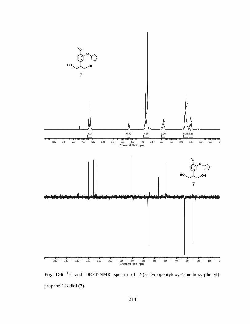
214
8.5 8.0 7.5 7.0 6.5 6.0 5.5 5.0 4.5 4.0 3.5 3.0 2.5 2.0 1.5 1.0 0.5 0
Chemical Shift (ppm)
2.156.211.907.360.993.14
O
O
OH OH
7
150 140 130 120 110 100 90 80 70 60 50 40 30 20 10 0
Chemical Shift (ppm)
O
O
OH OH
7
Fig. C-6 1H and DEPT-NMR spectra of 2-(3-Cyclopentyloxy-4-methoxy-phenyl)-
propane-1,3-diol (7).
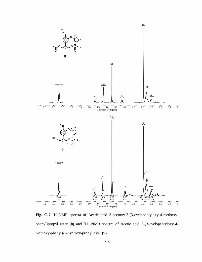
215
7.5 7.0 6.5 6.0 5.5 5.0 4.5 4.0 3.5 3.0 2.5 2.0 1.5 1.0 0.5 0
Chemical Shift (ppm)
Hd
Hh
Ha He Hc
Aromatic
Hb
Hf
O
O
OO
OO
a b
c
d
e
f
gh
8
7.5 7.0 6.5 6.0 5.5 5.0 4.5 4.0 3.5 3.0 2.5 2.0 1.5 1.0 0.5 0
Chemical Shift (ppm)
2.276.053.211.004.801.850.942.89
h
f
a
D & I
e
Aromatic
c
b
O
O
O
O
OH
a b
c
d
ef
g
h
9
i
Fig. C-7 1H NMR spectra of Acetic acid 3-acetoxy-2-(3-cyclopentyloxy-4-methoxy-
phenyl)propyl ester (8) and 1H -NMR spectra of Acetic acid 2-(3-cyclopentyloxy-4-
methoxy-phenyl)-3-hydroxy-propyl ester (9).

216
9 8 7 6 5 4 3 2 1 0
Chemical Shift (ppm)
Hd
Ha
Hh
Hf'Hc
Hj
Hb
Hf
Aromatic
O
O
O O
O
H
a b
c
d
ef
g h
i
j
10
O
O
O O
O
H
a b
c
d
ef
g h
i
j
10
180 160 140 120 100 80 60 40 20 0
Chemical Shift (ppm)
h
i
c
g
d & ea
f
b
Aromatic
Aromatic
Fig. C-8: 1H and 13C-NMR spectra of Acetic acid 2-(3-cyclopentyloxy-4-methoxy-
phenyl)-3-oxo-propyl ester (10).

217
OO
O
O
H
O
CH3
a b
c
d
ef
g h
i
11
*
7.5 7.0 6.5 6.0 5.5 5.0 4.5 4.0 3.5 3.0 2.5 2.0 1.5 1.0 0.5
Chemical Shift (ppm)
CDCl3
HdHh
Hk
Hj'
Hf
Hi'
HaHj'
Hi
Aromatic
Hc
Hb
7.5 7.0 6.5 6.0 5.5 5.0 4.5 4.0 3.5 3.0 2.5 2.0 1.5 1.0 0.5
Chemical Shift (ppm)
2.267.231.002.352.443.080.930.880.763.00
Alkene
d
a
i
Alkene
Alkene
f
e
Aromatic
c
b
OO
H
O
CH3
OH
a b
c
d
ef
i
12
*
Fig. C-9 1H NMR spectra of (E/Z)-Acetic acid 2-(3-cyclopentyloxy-4-methoxy-phenyl)-
4-methoxy-but-3-enyl ester (11) and 1H -NMR spectra of (E/Z)-2-(3-Cyclopentyloxy-4-
methoxy-phenyl)-4-methoxy-but-3-en-1-ol (12).
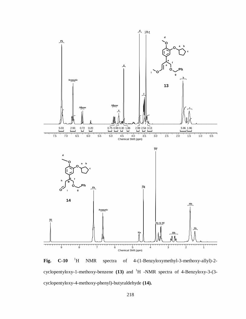
218
a b
c
d
ef
i
13
O
O
OO
Ph
*
g
7.5 7.0 6.5 6.0 5.5 5.0 4.5 4.0 3.5 3.0 2.5 2.0 1.5 1.0 0.5
Chemical Shift (ppm)
1.965.963.132.542.991.860.300.990.750.200.722.935.03
aAlkene
g
f
d
Aromatic
Alkene
i & e
c
Ph
b
O
O
O
O
Ph
a b
c
d
ef
i
14
*
g
h
9 8 7 6 5 4 3 2 1
Chemical Shift (ppm)
Ha
Hd
Hi
Hg
Aromatic
Hc
Ph
Hb
He & Hf
Hh
Fig. C-10 1H NMR spectra of 4-(1-Benzyloxymethyl-3-methoxy-allyl)-2-
cyclopentyloxy-1-methoxy-benzene (13) and 1H -NMR spectra of 4-Benzyloxy-3-(3-
cyclopentyloxy-4-methoxy-phenyl)-butyraldehyde (14).
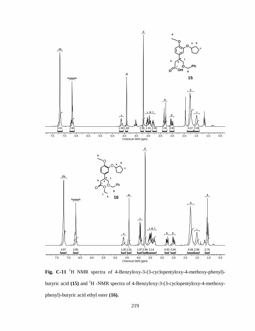
219
7.5 7.0 6.5 6.0 5.5 5.0 4.5 4.0 3.5 3.0 2.5 2.0 1.5 1.0 0.5
Chemical Shift (ppm)
2.086.070.901.910.901.912.961.841.002.855.01
g
d
h'
Aromatic
a
h
Ph
c
b
e & f
O
O
O
O
PhOH
a b
c
d
ef
i
15
*
g
h
OO
OO
PhO
7.5 7.0 6.5 6.0 5.5 5.0 4.5 4.0 3.5 3.0 2.5 2.0 1.5 1.0 0.5
Chemical Shift (ppm)
2.762.086.060.940.933.142.991.871.911.002.854.97
d
kg
j
a
Aromatic
h h'
c
Ph
b
e & f
ab
c
d
ef
i
16
*
g
h
jk
Fig. C-11 1H NMR spectra of 4-Benzyloxy-3-(3-cyclopentyloxy-4-methoxy-phenyl)-
butyric acid (15) and 1H -NMR spectra of 4-Benzyloxy-3-(3-cyclopentyloxy-4-methoxy-
phenyl)-butyric acid ethyl ester (16).

220
8.5 8.0 7.5 7.0 6.5 6.0 5.5 5.0 4.5 4.0 3.5 3.0 2.5 2.0 1.5 1.0 0.5
Chemical Shift (ppm)
2.912.386.301.060.970.872.223.111.801.003.01
17
O
O
O O
CH3
OH
*
6.5 6.0 5.5 5.0 4.5 4.0 3.5 3.0 2.5 2.0 1.5 1.0 0.5
Chemical Shift (ppm)
2.955.466.950.870.992.743.741.791.003.65
d
c
a
k
h h'
j
Aromatic
b
e & f
ab
c
d
ef
i
18
*h
j
k
O
O
O OBr
Fig. C-12 1H NMR spectra of 3-(3-Cyclopentyloxy-4-methoxy-phenyl)-4-hydroxy-
butyric acid ethyl ester (17) and 1H -NMR spectra of 4-Bromo-3-(3-cyclopentyloxy-4-
methoxy-phenyl)-butyric acid ethyl ester (18).

221
150 140 130 120 110 100 90 80 70 60 50 40 30 20 10
Chemical Shift (ppm)
j
ea
cb
d
k
f & h
Aromatic
a b
c
d
ef
i
18
*h
j
k
O
O
O OBr
7.5 7.0 6.5 6.0 5.5 5.0 4.5 4.0 3.5 3.0 2.5 2.0 1.5 1.0 0.5
Chemical Shift (ppm)
He
CDCl3
Hd
HaHg
Hg'
Hj
Hj
Hf
Hc
Aromatic
Hb
ab
c
d
ef
i
19
*h
j
k
O
O
O ON3
Fig. C-13 DEPT- NMR spectra of 4-Bromo-3-(3-cyclopentyloxy-4-methoxy-phenyl)-
butyric acid ethyl ester (18) and 1H -NMR spectra of 4-Azido-3-(3-cyclopentyloxy-4-
methoxy-phenyl)-butyric acid ethyl ester (19).

222
150 140 130 120 110 100 90 80 70 60 50 40 30 20 10
Chemical Shift (ppm)
j
e
d
h
b
f
k
a
c
Aromatic
O
O
O O
CH3
N3
a
b
c
d
ef
i
19
*h
j
k
Fig. C-14 DEPT- NMR spectra of 4-Azido-3-(3-cyclopentyloxy-4-methoxy-phenyl)-
butyric acid ethyl ester (19).

223
7.5 7.0 6.5 6.0 5.5 5.0 4.5 4.0 3.5 3.0 2.5 2.0 1.5 1.0 0.5 0
Chemical Shift (ppm)
2.506.323.051.000.892.05
OO
I
21
170 160 150 140 130 120 110 100 90 80 70 60 50 40 30 20 10 0
Chemical Shift (ppm)
OO
I
21
Fig. C-15 1H and
13C-NMR spectra of 2-Cyclopentyloxy-4-iodo-1-methoxy-benzene
(21).

224
8.5 8.0 7.5 7.0 6.5 6.0 5.5 5.0 4.5 4.0 3.5 3.0 2.5 2.0 1.5 1.0 0.5 0
Chemical Shift (ppm)
1.464.872.306.343.061.171.940.921.000.432.010.480.92
OO
O
O
OO
22
170 160 150 140 130 120 110 100 90 80 70 60 50 40 30 20 10 0
Chemical Shift (ppm)
OO
O
O
OO
22
Fig. C-16 1H and
13C-NMR spectra of 2-(3-Cyclopentyloxy-4-methoxy-phenyl)-but-2-
enedioic acid diethyl ester (22).
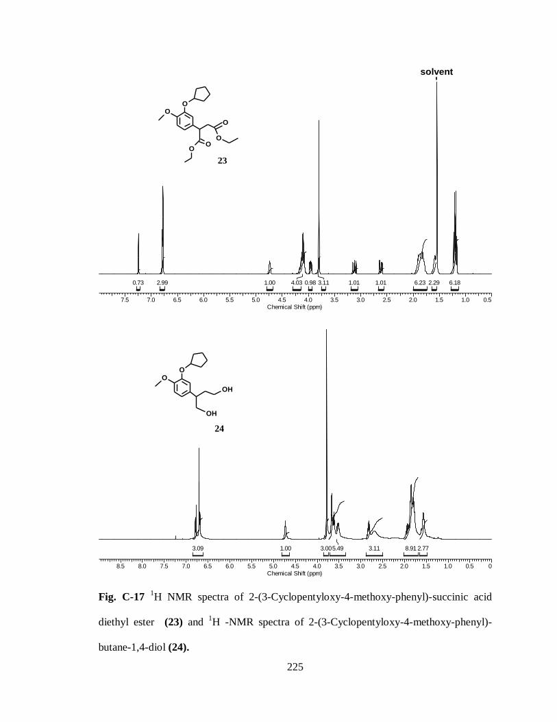
225
7.5 7.0 6.5 6.0 5.5 5.0 4.5 4.0 3.5 3.0 2.5 2.0 1.5 1.0 0.5
Chemical Shift (ppm)
6.182.296.231.011.013.110.984.031.002.990.73
solvent
23
OO
OO
O
O
8.5 8.0 7.5 7.0 6.5 6.0 5.5 5.0 4.5 4.0 3.5 3.0 2.5 2.0 1.5 1.0 0.5 0
Chemical Shift (ppm)
2.778.913.115.493.001.003.09
24
OO
OH
OH
Fig. C-17 1H NMR spectra of 2-(3-Cyclopentyloxy-4-methoxy-phenyl)-succinic acid
diethyl ester (23) and 1H -NMR spectra of 2-(3-Cyclopentyloxy-4-methoxy-phenyl)-
butane-1,4-diol (24).
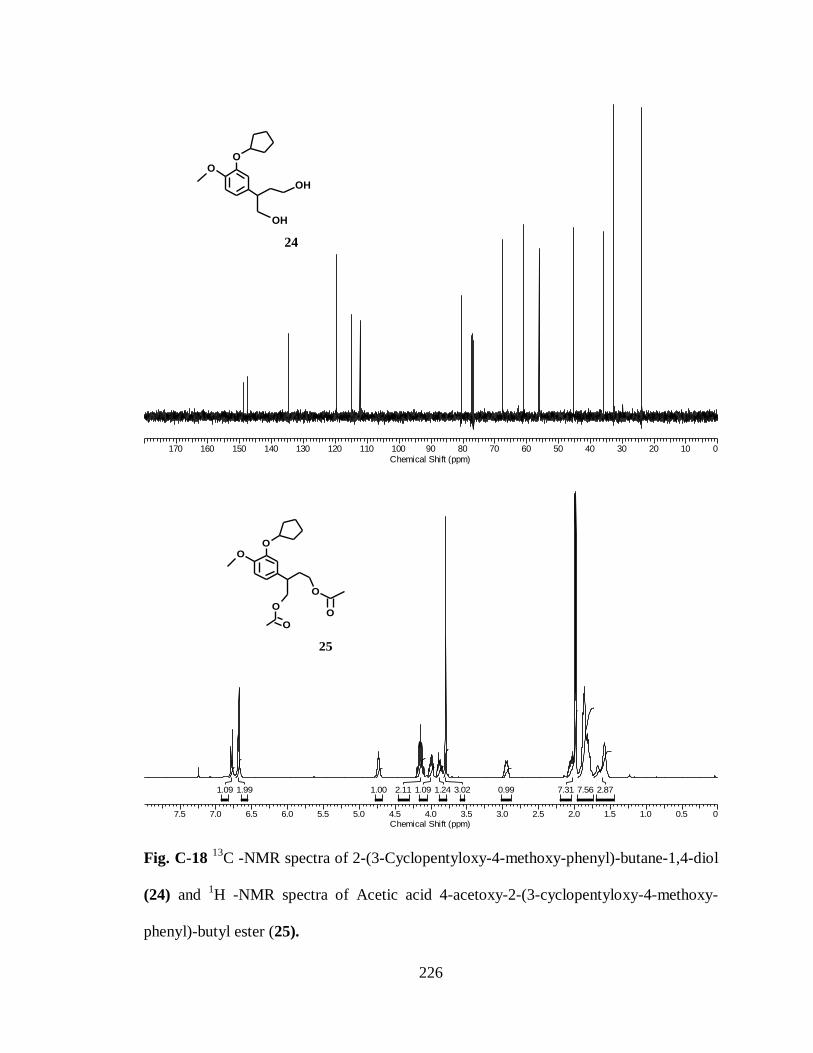
226
170 160 150 140 130 120 110 100 90 80 70 60 50 40 30 20 10 0
Chemical Shift (ppm)
24
OO
OH
OH
7.5 7.0 6.5 6.0 5.5 5.0 4.5 4.0 3.5 3.0 2.5 2.0 1.5 1.0 0.5 0
Chemical Shift (ppm)
2.877.567.310.993.021.241.092.111.001.991.09
25
OO
O
O
O
O
Fig. C-18 13
C -NMR spectra of 2-(3-Cyclopentyloxy-4-methoxy-phenyl)-butane-1,4-diol
(24) and 1H -NMR spectra of Acetic acid 4-acetoxy-2-(3-cyclopentyloxy-4-methoxy-
phenyl)-butyl ester (25).

227
170 160 150 140 130 120 110 100 90 80 70 60 50 40 30 20 10 0
Chemical Shift (ppm)
25
OO
O
O
O
O
Fig. C-19 13
C -NMR spectra of Acetic acid 4-acetoxy-2-(3-cyclopentyloxy-4-methoxy-
phenyl)-butyl ester (25).

228
Fig. C-20 1H and
13C-NMR spectra of Acetic acid 2-(3-cyclopentyloxy-4-methoxy-
phenyl)-4-hydroxy-butyl ester (26).
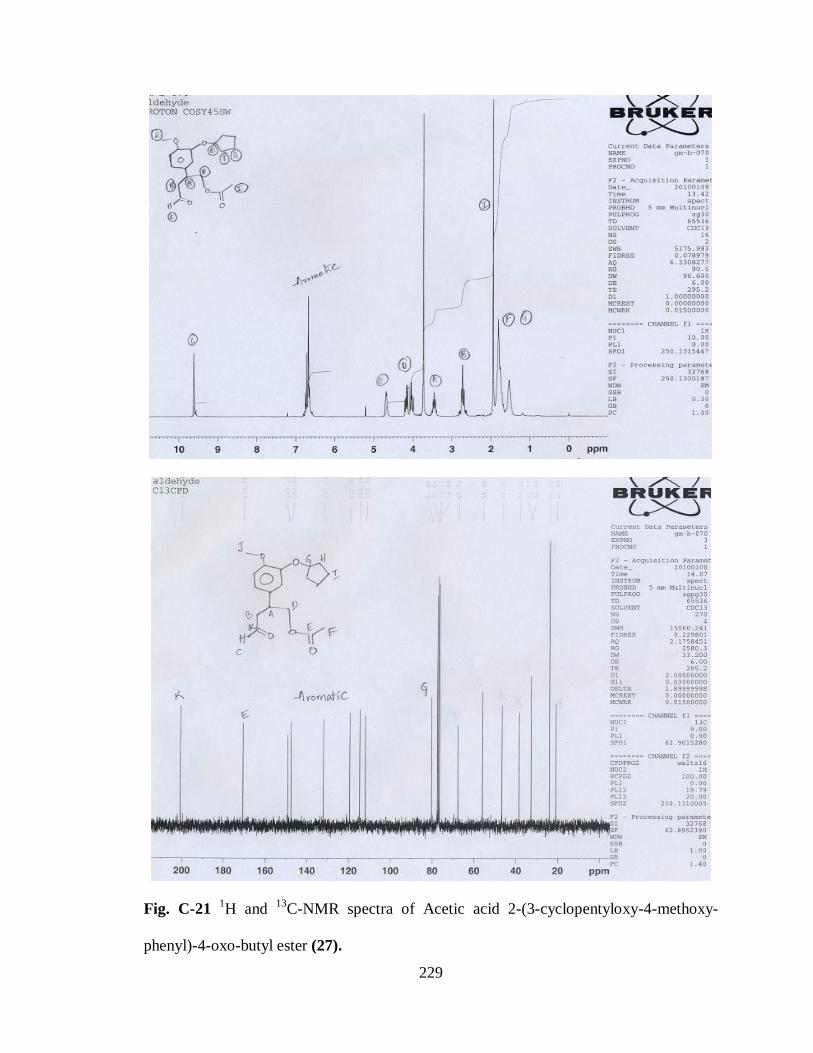
229
Fig. C-21 1H and
13C-NMR spectra of Acetic acid 2-(3-cyclopentyloxy-4-methoxy-
phenyl)-4-oxo-butyl ester (27).
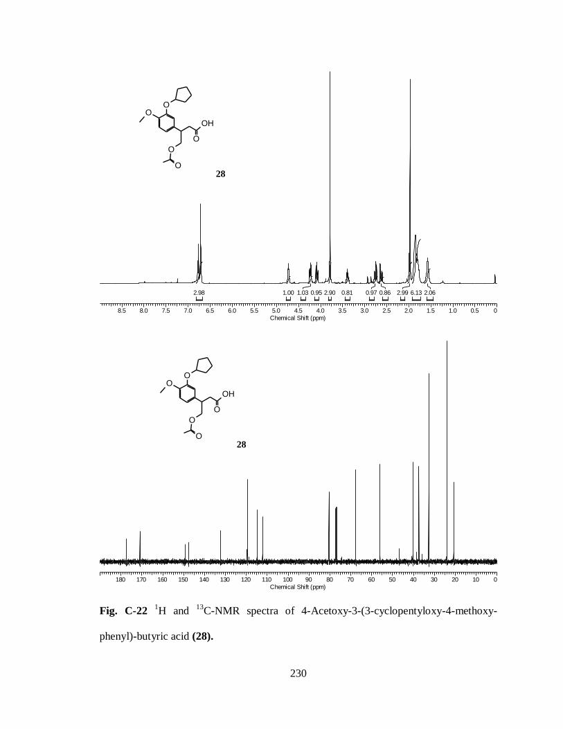
230
8.5 8.0 7.5 7.0 6.5 6.0 5.5 5.0 4.5 4.0 3.5 3.0 2.5 2.0 1.5 1.0 0.5 0
Chemical Shift (ppm)
2.066.132.990.860.970.812.900.951.031.002.98
OO
O
O
O
OH
28
180 170 160 150 140 130 120 110 100 90 80 70 60 50 40 30 20 10 0
Chemical Shift (ppm)
OO
O
O
O
OH
28
Fig. C-22 1H and
13C-NMR spectra of 4-Acetoxy-3-(3-cyclopentyloxy-4-methoxy-
phenyl)-butyric acid (28).
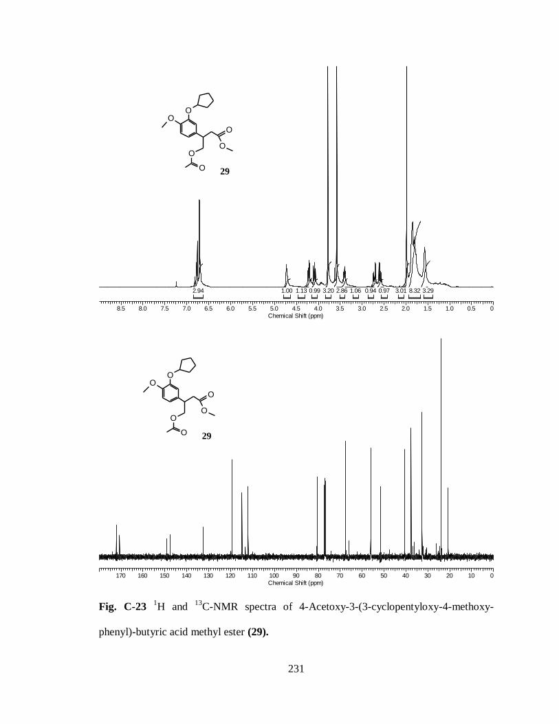
231
8.5 8.0 7.5 7.0 6.5 6.0 5.5 5.0 4.5 4.0 3.5 3.0 2.5 2.0 1.5 1.0 0.5 0
Chemical Shift (ppm)
3.298.323.010.970.941.062.863.200.991.131.002.94
OO
O
O
O
O 29
170 160 150 140 130 120 110 100 90 80 70 60 50 40 30 20 10 0
Chemical Shift (ppm)
OO
O
O
O
O 29
Fig. C-23 1H and
13C-NMR spectra of 4-Acetoxy-3-(3-cyclopentyloxy-4-methoxy-
phenyl)-butyric acid methyl ester (29).

232
Fig. C-24 1H NMR spectra of 4-(3-Cyclopentyloxy-4-methoxy-phenyl)-dihydro-furan-2-
one (30).