molecular imaging - labindia · molecular imaging ... vilber is the leading european provider of...
TRANSCRIPT
MOLECULAr IMAGINGCHEMILUMINESCENCE • FLUOrESCENCE • ANALySIS SOFTWArE
VILBEr is the leading European provider of
molecular imaging systems, analysis software and
UV fluorescence equipment. Founded over 50
years ago to serve the research, VILBEr has
pioneered the post electrophoresis market and
introduced breakthrough products such as stand
alone gel-documentation, Bio-1D imaging
software, Super-Bright UV technology, dedicated
chemiluminescence imaging system and 3D
approach to 1D gel analysis.
Through a network of owned subsidiary offices and
local distributors located in over 60 countries
around the world, VILBEr offers a broad range of
products:
> Gel documentation systems
> Chemiluminescence imaging systems
> Image analysis software
> UV instruments for molecular biology such as
transilluminators, crosslinkers and UV lamps.
For more information about VILBEr, visit our
website at www.vilber.com
HEADQUARTER
Vilber Lourmat
BP-66 – ZI Sud Torcy
F-77202 Marne-la-Vallée cedex 1
France
T.: 33 (0) 1 60 06 07 71
F.:33 (0) 1 64 80 48 59
GERMANY OFFICE
Vilber Lourmat Deutschland GmbH
Wielandstrasse 2
D-88436 Eberhardzell
Deutschland
T.: 49 (0) 7355 931 380
F.: 49 (0) 7355 931 379
www.vilber.com
CAR
REM
ENT
CO
M 0
1 64
72
11 5
2 -
07/2
010
C H E M I L U M I N E S C E N C E
I Working principles
and key features
I Fusion FX7
I Fusion FX5
I Fusion SL
I Specifications
F L U O R E S C E N C E
I Working principles
and key features
I Infinity VX2
I Quantum ST4
I Bio-Print Mega
I E-Box VX2
I Doc-Print VX2
I Accessories
I Darkrooms
A N A L Y S I S S O F T W A R E
I Bio-1D
I Bio-Gene
I Bio-1D++
I Bio-2D
I The Capt series
04
06
08
10
11
12
14
18
22
24
28
30
31
32
34
36
38
39
32
VILBEr is the leading European provider of
molecular imaging systems, analysis software
and UV fluorescence equipment. Founded
over 50 years ago to serve the research, VILBEr
has pioneered the post electrophoresis market
and introduced breakthrough products such as
stand alone gel-documentation, Bio-1D
imaging software, Super-Bright UV technology,
dedicated chemiluminescence imaging
system and 3D approach to 1D gel analysis.
Through a network of owned subsidiary offices
and local distributors located in over 60
countries around the world, VILBEr offers a
broad range of products:
> Gel documentation systems
> Chemiluminescence imaging systems
> Image analysis software
> UV instruments for molecular biology such as
transilluminators, crosslinkers and UV lamps.
For more information about VILBEr, visit our
website at www.vilber.com
Turn your searchinto answers
HEADQUARTER
Vilber Lourmat
BP-66 – ZI Sud Torcy
F-77202 Marne-la-Vallée cedex 1
France
T.: 33 (0) 1 60 06 07 71
F.: 33 (0) 1 64 80 48 59
GERMANY OFFICE
Vilber Lourmat Deutschland GmbH
Wielandstrasse 2
D-88436 Eberhardzell
Deutschland
T.: 49 (0) 7355 931 380
F.: 49 (0) 7355 931 379
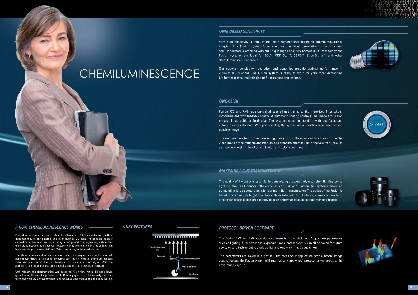
Very high sensitivity is one of the main requirements regarding chemiluminescence imaging. The Fusion systems’ cameras use the latest generation of sensors and semi-conductors. Combined with our unique High Sensitivity Camera (HSC) technology, the Fusion systems are ideal for ECLTM, CDP StarTM, CSPDTM, SuperSignalTM and other chemiluminescent enhancers.
Our superior sensitivity, resolution and dynamics provide optimal performance in virtually all situations. The Fusion system is ready to work for your most demanding bio-luminescence, multiplexing or fluorescence applications.
The quality of the optics is essential in transmitting the extremely weak chemiluminescencelight to the CCD sensor efficiently. Fusion FX and Fusion SL systems have an outstanding large-aperture lens for optimum light transmission. The optics of the Fusion isbased on a supremely bright fixed lens with an f-stop of 0.95. Unlike an ordinary camera lens,it has been specially designed to provide high performance at an extremely short distance.
The Fusion FX7 and FX5 acquisition software is protocol-driven. Acquisition parameterssuch as lighting, filter selections, exposure times, and sensitivity can all be saved for futureuse to ensure instrument reproducibility and one-click image acquisition.
The parameters are saved in a profile. Just recall your application profile before imageacquisition and the Fusion system will automatically apply your protocol-driven set-up to thenext image capture.
54
> HOW CHEMILUMINESCENCE WORKS
Chemiluminescence is used to detect proteins or DNA. This detection methoddoes not require any external excitation such as UV light. The light emission is caused by a chemical reaction exciting a compound to a high-energy state. Thisunstable compound rapidly looses its excess energy by emitting light. The emitted lighthas a wavelength between 400 and 500 nm according to the substrate used.
The chemiluminescent reaction occurs when an enzyme such as horseradish peroxidase (HRP) or alkaline phosphatase reacts with a chemiluminescent substrate (such as luminol or dioxetane) to produce a weak signal. With the addition of an enhancer, the light intensity and the light duration increase.
Until recently, the documentation was based on X-ray film, which did not allowed quantification. The recent improvements of CCD imaging in terms of sensitivity make thistechnology simply perfect for chemiluminescence documentation and quantification.
CHEMILUMINESCENCE
> KEY FEATURES
UNRIVALLED SENSITIVITY
MAXIMUM LIGHT TRANSMITTANCE
ONE-CLICK
PROTOCOL DRIVEN SOFTWARE
Fusion FX7 and FX5 have unrivalled ease of use thanks to the motorised filter wheel, motorised lens with feedback control, & automatic lighting controls. The image acquisition process is as quick as instinctive. The systems come in standard with autofocus and autoexposure as standard. With just one click, the system will automatically capture the bestpossible image.
The user-interface has rich features and guides you into the advanced functions such as thevideo mode or the multiplexing module. Our software offers multiple analysis features suchas molecular weight, band quantification and colony counting.
76
> KEY FEATURES
•The ultimate chemiluminescence imaging system
• Last generation of cooled CCD sensor
• Image Master assistant to easily get the optimum image
• Ultimate sensitivity
• 70% quantum efficiency at 450nm for unparalleled sensitivity
• Power without compromise
• 4.2 megapixels imaging
• Pure image integrity and access to the raw data
• Large sample size and ingenious tray system
• Free software for image acquisition & analysis
> APPLICATIONS
• CHEMILUMINESCENCE DETECTION
ECLTM, ECL PlusTM, ECL AdvanceTM, ECL PlexTM
CDP StarTM , Super SignalTM, CSPDTM
> TESTIMONIALS
I have been astonished by our FusionFX7. Our system is fully motorised andjust one click is necessary for imageacquisition.
The protocol-driven image acquisitionprocess delivers incredible results andversatility. This really helps to cover amultitude of applications.
The sensitivity is simply top class andour images are ideal for quantification.
“ ”
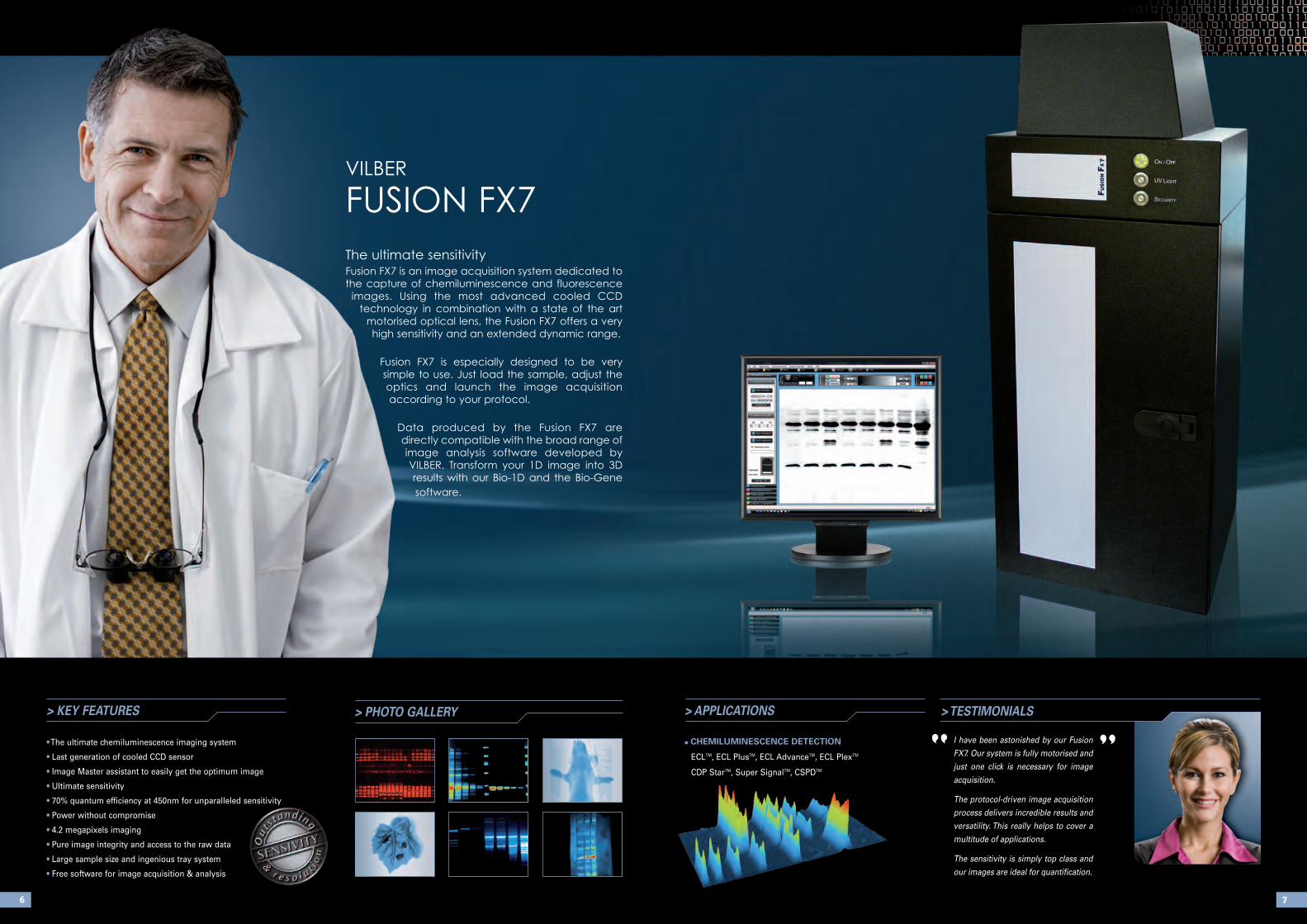
VILBEr
FUSION FX7
The ultimate sensitivityFusion FX7 is an image acquisition system dedicated to
the capture of chemiluminescence and fluorescence
images. Using the most advanced cooled CCD
technology in combination with a state of the art
motorised optical lens, the Fusion FX7 offers a very
high sensitivity and an extended dynamic range.
Fusion FX7 is especially designed to be very
simple to use. Just load the sample, adjust the
optics and launch the image acquisition
according to your protocol.
Data produced by the Fusion FX7 are
directly compatible with the broad range of
image analysis software developed by
VILBEr. Transform your 1D image into 3D
results with our Bio-1D and the Bio-Gene
software.
> PHOTO GALLERY

> TESTIMONIALS
The FUSION FX5 delivers very high
performance for a large number of
applications. This is an incredibly
versatile system.
Together with the StarLight module
for red, blue and green, we use it for
applications such as GFP, SYBR
Green, Western-blot, Luciferase and
standard gel documentation. This is
really the all-in-one necessary in our lab.
“ ”
98
> KEY FEATURES
• Unparalleled sensitivity
• One instrument for chemiluminescence and fluorescence
• Latest generation of cooled CCD sensor
• 2 Megapixels imaging / FireWire® interface
• Pure image integrity and access to the raw data
• Lens feedback controls
• Exclusive VILBER’s UV MasterTM technology
• Image Master assistant to easily get the optimum image
• Convenient versatile darkroom with multi-positions filter slide
• Compatible with the Super-Bright transilluminator technology
> PHOTO GALLERY
• CHEMILUMINESCENCEDETECTION
ECLTM, ECL PlusTM,
ECL AdvanceTM, ECL PlexTM,
CDP StarTM, QdotTM, Super
SignalTM, CSPDTM
• NUCLEIC ACID DETECTION
Ethidium bromide, SYBR™ Green,
SYBR™ Gold, Texas Red™ Gel Star™
• PROTEIN DETECTION
Coomassie blue, Sypro™ Ruby,
Sypro™ Orange, Sypro™ Red,
Silver Star™, Fluorescein,
QdotTM
• OTHER
Petri dish imaging
Microplate imaging
Autoradiograph imaging
> APPLICATIONS
VILBEr
FUSION FX5Power and versatility
The Fusion FX5 is an image acquisition system for
the capture of chemiluminescent and fluorescent
gel images.
The 2 megapixels cooled scientific CCD camera
acquires high sensitivity and high-resolution
images for multiple applications. Our unique
darkroom accommodates a transilluminator for
fluorescence applications and a separate tray for
Western-blot imaging.
Together with Bio-Profil software, Fusion FX5 forms
a versatile and powerful system for quantification
and documentation.
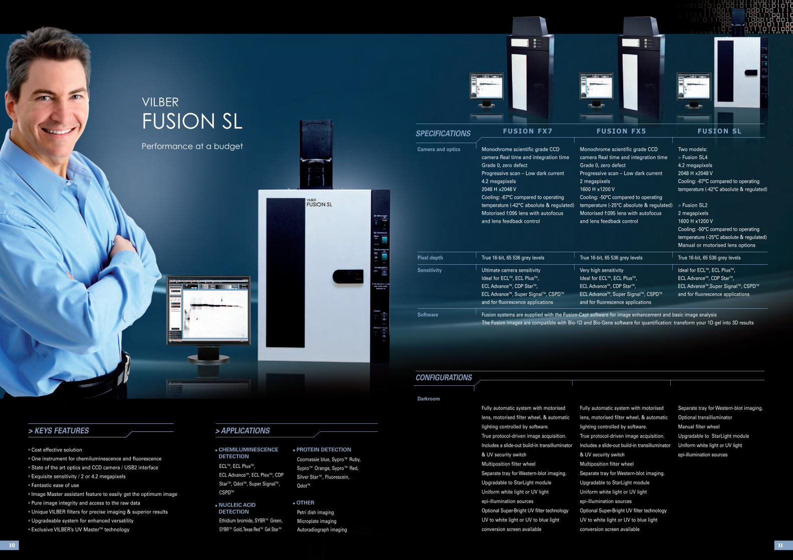
FUSION FX7 FUSION FX5 FUSION SL
> KEYS FEATURES
• Cost effective solution
• One instrument for chemiluminescence and fluorescence
• State of the art optics and CCD camera / USB2 interface
• Exquisite sensitivity / 2 or 4.2 megapixels
• Fantastic ease of use
• Image Master assistant feature to easily get the optimum image
• Pure image integrity and access to the raw data
• Unique VILBER filters for precise imaging & superior results
• Upgradeable system for enhanced versatility
• Exclusive VILBER’s UV MasterTM technology
• CHEMILUMINESCENCEDETECTION
ECLTM, ECL PlusTM,
ECL AdvanceTM, ECL PlexTM, CDP
StarTM, QdotTM, Super SignalTM,
CSPDTM
• NUCLEIC ACID DETECTION
Ethidium bromide, SYBR™ Green,
SYBR™ Gold, Texas Red™ Gel Star™
• PROTEIN DETECTION
Coomassie blue, Sypro™ Ruby,
Sypro™ Orange, Sypro™ Red,
Silver Star™, Fluorescein,
QdotTM
• OTHER
Petri dish imaging
Microplate imaging
Autoradiograph imaging
> APPLICATIONS
1110
CONFIGURATIONS
Darkroom
Fully automatic system with motorised Fully automatic system with motorised Separate tray for Western-blot imaging.
lens, motorised filter wheel, & automatic lens, motorised filter wheel, & automatic Optional transilluminator
lighting controlled by software. lighting controlled by software. Manual filter wheel
True protocol-driven image acquisition. True protocol-driven image acquisition. Upgradable to StarLight module
Includes a slide-out build-in transilluminator Includes a slide-out build-in transilluminator Uniform white light or UV light
& UV security switch & UV security switch epi-illumination sources
Multiposition filter wheel Multiposition filter wheel
Separate tray for Western-blot imaging. Separate tray for Western-blot imaging.
Upgradable to StarLight module Upgradable to StarLight module
Uniform white light or UV light Uniform white light or UV light
epi-illumination sources epi-illumination sources
Optional Super-Bright UV filter technology Optional Super-Bright UV filter technology
UV to white light or UV to blue light UV to white light or UV to blue light
conversion screen available conversion screen available
Camera and optics Monochrome scientific grade CCD Monochrome scientific grade CCD Two models:
camera Real time and integration time camera Real time and integration time > Fusion SL4
Grade 0, zero defect Grade 0, zero defect 4.2 megapixels
Progressive scan – Low dark current Progressive scan – Low dark current 2048 H x2048 V
4.2 megapixels 2 megapixels Cooling: -67°C compared to operating
2048 H x2048 V 1600 H x1200 V temperature (-42°C absolute & regulated)
Cooling: -67°C compared to operating Cooling: -50°C compared to operating
temperature (-42°C absolute & regulated) temperature (-25°C absolute & regulated) > Fusion SL2
Motorised f:095 lens with autofocus Motorised f:095 lens with autofocus 2 megapixels
and lens feedback control and lens feedback control 1600 H x1200 V
Cooling: -50°C compared to operating
temperature (-25°C absolute & regulated)
Manual or motorised lens options
Pixel depth True 16-bit, 65 536 grey levels True 16-bit, 65 536 grey levels True 16-bit, 65 536 grey levels
Sensitivity Ultimate camera sensitivity Very high sensitivity Ideal for ECLTM, ECL PlusTM,
Ideal for ECLTM, ECL PlusTM, Ideal for ECLTM, ECL PlusTM, ECL AdvanceTM, CDP StarTM,
ECL AdvanceTM, CDP StarTM, ECL AdvanceTM, CDP StarTM, ECL AdvanceTM,Super SignalTM, CSPDTM
ECL AdvanceTM, Super SignalTM, CSPDTM ECL AdvanceTM, Super SignalTM, CSPDTM and for fluorescence applications
and for fluorescence applications and for fluorescence applications
Software Fusion systems are supplied with the Fusion-Capt software for image enhancement and basic image analysis
The Fusion images are compatible with Bio-1D and Bio-Gene software for quantification: transform your 1D gel into 3D results
SPECIFICATIONS
VILBEr
FUSION SLPerformance at a budget

1312
Our exclusive Image Master assistant helps you to obtain the optimum image at a glance.For instance, it automatically monitors the maximum and the minimum intensity obtainedon the image, indicates its dynamic and warns you about pixel saturation. Image Master issimply perfect for quantification and publication. It helps you to keep the control on theimage, making sure your image is always appropriate whatever its use.
The new VILBER SKYlight Super-Blue transilluminator eliminates the damage caused by UV light onDNA and RNA gels. It also improves cloning efficiency dramatically by eliminating the effects of UV-induced nicking or crosslinking, often encountered during the purification of DNA from gels for fur-ther use.
The SKYlight Super-Blue table is based on the latest blue LED technology for an unparalleled lightuniformity. The table incorporates 270 Light Emitting Devices in an optimized array to give consis-tent intensity across the table. This uniform light is then filtered with a narrow excitation filter toobtain an excitation peak at 470nm and to eliminate light interference on the resulting image. On thesurface, the protection glass allows you to cut the gel without damaging the table.
The VILBER SKYlight Super-Blue is a new technology ideal for Sybr Safe®, Gel-Red®, Sypro Ruby®,Gel-Star®, Sypro Orange®, Sybr Gold®, Sybr Green® I & II and eGFP®, amongst others.
IMAGE MASTER
> LEADING CLASS TECHNOLOGIE
FREE SOFTWARE
UV MASTER TECHNOLOGY
All our systems are supplied with complimentary software to perform analysis such as molecular weight, band quantification, colony counting and distance calculation. It also includes image enhancement features to enable editing of comments, inversion, contrast /brightness adjustment as well as colorimetry.Designed by molecular biologists, our software are intuitive and very easy to use: just fewclicks are necessary to obtain sophisticated results. Please refer to page 39 for a completelist of functions.
VILBER is the UV fluorescence expert since 1951. Our own UV lamps emit highly concentratedUV radiation. This output is reinforced with the use of our exclusive Ondulex® reflector, especially polished to reflect the maximum of the light to the outside. Combined with our innovative range of special UV filters, our unique fluorescence sourcesdramatically improve the quality of gel visualization and documentation and create unrivalledmulti fluorescence application capabilities.
> HOW FLUORESCENCE WORKS
Fluorescence results from a process that occurs under certain
conditions in molecules known as fluophores, or fluorescent dyes.
The fluophore absorbs the UV light coming from the
transilluminator, reaching a higher energy state. By returning to
its former state, it emits fluorescent light.
The emission wavelength is always higher than the
excitation wavelength (UV light). The aim of the gel
documentation system is to separate the emitted light from the
excitation light in order to obtain an optimum gel image.
> FLUORESCENCE IMAGING
FLUOrESCENCE
Standardtransilluminator
Super-Bright MX
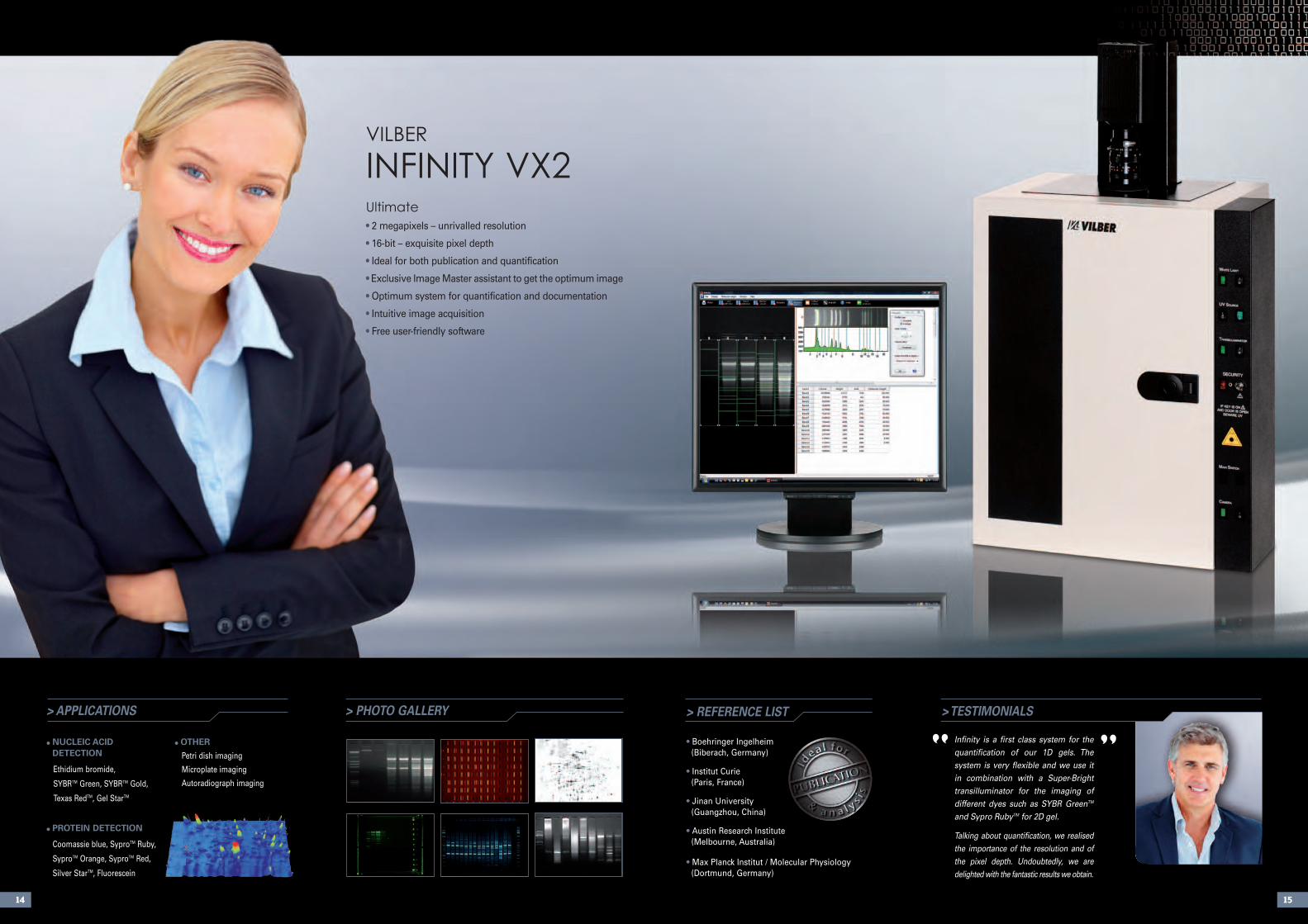
> PHOTO GALLERY > REFERENCE LIST
• Boehringer Ingelheim(Biberach, Germany)
• Institut Curie(Paris, France)
• Jinan University(Guangzhou, China)
• Austin Research Institute (Melbourne, Australia)
• Max Planck Institut / Molecular Physiology(Dortmund, Germany)
> TESTIMONIALS
Infinity is a first class system for thequantification of our 1D gels. The system is very flexible and we use itin combination with a Super-Brighttransilluminator for the imaging ofdifferent dyes such as SYBR GreenTM
and Sypro RubyTM for 2D gel.
Talking about quantification, we realisedthe importance of the resolution and ofthe pixel depth. Undoubtedly, we are delighted with the fantastic results we obtain.
”• NUCLEIC ACIDDETECTION
Ethidium bromide,
SYBRTM Green, SYBRTM Gold,
Texas RedTM, Gel StarTM
• PROTEIN DETECTION
Coomassie blue, SyproTM Ruby,
SyproTM Orange, SyproTM Red,
Silver StarTM, Fluorescein
• OTHER
Petri dish imaging
Microplate imaging
Autoradiograph imaging
> APPLICATIONS
14 15
“
VILBEr
INFINITy VX2Ultimate
• 2 megapixels – unrivalled resolution
• 16-bit – exquisite pixel depth
• Ideal for both publication and quantification
• Exclusive Image Master assistant to get the optimum image
• Optimum system for quantification and documentation
• Intuitive image acquisition
• Free user-friendly software
Darkroom CN-3000 darkroom CN-1100 darkroom
Includes a slide-out build-in transilluminator Includes a slide-out build-in transilluminator
& UV security switch & UV security switch
Multiposition filter slide Multiposition filter slide
Upgradable to StarLight module
Uniform white light or UV light epi-illumination sources Overhead white light by fluorescent tubes
Single or dual wavelength transilluminator available Single or dual wavelength transilluminator available
Filter size : 20x20cm or 21x26cm Filter size : 20x20cm or 21x26cm
Optional Super-Bright UV filter technology Optional Super-Bright UV filter technology
UV to white light or UV to blue light conversion UV to white light or UV to blue light conversion
screen available screen available
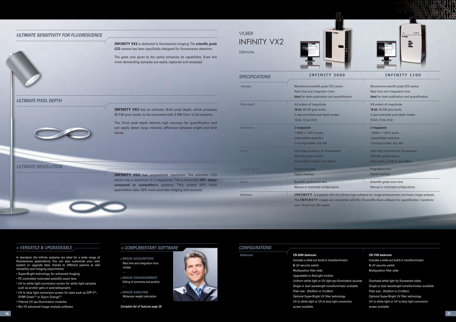
INFINITY VX2 is dedicated to fluorescence imaging. The scientific gradeCCD camera has been specifically designed for fluorescence detection.
The great care given to the optics enhances its capabilities. Even themost demanding samples are easily captured and analyzed.
INFINITY VX2 has an ultimate 16-bit pixel depth, which produces 65 536 grey levels, to be compared with 4 096 from 12-bit systems.
The 16-bit pixel depth delivers high accuracy for quantification andcan easily detect large intensity difference between bright and faintbands.
INFINITY VX2 has unparalleled resolution. The scientific CCD sensor has a resolution of 2 megapixels. This is more than 50% better compared to competitor’s systems. This means 50% more quantitative data, 50% more accurate imaging and analysis.
Camera Monochrome scientific grade CCD camera Monochrome scientific grade CCD camera
Real time and integration time Real time and integration time
Ideal for both publication and quantification Ideal for both publication and quantification
Pixel depth 4.8 orders of magnitude 4.8 orders of magnitude
16-bit, 65 536 grey levels. 16-bit, 65 536 grey levels.
3 user-controlled pixel depth modes : 3 user-controlled pixel depth modes :
16-bit, 12-bit, 8-bit 16-bit, 12-bit, 8-bit
Resolution 2 megapixels 2 megapixels
1 600H x 1 200 V pixels 1 600H x 1 200 V pixels
Unparalleled resolution Unparalleled resolution
2 binning modes: 2x2; 4x4 2 binning modes: 2x2; 4x4
Grade Ultra high sensitivity for fluorescence Ultra high sensitivity for fluorescence
Scientific grade camera Scientific grade camera
Chip quality: Grade 0, zero defect Chip quality: Grade 0, zero defect
Camera device Progressive scan Progressive scan
Digital interface Digital interface
Zoom Scientific grade zoom lens Scientific grade zoom lens
Manual or motorized configurations Manual or motorized configurations
Software INFINITY is supplied with the Infinity-Capt software for image enhancement and basic image analysis.
The INFINITY images are compatible with Bio-1D and Bio-Gene software for quantification: transform
your 1D gel into 3D results.
1716
INFINITY 3000 INFINITY 1100
> COMPLIMENTARY SOFTWARE> VERSATILE & UPGRADEABLE
In standard, the Infinity systems are ideal for a wide range of fluorescence applications. You can also customize your own system or upgrade later, thanks to different options to add versatility and imaging experiments:
• Super-Bright technology for enhanced imaging
• PC controlled motorized scientific zoom lens
• UV to white light conversion screen for white light samplessuch as protein gels or autoradiographs
• UV to blue light conversion screen for dyes such as GFP IITM,SYBR GreenTM or Sypro OrangeTM.
• Filtered UV epi-illumination modules
• Bio-1D advanced image analysis software
• IMAGE ACQUISITION
Real time and integration time modes. . .
• IMAGE ENHANCEMENTEditing of comments and symbols. . .
• IMAGE ANALYSISMolecular weight calculation. . .
Complete list of features page 39
SPECIFICATIONS
CONFIGURATIONS
ULTIMATE SENSITIVITY FOR FLUORESCENCE
ULTIMATE PIXEL DEPTH
ULTIMATE RESOLUTION
VILBEr
INFINITy VX2Ultimate
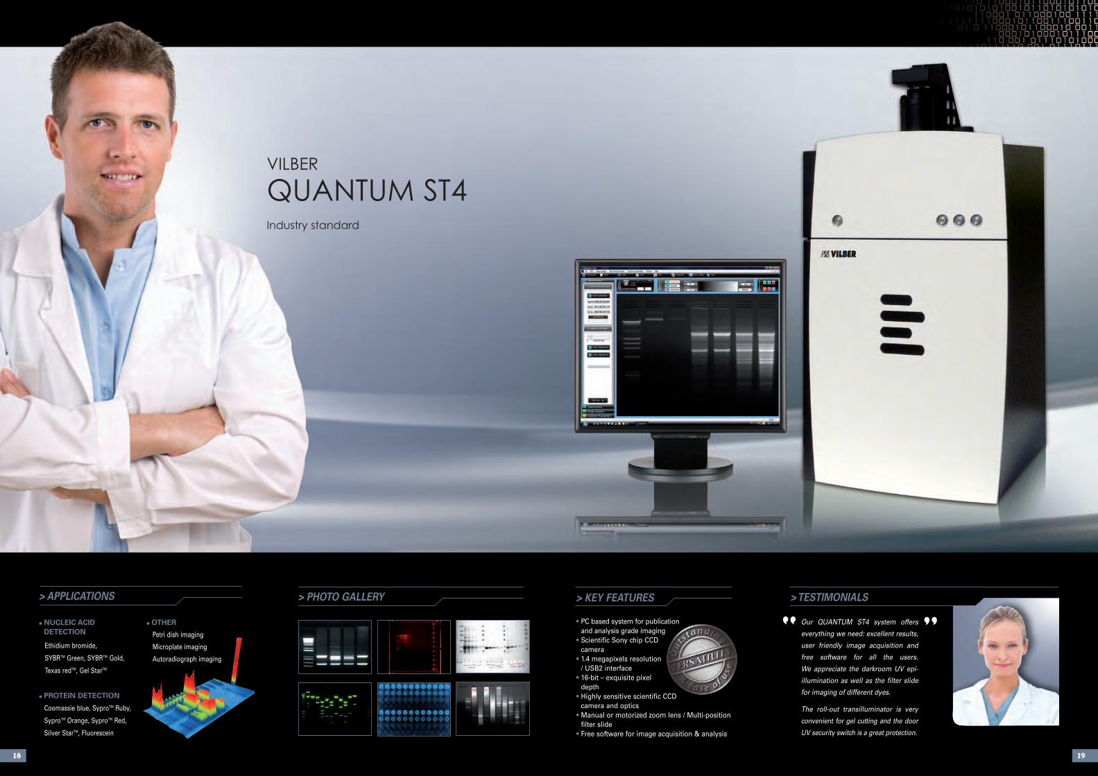
> KEY FEATURES
• PC based system for publicationand analysis grade imaging
• Scientific Sony chip CCDcamera
• 1.4 megapixels resolution/ USB2 interface
• 16-bit – exquisite pixeldepth
• Highly sensitive scientific CCDcamera and optics
• Manual or motorized zoom lens / Multi-positionfilter slide
• Free software for image acquisition & analysis
> TESTIMONIALS
Our QUANTUM ST4 system offers
everything we need: excellent results,
user friendly image acquisition and
free software for all the users.
We appreciate the darkroom UV epi-
illumination as well as the filter slide
for imaging of different dyes.
The roll-out transilluminator is very
convenient for gel cutting and the door
UV security switch is a great protection.
“ ”> PHOTO GALLERY
18 19
> APPLICATIONS
• NUCLEIC ACIDDETECTION
Ethidium bromide,
SYBRTM Green, SYBRTM Gold,
Texas redTM, Gel StarTM
• PROTEIN DETECTION
Coomassie blue, SyproTM Ruby,
SyproTM Orange, SyproTM Red,
Silver StarTM, Fluorescein
• OTHER
Petri dish imaging
Microplate imaging
Autoradiograph imaging
VILBEr
QUANTUM ST4Industry standard
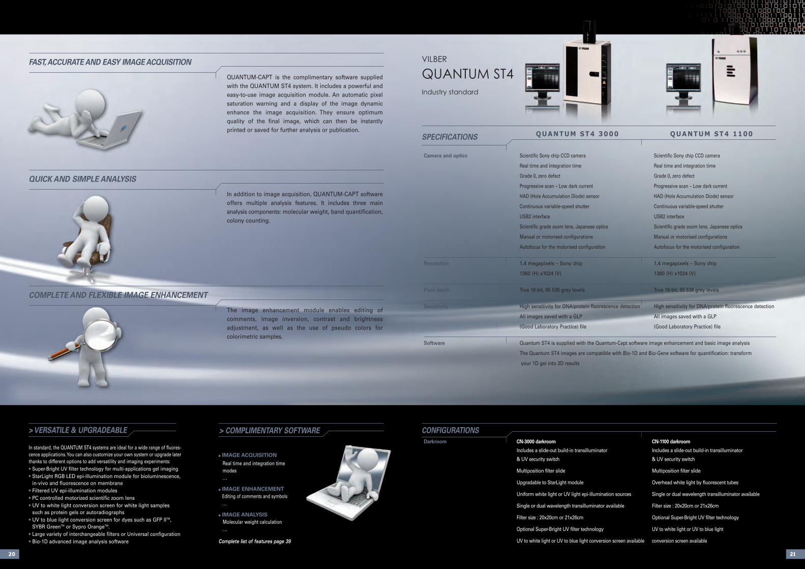
Camera and optics Scientific Sony chip CCD camera Scientific Sony chip CCD camera
Real time and integration time Real time and integration time
Grade 0, zero defect Grade 0, zero defect
Progressive scan – Low dark current Progressive scan – Low dark current
HAD (Hole Accumulation Diode) sensor HAD (Hole Accumulation Diode) sensor
Continuous variable-speed shutter Continuous variable-speed shutter
USB2 interface USB2 interface
Scientific grade zoom lens. Japanese optics Scientific grade zoom lens. Japanese optics
Manual or motorised configurations Manual or motorised configurations
Autofocus for the motorised configuration Autofocus for the motorised configuration
Resolution 1,4 megapixels – Sony chip 1,4 megapixels – Sony chip
1360 (H) x1024 (V) 1360 (H) x1024 (V)
Pixel depth True 16-bit, 65 536 grey levels True 16-bit, 65 536 grey levels
Sensitivity High sensitivity for DNA/protein fluorescence detection High sensitivity for DNA/protein fluorescence detection
All images saved with a GLP All images saved with a GLP
(Good Laboratory Practice) file (Good Laboratory Practice) file
Software Quantum ST4 is supplied with the Quantum-Capt software image enhancement and basic image analysis
The Quantum ST4 images are compatible with Bio-1D and Bio-Gene software for quantification: transform
your 1D gel into 3D results
2120
> VERSATILE & UPGRADEABLE
In standard, the QUANTUM ST4 systems are ideal for a wide range of fluores-cence applications. You can also customize your own system or upgrade laterthanks to different options to add versatility and imaging experiments:• Super-Bright UV filter technology for multi-applications gel imaging• StarLight RGB LED epi-illumination module for bioluminescence,
in-vivo and fluorescence on membrane• Filtered UV epi-illumination modules• PC controlled motorized scientific zoom lens• UV to white light conversion screen for white light samples
such as protein gels or autoradiographs• UV to blue light conversion screen for dyes such as GFP IITM,
SYBR GreenTM or Sypro OrangeTM.• Large variety of interchangeable filters or Universal configuration• Bio-1D advanced image analysis software
> COMPLIMENTARY SOFTWARE
• IMAGE ACQUISITION
Real time and integration time modes. . .
• IMAGE ENHANCEMENTEditing of comments and symbols. . .
• IMAGE ANALYSISMolecular weight calculation. . .
Complete list of features page 39
CONFIGURATIONSDarkroom CN-3000 darkroom CN-1100 darkroom
Includes a slide-out build-in transilluminator Includes a slide-out build-in transilluminator
& UV security switch & UV security switch
Multiposition filter slide Multiposition filter slide
Upgradable to StarLight module Overhead white light by fluorescent tubes
Uniform white light or UV light epi-illumination sources Single or dual wavelength transilluminator available
Single or dual wavelength transilluminator available Filter size : 20x20cm or 21x26cm
Filter size : 20x20cm or 21x26cm Optional Super-Bright UV filter technology
Optional Super-Bright UV filter technology UV to white light or UV to blue light
UV to white light or UV to blue light conversion screen available conversion screen available
QUANTUM ST4 3000 QUANTUM ST4 1100SPECIFICATIONS
QUANTUM-CAPT is the complimentary software suppliedwith the QUANTUM ST4 system. It includes a powerful andeasy-to-use image acquisition module. An automatic pixelsaturation warning and a display of the image dynamicenhance the image acquisition. They ensure optimum quality of the final image, which can then be instantly printed or saved for further analysis or publication.
FAST, ACCURATE AND EASY IMAGE ACQUISITION
In addition to image acquisition, QUANTUM-CAPT softwareoffers multiple analysis features. It includes three main analysis components: molecular weight, band quantification,colony counting.
QUICK AND SIMPLE ANALYSIS
The image enhancement module enables editing of comments, image inversion, contrast and brightness adjustment, as well as the use of pseudo colors for colorimetric samples.
COMPLETE AND FLEXIBLE IMAGE ENHANCEMENT
VILBEr
QUANTUM ST4Industry standard
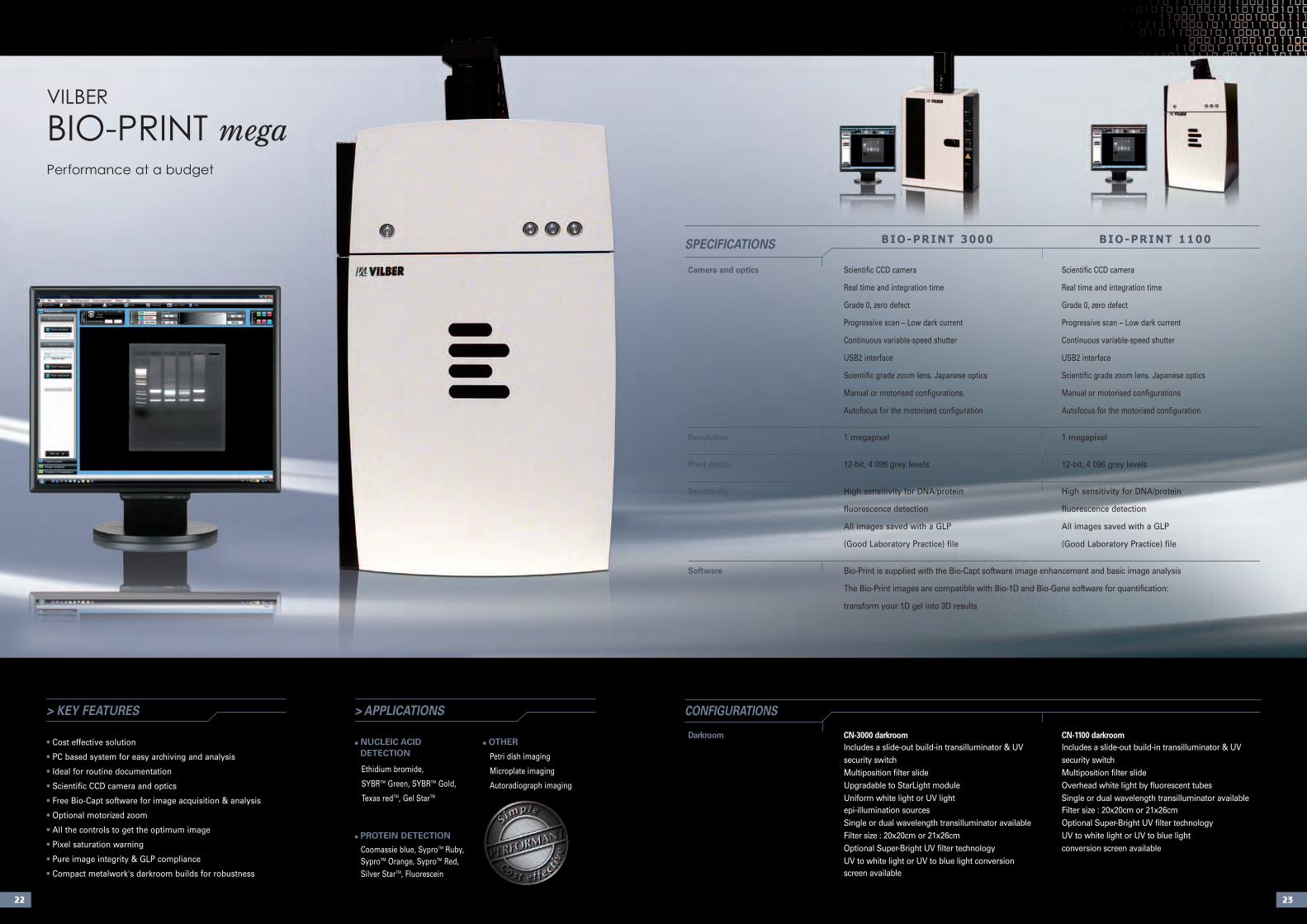
> KEY FEATURES
• Cost effective solution
• PC based system for easy archiving and analysis
• Ideal for routine documentation
• Scientific CCD camera and optics
• Free Bio-Capt software for image acquisition & analysis
• Optional motorized zoom
• All the controls to get the optimum image
• Pixel saturation warning
• Pure image integrity & GLP compliance
• Compact metalwork's darkroom builds for robustness
Camera and optics Scientific CCD camera Scientific CCD camera
Real time and integration time Real time and integration time
Grade 0, zero defect Grade 0, zero defect
Progressive scan – Low dark current Progressive scan – Low dark current
Continuous variable-speed shutter Continuous variable-speed shutter
USB2 interface USB2 interface
Scientific grade zoom lens. Japanese optics Scientific grade zoom lens. Japanese optics
Manual or motorised configurations. Manual or motorised configurations
Autofocus for the motorised configuration Autofocus for the motorised configuration
Resolution 1 megapixel 1 megapixel
Pixel depth 12-bit, 4 096 grey levels 12-bit, 4 096 grey levels
Sensitivity High sensitivity for DNA/protein High sensitivity for DNA/protein
fluorescence detection fluorescence detection
All images saved with a GLP All images saved with a GLP
(Good Laboratory Practice) file (Good Laboratory Practice) file
Software Bio-Print is supplied with the Bio-Capt software image enhancement and basic image analysis
The Bio-Print images are compatible with Bio-1D and Bio-Gene software for quantification:
transform your 1D gel into 3D results
Darkroom CN-3000 darkroom CN-1100 darkroomIncludes a slide-out build-in transilluminator & UV Includes a slide-out build-in transilluminator & UV security switch security switchMultiposition filter slide Multiposition filter slideUpgradable to StarLight module Overhead white light by fluorescent tubesUniform white light or UV light Single or dual wavelength transilluminator availableepi-illumination sources Filter size : 20x20cm or 21x26cmSingle or dual wavelength transilluminator available Optional Super-Bright UV filter technologyFilter size : 20x20cm or 21x26cm UV to white light or UV to blue lightOptional Super-Bright UV filter technology conversion screen availableUV to white light or UV to blue light conversion screen available
2322
CONFIGURATIONS
• NUCLEIC ACIDDETECTION
Ethidium bromide,
SYBRTM Green, SYBRTM Gold,
Texas redTM, Gel StarTM
• PROTEIN DETECTION
Coomassie blue, SyproTM Ruby,SyproTM Orange, SyproTM Red,Silver StarTM, Fluorescein
• OTHER
Petri dish imaging
Microplate imaging
Autoradiograph imaging
> APPLICATIONS
VILBEr
BIO-PrINT megaPerformance at a budget
BIO-PRINT 3000 BIO-PRINT 1100SPECIFICATIONS
> PHOTO GALLERY > TESTIMONIALS
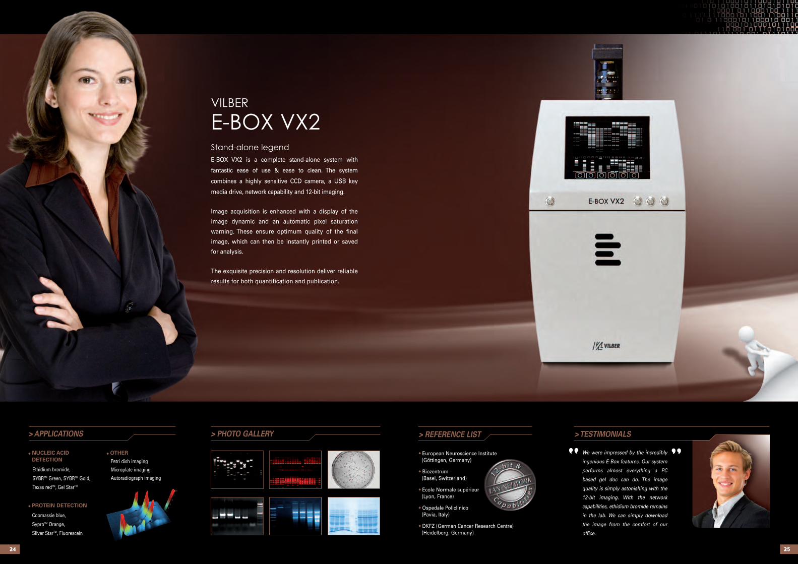
We were impressed by the incredibly
ingenious E-Box features. Our system
performs almost everything a PC
based gel doc can do. The image
quality is simply astonishing with the
12-bit imaging. With the network
capabilities, ethidium bromide remains
in the lab. We can simply download
the image from the comfort of our
office.
“ ”> APPLICATIONS
• NUCLEIC ACIDDETECTION
Ethidium bromide,
SYBRTM Green, SYBRTM Gold,
Texas redTM, Gel StarTM
• PROTEIN DETECTION
Coomassie blue,
SyproTM Orange,
Silver StarTM, Fluorescein
• OTHER
Petri dish imaging
Microplate imaging
Autoradiograph imaging
2524
> REFERENCE LIST
• European Neuroscience Institute(Göttingen, Germany)
• Biozentrum(Basel, Switzerland)
• Ecole Normale supérieur(Lyon, France)
• Ospedale Policlinico(Pavia, Italy)
• DKFZ (German Cancer Research Centre)(Heidelberg, Germany)
VILBEr
E-BOX VX2Stand-alone legend
E-BOX VX2 is a complete stand-alone system with
fantastic ease of use & ease to clean. The system
combines a highly sensitive CCD camera, a USB key
media drive, network capability and 12-bit imaging.
Image acquisition is enhanced with a display of the
image dynamic and an automatic pixel saturation
warning. These ensure optimum quality of the final
image, which can then be instantly printed or saved
for analysis.
The exquisite precision and resolution deliver reliable
results for both quantification and publication.
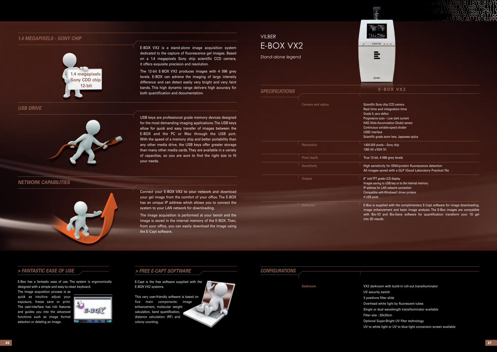
Camera and optics Scientific Sony chip CCD cameraReal time and integration timeGrade 0, zero defectProgressive scan – Low dark currentHAD (Hole Accumulation Diode) sensorContinuous variable-speed shutterUSB2 interfaceScientific grade zoom lens. Japanese optics
Resolution 1.400.000 pixels – Sony chip1360 (H) x1024 (V)
Pixel depth True 12-bit, 4 096 grey levels
Sensitivity High sensitivity for DNA/protein fluorescence detectionAll images saved with a GLP (Good Laboratory Practice) file
Output 8” inch TFT grade LCD displayImages saving to USB key or to the internal memoryIP address for LAN network connectionCompatible with Windows© driver printers4 USB ports
Software E-Box is supplied with the complimentary E-Capt software for image downloading, image enhancement and basic image analysis. The E-Box images are compatible with Bio-1D and Bio-Gene software for quantification: transform your 1D gelinto 3D results
Darkroom VX2 darkroom with build-in roll-out transilluminator
UV security switch
3 positions filter slide
Overhead white light by fluorescent tubes
Single or dual wavelength transilluminator available
Filter size : 20x20cm
Optional Super-Bright UV filter technology
UV to white light or UV to blue light conversion screen available
2726
E-BOX VX2SPECIFICATIONS
> FANTASTIC EASE OF USE
E-Box has a fantastic ease of use. The system is ergonomically
designed with a simple and easy-to-clean keyboard.
The image acquisition process is as
quick as intuitive: adjust your
exposure, freeze save or print.
The user-interface has rich features
and guides you into the advanced
functions such as image format
selection or deleting an image.
> FREE E-CAPT SOFTWARE CONFIGURATIONS
Connect your E-BOX VX2 to your network and downloadyour gel image from the comfort of your office. The E-BOXhas an unique IP address which allows you to connect the system to your LAN network for downloading.
The image acquisition is performed at your bench and theimage is saved in the internal memory of the E-BOX. Then,from your office, you can easily download the image usingthe E-Capt software.
NETWORK CAPABILITIES
E-BOX VX2 is a stand-alone image acquisition system dedicated to the capture of fluorescence gel images. Basedon a 1.4 megapixels Sony chip scientific CCD camera, it offers exquisite precision and resolution.
The 12-bit E-BOX VX2 produces images with 4 096 greylevels. E-BOX can achieve the imaging of large intensity difference and can detect easily very bright and very faintbands. This high dynamic range delivers high accuracy forboth quantification and documentation.
1.4 MEGAPIXELS - SONY CHIP
USB keys are professional grade memory devices designedfor the most demanding imaging applications. The USB keysallow for quick and easy transfer of images between theE-BOX and the PC or Mac through the USB port.With the speed of a memory chip and better portability thanany other media drive, the USB keys offer greater storagethan many other media cards. They are available in a varietyof capacities, so you are sure to find the right size to fityour needs.
USB DRIVE
VILBEr
E-BOX VX2Stand-alone legend
1.4 megapixelsSony CDD chip
12-bit
E-Capt is the free software supplied with the E-BOX VX2 systems.
This very user-friendly software is based on
five main components: image
enhancement, molecular weight
calculation, band quantification,
distance calculation (RF) and
colony counting.
> KEY FEATURES
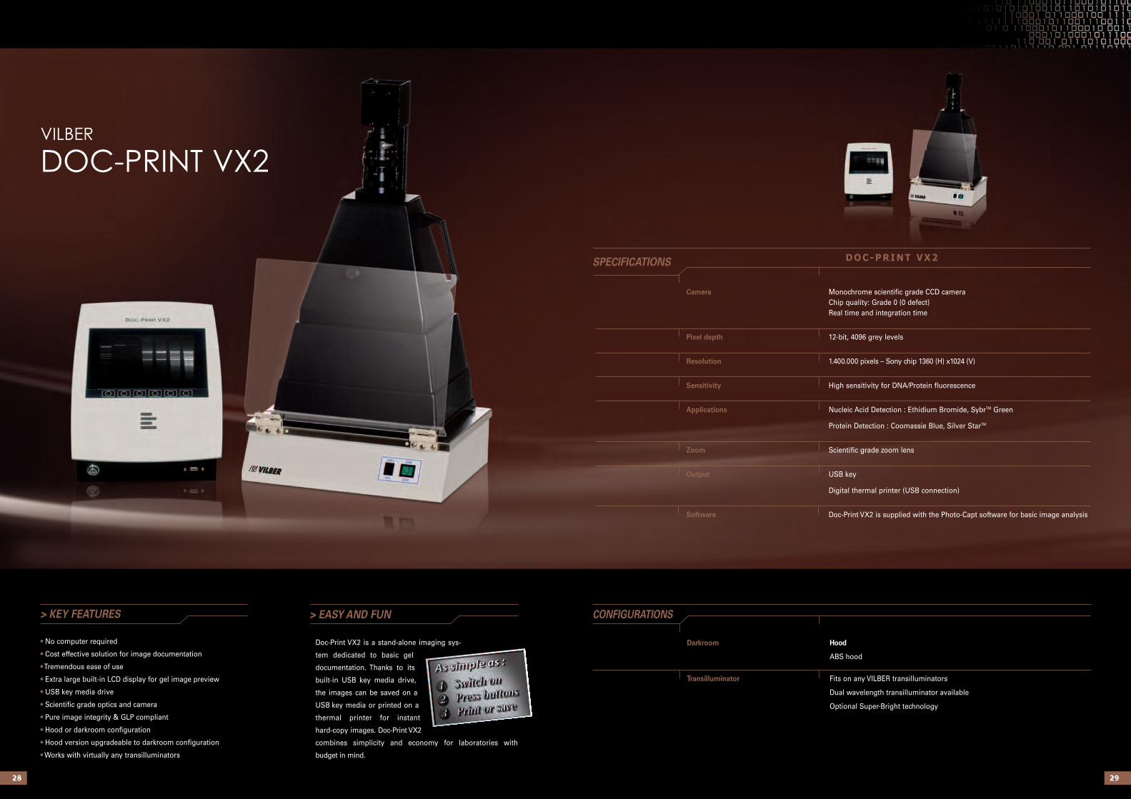
• No computer required
• Cost effective solution for image documentation
•Tremendous ease of use
• Extra large built-in LCD display for gel image preview
• USB key media drive
• Scientific grade optics and camera
• Pure image integrity & GLP compliant
• Hood or darkroom configuration
• Hood version upgradeable to darkroom configuration
• Works with virtually any transilluminators
> EASY AND FUN
2928
Doc-Print VX2 is a stand-alone imaging sys-
tem dedicated to basic gel
documentation. Thanks to its
built-in USB key media drive,
the images can be saved on a
USB key media or printed on a
thermal printer for instant
hard-copy images. Doc-Print VX2
combines simplicity and economy for laboratories with
budget in mind.
Darkroom Hood
ABS hood
Transilluminator Fits on any VILBER transilluminators
Dual wavelength transilluminator available
Optional Super-Bright technology
CONFIGURATIONS
VILBEr
DOC-PrINT VX2
Camera Monochrome scientific grade CCD cameraChip quality: Grade 0 (0 defect)Real time and integration time
Pixel depth 12-bit, 4096 grey levels
Resolution 1.400.000 pixels – Sony chip 1360 (H) x1024 (V)
Sensitivity High sensitivity for DNA/Protein fluorescence
Applications Nucleic Acid Detection : Ethidium Bromide, SybrTM Green
Protein Detection : Coomassie Blue, Silver StarTM
Zoom Scientific grade zoom lens
Output USB key
Digital thermal printer (USB connection)
Software Doc-Print VX2 is supplied with the Photo-Capt software for basic image analysis
DOC-PRINT VX2SPECIFICATIONS
3130
Standardtransilluminator
Super-BrightMX
300 400 500 600 700
Chemiluminescence lightSyber GreenTM
Ethidium bromide
Texas Red
F- 440 filter
ACCESSOrIES DArKrOOMS
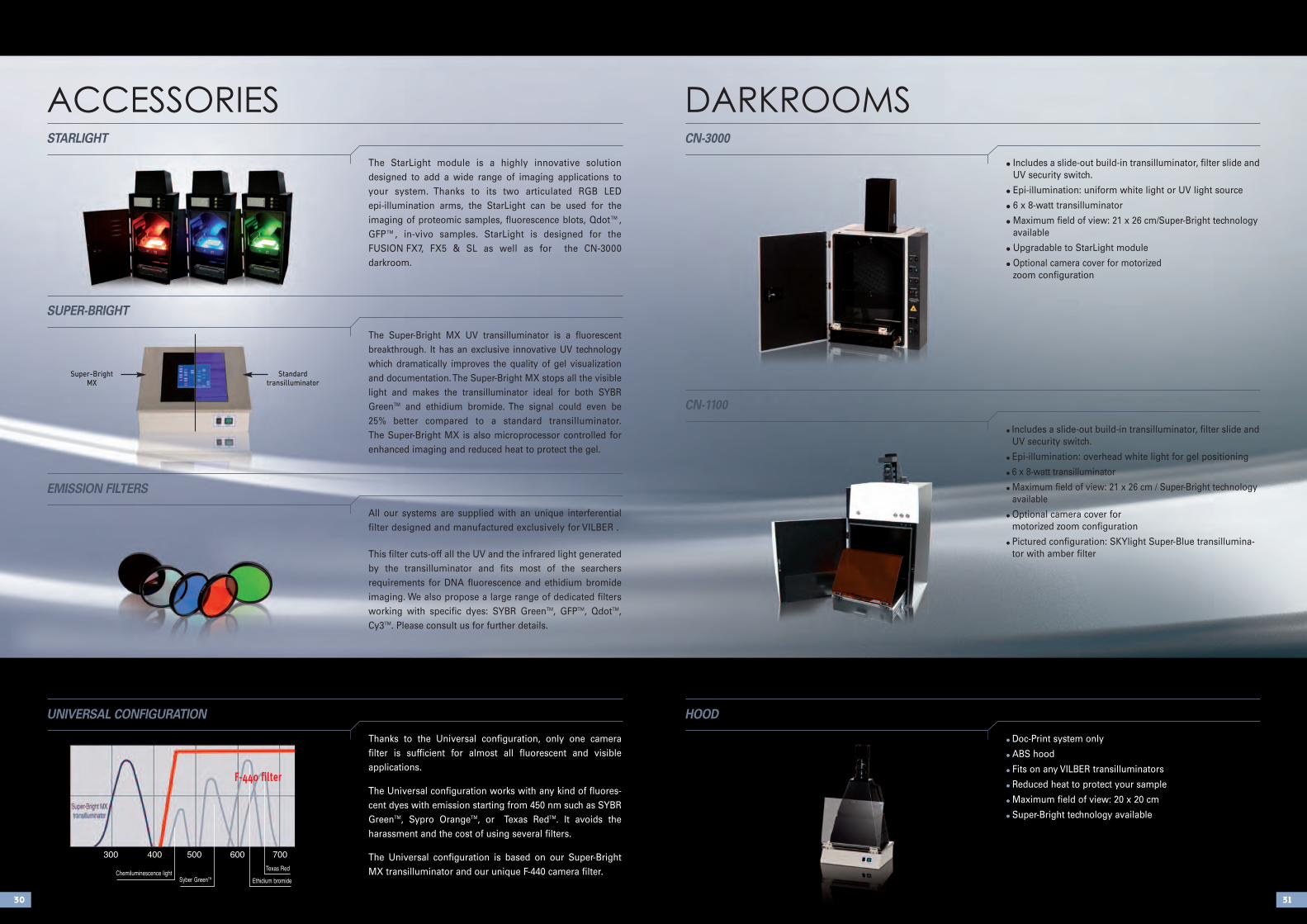
The StarLight module is a highly innovative solution designed to add a wide range of imaging applications toyour system. Thanks to its two articulated RGB LED epi-illumination arms, the StarLight can be used for the imaging of proteomic samples, fluorescence blots, Qdot™,GFP™, in-vivo samples. StarLight is designed for the FUSION FX7, FX5 & SL as well as for the CN-3000 darkroom.
STARLIGHT
The Super-Bright MX UV transilluminator is a fluorescent breakthrough. It has an exclusive innovative UV technologywhich dramatically improves the quality of gel visualizationand documentation. The Super-Bright MX stops all the visiblelight and makes the transilluminator ideal for both SYBRGreenTM and ethidium bromide. The signal could even be 25% better compared to a standard transilluminator. The Super-Bright MX is also microprocessor controlled forenhanced imaging and reduced heat to protect the gel.
SUPER-BRIGHT
All our systems are supplied with an unique interferential filter designed and manufactured exclusively for VILBER .
This filter cuts-off all the UV and the infrared light generatedby the transilluminator and fits most of the searchers requirements for DNA fluorescence and ethidium bromide imaging. We also propose a large range of dedicated filtersworking with specific dyes: SYBR GreenTM, GFPTM, QdotTM,Cy3TM. Please consult us for further details.
EMISSION FILTERS
Thanks to the Universal configuration, only one camera filter is sufficient for almost all fluorescent and visible applications.
The Universal configuration works with any kind of fluores-cent dyes with emission starting from 450 nm such as SYBRGreenTM, Sypro OrangeTM, or Texas RedTM. It avoids theharassment and the cost of using several filters.
The Universal configuration is based on our Super-BrightMX transilluminator and our unique F-440 camera filter.
UNIVERSAL CONFIGURATION
• Includes a slide-out build-in transilluminator, filter slide andUV security switch.
• Epi-illumination: uniform white light or UV light source
• 6 x 8-watt transilluminator
• Maximum field of view: 21 x 26 cm/Super-Bright technologyavailable
• Upgradable to StarLight module
• Optional camera cover for motorizedzoom configuration
CN-3000
• Includes a slide-out build-in transilluminator, filter slide andUV security switch.
• Epi-illumination: overhead white light for gel positioning
• 6 x 8-watt transilluminator
• Maximum field of view: 21 x 26 cm / Super-Bright technologyavailable
• Optional camera cover for motorized zoom configuration
• Pictured configuration: SKYlight Super-Blue transillumina-tor with amber filter
CN-1100
• Doc-Print system only
• ABS hood
• Fits on any VILBER transilluminators
• Reduced heat to protect your sample
• Maximum field of view: 20 x 20 cm
• Super-Bright technology available
HOOD
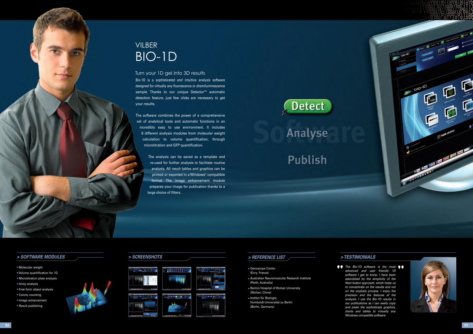
Bio-1D is a sophisticated and intuitive analysis software
designed for virtually any fluorescence or chemiluminescence
sample. Thanks to our unique DetectorTM automatic
detection feature, just few clicks are necessary to get
your results.
The software combines the power of a comprehensive
set of analytical tools and automatic functions in an
incredibly easy to use environment. It includes
8 different analysis modules from molecular weight
calculation to volume quantification, through
microtitration and GFP quantification.
The analysis can be saved as a template and
re-used for further analysis to facilitate routine
analysis. All result tables and graphics can be
printed or exported in a Windows® compatible
format. The image enhancement module
prepares your image for publication thanks to a
large choice of filters.
3332
> SCREENSHOTS > REFERENCE LIST
• Genoscope Center (Evry, France)
• Australian Neuromuscular Research Institute(Perth, Australia)
• Reimin Hospital of Wuhan University (Wuhan, China)
• Institut für Biologie, Humboldt-Universität zu Berlin (Berlin, Germany)
> SOFTWARE MODULES
• Molecular weight
• Volume quantification for 1D
• Microtitration plate analysis
• Array analysis
• Free form object analysis
• Colony counting
• Image enhancement
• Result publishing
> TESTIMONIALS
The Bio-1D software is the mostadvanced and user friendly 1D software I get to know. I have beenastonished by the simplicity of theNext button approach, which helps usto concentrate on the results and noton the analysis process. I enjoy theprecision and the features of theanalysis. I use the Bio-1D results inour publications as I can easily copyand paste the sophisticate graphics,charts and tables to virtually anyWindows compatible software.
“ ”
VILBEr
BIO-1DTurn your 1D gel into 3D results
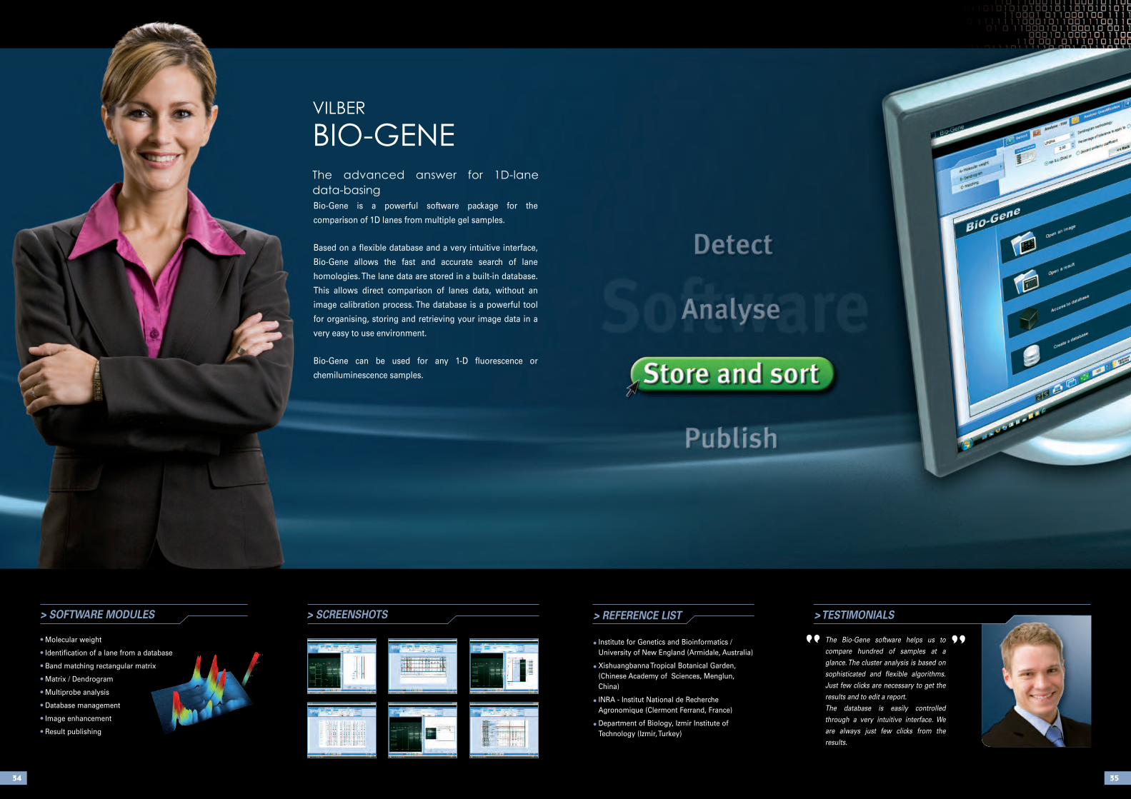
Bio-Gene is a powerful software package for the
comparison of 1D lanes from multiple gel samples.
Based on a flexible database and a very intuitive interface,
Bio-Gene allows the fast and accurate search of lane
homologies. The lane data are stored in a built-in database.
This allows direct comparison of lanes data, without an
image calibration process. The database is a powerful tool
for organising, storing and retrieving your image data in a
very easy to use environment.
Bio-Gene can be used for any 1-D fluorescence or
chemiluminescence samples.
3534
> SCREENSHOTS > TESTIMONIALS
The Bio-Gene software helps us tocompare hundred of samples at aglance. The cluster analysis is based onsophisticated and flexible algorithms.Just few clicks are necessary to get theresults and to edit a report.The database is easily controlledthrough a very intuitive interface. Weare always just few clicks from the results.
“ ”> REFERENCE LIST
• Institute for Genetics and Bioinformatics /University of New England (Armidale, Australia)
• Xishuangbanna Tropical Botanical Garden,(Chinese Academy of Sciences, Menglun,China)
• INRA - Institut National de RechercheAgronomique (Clermont Ferrand, France)
• Department of Biology, Izmir Institute ofTechnology (Izmir, Turkey)
> SOFTWARE MODULES
• Molecular weight
• Identification of a lane from a database
• Band matching rectangular matrix
• Matrix / Dendrogram
• Multiprobe analysis
• Database management
• Image enhancement
• Result publishing
VILBEr
BIO-GENEThe advanced answer for 1D-lane
data-basing
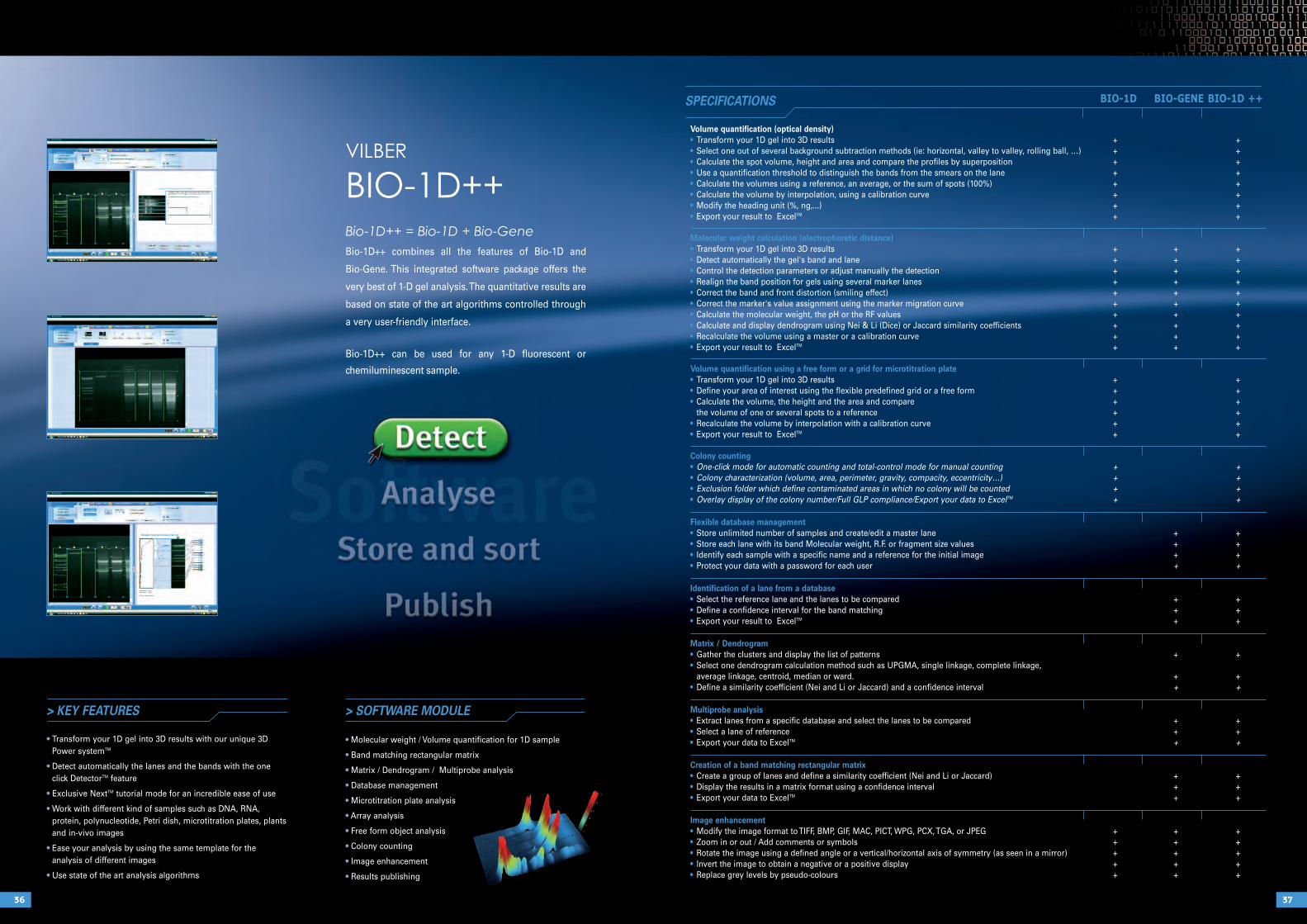
BIO-1D BIO-GENE BIO-1D ++
Volume quantification (optical density)• Transform your 1D gel into 3D results + +• Select one out of several background subtraction methods (ie: horizontal, valley to valley, rolling ball, …) + +• Calculate the spot volume, height and area and compare the profiles by superposition + +• Use a quantification threshold to distinguish the bands from the smears on the lane + +• Calculate the volumes using a reference, an average, or the sum of spots (100%) + +• Calculate the volume by interpolation, using a calibration curve + +• Modify the heading unit (%, ng,...) + +• Export your result to ExcelTM + +
Molecular weight calculation (electrophoretic distance)• Transform your 1D gel into 3D results + + +• Detect automatically the gel's band and lane + + +• Control the detection parameters or adjust manually the detection + + +• Realign the band position for gels using several marker lanes + + +• Correct the band and front distortion (smiling effect) + + +• Correct the marker's value assignment using the marker migration curve + + +• Calculate the molecular weight, the pH or the RF values + + +• Calculate and display dendrogram using Nei & Li (Dice) or Jaccard similarity coefficients + + +• Recalculate the volume using a master or a calibration curve + + +• Export your result to ExcelTM + + +
Volume quantification using a free form or a grid for microtitration plate• Transform your 1D gel into 3D results + +• Define your area of interest using the flexible predefined grid or a free form + +• Calculate the volume, the height and the area and compare + +
the volume of one or several spots to a reference + +• Recalculate the volume by interpolation with a calibration curve + +• Export your result to ExcelTM + +
Colony counting• One-click mode for automatic counting and total-control mode for manual counting + +• Colony characterization (volume, area, perimeter, gravity, compacity, eccentricity…) + +• Exclusion folder which define contaminated areas in which no colony will be counted + +• Overlay display of the colony number/Full GLP compliance/Export your data to ExcelTM + +
Flexible database management• Store unlimited number of samples and create/edit a master lane + +• Store each lane with its band Molecular weight, R.F. or fragment size values + +• Identify each sample with a specific name and a reference for the initial image + +• Protect your data with a password for each user + +
Identification of a lane from a database• Select the reference lane and the lanes to be compared + +• Define a confidence interval for the band matching + +• Export your result to ExcelTM + +
Matrix / Dendrogram• Gather the clusters and display the list of patterns + +• Select one dendrogram calculation method such as UPGMA, single linkage, complete linkage,
average linkage, centroid, median or ward. + +• Define a similarity coefficient (Nei and Li or Jaccard) and a confidence interval + +
Multiprobe analysis• Extract lanes from a specific database and select the lanes to be compared + +• Select a lane of reference + +• Export your data to ExcelTM + +
Creation of a band matching rectangular matrix• Create a group of lanes and define a similarity coefficient (Nei and Li or Jaccard) + +• Display the results in a matrix format using a confidence interval + +• Export your data to ExcelTM + +
Image enhancement• Modify the image format to TIFF, BMP, GIF, MAC, PICT, WPG, PCX, TGA, or JPEG + + +• Zoom in or out / Add comments or symbols + + +• Rotate the image using a defined angle or a vertical/horizontal axis of symmetry (as seen in a mirror) + + +• Invert the image to obtain a negative or a positive display + + +• Replace grey levels by pseudo-colours + + +
3736
SPECIFICATIONS
> KEY FEATURES
• Transform your 1D gel into 3D results with our unique 3DPower systemTM
• Detect automatically the lanes and the bands with the oneclick DetectorTM feature
• Exclusive NextTM tutorial mode for an incredible ease of use
• Work with different kind of samples such as DNA, RNA, protein, polynucleotide, Petri dish, microtitration plates, plantsand in-vivo images
• Ease your analysis by using the same template for the analysis of different images
• Use state of the art analysis algorithms
> SOFTWARE MODULE
• Molecular weight / Volume quantification for 1D sample
• Band matching rectangular matrix
• Matrix / Dendrogram / Multiprobe analysis
• Database management
• Microtitration plate analysis
• Array analysis
• Free form object analysis
• Colony counting
• Image enhancement
• Results publishing
VILBEr
BIO-1D++Bio-1D++ = Bio-1D + Bio-Gene
Bio-1D++ combines all the features of Bio-1D and
Bio-Gene. This integrated software package offers the
very best of 1-D gel analysis. The quantitative results are
based on state of the art algorithms controlled through
a very user-friendly interface.
Bio-1D++ can be used for any 1-D fluorescent or
chemiluminescent sample.
3938
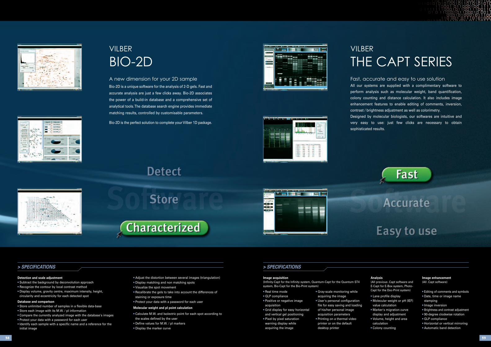
> SPECIFICATIONS
Detection and scale adjustment• Subtract the background by deconvolution approach• Recognize the contour by local contrast method• Display volume, gravity centre, maximum intensity, height,
circularity and eccentricity for each detected spot
Database and comparison• Store unlimited number of samples in a flexible data-base• Store each image with its M.W. / pI information• Compare the currently analyzed image with the database's images• Protect your data with a password for each user• Identify each sample with a specific name and a reference for the
initial image
> SPECIFICATIONS
Image acquisition(Infinity-Capt for the Infinity system, Quantum-Capt for the Quantum ST4system, Bio-Capt for the Bio-Print system)
• Real time mode• GLP compliance• Positive or negative image
acquisition• Grid display for easy horizontal
and vertical gel positioning• Pixel by pixel saturation
warning display while acquiring the image
• Gray-scale monitoring whileacquiring the image
• User's personal configurationfile for easy saving and loadingof his/her personal imageacquisition parameters
• Printing on a thermal videoprinter or on the default desktop printer
Analysis(All previous .Capt software and E-Capt for E-Box system, Photo-Capt for the Doc-Print system)
• Lane profile display• Molecular weight or pH (IEF)
value calculation• Marker's migration curve
display and adjustment• Volume, height and area
calculation• Colony counting
Image enhancement(All .Capt software)
• Editing of comments and symbols• Date, time or image name
stamping• Image inversion• Brightness and contrast adjustment• 90-degree clockwise rotation• GLP compliance• Horizontal or vertical mirroring • Automatic band detection
• Adjust the distortion between several images (triangulation)• Display matching and non matching spots• Visualize the spot movement• Recalibrate the gels to take into account the differences of
staining or exposure time• Protect your data with a password for each user
Molecular weight and pI point calculation
• Calculate M.W. and Isolectric point for each spot according tothe scales defined by the user
• Define values for M.W. / pI markers• Display the marker curve
VILBEr
BIO-2DA new dimension for your 2D sample
Bio-2D is a unique software for the analysis of 2-D gels. Fast and
accurate analysis are just a few clicks away. Bio-2D associates
the power of a build-in database and a comprehensive set of
analytical tools. The database search engine provides immediate
matching results, controlled by customisable parameters.
Bio-2D is the perfect solution to complete your Vilber 1D package.
VILBEr
THE CAPT SErIESFast, accurate and easy to use solution
All our systems are supplied with a complimentary software to
perform analysis such as molecular weight, band quantification,
colony counting and distance calculation. It also includes image
enhancement features to enable editing of comments, inversion,
contrast / brightness adjustment as well as colorimetry.
Designed by molecular biologists, our softwares are intuitive and
very easy to use: just few clicks are necessary to obtain
sophisticated results.
MOLECULAr IMAGINGCHEMILUMINESCENCE • FLUOrESCENCE • ANALySIS SOFTWArE
VILBEr is the leading European provider of
molecular imaging systems, analysis software and
UV fluorescence equipment. Founded over 50
years ago to serve the research, VILBEr has
pioneered the post electrophoresis market and
introduced breakthrough products such as stand
alone gel-documentation, Bio-1D imaging
software, Super-Bright UV technology, dedicated
chemiluminescence imaging system and 3D
approach to 1D gel analysis.
Through a network of owned subsidiary offices and
local distributors located in over 60 countries
around the world, VILBEr offers a broad range of
products:
> Gel documentation systems
> Chemiluminescence imaging systems
> Image analysis software
> UV instruments for molecular biology such as
transilluminators, crosslinkers and UV lamps.
For more information about VILBEr, visit our
website at www.vilber.com
HEADQUARTER
Vilber Lourmat
BP-66 – ZI Sud Torcy
F-77202 Marne-la-Vallée cedex 1
France
T.: 33 (0) 1 60 06 07 71
F.:33 (0) 1 64 80 48 59
GERMANY OFFICE
Vilber Lourmat Deutschland GmbH
Wielandstrasse 2
D-88436 Eberhardzell
Deutschland
T.: 49 (0) 7355 931 380
F.: 49 (0) 7355 931 379
www.vilber.comCA
RR
EMEN
T C
OM
01
64 7
2 11
52
- 07
/201
0