modification of biodistribution and brain uptake of copper
TRANSCRIPT

Modification of Biodistribution and Brain Uptake of CopperBis(thiosemicarbazonato) Complexes by the Incorporation of Amineand Polyamine Functional GroupsBrett M. Paterson,†,‡,§ Carleen Cullinane,⊥,∥ Peter J. Crouch,¶,∇,# Anthony R. White,¶,○
Kevin J. Barnham,‡,∇,# Peter D. Roselt,∥ Wayne Noonan,∥ David Binns,∥ Rodney J. Hicks,⊥,∥
and Paul S. Donnelly*,†,‡
†School of Chemistry, ‡Bio21 Molecular Science and Biotechnology Institute, ¶Department of Pathology, ∇Department ofPharmacology and Therapeutics, #Florey Institute of Neuroscience and Mental Health, and ⊥The Sir Peter MacCallum Departmentof Oncology, The University of Melbourne, Parkville, Victoria 3010, Australia∥The Centre for Molecular Imaging and Translational Research Laboratory, The Peter MacCallum Cancer Centre, Melbourne,Victoria 3000, Australia
*S Supporting Information
A B S T R A C T : T h e s y n t h e s i s o f n e w b i s -(thiosemicarbazonato)copper(II) complexes featuring poly-amine substituents via selective transamination reactions ispresented. Polyamines of different lengths, with differentionizable substituent groups, were used to modify and adjustthe hydrophilic/lipophilic balance of the copper complexes.The new analogues were radiolabeled with copper-64 andtheir lipophilicities estimated using distribution coefficients.The cell uptake of the new polyamine complexes wasinvestigated with preliminary in vitro biological studies usinga neuroblastoma cancer cell line. The in vivo biodistributionof three of the new analogues was investigated in vivo in miceusing positron-emission tomography imaging, and one of thenew complexes was compared to [64Cu]Cu(atsm) in an A431 squamous cell carcinoma xenograft model. Modification of thecopper complexes with various amine-containing functional groups alters the biodistribution of the complexes in mice. Onecomplex, with a pendent (N,N-dimethylamino)ethane functional group, displayed tumor uptake similar to that of[64Cu]Cu(atsm) but higher brain uptake, suggesting that this compound has the potential to be of use in the diagnosticbrain imaging of tumors and neurodegenerative diseases.
■ INTRODUCTION
Copper complexes of bis(thiosemicarbazonato) ligands derivedfrom 1,2-diones are of interest as potential diagnostic andtherapeutic agents. The ligands can be used to form copper(II)complexes with radioactive isotopes of copper that are stable(Ka = 1018), charge-neutral, lipophilic, and capable of crossingcell membranes.1 The positron-emitting isotopes, 60Cu (t1/2 =24 m), 61Cu (t1/2 = 3.33 h), 62Cu (t1/2 = 9.75 m), and 64Cu(t1/2 = 12.7 h) are of interest in the development of newimaging agents for positron-emission tomography (PET).Copper-64 also has a β− emission (Eβ
−max = 0.574 MeV,
40%) that is of potential use in targeted radiotherapy, as does67Cu (t1/2 = 62 h, β−, 100%, Eave = 0.12 MeV).The coppe r comp lex d i a ce t y lb i s (4 -me thy l - 3 -
thiosemicarbazonato)copper(II) [Cu(atsm); Figure 1] hasbeen investigated as a hypoxia imaging agent in tumors andmyocardial ischemia.2−6 Studies on [64Cu]Cu(atsm) haveprogressed to a phase II human trial as a PET imaging andtherapeutic agent for cervical cancer.7−10 The hypoxia
selectivity of Cu(atsm) is related to the CuII/I reductionpotential, which leads to selective reduction of the metal ionand subsequent trapping of the radioactivity in certaincells.3,5,11,12 The high lipophilicity of Cu(atsm) results inhigh nonspecific cell uptake and liver uptake. The nonspecific
Received: January 13, 2019Published: March 14, 2019
Figure 1. Copper complex Cu(atsm).
Article
pubs.acs.org/ICCite This: Inorg. Chem. 2019, 58, 4540−4552
© 2019 American Chemical Society 4540 DOI: 10.1021/acs.inorgchem.9b00117Inorg. Chem. 2019, 58, 4540−4552
Dow
nloa
ded
via
QU
EE
NSL
AN
D U
NIV
OF
TE
CH
NO
LO
GY
on
May
27,
202
0 at
23:
36:3
5 (U
TC
).Se
e ht
tps:
//pub
s.ac
s.or
g/sh
arin
ggui
delin
es f
or o
ptio
ns o
n ho
w to
legi
timat
ely
shar
e pu
blis
hed
artic
les.
uptake can compromise the image quality, and the high liveruptake results in dose-limiting radiotoxicity to the liver fortherapeutic applications.13
The ability of Cu(atsm) to cross the blood−brain barrier hasled to radiolabeled [62Cu]Cu(atsm) being investigated as aprobe for the redox status of the brain in mitochondrialdisease, Parkinson’s disease, and amyotrophic lateral sclerosis(ALS).14−16 In addition to radiopharmaceutical applications,nonradioactive Cu(atsm) has been investigated as a potentialtherapeutic agent for Parkinson’s disease and ALS.17−23 Thetherapeutic potential for Cu(atsm) in both ALS andParkinson’s disease is currently being investigated in twohuman clinical trials.The biodistribution, cellular accumulation, and metabolism
of bis(thiosemicarbazonato)copper(II) [Cu(btsc)] complexesis dependent on their lipophilicity and their CuII/I reductionpotentials.24,25 The lipophilicity of Cu(btsc) complexes can bealtered by changing the nature of both the substituent on theπ-conjugated backbone and the terminal (N4) amine of theligand.24,26 In general, changing the aliphatic substituents onthe N4 amine does not affect the redox potential significantly(±0.05 V), especially compared to the changes that can occuras a result of changes in the substituents on the diiminebackbone (±0.2 V).5 Hydrophilic analogues of Cu(atsm) havebeen identified as potential targets for new hypoxia-selectivecopper radiopharmaceuticals to reduce the level of liver andkidney uptake.27−30
We have prepared a series of new bis(thiosemicarbazones)with amine and polyamine functional groups with the goal ofproducing complexes with biodistribution profiles differentfrom those of Cu(atsm). The biogenic polyamines butane-1,4-diamine (putrescine), N-(3-aminopropyl)butane-1,4-diamine(spermidine), and N,N′-bis(3-aminopropyl)butane-1,4-dia-
mine (spermine) are ubiquitous in nearly every prokaryoticand eukaryotic cell type and have been used to increase theblood−brain barrier permeability of therapeutic peptides andproteins.31−33 In aqueous solution at pH 7.4, the polyaminesare fully protonated, giving them considerable water solubility.Polyamine side chains have been shown to enhance the cellularuptake and specificity of anticancer pharmaceuticals andimaging agents by a combination of utilization of polyaminetransport systems and the ability of polyamines and alkylatedamine functional groups to modify the hydrophobic−lipophiliccharacter as well as overall charge and solvation.34−43
The synthesis of new Cu(btsc) complexes featuringpolyamine substituents via selective transamination reactionsis presented. Polyamines of different lengths, numbers ofcharged groups, and substituent groups were used to modifyand adjust the hydrophilic/lipophilic balance of the coppercomplexes. The new analogues were radiolabeled with 64Cu,and their distribution coefficients were determined. The celluptake of the new polyamine complexes was investigated withpreliminary in vitro biological studies using a neuroblastomacancer cell line because hypoxia is a strong independent riskpredictor in neuroblastoma patients.44 We also examined the invivo biodistribution of three of the new analogues using in vivosmall-animal PET imaging and selected one complex forevaluation in an A431 squamous cell carcinoma xenograftmodel.
■ RESULTS AND DISCUSSION
Synthesis of Bis(thiosemicarbazone)−Polyaminesand Their Copper Complexes. Selective transaminationreactions of the nonsymmetric molecule H2L
1 with nucleo-philic amines resulting in the selective displacement ofdimethylamine from the dimethyl substituent have proven to
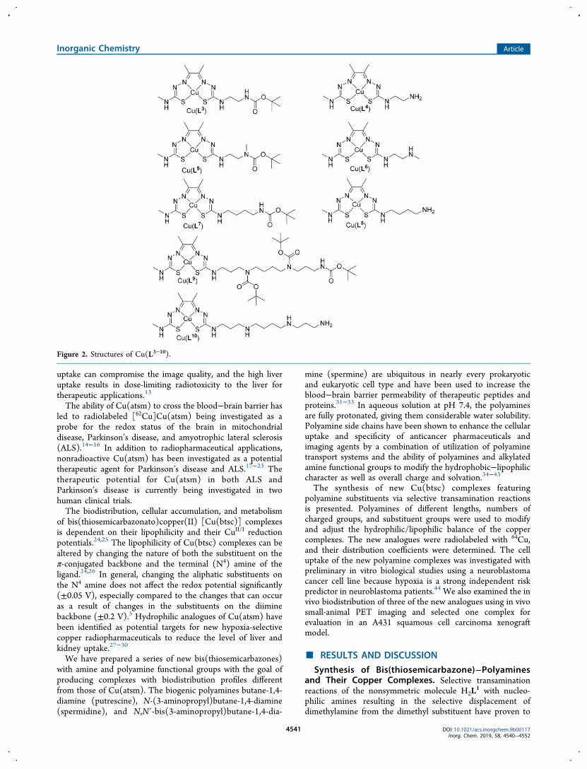
Figure 2. Structures of Cu(L3−10).
Inorganic Chemistry Article
DOI: 10.1021/acs.inorgchem.9b00117Inorg. Chem. 2019, 58, 4540−4552
4541

be a reliable method to prepare substituted bis-(thiosemicarbazone) (Figure 2).29,45−48 The incoming aminereacts with the electrophilic thiocarbonyl carbon atom ofN4,N4-dimethylthiosemicarbazone substituents with preferencetoward the N4-monosubstituted moiety, which is capable ofundergo ing t au tomer i z a t ion . A f ami l y o f b i s -(thiosemicarbazones) complexes were prepared where short-chain polyamines, 1,2-diaminoethane, N,N-dimethylethylene-diamine, and N-methylethylenediamine, as well as the biogenicpolyamines putrescine and spermine, were appended to theligand framework. The reaction of N,N-dimethylethylenedi-amine with H2L
1 allowed the isolation of H2L2 in high yield.
The copper complex was prepared by reacting H2L2 with
copper acetate monohydrate, which leads to double deproto-nation of the ligand and the formation of charge-neutralCu(L2) (Scheme 1).The reactions with polyamines that contain more than one
nucleophilic amine required selective protection, with N-tert-butoxycarbonyl (t-Boc) groups (Figure 2). The syntheses of[H3L
4][CF3CO2] and [Cu(H2L4)][CF3CO2]2 were described
previously (Figure 2) but are included.45 Both the primary andsecondary amines of N-methylethylenediamine are sufficientlynucleophilic to complicate a transamination reaction. Toprevent the secondary amine from reacting, it was protectedwith a t-Boc group. The primary amine was first protectedusing a trifluoroacetamide group.49 Ethyl trifluoroacetate reactspreferentially with the primary amine under controlledconditions. The t-Boc group was added to the secondaryamine before trifluoroacetamide was removed in a basicsolution. The resulting product was subjected to trans-amination to give the product H2L
5, and the copper complexCu(L5) was prepared by the addition of copper acetatemonohydrate (Figure 2).The room temperature 1H and 13C{1H} NMR spectra of
H2L5 contain broadened signals and signals that have been
divided in two. These signals correspond to proton and carbonenvironments near the carbamate bond. The signals due to thebis(thiosemicarbazone) groups were much sharper in compar-ison. In the 13C{1H} NMR spectrum, the signals are split intotwo peaks for the methylene (δ = 41.7, 42.5 and 46.6, 47.3),carbonyl (δ = 154.7, 155.5), N-methyl (δ = 34.0, 34.4), andquaternary (δ = 78.3, 78.6) carbon atoms (Figure S1). A likelyexplanation is that electron delocalization results in partialdouble-bond character for the carbamate group and hinderedrotation about the C(carbonyl)−N bond (Figure S2). Thepartial double-bond character renders the carbamate groupplanar, with it existing in either the s-cis or s-trans rotamer.Hindered rotation about secondary amide peptide bonds andcarbamates is known.50−53 At room temperature, both isomersare observed in the NMR spectrum as a result of slowinterconversion between the two forms relative to the NMRtime scale. Coalescence of the isomeric signals was observed asthe temperature was increased. At 70 °C, a single broad peak
was observed for the methylene (δ = 42.0 and 46.8) carbonyl(δ = 155.0), N-methyl (δ = 34.0), and quaternary (δ = 78.3)carbon atoms, which indicates that the rate of interconversionwas now faster than the time scale of the NMR experiment.The electron-donating N-methyl group of the carbamate onH2L
5 may be stabilizing the C(carbonyl)−N double-bondform, thus increasing the barrier to C(carbonyl)−N bondrotation.54,55 In contrast, isomerization was not observed in theroom temperature NMR spectra of H2L
3 because the partialdouble bond is less stabilized, leading to faster interconversionbetween the s-cis and s-trans isomers.The t-Boc protecting group of H2L
5 and Cu(L5) wasremoved with trifluoroacetic acid to give [H3L
6][CF3CO2] and[Cu(HL6)][CF3CO2]·0.8CF3CO2H, respectively. The electro-spray ionization mass spectrometry (ESI-MS) spectrum of[Cu(HL6)][CF3CO2]·0.8CF3CO2H (m/z 365.05; 100%)corresponded to that of [Cu(HL6)]+, and reverse-phase high-performance liquid chromatography (RP-HPLC; RT = 7.424min) indicated successful deprotection, while the microanalysissuggested the presence of trifluoroacetate as the counterion.One of the primary amines of the polyamine putrescine was
protected with t-Boc using a literature procedure.56 Trans-amination of H2L
1 with t-Boc-protected polyamine tert-butyl 4-aminobutylcarbamate produced H2L
7 in high yield (Figure 2).To ensure a selective transamination reaction with one of theprimary amine groups of spermine, one of the primary aminesand both secondary amines were protected using t-Boc. Thetri-t-Boc spermine compound was prepared using a literatureprocedure.57 The transamination reaction between H2L
1 and(N1,N4,N9-tri-tert-butoxycarbonyl)-N,N′-bis(3-aminopropyl)-butane-1,4-diamine gave H2L
9 in good yield following silicachromatography. Signals in the 1H and 13C{1H} NMR spectrawere broad when obtained at room temperature, possiblybecause of cis/trans isomerism of the three carbamate bonds.Increasing the temperature to 70 °C sharpened some of thesignals, allowing the spectra to be more easily assigned withtwo-dimensional NMR spectroscopy. The ESI-MS spectrumgave a peak at m/z 732.43 (100%), corresponding to [H2L
9 +H+], and the RP-HPLC had a single peak (RT = 18.16 min).The copper complex Cu(L9) (m/z 793.34; 100%; RT = 17.40min) was prepared with the addition of copper acetatemonohydrate to a solution of H2L
9 in ethanol (Figure 2).Deprotection of H2L
9 and Cu(L9) with trifluoroacetic acidgave the trications [H5L
10][CF3CO2]3 and [Cu(H3L10)]-
[CF3CO2]3·2H2O (Figure 2), respectively. ESI-MS spectrafor [Cu(H3L
10)][CF3CO2]3·2H2O (m/z 493.18 and 247.10;100%) corresponded to [Cu(HL10)]+ and [Cu(H2L
10)]2+,respectively.
Electrochemical Characterization, Radiolabeling with64Cu, and Distribution Coefficients. The hypoxia selectivityand biological activity of the Cu(btsc) complexes stronglycorrelate with the CuII/I reduction potential. The newcomplexes Cu(L2−10) all retain the methyl substituents on
Scheme 1. Synthesis of H2L2 and Cu(L2)a
a(a) N,N-Dimethylethylenediamine (1.3 equiv), acetonitrile, reflux, 6.5 h, 74%. (b) Cu(CH3CO2)2·H2O (1 equiv), acetonitrile, reflux, 1.5 h, 75%.
Inorganic Chemistry Article
DOI: 10.1021/acs.inorgchem.9b00117Inorg. Chem. 2019, 58, 4540−4552
4542

the diimine-like backbone to ensure that they display CuII/I
couples similar to those of Cu(atsm). The electrochemistry ofthe new complexes was investigated by cyclic voltammetry.The median potentials (E) and peak separations (Epa − Epc;Table 1) reveal no significant differences between the
complexes. For example, Cu(L2) has a quasi-reversiblereduction with a median potential58−60 E = −0.65 V [vsstandard calomel electrode (SCE), where E = (Epc + Epa)/2and ferrocene/ferrocinium (Fc/Fc+) = 0.54 V] with an anodic-to-cathodic peak separation of 105 mV in N,N-dimethylforma-mide (DMF) at a glassy carbon working electrode, which wasattributed to a CuII/I reduction process (Figure 3A and Table1). Deprotection of the complexes with trifluoroacetic acid ledto their isolation as aminium salts, and cyclic voltammetry ofthese protonated cationic salts revealed irreversible reductionwaves characteristic of the adsorption of a reduced species tothe working electrode (Figure S3A). Neutralization of theprotonated cations with triethylamine resulted in theobservation of quasi-reversible CuII/CuI couples (Figure S3B).The copper-64 complexes were prepared by the addition of
64Cu (0.02 M HCl, pH 1) to a buffered solution of the ligands(pH ∼7). The complexes were prepared at a specific activityrange of 0.74−3.7 GBq mg−1 of the ligand. The purities of thecomplexes were confirmed using HPLC coupled to aradioactivity detector and a comparison with the HPLC tracesof the nonradioactive analogues (λ = 280 nm). All of theradiolabeled complexes could be prepared at room temper-ature in minutes under mild conditions with >90% radio-chemical purity without additional purification, making themideal candidates for in vivo imaging.The respective retention times of the RP-HPLC traces
highlighted the differences in lipophilicity between thecomplexes and were quantified by obtaining the distributioncoefficients (log D at pH 7.4) in phosphate-buffered saline(PBS; Figure 4). Cu(atsm) possessed the highest log D, pH7.4, value of 1.49 ± 0.08 and a retention time of 10.12 min. Ofthe new derivatives, the ligand featuring a dimethylamine
substituent, Cu(L2), was the most lipophilic (0.83 ± 0.03 and8.91 min) and the spermine derivative the most hydrophilic(−1.29 ± 0.01). The complexes possessing either a singleprimary amine, Cu(L4) and Cu(L8), or a single secondaryamine, Cu(L6), had similar lipophilicities (−0.21 ± 0.02,−0.25 ± 0.01, and −0.14 ± 0.05, respectively).
Cytotoxicity and Cel lular Uptake of Bis-(thiosemicarbazonato)copper(II) Complexes. The coppercontent of SH-SY5Y cells, a neuroblastoma cell line, treatedwith either Cu(L2), Cu(L4), Cu(L6), Cu(L8), or Cu(L10) (10μM, 1 h) was measured by inductively coupled plasma massspectrometry (ICP-MS; Figure 5a). The highest level ofcellular copper was observed in cells treated with the complexCu(L2), where a (62 ± 2)-fold increase in the copper levelswas detected compared to vehicle-treated controls. This valuewas greater than the value detected for Cu(atsm) [(47 ± 5)-fold increase]. Significant increases in the copper content (35−37-fold) were observed for the complexes [Cu(H2L
4)]2+,[Cu(HL6)]+, and [Cu(HL8)]+, which have either a singleprimary or secondary amine. The complex [Cu(H3L
10)]3+,which has two secondary amines and one primary amine thatare likely protonated at biological pH, showed significantly lessuptake than the other complexes including unchelated Cu2+ (a3-fold compared to a 5-fold increase in the copper levels).
Table 1. Median Potentials and Peak Separations of theCyclic Voltammograms (Scan Rate 0.1 V s−1; Potentials AreQuoted Relative to a SCE)
compound Cu(atsm) Cu(L2) Cu(L6) Cu(L8) Cu(L10)
CuII/CuI E (V) −0.63 −0.65 −0.60 −0.65 −0.63CuII/CuIEpa−Epc (mV) 102 105 104 92 89
Figure 3. (A) Cyclic voltammogram of Cu(L2). (B) Cyclic voltammogram of Cu(L6) in the presence of triethylamine. Scan rate 0.1 V s−1.Potentials are quoted relative to a SCE, where Fc/Fc+ = 0.54 V.
Figure 4. RP-HPLC retention times (RT) versus the distributioncoefficients (log D at pH 7.4) of Cu(atsm) and the new polyaminecopper-64 complexes.
Inorganic Chemistry Article
DOI: 10.1021/acs.inorgchem.9b00117Inorg. Chem. 2019, 58, 4540−4552
4543
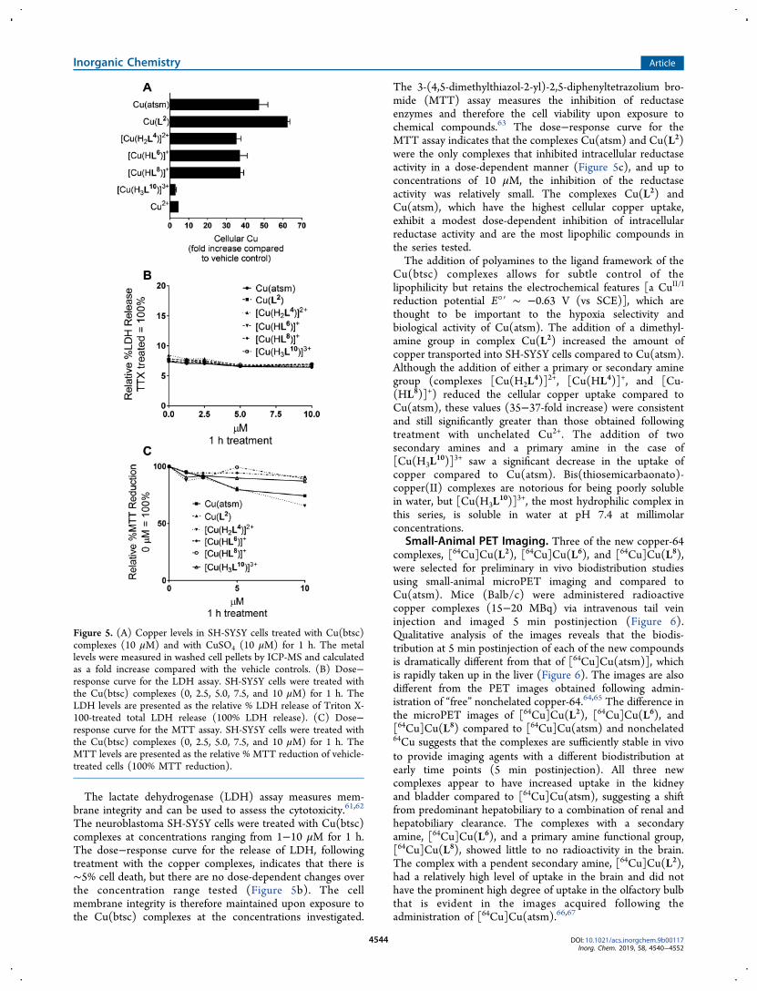
The lactate dehydrogenase (LDH) assay measures mem-brane integrity and can be used to assess the cytotoxicity.61,62
The neuroblastoma SH-SY5Y cells were treated with Cu(btsc)complexes at concentrations ranging from 1−10 μM for 1 h.The dose−response curve for the release of LDH, followingtreatment with the copper complexes, indicates that there is∼5% cell death, but there are no dose-dependent changes overthe concentration range tested (Figure 5b). The cellmembrane integrity is therefore maintained upon exposure tothe Cu(btsc) complexes at the concentrations investigated.
The 3-(4,5-dimethylthiazol-2-yl)-2,5-diphenyltetrazolium bro-mide (MTT) assay measures the inhibition of reductaseenzymes and therefore the cell viability upon exposure tochemical compounds.63 The dose−response curve for theMTT assay indicates that the complexes Cu(atsm) and Cu(L2)were the only complexes that inhibited intracellular reductaseactivity in a dose-dependent manner (Figure 5c), and up toconcentrations of 10 μM, the inhibition of the reductaseactivity was relatively small. The complexes Cu(L2) andCu(atsm), which have the highest cellular copper uptake,exhibit a modest dose-dependent inhibition of intracellularreductase activity and are the most lipophilic compounds inthe series tested.The addition of polyamines to the ligand framework of the
Cu(btsc) complexes allows for subtle control of thelipophilicity but retains the electrochemical features [a CuII/I
reduction potential E°′ ∼ −0.63 V (vs SCE)], which arethought to be important to the hypoxia selectivity andbiological activity of Cu(atsm). The addition of a dimethyl-amine group in complex Cu(L2) increased the amount ofcopper transported into SH-SY5Y cells compared to Cu(atsm).Although the addition of either a primary or secondary aminegroup (complexes [Cu(H2L
4)]2+, [Cu(HL4)]+, and [Cu-(HL8)]+) reduced the cellular copper uptake compared toCu(atsm), these values (35−37-fold increase) were consistentand still significantly greater than those obtained followingtreatment with unchelated Cu2+. The addition of twosecondary amines and a primary amine in the case of[Cu(H3L
10)]3+ saw a significant decrease in the uptake ofcopper compared to Cu(atsm). Bis(thiosemicarbaonato)-copper(II) complexes are notorious for being poorly solublein water, but [Cu(H3L
10)]3+, the most hydrophilic complex inthis series, is soluble in water at pH 7.4 at millimolarconcentrations.
Small-Animal PET Imaging. Three of the new copper-64complexes, [64Cu]Cu(L2), [64Cu]Cu(L6), and [64Cu]Cu(L8),were selected for preliminary in vivo biodistribution studiesusing small-animal microPET imaging and compared toCu(atsm). Mice (Balb/c) were administered radioactivecopper complexes (15−20 MBq) via intravenous tail veininjection and imaged 5 min postinjection (Figure 6).Qualitative analysis of the images reveals that the biodis-tribution at 5 min postinjection of each of the new compoundsis dramatically different from that of [64Cu]Cu(atsm)], whichis rapidly taken up in the liver (Figure 6). The images are alsodifferent from the PET images obtained following admin-istration of “free” nonchelated copper-64.64,65 The difference inthe microPET images of [64Cu]Cu(L2), [64Cu]Cu(L6), and[64Cu]Cu(L8) compared to [64Cu]Cu(atsm) and nonchelated64Cu suggests that the complexes are sufficiently stable in vivoto provide imaging agents with a different biodistribution atearly time points (5 min postinjection). All three newcomplexes appear to have increased uptake in the kidneyand bladder compared to [64Cu]Cu(atsm), suggesting a shiftfrom predominant hepatobiliary to a combination of renal andhepatobiliary clearance. The complexes with a secondaryamine, [64Cu]Cu(L6), and a primary amine functional group,[64Cu]Cu(L8), showed little to no radioactivity in the brain.The complex with a pendent secondary amine, [64Cu]Cu(L2),had a relatively high level of uptake in the brain and did nothave the prominent high degree of uptake in the olfactory bulbthat is evident in the images acquired following theadministration of [64Cu]Cu(atsm).66,67
Figure 5. (A) Copper levels in SH-SY5Y cells treated with Cu(btsc)complexes (10 μM) and with CuSO4 (10 μM) for 1 h. The metallevels were measured in washed cell pellets by ICP-MS and calculatedas a fold increase compared with the vehicle controls. (B) Dose−response curve for the LDH assay. SH-SY5Y cells were treated withthe Cu(btsc) complexes (0, 2.5, 5.0, 7.5, and 10 μM) for 1 h. TheLDH levels are presented as the relative % LDH release of Triton X-100-treated total LDH release (100% LDH release). (C) Dose−response curve for the MTT assay. SH-SY5Y cells were treated withthe Cu(btsc) complexes (0, 2.5, 5.0, 7.5, and 10 μM) for 1 h. TheMTT levels are presented as the relative % MTT reduction of vehicle-treated cells (100% MTT reduction).
Inorganic Chemistry Article
DOI: 10.1021/acs.inorgchem.9b00117Inorg. Chem. 2019, 58, 4540−4552
4544

Tumor Uptake and Biodistribution in a A431 TumorModel. The hypoxia-selective retention of radioactivity from[62Cu]Cu(atsm) in an ex vivo rat heart model of ischemiastimulated much interest in the potential to use radiolabeledversions of Cu(atsm) as hypoxia imaging agents.3 The hypoxia-selective retention of radioactivity following the treatment ofEMT6 cells with [64Cu]Cu(atsm) has been demonstrated invitro.68 The tumor uptake of copper-64 following injection of[64Cu]Cu(atsm) to BALB/c mice bearing EMT6 tumors was4.17 ± 1.03%IA/g (%IA/g = injected activity per gram oftissue) at 40 min postinjection, and the retention wasattributed to hypoxia.68 Injection of either [64Cu]Cu(atsm)or [64Cu]Cu(CH3CO2)2 in mice bearing CaNT or EMT6tumors resulted in similar degrees of copper-64 retention in therepresentative tumors at 2 and 16 h postinjection, and theareas of uptake correlated with the areas of hypoxia identifiedby immunohistochemistry. However, the copper uptakefollowing the administration of either [64Cu]Cu(atsm) or[64Cu]Cu(CH3CO2)2 at earlier time points (15 min and 2 hpostinjection) did not correlate to areas of hypoxia in thismodel.69 Despite the similarities between the biodistribution of[64Cu]Cu(atsm) and [64Cu]Cu(CH3CO2)2 at later timepo in t s , r ad ioa c t i v e coppe r comp l e xe s o f b i s -(thiosemicarbazonato) ligands do not behave like unchelatedcopper in the early phase of biodistribution. There is adramatic difference in the brain uptake of [64Cu]Cu(atsm) and[64Cu]Cu(CH3CO2)2.
66,67
The difference in the biodistribution of [64Cu]Cu(L2) and[64Cu]Cu(atsm) revealed by PET imaging at 5 min post-injection coupled with the relatively high brain uptakedisplayed by [64Cu]Cu(L2) encouraged us to select thiscompound for evaluation in a tumor model. The new tracer,[64Cu]Cu(L2), was compared with [64Cu]Cu(atsm) in anA431 squamous cell carcinoma xenograft model that leads tointratumoral hypoxia.70 Mice were administered with either[64Cu]Cu(L2) or [64Cu]Cu(atsm) via intravenous tail veininjection (10−15 MBq) and imaged with a small-animal PETscanner at 1, 3, and 22 after tracer injection. The microPETimaging data reveal that at each time point the standardizeduptake values (SUVs) in the tumor for [64Cu]Cu(L2) and[64Cu]Cu(atsm) are not significantly different (p > 0.08; Table2).Following the final scan, the mice were culled and tissues
excised for an ex vivo biodistribution analysis (Table 3). Theadministration of [64Cu]Cu(atsm) and [64Cu]Cu(L2) leads to
Figure 6. (A) Small-animal microPET images (maximum-intensity projection) of two BALB/c mice 5 min after a single intravenous administrationof 15−20 MBq of [64Cu]Cu(atsm), [64Cu]Cu(L2), [64Cu]Cu(L6), and [64Cu]Cu(L8). The animals were placed on the bed of a Philips Mosaicsmall-animal PET scanner 5 min postinjection and imaged over 10 min. (B) Representative false color images: close-up of the brain region (sagittalplane) and maximum-intensity projection.
Table 2. Tumor SUV Data (SUV ± Standard Error; n = 3)As Determined by Small-Animal PET Imaging
time (h) [64Cu]Cu(atsm) [64Cu]Cu(L2)
1 0.61 ± 0.05 0.79 ± 0.083 0.70 ± 0.02 0.81 ± 0.0622 0.78 ± 0.05 0.99 ± 0.08
Inorganic Chemistry Article
DOI: 10.1021/acs.inorgchem.9b00117Inorg. Chem. 2019, 58, 4540−4552
4545

tumor uptakes of 2.59 ± 0.20%IA/g and 3.35 ± 0.20%IA/g,respectively, 23 h after injection (p < 0.06). The degree ofuptake observed 16 h postinjection of [64Cu]Cu(atsm) to CBAmice bearing CaNT tumors was 1.32 ± 0.09%IA/g.69 TheA431 squamous cell carcinoma xenograft model used in thisstudy has been previously shown to lead to intratumoralhypoxia, but a limitation of this present study is that, in thisinstance, the tumors were not confirmed as hypoxic usingimmunohistochemistry.70 Considering that injection of either[64Cu]Cu(atsm) or [64Cu]Cu(CH3CO2)2 in mice bearingCaNT or EMT6 tumors resulted in similar degrees of copper-64 retention at 16 h postinjection, it would be pertinent tohave included [64Cu]Cu(CH3CO2)2 in the present study andto investigate the in vivo stability of the two complexes.69 It ispossible that the uptake of copper-64 in tumors at 16 hpostinjection actually reflects endogenous copper metabolicpathways involving specific chaperone and transport proteinsas well as cuproenzymes rather than specific uptake mediatedby the injected complex.71−73 Significant differences werefound between the biodistribution of the two tracers in thelungs, kidneys, heart, muscle, and brain (p < 0.05) at 23 hpostinjection. The ability of Cu(atsm) to cross the blood−brain barrier has led to radiolabeled [62Cu]Cu(atsm) beingused to distinguish the tumor grade in human gliomapatients,74 and treatment with [64Cu]Cu(atsm) was inves-tigated recently as a therapeutic option in a mouse model ofglioblastoma.75 Brain imaging with [62Cu]Cu(atsm) has alsobeen used to probe the redox status in human patients withmitochondrial disease,14 Parkinson’s disease,15 and ALS.16 Theadministration of [64Cu]Cu(L2) leads to more radioactivity inthe brain, 4.41 ± 0.23%ID/g, compared to that of [64Cu]Cu-(atsm), 2.43 ± 0.31%ID/g, (p < 0.01), suggesting that this newvariant could be of interest as a brain imaging agent. Theaddition of the same functional group to copper complexes offunctionalized pyridylthiosemicarbazone ligands also improvedbrain uptake.76
■ CONCLUSIONA range of new Cu(btsc) complexes with amine and polyaminefunctional groups have been prepared using a selectivetransamination reaction. The addition of the amine functionalgroups reduces the lipophilicity of these derivatives ofCu(atsm) but does not significantly alter the CuII/I reductionpotential that is thought to be central to the biological activityof this type of complex. With the exception of the complex thatincorporates a spermine functional group, [Cu(H3L
10)]3+, thecomplexes were all capable of dramatically increasing theintracellular copper content of neuroblastoma SH-SY5Y cells,
suggesting that they retain the ability of Cu(atsm) to cross cellmembranes, which is also thought to be crucial to thebiological activity of this family of complexes. The biodis-tribution in mice of three of the new complexes, [64Cu]Cu-(L2), [64Cu]Cu(L6), and [64Cu]Cu(L8), was investigated usingmicroPET imaging and revealed that each of these lesslipophilic derivatives of [64Cu]Cu(atsm) has a different earlybiodistribution compared to [64Cu]Cu(atsm). The differencein the biodistribution of [64Cu]Cu(L2) and [64Cu]Cu(atsm)revealed by a preliminary PET imaging study and the relativelyhigh brain uptake encouraged us to evaluate this compound inan A431 tumor model. In this model, the administration of[64Cu]Cu(L2) leads to tumor uptake (3.35 ± 0.20%IA/g) 23 hpostinjection similar to that of the administration of [64Cu]-Cu(atsm) (2.59 ± 0.20%IA/g; p < 0.06). The administrationof [64Cu]Cu(L2), possessing a pendent N,N-dimethylamino-ethane functional group to mice results in a significantly higherbrain uptake than the administration of [64Cu]Cu(atsm),suggesting that the compound has the potential to be used inimaging brain tumors as well as ALS and Parkinson’s disease.
■ EXPERIMENTAL SECTIONGeneral Procedures. The following reagents were used as
received: spermine (Sigma-Aldrich), butane-1,4-diamine (Sigma-Aldrich), di-tert-butyl dicarbonate (Sigma-Aldrich), N,N-dimethyle-thylenediamine (Aldrich Chemicals), and N-methylethylenediamine(Aldrich Chemicals). All solvents were obtained from standardcommercial sources and used as received. NMR spectra were acquiredon Varian FT-NMR 500 and FT-NMR 400 spectrometers. 1H NMRspectra were acquired at 500 or 400 MHz, and 13C{1H} NMR spectrawere acquired at 125.7 MHz. All NMR spectra were recorded at 25°C unless otherwise indicated. 1H and 13C{1H} chemical shifts werereferenced to residual solvent peaks and are quoted in parts permillion relative to tetramethylsilane . MS spectra were recorded on anAgilent 6510 ESI-TOF LC/MS mass spectrometer. Cyclic voltammo-grams were recorded on an Autolab PGSTAT100 electrochemicalworkstation using GPES V4.9 software and employing a glassy carbonworking electrode, a platinum counter electrode, and an Ag/Ag+
reference electrode [silver wire in CH3CN (AgNO3; 0.01 M)]. Allmeasurements were carried out in DMF. All solutions were 5 mManalyte in a 0.1 M tetrabutylammoniumtetrafluoroborate solution.DMF was obtained from commercial sources and dried over 3 Åsieves before use. Each solution was purged with N2 prior to analysisand measured at ambient temperatures under a N2 atmosphere. Thepeak (Ep) and median (E) potentials were referenced to the Fc/Fc+
couple, +0.54 V in DMF versus SCE. The Fc/Fc+ median potentialunder the conditions used was +0.07 V. Microanalyses for carbon,hydrogen, and nitrogen were carried out by Chemical & Micro-analytical Services (CMAS) Pty. Ltd., Belmont, Victoria, Canada. RP-HPLC utilized an Agilent 1200 series HPLC system using an AgilentZorbax Eclipse XDB-C18 column (4.6 × 150 mm, 5 μm) with a 1mL/min flow rate, gradient elution of buffer A = 0.1% TFA in H2Oand buffer B = 0.1% TFA in acetonitrile (0−100% B in A at 20 min)and detection at 220, 254, and 275 nm. 64Cu was produced via the64Ni(p,n)64Cu reaction, using a custom-manufactured solid targetassembly positioned externally to a Cyclone 18/9 (IBA) cyclotron.The target consisted of 64Ni metal (enriched to 94.8−99.07%)electroplated onto a gold foil (15 mm × 125 μm) backing, housed in acustom-made aluminum cradle. The primary proton beam wasdegraded to 11.7 MeV using a graphite degrader built into a graphitecollimator. Helium cooling was on the target holder at beam entryand chilled water upon beam exit. All targets were irradiated at 40 μA,for up to 2 h. After irradiation, the target was transferred to thelaboratory for further chemical processing, in which 64Cu was isolatedusing ion-exchange chromatography using low concentrations of HClin alcohol solutions. Final reconstitution of the 64Cu fraction in
Table 3. Biodistribution (%IA/g ± Standard Error; n = 3) at23 h Postinjection
[64Cu]Cu(atsm) [64Cu]Cu(L2)
blood 2.34 ± 0.28 2.98 ± 0.56lungs 6.79 ± 0.44 15.53 ± 1.47heart 3.58 ± 0.42 5.42 ± 0.35liver 14.27 ± 1.96 16.59 ± 3.08kidneys 5.98 ± 0.55 8.97 ± 0.45muscle 0.74 ± 0.07 1.42 ± 0.05spleen 5.04 ± 0.22 4.62 ± 0.77brain 2.43 ± 0.31 4.41 ± 0.23eyes 1.06 ± 0.14 1.97 ± 0.60tumor 2.59 ± 0.20 3.35 ± 0.20
Inorganic Chemistry Article
DOI: 10.1021/acs.inorgchem.9b00117Inorg. Chem. 2019, 58, 4540−4552
4546

aqueous HCl yielded 1−2.6 GBq of 64Cu as 64CuCl2 (specific activity,28.9 GBq μmol−1 (μA·h/mg of 64Ni)−1; radionuclidic purity, 99%).Cu(atsm) and H2L
1, H2L3, Cu(L3), [H3L
4][CF3CO2], and[Cu(H2L
4)][CF3CO2]2 were prepared as previously published.45
Synthesis. Diacetyl-4-ethylenedimethylamine-4′-methylbis-(thiosemicarbazone), H2L
2. To a stirring suspension of H2L1 (0.21
g, 0.85 mmol) in acetonitrile (30 mL) was added N-dimethylethy-lenediamine (0.01 g, 1.12 mmol). The resulting yellow suspensionwas heated at reflux for 6.5 h under an atmosphere of N2. Theresulting orange solution was cooled to room temperature, resulting inthe precipitation of colorless crystals, which were collected byfiltration, washed with acetonitrile (1×) and diethyl ether (3×), anddried to give H2L
2 (0.18 g, 0.55 mmol, 74%). Elem anal. Found: C,41.69; H, 7.36; N, 30.74. Calcd for C11H23N7S2: C, 41.61; H, 7.30; N,30.88. 1H NMR (DMSO-d6, 500 MHz): δ 2.16, 3H, s, CH3; 2.19, 6H,s, CH3; 2.21, 3H, s, CH3; 2.46, 2H, t,
3JHH = 6.5 Hz, CH2; 3.03, 3H, d,3JHH = 4 Hz, NHCH3; 3.61, 2H, m, CH2; 8.34−8.37, 2H, m, NHCH2,NHCH3; 10.24, 2H, br s, NH.
13C{1H} NMR (125.7 MHz): δ 11.3,11.7, CH3; 31.2, NHCH3; 41.4, CH2; 45.0, N(CH3)2; 57.1, CH2;147.5, 147.9, CN; 177.6, 178.5, CS. ESI-MS (positive ion; 100%,[M + H+]): m/z 318.15 (experimental), 318.15 (calcd). RP-HPLC:RT = 7.58 min.D ia c e t y l - 4 - e t h y l en ed ime thy l am ine - 4 ′ -me th y l b i s -
(thiosemicarbazonato)copper(II), Cu(L2). To a solution of H2L2
(0.10 g, 0.3 mmol) in acetonitrile (10 mL) was added Cu(OAc)2·H2O (0.07 g, 0.3 mmol), and the resulting red/brown suspension wasstirred at reflux for 1.5 h and then allowed to cool to roomtemperature. The solid was collected by filtration, washed withacetonitrile (1×) and diethyl ether (3×), and dried to give Cu(L2)(0.09 g, 75%). Elem anal. Found: C, 34.87; H, 5.62; N, 25.79. Calcdfor C11H21CuN7S2: C, 34.86; H, 5.58; N, 25.87. ESI-MS: (positiveion; 100%, [M + H+]): m/z 379.07 (experimental), 379.07 (calcd).RP-HPLC: RT = 7.67 min.tert-Butyl Methyl[2-(2,2,2-trifluoroacetylamino)ethyl]-
carbamate. The title compound was prepared according to aliterature procedure and isolated as a white crystalline solid (3.90 g,72%).49 1H NMR (CDCl3, 500 MHz): δ 1.46, 9H, s, (CH3)3; 2.90,3H, s, CH3; 3.48, 4H, br m, CH2; 7.95, br s, NH.tert-Butyl (2-Aminoethyl)methylcarbamate. The title compound
was prepared according to a literature procedure and isolated as ayellow oil (1.71 g, 68%).49 1H NMR (CDCl3, 500 MHz): δ 1.45, 9H,s, (CH3)3; 2.82, 2H, t, NH2CH2; 2.87, 3H, s, CH3; 3.27, br m, 2H,CH2.tert-Butyl 4-Aminobutylcarbamate. The title compound was
prepared according to a literature procedure and isolated as a colorlessoil.56 1H NMR (CDCl3, 500 MHz): δ 1.43, 9H, s, (CH3)3; 1.45−1.53,4H, m, CH2; 2.70, 2H, t,
3JHH = 7 Hz, CH2; 3.12, 2H, q,3JHH = 6 Hz,
CH2.(N1,N4,N9-Tri-tert-butoxycarbonyl)-N,N′-bis(3-aminopropyl)-
butane-1,4-diamine. The title compound was prepared according toa literature procedure and obtained as a colorless, homogeneous oil(1.33 g, 51%), Rf 0.5 [CH2Cl2−MeOH−concentrated aqueous NH3,5:1:0.1 (v/v/v)] after purification over silica gel [CH2Cl2−MeOH−concentrated aqueous NH3, 100:0:0 to 5:1:0.1 (v/v/v)].57 1H NMR(CDCl3, 500 MHz): δ 1.42−1.50, 31H, m, CH3 × 3, CH2 × 2; 1.61−1.67, 4H, m, CH2 × 2; 3.08−3.31, 10H, m, CH2 × 5. ESI-MS(positive ion; 100%, [M + H+]): m/z 503.41 (experimental), 503.41(calcd).Diacetyl-tert-butyl-4-ethylmethylcarbamate-4′-methylbis-
(thiosemicarbazone), H2L5·0.5CH3CN. Following the same procedure
employed for the synthesis of H2L2, H2L
1 (0.55 g, 2.0 mmol) and tert-butyl (2-aminoethyl)methylcarbamate (0.42 g, 2.4 mmol) were usedto prepare H2L
5 (0.70 g, 86%). Elem anal. Found: C, 45.30; H, 7.17;N, 25.00. Calcd for C15H29N7O2S2·0.5CH3CN: C, 45.31; H, 7.25; N,24.77. 1H NMR (DMSO-d6, 500 MHz, 343 K): δ 1.38, 9H, s, (CH3)3;2.05, CH3CN; 2.21, 3H, s, CH3; 2.22, 3H, s, CH3; 2.84, 3H, s, CH3;3.05, 3H, d, 3JHH = 4.5 Hz, NHCH3; 3.43, 2H, t,
3JHH = 6.0 Hz, CH2;3.71−3.77, 2H, m, CH2; 8.25−8.35, 2H, br m, NHCH2, NHCH3;10.07, 2H, s, NH. 13C{1H} NMR (125.7 MHz, 343 K): δ 11.2, 11.3,CH3; 27.8, (CH3)3; 30.9, NHCH3; 34.0, NCH3; 42.0, CH2; 46.8,
CH2; 78.3, C(CH3)3; 147.4, 147.8, CN; 155.0, CO; 178.1, 178.6,CS. ESI-MS (positive ion; 100%, [M + H+]): m/z 404.19(experimental), 404.19 (calcd).
Diacetyl-tert-butyl-4-ethylmethylcarbamate-4′-methylbis-(thiosemicarbazonato)copper(II), Cu(L5)·H2O. To a solution of H2L
5
(0.18 g, 0.5 mmol) in ethanol (10 mL) was added Cu(OAc)2·H2O(0.10 g, 0.5 mmol), and the resulting red/brown suspension wasstirred at reflux for 2 h. The solvent was removed in vacuo, and thebrown residue was dissolved in acetone (3 mL) and precipitated withhexane (30 mL). The solid was collected by filtration, washed withhexane, and dried to give Cu(L5) (0.16 g, 78%). Elem anal. Found: C,36.84; H, 5.51; N, 19.82. Calcd for C15H27CuN7O2S2·H2O: C, 37.29;H, 6.05; N, 20.29. ESI-MS (positive ion; 100%, [M + H+]): m/z465.10 (experimental), 465.10 (calcd). RP-HPLC: RT = 13.01 min.
Diace t y l - 4 - e t hy l eneme thy lam in ium-4 ′ -me thy lb i s -(thiosemicarbazone) Trifluoroacetate, [H3L
6][CF3CO2]. A solution ofH2L
5(0.11 g, 0.3 mmol) in CH2Cl2 (4 mL) was added dropwise over20 min to TFA (4 mL) with stirring in an ice bath. The resultingsolution was left to warm to room temperature before the solvent wasremoved in vacuo. To the orange oily residue was added diethyl ether(25 mL), resulting in a precipitate, which was collected by filtration,washed with diethyl ether, and dried to give [H3L
6][CF3CO2] as awhite solid (0.09 g, 0.22 mmol). Elem anal. Found: C, 34.46; H, 5.21;N, 23.35. Calcd for C12H22F3N7O2S2: C, 34.52; H, 5.31; N, 23.49.
1HNMR (DMSO-d6, 500 MHz): δ 2.23, 6H, s, CH3; 2.61, 3H, s, CH3;3.02, 3H, d, CH3; 3.16, 2H, br, CH2; 3.89, br, 2H, CH2; 8.40, 1H, br,NH; 8.50, br, 3H, NH, NH2
+CH3; 10.25, s, 1H, NH; 10.54, s, 1H,NH. 13C{1H} NMR (125.7 MHz): δ 11.7, 11.9, CH3; 31.2, CH3;32.9, CH3; 40.5, 47.5, CH2; 117.2, q,
1JCF = 299.5 Hz, CF3; 147.7,148.9, CN; 158.2, q, 2JCF = 31.3 Hz, CCF3; 178.5, CS. ESI-MS(positive ion; 100%, [M + H+]): m/z 304.14 (experimental), 304.14(calcd).
Diace t y l - 4 - e t hy l eneme thy lam in ium-4 ′ -me thy lb i s -(thiosemicarbazonato)copper(II) Trifluoroacetate, [Cu(HL6)]-[CF3CO2]·0.8CF3CO2H. To a solution of trifluoroacetic acid (5 mL,0 °C) cooled in an ice bath was added Cu(L5) (0.07 g, 0.14 mmol) inportions over 20 min. The solution mixture was warmed to roomtemperature and stirred for 1.5 h. The solvent was removed in vacuoto give a brown oil. Diethyl ether was added, and a brown solidprecipitated, which was collected by filtration, washed with diethylether, and dried to give [Cu(HL6)][CF3CO2]·0.8CF3CO2H (0.05 g,65%). Elem anal. Found: C, 28.38; H, 3.87; N, 17.49. Calcd forC12H19CuF3N7O2S2·0.8C2HF3O2: C, 28.65; H, 3.68; N, 17.19. ESI-MS (positive ion; 100%, [M + H+]): m/z 365.05 (experimental),365.05 (calcd). RP-HPLC: RT = 7.42 min.
Diacety l tert -Buty l -4-buty lcarbamate-4 ′ -methylbis -(thiosemicarbazone), H2L
7. Following the same procedure that wasemployed for the synthesis of H2L
2, H2L1 (0.20 g, 0.74 mmol) and
tert-butyl 4-aminobutylcarbamate (0.21 g, 1.1 mmol) were used toprepare H2L
7 (0.27 g, 87%). Elem anal. Found: C, 45.63; H, 7.56; N,23.31. Calcd for C16H31N7O2S2: C, 46.02; H, 7.48; N, 23.48.
1HNMR (DMSO-d6, 500 MHz): δ 1.35−1.42, 11H, br, (CH3)3, CH2;1.54, 2H, m, CH2; 2.20, 6H, s, CH3; 2.93, 2H, m, CH2; 3.02, 3H, d,3JHH = 6 Hz, NHCH3; 3.55, 2H, m, CH2; 6.77, 1H, s, NHCO; 8.35−8.40, 2H, br m, NHCH3, NHCH2; 10.13, 2H, br s, NH. 13C{1H}NMR (125.7 MHz): δ 11.6, 11.7, CH3; 26.2, CH2; 27.0, CH2; 28.3,(CH3)3; 31.2, CH3NH; 40.2, CH2; 43.5, CH2
1; 77.3, C(CH3)3; 147.8,148.0, CN; 155.6, CO; 177.6, 178.5, CS. ESI-MS (positiveion; 100%, [M + H+]): m/z 418.21 (experimental), 418.21 (calcd).RP-HPLC: RT = 16.40 min.
Diacety l tert -Buty l -4-buty lcarbamate-4 ′ -methylbis -(thiosemicarbazonato)copper(II), Cu(L7)·H2O·0.5CH3CN. Followingthe same procedure that was employed for the synthesis of Cu(L2),H2L
7 (0.10 g, 0.25 mmol) and Cu(OAc)2·H2O (0.05 g, 0.25 mmol)were used to prepare Cu(L7) (0.10 g, 81%). (Elem anal. Found: C,39.25; H, 6.24; N, 20.83. Calcd for C16H29CuN7O2S2·H2O·0.5CH3CN: C, 39.44; H, 6.33; N, 20.29. ESI-MS (positive ion;100%, [M + H+]): m/z 479.12 (experimental), 479.12 (calcd). RP-HPLC: RT = 15.13 min.
Inorganic Chemistry Article
DOI: 10.1021/acs.inorgchem.9b00117Inorg. Chem. 2019, 58, 4540−4552
4547

Diacetyl-4-butyleneaminium-4′-methylbis(thiosemicarbazone)Trifluoroacetate, [HL8][CF3CO2]. Following the same procedure thatwas employed for the synthesis of [H3L
6][CF3CO2], H2L7 (0.06 g,
0.13 mmol) was used to prepare [HL8][CF3CO2] (0.05 g, 86%).Elem anal. Found: C, 36.30; H, 5.71; N, 21.59. Calcd forC13H24CuN7F3O2S2: C, 36.19; H, 5.61; N, 22.72. 1H NMR(DMSO-d6, 500 MHz): δ 1.52−1.67, 4H, m, CH2; 2.21, 6H, s,CH3; 2.82, 2H, m, CH2; 3.02, 3H, d, CH3,
3JHH = 4 Hz; 3.60, m, 2H,CH2; 7.69, 3H, br s, NH3
+; 8.35−8.47, m, 2H, NH; 10.21, br s, 2H,NH. ESI-MS (positive ion; 100%, [M+]): m/z 318.15 (experimental),318.15 (calcd). RP-HPLC: RT = 10.02 min.D i a c e t y l - 4 - b u t y l e n e a m i n i u m - 4 ′ - m e t h y l b i s -
(thiosemicarbazonato)copper(II) Trifluoroacetate, [Cu(HL8)]-[CF3CO2]. Following the same procedure that was employed for thesynthesis of [Cu(HL6)][CF3CO2]·0.8CF3CO2H, Cu(L
7) (0.04 g, 0.1mmol) was used to prepare [Cu(HL8)][CF3CO2] (0.02 g, 51%).Elem anal. Found: C, 31.64; H, 4.56; N, 19.81. Calcd forC13H22CuN7F3O2S2: C, 31.67; H, 4.50; N, 19.89. ESI-MS (positiveion; 100%, [M+]): m/z 379.07 (experimental), 379.07 (calcd). RP-HPLC: RT = 7.62 min.Diacetyl 4-(N1,N4,N9-Tri-tert-butoxycarbonyl)-N,N′-bis(3-
am i n o p r o p y l ) b u t a n e - 1 , 4 - d i am i n e - 4 ′ - m e t h y l b i s -(thiosemicarbazone), H2L
9. To a stirring suspension of H2L1 (0.30 g,
1.1 mmol) in acetonitrile (20 mL) was added (N1,N4,N9-tri-tert-butoxycarbonyl)-N,N′-bis(3-aminopropyl)butane-1,4-diamine (0.56g, 1.1 mmol). The mixture was heated at reflux for 3 h andmonitored by thin-layer chromatography analysis [5% MeOH−CH2Cl2 (v/v)]. The resulting suspension was cooled to roomtemperature and filtered to remove the white precipitate. The solventwas removed in vacuo to give a light-yellow, glassy solid, which waspurified over silica [CH2Cl2−MeOH−Et3N 100:0:0 to 100:1:0.1 to100:4:0.1 (v/v/v)] to afford H2L
9 as a white glassy solid (0.66 g, 0.9mmol, 81%), Rf = 0.6 [5% MeOH−CH2Cl2 (v/v)]. Elem anal. Found:C, 52.37; H, 8.46; N, 17.17. Calcd for C32H61N9O6S2: C, 52.50; H,8.40; N, 17.22. 1H NMR (DMSO-d6, 500 MHz, 343 K): δ 1.38, 9H, s,(CH3)3; 1.39, 9H, s, (CH3)3; 1.41, 9H, s, (CH3)3; 1.42−1.47, 4H, brm, CH2
5, CH26; 1.59, 2H, p, 3JHH = 7 Hz, CH2; 1.73−1.81, 2H, br m,
CH2; 2.21, 3H, s, CH3; 2.22, 2H, s, CH3; 2.91, 2H, q,3JHH = 6 Hz,
CH2; 3.05, 3H, d,3JHH = 4.5 Hz, CH3NH; 3.10−3.17, 6H, m, CH2,
CH2, CH2; 3.21, 2H, t,3JHH = 7 Hz, CH2; 3.57, 2H, q,
3JHH = 6.5 Hz,CH2; 6.44−6.60, 1H, br s, NH; 8.28, 1H, m, NHCH3; 8.39−8.60, 1H,br s, NHCH2; 9.95−10.10, 2H, br s, NH. 13C{1H} NMR (125.7MHz, 343 K): δ 11.1, 11.3, CH3; 25.3, CH2, CH2; 27.8, 27.8, 28.0,(CH3)3, CH2; 28.5, CH2; 30.1, CH3NH; 37.5, CH2; 40.8, CH2; 43.5,CH2; 44.2, CH2; 46.1, CH2, CH2; 77.2, 78.0, 78.3, C(CH3)3; 147.5,147.7, CN; 154.4, 155.2, CO; 177.7, 178.5, CS. ESI-MS(positive ion; 100%, [M + H+]): m/z 732.43 (experimental), 732.43(calcd). RP-HPLC: RT = 18.16 min.Diacetyl 4-(N1,N4,N9-tri-tert-butoxycarbonyl)-N,N′-bis(3-
am i n o p r o p y l ) b u t a n e - 1 , 4 - d i am i n e - 4 ′ - m e t h y l b i s -(thiosemicarbazonato)copper(II), Cu(L9). To a solution of H2L
9
(0.31 g, 0.4 mmol) in ethanol (5 mL) was added Cu(OAc)2·H2O(0.09 g, 0.4 mmol). The red/brown solution was stirred at roomtemperature for 19 h. The solvent was removed in vacuo, and thebrown residue was dissolved in dichloromethane (3 mL) and filtered.The solvent was removed in vacuo to give Cu(L9) as a brown/redglassy solid (0.29 g, 88%). Elem anal. Found: C, 48.38; H, 7.53; N,15.92. Calcd for CuC32H59N9O6S2: C, 48.43; H, 7.49; N, 15.89. ESI-MS (positive ion; 100%, [M + H+]): m/z 793.34 (experimental),793.34 (calcd). RP-HPLC: RT = 17.40 min.Diacetyl 4-N-(3-Aminiumpropyl)-N′-(3-aminopropyl)butane-1,4-
diaminium-4′-methylbis(thiosemicarbazone), [H5L10][CF3CO2]3.
Following the same procedure that was employed for the synthesisof [H3L
6][CF3CO2], H2L9 (0.23 g, 0.3 mmol) was used to prepare
[H5L10][CF3CO2]3 (0.22 g, 89%). Elem anal. Found: C, 35.51; H,
5.31; N, 16.20. Calcd for C23H40F9N9O6S2: C, 35.70; H, 5.21; N,16.29. 1H NMR (DMSO-d6, 500 MHz): δ 1.63, 4H, m, CH2, CH2;1.87−1.96, 4H, m, CH2, CH2; 2.20, 6H, s, CH3 × 2; 2.86−3.00, 10H,br, CH2, CH2, CH2, CH2, CH2; 3.02, 3H, d,
3JHH = 4.5 Hz, CH3NH;3.65, 2H, q, 3JHH = 6 Hz, CH2; 7.98−8.09, 3H, br s, H3N
+; 8.39, 1H,q, 3JHH = 4.5 Hz, NHCH3; 8.53, 1H, t,
3JHH = 6 Hz, NHCH2; 8.70−
8.80, 2H, br, H2N+; 8.86−8.95, 2H, br, H2N
+; 10.21, 1H, s, NH;10.30, 1H, s, NH. 13C{1H} NMR (125.7 MHz): δ 11.7, 11.8, CH3;22.6, 22.7, CH2, CH2; 23.8, CH2; 25.7, CH2; 31.2, NHCH3; 36.2,CH2; 40.8, CH2; 43.9, CH2; 44.7, CH2; 46.1, 46.2, CH2, CH2; 117.0,q, 1JCF = 298.9 Hz, CF3; 147.8, 148.6, CN; 158.7, q, 2JCF = 32.0 Hz,CCF3; 178.1, 178.5, CS. ESI-MS (positive ion; 100%, [M3+−2H+]): m/z 432.27 (experimental), 432.27 (calcd). RP-HPLC: RT =6.97 min.
Diacetyl 4-N-(3-Aminiumpropyl)-N′-(3-aminopropyl)butane-1,4-diaminium-4′-methylbis(thiosemicarbazonato)copper(II), [Cu-(H3L
10)][CF3CO2]3·2H2O. Following the same procedure that wasemployed for the synthesis of [H5L
10][CF3CO2]3, Cu(L9) (0.14 g,
0.17 mmol) was used to prepare [Cu(H3L10)][CF3CO2]3·2H2O (0.13
g, 89%). Elem anal. Found: C, 31.69; H, 4.43; N, 14.47. Calcd forC23H42CuF9N9O8S2: C, 31.71; H, 4.86; N, 14.47. ESI-MS (positiveion; 100%, [M3+− 2H+]): m/z 493.18 (experimental), 493.18 (calcd).ESI-MS (positive ion; 100%, [M3+− H+]): m/z 247.10 (exper-imental), 247.10 (calcd). RP-HPLC: RT = 7.05 min.
Radiochemistry. An aliquot of [64Cu]CuCl2 (95 μL, ∼60 MBq, pH1) was added to a solution containing the ligand (5 μL, 1 mg/mLDMSO) and PBS (210 μL, 0.1 M). The reaction was left for 30 min atroom temperature before 5 μL of the reaction solution was injectedonto a C18 analytical RP-HPLC column. A DMSO solution of thenonradioactive copper complex (1 mg/mL) was injected (8 μL)under the same conditions (λ = 275 nm) to verify the identity of theradiolabeled complex. HPLC were performed using a Shimadzu SPD-10ATvP HPLC system equipped with a Phenomenex Luna C18 100Å column (4.6 × 150 mm, 5 μm) with a 1 mL/min flow rate and withscintillation and UV−vis detectors in series (280 nm). Retentiontimes (RT/min) were recorded using a gradient elution method of 5−100% B over 10 min; solution A consisted of water (buffered with0.1% trifluoroacetic acid), and solution B consisted of acetonitrile(buffered with 0.1% trifluoroacetic acid).
Distribution Coefficients. Octanol/water distribution coefficientswere measured by vortex mixing 0.5 mL of 1-octanol and 0.5 mL ofisotonic PBS (pH 7.4) with a 25−50 μL sample of an aqueoussolution of the copper-64 complex. Following centrifugation, 100 μLeach of the octanol and aqueous phases was sampled, diluted to 1 mL,and counted in an automatic well counter using a window centered at511 keV. Distribution coefficients, D, are reported as counts per gramof octanol divided by counts per gram of water.
Exposure of Bis(thiosemicarbazonato)copper(II) Complexes toSH-SY5Y Cells and ICP-MS. SH-SY5Y neuroblastoma cells wereseeded into 10 cm plates at a density of 5 × 104 cm−2 and then grownfor 4 days before treatment with the Cu(btsc) complexes. Attreatment, the cells were ∼90% confluent. The copper complexeswere prepared as 10 mM stock solutions in DMSO. CuSO4 wasprepared as a 10 mM stock solution in PBS. An aliquot (50 μL) of thestock solution was added to 50 mL of a DMEM:F12 medium to give afinal complex concentration of 10 μM. Existing media were removedfrom the cells and replaced by the media/copper complex mixture (15mL/plate). Each compound was treated in triplicate. The cells wereincubated for 1 h at 37 °C. The cells were scraped into the media, andthen the cells/media mixture was centrifuged at 1000g for 3 min topellet cells. The media were removed, and the cells were resuspendedin PBS (pH 7.4) and centrifuged at 1000g for 3 min. The cells wereagain resuspended in PBS, an aliquot was taken for proteindetermination (Protein Microassay, Bio-Rad), the remaining cellswere centrifuged at 3000 rpm for 5 min, and the cell pellets werestored at −70 °C. The metal levels were determined in cell pelletsusing ICP-MS. An Agilent 7700 series ICP-MS instrument was usedunder routine multielement operating conditions with a heliumreaction gas cell as described previously.77 Briefly, concentrated nitricacid (65%, 50 μL; Suprapur, Merck) was added to each cell pellet andallowed to digest overnight at ambient temperature. The samples werethen heated at 90 °C for 25 min using a heating block to complete thedigestion. To each sample was added 1% (v/v) nitric acid (1.0 mL).The metal content of the samples was calculated relative to the sampleprotein content and then converted to a fold increase in the cellular
Inorganic Chemistry Article
DOI: 10.1021/acs.inorgchem.9b00117Inorg. Chem. 2019, 58, 4540−4552
4548

metal compared with vehicle-treated controls. The data are the mean± standard error of the mean (SEM) from triplicate samples.LDH and MTT Cytotoxicity Assays. SH-SY5Y cells were cultured as
above. After 4 days of growth, once the cultures had reached 90%confluency, cultures were treated with Cu(btsc) complexes atconcentrations of 0, 1.25, 2.5, 5, and 10 μM for 1 h. The LDH andMTT assays were performed as previously described.61−63 Thespectrophotometric absorbance was 490 nm for the LDH assay and560 nm for the MTT assay. The relative % LDH release wascalculated by treating some cells with the detergent Triton X-100,which was added to the media to a final concentration of 1% v/v topermeabilize the cells. This treatment completely permeabilizes thecell membranes and therefore induces the total release of cellularLDH into the media (100% LDH release). Relative % MTT reductionwas calculated relative to vehicle-treated cells (100% MTTreduction). All absorbance readings for the LDH and MTT assayswere adjusted using relevant controls (i.e., absorbance values for cell-free media were subtracted).Small-Animal PET Imaging and Biodistribution Studies. All
mouse experiments were performed with approval from the PeterMacCallum Cancer Centre Animal Experimentation Ethics Commit-tee. For brain imaging studies, Balb/c mice were anaesthetized usingisoflurane in 50% oxygen in air before being injected intravenouslywith 15−20 MBq of activity. The animals were then placed on the bedof a Philips Mosaic small-animal PET scanner 5 min postinjection andimaged over 10 min. The PET images were reconstructed using a 3DRAMLA algorithm as described previously.78 For the A431 xenograftmodel, female Balb/C nude mice were injected subcutaneously on theright flank with 3 × 10⁶ of A431 cells. Once the tumors reached avolume of 250−500 mm3, the mice were assigned to two groups ofthree mice and injected with either [64Cu]Cu(L2) or [64Cu]Cu(atsm)(10−15 MBq) via intravenous tail vein injection. Animals wereimaged as described previously at 1, 3, and 22 h after tracer injection.The images were reconstructed using a 3D RAMLA algorithm asdescribed previously.78 Following the final scan, the mice wereeuthanized and tissues excised for ex vivo biodistribution analysis.Data are presented as mean ± standard error. The statisticalsignificance was determined using two-tailed unpaired t tests withWelch’s correction.
■ ASSOCIATED CONTENT*S Supporting InformationThe Supporting Information is available free of charge on theACS Publications website at DOI: 10.1021/acs.inorg-chem.9b00117.
Portion of the 13C{1H} NMR spectra of H2L5 at 25 and
70 °C and the s-cis/s-trans equilibrium and double-bondcharacter of the carbamate bond of H2L
5 (PDF)
■ AUTHOR INFORMATIONCorresponding Author*E-mail: [email protected].
ORCIDBrett M. Paterson: 0000-0002-7768-811XPaul S. Donnelly: 0000-0001-5373-0080Present Addresses§B.M.P.: School of Chemistry, Monash University, Clayton,Victoria 3800, Australia.○A.R.W.: QIMR Berghofer Medical Research Institute,Brisbane 4006, Australia.
Author ContributionsThe manuscript was written through contributions of allauthors. All authors have given approval to the final version ofthe manuscript.
NotesThe authors declare the following competing financialinterest(s): P.S.D. and B.M.P. are listed as inventors on theintellectual property related to this research, which has beenlicensed from the University of Melbourne to eitherCollaborative Medicinal Chemistry or Clarity Pharmaceuticals.
■ ACKNOWLEDGMENTS
We thank the Australian Research Council for funding aspectsof this research. We thank Irene Volitakis for her expertise inICP-MS analysis of the cells. We thank Kerry Ardley and SusanJackson for their technical expertise in performing the PETscanning. We thank Charmaine M. Jeffery and Roger I. Price(Department of Medical Technology and Physics, Sir CharlesGairdner Hospital, Nedlands, Western Australia, Australia) forthe provision of copper-64.
■ REFERENCES(1) Paterson, B. M.; Donnelly, P. S. Copper complexes ofbis(thiosemicarbazones): from chemotherapeutics to diagnostic andtherapeutic radiopharmaceuticals. Chem. Soc. Rev. 2011, 40, 3005−3018.(2) Vavere, A. L.; Lewis, J. S. Cu-ATSM: A radiopharmaceutical forthe PET imaging of hypoxia. Dalton Trans. 2007, 4893−4902.(3) Fujibayashi, Y.; Taniuchi, H.; Yonekura, Y.; Ohtani, H.; Konishi,J.; Yokoyama, A. Copper-62-ATSM: a new hypoxia imaging agentwith high membrane permeability and low redox potential. J. Nucl.Med. 1997, 38, 1155−1160.(4) Dearling, J. L. J.; Lewis, J. S.; Mullen, G. E. D.; Rae, M. T.; Zweit,J.; Blower, P. J. Design of hypoxia-targeting radiopharmaceuticals:selective uptake of copper-64 complexes in hypoxic cells in vitro. Eur.J. Nucl. Med. Mol. Imaging 1998, 25, 788−792.(5) Dearling, J. L. J.; Blower, P. J. Redox-active metal complexes forimaging hypoxic tissues: structure-activity relationships in copper(II)bis(thiosemicarbazone) complexes. Chem. Commun. 1998, 2531−2532.(6) Lewis, J. S.; McCarthy, D. W.; McCarthy, T. J.; Fujibayashi, Y.;Welch, M. J. Evaluation of 64Cu-ATSM in vitro and in vivo in ahypoxic tumor model. J. Nucl. Med. 1999, 40, 177−183.(7) Lewis, J. S.; Laforest, R.; Dehdashti, F.; Grigsby, P. W.; Welch,M. J.; Siegel, B. A. An imaging comparison of 64Cu-ATSM and 60Cu-ATSM in cancer of the uterine cervix. J. Nucl. Med. 2008, 49, 1177−82.(8) Dehdashti, F.; Mintun, M. A.; Lewis, J. S.; Bradley, J.; Govindan,R.; Laforest, R.; Welch, M. J.; Siegel, B. A. In vivo assessment oftumor hypoxia in lung cancer with 60Cu-ATSM. Eur. J. Nucl. Med.Mol. Imaging 2003, 30, 844−850.(9) Dehdashti, F.; Grigsby, P. W.; Lewis, J. S.; Laforest, R.; Siegel, B.A.; Welch, M. Assessing tumor hypoxia in cervical cancer by PET with60Cu-labeled diacetyl-bis(N4-methylthiosemicarbazone). J. Nucl. Med.2008, 49, 201−205.(10) Lewis, J. S.; Laforest, R.; Buettner, T. L.; Song, S.-K.;Fujibayashi, Y.; Connett, J. M.; Welch, M. J. Copper-64-diacetyl-bis(N4-methylthiosemicarbazone): an agent for radiotherapy. Proc.Natl. Acad. Sci. U. S. A. 2001, 98, 1206−1211.(11) Xiao, Z.; Donnelly, P. S.; Zimmermann, M.; Wedd, A. G.Transfer of Copper between Bis(thiosemicarbazone) Ligands andIntracellular Copper-Binding Proteins. Insights into Mechanisms ofCopper Uptake and Hypoxia Selectivity. Inorg. Chem. 2008, 47,4338−4347.(12) Donnelly, P. S.; Liddell, J. R.; Lim, S.; Paterson, B. M.; Cater,M. A.; Savva, M. S.; Mot, A. I.; James, J. L.; Trounce, I. A.; White, A.R.; Crouch, P. J. An impaired mitochondrial electron transport chainincreases retention of the hypoxia imaging agent diacetylbis(4-methylthiosemicarbazonato)copperII. Proc. Natl. Acad. Sci. U. S. A.2012, 109, 47−52.
Inorganic Chemistry Article
DOI: 10.1021/acs.inorgchem.9b00117Inorg. Chem. 2019, 58, 4540−4552
4549

(13) Laforest, R.; Dehdashti, F.; Lewis, J. S.; Schwarz, S. W.Dosimetry of 60/61/62/64Cu-ATSM: a hypoxia imaging agent forPET. Eur. J. Nucl. Med. Mol. Imaging 2005, 32, 764−770.(14) Ikawa, M.; Okazawa, H.; Arakawa, K.; Kudo, T.; Kimura, H.;Fujibayashi, Y.; Kuriyama, M.; Yoneda, M. PET imaging of redox andenergy states in stroke-like episodes of MELAS. Mitochondrion 2009,9, 144−148.(15) Ikawa, M.; Okazawa, H.; Kudo, T.; Kuriyama, M.; Fujibayashi,Y.; Yoneda, M. Evaluation of striatal oxidative stress in patients withParkinson’s disease using [62Cu]ATSM PET. Nucl. Med. Biol. 2011,38, 945−951.(16) Ikawa, M.; Okazawa, H.; Tsujikawa, T.; Matsunaga, A.;Yamamura, O.; Mori, T.; Hamano, T.; Kiyono, Y.; Nakamoto, Y.;Yoneda, M. Increased oxidative stress is related to disease severity inthe ALS motor cortex: A PET study. Neurology 2015, 84, 2033−2039.(17) Hung, L. W.; Villemagne, V. L.; Cheng, L.; Sherratt, N. A.;Ayton, S.; White, A. R.; Crouch, P. J.; Lim, S.; Leong, S. L.; Wilkins,S.; George, J.; Roberts, B. R.; Pham, C. L. L.; Liu, X.; Chiu, F. C. K.;Shackleford, D. M.; Powell, A. K.; Masters, C. L.; Bush, A. I.; O’Keefe,G.; Culvenor, J. G.; Cappai, R.; Cherny, R. A.; Donnelly, P. S.; Hill, A.F.; Finkelstein, D. I.; Barnham, K. J. The hypoxia imaging agentCuII(atsm) is neuroprotective and improves motor and cognitivefunctions in multiple animal models of Parkinson’s disease. J. Exp.Med. 2012, 209, 837−854.(18) Soon, C. P. W.; Donnelly, P. S.; Turner, B. J.; Hung, L. W.;Crouch, P. J.; Sherratt, N. A.; Tan, J. L.; Lim, N. K. H.; Lam, L.; Bica,L.; Lim, S. C.; Hickey, J. L.; Morizzi, J.; Powell, A.; Finkelstein, D. I.;Culvenor, J. G.; Masters, C. L.; Duce, J.; White, A. R.; Barnham, K. J.;Li, Q. X. Diacetylbis(N(4)-methylthiosemicarbazonato) Copper(II)(CuII(atsm)) Protects against Peroxynitrite-induced NitrosativeDamage and Prolongs Survival in Amyotrophic Lateral SclerosisMouse Model. J. Biol. Chem. 2011, 286, 44035−44044.(19) Parker, S. J.; Meyerowitz, J.; James, J. L.; Liddell, J. R.; Nonaka,T.; Hasegawa, M.; Kanninen, K. M.; Lim, S. C.; Paterson, B. M.;Donnelly, P. S.; Crouch, P. J.; White, A. R. Inhibition of TDP-43accumulation by Bis(thiosemicarbazonato)-copper complexes. PLoSOne 2012, 7, No. e42277.(20) McAllum, E. J.; Lim, N. K. H.; Hickey, J. L.; Paterson, B. M.;Donnelly, P. S.; Li, Q.-X.; Liddell, J. R.; Barnham, K. J.; White, A. R.;Crouch, P. J. Therapeutic effects of CuII(atsm) in the SOD1-G37Rmouse model of amyotrophic lateral sclerosis. Amyotrophic LateralScler. Frontotemporal Degener. 2013, 14, 586−590.(21) Roberts, B. R.; Turner, B. J.; Bush, A. I.; Masters, C. L.; Li, Q.-X.; Lim, N. K. H.; McAllum, E. J.; Price, K. A.; Kanninen, K. M.;Liddell, J. R.; Grubman, A.; Donnelly, P. S.; Chun Lim, S.; Paterson,B. M.; Hickey, J. L.; Hare, D. J.; Doble, P. A.; Rhoads, T. W.;Williams, J. R.; Hung, L. W.; Monty, J.-F.; Llanos, R. M.; Mercer, J. F.B.; Kramer, D. R.; Duce, J. A.; Beckman, J. S.; Barnham, K. J.; White,A. R.; Crouch, P. J. Oral Treatment with CuII(atsm) IncreasesMutant SOD1 In Vivo but Protects Motor Neurons and Improves thePhenotype of a Transgenic Mouse Model of Amyotrophic LateralSclerosis. J. Neurosci. 2014, 34, 8021−8031.(22) McAllum, E. J.; Roberts, B. R.; Hickey, J. L.; Dang, T. N.;Grubman, A.; Donnelly, P. S.; Liddell, J. R.; White, A. R.; Crouch, P.J. ZnII(atsm) is protective in amyotrophic lateral sclerosis model micevia a copper delivery mechanism. Neurobiol. Dis. 2015, 81, 20−24.(23) Hilton, J. B.; Mercer, S. W.; Lim, N. K. H.; Faux, N. G.; Buncic,G.; Beckman, J. S.; Roberts, B. R.; Donnelly, P. S.; White, A. R.;Crouch, P. J. CuII(atsm) improves the neurological phenotype andsurvival of SOD1G93A mice and selectively increases enzymaticallyactive SOD1 in the spinal cord. Sci. Rep. 2017, 7, 42292.(24) John, E. K.; Green, M. A. Structure-activity relationships formetal-labeled blood flow tracers: comparison of keto aldehydebis(thiosemicarbazonato)copper(II) derivatives. J. Med. Chem. 1990,33, 1764−70.(25) Price, K. A.; Crouch, P. J.; Volitakis, I.; Paterson, B. M.; Lim, S.;Donnelly, P. S.; White, A. R. Mechanisms Controlling the CellularAccumulation of Copper Bis(thiosemicarbazonato) Complexes. Inorg.Chem. 2011, 50, 9594−9605.
(26) Green, M. A.; Klippenstein, D. L.; Tennison, J. R. Copper(II)bis(thiosemicarbazone) complexes as potential tracers for evaluationof cerebral and myocardial blood flow with PET. J. Nucl. Med. 1988,29, 1549−57.(27) Bayly, S. R.; King, R. C.; Honess, D. J.; Barnard, P. J.; Betts, H.M.; Holland, J. P.; Hueting, R.; Bonnitcha, P. D.; Dilworth, J. R.;Aigbirhio, F. I.; Christlieb, M. In vitro and in vivo evaluations of ahydrophilic 64Cu-bis(thiosemicarbazonato)-glucose conjugate forhypoxia imaging. J. Nucl. Med. 2008, 49, 1862−1868.(28) Holland, J. P.; Aigbirhio, F. I.; Betts, H. M.; Bonnitcha, P. D.;Burke, P.; Christlieb, M.; Churchill, G. C.; Cowley, A. R.; Dilworth, J.R.; Donnelly, P. S.; Green, J. C.; Peach, J. M.; Vasudevan, S. R.;Warren, J. E. Functionalized Bis(thiosemicarbazonato) Complexes ofZinc and Copper: Synthetic Platforms Toward Site-SpecificRadiopharmaceuticals. Inorg. Chem. 2007, 46, 465−485.(29) Buncic, G.; Hickey, J. L.; Schieber, C.; White, J. M.; Crouch, P.J.; White, A. R.; Xiao, Z.; Wedd, A. G.; Donnelly, P. S. Water-solubleBis(thiosemicarbazonato)copper(II) Complexes. Aust. J. Chem. 2011,64, 244−252.(30) Hickey, J. L.; Crouch, P. J.; Mey, S.; Caragounis, A.; White, J.M.; White, A. R.; Donnelly, P. S. Copper(II) complexes of hybridhydroxyquinoline-thiosemicarbazone ligands: GSK3β inhibition dueto intracellular delivery of copper. Dalton Trans. 2011, 40, 1338−1347.(31) Poduslo, J. F.; Curran, G. L.; Gill, J. S. Putrescine-modifiednerve growth factor: bioactivity, plasma pharmacokinetics, blood-brain/nerve barrier permeability, and nervous system biodistribution.J. Neurochem. 1998, 71, 1651−1660.(32) Zhang, L.; Lee, H.-K.; Pruess, T. H.; White, H. S.; Bulaj, G.Synthesis and applications of polyamine amino acid residues:improving the bioactivity of an analgesic neuropeptide, neurotensin.J. Med. Chem. 2009, 52, 1514−1517.(33) Zorko, M.; Langel, U. Cell-penetrating peptides: mechanismand kinetics of cargo delivery. Adv. Drug Delivery Rev. 2005, 57, 529−545.(34) Phanstiel, O.; Kaur, N.; Delcros, J. G. Structure-activityinvestigations of polyamine-anthracene conjugates and their uptakevia the polyamine transporter. Amino Acids 2007, 33, 305−313.(35) Delcros, J.-G.; Tomasi, S.; Duhieu, S.; Foucault, M.; Martin, B.;Le Roch, M.; Eifler-Lima, V.; Renault, J.; Uriac, P. Effect of polyaminehomologation on the transport and biological properties ofheterocyclic amidines. J. Med. Chem. 2006, 49, 232−245.(36) Wang, C.; Delcros, J.-G.; Biggerstaff, J.; Phanstiel, O. Synthesisand Biological Evaluation of N1-(Anthracen-9-ylmethyl)triamines asMolecular Recognition Elements for the Polyamine Transporter. J.Med. Chem. 2003, 46, 2663−2671.(37) Barret, J.-M.; Kruczynski, A.; Vispe, S.; Annereau, J.-P.; Brel, V.;Guminski, Y.; Delcros, J.-G.; Lansiaux, A.; Guilbaud, N.; Imbert, T.;Bailly, C. F14512, a Potent Antitumor Agent Targeting Topoisomer-ase II Vectored into Cancer Cells via the Polyamine TransportSystem. Cancer Res. 2008, 68, 9845−9853.(38) Delcros, J.-G.; Tomasi, S.; Carrington, S.; Martin, B.; Renault,J.; Blagbrough, I. S.; Uriac, P. Effect of Spermine Conjugation on theCytotoxicity and Cellular Transport of Acridine. J. Med. Chem. 2002,45, 5098−5111.(39) Holley, J. L.; Mather, A.; Wheelhouse, R. T.; Cullis, P. M.;Hartley, J. A.; Bingham, J. P.; Cohen, G. M. Targeting of tumor cellsand DNA by a chlorambucil-spermidine conjugate. Cancer Res. 1992,52, 4190−5.(40) Bergeron, R. J.; McManis, J. S.; Weimar, W. R.; Schreier, K.;Gao, F.; Wu, Q.; Ortiz-Ocasio, J.; Luchetta, G. R.; Porter, C.; Vinson,J. R. T. The role of charge in polyamine analog recognition. J. Med.Chem. 1995, 38, 2278−85.(41) Wolf, M.; Hull, W. E.; Mier, W.; Heiland, S.; Bauder-Wuest, U.;Kinscherf, R.; Haberkorn, U.; Eisenhut, M. Polyamine-SubstitutedGadolinium Chelates: A New Class of Intracellular Contrast Agentsfor Magnetic Resonance Imaging of Tumors. J. Med. Chem. 2007, 50,139−148.
Inorganic Chemistry Article
DOI: 10.1021/acs.inorgchem.9b00117Inorg. Chem. 2019, 58, 4540−4552
4550
(42) Agostinelli, E.; Marques, M. P. M.; Calheiros, R.; Gil, F. P. S.C.; Tempera, G.; Viceconte, N.; Battaglia, V.; Grancara, S.; Toninello,A. Polyamines: fundamental characters in chemistry and biology.Amino Acids 2010, 38, 393−403.(43) Casero, R. A.; Woster, P. M. Recent Advances in theDevelopment of Polyamine Analogues as Antitumor Agents. J. Med.Chem. 2009, 52, 4551−4573.(44) Fardin, P.; Barla, A.; Mosci, S.; Rosasco, L.; Verri, A.; Versteeg,R.; Caron, H. N.; Molenaar, J. J.; Øra, I.; Eva, A.; Puppo, M.; Varesio,L. A biology-driven approach identifies the hypoxia gene signature asa predictor of the outcome of neuroblastoma patients. Mol. Cancer2010, 9, 185.(45) Paterson, B. M.; Karas, J. A.; Scanlon, D. B.; White, J. M.;Donnelly, P. S. Versatile New Bis(thiosemicarbazone) BifunctionalChelators: Synthesis, Conjugation to Bombesin(7−14)-NH2, andCopper-64 Radiolabeling. Inorg. Chem. 2010, 49, 1884−1893.(46) Buncic, G.; Donnelly, P. S.; Paterson, B. M.; White, J. M.;Zimmermann, M.; Xiao, Z.; Wedd, A. G. A Water-soluble bis-(thiosemicarbazone) ligand. A sensitive probe and metal buffer forzinc. Inorg. Chem. 2010, 49, 3071−3073.(47) Hickey, J. L.; James, J. L.; Henderson, C. A.; Price, K. A.; Mot,A. I.; Buncic, G.; Crouch, P. J.; White, J. M.; White, A. R.; Smith, T.A.; Donnelly, P. S. Intracellular Distribution of Fluorescent Copperand Zinc Bis(thiosemicarbazonato) Complexes Measured withFluorescence Lifetime Spectroscopy. Inorg. Chem. 2015, 54, 9556−9567.(48) Xie, D.; Kim, S.; Kohli, V.; Banerjee, A.; Yu, M.; Enriquez, J. S.;Luci, J. J.; Que, E. L. Hypoxia-Responsive 19F MRI Probes withImproved Redox Properties and Biocompatibility. Inorg. Chem. 2017,56, 6429−6437.(49) Martins, E. T.; Baruah, H.; Kramarczyk, J.; Saluta, G.; Day, C.S.; Kucera, G. L.; Bierbach, U. Design, Synthesis, and BiologicalActivity of a Novel Non-Cisplatin-type Platinum-Acridine Pharmaco-phore. J. Med. Chem. 2001, 44, 4492−4496.(50) Cox, C.; Lectka, T. Solvent effects on the barrier to rotation incarbamates. J. Org. Chem. 1998, 63, 2426−2427.(51) Rablen, P. R. Computational Analysis of the Solvent Effect onthe Barrier to Rotation about the Conjugated C-N Bond in MethylN,N-Dimethylcarbamate. J. Org. Chem. 2000, 65, 7930−7937.(52) Stewart, W. E.; Siddall, T. H., III Nuclear magnetic resonancestudies of amides. Chem. Rev. 1970, 70, 517−51.(53) Scherer, G.; Kramer, M. L.; Schutkowski, M.; Reimer, U.;Fischer, G. Barriers to Rotation of Secondary Amide Peptide Bonds. J.Am. Chem. Soc. 1998, 120, 5568−5574.(54) Deetz, M. J.; Forbes, C. C.; Jonas, M.; Malerich, J. P.; Smith, B.D.; Wiest, O. Unusually Low Barrier to Carbamate C-N Rotation. J.Org. Chem. 2002, 67, 3949−3952.(55) Smith, B. D.; Goodenough-Lashua, D. M.; D’Souza, C. J. E.;Norton, K. J.; Schmidt, L. M.; Tung, J. C. Substituent effects on thebarrier to carbamate C-N rotation. Tetrahedron Lett. 2004, 45, 2747−2749.(56) Poole, L. B.; Klomsiri, C.; Knaggs, S. A.; Furdui, C. M.; Nelson,K. J.; Thomas, M. J.; Fetrow, J. S.; Daniel, L. W.; King, S. B.Fluorescent and Affinity-Based Tools To Detect Cysteine SulfenicAcid Formation in Proteins. Bioconjugate Chem. 2007, 18, 2004−2017.(57) Wellendorph, P.; Jaroszewski, J. W.; Hansen, S. H.; Franzyk, H.A sequential high-yielding large-scale solution-method for synthesis ofphilanthotoxin analogues. Eur. J. Med. Chem. 2003, 38, 117−22.(58) Lovric, M.; Scholz, F. Modeling cyclic voltammograms ofsimultaneous electron and ion transfer reactions at a conic film three-phase electrode. J. Electroanal. Chem. 2003, 540, 89−96.(59) Lovric, M.; Komorsky-Lovric, S. Distribution of three ions inthe thin film experiment. Electrochem. Commun. 2003, 5, 637−643.(60) Rieke, P. C.; Armstrong, N. R. Light-assisted, aqueous redoxreactions at chlorogallium phthalocyanine thin-film photoconductors:dependence of the photopotential on the formal potential of the redoxcouple and evidence for photoassisted hydrogen evolution. J. Am.Chem. Soc. 1984, 106, 47−50.
(61) Korzeniewski, C.; Callewaert, D. M. An enzyme-release assayfor natural cytotoxicity. J. Immunol. Methods 1983, 64, 313−20.(62) Decker, T.; Lohmann-Matthes, M. L. A quick and simplemethod for the quantitation of lactate dehydrogenase release inmeasurements of cellular cytotoxicity and tumor necrosis factor(TNF) activity. J. Immunol. Methods 1988, 115, 61−9.(63) Alley, M. C.; Scudiero, D. A.; Monks, A.; Hursey, M. L.;Czerwinski, M. J.; Fine, D. L.; Abbott, B. J.; Mayo, J. G.; Shoemaker,R. H.; Boyd, M. R. Feasibility of drug screening with panels of humantumor cell lines using a microculture tetrazolium assay. Cancer Res.1988, 48, 589−601.(64) Knight, J. C.; Wuest, M.; Saad, F. A.; Wang, M.; Chapman, D.W.; Jans, H.-S.; Lapi, S. E.; Kariuki, B. M.; Amoroso, A. J.; Wuest, F.Synthesis, characterisation and evaluation of a novel copper-64complex with selective uptake in EMT-6 cells under hypoxicconditions. Dalton Trans. 2013, 42, 12005−12014.(65) Blower, P. J. A nuclear chocolate box: the periodic table ofnuclear medicine. Dalton Trans. 2015, 44, 4819−4844.(66) Fodero-Tavoletti, M. T.; Villemagne, V. L.; Paterson, B. M.;White, A. R.; Li, Q.-X.; Camakaris, J.; O’Keefe, G.; Cappai, R.;Barnham, K. J.; Donnelly, P. S. Bis (thiosemicarbazonato) Cu-64Complexes for Positron Emission Tomography Imaging ofAlzheimer’s Disease. J. Alzheimer's Dis. 2010, 20, 49−55.(67) Torres, J. B.; Andreozzi, E. M.; Dunn, J. T.; Siddique, M.;Szanda, I.; Howlett, D. R.; Sunassee, K.; Blower, P. J. PET imaging ofcopper trafficking in a mouse model of Alzheimer disease. J. Nucl.Med. 2016, 57, 109−114.(68) Lewis, J. S.; McCarthy, D. W.; McCarthy, T. J.; Fujibayashi, Y.;Welch, M. J. Evaluation of 64Cu-ATSM in vitro and in vivo in ahypoxic tumor model. J. Nucl. Med. 1999, 40, 177−183.(69) Hueting, R.; Kersemans, V.; Cornelissen, B.; Tredwell, M.;Hussien, K.; Christlieb, M.; Gee, A. D.; Passchier, J.; Smart, S. C.;Dilworth, J. R.; Gouverneur, V.; Muschel, R. J. A comparison of thebehavior of 64Cu-acetate and 64Cu-ATSM in vitro and in vivo. J. Nucl.Med. 2014, 55, 128−134.(70) Solomon, B.; Binns, D.; Roselt, P.; Weibe, L. I.; McArthur, G.A.; Cullinane, C.; Hicks, R. J. Modulation of intratumoral hypoxia bythe epidermal growth factor receptor inhibitor gefitinib detected usingsmall animal PET imaging. Mol. Cancer Ther. 2005, 4, 1417−1422.(71) Donnelly, P. S.; Xiao, Z.; Wedd, A. G. Copper and Alzheimer’sdisease. Curr. Opin. Chem. Biol. 2007, 11, 128−133.(72) Xiao, Z.; Donnelly, P. S.; Zimmermann, M.; Wedd, A. G.Transfer of Copper between Bis(thiosemicarbazone) Ligands andIntracellular Copper-Binding Proteins. Insights into Mechanisms ofCopper Uptake and Hypoxia Selectivity. Inorg. Chem. 2008, 47,4338−4347.(73) Rubino, J. T.; Franz, K. J. Coordination chemistry of copperproteins: How nature handles a toxic cargo for essential function. J.Inorg. Biochem. 2012, 107, 129−143.(74) Tateishi, K.; Tateishi, U.; Sato, M.; Yamanaka, S.; Kanno, H.;Murata, H.; Inoue, T.; Kawahara, N. Application of 62Cu-diacetyl-bis(N4-methylthiosemicarbazone) PET imaging to predict highlymalignant tumor grades and hypoxia-inducible factor-1α expressionin patients with glioma. AJNR. Am. J. Neuroradiol. 2013, 34, 92−9.(75) Yoshii, Y.; Matsumoto, H.; Yoshimoto, M.; Zhang, M.-R.; Oe,Y.; Jin, Z.-H.; Tsuji, A. B.; Yoshinaga, K.; Fujibayashi, Y.; Higashi, T.;Kurihara, H.; Narita, Y. Multiple Administrations of 64Cu-ATSM as aNovel Therapeutic Option for Glioblastoma: a Translational StudyUsing Mice with Xenografts. Transl. Oncol. 2018, 11, 24−30.(76) Hickey, J. L.; Lim, S. C.; Hayne, D. J.; Paterson, B. M.; White, J.M.; Villemagne, V. L.; Roselt, P.; Binns, D.; Cullinane, C.; Jeffery, C.M.; Price, R. I.; Barnham, K. J.; Donnelly, P. S. Diagnostic ImagingAgents for Alzheimer’s Disease: Copper Radiopharmaceuticals thatTarget Aβ Plaques. J. Am. Chem. Soc. 2013, 135, 16120−16132.(77) Caragounis, A.; Du, T.; Filiz, G.; Laughton, K. M.; Volitakis, I.;Sharples, R. A.; Cherny, R. A.; Masters, C. L.; Drew, S. C.; Hill, A. F.;Li, Q.-X.; Crouch, P. J.; Barnham, K. J.; White, A. R. Differentialmodulation of Alzheimer’s disease amyloid-β peptide accumulation bydiverse classes of metal ligands. Biochem. J. 2007, 407, 435−450.
Inorganic Chemistry Article
DOI: 10.1021/acs.inorgchem.9b00117Inorg. Chem. 2019, 58, 4540−4552
4551

(78) Dorow, D. S.; Cullinane, C.; Conus, N.; Roselt, P.; Binns, D.;McCarthy, T. J.; McArthur, G. A.; Hicks, R. J. Multi-tracer smallanimal PET imaging of the tumor response to the novel pan-Erb-Binhibitor CI-1033. Eur. J. Nucl. Med. Mol. Imaging 2006, 33, 441−52.
Inorganic Chemistry Article
DOI: 10.1021/acs.inorgchem.9b00117Inorg. Chem. 2019, 58, 4540−4552
4552