modeling the cerebellar microcircuit: new strategies for a ...jesusgarrido/pdf/dangelo2016b.pdf ·...
TRANSCRIPT
REVIEWpublished: 08 July 2016
doi: 10.3389/fncel.2016.00176
Modeling the CerebellarMicrocircuit: New Strategies for aLong-Standing IssueEgidio D’Angelo 1,2*, Alberto Antonietti 3, Stefano Casali 1, Claudia Casellato 3,Jesus A. Garrido 4, Niceto Rafael Luque 4, Lisa Mapelli 1, Stefano Masoli 1,Alessandra Pedrocchi 3, Francesca Prestori 1, Martina Francesca Rizza 1,5
and Eduardo Ros 4
1 Department of Brain and Behavioral Sciences, University of Pavia, Pavia, Italy, 2 Brain Connectivity Center, C. MondinoNational Neurological Institute, Pavia, Italy, 3 NearLab - NeuroEngineering and Medical Robotics Laboratory, Department ofElectronics, Information and Bioengineering, Politecnico di Milano, Milano, Italy, 4 Department of Computer Architecture andTechnology, University of Granada, Granada, Spain, 5 Dipartimento di Informatica, Sistemistica e Comunicazione, Universitàdegli Studi di Milano-Bicocca, Milan, Italy
Edited by:Tycho M. Hoogland,
Erasmus MC, Netherlands
Reviewed by:James M. Bower,
Numedeon Inc., USADiasynou Fioravante,
University of California, Davis, USAIan Duguid,
The University of Edinburgh, UK
*Correspondence:Egidio D’[email protected]
Received: 12 April 2016Accepted: 23 June 2016Published: 08 July 2016
Citation:D’Angelo E, Antonietti A, Casali S,
Casellato C, Garrido JA, Luque NR,Mapelli L, Masoli S, Pedrocchi A,
Prestori F, Rizza MF and Ros E (2016)Modeling the Cerebellar Microcircuit:
New Strategies for aLong-Standing Issue.
Front. Cell. Neurosci. 10:176.doi: 10.3389/fncel.2016.00176
The cerebellar microcircuit has been the work bench for theoretical and computationalmodeling since the beginning of neuroscientific research. The regular neural architectureof the cerebellum inspired different solutions to the long-standing issue of howits circuitry could control motor learning and coordination. Originally, the cerebellarnetwork was modeled using a statistical-topological approach that was later extendedby considering the geometrical organization of local microcircuits. However, withthe advancement in anatomical and physiological investigations, new discoverieshave revealed an unexpected richness of connections, neuronal dynamics andplasticity, calling for a change in modeling strategies, so as to include the multitudeof elementary aspects of the network into an integrated and easily updatablecomputational framework. Recently, biophysically accurate “realistic” models usinga bottom-up strategy accounted for both detailed connectivity and neuronal non-linear membrane dynamics. In this perspective review, we will consider the stateof the art and discuss how these initial efforts could be further improved.Moreover, we will consider how embodied neurorobotic models including spikingcerebellar networks could help explaining the role and interplay of distributedforms of plasticity. We envisage that realistic modeling, combined with closed-loop simulations, will help to capture the essence of cerebellar computationsand could eventually be applied to neurological diseases and neurorobotic controlsystems.
Keywords: cerebellum, cellular neurophysiology, microcircuit, computational modeling, motor learning, neuralplasticity, spiking neural network, neurorobotics
Abbreviations: aa, ascending axon; APN, anterior pontine nucleus; ATN, anterior thalamic nuclei; BC, basket cell; BG,basal ganglia; cf, climbing fiber; Ca2+, calcium ions; cGMP, cyclic GMP; DCN, deep cerebellar nuclei; DAG, diacyl-glycerol;GoC, Golgi cell; glu, glutamate; GC, guanyl cyclase; GCL, granular cell layer; GrC, granule cell; IO, inferior olive; IP3,inositol-triphosphate; LC, Lugaro cell; ML, molecular layer; MLI, molecular layer interneuron; mf, mossy fiber; MC,motor cortex; NO, nitric oxide; NOS, nitric oxide synthase; PKC, protein kinase C; pf, parallel fiber; PC, Purkinje cell;PC, parietal cortex; PIP, phosphatidyl-inositol-phosphate; PFC, prefrontal cortex; PCL, Purkinje cell layer; RN, reticularnucleus; SC, stellate cell; TC, temporal cortex; STN, subthalamic nucleus; UBC, unipolar brush cell.
Frontiers in Cellular Neuroscience | www.frontiersin.org 1 July 2016 | Volume 10 | Article 176

D’Angelo et al. Cerebellum Modeling
INTRODUCTION
The “Realistic” Modeling ApproachIn contrast to the classical top-down modeling strategiesguided by researcher’s intuitions about the structure-functionrelationship of brain circuits, much attention has recentlybeen given to bottom-up strategies. In the construction ofbottom-up models, the system is first reconstructed througha reverse engineering process integrating available biologicalfeatures. Then, the models are carefully validated againsta complex dataset not used to construct them, and finallytheir performance is analyzed as they were the real system.The biological precision of these models can be ratherhigh so that they merit the name of realistic models.The advantage of realistic models is two-fold. First, thereis limited selection of biological details that might berelevant to function (this issue will be important in thesimplification process considered below). Secondly, with thesemodels it is possible to monitor the impact of microscopicvariables on the whole system. A drawback is that somedetails may be missing, although they can be introducedat a later stage providing proofs on their relevance tocircuit functioning (model upgrading). Another potentialdrawback of realistic models is that they may lose insightinto the function being modeled. However, this insightcan be recovered at a later stage, since realistic modelscan incorporate sufficient details to generate microcircuitspatio-temporal dynamics and explain them on the basis ofelementary neuronal and connectivity mechanisms (Brette et al.,2007).
Realistic modeling responds to the general intuition thatcomplexity in biological systems should be exploited ratherthat rejected (Pellionisz and Szentágothai, 1974; Jaeger et al.,1997; De Schutter, 1999; Fernandez et al., 2007; Bower, 2015).For example, the essential computational aspects of a complexadaptive system may reside in its dynamics rather than just inthe structure-function relationship (Arbib et al., 1997, 2008),and require therefore closed-loop testing and the extractionof rules from models running in a virtual environment (seebelow). Moreover, the multilevel organization of the brainoften prevents from finding a simple relationship betweenelementary properties (e.g., neuronal firing) and higher functions(e.g., motor control or cognition). Network connectivityon different scales exploits local neuronal computationsand eventually generates the algorithms subtending brainoperations. An important new aspect of the realistic modelingapproach is that it is now much more affordable thanin the past, when it was less used due to the lack ofsufficient biophysical data on one hand and of computationalpower and infrastructures on the other. Now that theseall are becoming available, the realistic modeling approachrepresents a new exciting opportunity for understandingthe inner nature of brain functioning. In a sense, realisticmodeling is emerging as one of the most powerful tools inthe hands of neuroscientists (Davison, 2012; Gerstner et al.,2012; Markram, 2013). The cerebellum has actually beenthe work bench for the development of ideas and tools
fuelling realistic modeling over almost 40 years (for reviewsee Bhalla et al., 1992; Baldi et al., 1998; Cornelis et al.,2012a; D’Angelo et al., 2013a; Bower, 2015; Sudhakar et al.,2015).
Cerebellar Microcircuit Modeling:FoundationsIn the second half of the 20th century David Marr, in aclassical triad, developed theoretical models for the neocortex,the hippocampus and the cerebellum, setting landmarks for thedevelopment of theoretical and computational neuroscience (forreview see, Ito, 2006; Honda et al., 2013). Since then, the modelshave advanced alternatively in either one or the other of thesebrain areas.
The striking anatomical organization of the cerebellar circuithas been the basis for initial models. In 1967, the future NobelLaureate J.C. Eccles envisaged that the cerebellum could operateas a neuronal ‘‘timing’’ machine (Eccles, 1967). This predictionwas soon followed by the theoretical models of Marr and Albus,who proposed the Motor Learning Theory (Marr, 1969; Albus,1971) emphasizing the cerebellum as a ‘‘learning machine’’ (fora critical vision on this issue, see Llinás, 2011). These lattermodels integrated a statistical description of circuit connectivitywith intuitions about the function the circuit has in behavior(Marr, 1969; Albus, 1971). These models have actually beenonly partially implemented and simulated as such (Tyrrell andWillshaw, 1992; see below) or transformed into mathematicallytractable versions like the adaptive filter model (AFM; Dean andPorrill, 2010, 2011; Porrill et al., 2013).
While Marr himself framed his own efforts to understandbrain function by contrasting ‘‘bottom up’’ and ‘‘top down’’approaches (he believed his approach was ‘‘bottom up’’), ininitial models the level of realism was limited (at that time,little was known on the ionic channels and receptors of theneuronal membrane, by the way). Since then, several models ofthe cerebellum and cerebellar subcircuits have been developedincorporating realistic details to a different extent (Maex and DeSchutter, 1998; Medina et al., 2000; Solinas et al., 2010). In themost recent models, neurons and synapses incorporate Hodgkin-Huxley-style mechanisms and neurotransmission dynamics(Yamada et al., 1989; Tsodyks et al., 1998; D’Angelo et al.,2013a). As far as microcircuit connectivity is concerned, thishas been reconstructed by applying combinatorial rules similarto those that have inspired the original Marr’s model. Recently,an effort has allowed the reconstruction and simulation ofthe neocortical microcolumn (Markram et al., 2015) showingconstruction rules that may also be used for different brainareas. The approach used for the neocortical microcircuitis based on precise determination of cell densities, on cellmorphologies and on a set of rules for synaptic connectivity basedon proximity of the neuronal processes (density-morphology-proximity or DMP rule). One question is now whether theconstruction rules used for the neocortex can also be appliedto the cerebellar network. Moreover, since ontogenetic factorsplay a critical role in network formation, taking a snapshotof the actual state of the mature cerebellar network may
Frontiers in Cellular Neuroscience | www.frontiersin.org 2 July 2016 | Volume 10 | Article 176

D’Angelo et al. Cerebellum Modeling
not be enough to implement its connectivity and investigateits function. Again, while developmental models have beendevised for the cerebral cortex (Zubler et al., 2013; Robertset al., 2014), their application to the cerebellum remains tobe investigated. Therefore, advancement on the neocorticalfront may now inspire further development in cerebellarmodeling.
The most recent realistic computational models of thecerebellum have been built using an extensive amount ofinformation taken from the anatomical and physiologicalliterature and incorporate neuronal and synaptic modelscapable of responding to arbitrary input patterns and ofgenerating multiple response properties (Maex and De Schutter,1998; Medina et al., 2000; Santamaria et al., 2002, 2007;Santamaria and Bower, 2005; Solinas et al., 2010; Kennedyet al., 2014). Each neuron model is carefully reconstructedthrough repeated validation steps at different levels: at present,accurate models of the GrCs, GoCs, UBCs, PCs, DCN neuronsand IOs neurons are available (De Schutter and Bower,1994a,b; D’Angelo et al., 2001, 2016; Nieus et al., 2006,2014; Solinas et al., 2007a,b; Vervaeke et al., 2010; Luthmanet al., 2011; Steuber et al., 2011; De Gruijl et al., 2012;Subramaniyam et al., 2014; Masoli et al., 2015). Clearly,realistic models have the intrinsic capacity to resolve the stillpoorly understood issue of brain dynamics, an issue critical tounderstand how the cerebellum operates (for e.g., see Llinás,2014).
That understanding cerebellar neuron dynamics can bringbeyond a pure structure-function relationships was earlyrecognized but the issue is not resolved yet. There are severalcorrelated aspects that, in cascade from macroscopic tomicroscopic, need to be considered in detail (see below).Eventually, cerebellar functioning may exploit internaldynamics to regulate spike-timing and to store relevantnetwork configurations through distributed plasticity (Ito,2006; D’Angelo and De Zeeuw, 2009; Gao et al., 2012). Thetesting of integrated hypotheses of this kind is exactly what arealistic computational model, once properly reconstructed andvalidated, should be able to promote.
A further crucial consideration is that the cerebellumhas a similar microcircuit structure in all its parts, whosefunctions differentiate over a broad range of sensori-motor andcognitive control functions depending on the specific anatomicalconnections (Schmahmann and Sherman, 1998; Schmahmann,2004; Ito, 2006; Schmahmann and Caplan, 2006; D’Angelo andCasali, 2013; Koziol et al., 2014). It appears therefore that theintuition about the network role in learning and behavior ofthe original models of Marr-Albus-Ito can be implementednow by integrating realistic models into a closed-loop roboticenvironment. This allows the interaction of the microcircuit withongoing actions and movements and the subsequent learningand extraction of rules from the analysis of neuronal andsynaptic properties under closed-loop testing (Caligiore et al.,2013, 2016). In this article, we are reviewing an extended setof critical data that could impact on realistic modeling andare proposing a framework for cerebellar model developmentand testing. Since not all the aspects of cerebellar modeling
have evolved at similar rate, more emphasis has been givento those that will help more in exemplifying prototypicalcases.
Realistic Modeling Techniques: TheCerebellum as WorkbenchRealistic modeling allows reconstruction of neuronal functionsthrough the application of principles derived from membranebiophysics. The membrane and cytoplasmic mechanisms can beintegrated in order to explain membrane potential generationand intracellular regulation processes (Koch, 1998; De Schutter,2000; D’Angelo et al., 2013a). Once validated, neuronal modelscan be used for reconstructing entire neuronal microcircuits.The basis of realistic neuronal modeling is the membraneequation, in which the first time derivative of potential isrelated to the conductances generated by ionic channels. These,in turn, are voltage- and time-dependent and are usuallyrepresented either through variants of the Hodgkin-Huxleyformalism, through Markov chain reaction models, or usingstochastic models (Hodgkin and Huxley, 1952; Connor andStevens, 1971; Hepburn et al., 2012). All these mechanisms canbe arranged into a system of ordinary differential equations,which are solved by numerical methods. The model cancontain all the ion channel species that are thought to berelevant to explain the function of a given neuron, which caneventually generate all the known firing patterns observed inreal cells. In general, this formalism is sufficient to explainthe properties of a membrane patch or of a neuron withvery simple geometry, so that one such model may collapseall properties into a single equivalent electrical compartment.In most cases, however, the properties of neurons cannot beexplained so easily, and multiple compartments (representingsoma, dendrites and axon) have to be included thus generatingmulticompartment models. This strategy requires an extensionof the theory based on Rall’s equation for muticompartmentalneuronal structures (Rall et al., 1992; Segev and Rall, 1998).Eventually, the ionic channels will be distributed over numerousdifferent compartments communicating one with each otherthrough the cytoplasmic resistance. Up to this point, themodels can usually be satisfactorily constrained by biologicaldata on neuronal morphology, ionic channel properties andcompartmental distribution. However, the main issue thatremains is to appropriately calibrate the maximum ionicconductances of the different ionic channels. To this aim,recent techniques have made use of genetic algorithms thatcan determine the best data set of multiple conductancesthrough a mutation/selection process (Druckmann et al., 2007,2008).
As well as membrane excitation, synaptic transmissionmechanisms can also be modeled at a comparable level ofdetail. Differential equations can be used to describe thepresynaptic vesicle cycle and the subsequent processes ofneurotransmitter diffusion and postsynaptic receptor activation(Tsodyks et al., 1998). This last step consists of neurotransmitterbinding to receptors, followed by the opening ion channels ormodulation of intracellular cascades, and it is often accounted
Frontiers in Cellular Neuroscience | www.frontiersin.org 3 July 2016 | Volume 10 | Article 176

D’Angelo et al. Cerebellum Modeling
by stochastic receptor models. The synapses can also beendowed with mechanisms generating various forms of short-and long-term plasticity (Migliore et al., 1995). Appropriatesynaptic modeling provides the basis for assembling neuronalcircuits.
In all these cases, the cerebellum has provided a workbench that has remarkably contributed to write the historyof realistic modeling. Examples are the development ofintegrated simulation platforms (Bhalla et al., 1992; Bowerand Beeman, 2007), the definition of model optimization andevaluation strategies (Baldi et al., 1998; Vanier and Bower,1999; Cornelis et al., 2012a,b; Bower, 2015), the generationof complex neuron models as exemplified by the Purkinjecells (De Schutter and Bower, 1994a,b; Bower, 2015; Masoliet al., 2015) and the GrCs (D’Angelo et al., 2001; Nieuset al., 2006; Diwakar et al., 2009) and the generation ofcomplex microcircuit models (Maex and De Schutter, 1998;Medina and Mauk, 2000; Solinas et al., 2010). Now, thecerebellar neurons, synapses and network pose new challengesfor realistic modeling depending on recent discoveries onneuron and circuit biology and on the possibility of includinglarge-scale realistic circuit models into closed loop roboticsimulations.
CRITICAL STRUCTURAL PROPERTIES OFTHE CEREBELLAR NETWORK
In the Marr-Albus models, the core hypothesis was that theGCL performs sparse coding of mf information, so that thespecific patterns of activity presented to PCs can be optimallylearned at the pf-PC synapse under cf control. In these modelsthe cerebellar cortex processes incoming information serially(Altman and Bayer, 1997; Sotelo, 2004) and its output impingeson the DCN, while the IO plays an instructing or teachingrole by activating PCs through the cfs. These models reflectthe anatomical concept of the cerebellar cortical microzone,which, once connected to the DCN and IO, forms the cerebellarmicrocomplex (Ito, 1984) representing the functional unit ofthe cerebellum. Recently, this fundamental modular organizationhas been extended by including recurrent loops between DCNand GCL and also between the DCN and IO. Moreover, thecerebellum turns out to be divided into longitudinal stripesthat intersect the transverse lamella of the folia and can besubdivided into various anatomo-functional regions connectedto specific brain structures forming nested and multiple feed-forward and feed-back loops with the spinal cord, brainstem and cerebral cortex. Thus, the cerebellar connectivity,both on the micro-scale, meso-scale and macro-scale, is farfrom being as simple as originally assumed but it ratherappears to generate a complex multidimensional hyperspace.A main challenge for future modeling efforts is thus toconsider these different scales of complexity and recurrentconnectivity.
Microscale OrganizationThe cerebellar inputs are elaborated in the GCL beforebeing further processed in the ML and distributed to PCs,
from which signals are sent to DCN. While signals flowalong the GrC → PC → DCN neuronal chain, they arethought to undergo an initial ‘‘expansion recoding’’ in theGCL followed by a ‘‘perceptron-like’’ sampling in PCs beforeconverging onto the DCN (the validity of these assumptions isfurther considered below). Local computations in the cerebellarcortex are regulated by two extended inhibitory interneuronnetworks, one in the GCL and one in the ML. Since theDCN is also activated by mf collaterals, the cerebellar cortexde facto operates as a modulator of DCN activity. Finally,the IO → PC → DCN neuronal chain forms anotherpathway probably implied in controlling network learningand timing capabilities. Recently, relevance has been givento recurrent DCN → GrC and DCN → IO connections,which can directly send output information back to theinput. Of great importance for network conceptualizationand modeling are not just the convergence/divergence ratiosand cell densities reported in Table 1 but also the specificgeometries of connectivity reported in Figures 1, 2 (neuronand microcircuit dynamics are considered in the next chapter).It turns out that, differently from the neocortex that hasneurons almost isotropically organized inside microcolumns, thecerebellar cortex shows precisely oriented neuronal structuresand connections.
The Double mf and cf InputThe main input to the cerebellum comes through the mfs. Themfs originate from neurons located in the brain-stem nuclei(including the cuneate nucleus, vestibular nucleus, reticularnucleus, red nucleus and APN) and spinal cord (dorsalcolumns). Moreover, relevant to external connectivity, GrCshave recently been shown to receive a blend of modalitiesfrom brain-stem and cortical afferences (Huang et al., 2013;Ishikawa et al., 2015). In the GCL, mfs, GrC dendrites, GoCdendrites and axons interact into specialized structures calledglomeruli. The mfs emit collaterals forming synapses in theDCN. The other important input originates from a brain-stemnucleus, the IO, giving rise to the cfs contacting PCs andDCNs.
The Geometry of Microcircuit ConnectivityThe mfs, after entering the GCL, branch longitudinally (i.e.,orthogonally to the main axis of the folia) generating numerous‘‘rosettes’’ (i.e., clusters of glomeruli). The basal GoC dendritesspread around the soma, while the apical dendrites ascendinto the ML and the GoC axons remains confined into theGCL also spreading longitudinally (Wu et al., 1999; Sultan,2001; Sultan and Heck, 2003). There are just 3–5 short GrCdendrites that are connected to as many different glomeruli,whereas the GrC axons pass vertically the PCL and the MLuntil they divide into pfs running transversally (i.e., along themain axis of the folia). The flattened dendritic trees of PCsform an ordered palisade perpendicular to the folia (Personand Raman, 2012a) and are crossed by pfs connecting arraysof PCs aligned along the pf bundle. The SCs are locatedin the upper part of the ML and the BCs in the lower ofthe ML (Briatore et al., 2010; Alcami and Marty, 2013) with
Frontiers in Cellular Neuroscience | www.frontiersin.org 4 July 2016 | Volume 10 | Article 176

D’Angelo et al. Cerebellum Modeling
TABLE 1 | Statistics of connectivity.
Source cell Density Target cell Divergence Convergence Reference Species
Glomeruli 3 ∗105/mm3 GrC 1:53 4:1 Solinas et al. (2010) RatGoC 1:3.6 50:1 Solinas et al. (2010)
Mf not known Glomeruli not known not known Rat
GrC 4 ∗106/mm3 GoC see aa and pf see aa and pf Korbo et al. (1993) RatPC see aa and pf see aa and pf
GoC 9.000/mm3 GrC 1:600 4:1 Korbo et al. (1993) Rat
Aa not known GoC 400:1 Cesana et al. (2013) RatPC 1:1 n (not known):1
Pf not known GoC 1:1.9 1000:1 Walter et al. (2009) RatPC 1:1 1000:1 Ito (1984)BC/SC not known not known
Cf PC 1:37 ± 11 1:1 Brown et al. (2012) Rat
PC 10.100/mm3 DCN 1:1 40:1 Korbo and Andersen (1995)and Person and Raman (2012a,b)
Rat
SC 1 ∗105/mm3 PC 1:10∼17 7:1 Briatore et al. (2010),Wadleigh and Valenzuela (2012)and Kim et al. (2014)
Mouse
BC 1 ∗105/mm3 PC 1:30 7:1 Briatore et al. (2010),Wadleigh and Valenzuela (2012)and Kim et al. (2014)
Mouse
DCN 50.000–100.000/mm3 IO not known not known Baumel et al. (2009) MouseGoC not known not known Najac and Raman (2015)GrC 1:6 4:1 Ankri et al. (2015)
Houck and Person (2015)
IO 43.900/mm3 DCN 1:4 1:1 Schild (1970) MouseUusisaari and Knöpfel (2011)
The table reports the connectivity between the source and the target cell in the cerebellar circuit, the density of the cerebellar neurons and the divergence/convergence
ratios. (Data extracted from Solinas et al., 2010).
the dendritic trees perpendicular to the folium and axonsspreading to some distance both along and across the pfbundle. In turn, the cfs branch longitudinally and contactthe dendrites of clusters of PCs. Therefore, perhaps the moststriking aspect in the cerebellar microcircuit is that, while mfs,cfs, GoC axons and PC dendrites are oriented longitudinally,they are orthogonal to the pfs that cross the PC dendritictrees.
The Inhibitory Interneuron NetworksThe cerebellum is characterized by two extended inhibitoryinterneuron networks. The GCL layer inhibitory network is madeof feedforward and feedback loops driven by mfs: (i) the mfsexcite GrC and GoC dendrites and these latter inhibit GrCs ina feedforward loop, and (ii) the mfs excite GrCs and then pfsexcite GoCs and these latter inhibit GrCs in a feedback loop(Simões de Souza andDe Schutter, 2011;Mapelli et al., 2014). TheGoCs are interconnected through gap-junctions and reciprocalinhibitory synapses. The ML inhibitory network is formed by aseries of MLIs (SCs and BCs) activated by pfs and inhibiting PCsin feed-forward (Santamaria et al., 2002, 2007). The MLIs areinterconnected through gap-junctions and reciprocal inhibitorysynapses (Astori et al., 2009; Alcami and Marty, 2013).
Mesoscale OrganizationBeyond the combinatorial and geometrical architecturedescribed above, which is valid for the whole cerebellarcortex, there are higher orders of organization.
Cortical Microzones and Cerebellar ModulesTracing studies have revealed longitudinal zones that elongatein the rostro-caudal direction and run perpendicular to thelong axis of the lobules. The longitudinal zones include theolivocerebellar afferents (cfs) and the corticonuclear (PC)efferents. The somatotopic distribution of cfs are directed toone or two longitudinal zones, while mfs have a more extendedtransverse branching and terminate in multiple longitudinalzones. Some longitudinal zones can be split into smaller unitscalled microzones. The microzones receiving the same cf inputsfrom the multizonal microcomplexes and are important forthe parallel processing and integration of information comingfrom mf inputs. Thus, while the neocortex is characterizedby microcolums and columns, the cerebellum can be dividedinto anatomo-functional modules deriving from the assembly ofmicrozones (Cerminara, 2010). Recently, by combining in vitrorecordings with optogenetics, it has been possible to identifystereotyped patterns of functional synaptic organization between
Frontiers in Cellular Neuroscience | www.frontiersin.org 5 July 2016 | Volume 10 | Article 176
D’Angelo et al. Cerebellum Modeling
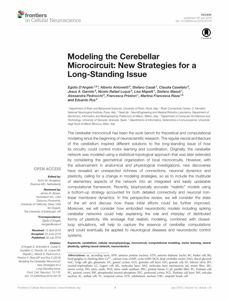
FIGURE 1 | The multi-level organization of the cerebellum. This schematic representation shows how the core cerebellar microcircuit is wired inside the wholebrain and how it can be further dissected into levels of increasing cellular and molecular complexity. The drawing at the center shows the cerebellar cortex subdividedinto three layers (GCL, granular cell layer; PCL, Purkinje cell layer; ML, Molecular layer), which contain different types of excitatory and inhibitory neurons (cf, climbingfiber; DCN, deep cerebellar nuclei; GoC, Golgi cell; GrC, granule cell; IO, inferior olive; APN, anterior pontine nucleus; RN, reticular nucleus; MLI, molecular layerinterneuron; mf, mossy fiber; pf, parallel fiber; PC, Purkinje cell; the signs indicate the excitatory or inhibitory nature of the cell or fiber). A cortical microzone isconnected to IO and DCN to form a cerebellar microcomplex. The expansion to the top, which shows a flattened representation of the cerebellar cortex, indicateshow a cerebellar microcomplex can extend to include several microzones located in separated cerebellar regions. A further expansion to the top shows the maincircuit loops formed by the cerebellum with the cerebral cortex (PFC, prefrontal cortex; MC, motor cortex; PC, parietal cortex; TC, temporal cortex) through the DCNand the anterior thalamic nuclei (ATN) on the efferent pathway and through the anterior pontine nuclei (APN) on the afferent pathway. The connection with basal
(Continued)
Frontiers in Cellular Neuroscience | www.frontiersin.org 6 July 2016 | Volume 10 | Article 176

D’Angelo et al. Cerebellum Modeling
FIGURE 1 | Continuedganglia (BG) and subthalamic nucleus (STN) is also indicated. The insets tothe bottom show, expand in cascade the wiring in the granular layer to showglomerular connectivity, glomerular neurotransmission and synaptictransduction mechanisms. The receptors involved (labeled in the inset) and theintracellular cascades include several identified molecular elements (glu,glutamate; PKC, protein kinase C; DAG, diacyl-glycerol; IP3,inositol-triphosphate; PIP, phosphatidyl-inositol-phosphate; NO, nitric oxidesynthase; NOS, nitric oxide synthase; NO, nitric oxide; Ca2+, calcium ions;GC, guanyl cyclase; cGMP, cyclic GMP; Modified from D’Angelo and Peres,2011; Mapelli et al., 2014).
GrCs and PCs, GoCs and MLIs. All these connections displayedposition-specific patterns of GrC synaptic inputs that did notstrictly match with anatomical boundaries and could connectdistant cortical modules, indicating that specific microcircuitconnectivity rules have also to be taken into account (Valera et al.,2016).
Longitudinal Organization: The Zebrin StripesThe so-called zones are long cerebellar stripes ranging from theanterior to posterior poles of the cerebellum and can be identifiedhistochemically and functionally (Andersson and Oscarsson,1978; Apps and Garwicz, 2005; Apps and Hawkes, 2009; Voogd,
2011). Each stripe is defined by the PC type depending on theexpression of Aldolase-C (Zebrin II) as well as of other enzymes(e.g., NOS and PKC isoforms) and ionic channels (e.g., TRIP).PCs expressing Zebrin II (Z+) show a slower spontaneous firing(40 Hz) compared to PCs not expressing Zebrin II (Z−; 90–100Hz; Zhou et al., 2014). Moreover, Z+ and Z− PCs differ as fortheir ability to generate plasticity at the pf-PC synapse (Wadicheand Jahr, 2005; Wang et al., 2011). It has recently been shownthat GoC somata and dendrites are restricted to the same PCZebrin II stripe (Sillitoe et al., 2008). The restriction of GoCsin specific stripes may influence network activity, since GoCsare connected through gap junctions (Vervaeke et al., 2010) andcould have a role in controlling GCL oscillations (Simões deSouza and De Schutter, 2011). The PCs output on specific DCNsis then retransmitted to the IO trough the nucleo-olivary pathwayand this pathway has been seen to influence the responses of theIO to their target PCs (Voogd, 2011).
Macroscale OrganizationMajor Anatomical SubdivisionsThe cerebellum, on each side of the midline, is divided into threeregions running along the rostral to caudal axis: the vermis, the
FIGURE 2 | Special properties of GCL connectivity. The figure shows schematically the most important properties of GCL connectivity that have emerged from acomplex set of physiological and structural experiments. (1) Divergence of mossy fibers onto different cell types. Formation of multiple glomeruli per mossy fiber.Multiple inputs onto the same GrC but different inputs on each granule cell dendrite. (2) Glomerular integration: a cerebellar glomerulus contains a mossy fiberterminal as well as GoC axonal terminals and dendrites. (3) Feed-forward inhibitory loops pass through the MF→GoC→GrC circuit. (4) Feed-back inhibitory loopspass through the MF→GC→GoC→GrC circuit. (5) GrCs activate GoCs both on basal dendrites and apical dendrites (4). (6) GoC→GoC reciprocal inhibition throughreciprocal synapses. (7) GoC→GoC communication through gap-junctions. (8) UBC pathway: MF→UBC→ GrC. (9) Lugaro Cell pathway: MF→LC→ GoC. (aa,Ascending axon; other labels and symbols as in Figure 1). Modified from Mapelli et al. (2014).
Frontiers in Cellular Neuroscience | www.frontiersin.org 7 July 2016 | Volume 10 | Article 176

D’Angelo et al. Cerebellum Modeling
paravermis and the hemisphere. Each of these regions is foldedinto lobules and each lobule is subdivided into folia. Remarkably,the afferent and efferent connections of the cerebellar cortex,as well as the corresponding DCNs, are strictly related to thisanatomical arrangement, as recently confirmed by viral tracingin experimental animals (Huang et al., 2013; Watson et al., 2014)and MRI data in humans (Balsters et al., 2010; Diedrichsenet al., 2011; Sokolov et al., 2012; Palesi et al., 2015). Projectionsfrom the cerebral cortex are conveyed to the anterior pontinenuclei and then relayed mostly to the posterior-lateral partsof the cerebellum through the medium cerebellar peduncle.Projections from the pons and spinal cord are relayed mostlyto the vermis and anterior cerebellum through the inferiorand superior cerebellar peduncle. These same cerebellar regionsproject to the spinal cord, brainstem and cerebral cortex throughdifferent subdivisions of the DCNs (e.g., see Eccles, 1967; Ito,1984).
Extracerebellar Connectivity and Recurrent LoopsBeyond anatomical details, what is relevant here is that thecerebellum is involved in major connections with brainstem,spinal cord and cerebral cortex as well as with basal ganglia (BG)and hippocampus. These connections generate multiple loops, inwhich the cerebellum is wired as a pivotal node (Caligiore et al.,2013, 2016; D’Angelo and Casali, 2013).
– The most renowned recurrent loop passes through the IO. Thesmall DCN GABAergic neurons inhibit the IO cells regulatingtheir coupling and oscillations (Najac and Raman, 2015).
– The DCNs are involved in the cerebellar circuitry with a oneway connection between the glycinergic DCN, projecting tothe GCL, inhibiting GABAergic GoCs and the glutamatergicDCN that excite the GRCs and GOCs (Ankri et al., 2015;Houck and Person, 2015; Gao et al., 2016). A similarconnectivity characterizes the medial vestibular nucleus in thevestibulo-cerebellum.
– There are several loops formed with the cerebellum by thebrainstem, passing through different cerebellar nuclei (exceptthe dentate) and involving the red nucleus and the reticularnucleus.
– The major loops connecting the cerebellum to the forebrain,start from the dentate nucleus and pass through the anteriorventrolateral thalamus mostly to reach the cerebral cortex,then return through the anterior pontine nuclei and themedialcerebellum peduncle.
– Afferent sensory fibers are relayed to the cerebellum throughnuclei located in the spinal cord (e.g., in the Deiter’s columns),brain stem (e.g., the cuneate nucleus), and superior andinferior colliculi.
Functionally, it is important to note that all these loopsare normally closed, in that fibers leave and then returnto the cerebellum through a different pathway. The mostremarkable loops are formed with the cerebral cortex andwith the peripheral motor system, so that the cerebellum isactually embedded in loops controlling movement planningand the sensory consequences of movement execution. Theseloops are the substrates of what are usually referred to as
the cerebellar ‘‘feed-forward’’ and ‘‘feed-back’’ controllers (seebelow).
CRITICAL DYNAMIC PROPERTIES OF THECEREBELLAR MICROCIRCUIT
The neurons and synapses of cerebellum are amongst themost intensely studied in the whole brain and biophysicallydetailed models of several cerebellar neurons and synapsesare available (Figures 3, 4; Table 2). These models arebased on realistic multicompartmental morphologies andincorporate a detailed description of membrane mechanismsincluding various ionic channels, synaptic receptors, ionicpumps, intracellular calcium dynamics and some cytoplasmicprocesses. These models, together with detailed connectivityrules, are fundamental to reconstruct realistic microcircuitdynamics.
Neuronal Intrinsic ExcitabilityNeurons of the cerebellum show complex nonlinear propertiesthat are likely to play a key role in controlling networkfunctions. Firstly, several neurons are autorhythmic, withfrequencies varying between a few up to around 100 Hz.The spikes have different shapes and properties and canconfigure various patterns in response to current injectionor synaptic activation. Secondly, for some neurons, evidencefor resonance in the theta-frequency band has emerged.Thirdly, neurons express non-linear firing properties suitablefor processing burst generation and burst-pause responses.Finally, several neurons have inward rectification controllingresting membrane potential and rebound excitation. Theseproperties emerge from the specific ionic channel complementand involve differentially the soma, dendrites and axons.For most of these neurons, there are advanced Hodgkin-Huxley style models, which have helped understanding howthe specific electroresponsive properties are generated andas noted above, have set landmarks for realistic modelingstrategy (for an extended review see D’Angelo et al.,2016).
The Purkinje cell is probably the most apparent exampleof this (for a recent review, see Bower, 2015). Early inthe 60’s, Rodolfo Llinas claimed that Purkinje cell dendriteswere electrically active (Llinás et al., 1968). Following a livelyscientific debate, the demonstration came from a double proofprovided by the advent of intracellular PC recordings (Llinás andSugimori, 1980) followed by the first model of active dendrites(Pellionisz and Szentágothai, 1973,1974morpho-electrical reconstruction of a guinea-pig PC (Rappet al., 1994), the first PC model based on realistic constructionprinciples was presented (De Schutter and Bower, 1994a,b)and then widely used for network simulations for over 20years (Santamaria et al., 2002; Steuber et al., 2007; Bower,2010; Maex and Steuber, 2013). Recently, based on the samemorphology, a new PC model has been developed using anupdated set of ionic channels and accounting for the axonalgeneration mechanism of simple spikes (Masoli et al., 2015).
Frontiers in Cellular Neuroscience | www.frontiersin.org 8 July 2016 | Volume 10 | Article 176
). Then, following precise

D’Angelo et al. Cerebellum Modeling
FIGURE 3 | Ionic channel types, distribution and gating properties in a PC model. The investigation of cerebellar neurons physiology and biophysics hasclassically followed the same procedures used for other central neurons. Most experiments have been carried out in mice and rats in acute brain slice preparationswith the aim of determining their intrinsic electroresponsiveness. Voltage-clamp analysis of membrane currents has mostly been dedicated to synaptic events, sincespace-clamp problems have in most cases hindered an accurate determination of current kinetics (except for GrCs, which are electrotonically compact). In someneurons, relevant information has been gained through single-unit and even patch-clamp recordings in vivo. Modeling reconstruction has, in most cases, exploitedthe knowledge of ionic currents identified kinetically and pharmacologically and the corresponding gating models have been derived from ion-channel libraries. Themaximum ionic channel conductances have been iteratively adjusted by fitting complex sets of experimental data derived from current-clamp recordings. (Top) Thediagram shows a 3D representation of PC morphology. This has been divided into eight distinct sections illustrated in the table on the right and endowed with ionicmechanisms according to immunohistochemical data. The ionic mechanisms include the sodium channel (Nav1.6), LVA and HVA calcium channels (Cav2.1, Cav3.1,
(Continued)
Frontiers in Cellular Neuroscience | www.frontiersin.org 9 July 2016 | Volume 10 | Article 176

D’Angelo et al. Cerebellum Modeling
FIGURE 3 | ContinuedCav3.2, Cav3.3), potassium channels (Kv3.4, Kv1.1, Kv4.3, Kv1.5, Kv3.3),potassium calcium dependent channels (KCa1.1, KCa3.1, KCa2.2), inwardrectified potassium channel (Kir2.x), cationic channel (HCN1) and a Cabuffering system composed by Calbindin and Parvalbumin (CDP5). The graphrepresents the state variables of the Nav1.6 channel during an actionpotential. C, I, O, B, indicate closed, inactivated, open and blocked states.Vertical dashed lines indicate the approximate action potential threshold (−50mV). (Bottom) The drawings show PC membrane potential at different times(arrows) during complex bursting (membrane potential is color-coded) in distaldendrites, soma and third node of Ranvier (3NR). At the end of the spikeburst, the PC model depolarizes starting from distal dendrites before thedepolarization invades the whole dendritic tree. A large Ca spike is the mostrelevant depolarizing event in terminal dendrites, while fast Na spikes are mostevident in AIS. In the 3RN, there is no firing pause during the dendritic Caspike. (Modified from Masoli et al., 2015).
A compressed version has also been presented (Marasco et al.,2013).
The granule cell has been first approximated to a McCulloc-Pitt neuron by a realistic model based on a limited set of ioniccurrents (Gabbiani et al., 1994). Then GrCs were shown togenerate non-linear input-output relationships and were fullymodeled based on a more complex set of ionic currents andvalidated against a rich repertoire of electroresponsive propertiesincluding near-threshold oscillations and resonance (D’Angeloet al., 2001). Interestingly, this last model still represents aunique example of full Hodgkin-Huxley style reconstructionbased on ionic currents recorded directly from the same neuron,therefore implying minimal assumptions even for the calibrationof maximum ionic conductances. The model has subsequentlybeen updated to incorporate detailed synaptic inputs (Nieuset al., 2006, 2014) and to include the dendrites and axondemonstrating the mechanisms of action potential initiationand spike back-propagation (Diwakar et al., 2009). The modelhas then been used for network simulations (Solinas et al.,2010).
The DCN cells have been modeled, although not for allthe neuronal subtypes. A model of the glutamatergic DCNneurons, based on realistic morphological reconstruction withactive channels (Steuber et al., 2011), was used to analyze synapticintegration and DCN rebound firing after inhibition. Moreadvanced versions have been used to study the dependence ofneuronal encoding on short-term synaptic plasticity (Luthmanet al., 2011) and the impact of Kv1 channels in spontaneous spikegeneration (Ovsepian et al., 2013). These models have been usedto predict the impact of the cerebellar output on extracerebellarcircuits (Kros et al., 2015).
The IO neurons were modeled to investigate the interaction ofdifferent ionic currents in mono compartmental models (Manoret al., 1997; Torben-Nielsen et al., 2012) showing modificationsto sub threshold oscillations (STO) when two neurons whereconnected through gap junctions. A bi-compartment model(Schweighofer et al., 1999) was able to reproduce the typical STOand the particular spikes generated by the interaction of sodiumand calcium currents in the soma/dendritic compartments.A three compartment model was then built to account forthe interaction between the dendrites, soma and the AIS in
generating the STO and spike output of the IO neurons(De Gruijl et al., 2012). Different versions of IO neuron modelshave been used to simulate the properties of the IO network(Manor et al., 1997; Torben-Nielsen et al., 2012).
InterneuronsThe Golgi cells were modeled reproducing the basis of theirintrinsic electroreponsiveness, showing complex non linearbehaviors such as pacemaking, resonance and phase resetand uncovering the role of gap junctions in oscillatorysynchronization (Solinas et al., 2007a,b; Dugué et al., 2009;Vervaeke et al., 2010). The model of UBCs reproducedthe nonlinear behaviors of this neuron including bursts,rebounds and the late-onset burst response. This latter propertycontributes to generate transmission delays in the circuit(Subramaniyam et al., 2014). Concerning MLIs (Llano andGerschenfeld, 1993; Alcami and Marty, 2013) no detailedconductance-based models are available yet and simplified IFmodels of these neurons were connected with the PCs toinvestigate the ML subcircuit (Santamaria et al., 2007; Lennonet al., 2014).
Synaptic Transmission and PlasticityA wealth of experimental investigations has addressed thefunctional properties of cerebellar synapses and will not beconsidered in detail here (for review see e.g., Mapelli et al.,2014; for the granular layer, Barmack and Yakhnitsa, 2008;for ML). Almost all cerebellar synapses present different formsof short-term plasticity (short-term facilitation: STF; short-term depression: STD) and long-term plasticity (LTP, LTD;De Zeeuw et al., 2011; Gao et al., 2012). In general, short-term plasticity is suitable to regulate transmission during bursts.STD prevails at the mf-GrC synapse, STF prevails at the pf-PCsynapse, and STD occurs at the PC-DCN synapses (Häusser andClark, 1997; Mitchell and Silver, 2000a,b; Nielsen et al., 2004;Sargent et al., 2005; Nieus et al., 2006; DiGregorio et al., 2007;Szapiro and Barbour, 2007; Kanichay and Silver, 2008; Duguidet al., 2012; Powell et al., 2015; Wilms and Häusser, 2015; vanWelie et al., 2016). While neurotransmitter dynamics involvingvesicular release as well as postsynaptic receptor desensitizationproved critical for controlling neurotransmission dynamics, anintriguing observation has been that spillover in the cerebellarglomerulus and in the ML might have a more important rolethan expected (e.g., see Mitchell and Silver, 2000a,b; Szapiro andBarbour, 2007).
Likewise, there are more than 15 forms of long-term synapticplasticity in the cerebellar network, appearing both as LTP orLTD with multiple and different mechanisms of induction andexpression (for review, see Ito, 2002; Gao et al., 2012; D’Angelo,2014). Plasticity has been reported not just in acute brain slicesbut also in vivo (Jörntell and Ekerot, 2002; Roggeri et al., 2008;Diwakar et al., 2011; Johansson et al., 2014; Ramakrishnanet al., 2016), revealing that patterned sensory inputs candetermine a complex set of changes encompassing multiplesynaptic relays. Importantly several of the cerebellar synapsesmay show forms of spike-timing-dependent plasticity (STDP),linking intracerebellar oscillations to the ability of generating
Frontiers in Cellular Neuroscience | www.frontiersin.org 10 July 2016 | Volume 10 | Article 176
D’Angelo et al. Cerebellum Modeling
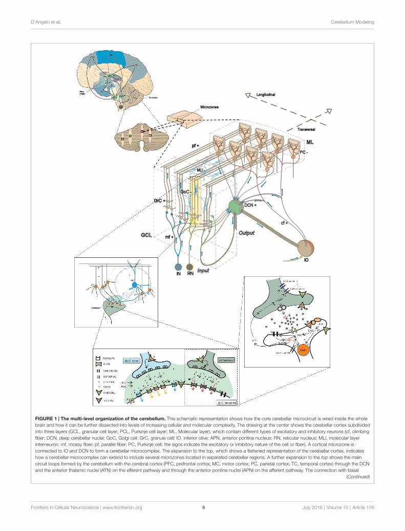
FIGURE 4 | Different electrophysiological properties of cerebellar neurons and their biophysical modeling. At present, accurate realistic models have beenconstructed for most cerebellar neurons, except for MLIs and Lugaro cells. In the different panels, the figure shows schematically the most important properties ofcerebellar neurons (left) and their biophysical reconstruction (right). For GCL and IO neurons, example tracings are taken from intracellular current-clamp recordings.For PC, MLI and DCN neurons, example tracings are reported along with raster plots and PSTH of activity. The traces are modified from: (GrC) Experiments: Nieuset al. (2014). Model: Solinas et al. (2010). (UBC) Experiments: Locatelli et al. (2013). Model: Subramaniyam et al. (2014). (GoC) Experiments: Bureau et al. (2000);Forti et al. (2006); D’Angelo et al. (2013b). Model: Solinas et al. (2010). (PC) Experiments: Ramakrishnan et al. (2016). Model: Masoli et al. (2015). (MLI) Experiments:Ramakrishnan et al. (2016). (DCN) Experiments: Rowland and Jaeger (2005); Uusisaari et al. (2007). Model: Luthman et al. (2011). (IO) Experiments: Lampl andYarom (1997); Lefler et al. (2014). Model: De Gruijl et al. (2012).
plasticity (D’Angelo et al., 2015; Garrido et al., 2016; Luqueet al., 2016). Understanding the importance of these forms ofplasticity may greatly benefit from integrated network modeling.At present, models incorporating dynamics presynaptic vesiclecycling (Tsodyks et al., 1998) have been developed for the mf-GrC, mf-GoC, GoC-GrC and GrC-GoC synapses (Nieus et al.,2006, 2014).
Microcircuit Dynamics: Timing andLearningThe cerebellar microcircuit has been shown to develop dynamicbehaviors, although their investigation is still limited. The EEG
cannot normally be recorded from the cerebellum, althoughsome MEG data have been reported showing increased powerin the theta-band during motor processing (Gross et al., 2001,2002). Recordings in the experimental animal in vivo havefocused on PC discharge patterns. PCs have been shown toactivate in spots forming transient clusters (Velarde et al.,2004), to exploit burst-pause coding (Herzfeld et al., 2015)and to encode the prediction of ongoing motor states (Balsterset al., 2010). A recent report has shown that locomotionwas associated with widespread increased activity in GrCsand interneurons, consistent with an increase in mossy fiberdrive, and that dendrites of different PC showed increasedco-activation, reflecting increased synchrony of climbing fiber
Frontiers in Cellular Neuroscience | www.frontiersin.org 11 July 2016 | Volume 10 | Article 176
D’Angelo et al. Cerebellum Modeling
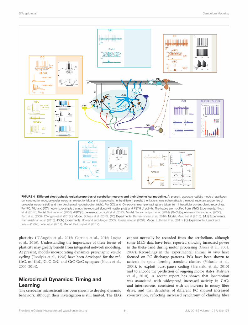
TABLE 2 | Neuronal electroresponsive properties.
Realistic Compartments Spontaneous Firing Inward Resonancemodel number frequency properties rectification frequency
GrC D’Angelo et al. (2001), Nieuset al. (2006) and Diwakar et al.(2009)
Single Multi No Fast spiking, variable presence ofadaptation
Fast ∼6 Hz
GoC Solinas et al. (2007a,b) andVervaeke et al. (2010)
Multi 6 Hz Fast spiking, adaptation, slow AHP,post-inhibitory rebound
Slow ∼6 Hz
UBC Subramaniyam et al. (2014) Multi No Fast spiking, adaptation, delayedbursting, slow AHP
Slow –
PC Masoli et al. (2015) Multi 40–80 Hz Fast spiking, adaptation, complexbursting, slow AHP
Slow –
SC/BC Multi 20 Hz Fast spiking, post-inhibitory rebound Slow –
DCN Luthman et al. (2011) Multi 10–30 Hz Fast spiking, post-inhibitory rebound Slow –
IO De Gruijl et al. (2012) Multi No Slow spiking, calcium spikes,subthreshold oscillations
Slow 3–10 Hz
The table reports details about the models available for each type of cerebellar neuron along with a short summary of their characterizing electroresponsive properties.
activity. At the same time, responses to external stimuli inall three cell types were strongly suppressed showing thatclimbing and mossy fiber representations can shift togetherwithin a fraction of a second between responses to movement-associated or external stimuli (Ozden et al., 2012). However,the spatio-temporal reconfiguration of signals expected to occurin the GCL remains to be fully addressed in vivo and it isnot fully clear how signals coming from different sources areredistributed through the different internal channels of thecerebellum.
Relevant to cerebellar circuit dynamics are its oscillating andresonant properties. On one hand, the GCL can be entrainedinto coherent oscillations by external inputs, possibly exploitingthe resonance properties of its neurons (Pellerin and Lamarre,1997; Hartmann and Bower, 1998; D’Angelo et al., 2001;Courtemanche et al., 2002, 2013; Solinas et al., 2007a; D’Angeloand De Zeeuw, 2009; Gandolfi et al., 2013; Garrido et al.,2016). On the other hand, spontaneous oscillations occur inthe IO, that might have the role of coordinating cerebellaractivity generating patterns that could be used for timing motor,sensory and cognitive tasks (Lampl and Yarom, 1997; Jacobsonet al., 2008; Llinás, 2014). In 2011, these two observationshave been merged with a large set of experimental data topropose a 3-level hypothesis, in which: (1) the spatio-temporalreconfiguration of incoming signals in the GCL is followed by;(2) their synthesis in theML andDCN; while (3) the DCN/PC/IOloop controls a modular synchronization of cerebellar sub-fieldsbased on circuit recurrent dynamics and selective frequency-dependent signal transmission (D’Angelo, 2011). The issue ofoscillations is particularly relevant not just for microcircuitcomputation but also for microcircuit learning through STDPrules (see also ‘‘Model Simplification and Implementation inClosed-loop Robotic Testing’’ Section below). Once again, timingto learning appear as complementary aspects of the samemechanisms rather than alternative mechanisms of function, asit was suggested by the original models (Marr, 1969; Eccles,1973).
Signal Transmission in Local MicrocircuitsDespite its extensive investigation, several fundamental issuesabout signal transmission in local microcircuits are stillincompletely understood.
There has been a long debate, which is not fully resolvedyet, on the modality of PC activation by GCL inputs. Whilepunctuate peripheral stimulation in vivo generates activity spotson the cerebellar surface (Bower and Woolston, 1983; Rokniet al., 2007), local pf stimulation elicits stripes of activity alongthe pf bundles (Ebner and Pasalar, 2008; Ebner, 2013). A recentwork using localized Glu uncaging in acute cerebellar slicessuggests that the organization of connections between the GCLand PCs may actually be even more complex than originallythought (Valera et al., 2016). From a functional viewpoint,following GCL stimulation, high-frequency modulated burstsare reliably transmitted vertically from the GCl to PCs, whileonly low-frequencies are transmitted transversally along thepfs (Mapelli et al., 2010). This observation suggested that afrequency-dependent selection of transmission lines, togetherwith a specific micro-connectivity, may allow the formation offunctional modules of active spots emerging vertically at theintersection of multiple pf bundles running along the folia withcfs fibers branching orthogonally to them (D’Angelo, 2011).At these intersection points, PCs may be able to decode thephase of IO oscillations and regulate pf gain (Ohtsuki et al.,2009).
A correlated issue concerns signal spread in the ML andPC inhibition. The pure feed-forward inhibition of PCs hasinspired initial functional models taking the move from theobservation that SCs and BCs inhibit PC activity with specificspatial organization and timing along and across the pf bundle(Eccles, 1967; Ito, 1984). This structural-functional relationshiphas recently been revisited highlighting the differential effectof inhibition on PC excitation mediated by aa and parallelfiber synapse (Mann-Metzer and Yarom, 1999, 2000, 2002;Santamaria et al., 2002, 2007; Mittmann et al., 2005; Santamariaand Bower, 2005; Mittmann and Häusser, 2007; Rieubland
Frontiers in Cellular Neuroscience | www.frontiersin.org 12 July 2016 | Volume 10 | Article 176
D’Angelo et al. Cerebellum Modeling
et al., 2014). Several dynamic phenomena have been reportedto intervene in determining how the ML actually operates.SCs are pacemaking and are electrically coupled thus formingan oscillating interneuron network (Mann-Metzer and Yarom,1999, 2000, 2002; Alcami and Marty, 2013). The analysis ofthese electrical and chemical SC microcircuits has recentlyrevealed that transitivity of chemical connectivity is directedvertically in the sagittal plane, and electrical synapses appearstrictly confined to the sagittal plane (Rieubland et al.,2014). The effect of ML inhibition is not confined toregulate PC activity, but it can also regulate generation ofLTD and LTP at pf-PC synapses (Mittmann et al., 2005;Mittmann and Häusser, 2007). On the side of ML coding,SC inhibition deeply affects the burst-pause pattern of PCoutput (Steuber et al., 2007; Herzfeld et al., 2015). Moreover,a form of interconnectivity between PCs has been proposedto generate traveling waves of activity in the ML (Watt et al.,2009).
Finally, the dynamics of the IO-PC-DCN subcircuit remainstill incompletely understood. The well-known contention aboutthe role of cfs, that has been proposed either to control cerebellarlearning or timing (Ito, 2000; Jacobson et al., 2008; Llinás,2009, 2011, 2014), is not yet over. What is becoming clearis that this subcircuit has all the ingredients to subserve bothfunctions. The IO operates as a pattern generator exploitinggap-junctions and local synaptic inhibition coming from theDCN in order to organize internal activity patterns that arethen conveyed to PCs (Jacobson et al., 2008; Chen et al.,2010; Libster et al., 2010; Lefler et al., 2013; Libster andYarom, 2013). This cf pattern, in turn, could be used toselect mossy fiber patterns in specific groups of PCs. It canbe argued that the coincidence of these cf and mf patternscould be instrumental to generate various forms of plasticityat PC and DCN synapses (see D’Angelo, 2014) raising againthe duality of the timing-plasticity issue in the cerebellarcircuit.
REALISTIC MODELS OF THECEREBELLAR MICROCIRCUIT
Realistic models of the cerebellar network have to take intoaccount a series of experimental observations, some used forconstruction, others for validation. In general, morphologicalmeasurements are themost relevant for constructing the networkstructure, electrophysiological data are needed to implementneurons and synaptic models, microcircuit-scale functionalmeasurements (imaging and electrophysiology) are fundamentalfor validation.
The Most Compelling Example: The Modelof the GCL SubcircuitConstructionThe wealth of anatomical data reported above (Figures 1, 2)and of cellular data (Figures 3, 4) provides the basis forreconstructing the cerebellar microcircuit (Figure 5). The stateof the art for the cerebellar GCL is currently set by the
2010 model (Solinas et al., 2010), which was intended togenerate a core computational element of the GCL microcircuit(about 10,000 neurons). This model was built by carefullyreproducing the cerebellar GCL network anatomical propertiesand then validating the response against a large set of availablephysiological data. A peculiarity of the cerebellar network isthat of being highly defined in terms of number of elements,convergence/divergence ratios and even in the number ofsynapses impinging on individual neurons. Moreover, thegeometric orientation of processes is not isotropic but rathergeometrically oriented, so that this network is quasi-crystalline innature. This has allowed the application of a ‘‘direct approach’’,in which:
– The appropriate number of neuronal elements has beenrandomly dislocated in a 3D space (density).
– The connectivity rules have been implemented to respect theconvergence/divergence ratios.
– The connections have been limited to specific network sub-spaces with well defined innervation territories. This, togetherwith the estimates of cell densities and of the number ofsynapses, allowed to implement an equivalent 3D connectivityeven if the axonal plexus was not represented explicitly.
– The neurons, though very accurate, had an equivalent ratherthan a realistic morphology, either monocompartmental(GrCs) or multicompartmental (GoCs).
Given that the data were sufficient to determine microcircuitconnectivity, it was not necessary to implement DMPrules (see below). Moreover, since the neurons were veryaccurate in reproducing the neuronal electrophysiologicalproperties (Table 2), there was no need to implement realisticmorphologies. Therefore, this network represents a ‘‘specialcase’’ of a more general network reconstruction procedure, asexplained below.
ValidationNetwork validation has been performed against a relevantexperimental dataset:
– First of all, it was considered whether the model neurons,which were calibrated beforehand on acute slice data(D’Angelo et al., 2001; Nieus et al., 2006; Solinas et al.,2007a,b), showed properties observed using patch-clamprecordings in vivo (Rancz et al., 2007; Arenz et al., 2008;Duguid et al., 2012, 2015; Chadderton et al., 2014). Thisactually happened, suggesting that a simulation of the roleplayed by specific ionic channels during network processingis actually possible.
– Secondly, it was assessed how the model network reacted torandom inputs distributed across the mfs. The model correctlygenerated coherent GrC oscillations in the theta band (Pellerinand Lamarre, 1997; Hartmann and Bower, 1998) provided thatan appropriate balance between the MF and PF input to GoCwas maintained.
– Thirdly, it was considered whether the high-pass filteringproperties of the GCL emerged. Again this happened, witha correct cut-off around 50 Hz. Importantly, this property
Frontiers in Cellular Neuroscience | www.frontiersin.org 13 July 2016 | Volume 10 | Article 176

D’Angelo et al. Cerebellum Modeling
FIGURE 5 | GCL modeling. The reconstruction of the microcircuit model of the GCL involves a precise representation of neurons, synapses and networkconnectivity. Interestingly, the model accounted for all the spatio-temporal dynamics of the GCL known at the moment. The model can therefore provide relevantinformation about the inner structure of neuronal activity during specific patterns of activity and reveal the relationship between individual synaptic and neuronalelements and the ensemble network response. (Top) synaptic currents in the dendrites of two different GrCs and receptor-specific components (AMPA, A; NMDA, N;GABA, G). (Bottom) Spatio-temporal dynamics of the network under noisy inputs reveal coherent low-frequency oscillations in the GC populations (left). Spatialresponse of GCs to a collimated mf bursts reveal a center-surround structure (right). (Modified from Solinas et al., 2010).
depended on NMDA receptors but much less so on GABA-Areceptors, as observed experimentally (Mapelli et al., 2010).
– Finally, the network response to collimated mf bursts wastested. According to previous observations using MEA
recordings, the typical center-surround organization of GCLresponses emerged (Mapelli and D’Angelo, 2007).
Therefore, the GCL network model successfully reproducedthe whole set of functional properties known at that time,
Frontiers in Cellular Neuroscience | www.frontiersin.org 14 July 2016 | Volume 10 | Article 176
D’Angelo et al. Cerebellum Modeling
suggesting that it could be used for predicting emerging networkbehaviors. Nonetheless, several issues remained unresolved,mostly concerning the GoC inhibitory network, and the range ofnetwork properties has in the meantime been extended by newfindings.
– The relative weight of the feed-forward and feed-backinhibitory loop generated by GoCs was a free parameter,whose impact was explored explicitly. A strong feed-back loopfavored coherent GCL network oscillations, as predicted bya previous modeling layout (Maex and De Schutter, 1998),while a strong fed-forward loop was needed to implement thetime-windowing effect (D’Angelo and De Zeeuw, 2009). It stillremains unclear how the two loops balanced. It is possible thatthe oscillating mode dominates over large network areas andthat selective mf inputs to GoCs project restricted regions intothe time-window mode, a hypothesis that needs to be tested(Duguid et al., 2015).
– The inhibitory input to GoCs was supposed to derive fromMLIs, but now this hypothesis is less creditable, since recentdata support the existence of inhibitory GoC-GoC connections(Hull and Regehr, 2012).
– The excitatory input to GoCs is more complex than previouslythought, GrCs form contacts onto GoC dendrites (Cesanaet al., 2013), and GoCs are connected through gap-junctions(Dugué et al., 2009; Vervaeke et al., 2010).
– The modality of GoC-GrC connectivity in the glomerulus isnot clear yet. While each GrC receives a single inhibitorycontact from GoCs, it is not clear if all the GrCs ineach individual glomerulus receive inhibition from the sameGoC axon or rather if they receive connections fromdifferent GoCs.
– Finally, the small-scale of the 2010 network precluded theanalysis of extended spatio-temporal effects, for example ofthose concerning interaction of different active clusters andthe spatial distribution of responses along the pf axis.
– The microscopic structure of GCL network activation can nowbe compared with the multispot two-photon microscopy data,which provide a new level of microcircuit validation (Gandolfiet al., 2014).
Eventually, improvements of specific structural propertiesand of membrane and intracellular mechanisms could alsobe considered. For example, the dendrites of GoCs arelikely to be active and this has to be accounted for infuture models (Rudolph et al., 2015). MulticompartmentGrC models perform better than monocompartment onesin controlling spike properties and delays (Diwakar et al.,2009) and so they should be developed and adopted forall neurons in the network model. Specific issues concernthe cerebellar glomerulus: at present, this structure has afictive morphology but it could be designed to incorporate aclosed diffusion space allowing the generation of glomerularhomeostatic mechanisms balancing excitatory and inhibitoryneurotransmitter release during repetitive synaptic activity(Mapelli et al., 2014; Nieus et al., 2014). Another specificissue concerns the mechanisms of postsynaptic calciumregulation, signal transduction and plasticity in GrCs
and GoCs dendrites, for reason that will become evidentbelow.
The model of the GCL is fundamental since it generatesthe input to the subsequent stages of the cerebellar cortex.Although, in a local perspective, a microcircuit made of GrCsand GoCs is enough to generate meaningful outputs forML and PCs, the incorporation of the GCL in an extendedmacrocircuit requires a set of extensions. These concernadditional control subcircuits that include the UBC subcircuit,that predicted to play an important role in generating delaylines inside the GCL (Kennedy et al., 2014), and the LCsubcircuit, that provides a control loop regulating GoC activity(Dieudonné and Dumoulin, 2000; Barmack and Yakhnitsa,2008).
Perspectives for Modeling OtherCerebellar Network Subcircuits andThe Whole Cerebellar NetworkThe GCL network provides the most advanced computationalmodel of the cerebellum at the moment. The impact of GCLmodeling becomes even more relevant once the GCL outputis used to activate the ML. At this level, mapping of GCLactivity onto PCs and MLIs occurs serially, as there is noevidence of direct feed-back from the ML to the GCL (thoughit occurs through DCN and extracerebellar loops, see alsobelow). A reference model for the ML has been proposedover 10 years ago to explain PC activation (Santamaria et al.,2007), but the main connectivity aspects of BCs and SCswith PCs need now to updated with recent data that revealedpotentially important physiological and molecular details. Forexample, ephaptic synapses need to be added on the PCaxonal initial segment (Blot and Barbour, 2014) and short-term plasticity needs to be implemented at all the ML synapses(Liu et al., 2008; Lennon et al., 2015). Likewise, while modelsfor the fundamental properties of IO and DCN neuronsare available, they also need to be updated. For example,IO neuron axonal burst generation (Mathy et al., 2009) stillneeds to be resolved. All these properties are likely to havea relevant impact on cerebellar computation dynamics. Thesame connectivity inside the IO-DCN-PC subcircuit has neverbeen modeled in full although relevant progress has been done(De Schutter and Steuber, 2009; Steuber and Jaeger, 2013).In principle, the IO-DCN-PC subcircuit should be modeledindependently and tested and then wired with the cerebellarcortical model.
A first series of effects is expected from the integration of thedifferent subcircuits (granular, molecular and IO-DCN-PC) intoa whole-cerebellum network model. This assembly, by includinga set of recurrent loops, breaks down the serial processing schemeadopted when modeling the cerebellar subcircuits separately. Inthis way, the intrinsic dynamics of the IO-DCN-PC subsystemwill be integrated with the activity patterns carried by themfs andprocessed in the GCL andML. Eventually, this whole-cerebellumnetwork model will help facing the basic question of how PCand DCN firing is regulated by the cerebellar cortical circuitactivity.
Frontiers in Cellular Neuroscience | www.frontiersin.org 15 July 2016 | Volume 10 | Article 176
D’Angelo et al. Cerebellum Modeling
A second series of effects is expected from the integrationof the whole-cerebellum network model into extracerebellarloops. This step is essential to analyze how the cerebellarnetwork operates. For example, properties like resonanceor STDP are relevant only in the context of rhythmicpatterns of activity in closed-loop circuits formed by thecerebellum with the DCN (Kistler and De Zeeuw, 2003),the cerebral cortex, brain stem and spinal-cord. The needingof connecting the cerebellum model with external brainstructures brings about a series of additional modelingquestions.
Relevant Properties of the mf InputSeveral anatomical and functional observations become relevantwhen considering the internal and external connectivity ofthe cerebellum. The mfs connecting to a certain GrC areprobably not all of the same nature but rather they comefrom different sources. For example, there are GrCs receivingcombinations of cortical and spinal afferences and someshow a multimodal response to sensory stimulation (Huanget al., 2013; Ishikawa et al., 2015). Thus, each GrC maywork as a coincidence detector of different signal sources.However, in some areas GrCs may operate as thresholddetectors for the intensity of signal sources deriving froma specific modality or somatic subregions (Bengtsson andJörntell, 2009). Implementing these connections requires toknow how mfs from different sources combine in individualGrC and requires therefore a specific redistribution ofglomeruli inside the GCL (Billings et al., 2014). Ideally,the combination of different fibers in GrCs allows directcoincidence detection of signals from different areas carrying‘‘congruent’’ information that needs to be associated beforefurther processing in the cerebellum. Some mfs also comefrom the DCN imposing further constraints on the internaldistribution of connections. The GrCs receiving the internalfeed-back from DCN may be able to associate the coincidencebetween DCN and extracerebellar inputs. These observationssuggest that understanding the cerebellar GCL should considerthe distribution of glomeruli deriving from mfs originating fromvarious sources.
Relevant Properties of Zonal and RegionalOrganizationPerhaps the aspect most relevant to cerebellar modeling on themesoscale is the organization of subcircuits, in which the cfs andthe mfs contacting a certain group of PCs and DCN neuronsare connected to the same area of origin to form fully connectedcerebellar modules. Furthermore, the cerebellar modules can beorganized according to the longitudinal stripes, in which someneuronal and synaptic mechanisms are differentiated dependingon the type (Z+ or Z−) of the stripe (Wadiche and Jahr, 2005;Wang et al., 2011; Zhou et al., 2014). In turn, a model on themacroscale has to be composed of multiple modules, each oneconnected to specific extracerebellar regions. These aspects willhave to be considered once the cerebellum model will be wiredwith extracerebellar areas (see below).
NEW MODELING STRATEGIES FOR NEWCHALLENGING QUESTIONS
Realistic cerebellar modeling has to face two mainchallenges. First, it has to able to incorporate realisticmorphologies and to improve details on the molecularand cellular microscale. Secondly, it has to be expandedtoward the mesoscale and macroscale. In order to do so,a general and flexible implementation strategy is needed,and in this process cerebellar modeling has once againbeen acting to promoting the development of generalmodel strategies (Bhalla et al., 1992; Bower and Beeman,2007).
The cerebellar network is probably the most orderedstructure of the brain, and this has allowed a precise modelingreconstruction of its internal connectivity based on extendeddatasets derived from mice and rats (Maex and De Schutter,1998; Medina and Mauk, 2000; Medina et al., 2000; Solinas et al.,2010). A further advancement would benefit of an approachbased on structured multiscale simulators (Hines and Carnevale,2001; Bower and Beeman, 2003; Gleeson et al., 2007; Ramaswamyet al., 2015). This would allow to extend cerebellum modelingperformed in mice and rats to other species (e.g., humans) and toparacerebellar structures, including the dorsal cochlear nucleusin all vertebrates and the paracerebellar organs in electric fishes(Oertel and Young, 2004; Requarth and Sawtell, 2011; Kennedyet al., 2014). This approach would facilitate the incorporationof new cell types (like the UBCs or the LCs), provided thattheir detailed single neuron models are available. This approachcan host morphological and functional variants of the differentneurons, thusmoving from canonical neuronalmodels to neuronmodel families expressing all the richness of electrophysiologicalproperties that characterize biological networks.
The cerebellum is fundamentally a plastic structure and itsfunction is hard to understand if plasticity is not considered.The cerebellum drives adaptation through plasticity. Moreover,the cerebellum attains the adult network organization througha blend of plastic processes guided by the interaction of geneticprograms with epigenetic cues. Thus the interaction of thecerebellar network with the rest of the brain and with ongoingbehavior is key not just to determine how the cerebellum operatesbut also how the cerebellum forms its internal structure andconnections. Plasticity during development and in adulthood areprobably the most fascinating aspects of the cerebellum and posechallenging questions for modeling.
In adulthood, the cerebellar synapses express various formsof plasticity with learning rules showing different patternsensitivity, induction and expression mechanisms (D’Angelo,2014). The corresponding learning rules are embedded intothese mechanisms and although it would be desirable thatthese are eventually represented using dynamics synaptic models(Migliore et al., 1995, 1997, 2015; Tsodyks et al., 1998; Miglioreand Lansky, 1999; Rothman and Silver, 2014) at present no suchmodels are available. Nonetheless, theoretical rules based onHebbian coincidence detectors and STDP have been developed insome cases (Garrido et al., 2016; see below). Eventually a realisticmodel incorporating learning rules resolved at the molecular
Frontiers in Cellular Neuroscience | www.frontiersin.org 16 July 2016 | Volume 10 | Article 176

D’Angelo et al. Cerebellum Modeling
level should be able to give insight on the adaptable propertiesof the network.
As far as ontogenetic network self-organization isconcerned, a reference model has been developed for thecerebral cortex accounting for synapse formation throughan interaction/pruning process guided by Hebbian rules(Zubler et al., 2013). The dendrite extension/pruningprocess would by itself solve problems like the crystallineconvergence/divergence ratio of the mf-GrC relay and ofthe cf-PC connectivity. In a way, it can be envisaged thatthe selection rules of DMP algorithm will eventually beimplemented using growing plastic rules. Moreover, onceconnection pathways are prescribed, the self-organizingsystem should be able to generate the appropriate distributionof the mf-glomeruli into the cerebellar GCL and to primethe ontogenetic development of the whole network,aligning transmission channels and optimizing circuitperformance by setting the appropriate associations of fibertypes.
Thus the problem is not just to determine and model theplasticity rules, but also to apply them to the network, as thiswould require the cerebellum model to be inserted in a whole-brain system interacting with the environment.
MODEL SIMPLIFICATION ANDIMPLEMENTATION IN CLOSED-LOOPROBOTIC TESTING
The ultimate challenge appears then to run the whole-cerebellumnetwork model in a simulated brain operating in closed-loop.While a radical approach is out of reach at the moment (it wouldrequire, in addition to fully developed cerebellum models, alsorealistic models of large brain sections outside the cerebellum),a first attempt has been done by reducing the complexity ofcerebellar models and using simplified versions to run closed-loop robotic simulations (Casellato et al., 2012, 2014, 2015;Garrido et al., 2013; Luque et al., 2014, 2016).
Complexity ReductionThe way complexity reduction is achieved is critical, since ithas to be performed in a way that preserves the fundamentalbiological properties relevant to the process under investigation.Two recent approaches have been proposed. Realistic PCmodels currently involve about 1500 electrical compartmentsand up to 15 active ionic conductances (De Schutter andBower, 1994a,b). This complexity has been remarkably reducedby applying Strahler’s analysis to reduce up to 200-fold therun time but yet maintaining an appropriate response tosynaptic inputs (Marasco et al., 2012, 2013). Likewise, thegranular layer network has been simplified using analyticaltools by increasing the simulation speed at least 270 times butyet reproducing salient features of neural network dynamicssuch as local microcircuit synchronization, traveling waves,center-surround, and time-windowing (Cattani et al., 2016).In all these cases, a well defined relationship is maintainedbetween the simplified models and their more complex realistic
counterparts. These attempts open the way to a guidedsimplification procedure, at least for some cerebellar neuronsand subnetworks. When the whole cerebellar network has tobe represented in a macro-scale model, simplifications thatare computationally efficient may be preferable in a firstinstance. Clearly, in this case a top-down approach is adoptedand the relationship of the simplified model with the realsystem is a matter of speculation. This approach has beenused to generate cerebellar spiking networks (SNN) allowingto reproduce a single basic cerebellar module running withhigh efficiency in a robotic controller yet maintaining somefundamental features of neurons and connections (Casellatoet al., 2012, 2014, 2015; Garrido et al., 2013; Luque et al., 2014,2016). For example, in these models, neurons were representedby integrate-and-fire single-compartment elements, the localinhibitory interneuron networks were not included and theGCL was not fully implemented resorting to the concept of anon-recurrent states in a liquid-state machine (Yamazaki andTanaka, 2007). Nonetheless, the model incorporated multipleforms of bidirectional plasticity at the PC and DCN synapses.This compromise had to be accepted in order to generate aspiking cerebellum model running in real-time inside a closed-loop robotic control system and to perform system level analysisof complex tasks like active manipulation.
Spiking Neural Networks of the CerebellumDespite the simplicity of the cerebellar SNN (Figure 6),the robots that incorporated it revealed remarkable emergingproperties (Casellato et al., 2012, 2014, 2015). The SNNrobots correctly performed multiple associative learning andcorrection tasks, which ranged from eye-blink conditioningto vestibulo-ocular reflex (VOR) and force-field correction.Importantly, the robots were not designed for any specificone of these tasks but could cope equally well with all ofthem demonstrating generalized learning and computationalcapabilities. The robots could also generalize their previousstored patterns to analogous cases with a learning rateapproaching that observed in real life. This system could easilyfit human EBCC data predicting dual-rate learning in thenetwork. Again, the outcome of the closed-loop simulation havebeen validated against real experiments carried out in humans(Monaco et al., 2014; D’Angelo et al., 2015) and the challengeis now to see whether it is predictive with respect to humanpathologies.
An important aspect of thesemodels is to incorporate learningrules that allow to test the impact of learning on cerebellarcomputation. While a precise correspondence with long-termsynaptic plasticity is not at the level of molecular mechanisms(we are dealing with simplifiedmodels by the way), these learningrules can effectively capture the learning dynamics of the system.Importantly, faster learning rates at PC than DCN synapsesallow fast acquisition and subsequent transfer of memory in aconsolidated state (Luque et al., 2014) and STDP rules allowlearning to accurately match the network temporal dynamics(Luque et al., 2016). These models allowed to evaluate theimpact of known forms of bidirectional LTP/LTD at pf-PC,
Frontiers in Cellular Neuroscience | www.frontiersin.org 17 July 2016 | Volume 10 | Article 176

D’Angelo et al. Cerebellum Modeling
FIGURE 6 | Simulating an associative learning task using a cerebellar spiking neural network (SNN). The cerebellum circuit was simplified and embeddedinto a robotic control system, in which it provided the substrate to integrate spatio-temporal information in different associative learning tasks. Real robot paradigms(top left panel): eye blink classical conditioning (EBCC)-like, vestibulo-ocular reflex (VOR) and upper limb reaching perturbed by force fields. The EBCC-like Pavloviantask is reproduced into the robotic platform as a collision-avoidance task. The conditioned stimulus (CS) onset is based on the distance between the moving robotend-effector and the fixed obstacle placed along the trajectory, detected by the optical tracker. The unconditioned stimulus (US) is the collision event. The DCNstrigger the conditioned response (anticipated stop). The VOR is reproduced into the robotic platform by using the second joint of the robotic arm as the head(imposed rotation) and the third joint (determining the orientation of the second link) as the eye. The misalignment between the gaze direction and the environmentaltarget to be looked at is computed through geometric equations from the optical tracker recording. The DCNs modulate the eye compensatory motion. Theperturbed reaching is reproduced into the robotic platform by applying a viscous force field on the moving robotic arm by means of the other robotic device attachedat its end-effector. The DCNs modulate the anticipatory corrective torque. (Modified from Casellato et al., 2014). EBCC-like control system embedding spikingcerebellar network (top right panel). US is fed into the cf pathway; CS into the mf pathway. CS and US co-terminate (as in the “delay” EBCC). The SNN learns toproduce conditioned responses (CRs), i.e., a stop of the robotic arm (collision avoidance) anticipating the US onset. The figure highlights the major forms of plasticityembedded in the cerebellar network and driving the learning, namely synaptic long-term potentiation (LTP) and synaptic long-term depression (LTD), both at cortical
(Continued)
Frontiers in Cellular Neuroscience | www.frontiersin.org 18 July 2016 | Volume 10 | Article 176

D’Angelo et al. Cerebellum Modeling
FIGURE 6 | Continuedand nuclear levels (distributed plasticity). The protocol is made up ofacquisition and extinction phases; in the acquisition trials CS-US pairs arepresented at a constant Inter-Stimuli Interval (ISI); in the extinction trials CSalone is presented. Each trial lasts 600 ms. The number of cell in the circuit isindicated. All labels as in previous figures. (Modified from D’Angelo et al.,2015). Network activity and output behavior during EBCC training (bottompanel). After learning, the response of PCs to inputs decreases, and thisincreases the discharge in DCN neurons (raster plot and integral of neuronalactivity, left). Since the DCN spike pattern changes occur before the US arrival,the DCN discharge accurately predicts the US and therefore facilitates therelease of an anticipatory behavioral response. Number of CRs (%) along trials(80 acquisition trials and 20 extinction trials for two sessions in a row; CR% iscomputed as percentage number of CR occurrence within blocks of 10 trialseach). The black curve (right plot) represents the behavior generated by thecerebellar SNN equipped with only one plasticity site at the cortical layer(median on 15 tests with interquartile intervals). Despite uncertainty andvariability introduced by the direct interaction with a real environment, the SNNprogressively learns to generate CRs anticipating the US, to rapidly extinguishthem and to consolidate the learnt association to be exploited in the re-testsession. (Modified from Casellato et al., 2015; D’Angelo et al., 2015; Antoniettiet al., 2016).
PC-DCN and mf-DCN synapses and to predict a critical rolefor plasticity at IO-DCN synapses. The implementation of GCLplasticity poses a formidable problem as it is hard to determineits supervision process. A recent proposal suggests that the issuecould be solved by exploiting multi-step learning with an initialpattern storage in the inhibitory interneuron network formed byGolgi cells (Garrido et al., 2016).
Advanced Robotic Simulations ofManipulation TasksWhen manipulating a tool, the cerebellar network acquires adynamic and kinematic model of the tool. In this way, themanipulated tool becomes de facto as an extension of the armallowing to perform accurate movements of the arm-objectsystem as a whole. This unique capability is to a large extentbased on the cerebellum sensory-motor integration properties. Inorder to establish a functional link between specific properties ofneurons, network organization, plasticity rules and behavior, thecerebellar model needs to be integrated with a body (a simulatedor real robotic sensory-motor system). Sensory signals need tobe translated into biologically plausible codes to be deliveredto the cerebellar network, and also cerebellar outputs need tobe translated into representations suitable to be transferred toactuators (Luque et al., 2012). The experimental set-up is definedso as to monitor how accurately the system performs pre-definedmovements when manipulating objects that significantly affectthe arm/object kinematics and dynamics (Figure 7).
At this level, the cerebellar network is assumed to integratesensory-motor signals by delivering corrective terms duringmovement execution (here a top-down approach is applied). Inthe framework of a biologically relevant task such as accurateobject manipulation, different issues need to be addressedand defined by adopting specific working hypothesis andsimplifications. For example: (i) PCs and DCN can be arrangedin microcomplexes dealing with different degrees of freedom;(ii) error-related signal coming from the IO are delivered to
PCs and drive learning at pf-PC synapses; (iii) neurons andconnection can be simplified still maintaining the fundamentalcerebellar network structure and functionality. There aredifferent modeling approaches that have been simulated andtested (Luque et al., 2011a,b):
(1) Integrating the cerebellum in a feed-forward schemedelivering corrective terms to the spinal cord. In thiscase the cerebellum receives sensory inputs and producesmotor corrective terms (the cerebellum implements an‘‘inverse model’’). Thus in this case the input and outputrepresentation spaces are different and the sensori-motortransformation needs to be performed also in the cerebellarnetwork.
(2) Integrating the cerebellum in a feed-back (recurrent) schemedelivering corrective terms to the cerebellar cortex. In this casethe cerebellum receives sensory-motor inputs and producessensory corrective terms (the cerebellum implements a‘‘forward model’’; Kawato et al., 1988; Miyamoto et al., 1988;Gomi and Kawato, 1993; Yamazaki et al., 2015; Hausknechtet al., 2016).
Eventually, closed-loop robotic simulations allow toinvestigate the original issue of how the cerebellar microcircuitcontrols behavior in a novel manner. Here neurons and SNN arerunning in the robot. The challenge is clearly now to substitutethe current simplified models of neurons and microcircuitswith more realistic ones, so that from their activity during aspecific behavioral task, the scientists should be able to infer theunderlying coding strategies at the microscopic level.
CURRENT PERSPECTIVES FORREALISTIC CEREBELLAR MODELING
On one hand, realistic cerebellar modeling is now advancedenough to generate predictions that may guide the subsequentsearch for critical physiological phenomena amongst the manythat could be otherwise investigated. On the other hand, severalnew challenges await to be faced in terms of model constructionand validation in order to explore physiological phenomena thathave emerged from experiments. Realistic modeling is thereforebecoming more and more an interactive tool for cerebellarresearch.
Predictions of Realistic CerebellarModeling and their Experimental TestingCerebellar modeling is providing new opportunities forpredicting biological phenomena that can be subsequentlysearched for experimentally. This procedure is relevant forseveral reasons. First, as discussed above, the computationalmodels implicitly generate hypotheses providing the wayfor their subsequent validation or rejection. Secondly, thecomputational models can help focusing researcher’s interesttoward specific questions. There are several examples that applyto different levels of cerebellar physiology.
In 2001, an advanced GrC model, based on the ionicconductance complement of the same neuron, predicted that
Frontiers in Cellular Neuroscience | www.frontiersin.org 19 July 2016 | Volume 10 | Article 176
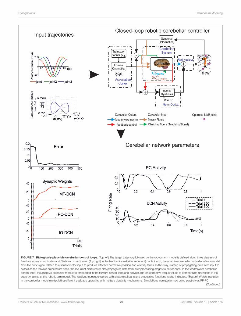
D’Angelo et al. Cerebellum Modeling
FIGURE 7 | Biologically plausible cerebellar control loops. (Top left) The target trajectory followed by the robotic arm model is defined along three degrees offreedom in joint coordinates and Cartesian coordinates. (Top right) In the feedback cerebellar (recurrent) control loop, the adaptive cerebellar controller infers a modelfrom the error signal related to a sensorimotor input to produce effective corrective position and velocity terms. In this way, instead of propagating data from input tooutput as the forward architecture does, the recurrent architecture also propagates data from later processing stages to earlier ones. In the feedforward cerebellarcontrol loop, the adaptive cerebellar module is embedded in the forward control loop and delivers add-on corrective torque values to compensate deviations in thebase dynamics of the robotic arm model. The idealized correspondence with anatomical parts and processing functions is also indicated. (Bottom) Weight evolutionin the cerebellar model manipulating different payloads operating with multiple plasticity mechanisms. Simulations were performed using plasticity at PF-PC,
(Continued)
Frontiers in Cellular Neuroscience | www.frontiersin.org 20 July 2016 | Volume 10 | Article 176

D’Angelo et al. Cerebellum Modeling
FIGURE 7 | ContinuedMF-DCN, and PC-DCN synapses and a custom-configured IO-DCNconnection for manipulating 2 kg external payloads during 500 trials. The initialcerebellar system gain was properly set to operate with no payload. Evolutionof the average error (MAE, black curve on the left) of the three robot jointsduring the learning process for 2 kg payload. The red curves on the leftindicate the evolution of synaptic weight at the different synapses. Note thatweights change rapidly at the beginning but then the cerebellar system worksalmost in open loop and no remarkable corrective action are applied by thecerebellar adapting system. PC and DCN neuron activity during a single trialshow oscillations dictating the precise timing of force delivery to the joints indifferent trials. (Modified from Luque et al., 2011a, 2014).
a slow K current was needed to explain certain aspectsof GrC firing and intrinsic GrC theta-band resonance. Thiscurrent has been then looked for experimentally and itssubsequent identification allowed to successfully complete themodel and explain bursting and resonance in mechanistic terms(D’Angelo et al., 2001). In 2006, a mossy fiber-granule cellneurotransmission model, based on specific quantal release andreceptor properties (Nieus et al., 2006), predicted that plasticityof intrinsic excitability could control rate coding while plasticityof release probability could control spike timing, as indeedverified experimentally. In 2007, a Golgi cell model actuallypredicted that Golgi cells were resonant in the theta-band aproperty that was then demonstrated experimentally (Solinaset al., 2007a,b). In 2007, a PC model predicted the codingproperties of PCs in relation to LTD (Steuber et al., 2007). In2009–2010 two models of the Golgi cell network predicted theimpact of gap-junctions in regulating local GrC discharge andGolgi cell synchronization (Dugué et al., 2009; Vervaeke et al.,2010). In 2013, a theoretical article predicted that bidirectionalplasticity had to exist at the mossy fiber—Golgi cell synapse(Garrido et al., 2013). This plasticity has subsequently beendemonstrated (Locatelli et al., 2015). In 2014, a model includingboth excitatory and inhibitory neurotransmission predicted thatphasic inhibitory mechanisms can dynamically regulate outputspike patterns, as well as calcium influx and NMDA currents,at the mossy fiber-granule cell relay of cerebellum (Nieus et al.,2014). Again this prediction was accurately matched by theexperiments. In 2015, a computational model predicted that thenumber of GrC dendrites that maximizes information transferis actually coincident with that measured anatomically (Billingset al., 2014).
Yet other predictions are awaiting for experimentalverification. In 2014, a closed-loop simulation predictedthat cerebellar learning would accelerate toward biologicallevels if a form of mid-term plasticity would exist between theIO and DCN neurons (Luque et al., 2014). In 2016, anotherwork predicted that STDP has the intrinsic capacity of bindinglearning to temporal network dynamics (Luque et al., 2016).Finally, very recently a mechanism of STDP learning involvingthe inhibitory interneuron network has been proposed (Garridoet al., 2016), that could be applicable to the GCL and explainhow learning takes place in this cerebellar subnetwork. Thus, anew perspective for the near future is to extend the feed-backbetween computational models and experiments generating defacto a new powerful tool for cerebellar network investigation.
New Challenges for Cerebellar Physiologyand their Realistic ModelingAmongst the new challenges that may benefit from enhancedand extended realistic models of the cerebellum, some have beenhighlighted in the present review and are summarized here.
There is a wealth of molecular and cellular phenomena, whosebiological significance has been inferred experimentally, thatcould be incorporated into a realistic cerebellar model in orderto investigate their implications for function. These include: therole of specific ionic channel properties in regulating neuronalexcitation (amongst known examples see Jaeger et al., 1997;Bower and Beeman, 1998; Kubota and Bower, 2001; Ovsepianet al., 2013); the role of synaptic receptor properties in neuronalexcitation and plasticity, like the voltage-dependence of NMDAreceptor subtypes (Schwartz et al., 2012); the role of diffusiblemessengers like nitric oxide in coordinating long-term synapticplasticity (Garthwaite, 2016); the role of intracellular biochemicalcascades in the induction and expression of long-term synapticplasticity (Tsukada et al., 1995; Schweighofer and Ferriol, 2000;Billings et al., 2014).
There are several properties of local microcircuits thatare being discovered and that could be further understoodby realistic cerebellar modeling. We have already mentionedthe critical issue on how the cerebellum processes incominginformation involving numerous molecular and cellularmechanisms that are only partially known. An issue that needsto be revisited, as it appears critical to understand the wholecerebellar functioning, is how the PC are activated by GrCthrough their aa (Gundappa-Sulur et al., 1999; Huang et al.,2006). Moreover, recent discoveries have opened new issues:ephaptic synapses have recently been revealed between basketcells (BCs) and PCs (Blot and Barbour, 2014), the connectivityof MLI involves complex spatial rules (Bower, 2010; Rieublandet al., 2014), the inhibitory network in the cerebellar granularlayer involves gap junctions and reciprocal inhibitory synapses(Dugué et al., 2009; Szoboszlay et al., 2016; van Welie et al.,2016), the inferior olivary neurons are connected through gapjunctions (Rothman et al., 2009; Rancz and Häusser, 2010; Lefleret al., 2014).
There are aspects of intracerebellar organization andconnectivity that remain to be incorporated into large-scalerealistic models, including the granular layer-molecular layerprojections (Valera et al., 2016), the PC-DCN convergence(Person and Raman, 2012b), the DCN-granular layer projections(Houck and Person, 2015), the PC-DCN-IO loops (Libster andYarom, 2013). Beyond this, these are needed for guided cerebellarmodel simplification and incorporation into large-scale networksrunning into robotic controllers and simulated environments(Garrido et al., 2013; Casellato et al., 2015; Yamazaki et al.,2015).
On the pathophysiological side (Chen et al., 2010; Libsteret al., 2010; Ovsepian et al., 2013; Kros et al., 2015), thereis a wealth of hypothesis that have or would benefit ofrealistic modeling. Ataxia has long been attributed to cerebellardysfunction. Recently, several ionic channel and neuronalalterations have been linked to ataxia (Libster et al., 2010) andto the disruption of dynamics in the olivo-cerebellar circuit
Frontiers in Cellular Neuroscience | www.frontiersin.org 21 July 2016 | Volume 10 | Article 176
D’Angelo et al. Cerebellum Modeling
(Chen et al., 2010). There are specific properties of the cerebellaroutput that are critical for controlling extracerebellar networksand their pathological states, like in cebro-cortical spike-and-wave discharge (e.g., see Ovsepian et al., 2013; Kros et al., 2015).This kind of observations may provide critical test-benches forrealistic model validation and prediction.
Finally, in perspective, the connectivity of the cerebellarnetwork in long-range loops appears to be critical to understandmicrocircuit functions. Following the fundamental recognitionof its involvement in sensory-motor coordination and learning,the cerebellum is now also believed to take part in theprocessing of cognition and emotion (Schmahmann, 2004)by exploiting the connectivity of the cerebellar modules withspecific brain structures through different cerebro-cerebellarloops. It has been proposed that a similar circuit structurein all cerebellar areas may carry out various operationsusing a common computational scheme (D’Angelo and Casali,2013). Since there is an intimate interplay between timingand learning at the cellular level that is reminiscent of the‘‘timing and learning machine’’ capabilities long attributedto the cerebellum, it is conceivable that realistic modelsdeveloped for sensori-motor control might also apply tocognitive-emotional control once integrated into the appropriateloops.
A MANIFESTO FOR COLLABORATIVECEREBELLAR Modeling
This review has summarized some relevant aspects characterizingthe cerebellar circuit showing how these have beenconceptualized and modeled. Still, there are several issuesthat deserve attention, ranging from molecular to neuronal,microcircuit, macrocircuit and integrative aspects, and evenmore it is clear that all these aspects are tightly bound. Thereis no solution through a single experiment or model, so thatunderstanding the structure-function-dynamics relationship ofthe cerebellum requires a continuous bottom-up top-downdialog (Akemann et al., 2009).
Realistic modeling is now opening new perspectives. Themain challenge is to join precise network wiring withaccurate representations of neuronal and synaptic propertiesin order to be able to simulate local network dynamics.The introduction of synaptic and non-synaptic plasticityin its various forms and locations could then allow tounderstand how input patterns can reconfigure the networkduring ontogenetic development and in the mature state.Finally, full exploitation of cerebellar network capabilitieswould require simulations operated in closed-loop in robotic
systems. It is envisaged that such systems will be able inthe future to emulate physiological and pathological states,providing the basis for protocols of network-guided roboticneurorehabilitation.
Large-scale simulations running efficiently onsupercomputers are now possible, and software developmentsystems have been designed and tested (Bhalla et al., 1992; Hinesand Carnevale, 1997; Bower and Beeman, 2007; Gleeson et al.,2007, 2010; Davison et al., 2009; Hines et al., 2009; Corneliset al., 2012a). While this may be sufficient for elaboratingcomplex codes in an iterative reconstruction/validation process,simulating network adaptation during learning would requireseveral repetitions over prolonged time periods. In this scenario,a large-scale cerebellar network embedding synaptic learningrules should be running inside a whole sensory-motor controlsystem generating a massive computational load and leadingto unaffordable simulation times. To this aim, efficient codeshave been developed (Eppler et al., 2008; Bednar, 2009; Zaytsevand Morrison, 2014). The problem that remains will be thatof providing efficient model simplifications still maintainingthe salient computational properties of the network (e.g., seethe chapter above Casellato et al., 2012, 2014, 2015; Garridoet al., 2013; Luque et al., 2014). Eventually, neuromorphichardware platforms will have to be considered for the cerebellumas well as for the cerebral cortex (Pfeil et al., 2013; Galluppiet al., 2015; Lagorce et al., 2015). It can be envisaged thatrealistic modeling of the cerebellum, with the reconstructionof neurons and large-scale networks based on extendeddata-sets and running on supercomputing infrastructures,will require a world-wide collaborative effort as it has beenproposed for other brain structures like the neocortex andhippocampus (Markram, 2006; Cornelis et al., 2012a; Crooket al., 2012; Kandel et al., 2013; Bower, 2015; Ramaswamy et al.,2015).
AUTHOR CONTRIBUTIONS
ED’A coordinated and wrote the article helped by all the otherauthors.
ACKNOWLEDGMENTS
The authors acknowledge the REALNET (FP7-ICT270434)and CEREBNET (FP7-ITN238686) consortium for the fruitfulinteractions that fueled cerebellar research in the last yearsand posed the grounds for the present article. The article wassupported by Human Brain Project (HBP-604102) to ED’A andER and by HBP-RegioneLombardia to AP.
REFERENCES
Akemann, W., Middleton, S. J., and Knöpfel, T. (2009). Optical imaging as a linkbetween cellular neurophysiology and circuit modeling. Front. Cell. Neurosci.3:5. doi: 10.3389/neuro.03.005.2009
Albus, J. S. (1971). A theory of cerebellar function.Math. Biosci. 10, 25–61. doi: 10.1016/0025-5564(71)90051-4
Alcami, P., and Marty, A. (2013). Estimating functional connectivity in anelectrically coupled interneuron network. Proc. Natl. Acad. Sci. U S A 110,E4798–E4807. doi: 10.1073/pnas.1310983110
Altman, J., and Bayer, S. A. (1997). Development of the Cerebellar System inRelation to its Evolution, Structure and Function. Boca Raton, FL: CRC Press.
Andersson, G., and Oscarsson, O. (1978). Climbing fiber microzones incerebellar vermis and their projection to different groups of cells in the
Frontiers in Cellular Neuroscience | www.frontiersin.org 22 July 2016 | Volume 10 | Article 176
D’Angelo et al. Cerebellum Modeling
lateral vestibular nucleus. Exp. Brain Res. 32, 565–579. doi: 10.1007/bf00239553
Ankri, L., Husson, Z., Pietrajtis, K., Proville, R., Léna, C., Yarom, Y., et al. (2015).A novel inhibitory nucleo-cortical circuit controls cerebellar Golgi cell activity.Elife 4:e06262. doi: 10.7554/eLife.06262
Antonietti, A., Casellato, C., Garrido, J. A., Luque, N. R., Naveros, F., Ros, E., et al.(2016). Spiking neural network with distributed plasticity reproduces cerebellarlearning in eye blink conditioning paradigms. IEEE Trans. Biomed. Eng. 63,210–219. doi: 10.1109/TBME.2015.2485301
Apps, R., and Garwicz, M. (2005). Anatomical and physiological foundationsof cerebellar information processing. Nat. Rev. Neurosci. 6, 297–311. doi: 10.1038/nrn1646
Apps, R., and Hawkes, R. (2009). Cerebellar cortical organization: aone-map hypothesis. Nat. Rev. Neurosci. 10, 670–681. doi: 10.1038/nrn2698
Arbib, M. A., Erdi, P., and Szentagothai, J. (1997). Neural Organization: Structure,Function and Dynamics. Cambridge, MA: MIT Press.
Arbib, M. A., Metta, G., and van der Smagt, P. (2008). ‘‘Neurorobotics: from visionto action,’’ in Springer Handbook of Robotics, eds B. Siciliano and O. Khatib(New York, NY: Springer-Verlag), 1453–1480.
Arenz, A., Silver, R. A., Schaefer, A. T., and Margrie, T. W. (2008). Thecontribution of single synapses to sensory representation in vivo. Science 321,977–980. doi: 10.1126/science.1158391
Astori, S., Luján, R., and Köhr, G. (2009). GABA release from cerebellar stellatecells is developmentally regulated by presynaptic GABA(B) receptors in atarget-cell-specific manner. Eur. J. Neurosci. 30, 551–559. doi: 10.1111/j.1460-9568.2009.06856.x
Baldi, P., Vanier, M. C., and Bower, J. M. (1998). On the use of Bayesian methodsfor evaluating compartmental neural models. J. Comput. Neurosci. 5, 285–314.doi: 10.1023/A:1008887028637
Balsters, J. H., Cussans, E., Diedrichsen, J., Phillips, K. A., Preuss, T. M.,Rilling, J. K., et al. (2010). Evolution of the cerebellar cortex: theselective expansion of prefrontal-projecting cerebellar lobules. Neuroimage 49,2045–2052. doi: 10.1016/j.neuroimage.2009.10.045
Barmack, N. H., and Yakhnitsa, V. (2008). Functions of interneurons in mousecerebellum. J. Neurosci. 28, 1140–1152. doi: 10.1523/JNEUROSCI.3942-07.2008
Baumel, Y., Jacobson, G. A., and Cohen, D. (2009). Implications of functionalanatomy on information processing in the deep cerebellar nuclei. Front. Cell.Neurosci. 3:14. doi: 10.3389/neuro.03.014.2009
Bednar, J. A. (2009). Topographica: building and analyzing map-level simulationsfrom python, C/C++, MATLAB, NEST, or NEURON components. Front.Neuroinform. 3:8. doi: 10.3389/neuro.11.008.2009
Bengtsson, F., and Jörntell, H. (2009). Sensory transmission in cerebellar granulecells relies on similarly coded mossy fiber inputs. Proc. Natl. Acad. Sci. U S A106, 2389–2394. doi: 10.1073/pnas.0808428106
Bhalla, U. S., Bilitch, D. H., and Bower, J. M. (1992). Rallpacks: a set of benchmarksfor neuronal simulators. Trends Neurosci. 15, 453–458. doi: 10.1016/0166-2236(92)90009-w
Billings, G., Piasini, E., Lorincz, A., Nusser, Z., and Silver, R. A. (2014). Networkstructure within the cerebellar input layer enables lossless sparse encoding.Neuron 83, 960–974. doi: 10.1016/j.neuron.2014.07.020
Blot, A., and Barbour, B. (2014). Ultra-rapid axon-axon ephaptic inhibition ofcerebellar Purkinje cells by the pinceau. Nat. Neurosci. 17, 289–295. doi: 10.1038/nn.3624
Bower, J. M. (2010). Model-founded explorations of the roles of molecular layerinhibition in regulating purkinje cell responses in cerebellar cortex: moretrouble for the beam hypothesis. Front. Cell. Neurosci. 4:27. doi: 10.3389/fncel.2010.00027
Bower, J. M. (2015). The 40-year history of modeling active dendrites in cerebellarPurkinje cells: emergence of the first single cell ‘‘community model’’. Front.Comput. Neurosci. 9:129. doi: 10.3389/fncom.2015.00129
Bower, J. M., and Beeman, D. (1998). ‘‘Ion channels in bursting neurons,’’ in TheBook of GENESIS, eds J. M. Bower and D. Beeman (New York, NY: SpringerNew York), 97–130.
Bower, J. M., and Beeman, D. (2003). The Book of GENESIS: Exploring RealisticNeural Models with the General Neural Simulation System. New York, NY:Springer-Verlag New York, Inc.
Bower, J. M., and Beeman, D. (2007). Constructing realistic neural simulationswith GENESIS. Methods Mol. Biol. 401, 103–125. doi: 10.1007/978-1-59745-520-6_7
Bower, J. M., and Woolston, D. C. (1983). Congruence of spatial organizationof tactile projections to granule cell and Purkinje cell layers of cerebellarhemispheres of the albino rat: vertical organization of cerebellar cortex.J. Neurophysiol. 49, 745–766.
Brette, R., Rudolph, M., Carnevale, T., Hines, M., Beeman, D., Bower, J. M.,et al. (2007). Simulation of networks of spiking neurons: a review of tools andstrategies. J. Comput. Neurosci. 23, 349–398. doi: 10.1007/s10827-007-0038-6
Briatore, F., Patrizi, A., Viltono, L., Sassoè-Pognetto, M., and Wulff, P. (2010).Quantitative organization of GABAergic synapses in the molecular layer ofthe mouse cerebellar cortex. PLoS One 5:e12119. doi: 10.1371/journal.pone.0012119
Brown, K. M., Sugihara, I., Shinoda, Y., and Ascoli, G. A. (2012). Digitalmorphometry of rat cerebellar climbing fibers revals distinct branch andbouton types. J. Neurosci. 32, 14670–14684. doi: 10.1523/JNEUROSCI.2018-12.2012
Bureau, I., Dieudonne, S., Coussen, F., and Mulle, C. (2000). Kainate receptor-mediated synaptic currents in cerebellar Golgi cells are not shaped by diffusionof glutamate. Proc. Natl. Acad. Sci. U S A 97, 6838–6843. doi: 10.1073/pnas.97.12.6838
Caligiore, D., Pezzulo, G., Baldassarre, G., Bostan, A. C., Strick, P. L., Doya, K., et al.(2016). Consensus paper: towards a systems-level view of cerebellar function:the interplay between cerebellum, basal ganglia and cortex. Cerebellum doi: 10.1007/s12311-016-0763-3 [Epub ahead of print].
Caligiore, D., Pezzulo, G., Miall, R. C., and Baldassarre, G. (2013). Thecontribution of brain sub-cortical loops in the expression and acquisition ofaction understanding abilities. Neurosci. Biobehav. Rev. 37, 2504–2515. doi: 10.1016/j.neubiorev.2013.07.016
Casellato, C., Antonietti, A., Garrido, J. A., Carrillo, R. R., Luque, N. R.,Ros, E., et al. (2014). Adaptive robotic control driven by a versatilespiking cerebellar network. PLoS One 9:e112265. doi: 10.1371/journal.pone.0112265
Casellato, C., Antonietti, A., Garrido, J. A., Ferrigno, G., D’Angelo, E., andPedrocchi, A. (2015). Distributed cerebellar plasticity implements generalizedmultiple-scale memory components in real-robot sensorimotor tasks. Front.Comput. Neurosci. 9:24. doi: 10.3389/fncom.2015.00024
Casellato, C., Pedrocchi, A., Garrido, J. A., Luque, N. R., Ferrigno, G., D’Angelo, E.,et al. (2012). ‘‘An integrated motor control loop of a human-like roboticarm: feedforward, feedback and cerebellum-based learning,’’ in 2012 4thIEEE RAS and EMBS International Conference on Biomedical Robotics andBiomechatronics Rome, 562–567.
Cattani, A., Solinas, S., and Canuto, C. (2016). A hybrid model for thecomputanionally-efficient simulation of the cerebellar granular layer. Front.Comput. Neurosci. 10:30 doi: 10.3389/fncom.2016.00030
Cerminara, N. L. (2010). Cerebellar modules: individual or composite entities?J. Neurosci. 30, 16065–16067. doi: 10.1523/JNEUROSCI.4823-10.2010
Cesana, E., Pietrajtis, K., Bidoret, C., Isope, P., D’Angelo, E., Dieudonné, S.,et al. (2013). Granule cell ascending axon excitatory synapses onto Golgi cellsimplement a potent feedback circuit in the cerebellar granular layer. J. Neurosci.33, 12430–12446. doi: 10.1523/JNEUROSCI.4897-11.2013
Chadderton, P., Schaefer, A. T., Williams, S. R., and Margrie, T. W. (2014).Sensory-evoked synaptic integration in cerebellar and cerebral corticalneurons. Nat. Rev. Neurosci. 15, 71–83. doi: 10.1038/nrn3648
Chen, X., Kovalchuk, Y., Adelsberger, H., Henning, H. A., Sausbier, M.,Wietzorrek, G., et al. (2010). Disruption of the olivo-cerebellar circuit byPurkinje neuron-specific ablation of BK channels. Proc. Natl. Acad. Sci. U S A107, 12323–12328. doi: 10.1073/pnas.1001745107
Connor, J. A., and Stevens, C. F. (1971). Prediction of repetitive firing behaviourfrom voltage clamp data on an isolated neurone soma. J. Physiol. 213, 31–53.doi: 10.1113/jphysiol.1971.sp009366
Cornelis, H., Coop, A. D., and Bower, J. M. (2012a). A federated design for aneurobiological simulation engine: the CBI federated software architecture.PLoS One 7:e28956. doi: 10.1371/journal.pone.0028956
Cornelis, H., Rodriguez, A. L., Coop, A. D., and Bower, J. M. (2012b). Python asa federation tool for GENESIS 3.0. PLoS One 7:e29018. doi: 10.1371/journal.pone.0029018
Frontiers in Cellular Neuroscience | www.frontiersin.org 23 July 2016 | Volume 10 | Article 176
D’Angelo et al. Cerebellum Modeling
Courtemanche, R., Pellerin, J.-P., and Lamarre, Y. (2002). Local field potentialoscillations in primate cerebellar cortex: modulation during active and passiveexpectancy. J. Neurophysiol. 88, 771–782. doi: 10.1152/jn.00718.2001
Courtemanche, R., Robinson, J. C., and Aponte, D. I. (2013). Linking oscillationsin cerebellar circuits. Front. Neural Circuits 7:125. doi: 10.3389/fncir.2013.00125
Crook, S. M., Bednar, J. A., Berger, S., Cannon, R., Davison, A. P., Djurfeldt, M.,et al. (2012). Creating, documenting and sharing network models. Network 23,131–149. doi: 10.3109/0954898X.2012.722743
D’Angelo, E. (2011). Neural circuits of the cerebellum: hypothesis forfunction. J. Integr. Neurosci. 10, 317–352. doi: 10.1142/s0219635211002762
D’Angelo, E. (2014). The organization of plasticity in the cerebellar cortex: fromsynapses to control. Prog. Brain Res. 210, 31–58. doi: 10.1016/B978-0-444-63356-9.00002-9
D’Angelo, E., and Casali, S. (2013). Seeking a unified framework for cerebellarfunction and dysfunction: from circuit operations to cognition. Front. NeuralCircuits 6:116. doi: 10.3389/fncir.2012.00116
D’Angelo, E., and De Zeeuw, C. I. (2009). Timing and plasticity in the cerebellum:focus on the granular layer.Trends Neurosci. 32, 30–40. doi: 10.1016/j.tins.2008.09.007
D’Angelo, E., Mapelli, L., Casellato, C., Garrido, J. A., Luque, N., Monaco, J., et al.(2015). Distributed circuit plasticity: new clues for the cerebellar mechanismsof learning. Cerebellum 15, 139–151. doi: 10.1007/s12311-015-0711-7
D’Angelo, E., Masoli, S., Rizza, M., and Casali, S. (2016). ‘‘Single-neuron andnetwork computation in realistic models of the cerebellar cortex,’’ in TheNeuronal Codes of the Cerebellum, ed. D. H. Heck. (San Diego, CA: AcademicPress), 239–260.
D’Angelo, E., Nieus, T., Maffei, A., Armano, S., Rossi, P., Taglietti, V., et al.(2001). Theta-frequency bursting and resonance in cerebellar granule cells:experimental evidence and modeling of a slow k+-dependent mechanism.J. Neurosci. 21, 759–770.
D’Angelo, E., and Peres, A. (2011). Fisiologia. ed. Ediermes.D’Angelo, E., Solinas, S., Garrido, J., Casellato, C., Pedrocchi, A., Mapelli, J., et al.
(2013a). Realisticmodeling of neurons and networks: towards brain simulation.Funct. Neurol. 28, 153–166. doi: 10.11138/FNeur/2013.28.3.153
D’Angelo, E., Solinas, S., Mapelli, J., Gandolfi, D., Mapelli, L., and Prestori, F.(2013b). The cerebellar Golgi cell and spatiotemporal organization ofgranular layer activity. Front. Neural Circuits 7:93. doi: 10.3389/fncir.2013.00093
Davison, A. P. (2012). Collaborative Modelling: the future of computationalneuroscience? Network 23, 157–166. doi: 10.3109/0954898X.2012.718482
Davison, A. P., Brüderle, D., Eppler, J., Kremkow, J., Muller, E., Pecevski, D., et al.(2009). PyNN: a common interface for neuronal network simulators. Front.Neuroinform. 2:11. doi: 10.3389/neuro.11.011.2008
Dean, P., and Porrill, J. (2010). The cerebellum as an adaptive filter: a generalmodel? Funct. Neurol. 25, 173–180.
Dean, P., and Porrill, J. (2011). Evaluating the adaptive-filter model of thecerebellum. J. Physiol. 589, 3459–3470. doi: 10.1113/jphysiol.2010.201574
De Gruijl, J. R., Bazzigaluppi, P., de Jeu, M. T. G., and De Zeeuw, C. I.(2012). Climbing fiber burst size and olivary sub-threshold oscillations in anetwork setting. PLoS Comput. Biol. 8:e1002814. doi: 10.1371/journal.pcbi.1002814
De Schutter, E. (1999). Using realistic models to study synaptic integration incerebellar Purkinje cells. Rev. Neurosci. 10, 233–245. doi: 10.1515/revneuro.1999.10.3-4.233
De Schutter, E. (2000). ‘‘Computational neuroscience: realistic modeling forexperimentalists,’’ in Methods and New Frontiers in Neuroscience, edsE. D. Schutter and R. C. Cannon (Boca Raton, FL: CRC Press), 347–348.
De Schutter, E., and Bower, J. M. (1994a). An active membrane model of thecerebellar Purkinje cell. I. Simulation of current clamps in slice. J. Neurophysiol.71, 375–400.
De Schutter, E., and Bower, J. M. (1994b). Simulated responses of cerebellarPurkinje cells are independent of the dendritic location of granule cell synapticinputs. Proc. Natl. Acad. Sci. U S A 91, 4736–4740. doi: 10.1073/pnas.91.11.4736
De Schutter, E., and Steuber, V. (2009). Patterns and pauses in Purkinje cell simplespike trains: experiments, modeling and theory. Neuroscience 162, 816–826.doi: 10.1016/j.neuroscience.2009.02.040
De Zeeuw, C. I., Hoebeek, F. E., Bosman, L. W. J., Schonewille, M., Witter, L., andKoekkoek, S. K. (2011). Spatiotemporal firing patterns in the cerebellum. Nat.Rev. Neurosci. 12, 327–344. doi: 10.1038/nrn3011
Diedrichsen, J., Maderwald, S., Küper, M., Thürling, M., Rabe, K., Gizewski, E. R.,et al. (2011). Imaging the deep cerebellar nuclei: a probabilistic atlasand normalization procedure. Neuroimage 54, 1786–1794. doi: 10.1016/j.neuroimage.2010.10.035
Dieudonné, S., and Dumoulin, A. (2000). Serotonin-driven long-range inhibitoryconnections in the cerebellar cortex. J. Neurosci. 20, 1837–1848.
DiGregorio, D. A., Rothman, J. S., Nielsen, T. A., and Silver, R. A. (2007).Desensitization properties of AMPA receptors at the cerebellar mossy fibergranule cell synapse. J. Neurosci. 27, 8344–8357. doi: 10.1523/JNEUROSCI.2399-07.2007
Diwakar, S., Lombardo, P., Solinas, S., Naldi, G., and D’Angelo, E. (2011). Localfield potential modeling predicts dense activation in cerebellar granule cellsclusters under LTP and LTD control. PLoS One 6:e21928. doi: 10.1371/journal.pone.0021928
Diwakar, S., Magistretti, J., Goldfarb, M., Naldi, G., and D’Angelo, E. (2009).Axonal Na+ channels ensure fast spike activation and back-propagation incerebellar granule cells. J. Neurophysiol. 101, 519–532. doi: 10.1152/jn.90382.2008
Druckmann, S., Banitt, Y., Gidon, A., Schürmann, F., Markram, H., and Segev, I.(2007). A novel multiple objective optimization framework for constrainingconductance-based neuron models by experimental data. Front. Neurosci. 1,7–18. doi: 10.3389/neuro.01.1.1.001.2007
Druckmann, S., Berger, T. K., Hill, S., Schürmann, F., Markram, H., and Segev, I.(2008). Evaluating automated parameter constraining procedures of neuronmodels by experimental and surrogate data. Biol. Cybern. 99, 371–379. doi: 10.1007/s00422-008-0269-2
Dugué, G. P., Brunel, N., Hakim, V., Schwartz, E., Chat, M., Lévesque, M., et al.(2009). Electrical coupling mediates tunable low-frequency oscillations andresonance in the cerebellar golgi cell network. Neuron 61, 126–139. doi: 10.1016/j.neuron.2008.11.028
Duguid, I., Branco, T., Chadderton, P., Arlt, C., Powell, K., and Häusser, M.(2015). Control of cerebellar granule cell output by sensory-evoked Golgi cellinhibition. Proc. Natl. Acad. Sci. U S A 112, 13099–13104. doi: 10.1073/pnas.1510249112
Duguid, I., Branco, T., London, M., Chadderton, P., and Hausser, M. (2012).Tonic inhibition enhances fidelity of sensory information transmission inthe cerebellar cortex. J. Neurosci. 32, 11132–11143. doi: 10.1523/JNEUROSCI.0460-12.2012
Ebner, T. J. (2013). ‘‘Cerebellum and internal models,’’ in Handbook of theCerebellum and Cerebellar Disorders, eds M. Manto, J. D. Schmahmann, F.Rossi, D. L. Gruol, and N. Koibuchi (Dordrecht: Springer, Netherlands),1279–1295.
Ebner, T. J., and Pasalar, S. (2008). Cerebellum predicts the future motor state.Cerebellum 7, 583–588. doi: 10.1007/s12311-008-0059-3
Eccles, J. C. (1967). Circuits in the cerebellar control of movement. Proc. Natl.Acad. Sci. U S A 58, 336–343. doi: 10.1073/pnas.58.1.336
Eccles, J. C. (1973). The cerebellum as a computer: patterns in space and time.J. Physiol. 229, 1–32. doi: 10.1113/jphysiol.1973.sp010123
Eppler, J. M., Helias, M., Muller, E., Diesmann, M., and Gewaltig, M.-O. (2008).PyNEST: a convenient interface to the NEST simulator. Front. Neuroinform.2:12. doi: 10.3389/neuro.11.012.2008
Fernandez, F. R., Engbers, J. D. T., and Turner, R. W. (2007). Firing dynamics ofcerebellar purkinje cells. J. Neurophysiol. 98, 278–294. doi: 10.1152/jn.00306.2007
Forti, L., Cesana, E., Mapelli, J., and D’Angelo, E. (2006). Ionic mechanisms ofautorhythmic firing in rat cerebellar Golgi cells. J. Physiol. 574, 711–729. doi: 10.1113/jphysiol.2006.110858
Gabbiani, F., Midtgaard, J., and Knöpfel, T. (1994). Synaptic integration in amodelof cerebellar granule cells. J. Neurophysiol. 72, 999–1009.
Galluppi, F., Lagorce, X., Stromatias, E., Pfeiffer, M., Plana, L. A., Furber, S. B.,et al. (2015). A framework for plasticity implementation on the SpiNNakerneural architecture. Front. Neurosci. 8:429. doi: 10.3389/fnins.2014.00429
Gandolfi, D., Lombardo, P., Mapelli, J., Solinas, S., and D’Angelo, E. (2013).θ-frequency resonance at the cerebellum input stage improves spike timing on
Frontiers in Cellular Neuroscience | www.frontiersin.org 24 July 2016 | Volume 10 | Article 176
D’Angelo et al. Cerebellum Modeling
the millisecond time-scale. Front. Neural Circuits 7:64. doi: 10.3389/fncir.2013.00064
Gandolfi, D., Pozzi, P., Tognolina, M., Chirico, G., Mapelli, J., and D’Angelo, E.(2014). The spatiotemporal organization of cerebellar network activity resolvedby two-photon imaging of multiple single neurons. Front. Cell. Neurosci. 8:92.doi: 10.3389/fncel.2014.00092
Gao, Z., Proietti-Onori, M., Lin, Z., Ten Brinke, M. M., Boele, H.-J., Potters, J.-W.,et al. (2016). Excitatory cerebellar nucleocortical circuit provides internalamplification during associative conditioning. Neuron 89, 645–657. doi: 10.1016/j.neuron.2016.01.008
Gao, Z., van Beugen, B. J., and De Zeeuw, C. I. (2012). Distributed synergisticplasticity and cerebellar learning. Nat. Rev. Neurosci. 13, 619–635. doi: 10.1038/nrn3312
Garrido, J. A., Luque, N. R., D’Angelo, E., and Ros, E. (2013). Distributed cerebellarplasticity implements adaptable gain control in a manipulation task: a closed-loop robotic simulation. Front. Neural Circuits 7:159. doi: 10.3389/fncir.2013.00159
Garrido, J. A., Luque, N. R., Tolu, S., and D’Angelo, E. (2016). Oscillation-driven spike-timing dependent plasticity allows multiple overlapping patternrecognition in inhibitory interneuron networks. Int. J. Neural Syst. 26:1650020.doi: 10.1142/s0129065716500209
Garthwaite, J. (2016). From synaptically localized to volume transmission by nitricoxide. J. Physiol. 594, 9–18. doi: 10.1113/JP270297
Gerstner, W., Sprekeler, H., and Deco, G. (2012). Theory and simulation inneuroscience. Science 338, 60–65. doi: 10.1126/science.1227356
Gleeson, P., Crook, S., Cannon, R. C., Hines, M. L., Billings, G. O., Farinella, M.,et al. (2010). NeuroML: a language for describing data driven models ofneurons and networks with a high degree of biological detail. PLoS Comput.Biol. 6:e1000815. doi: 10.1371/journal.pcbi.1000815
Gleeson, P., Steuber, V., and Silver, R. A. (2007). neuroConstruct: a tool formodeling networks of neurons in 3D space.Neuron 54, 219–235. doi: 10.1016/j.neuron.2007.03.025
Gomi, H., and Kawato,M. (1993). Neural network control for a closed-loop systemusing feedback-error-learning. Neural Netw. 6, 933–946. doi: 10.1016/s0893-6080(09)80004-x
Gross, J., Kujala, J., Hamalainen, M., Timmermann, L., Schnitzler, A., andSalmelin, R. (2001). Dynamic imaging of coherent sources: studying neuralinteractions in the human brain. Proc. Natl. Acad. Sci. U S A 98, 694–699.doi: 10.1073/pnas.98.2.694
Gross, J., Timmermann, L., Kujala, J., Dirks, M., Schmitz, F., Salmelin, R.,et al. (2002). The neural basis of intermittent motor control in humans.Proc. Natl. Acad. Sci. U S A 99, 2299–2302. doi: 10.1073/pnas.032682099
Gundappa-Sulur, G., De Schutter, E., and Bower, J. M. (1999). Ascendinggranule cell axon: an important component of cerebellar corticalcircuitry. J. Comp. Neurol. 408, 580–596. doi: 10.10.1002/(SICI)1096-9861(19990614)408:4<580::AID-CNE11>3.0.CO;2-O
Hartmann, M. J., and Bower, J. M. (1998). Oscillatory activity in the cerebellarhemispheres of unrestrained rats. J. Neurophysiol. 80, 1598–1604.
Hausknecht, M., Li, W.-K., Mauk, M., and Stone, P. (2016). Machinelearning capabilities of a simulated cerebellum. IEEE Trans.Neural Netw. Learn. Syst. 99, 1–13. doi: 10.1109/tnnls.2015.2512838 [Epub ahead of print].
Häusser, M., and Clark, B. A. (1997). Tonic synaptic inhibition modulatesneuronal output pattern and spatiotemporal synaptic integration. Neuron 19,665–678. doi: 10.1016/s0896-6273(00)80379-7
Hepburn, I., Chen, W., Wils, S., and De Schutter, E. (2012). STEPS: efficientsimulation of stochastic reaction-diffusion models in realistic morphologies.BMC Syst. Biol. 6:36. doi: 10.1186/1752-0509-6-36
Herzfeld, D. J., Kojima, Y., Soetedjo, R., and Shadmehr, R. (2015). Encoding ofaction by the Purkinje cells of the cerebellum. Nature 526, 439–442. doi: 10.1038/nature15693
Hines, M. L., and Carnevale, N. T. (1997). The NEURON simulation environment.Neural Comput. 9, 1179–1209. doi: 10.1162/neco.1997.9.6.1179
Hines, M. L., and Carnevale, N. T. (2001). NEURON: a tool for neuroscientists.Neuroscientist 7, 123–135. doi: 10.1177/107385840100700207
Hines, M. L., Davison, A. P., and Muller, E. (2009). NEURON and python. Front.Neuroinform. 3:1. doi: 10.3389/neuro.11.001.2009
Hodgkin, A., and Huxley, A. (1952). A quantitative description of membranecurrent and its application to conduction and excitation in nerve. J. Physiol.117, 500–544. doi: 10.1113/jphysiol.1952.sp004764
Honda, M., Urakubo, H., Koumura, T., and Kuroda, S. (2013). A commonframework of signal processing in the induction of cerebellar LTD andcortical STDP. Neural Netw. 43, 114–124. doi: 10.1016/j.neunet.2013.01.018
Houck, B. D., and Person, A. L. (2015). Cerebellar premotor output neuronscollateralize to innervate the cerebellar cortex. J. Comp. Neurol. 523, 2254–2271.doi: 10.1002/cne.23787
Huang, C. C., Sugino, K., Shima, Y., Guo, C., Bai, S., Mensh, B. D., et al.(2013). Convergence of pontine and proprioceptive streams onto multimodalcerebellar granule cells. Elife 2013:e00400. doi: 10.7554/eLife.00400
Huang, C., Wang, L., and Huang, R. H. (2006). Cerebellar granule cell: ascendingaxon and parallel fiber. Eur. J. Neurosci. 23, 1731–1737. doi: 10.1111/j.1460-9568.2006.04690.x
Hull, C., and Regehr, W. G. (2012). Identification of an inhibitory circuit thatregulates cerebellar golgi cell activity. Neuron 73, 149–158. doi: 10.1016/j.neuron.2011.10.030
Ishikawa, T., Shimuta, M., and Häusser, M. (2015). Multimodal sensoryintegration in single cerebellar granule cells in vivo. Elife 4:e12916. doi: 10.7554/eLife.12916
Ito, M. (1984). The Cerebellum and Neural Control. New York, NY: Raven Press.Ito, M. (2000). Mechanisms of motor learning in the cerebellum. Brain Res. 886,
237–245. doi: 10.1016/s0006-8993(00)03142-5Ito, M. (2002). The molecular organization of cerebellar long-term depression.
Nat. Rev. Neurosci. 3, 896–902. doi: 10.1038/nrn962Ito, M. (2006). Cerebellar circuitry as a neuronal machine. Prog. Neurobiol. 78,
272–303. doi: 10.1016/j.pneurobio.2006.02.006Jacobson, G. A., Rokni, D., and Yarom, Y. (2008). A model of the olivo-cerebellar
system as a temporal pattern generator. Trends Neurosci. 31, 617–625. doi: 10.1016/j.tins.2008.09.005
Jaeger, D., De Schutter, E., and Bower, J. M. (1997). The role of synaptic andvoltage-gated currents in the control of Purkinje cell spiking: a modeling study.J. Neurosci. 17, 91–106.
Johansson, F., Jirenhed, D.-A., Rasmussen, A., Zucca, R., and Hesslow, G. (2014).Memory trace and timing mechanism localized to cerebellar Purkinje cells.Proc. Natl. Acad. Sci. U S A 111, 14930–14934. doi: 10.1073/pnas.1415371111
Jörntell, H., and Ekerot, C. F. (2002). Reciprocal bidirectional plasticity of parallelfiber receptive fields in cerebellar Purkinje cells and their afferent interneurons.Neuron 34, 797–806. doi: 10.1016/s0896-6273(02)00713-4
Kandel, E. R., Markram, H., Matthews, P. M., Yuste, R., and Koch, C. (2013).Neuroscience thinks big (and collaboratively). Nat. Rev. Neurosci. 14, 659–664.doi: 10.1038/nrn3578
Kanichay, R. T., and Silver, R. A. (2008). Synaptic and cellular properties of thefeedforward inhibitory circuit within the input layer of the cerebellar cortex.J. Neurosci. 28, 8955–8967. doi: 10.1523/JNEUROSCI.5469-07.2008
Kawato, M., Uno, Y., Isobe, M., and Suzuki, R. (1988). Hierarchical neural networkmodel for voluntary movement with application to robotics. IEEE Control Syst.Mag. 8, 8–15. doi: 10.1109/37.1867
Kennedy, A., Wayne, G., Kaifosh, P., Alviña, K., Abbott, L. F., and Sawtell, N. B.(2014). A temporal basis for predicting the sensory consequences of motorcommands in an electric fish. Nat. Neurosci. 17, 416–422. doi: 10.1038/nn.3650
Kim, J., Lee, S., Tsuda, S., Zhang, X., Asrican, B., Gloss, B., et al. (2014).Optogenetic mapping of cerebellar inhibitory circuitry reveals spatially biasedcoordination of interneurons via electrical synapses. Cell Rep. 7, 1601–1613.doi: 10.1016/j.celrep.2014.04.047
Kistler,W.M., and De Zeeuw, C. I. (2003). Time windows and reverberating loops:a reverse-engineering approach to cerebellar function. Cerebellum 2, 44–54.doi: 10.1080/14734220309426
Koch, C. (1998). Biophysics of Computation: Information Processing in SingleNeurons. New York, NY: Oxford Unit.
Korbo, L., and Andersen, B. B. (1995). The distributions of Purkinje cellperikaryon and nuclear volume in human and rat cerebellum with thenucleator method. Neuroscience 69, 151–158. doi: 10.1016/0306-4522(95)00223-6
Korbo, L., Andersen, B. B., Ladefoged, O., and Møller, A. (1993). Totalnumbers of various cell types in rat cerebellar cortex estimated using an
Frontiers in Cellular Neuroscience | www.frontiersin.org 25 July 2016 | Volume 10 | Article 176
D’Angelo et al. Cerebellum Modeling
unbiased stereological method. Brain Res. 609, 262–268. doi: 10.1016/0006-8993(93)90881-m
Koziol, L. F., Budding, D., Andreasen, N., D’Arrigo, S., Bulgheroni, S.,Imamizu, H., et al. (2014). Consensus paper: the cerebellum’s role inmovement and cognition. Cerebellum 13, 151–177. doi: 10.1007/s12311-013-0511-x
Kros, L., Eelkman Rooda, O. H. J., Spanke, J. K., Alva, P., van Dongen, M. N.,Karapatis, A., et al. (2015). Cerebellar output controls generalized spike-and-wave discharge occurrence. Ann. Neurol. 77, 1027–1049. doi: 10.1002/ana.24399
Kubota, Y., and Bower, J. M. (2001). Transient versus asymptotic dynamics ofCaM kinase II: possible roles of phosphatase. J. Comput. Neurosci. 11, 263–279.doi: 10.1023/A:1013727331979
Lagorce, X., Stromatias, E., Galluppi, F., Plana, L. A., Liu, S. C., Furber, S. B.,et al. (2015). Breaking the millisecond barrier on spiNNaker: implementingasynchronous event-based plastic models with microsecond resolution. Front.Neurosci. 9:206. doi: 10.3389/fnins.2015.00206
Lampl, I., and Yarom, Y. (1997). Subthreshold oscillations and resonant behavior:two manifestations of the same mechanism. Neuroscience 78, 325–341. doi: 10.1016/s0306-4522(96)00588-x
Lefler, Y., Torben-Nielsen, B., and Yarom, Y. (2013). Oscillatory activity, phasedifferences and phase resetting in the inferior olivary nucleus. Front. Syst.Neurosci. 7:22. doi: 10.3389/fnsys.2013.00022
Lefler, Y., Yarom, Y., and Uusisaari, M. (2014). Cerebellar inhibitory input to theinferior olive decreases electrical coupling and blocks subthreshold oscillations.Neuron 81, 1389–1400. doi: 10.1016/j.neuron.2014.02.032
Lennon, W., Hecht-Nielsen, R., and Yamazaki, T. (2014). A spiking networkmodel of cerebellar Purkinje cells and molecular layer interneurons exhibitingirregular firing. Front. Comput. Neurosci. 8:157. doi: 10.3389/fncom.2014.00157
Lennon, W., Yamazaki, T., and Hecht-Nielsen, R. (2015). A model of in vitroplasticity at the parallel fiber–molecular layer interneuron synapses. Front.Comput. Neurosci. 9:150. doi: 10.3389/fncom.2015.00150
Libster, A. M., Lefler, Y., Yaron-Jakoubovitch, A., and Yarom, Y. (2010). Ataxiaand the olivo-cerebellar module. Funct. Neurol. 25, 129–133.
Libster, A. M., and Yarom, Y. (2013). In and out of the loop: external and internalmodulation of the olivo-cerebellar loop. Front. Neural Circuits 7:73. doi: 10.3389/fncir.2013.00073
Liu, S. J., Lachamp, P., Liu, Y., Savtchouk, I., and Sun, L. (2008). Long-termsynaptic plasticity in cerebellar stellate cells. Cerebellum 7, 559–562. doi: 10.1007/s12311-008-0057-5
Llano, I., and Gerschenfeld, H. M. (1993). Inhibitory synaptic currents in stellatecells of rat cerebellar slices. J. Physiol. 468, 177–200. doi: 10.1113/jphysiol.1993.sp019766
Llinás, R. R. (2009). Inferior olive oscillation as the temporal basis for motricityand oscillatory reset as the basis for motor error correction. Neuroscience 162,797–804. doi: 10.1016/j.neuroscience.2009.04.045
Llinás, R. R. (2011). Cerebellar motor learning versus cerebellar motor timing:the climbing fibre story. J. Physiol. 589, 3423–3432. doi: 10.1113/jphysiol.2011.207464
Llinás, R. R. (2014). The olivo-cerebellar system: a key to understanding thefunctional significance of intrinsic oscillatory brain properties. Front. NeuralCircuits 7:96. doi: 10.3389/fncir.2013.00096
Llinás, R., Nicholson, C., Freeman, J. A., and Hillman, D. E. (1968). Dendriticspikes and their inhibition in alligator Purkinje cells. Science 160, 1132–1135.doi: 10.1126/science.160.3832.1132
Llinás, R., and Sugimori, M. (1980). Electrophysiological properties of in vitroPurkinje cell dendrites in mammalian cerebellar slices. J. Physiol. 305, 197–213.doi: 10.1113/jphysiol.1980.sp013358
Locatelli, F., Bottà, L., Prestori, F., Masetto, S., and D’Angelo, E. (2013). Late-onset bursts evoked by mossy fiber bundle stimulation in unipolar brush cells:evidence for the involvement of H- and TRP-currents. J. Physiol. 591, 899–918.doi: 10.1113/jphysiol.2012.242180
Locatelli, F., Soda, T., Mapelli, L., Prestori, F., and D’Angelo, E. (2015). ‘‘Evidencefor long-term synaptic plasticity at the mossy fiber–Golgi cell synapse ofcerebellum,’’ in 66th SIF National Congress: Programme and Abstracts (Genoa).
Luque, N. R., Garrido, J. A., Carrillo, R. R., Coenen, O. J. D., and Ros, E.(2011a). Cerebellar input configuration toward object model abstraction
in manipulation tasks. IEEE Trans. Neural Netw. 22, 1321–1328. doi: 10.1109/TNN.2011.2156809
Luque, N. R., Garrido, J. A., Carrillo, R. R., Tolu, S., and Ros, E. (2011b).Adaptive cerebellar spiking model embedded in the control loop: contextswitching and robustness against noise. Int. J. Neural Syst. 21, 385–401. doi: 10.1142/s0129065711002900
Luque, N. R., Garrido, J. A., Carrillo, R. R., D’Angelo, E., and Ros, E. (2014).Fast convergence of learning requires plasticity between inferior olive anddeep cerebellar nuclei in a manipulation task: a closed-loop robotic simulation.Front. Comput. Neurosci. 8:97. doi: 10.3389/fncom.2014.00097
Luque, N. R., Garrido, J. A., Naveros, F., Carrillo, R. R., D’Angelo, E., and Ros, E.(2016). Distributed cerebellar motor learning: a spike-timing-dependentplasticity model. Front. Comput. Neurosci. 10:17. doi: 10.3389/fncom.2016.00017
Luque, N. R., Garrido, J. A., Ralli, J., Laredo, J. J., and Ros, E. (2012). From sensorsto spikes: evolving receptive fields to enhance sensorimotor informationin a robot-arm. Int. J. Neural Syst. 22:1250013. doi: 10.1142/S012906571250013X
Luthman, J., Hoebeek, F. E., Maex, R., Davey, N., Adams, R., De Zeeuw, C. I.,et al. (2011). STD-dependent and independent encoding of input irregularity asspike rate in a computational model of a cerebellar nucleus neuron. Cerebellum10, 667–682. doi: 10.1007/s12311-011-0295-9
Maex, R., and De Schutter, E. (1998). Synchronization of golgi and granulecell firing in a detailed network model of the cerebellar granule cell layer.J. Neurophysiol. 80, 2521–2537.
Maex, R., and Steuber, V. (2013). An integrator circuit in cerebellar cortex. Eur. J.Neurosci. 38, 2917–2932. doi: 10.1111/ejn.12272
Mann-Metzer, P., and Yarom, Y. (1999). Electrotonic coupling interacts withintrinsic properties to generate synchronized activity in cerebellar networks ofinhibitory interneurons. J. Neurosci. 19, 3298–3306.
Mann-Metzer, P., and Yarom, Y. (2000). Electrotonic coupling synchronizesinterneuron activity in the cerebellar cortex. Prog. Brain Res. 124, 115–122.doi: 10.1016/s0079-6123(00)24012-0
Mann-Metzer, P., and Yarom, Y. (2002). Pre- and postsynaptic inhibitionmediated by GABA(B) receptors in cerebellar inhibitory interneurons.J. Neurophysiol. 87, 183–190. doi: 10.1152/jn.00344.2001
Manor, Y., Rinzel, J., Yarom, Y., and Segev, I. (1997). Low-amplitude oscillationsin the inferior olive: a model based on electrical coupling of neurons withheterogeneous channel densities. J. Neurophysiol. 77, 2736–2752.
Mapelli, J., andD’Angelo, E. (2007). The spatial organization of long-term synapticplasticity at the input stage of cerebellum. J. Neurosci. 27, 1285–1296. doi: 10.1523/jneurosci.4873-06.2007
Mapelli, J., Gandolfi, D., and D’Angelo, E. (2010). Combinatorial responsescontrolled by synaptic inhibition in the cerebellum granular layer.J. Neurophysiol. 103, 250–261. doi: 10.1152/jn.00642.2009
Mapelli, L., Solinas, S., and D’Angelo, E. (2014). Integration and regulationof glomerular inhibition in the cerebellar granular layer circuit. Front. Cell.Neurosci. 8:55. doi: 10.3389/fncel.2014.00055
Marasco, A., Limongiello, A., and Migliore, M. (2012). Fast and accurate low-dimensional reduction of biophysically detailed neuron models. Sci. Rep. 2:928.doi: 10.1038/srep00928
Marasco, A., Limongiello, A., and Migliore, M. (2013). Using strahler’s analysis toreduce up to 200-fold the run time of realistic neuron models. Sci. Rep. 3:2934.doi: 10.1038/srep02934
Markram, H. (2006). The blue brain project. Nat. Rev. Neurosci. 7, 153–160.doi: 10.1038/nrn1848
Markram, H. (2013). Seven challenges for neuroscience. Funct. Neurol. 28,145–151. doi: 10.11138/FNeur/2013.28.3.144
Markram, H., Muller, E., Ramaswamy, S., Reimann, M. W., Abdellah, M.,Sanchez, C. A., et al. (2015). Reconstruction and simulation ofneocortical microcircuitry. Cell 163, 456–492. doi: 10.1016/j.cell.2015.09.029
Marr, B. Y. D. (1969). A theory of cerebellar cortex. J. Physiol. 202, 437–470.doi: 10.1113/jphysiol.1969.sp008820
Masoli, S., Solinas, S., and D’Angelo, E. (2015). Action potential processingin a detailed Purkinje cell model reveals a critical role for axonalcompartmentalization. Front. Cell. Neurosci. 9:47. doi: 10.3389/fncel.2015.00047
Frontiers in Cellular Neuroscience | www.frontiersin.org 26 July 2016 | Volume 10 | Article 176
D’Angelo et al. Cerebellum Modeling
Mathy, A., Ho, S. S. N., Davie, J. T., Duguid, I. C., Clark, B. A., and Häusser, M.(2009). Encoding of oscillations by axonal bursts in inferior olive neurons.Neuron 62, 388–399. doi: 10.1016/j.neuron.2009.03.023
Medina, J. F., Garcia, K. S., Nores, W. L., Taylor, N. M., and Mauk, M. D. (2000).Timing mechanisms in the cerebellum: testing predictions of a large-scalecomputer simulation. J. Neurosci. 20, 5516–5525.
Medina, J. F., and Mauk, M. D. (2000). Computer simulation of cerebellarinformation processing. Nat. Neurosci. 3, 1205–1211. doi: 10.1038/81486
Migliore, M., Alicata, F., and Ayala, G. F. (1995). A model for long-termpotentiation and depression. J. Comput. Neurosci. 2, 335–343. doi: 10.1007/BF00961444
Migliore, M., Alicata, F., and Ayala, G. F. (1997). Possible roles of retrogrademessengers on LTP, LTD and associative memory. Biosystems 40, 127–132.doi: 10.1016/0303-2647(96)01638-3
Migliore, M., De Simone, G., and Migliore, R. (2015). Effect of the initial synapticstate on the probability to induce long-term potentiation and depression.Biophys. J. 108, 1038–1046. doi: 10.1016/j.bpj.2014.12.048
Migliore, M., and Lansky, P. (1999). Long-term potentiation and depressioninduced by a stochastic conditioning of a model synapse. Biophys. J. 77,1234–1243. doi: 10.1016/s0006-3495(99)76975-4
Mitchell, S. J., and Silver, R. A. (2000a). GABA spillover from single inhibitoryaxons suppresses low-frequency excitatory transmission at the cerebellarglomerulus. J. Neurosci. 20, 8651–8658.
Mitchell, S. J., and Silver, R. A. (2000b). Glutamate spillover suppresses inhibitionby activating presynaptic mGluRs. Nature 404, 498–502. doi: 10.1038/35006649
Mittmann, W., and Häusser, M. (2007). Linking synaptic plasticity and spikeoutput at excitatory and inhibitory synapses onto cerebellar Purkinje cells.J. Neurosci. 27, 5559–5570. doi: 10.1523/jneurosci.5117-06.2007
Mittmann, W., Koch, U., and Häusser, M. (2005). Feed-forward inhibition shapesthe spike output of cerebellar Purkinje cells. J. Physiol. 563, 369–378. doi: 10.1113/jphysiol.2004.075028
Miyamoto, H., Kawato, M., Setoyama, T., and Suzuki, R. (1988). Feedback-error-learning neural network for trajectory control of a robotic manipulator. NeuralNetw. 1, 251–265. doi: 10.1016/0893-6080(88)90030-5
Monaco, J., Casellato, C., Koch, G., and D’Angelo, E. (2014). Cerebellar theta burststimulation dissociates memory components in eyeblink classical conditioning.Eur. J. Neurosci. 40, 3363–3370. doi: 10.1111/ejn.12700
Najac, M., and Raman, I. M. (2015). Integration of Purkinje cell inhibitionby cerebellar nucleo-olivary neurons. J. Neurosci. 35, 544–549. doi: 10.1523/JNEUROSCI.3583-14.2015
Nielsen, T. A., DiGregorio, D. A., and Silver, R. A. (2004).Modulation of glutamatemobility reveals the mechanism underlying slow-rising AMPAR EPSCs and thediffusion coefficient in the synaptic cleft. Neuron 42, 757–771. doi: 10.1016/j.neuron.2004.04.003
Nieus, T. R., Mapelli, L., and D’Angelo, E. (2014). Regulation of output spikepatterns by phasic inhibition in cerebellar granule cells. Front. Cell. Neurosci.8:246. doi: 10.3389/fncel.2014.00246
Nieus, T., Sola, E., Mapelli, J., Saftenku, E., Rossi, P., and D’Angelo, E. (2006). LTPregulates burst initiation and frequency at mossy fiber-granule cell synapsesof rat cerebellum: experimental observations and theoretical predictions.J. Neurophysiol. 95, 686–699. doi: 10.1152/jn.00696.2005
Oertel, D., and Young, E. D. (2004). What’s a cerebellar circuit doing inthe auditory system? Trends Neurosci. 27, 104–110. doi: 10.1016/j.tins.2003.12.001
Ohtsuki, G., Piochon, C., and Hansel, C. (2009). Climbing fiber signaling andcerebellar gain control. Front. Cell. Neurosci. 3:4. doi: 10.3389/neuro.03.004.2009
Ovsepian, S. V., Steuber, V., Le Berre, M., O’Hara, L., O’Leary, V. B., andDolly, J. O. (2013). A defined heteromeric KV1 channel stabilizes the intrinsicpacemaking and regulates the output of deep cerebellar nuclear neuronsto thalamic targets. J. Physiol. 591, 1771–1791. doi: 10.1113/jphysiol.2012.249706
Ozden, I., Dombeck, D. A., Hoogland, T. M., Tank, D. W., and Wang, S. S.-H.(2012). Widespread state-dependent shifts in cerebellar activity in locomotingmice. PLoS One 7:e42650. doi: 10.1371/journal.pone.0042650
Palesi, F., Tournier, J. D., Calamante, F., Muhlert, N., Castellazzi, G.,Chard, D., et al. (2015). Contralateral cerebello-thalamo-cortical pathways with
prominent involvement of associative areas in humans in vivo. Brain Struct.Funct. 220, 3369–3384. doi: 10.1007/s00429-014-0861-2
Pellerin, J. P., and Lamarre, Y. (1997). Local field potential oscillations inprimate cerebellar cortex during voluntary movement. J. Neurophysiol. 78,3502–3507.
Pellionisz, A., and Szentágothai, J. (1973). Dynamic single unit simulation of arealistic cerebellar network model. Brain Res. 49, 83–99. doi: 10.1016/0006-8993(73)90403-4
Pellionisz, A., and Szentágothai, J. (1974). Dynamic single unit simulation of arealistic cerebellar network model. II. Purkinje cell activity within the basiccircuit and modified by inhibitory systems. Brain Res. 68, 19–40. doi: 10.1016/0006-8993(74)90531-9
Person, A. L., and Raman, I. M. (2012a). Synchrony and neural codingin cerebellar circuits. Front. Neural Circuits 6:97. doi: 10.3389/fncir.2012.00097
Person, A., and Raman, I. (2012b). Purkinje neuron synchrony elicits time-lockedspiking in the cerebellar nuclei. Nature 481, 502–505. doi: 10.1038/nature10732
Pfeil, T., Grübl, A., Jeltsch, S., Müller, E., Müller, P., Petrovici, M. A., et al.(2013). Six networks on a universal neuromorphic computing substrate. Front.Neurosci. 7:11. doi: 10.3389/fnins.2013.00011
Porrill, J., Dean, P., and Anderson, S. R. (2013). Adaptive filters and internalmodels: multilevel description of cerebellar function.Neural Netw. 47, 134–149.doi: 10.1016/j.neunet.2012.12.005
Powell, K., Mathy, A., Duguid, I., and Häusser, M. (2015). Synaptic representationof locomotion in single cerebellar granule cells. Elife 4:e07290. doi: 10.7554/eLife.07290
Rall, W., Burke, R. E., Holmes, W. R., Jack, J. J., Redman, S. J., and Segev, I.(1992). Matching dendritic neuron models to experimental data. Physiol. Rev.72, S159–S186.
Ramakrishnan, K. B., Voges, K., De Propris, L., De Zeeuw, C. I., and D’Angelo, E.(2016). Tactile stimulation evokes long-lasting potentiation of purkinjecell discharge in vivo. Front. Cell. Neurosci. 10:36. doi: 10.3389/fncel.2016.00036
Ramaswamy, S., Courcol, J.-D., Abdellah, M., Adaszewski, S. R., Antille, N.,Arsever, S., et al. (2015). The neocortical microcircuit collaboration portal:a resource for rat somatosensory cortex. Front. Neural Circuits 9:44. doi: 10.3389/fncir.2015.00044
Rancz, E. A., and Häusser, M. (2010). Dendritic spikes mediate negative synapticgain control in cerebellar Purkinje cells. Proc. Natl. Acad. Sci. U S A 107,22284–22289. doi: 10.1073/pnas.1008605107
Rancz, E. A., Ishikawa, T., Duguid, I., Chadderton, P., Mahon, S., andHäusser,M. (2007). High-fidelity transmission of sensory information by singlecerebellar mossy fibre boutons. Nature 450, 1245–1248. doi: 10.1038/nature05995
Rapp, M., Segev, I., and Yarom, Y. (1994). Physiology, morphology and detailedpassive models of guinea-pig cerebellar Purkinje cells. J. Physiol. 474, 101–118.doi: 10.1113/jphysiol.1994.sp020006
Requarth, T., and Sawtell, N. B. (2011). Neural mechanisms for filtering self-generated sensory signals in cerebellum-like circuits. Curr. Opin. Neurobiol. 21,602–608. doi: 10.1016/j.conb.2011.05.031
Rieubland, S., Roth, A., and Häusser, M. (2014). Structured connectivity incerebellar inhibitory networks. Neuron 81, 913–929. doi: 10.1016/j.neuron.2013.12.029
Roberts, A., Conte, D., Hull, M., Merrison-Hort, R., al Azad, A. K., Buhl, E.,et al. (2014). Can simple rules control development of a pioneer vertebrateneuronal network generating behavior? J. Neurosci. 34, 608–621. doi: 10.1523/JNEUROSCI.3248-13.2014
Roggeri, L., Rivieccio, B., Rossi, P., and D’Angelo, E. (2008). Tactilestimulation evokes long-term synaptic plasticity in the granular layer ofcerebellum. J. Neurosci. 28, 6354–6359. doi: 10.1523/JNEUROSCI.5709-07.2008
Rokni, D., Llinas, R., and Yarom, Y. (2007). Stars and stripes in the cerebellarcortex: a voltage sensitive dye study. Front. Syst. Neurosci. 1:1. doi: 10.3389/neuro.06.001.2007
Rothman, J. S., Cathala, L., Steuber, V., and Silver, R. A. (2009). Synapticdepression enables neuronal gain control. Nature 457, 1015–1018. doi: 10.1038/nature07604
Frontiers in Cellular Neuroscience | www.frontiersin.org 27 July 2016 | Volume 10 | Article 176

D’Angelo et al. Cerebellum Modeling
Rothman, J. S., and Silver, R. A. (2014). Data-driven modeling of synaptictransmission and integration. Prog. Mol. Biol. Transl. Sci. 123, 305–350. doi: 10.1016/B978-0-12-397897-4.00004-8
Rowland, N. C., and Jaeger, D. (2005). Coding of tactile response properties inthe rat deep cerebellar nuclei. J. Neurophysiol. 94, 1236–1251. doi: 10.1152/jn.00285.2005
Rudolph, S., Hull, C., and Regehr, W. G. (2015). Active dendrites and differentialdistribution of calcium channels enable functional compartmentalization ofGolgi cells. J. Neurosci. 35, 15492–15504. doi: 10.1523/JNEUROSCI.3132-15.2015
Santamaria, F., and Bower, J. M. (2005). Background synaptic activitymodulates the response of a modeled purkinje cell to pairedafferent input. J. Neurophysiol. 93, 237–250. doi: 10.1152/jn.00458.2004
Santamaria, F., Jaeger, D., De Schutter, E., and Bower, J. M. (2002). Modulatoryeffects of parallel fiber and molecular layer interneuron synaptic activityon Purkinje cell responses to ascending segment input: a modeling study.J. Comput. Neurosci. 13, 217–235. doi: 10.1023/A:1020266315730
Santamaria, F., Tripp, P. G., and Bower, J. M. (2007). Feedforward inhibitioncontrols the spread of granule cell-induced Purkinje cell activity inthe cerebellar cortex. J. Neurophysiol. 97, 248–263. doi: 10.1152/jn.01098.2005
Sargent, P. B., Saviane, C., Nielsen, T. A., DiGregorio, D. A., and Silver, R. A.(2005). Rapid vesicular release, quantal variability and spillover contribute tothe precision and reliability of transmission at a glomerular synapse. J. Neurosci.25, 8173–8187. doi: 10.1523/jneurosci.2051-05.2005
Schild, R. F. (1970). On the inferior olive of the albino rat. J. Comp. Neurol. 140,255–260. doi: 10.1002/cne.901400302
Schmahmann, J. D. (2004). Disorders of the cerebellum: ataxia, dysmetria ofthought and the cerebellar cognitive affective syndrome. J. NeuropsychiatryClin. Neurosci. 16, 367–378. doi: 10.1176/jnp.16.3.367
Schmahmann, J. D., and Caplan, D. (2006). Cognition, emotion and thecerebellum. Brain 129, 290–292. doi: 10.1093/brain/awh729
Schmahmann, J. D., and Sherman, J. C. (1998). The cerebellar cognitive affectivesyndrome. Brain 121, 561–579. doi: 10.1093/brain/121.4.561
Schwartz, E. J., Rothman, J. S., Dugué, G. P., Diana, M., Rousseau, C.,Silver, R. A., et al. (2012). NMDA receptors with incomplete Mg2+
block enable low-frequency transmission through the cerebellarcortex. J. Neurosci. 32, 6878–6893. doi: 10.1523/JNEUROSCI.5736-11.2012
Schweighofer, N., Doya, K., and Kawato, M. (1999). Electrophysiologicalproperties of inferior olive neurons: a compartmental model. J. Neurophysiol.82, 804–817.
Schweighofer, N., and Ferriol, G. (2000). Diffusion of nitric oxide can facilitatecerebellar learning: a simulation study. Proc. Natl. Acad. Sci. U S A 97,10661–10665. doi: 10.1073/pnas.97.19.10661
Segev, I., and Rall, W. (1998). Excitable dendrites and spines: earlier theoreticalinsights elucidate recent direct observations. Trends Neurosci. 21, 453–460.doi: 10.1016/s0166-2236(98)01327-7
Sillitoe, R. V., Chung, S.-H., Fritschy, J.-M., Hoy, M., and Hawkes, R. (2008).Golgi cell dendrites are restricted by Purkinje cell stripe boundaries in the adultmouse cerebellar cortex. J. Neurosci. 28, 2820–2826. doi: 10.1523/JNEUROSCI.4145-07.2008
Simões de Souza, F. M., and De Schutter, E. (2011). Robustness effect of gapjunctions between Golgi cells on cerebellar cortex oscillations. Neural Syst.Circuits 1:7. doi: 10.1186/2042-1001-1-7
Sokolov, A. A., Erb, M., Gharabaghi, A., Grodd, W., Tatagiba, M. S., andPavlova, M. A. (2012). Biological motion processing: the left cerebellumcommunicates with the right superior temporal sulcus. Neuroimage 59,2824–2830. doi: 10.1016/j.neuroimage.2011.08.039
Solinas, S., Forti, L., Cesana, E., Mapelli, J., De Schutter, E., and D’Angelo, E.(2007a). Computational reconstruction of pacemaking and intrinsicelectroresponsiveness in cerebellar Golgi cells. Front. Cell. Neurosci. 1:2.doi: 10.3389/neuro.03.002.2007
Solinas, S., Forti, L., Cesana, E., Mapelli, J., De Schutter, E., D’Angelo, E., et al.(2007b). Fast-reset of pacemaking and theta-frequency resonance patterns incerebellar golgi cells: simulations of their impact in vivo. Front. Cell. Neurosci.1:4. doi: 10.3389/neuro.03.004.2007
Solinas, S., Nieus, T., and D’Angelo, E. (2010). A realistic large-scale model of thecerebellum granular layer predicts circuit spatio-temporal filtering properties.Front. Cell. Neurosci. 4:12. doi: 10.3389/fncel.2010.00012
Sotelo, C. (2004). Cellular and genetic regulation of the development of thecerebellar system. Prog. Neurobiol. 72, 295–339. doi: 10.1016/j.pneurobio.2004.03.004
Steuber, V., and Jaeger, D. (2013). Modeling the generation of output bythe cerebellar nuclei. Neural Netw. 47, 112–119. doi: 10.1016/j.neunet.2012.11.006
Steuber, V., Mittmann, W., Hoebeek, F. E., Silver, R. A., De Zeeuw, C. I.,Häusser, M., et al. (2007). Cerebellar LTD and pattern recognition by Purkinjecells. Neuron 54, 121–136. doi: 10.1016/j.neuron.2007.03.015
Steuber, V., Schultheiss, N. W., Silver, R. A., De Schutter, E., and Jaeger, D. (2011).Determinants of synaptic integration and heterogeneity in rebound firingexplored with data-driven models of deep cerebellar nucleus cells. J. Comput.Neurosci. 30, 633–658. doi: 10.1007/s10827-010-0282-z
Subramaniyam, S., Solinas, S., Perin, P., Locatelli, F., Masetto, S., and D’Angelo, E.(2014). Computational modeling predicts the ionic mechanism of late-onsetresponses in unipolar brush cells. Front. Cell. Neurosci. 8:237. doi: 10.3389/fncel.2014.00237
Sudhakar, S. K., Torben-Nielsen, B., and De Schutter, E. (2015). Cerebellarnuclear neurons use time and rate coding to transmit purkinje neuronpauses. PLoS Comput. Biol. 11:e1004641. doi: 10.1371/journal.pcbi.1004641
Sultan, F. (2001). Distribution of mossy ber rosettes in the cerebellum of cats andmice: evidence for a parasagittal organization on the single ber level. Eur. J.Neurosci. 13, 2123–2130. doi: 10.1046/j.0953-816x.2001.01593.x
Sultan, F., and Heck, D. (2003). Detection of sequences in the cerebellar cortex:numerical estimate of the possible number of tidal-wave inducing sequencesrepresented. J. Physiol. Paris 97, 591–600. doi: 10.1016/j.jphysparis.2004.01.016
Szapiro, G., and Barbour, B. (2007). Multiple climbing fibers signal to molecularlayer interneurons exclusively via glutamate spillover. Nat. Neurosci. 10,735–742. doi: 10.1038/nn1907
Szoboszlay, M., Lõrincz, A., Lanore, F., Vervaeke, K., Silver, R. A., and Nusser, Z.(2016). Functional properties of dendritic gap junctions in cerebellar golgi cells.Neuron 90, 1043–1056. doi: 10.1016/j.neuron.2016.03.029
Torben-Nielsen, B., Segev, I., and Yarom, Y. (2012). The generation of phasedifferences and frequency changes in a network model of Inferior Olivesubthreshold oscillations. PLoS Comput. Biol. 8:e1002580. doi: 10.1371/journal.pcbi.1002580
Tsodyks, M., Pawelzik, K., and Markram, H. (1998). Neural networkswith dynamic synapses. Neural Comput. 10, 821–835. doi: 10.1162/089976698300017502
Tsukada, M., Yamazaki, Y., and Koizumi, A. (1995). Changes in nitric oxidesynthase activities in the cerebellum during development and aging of C57BL/6mice. Tohoku J. Exp. Med. 176, 69–74. doi: 10.1620/tjem.176.69
Tyrrell, T., and Willshaw, D. (1992). Cerebellar cortex: its simulation and therelevance ofMarr’s theory. Philos. Trans. R. Soc. Lond. B Biol. Sci. 336, 239–257.doi: 10.1098/rstb.1992.0059
Uusisaari, M., and Knöpfel, T. (2011). Functional classification of neurons in themouse lateral cerebellar nuclei. Cerebellum 10, 637–646. doi: 10.1007/s12311-010-0240-3
Uusisaari, M., Obata, K., and Knöpfel, T. (2007). Morphological andelectrophysiological properties of GABAergic and non-GABAergic cellsin the deep cerebellar nuclei. J. Neurophysiol. 97, 901–911. doi: 10.1152/jn.00974.2006
Valera, A. M., Binda, F., Pawlowski, S. A., Dupont, J.-L., Casella, J.-F.,Rothstein, J. D., et al. (2016). Stereotyped spatial patterns of functionalsynaptic connectivity in the cerebellar cortex. Elife 5:e09862. doi: 10.7554/eLife.09862
Vanier, M. C., and Bower, J. M. (1999). A comparative survey of automatedparameter-search methods for compartmental neural models. J. Comput.Neurosci. 7, 149–171. doi: 10.1023/A:1008972005316
van Welie, I., Roth, A., Ho, S. S. N., Komai, S., and Häusser, M. (2016).Conditional spike transmission mediated by electrical coupling ensuresmillisecond precision-correlated activity among interneurons in vivo. Neuron90, 810–823. doi: 10.1016/j.neuron.2016.04.013
Frontiers in Cellular Neuroscience | www.frontiersin.org 28 July 2016 | Volume 10 | Article 176
D’Angelo et al. Cerebellum Modeling
Velarde, M. G., Nekorkin, V. I., Makarov, V. A., Makarenko, V. I., and Llinàs, R. R.(2004). Clustering behavior in a three-layer system mimicking olivo-cerebellardynamics. Neural Netw. 17, 191–203. doi: 10.1016/j.neunet.2003.07.010
Vervaeke, K., LÖrincz, A., Gleeson, P., Farinella, M., Nusser, Z., and Silver, R. A.(2010). Rapid desynchronization of an electrically coupled interneuronnetwork with sparse excitatory synaptic input. Neuron 67, 435–451. doi: 10.1016/j.neuron.2010.06.028
Voogd, J. (2011). Cerebellar zones: a personal history. Cerebellum 10, 334–350.doi: 10.1007/s12311-010-0221-6
Wadiche, J. I., and Jahr, C. E. (2005). Patterned expression of Purkinje cellglutamate transporters controls synaptic plasticity.Nat. Neurosci. 8, 1329–1334.doi: 10.1038/nn1539
Wadleigh, A., and Valenzuela, C. F. (2012). Ethanol increases GABAergictransmission and excitability in cerebellar molecular layer interneuronsfrom GAD67-GFP knock-in mice. Alcohol Alcohol. 47, 1–8. doi: 10.1093/alcalc/agr147
Walter, J. T., Dizon, M.-J., and Khodakhah, K. (2009). The functional equivalenceof ascending and parallel fiber inputs in cerebellar computation. J. Neurosci. 29,8462–8473. doi: 10.1523/JNEUROSCI.5718-08.2009
Wang, X., Chen, G., Gao, W., and Ebner, T. J. (2011). Parasagittally aligned,mGluR1-dependent patches are evoked at long latencies by parallel fiberstimulation in the mouse cerebellar cortex in vivo. J. Neurophysiol. 105,1732–1746. doi: 10.1152/jn.00717.2010
Watson, T. C., Becker, N., Apps, R., and Jones, M. W. (2014). Back to front:cerebellar connections and interactions with the prefrontal cortex. Front. Syst.Neurosci. 8:4. doi: 10.3389/fnsys.2014.00004
Watt, A. J., Cuntz, H., Mori, M., Nusser, Z., Sjöström, P. J., andHäusser, M. (2009).Traveling waves in developing cerebellar cortex mediated by asymmetricalPurkinje cell connectivity. Nat. Neurosci. 12, 463–473. doi: 10.1038/nn.2285
Wilms, C. D., and Häusser, M. (2015). Reading out a spatiotemporal populationcode by imaging neighbouring parallel fibre axons in vivo. Nat. Commun.6:6464. doi: 10.1038/ncomms7464
Wu, H. S., Sugihara, I., and Shinoda, Y. (1999). Projection patterns of singlemossy fibers originating form the lateral reticular nucleus in the rat cerebellar
cortex and nuclei. J. Comp. Neurol. 411, 97–118. doi: 10.1002/(SICI)1096-9861(19990816)411:1<97::aid-cne8>3.0.CO;2-O
Yamada, W. M., Koch, C., and Adams, P. R. (1989). ‘‘Multiple channels andcalcium dynamics’’ in Methods Neuronal Model, eds C. Koch and I. Segev(Cambridge, MA: MIT Press), 97–133.
Yamazaki, T., Nagao, S., Lennon, W., and Tanaka, S. (2015). Modeling memoryconsolidation during posttraining periods in cerebellovestibular learning.Proc. Natl. Acad. Sci. U S A 112, 3541–3546. doi: 10.1073/pnas.1413798112
Yamazaki, T., and Tanaka, S. (2007). The cerebellum as a liquid state machine.Neural Netw. 20, 290–297. doi: 10.1016/j.neunet.2007.04.004
Zaytsev, Y. V., and Morrison, A. (2014). CyNEST: a maintainable Cython-basedinterface for the NEST simulator. Front. Neuroinform. 8:23. doi: 10.3389/fninf.2014.00023
Zhou, H., Lin, Z., Voges, K., Ju, C., Gao, Z., Bosman, L.W., et al. (2014). Cerebellarmodules operate at different frequencies. Elife 3:e02536. doi: 10.7554/eLife.02536
Zubler, F., Hauri, A., Pfister, S., Bauer, R., Anderson, J. C., Whatley, A. M., et al.(2013). Simulating cortical development as a self constructing process: a novelmulti-scale approach combiningmolecular and physical aspects. PLoS Comput.Biol. 9:e1003173. doi: 10.1371/journal.pcbi.1003173
Conflict of Interest Statement: The authors declare that the research wasconducted in the absence of any commercial or financial relationships that couldbe construed as a potential conflict of interest.
Copyright © 2016 D’Angelo, Antonietti, Casali, Casellato, Garrido, Luque, Mapelli,Masoli, Pedrocchi, Prestori, Rizza and Ros. This is an open-access article distributedunder the terms of the Creative Commons Attribution License (CC BY). Theuse, distribution and reproduction in other forums is permitted, provided theoriginal author(s) or licensor are credited and that the original publication inthis journal is cited, in accordance with accepted academic practice. No use,distribution or reproduction is permitted which does not comply with theseterms.
Frontiers in Cellular Neuroscience | www.frontiersin.org 29 July 2016 | Volume 10 | Article 176