mobile system for in-situ imaging of cultural objects
TRANSCRIPT

Journal of Instrumentation
Mobile system for in-situ imaging of culturalobjectsTo cite this article J Zemlicka et al 2012 JINST 7 C01108
View the article online for updates and enhancements
You may also likeApplication of Small Angle NeutronScattering for In Situ Measurements of Li-Ion BatteriesStefan Seidlmayer Veronika Zinth RalphGilles et al
-
In SITU Transmission Electron Microscopyon Operating Electrochemical CELLSFabrizio Gualandris Soslashren BredmoseSimonsen Mogens Bjerg Mogensen et al
-
Propagation mechanism of fracture zonesin single-hole rock mass under high in-situstressZhu Jin Yan Peng Lu Wen-Bo et al
-
Recent citationsApplication of X-ray fluorescence in aninvestigation of photographic heritageT echaacutek et al
-
Mobile depth profiling and sub-surfaceimaging techniques for historical paintingsndash A ReviewMatthias Alfeld et al
-
This content was downloaded from IP address 1097211186 on 21012022 at 0219
2012 JINST 7 C01108
PUBLISHED BY IOP PUBLISHING FOR SISSA
RECEIVED September 30 2011REVISED January 1 2012
ACCEPTED January 4 2012PUBLISHED January 31 2012
13th INTERNATIONAL WORKSHOP ON RADIATION IMAGING DETECTORS3ndash7 JULY 2011ETH ZURICH SWITZERLAND
Mobile system for in-situ imaging of cultural objects
J Zemlickaa1 J Jakubeka F Krejcia D Hradilbc J Hradilovac and H Mislerovac
aInstitute of Experimental and Applied Physics Czech Technical University PragueHorska 3a22 128 00 Prague 2 Czech Republic
bInstitute of Inorganic Chemistry AS CR vvi ALMA250 68 Husinec-Oslashez Czech Republic
cAcademy of Fine Arts in Prague ALMAU Akademie 4 170 2 Prague 7 Czech Republic
E-mail janzemlickautefcvutcz
ABSTRACT Non-invasive analytical techniques recently developed with the Timepix pixel detec-tor have shown great potential for the inspection of objects of cultural heritage We have developednew instrumentation and methodology for in-situ X-ray transmission radiography and X-ray fluo-rescence imaging and successfully tested and evaluated a mobile system for remote terrain tasksThe prototype portable imaging device comprises the radiation source tube and the spectral sen-sitive X-ray camera Both components can be moreover mounted on independent motorized po-sitioning systems allowing adaptation of irradiation geometry to the object shape Both parts areplaced onto a pair of universal portable holders (tripods) The detector is placed in a shielded boxwith exchangeable entrance window (beam filters and pinhole collimator) This adjustable setup al-lows performing in-situ measurements for both transmission and emission (XRF) radiography Theassembled system has been successfully tested in our laboratory with phantoms and real samplesThe obtained and evaluated results are presented in this paper Future work will include successiveadaptation of the current system for real in-situ utilization and preparation of software allowingsemi-automatic remote control of measurements
KEYWORDS Inspection with x-rays X-ray fluorescence (XRF) systems
1Corresponding author
ccopy 2012 IOP Publishing Ltd and SISSA doi1010881748-0221701C01108
2012 JINST 7 C01108
Contents
1 Introduction 1
2 Principles and methods 121 Imaging techniques 122 Portable setup 223 Prototype components 2
3 Results 331 Conventional transmission radiography 432 Energy sensitive X-ray radiography 433 X-ray fluorescence imaging 5
4 Conclusions and future work 5
1 Introduction
The construction of a portable non-destructive analytical and imaging system for the inspection ofthe painted arts results from requirements of restoration laboratories The radiation based methodswhich can be utilized for these type of studies can be divided into transmission and emission groupsaccording to the principle used Mobile methods which can be performed in-situ serve whenevera movable or immovable artwork is too large or expensive for transport to the laboratory Cur-rently used analytical methods are usually non-imaging solely focusing on analysis of the sampleat selected points [1]
We propose an imaging analytical method based on a position and energy sensitive pixel detec-tor We use the Timepix detector [2] which has already proven its applicability in energy sensitiveimaging [3]
In this paper we present a prototype system for inspection of painted arts The system is ableto perform X-ray radiography and surface elemental mapping This whole device is portable whichenables in-situ analysis of samples
2 Principles and methods
21 Imaging techniques
Conventional X-ray transmission radiography records the overall intensity of the X-ray beam be-hind the inspected sample Changes in the beam intensity are caused by the varying thickness andattenuation by the measured sample Based on the recorded changes we can deduce the struc-ture and material composition of the object Images obtained by pixel detectors can be corrected
ndash 1 ndash
2012 JINST 7 C01108
by using several different techniques mdash for images shown in this article a flat field correctionwas used [4]
Information obtained by forenamed method can be used as an input for the 3D reconstruc-tion methods such as computed tomography (CT) Taking into account that inspected samples areoften large and flat modified algorithms such as CT with limited angles and coplanar rotationallaminography (CRL) have to be used
Energy sensitive X-ray transmission radiography is an extension of conventional X-ray trans-mission radiography With this method the recorded energy of incident photons can be divided intoseveral energy windows representing colour channels mdash in analogy to colour photograph Fur-thermore changes in the recorded spectra in each pixel can be evaluated and used for materialdecomposition
Unlike transmission methods X-ray fluorescence imaging [5] is based on the detection ofthe characteristic photon radiation which is emitted from the irradiated sample This method canbe used for the study of elemental composition of object surface because every element emitsphotons of different but specific energies (characteristic radiation) Based on the energy spectrait is possible to deduce the concentration of contained elements This method can be used formapping the element distribution on the sample surface
22 Portable setup
We have designed a prototype of an imaging unit enhanced with analytical potentialities The an-alytical power of this technique is based on the usage of the state-of-the-art semiconductor pixeldetector namely Timepix The detection is based on single particle counting and the main advan-tage of this approach lies in the ability to perform high contrast imaging without noise or so calleddark current [6] Moreover the Timepix device is able to measure the energy of incident particleswhich can be used for the energy sensitive X-ray radiography and fluorescence imaging
The system was designed with respect to maximal adaptability in in-situ measurements Forthis purpose the system is divided into two independent parts mdash X-ray tube head and detector headBoth parts are mounted on motorized positioning systems placed onto universal tripod holdersThe detector is placed in a lead cased box to shield scattered radiation and the entrance window ismounted with holder for exchangeable filters and collimators
The setup with two independent heads enables measurement in several geometries (see fig-ure 1) enabling transmission X-ray radiography and tomography as well as XRF imaging Bothdetector and X-ray tube are integrated in the motorized positioning system for automatic scanningof the sample
23 Prototype components
The X-ray tube head is equipped with a sealed 60 W X-ray tube made by Oxford Instruments (max60 kV 1 mA tungsten target) Both power supply and control units are small enough to be mountedonto tripod legs The X-ray tube is controlled from a PC via a USB interface
The detector head contains the pixel detector Timepix which was developed at CERN bythe Medipix Collaboration [7] The device consists of a semiconductor detector chip (usually300 mm thick silicon) bump-bonded to a readout chip The detector chip is equipped with a single
ndash 2 ndash
2012 JINST 7 C01108
Figure 1 Illustration of the possible measurement setups (XT ndash X-ray tube S ndash sample D ndash detector) a)Conventional transmission radiography with scanning of the sample b) Coplanar rotational laminography(CRL) c) Computed tomography (CT) with limited angles d) X-ray fluorescence imaging All methods areorientated towards large flat samples (paintings)
Figure 2 Transmission setup with a replica of a historic painting from the 19th century (painted withauthentic pigments) The sealed X-ray tube (left) is aimed to the shielded detector head with copper entrancewindow
common back-side electrode and a front-side matrix of electrodes (256times 256 square pixels withpitch of 55 microm) Each element of the matrix (pixel) is connected to its respective preamplifierdiscriminator and digital counter integrated on the readout chip creating independent spectroscopicline The energy measurements utilize the ability of the device to directly measure the amount ofthe collected charge which is created by incident particle in each pixel Data from detector areacquired by FITPix readout interface [8] and processed with Pixelman software package [9]
3 Results
The prototype of the portable system has been tested with all described methods Figure 2 showsthe photograph of a measurement in transmission geometry The X-ray tube was set to 60 kV and05 mA (30 W)The obtained results can be divided into three groups mdash conventional transmissionradiography energy sensitive radiography and X-ray fluorescence imaging
ndash 3 ndash
2012 JINST 7 C01108
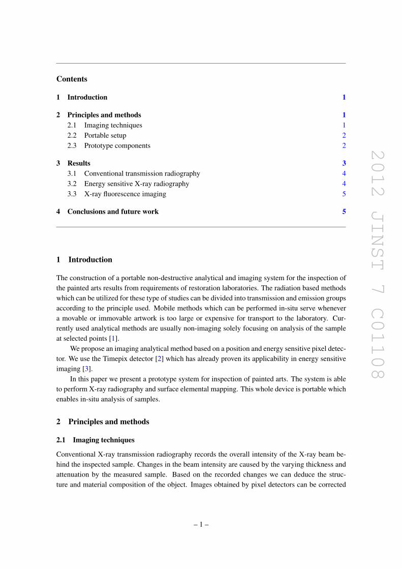
Figure 3 Transmission radiogram of a painted canvasMeasurement in several areas can reveal the innerstructure of the canvas paint layers and the wooden frame
31 Conventional transmission radiography
A real sample of modern painting on canvas from the 19th century by Friedrich von Amerling (copycreated by restorer Hana Mislerova) was chosen for the testing measurements Figure 3 shows aphotograph and the transmission images in several regions of interest The underlying structure offrame connections canvas and brush strokes can be recognized The exposure time was set to 1second (integral image from 10 frames of 01 s)
32 Energy sensitive X-ray radiography
While transmission images show high contrast and high spatial resolution (see figure 3) some in-teresting structures overlap To distinguish them energy sensitive transmission X-ray radiographycan be favourably used To test the energy sensitive radiography technique a simple phantom madeof metal foils was used The transmitted spectra were measured in each pixel to obtain a completematerial identification at all points [10] The photograph intensity image average energy imageand pseudocolored image are shown figure 4 The pseudocolors use RGB coding with three chosenenergy intervals (4ndash8 keV 10ndash15 keV and 20ndash30 keV)
To test this approach on a real sample a simplification of the multichannel approach usingonly two channels was used Although this method can distinguish only two material groups theobtained results bring new information about the composition of the painting The result of thismethod applied to painting is shown in figure 5 where two material components were separated-light structures (organic binders and canvas) and heavy structures (mineral pigments)
ndash 4 ndash
2012 JINST 7 C01108
Figure 4 Energy sensitive radiography on model samples photograph integral intensity image averageenergy and pseudocolored materials (RGB coding used colour channels correspond to energy intervals 4ndash8 keV 10ndash15 keV and 20ndash30 keV)
Figure 5 Material separation into two groups on a real sample - integral intensity (left) heavy materialgroup (middle) and light material group (right)
33 X-ray fluorescence imaging
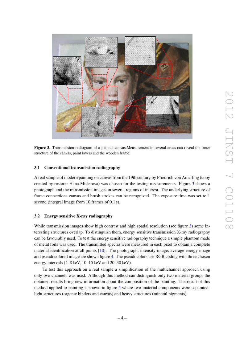
To demonstrate the applicability of this method on real samples we again used the same paintingFigure 6 is an X-ray fluorescence intensity image obtained in the geometry of pinhole camera Thisimage can be divided into the respective energy channels and on the basis of a proper calibrationa compound surface map can be created for the individual elements
4 Conclusions and future work
The prototype of a portable system has been built and tested and proposed imaging and analyticalmethods have been successfully implemented with this prototype The demonstration of conven-tional transmission radiography was done on real samples of painted artwork showing the widedynamic range and high spatial resolution The energy sensitive method was tested on model sam-ples and a simplified model for calculations of the response was used The separation of organic
ndash 5 ndash
2012 JINST 7 C01108
Figure 6 Intensity image (left) and pseudocolored material map (right) obtained by the X-ray fluorescenceimaging
and mineral components of the painting was done even with this simple evaluation method X-rayfluorescence imaging was tested to prove the feasibility of the measurements with the prototype
In future we plan to construct an extended prototype with several improvements which canbe divided into two groups mdash mechanical construction and software control The mechanical up-grades will focus on better shielding of the detector box head and the construction of a motorizedcarousel with different filters and collimators Some changes in the automation of the whole sys-tem will be necessary The upgrade of software control will be done by implementation of thecurrently used software This should allow us to prepare semiautomatic procedure which will cen-tre the X-ray beam on the detector and perform full sample scans needed for large samples or3D reconstruction The safe mode of operation of the device has to be approved by appropriateauthorities
Acknowledgments
This work was supported by the Ministry of Education Youth and Sports of the Czech Republicunder Research Center No LC06041 and by Grant No P105111551 of the Grant Agency of theCzech Republic Research carried out in frame of the Medipix Collaboration
References
[1] P Moioli and C Seccaroni Analysis of art objects using a portable x-ray fluorescence spectrometerX-Ray Spectrom 29 (2000) 48
[2] X Llopart R Ballabriga M Campbell L Tlustos and W Wong Timepix a 56 k programmablepixel readout chip for arrival time energy andor photon counting measurements Nucl InstrumMeth A 581 (2007) 485
[3] J Zemlicka J Jakubek M Kroupa D Hradil J Hradilova and H Mislerova Analysis of paintedarts by energy sensitive radiographic techniques with the Pixel DetectorTimepix 2011 JINST 6C01066
ndash 6 ndash
2012 JINST 7 C01108
[4] J Jakubek Data processing and image reconstruction methods for pixel detectors Nucl InstrumMeth A 576 (2007) 223
[5] J Zemlicka J Jakubek M Kroupa and V Tichy Energy and Position Sensitive Pixel DetectorTimepix for X-Ray Fluorescence Imaging Nucl Instrum Meth A 607 (2009) 202
[6] J Jakubek Semiconductor Pixel Detectors and their Applications in Life Sciences 2009 JINST 4P03013
[7] Medipix Collaboration httpmedipixwebcernchMEDIPIX
[8] V Kraus M Holik J Jakubek M Kroupa P Soukup and Z Vykydal FITPix mdash Fast Interface forTimepix Pixel Detectors 2011 JINST 6 C01079
[9] D Turecek T Holy J Jakubek S Pospısil and Z Vykydal Pixelman a multi-platform dataacquisition and processing software package for Medipix2 Timepix and Medipix3 detectors 2011JINST 6 C01046
[10] J Jakubek Energy Sensitive X-ray Radiography and Charge Sharing Effect in Pixelated DetectorNucl Instrum Meth A 607 (2009) 192
ndash 7 ndash
- Introduction
- Principles and methods
-
- Imaging techniques
- Portable setup
- Prototype components
-
- Results
-
- Conventional transmission radiography
- Energy sensitive X-ray radiography
- X-ray fluorescence imaging
-
- Conclusions and future work
-

2012 JINST 7 C01108
PUBLISHED BY IOP PUBLISHING FOR SISSA
RECEIVED September 30 2011REVISED January 1 2012
ACCEPTED January 4 2012PUBLISHED January 31 2012
13th INTERNATIONAL WORKSHOP ON RADIATION IMAGING DETECTORS3ndash7 JULY 2011ETH ZURICH SWITZERLAND
Mobile system for in-situ imaging of cultural objects
J Zemlickaa1 J Jakubeka F Krejcia D Hradilbc J Hradilovac and H Mislerovac
aInstitute of Experimental and Applied Physics Czech Technical University PragueHorska 3a22 128 00 Prague 2 Czech Republic
bInstitute of Inorganic Chemistry AS CR vvi ALMA250 68 Husinec-Oslashez Czech Republic
cAcademy of Fine Arts in Prague ALMAU Akademie 4 170 2 Prague 7 Czech Republic
E-mail janzemlickautefcvutcz
ABSTRACT Non-invasive analytical techniques recently developed with the Timepix pixel detec-tor have shown great potential for the inspection of objects of cultural heritage We have developednew instrumentation and methodology for in-situ X-ray transmission radiography and X-ray fluo-rescence imaging and successfully tested and evaluated a mobile system for remote terrain tasksThe prototype portable imaging device comprises the radiation source tube and the spectral sen-sitive X-ray camera Both components can be moreover mounted on independent motorized po-sitioning systems allowing adaptation of irradiation geometry to the object shape Both parts areplaced onto a pair of universal portable holders (tripods) The detector is placed in a shielded boxwith exchangeable entrance window (beam filters and pinhole collimator) This adjustable setup al-lows performing in-situ measurements for both transmission and emission (XRF) radiography Theassembled system has been successfully tested in our laboratory with phantoms and real samplesThe obtained and evaluated results are presented in this paper Future work will include successiveadaptation of the current system for real in-situ utilization and preparation of software allowingsemi-automatic remote control of measurements
KEYWORDS Inspection with x-rays X-ray fluorescence (XRF) systems
1Corresponding author
ccopy 2012 IOP Publishing Ltd and SISSA doi1010881748-0221701C01108
2012 JINST 7 C01108
Contents
1 Introduction 1
2 Principles and methods 121 Imaging techniques 122 Portable setup 223 Prototype components 2
3 Results 331 Conventional transmission radiography 432 Energy sensitive X-ray radiography 433 X-ray fluorescence imaging 5
4 Conclusions and future work 5
1 Introduction
The construction of a portable non-destructive analytical and imaging system for the inspection ofthe painted arts results from requirements of restoration laboratories The radiation based methodswhich can be utilized for these type of studies can be divided into transmission and emission groupsaccording to the principle used Mobile methods which can be performed in-situ serve whenevera movable or immovable artwork is too large or expensive for transport to the laboratory Cur-rently used analytical methods are usually non-imaging solely focusing on analysis of the sampleat selected points [1]
We propose an imaging analytical method based on a position and energy sensitive pixel detec-tor We use the Timepix detector [2] which has already proven its applicability in energy sensitiveimaging [3]
In this paper we present a prototype system for inspection of painted arts The system is ableto perform X-ray radiography and surface elemental mapping This whole device is portable whichenables in-situ analysis of samples
2 Principles and methods
21 Imaging techniques
Conventional X-ray transmission radiography records the overall intensity of the X-ray beam be-hind the inspected sample Changes in the beam intensity are caused by the varying thickness andattenuation by the measured sample Based on the recorded changes we can deduce the struc-ture and material composition of the object Images obtained by pixel detectors can be corrected
ndash 1 ndash
2012 JINST 7 C01108
by using several different techniques mdash for images shown in this article a flat field correctionwas used [4]
Information obtained by forenamed method can be used as an input for the 3D reconstruc-tion methods such as computed tomography (CT) Taking into account that inspected samples areoften large and flat modified algorithms such as CT with limited angles and coplanar rotationallaminography (CRL) have to be used
Energy sensitive X-ray transmission radiography is an extension of conventional X-ray trans-mission radiography With this method the recorded energy of incident photons can be divided intoseveral energy windows representing colour channels mdash in analogy to colour photograph Fur-thermore changes in the recorded spectra in each pixel can be evaluated and used for materialdecomposition
Unlike transmission methods X-ray fluorescence imaging [5] is based on the detection ofthe characteristic photon radiation which is emitted from the irradiated sample This method canbe used for the study of elemental composition of object surface because every element emitsphotons of different but specific energies (characteristic radiation) Based on the energy spectrait is possible to deduce the concentration of contained elements This method can be used formapping the element distribution on the sample surface
22 Portable setup
We have designed a prototype of an imaging unit enhanced with analytical potentialities The an-alytical power of this technique is based on the usage of the state-of-the-art semiconductor pixeldetector namely Timepix The detection is based on single particle counting and the main advan-tage of this approach lies in the ability to perform high contrast imaging without noise or so calleddark current [6] Moreover the Timepix device is able to measure the energy of incident particleswhich can be used for the energy sensitive X-ray radiography and fluorescence imaging
The system was designed with respect to maximal adaptability in in-situ measurements Forthis purpose the system is divided into two independent parts mdash X-ray tube head and detector headBoth parts are mounted on motorized positioning systems placed onto universal tripod holdersThe detector is placed in a lead cased box to shield scattered radiation and the entrance window ismounted with holder for exchangeable filters and collimators
The setup with two independent heads enables measurement in several geometries (see fig-ure 1) enabling transmission X-ray radiography and tomography as well as XRF imaging Bothdetector and X-ray tube are integrated in the motorized positioning system for automatic scanningof the sample
23 Prototype components
The X-ray tube head is equipped with a sealed 60 W X-ray tube made by Oxford Instruments (max60 kV 1 mA tungsten target) Both power supply and control units are small enough to be mountedonto tripod legs The X-ray tube is controlled from a PC via a USB interface
The detector head contains the pixel detector Timepix which was developed at CERN bythe Medipix Collaboration [7] The device consists of a semiconductor detector chip (usually300 mm thick silicon) bump-bonded to a readout chip The detector chip is equipped with a single
ndash 2 ndash
2012 JINST 7 C01108
Figure 1 Illustration of the possible measurement setups (XT ndash X-ray tube S ndash sample D ndash detector) a)Conventional transmission radiography with scanning of the sample b) Coplanar rotational laminography(CRL) c) Computed tomography (CT) with limited angles d) X-ray fluorescence imaging All methods areorientated towards large flat samples (paintings)
Figure 2 Transmission setup with a replica of a historic painting from the 19th century (painted withauthentic pigments) The sealed X-ray tube (left) is aimed to the shielded detector head with copper entrancewindow
common back-side electrode and a front-side matrix of electrodes (256times 256 square pixels withpitch of 55 microm) Each element of the matrix (pixel) is connected to its respective preamplifierdiscriminator and digital counter integrated on the readout chip creating independent spectroscopicline The energy measurements utilize the ability of the device to directly measure the amount ofthe collected charge which is created by incident particle in each pixel Data from detector areacquired by FITPix readout interface [8] and processed with Pixelman software package [9]
3 Results
The prototype of the portable system has been tested with all described methods Figure 2 showsthe photograph of a measurement in transmission geometry The X-ray tube was set to 60 kV and05 mA (30 W)The obtained results can be divided into three groups mdash conventional transmissionradiography energy sensitive radiography and X-ray fluorescence imaging
ndash 3 ndash
2012 JINST 7 C01108
Figure 3 Transmission radiogram of a painted canvasMeasurement in several areas can reveal the innerstructure of the canvas paint layers and the wooden frame
31 Conventional transmission radiography
A real sample of modern painting on canvas from the 19th century by Friedrich von Amerling (copycreated by restorer Hana Mislerova) was chosen for the testing measurements Figure 3 shows aphotograph and the transmission images in several regions of interest The underlying structure offrame connections canvas and brush strokes can be recognized The exposure time was set to 1second (integral image from 10 frames of 01 s)
32 Energy sensitive X-ray radiography
While transmission images show high contrast and high spatial resolution (see figure 3) some in-teresting structures overlap To distinguish them energy sensitive transmission X-ray radiographycan be favourably used To test the energy sensitive radiography technique a simple phantom madeof metal foils was used The transmitted spectra were measured in each pixel to obtain a completematerial identification at all points [10] The photograph intensity image average energy imageand pseudocolored image are shown figure 4 The pseudocolors use RGB coding with three chosenenergy intervals (4ndash8 keV 10ndash15 keV and 20ndash30 keV)
To test this approach on a real sample a simplification of the multichannel approach usingonly two channels was used Although this method can distinguish only two material groups theobtained results bring new information about the composition of the painting The result of thismethod applied to painting is shown in figure 5 where two material components were separated-light structures (organic binders and canvas) and heavy structures (mineral pigments)
ndash 4 ndash
2012 JINST 7 C01108
Figure 4 Energy sensitive radiography on model samples photograph integral intensity image averageenergy and pseudocolored materials (RGB coding used colour channels correspond to energy intervals 4ndash8 keV 10ndash15 keV and 20ndash30 keV)
Figure 5 Material separation into two groups on a real sample - integral intensity (left) heavy materialgroup (middle) and light material group (right)
33 X-ray fluorescence imaging
To demonstrate the applicability of this method on real samples we again used the same paintingFigure 6 is an X-ray fluorescence intensity image obtained in the geometry of pinhole camera Thisimage can be divided into the respective energy channels and on the basis of a proper calibrationa compound surface map can be created for the individual elements
4 Conclusions and future work
The prototype of a portable system has been built and tested and proposed imaging and analyticalmethods have been successfully implemented with this prototype The demonstration of conven-tional transmission radiography was done on real samples of painted artwork showing the widedynamic range and high spatial resolution The energy sensitive method was tested on model sam-ples and a simplified model for calculations of the response was used The separation of organic
ndash 5 ndash
2012 JINST 7 C01108
Figure 6 Intensity image (left) and pseudocolored material map (right) obtained by the X-ray fluorescenceimaging
and mineral components of the painting was done even with this simple evaluation method X-rayfluorescence imaging was tested to prove the feasibility of the measurements with the prototype
In future we plan to construct an extended prototype with several improvements which canbe divided into two groups mdash mechanical construction and software control The mechanical up-grades will focus on better shielding of the detector box head and the construction of a motorizedcarousel with different filters and collimators Some changes in the automation of the whole sys-tem will be necessary The upgrade of software control will be done by implementation of thecurrently used software This should allow us to prepare semiautomatic procedure which will cen-tre the X-ray beam on the detector and perform full sample scans needed for large samples or3D reconstruction The safe mode of operation of the device has to be approved by appropriateauthorities
Acknowledgments
This work was supported by the Ministry of Education Youth and Sports of the Czech Republicunder Research Center No LC06041 and by Grant No P105111551 of the Grant Agency of theCzech Republic Research carried out in frame of the Medipix Collaboration
References
[1] P Moioli and C Seccaroni Analysis of art objects using a portable x-ray fluorescence spectrometerX-Ray Spectrom 29 (2000) 48
[2] X Llopart R Ballabriga M Campbell L Tlustos and W Wong Timepix a 56 k programmablepixel readout chip for arrival time energy andor photon counting measurements Nucl InstrumMeth A 581 (2007) 485
[3] J Zemlicka J Jakubek M Kroupa D Hradil J Hradilova and H Mislerova Analysis of paintedarts by energy sensitive radiographic techniques with the Pixel DetectorTimepix 2011 JINST 6C01066
ndash 6 ndash
2012 JINST 7 C01108
[4] J Jakubek Data processing and image reconstruction methods for pixel detectors Nucl InstrumMeth A 576 (2007) 223
[5] J Zemlicka J Jakubek M Kroupa and V Tichy Energy and Position Sensitive Pixel DetectorTimepix for X-Ray Fluorescence Imaging Nucl Instrum Meth A 607 (2009) 202
[6] J Jakubek Semiconductor Pixel Detectors and their Applications in Life Sciences 2009 JINST 4P03013
[7] Medipix Collaboration httpmedipixwebcernchMEDIPIX
[8] V Kraus M Holik J Jakubek M Kroupa P Soukup and Z Vykydal FITPix mdash Fast Interface forTimepix Pixel Detectors 2011 JINST 6 C01079
[9] D Turecek T Holy J Jakubek S Pospısil and Z Vykydal Pixelman a multi-platform dataacquisition and processing software package for Medipix2 Timepix and Medipix3 detectors 2011JINST 6 C01046
[10] J Jakubek Energy Sensitive X-ray Radiography and Charge Sharing Effect in Pixelated DetectorNucl Instrum Meth A 607 (2009) 192
ndash 7 ndash
- Introduction
- Principles and methods
-
- Imaging techniques
- Portable setup
- Prototype components
-
- Results
-
- Conventional transmission radiography
- Energy sensitive X-ray radiography
- X-ray fluorescence imaging
-
- Conclusions and future work
-
2012 JINST 7 C01108
Contents
1 Introduction 1
2 Principles and methods 121 Imaging techniques 122 Portable setup 223 Prototype components 2
3 Results 331 Conventional transmission radiography 432 Energy sensitive X-ray radiography 433 X-ray fluorescence imaging 5
4 Conclusions and future work 5
1 Introduction
The construction of a portable non-destructive analytical and imaging system for the inspection ofthe painted arts results from requirements of restoration laboratories The radiation based methodswhich can be utilized for these type of studies can be divided into transmission and emission groupsaccording to the principle used Mobile methods which can be performed in-situ serve whenevera movable or immovable artwork is too large or expensive for transport to the laboratory Cur-rently used analytical methods are usually non-imaging solely focusing on analysis of the sampleat selected points [1]
We propose an imaging analytical method based on a position and energy sensitive pixel detec-tor We use the Timepix detector [2] which has already proven its applicability in energy sensitiveimaging [3]
In this paper we present a prototype system for inspection of painted arts The system is ableto perform X-ray radiography and surface elemental mapping This whole device is portable whichenables in-situ analysis of samples
2 Principles and methods
21 Imaging techniques
Conventional X-ray transmission radiography records the overall intensity of the X-ray beam be-hind the inspected sample Changes in the beam intensity are caused by the varying thickness andattenuation by the measured sample Based on the recorded changes we can deduce the struc-ture and material composition of the object Images obtained by pixel detectors can be corrected
ndash 1 ndash
2012 JINST 7 C01108
by using several different techniques mdash for images shown in this article a flat field correctionwas used [4]
Information obtained by forenamed method can be used as an input for the 3D reconstruc-tion methods such as computed tomography (CT) Taking into account that inspected samples areoften large and flat modified algorithms such as CT with limited angles and coplanar rotationallaminography (CRL) have to be used
Energy sensitive X-ray transmission radiography is an extension of conventional X-ray trans-mission radiography With this method the recorded energy of incident photons can be divided intoseveral energy windows representing colour channels mdash in analogy to colour photograph Fur-thermore changes in the recorded spectra in each pixel can be evaluated and used for materialdecomposition
Unlike transmission methods X-ray fluorescence imaging [5] is based on the detection ofthe characteristic photon radiation which is emitted from the irradiated sample This method canbe used for the study of elemental composition of object surface because every element emitsphotons of different but specific energies (characteristic radiation) Based on the energy spectrait is possible to deduce the concentration of contained elements This method can be used formapping the element distribution on the sample surface
22 Portable setup
We have designed a prototype of an imaging unit enhanced with analytical potentialities The an-alytical power of this technique is based on the usage of the state-of-the-art semiconductor pixeldetector namely Timepix The detection is based on single particle counting and the main advan-tage of this approach lies in the ability to perform high contrast imaging without noise or so calleddark current [6] Moreover the Timepix device is able to measure the energy of incident particleswhich can be used for the energy sensitive X-ray radiography and fluorescence imaging
The system was designed with respect to maximal adaptability in in-situ measurements Forthis purpose the system is divided into two independent parts mdash X-ray tube head and detector headBoth parts are mounted on motorized positioning systems placed onto universal tripod holdersThe detector is placed in a lead cased box to shield scattered radiation and the entrance window ismounted with holder for exchangeable filters and collimators
The setup with two independent heads enables measurement in several geometries (see fig-ure 1) enabling transmission X-ray radiography and tomography as well as XRF imaging Bothdetector and X-ray tube are integrated in the motorized positioning system for automatic scanningof the sample
23 Prototype components
The X-ray tube head is equipped with a sealed 60 W X-ray tube made by Oxford Instruments (max60 kV 1 mA tungsten target) Both power supply and control units are small enough to be mountedonto tripod legs The X-ray tube is controlled from a PC via a USB interface
The detector head contains the pixel detector Timepix which was developed at CERN bythe Medipix Collaboration [7] The device consists of a semiconductor detector chip (usually300 mm thick silicon) bump-bonded to a readout chip The detector chip is equipped with a single
ndash 2 ndash
2012 JINST 7 C01108
Figure 1 Illustration of the possible measurement setups (XT ndash X-ray tube S ndash sample D ndash detector) a)Conventional transmission radiography with scanning of the sample b) Coplanar rotational laminography(CRL) c) Computed tomography (CT) with limited angles d) X-ray fluorescence imaging All methods areorientated towards large flat samples (paintings)
Figure 2 Transmission setup with a replica of a historic painting from the 19th century (painted withauthentic pigments) The sealed X-ray tube (left) is aimed to the shielded detector head with copper entrancewindow
common back-side electrode and a front-side matrix of electrodes (256times 256 square pixels withpitch of 55 microm) Each element of the matrix (pixel) is connected to its respective preamplifierdiscriminator and digital counter integrated on the readout chip creating independent spectroscopicline The energy measurements utilize the ability of the device to directly measure the amount ofthe collected charge which is created by incident particle in each pixel Data from detector areacquired by FITPix readout interface [8] and processed with Pixelman software package [9]
3 Results
The prototype of the portable system has been tested with all described methods Figure 2 showsthe photograph of a measurement in transmission geometry The X-ray tube was set to 60 kV and05 mA (30 W)The obtained results can be divided into three groups mdash conventional transmissionradiography energy sensitive radiography and X-ray fluorescence imaging
ndash 3 ndash
2012 JINST 7 C01108
Figure 3 Transmission radiogram of a painted canvasMeasurement in several areas can reveal the innerstructure of the canvas paint layers and the wooden frame
31 Conventional transmission radiography
A real sample of modern painting on canvas from the 19th century by Friedrich von Amerling (copycreated by restorer Hana Mislerova) was chosen for the testing measurements Figure 3 shows aphotograph and the transmission images in several regions of interest The underlying structure offrame connections canvas and brush strokes can be recognized The exposure time was set to 1second (integral image from 10 frames of 01 s)
32 Energy sensitive X-ray radiography
While transmission images show high contrast and high spatial resolution (see figure 3) some in-teresting structures overlap To distinguish them energy sensitive transmission X-ray radiographycan be favourably used To test the energy sensitive radiography technique a simple phantom madeof metal foils was used The transmitted spectra were measured in each pixel to obtain a completematerial identification at all points [10] The photograph intensity image average energy imageand pseudocolored image are shown figure 4 The pseudocolors use RGB coding with three chosenenergy intervals (4ndash8 keV 10ndash15 keV and 20ndash30 keV)
To test this approach on a real sample a simplification of the multichannel approach usingonly two channels was used Although this method can distinguish only two material groups theobtained results bring new information about the composition of the painting The result of thismethod applied to painting is shown in figure 5 where two material components were separated-light structures (organic binders and canvas) and heavy structures (mineral pigments)
ndash 4 ndash
2012 JINST 7 C01108
Figure 4 Energy sensitive radiography on model samples photograph integral intensity image averageenergy and pseudocolored materials (RGB coding used colour channels correspond to energy intervals 4ndash8 keV 10ndash15 keV and 20ndash30 keV)
Figure 5 Material separation into two groups on a real sample - integral intensity (left) heavy materialgroup (middle) and light material group (right)
33 X-ray fluorescence imaging
To demonstrate the applicability of this method on real samples we again used the same paintingFigure 6 is an X-ray fluorescence intensity image obtained in the geometry of pinhole camera Thisimage can be divided into the respective energy channels and on the basis of a proper calibrationa compound surface map can be created for the individual elements
4 Conclusions and future work
The prototype of a portable system has been built and tested and proposed imaging and analyticalmethods have been successfully implemented with this prototype The demonstration of conven-tional transmission radiography was done on real samples of painted artwork showing the widedynamic range and high spatial resolution The energy sensitive method was tested on model sam-ples and a simplified model for calculations of the response was used The separation of organic
ndash 5 ndash
2012 JINST 7 C01108
Figure 6 Intensity image (left) and pseudocolored material map (right) obtained by the X-ray fluorescenceimaging
and mineral components of the painting was done even with this simple evaluation method X-rayfluorescence imaging was tested to prove the feasibility of the measurements with the prototype
In future we plan to construct an extended prototype with several improvements which canbe divided into two groups mdash mechanical construction and software control The mechanical up-grades will focus on better shielding of the detector box head and the construction of a motorizedcarousel with different filters and collimators Some changes in the automation of the whole sys-tem will be necessary The upgrade of software control will be done by implementation of thecurrently used software This should allow us to prepare semiautomatic procedure which will cen-tre the X-ray beam on the detector and perform full sample scans needed for large samples or3D reconstruction The safe mode of operation of the device has to be approved by appropriateauthorities
Acknowledgments
This work was supported by the Ministry of Education Youth and Sports of the Czech Republicunder Research Center No LC06041 and by Grant No P105111551 of the Grant Agency of theCzech Republic Research carried out in frame of the Medipix Collaboration
References
[1] P Moioli and C Seccaroni Analysis of art objects using a portable x-ray fluorescence spectrometerX-Ray Spectrom 29 (2000) 48
[2] X Llopart R Ballabriga M Campbell L Tlustos and W Wong Timepix a 56 k programmablepixel readout chip for arrival time energy andor photon counting measurements Nucl InstrumMeth A 581 (2007) 485
[3] J Zemlicka J Jakubek M Kroupa D Hradil J Hradilova and H Mislerova Analysis of paintedarts by energy sensitive radiographic techniques with the Pixel DetectorTimepix 2011 JINST 6C01066
ndash 6 ndash
2012 JINST 7 C01108
[4] J Jakubek Data processing and image reconstruction methods for pixel detectors Nucl InstrumMeth A 576 (2007) 223
[5] J Zemlicka J Jakubek M Kroupa and V Tichy Energy and Position Sensitive Pixel DetectorTimepix for X-Ray Fluorescence Imaging Nucl Instrum Meth A 607 (2009) 202
[6] J Jakubek Semiconductor Pixel Detectors and their Applications in Life Sciences 2009 JINST 4P03013
[7] Medipix Collaboration httpmedipixwebcernchMEDIPIX
[8] V Kraus M Holik J Jakubek M Kroupa P Soukup and Z Vykydal FITPix mdash Fast Interface forTimepix Pixel Detectors 2011 JINST 6 C01079
[9] D Turecek T Holy J Jakubek S Pospısil and Z Vykydal Pixelman a multi-platform dataacquisition and processing software package for Medipix2 Timepix and Medipix3 detectors 2011JINST 6 C01046
[10] J Jakubek Energy Sensitive X-ray Radiography and Charge Sharing Effect in Pixelated DetectorNucl Instrum Meth A 607 (2009) 192
ndash 7 ndash
- Introduction
- Principles and methods
-
- Imaging techniques
- Portable setup
- Prototype components
-
- Results
-
- Conventional transmission radiography
- Energy sensitive X-ray radiography
- X-ray fluorescence imaging
-
- Conclusions and future work
-

2012 JINST 7 C01108
Figure 1 Illustration of the possible measurement setups (XT ndash X-ray tube S ndash sample D ndash detector) a)Conventional transmission radiography with scanning of the sample b) Coplanar rotational laminography(CRL) c) Computed tomography (CT) with limited angles d) X-ray fluorescence imaging All methods areorientated towards large flat samples (paintings)
Figure 2 Transmission setup with a replica of a historic painting from the 19th century (painted withauthentic pigments) The sealed X-ray tube (left) is aimed to the shielded detector head with copper entrancewindow
common back-side electrode and a front-side matrix of electrodes (256times 256 square pixels withpitch of 55 microm) Each element of the matrix (pixel) is connected to its respective preamplifierdiscriminator and digital counter integrated on the readout chip creating independent spectroscopicline The energy measurements utilize the ability of the device to directly measure the amount ofthe collected charge which is created by incident particle in each pixel Data from detector areacquired by FITPix readout interface [8] and processed with Pixelman software package [9]
3 Results
The prototype of the portable system has been tested with all described methods Figure 2 showsthe photograph of a measurement in transmission geometry The X-ray tube was set to 60 kV and05 mA (30 W)The obtained results can be divided into three groups mdash conventional transmissionradiography energy sensitive radiography and X-ray fluorescence imaging
ndash 3 ndash
2012 JINST 7 C01108
Figure 3 Transmission radiogram of a painted canvasMeasurement in several areas can reveal the innerstructure of the canvas paint layers and the wooden frame
31 Conventional transmission radiography
A real sample of modern painting on canvas from the 19th century by Friedrich von Amerling (copycreated by restorer Hana Mislerova) was chosen for the testing measurements Figure 3 shows aphotograph and the transmission images in several regions of interest The underlying structure offrame connections canvas and brush strokes can be recognized The exposure time was set to 1second (integral image from 10 frames of 01 s)
32 Energy sensitive X-ray radiography
While transmission images show high contrast and high spatial resolution (see figure 3) some in-teresting structures overlap To distinguish them energy sensitive transmission X-ray radiographycan be favourably used To test the energy sensitive radiography technique a simple phantom madeof metal foils was used The transmitted spectra were measured in each pixel to obtain a completematerial identification at all points [10] The photograph intensity image average energy imageand pseudocolored image are shown figure 4 The pseudocolors use RGB coding with three chosenenergy intervals (4ndash8 keV 10ndash15 keV and 20ndash30 keV)
To test this approach on a real sample a simplification of the multichannel approach usingonly two channels was used Although this method can distinguish only two material groups theobtained results bring new information about the composition of the painting The result of thismethod applied to painting is shown in figure 5 where two material components were separated-light structures (organic binders and canvas) and heavy structures (mineral pigments)
ndash 4 ndash
2012 JINST 7 C01108
Figure 4 Energy sensitive radiography on model samples photograph integral intensity image averageenergy and pseudocolored materials (RGB coding used colour channels correspond to energy intervals 4ndash8 keV 10ndash15 keV and 20ndash30 keV)
Figure 5 Material separation into two groups on a real sample - integral intensity (left) heavy materialgroup (middle) and light material group (right)
33 X-ray fluorescence imaging
To demonstrate the applicability of this method on real samples we again used the same paintingFigure 6 is an X-ray fluorescence intensity image obtained in the geometry of pinhole camera Thisimage can be divided into the respective energy channels and on the basis of a proper calibrationa compound surface map can be created for the individual elements
4 Conclusions and future work
The prototype of a portable system has been built and tested and proposed imaging and analyticalmethods have been successfully implemented with this prototype The demonstration of conven-tional transmission radiography was done on real samples of painted artwork showing the widedynamic range and high spatial resolution The energy sensitive method was tested on model sam-ples and a simplified model for calculations of the response was used The separation of organic
ndash 5 ndash
2012 JINST 7 C01108
Figure 6 Intensity image (left) and pseudocolored material map (right) obtained by the X-ray fluorescenceimaging
and mineral components of the painting was done even with this simple evaluation method X-rayfluorescence imaging was tested to prove the feasibility of the measurements with the prototype
In future we plan to construct an extended prototype with several improvements which canbe divided into two groups mdash mechanical construction and software control The mechanical up-grades will focus on better shielding of the detector box head and the construction of a motorizedcarousel with different filters and collimators Some changes in the automation of the whole sys-tem will be necessary The upgrade of software control will be done by implementation of thecurrently used software This should allow us to prepare semiautomatic procedure which will cen-tre the X-ray beam on the detector and perform full sample scans needed for large samples or3D reconstruction The safe mode of operation of the device has to be approved by appropriateauthorities
Acknowledgments
This work was supported by the Ministry of Education Youth and Sports of the Czech Republicunder Research Center No LC06041 and by Grant No P105111551 of the Grant Agency of theCzech Republic Research carried out in frame of the Medipix Collaboration
References
[1] P Moioli and C Seccaroni Analysis of art objects using a portable x-ray fluorescence spectrometerX-Ray Spectrom 29 (2000) 48
[2] X Llopart R Ballabriga M Campbell L Tlustos and W Wong Timepix a 56 k programmablepixel readout chip for arrival time energy andor photon counting measurements Nucl InstrumMeth A 581 (2007) 485
[3] J Zemlicka J Jakubek M Kroupa D Hradil J Hradilova and H Mislerova Analysis of paintedarts by energy sensitive radiographic techniques with the Pixel DetectorTimepix 2011 JINST 6C01066
ndash 6 ndash
2012 JINST 7 C01108
[4] J Jakubek Data processing and image reconstruction methods for pixel detectors Nucl InstrumMeth A 576 (2007) 223
[5] J Zemlicka J Jakubek M Kroupa and V Tichy Energy and Position Sensitive Pixel DetectorTimepix for X-Ray Fluorescence Imaging Nucl Instrum Meth A 607 (2009) 202
[6] J Jakubek Semiconductor Pixel Detectors and their Applications in Life Sciences 2009 JINST 4P03013
[7] Medipix Collaboration httpmedipixwebcernchMEDIPIX
[8] V Kraus M Holik J Jakubek M Kroupa P Soukup and Z Vykydal FITPix mdash Fast Interface forTimepix Pixel Detectors 2011 JINST 6 C01079
[9] D Turecek T Holy J Jakubek S Pospısil and Z Vykydal Pixelman a multi-platform dataacquisition and processing software package for Medipix2 Timepix and Medipix3 detectors 2011JINST 6 C01046
[10] J Jakubek Energy Sensitive X-ray Radiography and Charge Sharing Effect in Pixelated DetectorNucl Instrum Meth A 607 (2009) 192
ndash 7 ndash
- Introduction
- Principles and methods
-
- Imaging techniques
- Portable setup
- Prototype components
-
- Results
-
- Conventional transmission radiography
- Energy sensitive X-ray radiography
- X-ray fluorescence imaging
-
- Conclusions and future work
-
2012 JINST 7 C01108
Figure 3 Transmission radiogram of a painted canvasMeasurement in several areas can reveal the innerstructure of the canvas paint layers and the wooden frame
31 Conventional transmission radiography
A real sample of modern painting on canvas from the 19th century by Friedrich von Amerling (copycreated by restorer Hana Mislerova) was chosen for the testing measurements Figure 3 shows aphotograph and the transmission images in several regions of interest The underlying structure offrame connections canvas and brush strokes can be recognized The exposure time was set to 1second (integral image from 10 frames of 01 s)
32 Energy sensitive X-ray radiography
While transmission images show high contrast and high spatial resolution (see figure 3) some in-teresting structures overlap To distinguish them energy sensitive transmission X-ray radiographycan be favourably used To test the energy sensitive radiography technique a simple phantom madeof metal foils was used The transmitted spectra were measured in each pixel to obtain a completematerial identification at all points [10] The photograph intensity image average energy imageand pseudocolored image are shown figure 4 The pseudocolors use RGB coding with three chosenenergy intervals (4ndash8 keV 10ndash15 keV and 20ndash30 keV)
To test this approach on a real sample a simplification of the multichannel approach usingonly two channels was used Although this method can distinguish only two material groups theobtained results bring new information about the composition of the painting The result of thismethod applied to painting is shown in figure 5 where two material components were separated-light structures (organic binders and canvas) and heavy structures (mineral pigments)
ndash 4 ndash
2012 JINST 7 C01108
Figure 4 Energy sensitive radiography on model samples photograph integral intensity image averageenergy and pseudocolored materials (RGB coding used colour channels correspond to energy intervals 4ndash8 keV 10ndash15 keV and 20ndash30 keV)
Figure 5 Material separation into two groups on a real sample - integral intensity (left) heavy materialgroup (middle) and light material group (right)
33 X-ray fluorescence imaging
To demonstrate the applicability of this method on real samples we again used the same paintingFigure 6 is an X-ray fluorescence intensity image obtained in the geometry of pinhole camera Thisimage can be divided into the respective energy channels and on the basis of a proper calibrationa compound surface map can be created for the individual elements
4 Conclusions and future work
The prototype of a portable system has been built and tested and proposed imaging and analyticalmethods have been successfully implemented with this prototype The demonstration of conven-tional transmission radiography was done on real samples of painted artwork showing the widedynamic range and high spatial resolution The energy sensitive method was tested on model sam-ples and a simplified model for calculations of the response was used The separation of organic
ndash 5 ndash
2012 JINST 7 C01108
Figure 6 Intensity image (left) and pseudocolored material map (right) obtained by the X-ray fluorescenceimaging
and mineral components of the painting was done even with this simple evaluation method X-rayfluorescence imaging was tested to prove the feasibility of the measurements with the prototype
In future we plan to construct an extended prototype with several improvements which canbe divided into two groups mdash mechanical construction and software control The mechanical up-grades will focus on better shielding of the detector box head and the construction of a motorizedcarousel with different filters and collimators Some changes in the automation of the whole sys-tem will be necessary The upgrade of software control will be done by implementation of thecurrently used software This should allow us to prepare semiautomatic procedure which will cen-tre the X-ray beam on the detector and perform full sample scans needed for large samples or3D reconstruction The safe mode of operation of the device has to be approved by appropriateauthorities
Acknowledgments
This work was supported by the Ministry of Education Youth and Sports of the Czech Republicunder Research Center No LC06041 and by Grant No P105111551 of the Grant Agency of theCzech Republic Research carried out in frame of the Medipix Collaboration
References
[1] P Moioli and C Seccaroni Analysis of art objects using a portable x-ray fluorescence spectrometerX-Ray Spectrom 29 (2000) 48
[2] X Llopart R Ballabriga M Campbell L Tlustos and W Wong Timepix a 56 k programmablepixel readout chip for arrival time energy andor photon counting measurements Nucl InstrumMeth A 581 (2007) 485
[3] J Zemlicka J Jakubek M Kroupa D Hradil J Hradilova and H Mislerova Analysis of paintedarts by energy sensitive radiographic techniques with the Pixel DetectorTimepix 2011 JINST 6C01066
ndash 6 ndash
2012 JINST 7 C01108
[4] J Jakubek Data processing and image reconstruction methods for pixel detectors Nucl InstrumMeth A 576 (2007) 223
[5] J Zemlicka J Jakubek M Kroupa and V Tichy Energy and Position Sensitive Pixel DetectorTimepix for X-Ray Fluorescence Imaging Nucl Instrum Meth A 607 (2009) 202
[6] J Jakubek Semiconductor Pixel Detectors and their Applications in Life Sciences 2009 JINST 4P03013
[7] Medipix Collaboration httpmedipixwebcernchMEDIPIX
[8] V Kraus M Holik J Jakubek M Kroupa P Soukup and Z Vykydal FITPix mdash Fast Interface forTimepix Pixel Detectors 2011 JINST 6 C01079
[9] D Turecek T Holy J Jakubek S Pospısil and Z Vykydal Pixelman a multi-platform dataacquisition and processing software package for Medipix2 Timepix and Medipix3 detectors 2011JINST 6 C01046
[10] J Jakubek Energy Sensitive X-ray Radiography and Charge Sharing Effect in Pixelated DetectorNucl Instrum Meth A 607 (2009) 192
ndash 7 ndash
- Introduction
- Principles and methods
-
- Imaging techniques
- Portable setup
- Prototype components
-
- Results
-
- Conventional transmission radiography
- Energy sensitive X-ray radiography
- X-ray fluorescence imaging
-
- Conclusions and future work
-

2012 JINST 7 C01108
Figure 4 Energy sensitive radiography on model samples photograph integral intensity image averageenergy and pseudocolored materials (RGB coding used colour channels correspond to energy intervals 4ndash8 keV 10ndash15 keV and 20ndash30 keV)
Figure 5 Material separation into two groups on a real sample - integral intensity (left) heavy materialgroup (middle) and light material group (right)
33 X-ray fluorescence imaging
To demonstrate the applicability of this method on real samples we again used the same paintingFigure 6 is an X-ray fluorescence intensity image obtained in the geometry of pinhole camera Thisimage can be divided into the respective energy channels and on the basis of a proper calibrationa compound surface map can be created for the individual elements
4 Conclusions and future work
The prototype of a portable system has been built and tested and proposed imaging and analyticalmethods have been successfully implemented with this prototype The demonstration of conven-tional transmission radiography was done on real samples of painted artwork showing the widedynamic range and high spatial resolution The energy sensitive method was tested on model sam-ples and a simplified model for calculations of the response was used The separation of organic
ndash 5 ndash
2012 JINST 7 C01108
Figure 6 Intensity image (left) and pseudocolored material map (right) obtained by the X-ray fluorescenceimaging
and mineral components of the painting was done even with this simple evaluation method X-rayfluorescence imaging was tested to prove the feasibility of the measurements with the prototype
In future we plan to construct an extended prototype with several improvements which canbe divided into two groups mdash mechanical construction and software control The mechanical up-grades will focus on better shielding of the detector box head and the construction of a motorizedcarousel with different filters and collimators Some changes in the automation of the whole sys-tem will be necessary The upgrade of software control will be done by implementation of thecurrently used software This should allow us to prepare semiautomatic procedure which will cen-tre the X-ray beam on the detector and perform full sample scans needed for large samples or3D reconstruction The safe mode of operation of the device has to be approved by appropriateauthorities
Acknowledgments
This work was supported by the Ministry of Education Youth and Sports of the Czech Republicunder Research Center No LC06041 and by Grant No P105111551 of the Grant Agency of theCzech Republic Research carried out in frame of the Medipix Collaboration
References
[1] P Moioli and C Seccaroni Analysis of art objects using a portable x-ray fluorescence spectrometerX-Ray Spectrom 29 (2000) 48
[2] X Llopart R Ballabriga M Campbell L Tlustos and W Wong Timepix a 56 k programmablepixel readout chip for arrival time energy andor photon counting measurements Nucl InstrumMeth A 581 (2007) 485
[3] J Zemlicka J Jakubek M Kroupa D Hradil J Hradilova and H Mislerova Analysis of paintedarts by energy sensitive radiographic techniques with the Pixel DetectorTimepix 2011 JINST 6C01066
ndash 6 ndash
2012 JINST 7 C01108
[4] J Jakubek Data processing and image reconstruction methods for pixel detectors Nucl InstrumMeth A 576 (2007) 223
[5] J Zemlicka J Jakubek M Kroupa and V Tichy Energy and Position Sensitive Pixel DetectorTimepix for X-Ray Fluorescence Imaging Nucl Instrum Meth A 607 (2009) 202
[6] J Jakubek Semiconductor Pixel Detectors and their Applications in Life Sciences 2009 JINST 4P03013
[7] Medipix Collaboration httpmedipixwebcernchMEDIPIX
[8] V Kraus M Holik J Jakubek M Kroupa P Soukup and Z Vykydal FITPix mdash Fast Interface forTimepix Pixel Detectors 2011 JINST 6 C01079
[9] D Turecek T Holy J Jakubek S Pospısil and Z Vykydal Pixelman a multi-platform dataacquisition and processing software package for Medipix2 Timepix and Medipix3 detectors 2011JINST 6 C01046
[10] J Jakubek Energy Sensitive X-ray Radiography and Charge Sharing Effect in Pixelated DetectorNucl Instrum Meth A 607 (2009) 192
ndash 7 ndash
- Introduction
- Principles and methods
-
- Imaging techniques
- Portable setup
- Prototype components
-
- Results
-
- Conventional transmission radiography
- Energy sensitive X-ray radiography
- X-ray fluorescence imaging
-
- Conclusions and future work
-
2012 JINST 7 C01108
Figure 6 Intensity image (left) and pseudocolored material map (right) obtained by the X-ray fluorescenceimaging
and mineral components of the painting was done even with this simple evaluation method X-rayfluorescence imaging was tested to prove the feasibility of the measurements with the prototype
In future we plan to construct an extended prototype with several improvements which canbe divided into two groups mdash mechanical construction and software control The mechanical up-grades will focus on better shielding of the detector box head and the construction of a motorizedcarousel with different filters and collimators Some changes in the automation of the whole sys-tem will be necessary The upgrade of software control will be done by implementation of thecurrently used software This should allow us to prepare semiautomatic procedure which will cen-tre the X-ray beam on the detector and perform full sample scans needed for large samples or3D reconstruction The safe mode of operation of the device has to be approved by appropriateauthorities
Acknowledgments
This work was supported by the Ministry of Education Youth and Sports of the Czech Republicunder Research Center No LC06041 and by Grant No P105111551 of the Grant Agency of theCzech Republic Research carried out in frame of the Medipix Collaboration
References
[1] P Moioli and C Seccaroni Analysis of art objects using a portable x-ray fluorescence spectrometerX-Ray Spectrom 29 (2000) 48
[2] X Llopart R Ballabriga M Campbell L Tlustos and W Wong Timepix a 56 k programmablepixel readout chip for arrival time energy andor photon counting measurements Nucl InstrumMeth A 581 (2007) 485
[3] J Zemlicka J Jakubek M Kroupa D Hradil J Hradilova and H Mislerova Analysis of paintedarts by energy sensitive radiographic techniques with the Pixel DetectorTimepix 2011 JINST 6C01066
ndash 6 ndash
2012 JINST 7 C01108
[4] J Jakubek Data processing and image reconstruction methods for pixel detectors Nucl InstrumMeth A 576 (2007) 223
[5] J Zemlicka J Jakubek M Kroupa and V Tichy Energy and Position Sensitive Pixel DetectorTimepix for X-Ray Fluorescence Imaging Nucl Instrum Meth A 607 (2009) 202
[6] J Jakubek Semiconductor Pixel Detectors and their Applications in Life Sciences 2009 JINST 4P03013
[7] Medipix Collaboration httpmedipixwebcernchMEDIPIX
[8] V Kraus M Holik J Jakubek M Kroupa P Soukup and Z Vykydal FITPix mdash Fast Interface forTimepix Pixel Detectors 2011 JINST 6 C01079
[9] D Turecek T Holy J Jakubek S Pospısil and Z Vykydal Pixelman a multi-platform dataacquisition and processing software package for Medipix2 Timepix and Medipix3 detectors 2011JINST 6 C01046
[10] J Jakubek Energy Sensitive X-ray Radiography and Charge Sharing Effect in Pixelated DetectorNucl Instrum Meth A 607 (2009) 192
ndash 7 ndash
- Introduction
- Principles and methods
-
- Imaging techniques
- Portable setup
- Prototype components
-
- Results
-
- Conventional transmission radiography
- Energy sensitive X-ray radiography
- X-ray fluorescence imaging
-
- Conclusions and future work
-