mitral valve anatomy-important anatomic relationship the functional components of the mitral valve...
TRANSCRIPT

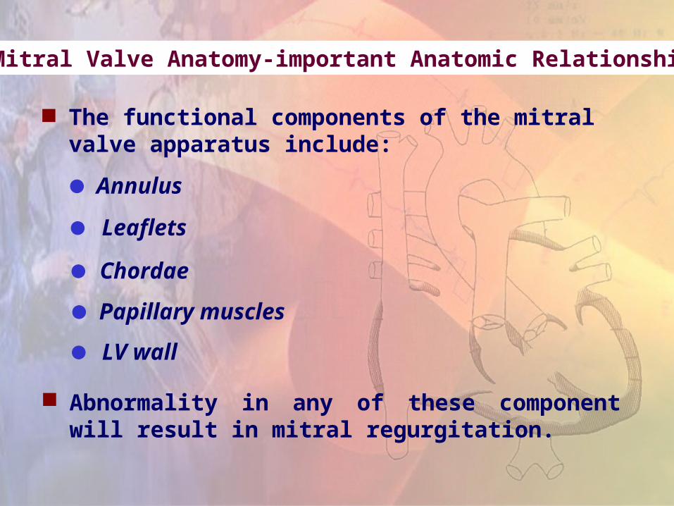
Mitral Valve Anatomy-important Anatomic Relationship
The functional components of the mitral valve apparatus include:
Annulus
Leaflets
Chordae
Papillary muscles
LV wall
Abnormality in any of these component will result in mitral regurgitation.
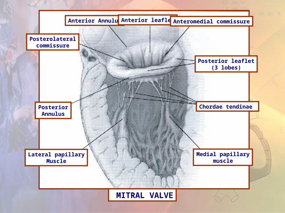
SLIDE 3 - PICTUREFIGURE 1
MITRAL VALVE
Lateral papillaryMuscle
PosteriorAnnulus
Posterolateralcommissure
Anterior Annulus Anterior leaflet Anteromedial commissure
Posterior leaflet(3 lobes)
Chordae tendinae
Medial papillarymuscle

Left mainCoronary artery
Intervalvulartrigone
Circumflexcoronary artery
Anterolateralpapillary muscle
Posterior leaflet
Left coronary
sinus
Noncoronarysinus
Anterior leaflet
SecondaryChorda tendinea
Coronary Sinus
Tertiary chordatendinea
Primary chordatendinea
Posteromedialpapillary muscle
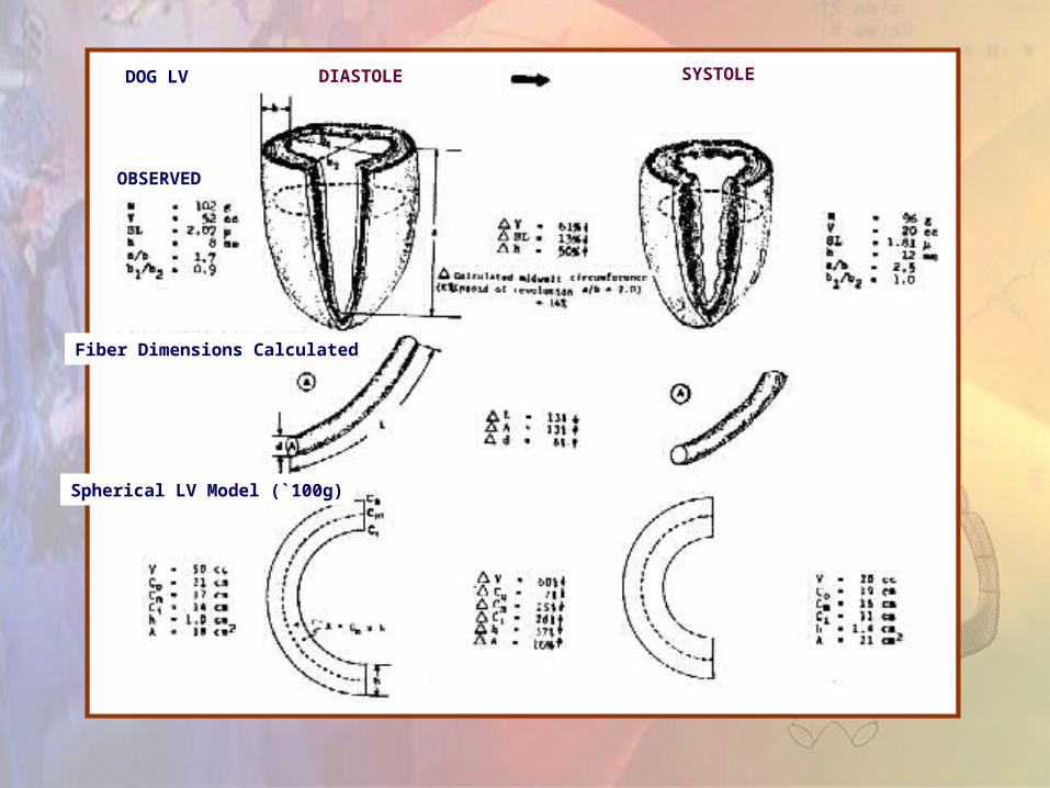
Fiber Dimensions Calculated
Spherical LV Model (`100g)
DIASTOLE SYSTOLE
OBSERVED
DOG LV

The left ventricular wall
composed of muslce fibers,
connective tissue, fat neuro-
vascular structures and
lymphatics

Photomontages assembled from electron micrographs of dog LV cells are from control (A), congestive heart failure due to mitral regurgitation (B), and recovery state after successful mitral valve surgery (C).
Electron micrograph of adult LV canine myocardium. Long axes of several cells cross the figure from left to right. Arrows mark the location of boundaries between adjacent cells. Large open spaces are capillaries perfused with fixative.
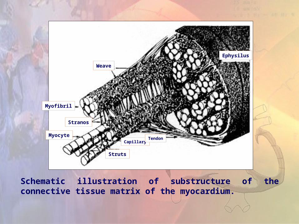
Schematic illustration of substructure of the connective tissue matrix of the myocardium.
Myocyte
Struts
CapillaryTendon
Stranos
Myofibril
Weave
Ephysilus

Systolic Torsion of LV
Asymmetry of fiber radii, sarcomere
length and electrical activation allows
torsion of the apex relative to the base.

Model for generation of torque for LV wall. Vectors for force generation at the epicardial and endocardial surfaces could neutralize each other. The epicardial fibers at the epicardial surface have a longer radius and a more powerful moment arm. Sarcomere lengths, activation time, and infolding of the wall contribute to heterogencity of the relation between structure and function between the inner and outer regions of the wall. Differences in the total force generated are believed responsible for the LV systolic twist.
Base
Endocardium
Apex
Epicardium

Mechanism of mitral regurgitation
Classification of Structural Valve Abnormalities
Type
I Normal leaflet motion
Annular dilation
Leaflet perforation
I I Leaflet prolapse
Chordal rupture
Chordal elongation
Papillary muscle rupture
Papillary musle elongation
I I I Restricted leaflet motion
Commissure fusion
Leaflet thickening
Chordal fusion and thickening
Abnormality
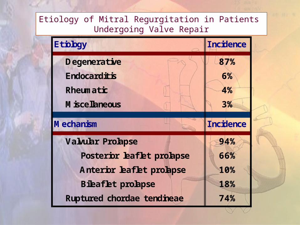
Etiology I ncidence
Degenerative 87%
Endocarditis 6%
Rheumatic 4%
Miscellaneous 3%
Mechanism I ncidence
Valvular Prolapse 94%
Posterior leaflet prolapse 66%
10%
Bileaflet prolapse 18%
Ruptured chordae tendineae 74%
Anterior leaflet prolapse
Etiology of Mitral Regurgitation in Patients Undergoing Valve Repair
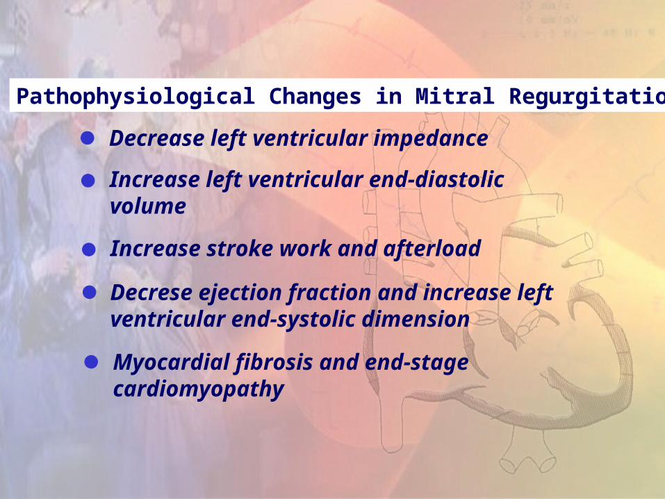
Pathophysiological Changes in Mitral Regurgitation
Decrease left ventricular impedance
Increase left ventricular end-diastolic volume
Increase stroke work and afterload
Decrese ejection fraction and increase left ventricular end-systolic dimension
Myocardial fibrosis and end-stage cardiomyopathy

The Natural History of Mitral Regurgitation
Mitral regurgitation is a progressive disease
With an increase on average of 7.5 ml/year for regurgitant volume and of 5.9 mm2/year for the effective regurgitant orifice.
The progression of mitral regurge also cause progression of LV remodelling at the same rate.
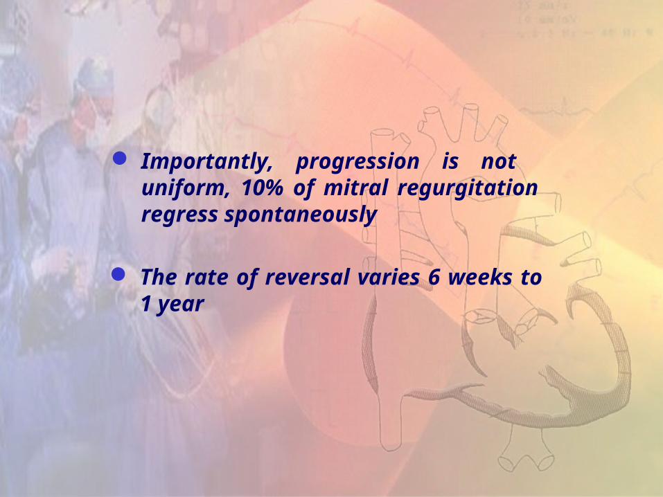
Importantly, progression is not uniform, 10% of mitral regurgitation regress spontaneously
The rate of reversal varies 6 weeks to 1 year
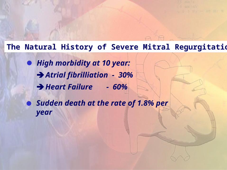
The Natural History of Severe Mitral Regurgitation
High morbidity at 10 year:
Atrial fibrilliation - 30%
Heart Failure - 60%
Sudden death at the rate of 1.8% per year

Timing of Surgery
What information is needed to define the timing of mitral surgery?
Symptoms-Functional Class Impact of Pre-operqtive symptoms on survival after mitral surgery

90+2
76+5
48+4
73+3
NYHA I-IINYHA III-IV
P<0.0001
100-
80-
60-
40-
20-
Su
rviv
al (
%)
N8I-II 199 192 187 184 181 169 125 95 63 42 34
III-IV 279 249 236 227 211 201 174 183 103 74 51
Figure 1. Overall postoperative survival compared between patients in NYHA Class I/II and patients in Class III/IV – Numbers at bottom indicate patients at risk.
0
0 1 2 3 4 5 6 7 8 9 10Years

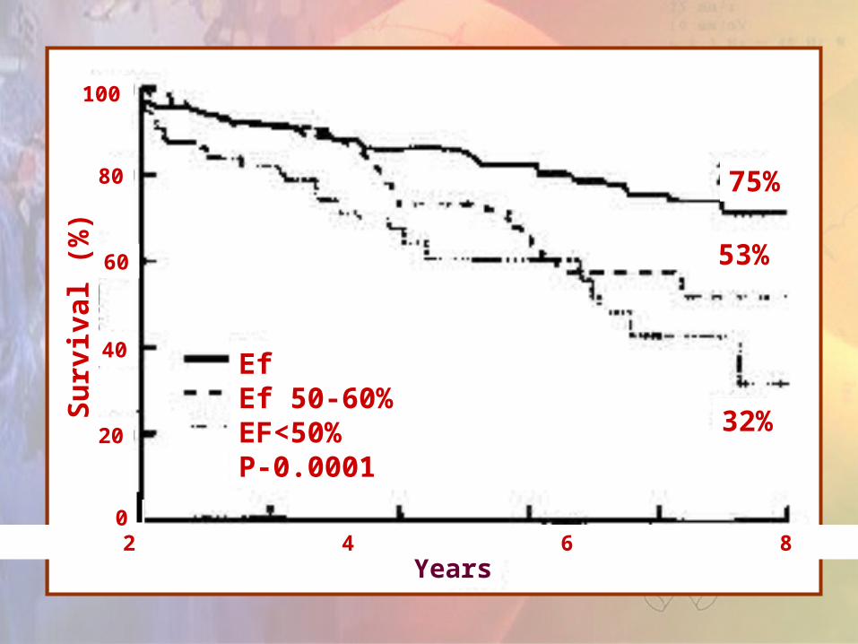
Left Ventricular Function
Ejection Fraction
Left ventricular and systolic dimension
Assessed by echocardiography
Su
rviv
al (%
)
53%
32%
75%
EfEf 50-60%EF<50%P-0.0001
Years0 2 4 6 8 10
100
80
60
40
20
0

Degree of mitral regurgitation-hymodynamics Regurgitant volume (R.Vol.)
Effective regurgitant orifice (ERO)
Assessed by quantitative doppler echo
The respective thresholds for severe mitral regurgitation are (R.Vol.) > 60 ml. and ERO > 40mm2

Timing of Surgery
Translate into_when the patient seen_promptly provided no major comonbidities
The concept of waiting fro signs of early LV dysfunction is not advised
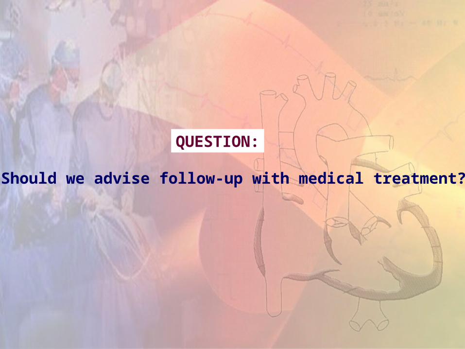
QUESTION:
Should we advise follow-up with medical treatment?
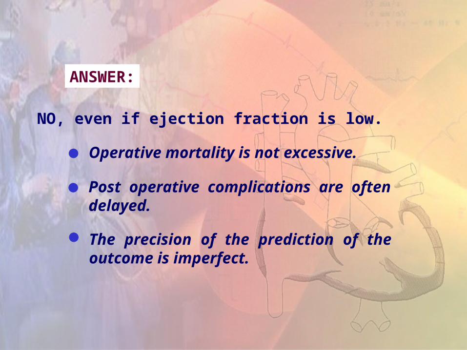
ANSWER:
NO, even if ejection fraction is low.
Operative mortality is not excessive.
Post operative complications are often delayed.
The precision of the prediction of the outcome is imperfect.
Mitral Valve Surgery
The optimal intervention for mitral surgery is valve repair.
Superior hemodynamics and ventricular function.
Less distortion of ventricular shape.
Avoidance of prosthetic valve and related complications.
Excellent long term clinical outcome.

Mitral Valve Repair
Leaflet plicationMcGoon Plication

Posterior leaflet excision [carpentier] Posterior leaflet prolapse
2 to ruptured or elongated chordae.
Posterior leaflet
Rupturedchordae
Repairleaflet
Supported repair
Repair annulus
Excise unsupportedleaflet

Subvalvar apparatus inspection with a nerve hook. The middle portion of the leaflets is identified
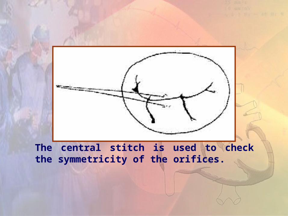
The central stitch is used to check the symmetricity of the orifices.

Edge-Edge Technique [Alfieri] Innovative method for mitral valve
repair.
A running suture along the free edge of the leaflets is done.
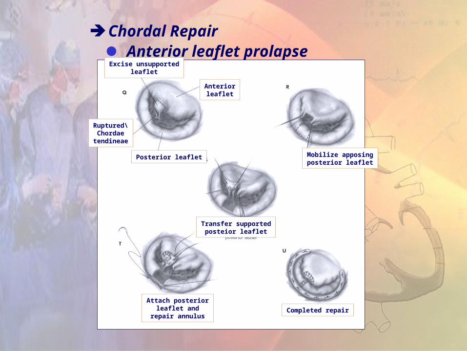
Chordal Repair Anterior leaflet prolapse
Excise unsupportedleaflet
Anteriorleaflet
Ruptured\Chordaetendineae
Posterior leaflet Mobilize apposingposterior leaflet
Transfer supportedposteior leaflet
Attach posteriorleaflet and
repair annulusCompleted repair
CONCLUSION:
Mitral regurgitation is a surgical issue.
Timing of mitral surgery still remained one of the most vexing problems of clinical cardiac science.
The concept of waiting for signs of early LV dysfunction not exists anymore.
The outlook is poor for patients who are treated medically.
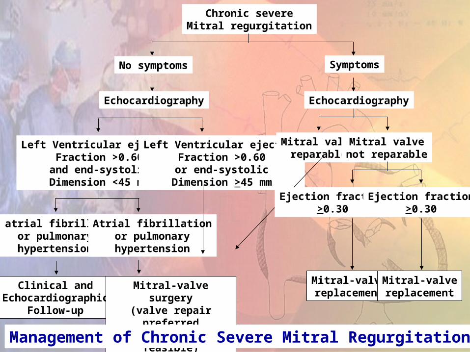
Chronic severeMitral regurgitation
No symptoms Symptoms
Echocardiography Echocardiography
Left Ventricular ejectionFraction >0.60
and end-systolicDimension <45 mm
Left Ventricular ejectionFraction >0.60or end-systolic
Dimension >45 mm
Mitral valvereparable
Mitral valvenot reparable
No atrial fibrillationor pulmonaryhypertension
Atrial fibrillationor pulmonaryhypertension
Clinical andEchocardiographic
Follow-up
Mitral-valve surgery(valve repair preferredif technically feasible)
Ejection fraction>0.30
Ejection fraction>0.30
Mitral-valvereplacement
Mitral-valvereplacement
Management of Chronic Severe Mitral Regurgitation
THANK YOU!!